Embed Size (px)
Citation preview
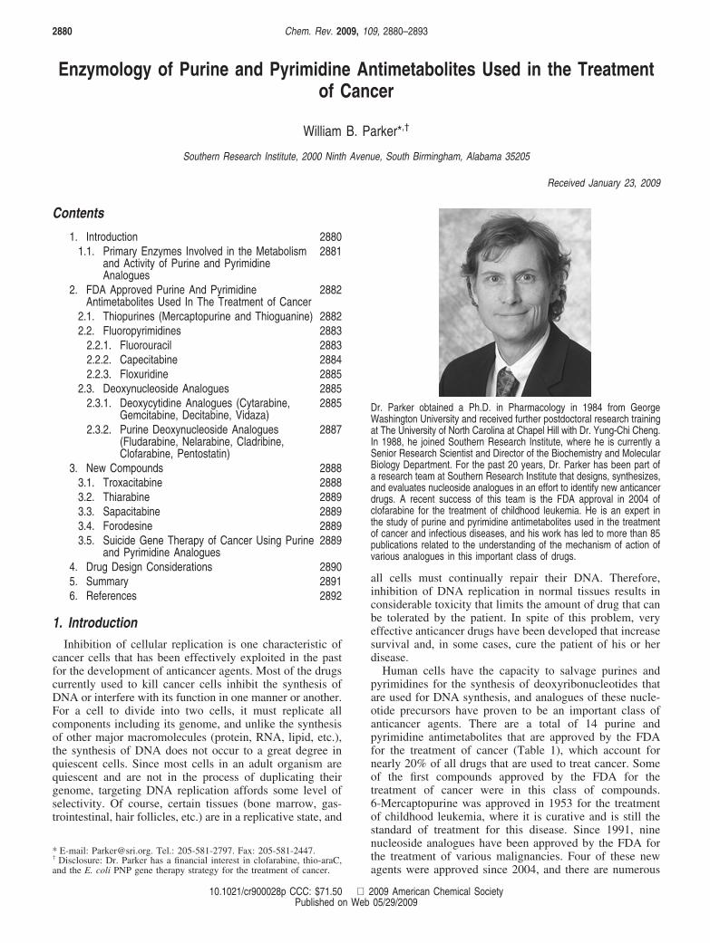
Enzymology of Purine and Pyrimidine Antimetabolites Used in the Treatmentof Cancer
William B. Parker*,†
Southern Research Institute, 2000 Ninth Avenue, South Birmingham, Alabama 35205
Received January 23, 2009
Contents
1. Introduction 28801.1. Primary Enzymes Involved in the Metabolism
and Activity of Purine and PyrimidineAnalogues
2881
2. FDA Approved Purine And PyrimidineAntimetabolites Used In The Treatment of Cancer
2882
2.1. Thiopurines (Mercaptopurine and Thioguanine) 28822.2. Fluoropyrimidines 2883
2.2.1. Fluorouracil 28832.2.2. Capecitabine 28842.2.3. Floxuridine 2885
2.3. Deoxynucleoside Analogues 28852.3.1. Deoxycytidine Analogues (Cytarabine,
Gemcitabine, Decitabine, Vidaza)2885
2.3.2. Purine Deoxynucleoside Analogues(Fludarabine, Nelarabine, Cladribine,Clofarabine, Pentostatin)
2887
3. New Compounds 28883.1. Troxacitabine 28883.2. Thiarabine 28893.3. Sapacitabine 28893.4. Forodesine 28893.5. Suicide Gene Therapy of Cancer Using Purine
and Pyrimidine Analogues2889
4. Drug Design Considerations 28905. Summary 28916. References 2892
1. IntroductionInhibition of cellular replication is one characteristic of
cancer cells that has been effectively exploited in the pastfor the development of anticancer agents. Most of the drugscurrently used to kill cancer cells inhibit the synthesis ofDNA or interfere with its function in one manner or another.For a cell to divide into two cells, it must replicate allcomponents including its genome, and unlike the synthesisof other major macromolecules (protein, RNA, lipid, etc.),the synthesis of DNA does not occur to a great degree inquiescent cells. Since most cells in an adult organism arequiescent and are not in the process of duplicating theirgenome, targeting DNA replication affords some level ofselectivity. Of course, certain tissues (bone marrow, gas-trointestinal, hair follicles, etc.) are in a replicative state, and
all cells must continually repair their DNA. Therefore,inhibition of DNA replication in normal tissues results inconsiderable toxicity that limits the amount of drug that canbe tolerated by the patient. In spite of this problem, veryeffective anticancer drugs have been developed that increasesurvival and, in some cases, cure the patient of his or herdisease.
Human cells have the capacity to salvage purines andpyrimidines for the synthesis of deoxyribonucleotides thatare used for DNA synthesis, and analogues of these nucle-otide precursors have proven to be an important class ofanticancer agents. There are a total of 14 purine andpyrimidine antimetabolites that are approved by the FDAfor the treatment of cancer (Table 1), which account fornearly 20% of all drugs that are used to treat cancer. Someof the first compounds approved by the FDA for thetreatment of cancer were in this class of compounds.6-Mercaptopurine was approved in 1953 for the treatmentof childhood leukemia, where it is curative and is still thestandard of treatment for this disease. Since 1991, ninenucleoside analogues have been approved by the FDA forthe treatment of various malignancies. Four of these newagents were approved since 2004, and there are numerous
* E-mail: [email protected]. Tel.: 205-581-2797. Fax: 205-581-2447.† Disclosure: Dr. Parker has a financial interest in clofarabine, thio-araC,and the E. coli PNP gene therapy strategy for the treatment of cancer.
Dr. Parker obtained a Ph.D. in Pharmacology in 1984 from GeorgeWashington University and received further postdoctoral research trainingat The University of North Carolina at Chapel Hill with Dr. Yung-Chi Cheng.In 1988, he joined Southern Research Institute, where he is currently aSenior Research Scientist and Director of the Biochemistry and MolecularBiology Department. For the past 20 years, Dr. Parker has been part ofa research team at Southern Research Institute that designs, synthesizes,and evaluates nucleoside analogues in an effort to identify new anticancerdrugs. A recent success of this team is the FDA approval in 2004 ofclofarabine for the treatment of childhood leukemia. He is an expert inthe study of purine and pyrimidine antimetabolites used in the treatmentof cancer and infectious diseases, and his work has led to more than 85publications related to the understanding of the mechanism of action ofvarious analogues in this important class of drugs.
Chem. Rev. 2009, 109, 2880–28932880
10.1021/cr900028p CCC: $71.50 2009 American Chemical SocietyPublished on Web 05/29/2009
agents that are currently being evaluated in clinical trials.These recent FDA approvals indicate that the design andsynthesis of new nucleoside analogues is still a productivearea for discovering new drugs for the treatment of cancer.In general, these compounds have been most useful in thetreatment of hematologic malignancies, and even thoughthere is still room for significant improvements in thetreatment of these diseases, some of the newer agents arefinding use in the treatment of solid tumors.
The basic mechanism of action of purine and pyrimidineantimetabolites is similar. These compounds diffuse into cells(usually with the aid of a membrane transporter1) and areconverted to analogues of cellular nucleotides by enzymesof the purine or pyrimidine metabolic pathway. Thesemetabolites then inhibit one or more enzymes that are criticalfor DNA synthesis, causing DNA damage and induction ofapoptosis.2 Even though the compounds in this class arestructurally similar and share many mechanistic details, it isclear that subtle quantitative and qualitative differences inthe metabolism of these agents and their interactions withtarget enzymes can have a profound impact on theirantitumor activity. As noted by Plunkett and Gandhi,3 “oneof the remarkable features of purine and pyrimidine nucleo-side analogues that remains unexplained is how drugs withsuch similar structural features, that share metabolic path-ways, and elements of their mechanism of action show suchdiversity in their clinical activities”. Possibly the bestexample of this fact is the newly approved drug, clofarabine,which differs from cladribine by only one fluorine atom,because it has demonstrated excellent efficacy in the treat-ment of relapsed and refractory pediatric acute lymphoblasticleukemia, whereas cladribine is not effective against thisdisease. These clinical results indicate that the biochemicalactions of clofarabine are sufficiently different from that ofcladribine to impart unique clinical activities. This and otherexamples indicate that small structural modifications ofnucleoside analogues can have profound effects on thechemical stability and biological activity of nucleosideanalogues.
1.1. Primary Enzymes Involved in the Metabolismand Activity of Purine and Pyrimidine Analogues
To adequately understand the mechanism of action of thisclass of compounds it is necessary to be familiar with theenzymes that are involved in the metabolism of naturalpurines and pyrimidines. Human cells have all the enzymes
needed for de novo synthesis of purine and pyrimidinenucleotides; however, other than orotate phosphoribosyltransferase with fluorouracil, these enzymes are not involvedin the activation of the purine and pyrimidine antimetabolitesand are only secondary targets responsible for antitumoractivity of these compounds. Although salvage of purinesand pyrimidines is not required for growth, human cellsexpress many enzymes that can utilize purines and pyrim-idines as substrates, and it is these enzymes (shown in Figures1 and 2) that are most important to the anabolism andcatabolism of the purine and pyrimidine antimetabolites thatare used in the treatment of cancer. The catabolic enzymesare important because they are often responsible for detoxi-fying the nucleoside analogues, and these enzymes areexpressed thoughout the body. Dihydropyrimidine dehydro-genase and xanthine oxidase are the initial enzymes in thedegradation pathways of pyrimidines and purines. Adenosinedeaminase and purine nucleoside phosphorylase are twoimportant enzymes in the inactivation of purine nucleosideanalogues but have also been successful targets of two agents,pentostatin and forodesine.
Phosphoribosyl transferases are responsible for activatingthe 3 base analogues (mercaptopurine, thioguanine, andfluorouracil), and there are five enzymes in human cells thatcan phosphorylate deoxynucleoside analogues4-6 (deoxycy-tidine kinase, thymidine kinase 1, thymidine kinase 2,deoxyguanosine kinase, and 5′-nucleotidase). The primaryrate-limiting enzyme for activation of most of the approvednucleoside analogues is deoxycytidine kinase. Althoughdeoxycytidine is the preferred natural substrate for thisenzyme, it also recognizes deoxyadenosine and deoxygua-nosine as substrates. The purine analogues are also substratesfor deoxyguanosine kinase expressed in mitochondria, andthis enzyme can contribute to the activation of these agents.Once formed, the monophosphate metabolites are phospho-rylated by the appropriate monophosphate kinases7 to thediphosphate metabolite, which is phosphorylated by nucleo-side diphosphate kinase. The first step in the formation ofthe 5′-triphosphates is typically the rate-limiting step and is,therefore, the most important step in activation of deoxy-nucleoside analogues. The X-ray crystal structure of deoxy-cytidine kinase has recently been solved,8 and given itsimportance in the activation of deoxynucleoside analogues,its structure is used for design of new agents.
The primary target of the deoxynucleoside analogues arethe DNA polymerases involved in DNA replication. Thereare at least 14 eukaryotic DNA polymerases expressed inhuman cells,9 three of which are primarily involved inchromosomal replication (DNA polymerases R, δ, and ε)and are the primary targets for the anticancer nucleosideanalogues. The other major cellular polymerases are DNApolymerase �, which is involved in DNA repair; DNApolymerase γ, which is the polymerase responsible formitochondrial DNA replication; and telomerase, which isresponsible for the replication of DNA telomeres, but theseenzymes are not primary targets for the anticancer antime-tabolites. Inhibition of DNA polymerase γ or telomeraseactivity does not result in the immediate inhibition of cellgrowth.
A deoxynucleotide triphosphate analogue could theoreti-cally interact with a DNA polymerase in one of three ways:(i) it could compete with the natural substrate, but not beused as a substrate; (ii) it could substitute for the naturalsubstrate with little effect on subsequent DNA synthesis; or
Table 1. FDA Approved Purine and Pyrimidine Antimetabolites
drug dateapproved
5-aza-2′-deoxycytidine (decitabine) 2006O6-methylarabinofuranosyl guanine (nelarabine) 20052′-fluoro-2′-deoxyarabinofuranosyl-2-chloroadenine
(clofarabine)2004
5-aza-cytidine (vidaza) 2004N4-pentyloxycarbonyl-5′-deoxy-5-fluorocytidine
(capecitabine)1998
2,2-difluoro-2′-deoxycytidine (gemcitabine) 19962-chloro-2′-deoxyadenosine (cladribine) 1992arabinofuranosyl-2-fluoroadenine (fludarabine) 19912′-deoxycoformycin (pentostatin) 19915-fluoro-2′-deoxyuridine (floxuridine) 1970arabinofuranosylcytosine (cytarabine) 19696-thioguanine 19665-fluorouracil 19626-mercaptopurine 1953
Enzymology of Purine and Pyrimidine Antimetabolites Chemical Reviews, 2009, Vol. 109, No. 7 2881
(iii) it could substitute for the natural substrate and interferewith subsequent DNA synthesis, causing chain termination.The second two possibilities are the primary manners inwhich the anticancer nucleotide analogues interact with DNApolymerases, and all of these analogues have been shown tobe good substrates for the replicative DNA polymerases. Theprimary differences in these compounds are (i) how easilythe DNA chain is elongated after the incorporation of theanalogue and (ii) how easily they can be removed from theDNA by the proof-reading exonucleases. The incorporationof these agents into DNA is one of the most important aspectsof their mechanism of action resulting in antitumor activity,because the incorporation is difficult to repair and causes alasting inhibition of DNA synthesis or disruption of DNAfunction. The inhibition of DNA synthesis by agents, suchas aphidicolin, that only inhibit DNA polymerase activitywithout being incorporated into the DNA chain have notmade good anticancer agents, because the DNA is notdamaged by these agents and DNA synthesis resumes afterthe removal of the agent. Indeed, aphidicolin is used tosynchronize cell populations,10 because of its ability totemporarily inhibit DNA synthesis without inducing celldeath.
2. FDA Approved Purine And PyrimidineAntimetabolites Used In The Treatment of Cancer
The FDA approved purine and pyrimidine antimetabolitescan be grouped into three primary classes (thiopurines,
fluoropyrimidines, and the deoxynucleoside analogues) basedon structural and mechanistic considerations. The deoxy-nucleoside analogues are the largest class and are where mostof the design of new compounds has occurred recently. Amassive amount of literature on the mechanism of action ofthese established agents is available, and there will be noattempt in this review to include all that has been done withthese compounds. Instead, a description of the importantmetabolic features of each compound, the primary enzymatictargets responsible for their antitumor activity, and the uniquefeatures of the various compounds will be presented.
2.1. Thiopurines (Mercaptopurine andThioguanine)
6-mercaptopurine (MP) was one of the first agents ap-proved by the FDA for the treatment of cancer,11 where itproved to be effective in the treatment of childhood acutelymphocytic leukemia. MP is an analogue of hypoxanthine(Figure 3), and like hypoxanthine, it is a good substrate forhypoxanthine/guanine phosphoribosyl transferase (Figure 4).The product of the reaction, 6-thio-inosine monophosphate(T-IMP), is a substrate for IMP dehydrogenase and issubsequently converted to guanine nucleotides. The primaryintracellular metabolite of MP is 6-thioguanosine-5′-triphos-phate, and it is readily incorporated into RNA. However,since specific inhibition of RNA synthesis does not affectthe activity of MP,12 the incorporation of thioguanine (TG)
Figure 1. Primary enzymes involved in the metabolism of pyrimidine analogues.
Figure 2. Primary enzymes involved in the metabolism of purineanalogues.
Figure 3. Structures of thiopurines.
2882 Chemical Reviews, 2009, Vol. 109, No. 7 Parker
into RNA does not appear to play an important role in theantitumor activity of MP.
MP is also converted via ribonucleotide reductase to6-thio-2′-deoxyguanosine-5′-triphosphate, which is incorpo-rated into DNA. Unlike most of the other cytotoxic purineand pyrimidine antimetabolites used in the treatment ofcancer, treatment of cells with MP does not result in theimmediate inhibition of DNA synthesis in that cells continueto divide before dying. This result is consistent with studiesthat indicate that T-dGTP is a good substrate for the DNApolymerases involved in DNA replication.14,15 It is utilizedas effectively as dGTP as a substrate for DNA polymeraseR, and once incorporated, it is readily extended by thepolymerase and is incorporated into internal positions in theDNA chain. Although treatment with MP does not inhibitDNA polymerase activity, its incorporation into DNAresulting in DNA damage is believed to be primarilyresponsible for the antitumor activity of MP. It is thoughtthat TG in DNA, as well as its methylated counterpart, isrecognized by mismatch repair enzymes, which causes afutile cycle of repair that results in lethal DNA damage.13
The sulfur atom of T-IMP is methylated by thiopurineS-methyltransferase (TPMT) present in mammalian tissues,and methyl mercaptopurine riboside monophosphate (methyl-T-IMP) is also an important metabolite in cells. Thismetabolite is a potent inhibitor of PRPP amidotransferase,the first enzyme in de novo purine biosynthesis, and itsinhibition results in a decrease in purine nucleotide pools.Therefore, there are two primary biochemical actions thatcontribute to the anticancer activity of MP; its inhibition ofde novo purine synthesis and its incorporation into DNA as6-thio-2′-deoxyguanosine.
No adenine nucleotide analogues of MP are formed incells, because T-IMP is not a substrate for adenylosuccinatesynthetase, the first enzyme in the formation of adeninenucleotides from IMP. Even if it were a substrate for thisenzyme, the mechanism of action of this enzyme wouldremove the 6 sulfur atom and replace it with an aspartic acidto form adenylosuccinic acid, which is the natural productof this reaction. A small amount of T-ITP is formed in cells,but this metabolite is not believed to be important in themechanism of activity of MP.
The metabolism of thioguanine (TG) is much simpler thanthat of MP. TG is also a substrate for hypoxanthine/guaninephosphoribosyl transferase and large concentrations of TGnucleotides accumulate in cells treated with TG. T-GMP isalso methylated by S-methyl transferase, but the product ofthe reaction, methyl-T-GMP, is not a potent inhibitor ofPRPP amidotransferase. Therefore, inhibition of de novopurine biosynthesis is less important to the action of TG,
and the mechanism of cytotoxicity of TG is believed to beprimarily due to its incorporation into DNA and subsequentDNA damage.13 Thioguanine (TG) is approved for use inacute myelogenous leukemia.
In patients, the methylation of the purine bases, MP andTG, by thiopurine S-methyltransferase (TPMT) is a majormechanism of detoxification of these agents.16,17 The productsof the reaction, S6-methyl-mercaptopurine and S6-methyl-thioguanine, are not substrates for hypoxanthine/guaninephosphoribosyl transferase (HGPRT) and are, therefore, nottoxic to human cells. Approximately 0.3% of the populationdoes not express functional TPMT activity, and treatmentof these people with either thiopurine can result in severetoxicity.
2.2. Fluoropyrimidines2.2.1. Fluorouracil
5-Fluorouracil (FUra, Figure 5) is one of the first examplesof an anticancer drug that was designed based on theavailable biochemical information. It was known that (i) afluorine atom was of similar size to a hydrogen atom; (ii) acarbon-fluorine bond was much stronger than a carbon-hydrogen bond; (iii) the reaction mechanism of thymidylatesynthase replaces the 5-hydrogen of deoxyuridine mono-phosphate with a methyl group obtained from methylenetetrahydrofolate to make thymidylate (TMP); and (iv) rathepatoma cells, but not normal liver cells, could utilize uracil(although it was subsequently found that this observationdid not extend to other cell types). Utilizing this information,Heidelberger18 and colleagues hypothesized that FUra wouldselectively kill tumor cells because of its selective metabo-lism in tumor cells to F-dUMP, which would inhibitthymidylate synthetase due to the inability of the enzyme toremove the 5-fluorine atom. Much of the original hypothesishas been shown to be true,19 and FUra is used for palliativetreatment of colorectal, breast, stomach, and pancreaticcancer. It also has utility as a topical treatment of superficialbasal cell carcinoma that cannot be treated with surgery andactinic keratosis, a precancerous skin condition. Much workhas been done since the approval of this agent that hasenhanced our understanding of its mechanism of action, andthis work has been extensively reviewed.20,21
As shown in Figure 6 the metabolism of FUra is verycomplex. FUra is converted into F-UMP by orotate phos-phoribosyl transferase, which is the first step in its activation.Nucleotide kinases then convert F-UMP to F-UTP, whichis the primary intracellular metabolite of FUra. F-UTP isused as a substrate for RNA synthesis in place of uridine
Figure 4. Metabolism of mercaptopurine and thioguanine.
Figure 5. Structures of fluoropyrimidines.
Enzymology of Purine and Pyrimidine Antimetabolites Chemical Reviews, 2009, Vol. 109, No. 7 2883
triphosphate (UTP), and a considerable amount of FUra isincorporated into all species of RNA. The incorporation ofFUra into various species of RNA has been shown to disruptthe function of these species of RNA, but these effects haveonly been observed at high concentrations. There are varioustypes of RNA molecules, and the effect of FUra on many ofthe newer functions of RNA has not yet been evaluated. Itis believed that the incorporation of FUra into RNA doescontribute to its cytotoxic activity, but because of thecomplexity of RNA, the precise RNA-directed action(s) hasnot been defined. It is likely that the incorporation into RNAcauses more than one defect and that inhibition of numerousRNA activities contribute to its RNA-directed activity.Although incorporation into RNA is an important componentof the mechanism of action of FUra, the RNA-directedactions are believed to be secondary to its DNA-directedactions described below, which is similar to the case withthe thiopurines.
F-UDP is a substrate for ribonucleotide reductase, whichremoves the 2′-OH group. F-dUDP is a good substrate fornucleoside diphosphate (NDP) kinase that forms F-dUTP,which is an excellent substrate for DNA polymerases.F-dUTP (as well as dUTP) is used by DNA polymerasesfor the synthesis of DNA as effectively as thymidinetriphosphate (TTP). Therefore, if F-dUTP accumulates incells, it will be incorporated into the DNA by the DNApolymerases. Human cells have developed a mechanism torecognize uracil in DNA and remove it, because a consider-able amount of uracil is formed in the DNA of any cell dueto the spontaneous deamination of cytosine and since uracilbase-pairs as thymine, this deamination of cytosine in DNAwould result in mutation. The enzyme responsible for theremoval of uracil from DNA is uracil glycosylase, and itrecognizes FUra in DNA as a substrate and readily removesit from the DNA, resulting in an apyrimidinic site, which isrecognized by apurinic/apyrimidinic endonuclease 1, causinga single strand break. The single strand break is recognizedby DNA repair enzymes, and in a manner similar to TG,
the repair and resynthesis of DNA sets up a futile cycle thatresults in inhibition of DNA synthesis and cell death.
Another mechanism the cell uses to keep uracil out ofDNA is to prevent its use as a substrate by DNA poly-merases. Since human cells contain the enzymes necessaryto make dUTP, human cells also express dUTPase, whichconverts dUTP to dUMP and keeps levels of dUTP verylow in the cell. dUMP is a substrate for thymidylate synthaseand is utilized for the synthesis of thymidine nucleotides.dUTPase also recognizes F-dUTP and is responsible for theformation of F-dUMP, which is a potent inhibitor ofthymidylate synthase, as hypothesized by Heidelberger. Theinhibition of thymidylate synthase results in decreases in TTPlevels and large increases in deoxyuridine nucleotides,including both dUTP and F-dUTP. As indicated above, anincrease in dUTP levels can result in the incorporation ofuracil in DNA and its subsequent removal by uracil glyco-sylase. Therefore, the inhibition of DNA synthesis in cellstreated with FUra is a result of two actions: depression ofintracellular TTP levels due to inhibition of thymidylatesynthetase and incorporation and removal of uracil (andFUra) in DNA. Therefore, inhibition of thymidylate synthesisby F-dUMP results in a nonproductive incorporation andremoval of uracil and FUra from DNA, which results ininhibition of DNA synthesis and DNA damage.
An important enzyme in the catabolism of FUra isdihydropyrimidine dehydrogenase. This enzyme is the rate-limiting enzyme in the conversion of FUra to fluoro-�-alanineand is, therefore, very important in the detoxification of FUra.Three to five percent of Caucasians express low levels ofdihydropyrimidine dehydrogenase, and if these people aretreated with FUra, severe toxicity, including death, canoccur.17
2.2.2. Capecitabine
Capecitabine (Figure 5) is a prodrug of FUra that isadministered orally.22 It has almost 100% oral bioavailability
Figure 6. Metabolism of fluoropyrimidines.
2884 Chemical Reviews, 2009, Vol. 109, No. 7 Parker
and is converted in three enzymatic reactions to FUra (Figure6). The N4-pentyloxycarbonyl moiety is first removed bycarboxylesterases in the liver to generate 5′-deoxy-5-fluo-rocytidine, which is a good substrate for cytidine deaminase,and is converted to 5′-deoxy-5-fluorouridine. Because of theabsence of a 5′-OH group, 5′-deoxy-5-fluorouridine cannotbe activated to FUra nucleotides by nucleoside kinases;however, it is a good substrate for thymidine phosphorylaseand is converted to FUra. Because thymidine phosphorylaseis overexpressed in tumor tissues, capecitabine should havea better selective index than FUra. In addition, thymidinephosphorylase activity is stimulated by radiation therapy, andcombination treatment with capecitabine plus radiation canfurther enhance selectivity of this compound for tumor cells.As a prodrug of FUra, capecitabine has two advantages overintravenous FUra: ease of administration (oral vs IV) and apotential increased therapeutic effect. It is currently approvedfor use in the treatment of stage III colon cancer andmetastatic breast cancer.
2.2.3. Floxuridine
Floxuridine (F-dUrd) is an excellent substrate for thymi-dine kinase, and it is converted by this enzyme directly toF-dUMP. In vitro, this compound is a much more potentinhibitor of cell growth than FUra and is not converted toribonucleotide metabolites to a significant degree at cytotoxicconcentrations. However, F-dUrd is also a good substratefor thymidine phosphorylase, which converts it to FUra, anda significant amount of F-dUrd is converted to FUra in vivo.Therefore, when used in the treatment of patients, F-dUrdis not a specific inhibitor of thymidylate synthesis. F-dUrdhas demonstrated some efficacy when given by hepaticarterial infusion to treat liver metastases.23 Although approvedby the FDA for this purpose, it is not widely used.
2.3. Deoxynucleoside AnaloguesThere are numerous deoxynucleoside analogues that are
useful in the treatment of cancer. Other than cytarabine,which was approved in 1969 for the treatment of acuteleukemias, these agents are relatively new, having been
approved for use since 1991, and except for deoxycoformy-cin, which is a potent inhibitor of adenosine deaminase, themechanisms of action of these agents are quite similar. Theyare converted to their respective nucleotide analogues, whichinhibit DNA synthesis by inhibition of DNA polymerasesand/or ribonucleotide reductase. However, in spite of thesesimilarities, there are differences in the interaction of theseagents and their metabolites with the various metabolicenzymes and intracellular targets that imparts unique proper-ties to each of these agents and results in unique clinicalactivity.
2.3.1. Deoxycytidine Analogues (Cytarabine, Gemcitabine,Decitabine, Vidaza)
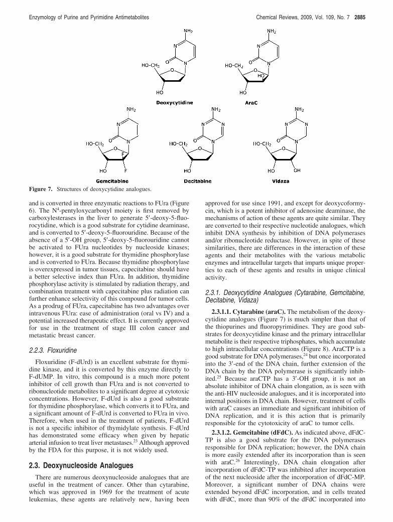
2.3.1.1. Cytarabine (araC). The metabolism of the deoxy-cytidine analogues (Figure 7) is much simpler than that ofthe thiopurines and fluoropyrimidines. They are good sub-strates for deoxycytidine kinase and the primary intracellularmetabolite is their respective triphosphates, which accumulateto high intracellular concentrations (Figure 8). AraCTP is agood substrate for DNA polymerases,24 but once incorporatedinto the 3′-end of the DNA chain, further extension of theDNA chain by the DNA polymerase is significantly inhib-ited.25 Because araCTP has a 3′-OH group, it is not anabsolute inhibitor of DNA chain elongation, as is seen withthe anti-HIV nucleoside analogues, and it is incorporated intointernal positions in DNA chain. However, treatment of cellswith araC causes an immediate and significant inhibition ofDNA replication, and it is this action that is primarilyresponsible for the cytotoxicity of araC to tumor cells.
2.3.1.2. Gemcitabine (dFdC). As indicated above, dFdC-TP is also a good substrate for the DNA polymerasesresponsible for DNA replication; however, the DNA chainis more easily extended after its incorporation than is seenwith araC.26 Interestingly, DNA chain elongation afterincorporation of dFdC-TP was inhibited after incorporationof the next nucleoside after the incorporation of dFdC-MP.Moreover, a significant number of DNA chains wereextended beyond dFdC incorporation, and in cells treatedwith dFdC, more than 90% of the dFdC incorporated into
Figure 7. Structures of deoxycytidine analogues.
Enzymology of Purine and Pyrimidine Antimetabolites Chemical Reviews, 2009, Vol. 109, No. 7 2885
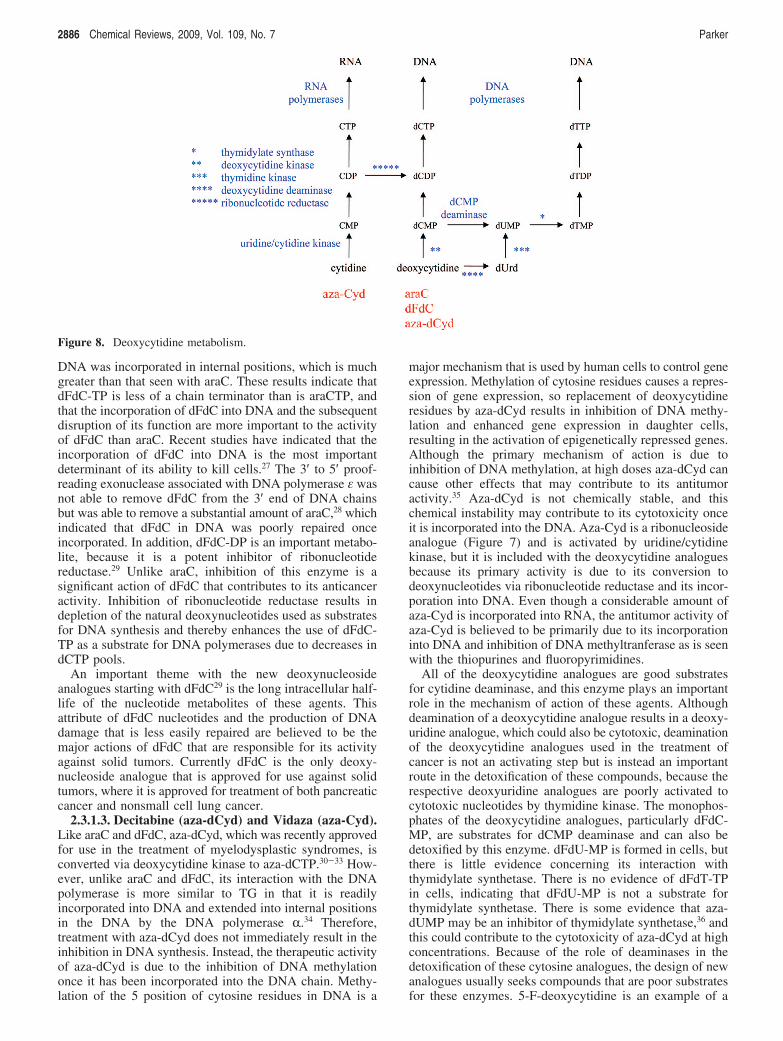
DNA was incorporated in internal positions, which is muchgreater than that seen with araC. These results indicate thatdFdC-TP is less of a chain terminator than is araCTP, andthat the incorporation of dFdC into DNA and the subsequentdisruption of its function are more important to the activityof dFdC than araC. Recent studies have indicated that theincorporation of dFdC into DNA is the most importantdeterminant of its ability to kill cells.27 The 3′ to 5′ proof-reading exonuclease associated with DNA polymerase ε wasnot able to remove dFdC from the 3′ end of DNA chainsbut was able to remove a substantial amount of araC,28 whichindicated that dFdC in DNA was poorly repaired onceincorporated. In addition, dFdC-DP is an important metabo-lite, because it is a potent inhibitor of ribonucleotidereductase.29 Unlike araC, inhibition of this enzyme is asignificant action of dFdC that contributes to its anticanceractivity. Inhibition of ribonucleotide reductase results indepletion of the natural deoxynucleotides used as substratesfor DNA synthesis and thereby enhances the use of dFdC-TP as a substrate for DNA polymerases due to decreases indCTP pools.
An important theme with the new deoxynucleosideanalogues starting with dFdC29 is the long intracellular half-life of the nucleotide metabolites of these agents. Thisattribute of dFdC nucleotides and the production of DNAdamage that is less easily repaired are believed to be themajor actions of dFdC that are responsible for its activityagainst solid tumors. Currently dFdC is the only deoxy-nucleoside analogue that is approved for use against solidtumors, where it is approved for treatment of both pancreaticcancer and nonsmall cell lung cancer.
2.3.1.3. Decitabine (aza-dCyd) and Vidaza (aza-Cyd).Like araC and dFdC, aza-dCyd, which was recently approvedfor use in the treatment of myelodysplastic syndromes, isconverted via deoxycytidine kinase to aza-dCTP.30-33 How-ever, unlike araC and dFdC, its interaction with the DNApolymerase is more similar to TG in that it is readilyincorporated into DNA and extended into internal positionsin the DNA by the DNA polymerase R.34 Therefore,treatment with aza-dCyd does not immediately result in theinhibition in DNA synthesis. Instead, the therapeutic activityof aza-dCyd is due to the inhibition of DNA methylationonce it has been incorporated into the DNA chain. Methy-lation of the 5 position of cytosine residues in DNA is a
major mechanism that is used by human cells to control geneexpression. Methylation of cytosine residues causes a repres-sion of gene expression, so replacement of deoxycytidineresidues by aza-dCyd results in inhibition of DNA methy-lation and enhanced gene expression in daughter cells,resulting in the activation of epigenetically repressed genes.Although the primary mechanism of action is due toinhibition of DNA methylation, at high doses aza-dCyd cancause other effects that may contribute to its antitumoractivity.35 Aza-dCyd is not chemically stable, and thischemical instability may contribute to its cytotoxicity onceit is incorporated into the DNA. Aza-Cyd is a ribonucleosideanalogue (Figure 7) and is activated by uridine/cytidinekinase, but it is included with the deoxycytidine analoguesbecause its primary activity is due to its conversion todeoxynucleotides via ribonucleotide reductase and its incor-poration into DNA. Even though a considerable amount ofaza-Cyd is incorporated into RNA, the antitumor activity ofaza-Cyd is believed to be primarily due to its incorporationinto DNA and inhibition of DNA methyltranferase as is seenwith the thiopurines and fluoropyrimidines.
All of the deoxycytidine analogues are good substratesfor cytidine deaminase, and this enzyme plays an importantrole in the mechanism of action of these agents. Althoughdeamination of a deoxycytidine analogue results in a deoxy-uridine analogue, which could also be cytotoxic, deaminationof the deoxycytidine analogues used in the treatment ofcancer is not an activating step but is instead an importantroute in the detoxification of these compounds, because therespective deoxyuridine analogues are poorly activated tocytotoxic nucleotides by thymidine kinase. The monophos-phates of the deoxycytidine analogues, particularly dFdC-MP, are substrates for dCMP deaminase and can also bedetoxified by this enzyme. dFdU-MP is formed in cells, butthere is little evidence concerning its interaction withthymidylate synthetase. There is no evidence of dFdT-TPin cells, indicating that dFdU-MP is not a substrate forthymidylate synthetase. There is some evidence that aza-dUMP may be an inhibitor of thymidylate synthetase,36 andthis could contribute to the cytotoxicity of aza-dCyd at highconcentrations. Because of the role of deaminases in thedetoxification of these cytosine analogues, the design of newanalogues usually seeks compounds that are poor substratesfor these enzymes. 5-F-deoxycytidine is an example of a
Figure 8. Deoxycytidine metabolism.
2886 Chemical Reviews, 2009, Vol. 109, No. 7 Parker
deoxycytidine analogue that is activated by deamination, andit has been suggested to be used as a prodrug of F-dUrd,37
but it has not been approved for human use.
2.3.2. Purine Deoxynucleoside Analogues (Fludarabine,Nelarabine, Cladribine, Clofarabine, Pentostatin)
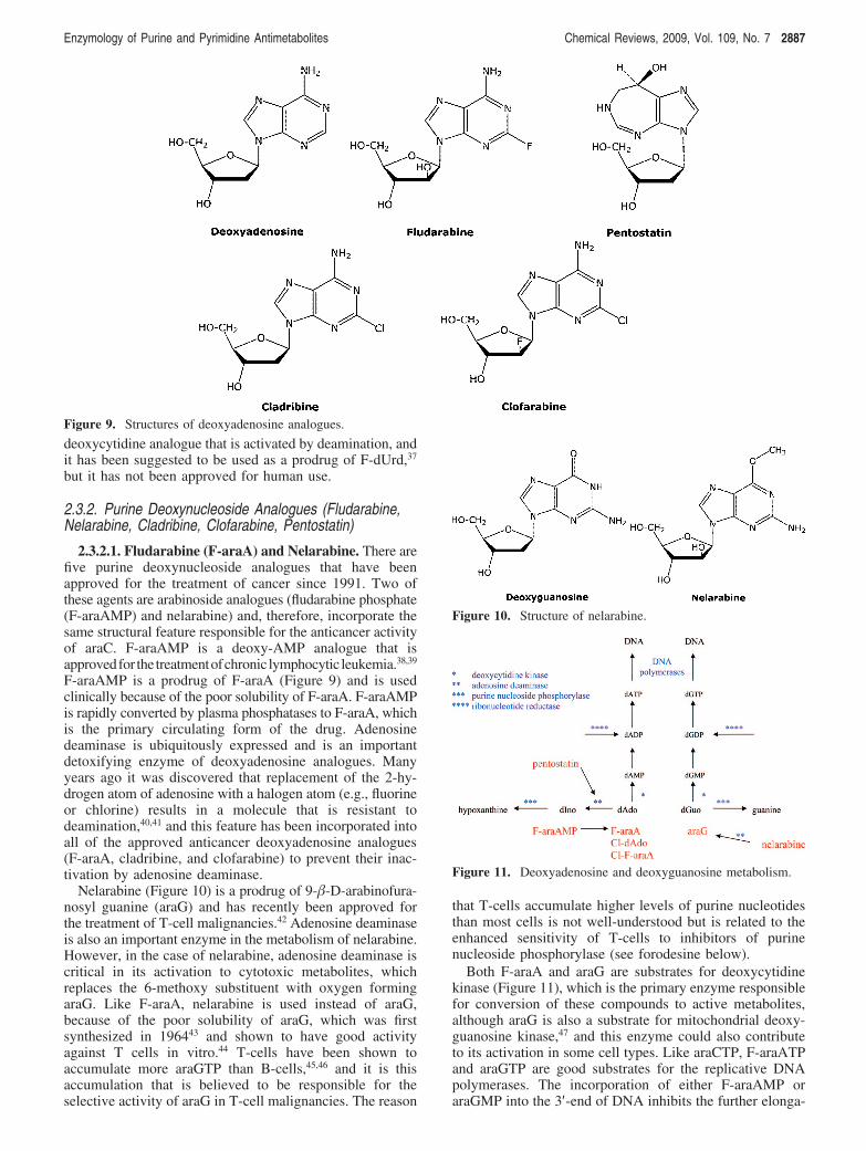
2.3.2.1. Fludarabine (F-araA) and Nelarabine. There arefive purine deoxynucleoside analogues that have beenapproved for the treatment of cancer since 1991. Two ofthese agents are arabinoside analogues (fludarabine phosphate(F-araAMP) and nelarabine) and, therefore, incorporate thesame structural feature responsible for the anticancer activityof araC. F-araAMP is a deoxy-AMP analogue that isapprovedfor the treatmentofchronic lymphocytic leukemia.38,39
F-araAMP is a prodrug of F-araA (Figure 9) and is usedclinically because of the poor solubility of F-araA. F-araAMPis rapidly converted by plasma phosphatases to F-araA, whichis the primary circulating form of the drug. Adenosinedeaminase is ubiquitously expressed and is an importantdetoxifying enzyme of deoxyadenosine analogues. Manyyears ago it was discovered that replacement of the 2-hy-drogen atom of adenosine with a halogen atom (e.g., fluorineor chlorine) results in a molecule that is resistant todeamination,40,41 and this feature has been incorporated intoall of the approved anticancer deoxyadenosine analogues(F-araA, cladribine, and clofarabine) to prevent their inac-tivation by adenosine deaminase.
Nelarabine (Figure 10) is a prodrug of 9-�-D-arabinofura-nosyl guanine (araG) and has recently been approved forthe treatment of T-cell malignancies.42 Adenosine deaminaseis also an important enzyme in the metabolism of nelarabine.However, in the case of nelarabine, adenosine deaminase iscritical in its activation to cytotoxic metabolites, whichreplaces the 6-methoxy substituent with oxygen formingaraG. Like F-araA, nelarabine is used instead of araG,because of the poor solubility of araG, which was firstsynthesized in 196443 and shown to have good activityagainst T cells in vitro.44 T-cells have been shown toaccumulate more araGTP than B-cells,45,46 and it is thisaccumulation that is believed to be responsible for theselective activity of araG in T-cell malignancies. The reason
that T-cells accumulate higher levels of purine nucleotidesthan most cells is not well-understood but is related to theenhanced sensitivity of T-cells to inhibitors of purinenucleoside phosphorylase (see forodesine below).
Both F-araA and araG are substrates for deoxycytidinekinase (Figure 11), which is the primary enzyme responsiblefor conversion of these compounds to active metabolites,although araG is also a substrate for mitochondrial deoxy-guanosine kinase,47 and this enzyme could also contributeto its activation in some cell types. Like araCTP, F-araATPand araGTP are good substrates for the replicative DNApolymerases. The incorporation of either F-araAMP oraraGMP into the 3′-end of DNA inhibits the further elonga-
Figure 9. Structures of deoxyadenosine analogues.
Figure 10. Structure of nelarabine.
Figure 11. Deoxyadenosine and deoxyguanosine metabolism.
Enzymology of Purine and Pyrimidine Antimetabolites Chemical Reviews, 2009, Vol. 109, No. 7 2887
tion of the DNA by these enzymes,48-50 resulting in theinhibition of DNA replication. Therefore, the mechanism ofcell kill of these three arabinofuranosyl analogues is similar.
F-araATP is also a weak inhibitor of ribonucleotidereductase.51 The activity of ribonucleotide reductase in cellsis tightly controlled by the natural deoxynucleoside triphos-phates to ensure that the cell has all of the deoxynucleotidesneeded for DNA synthesis in the correct concentrations.dATP is a potent regulator of ribonucleotide reductaseactivity and inhibits the reduction of ADP, UDP, and CDP.52
F-araATP binds to ribonucleotide reductase in the allostericbinding site as an analogue of dATP. As in the case withdFdC, inhibition of ribonucleotide reductase activity byF-araATP could potentiate the DNA polymerase directedactivity of this compound by reducing the intracellular levelsof dATP, the natural substrate that competes with F-araATPfor the DNA polymerase active site. Inhibition of ribonucle-otide reductase activity does not appear to play an importantrole in the anticancer activity of araG.45
2.3.2.2. Cladribine (Cl-dAdo). Cl-dAdo (Figure 9) is adeoxyadenosine analogue that was approved in 1992 for thetreatment of hairy-cell leukemia.53 The sugar component ofthis compound is the normal deoxyribose as opposed to anarabinose, and this compound is readily phosphorylated bydeoxycytidine kinase to Cl-dAdo nucleotides. Cl-dATP is agood substrate for DNA polymerases, where it is incorporatedinto the growing DNA chain and is extended better thanarabinoside analogues such as F-araA.48,53 DNA polymeraseR easily extended the DNA chain past the incorporation ofa single Cl-dAdo residue but was stopped by three successiveincorporated Cl-dAdo residues.53 Cl-dATP is a much morepotent inhibitor of ribonucleotide reductase than isF-araATP,48,53 and therefore, inhibition of this enzyme ismore important to its mechanism of action. As with dFdCand F-araA, inhibition of ribonucleotide reductase canpotentiate the inhibition of DNA polymerases by nucleotideanalogues. Since the incorporation of three successive dAdoresidues is a likely event in the replication of the genome,Cl-dAdo could still cause considerable chain termination.Like F-araA, Cl-dAdo is not a substrate for adenosinedeaminase, because of the presence of chlorine at the 2position.
2.3.2.3. Clofarabine (Cl-F-araA). Cl-F-araA was ap-proved for the treatment of relapsed and refractory pediatricacute lymphoblastic leukemia in 2004.54,55 The structure ofCl-F-araA differs from that of Cl-dAdo in that it contains afluorine atom at the 2′ position in the deoxyribose portionof the molecule (Figure 9). Comparison of these two FDAapproved drugs is the best example of how small structuraldifferences can result in dramatic clinical differences. Thismodest structural difference significantly increases the stabil-ity of the glycosidic bond, resulting in enhanced acid stabilityof the compound as well as good oral bioavailability. Themechanism of action of Cl-F-araA is similar to that ofCl-dAdo and F-araA in that it is activated by deoxycytidinekinase to Cl-F-araA 5′-triphosphate, which inhibits DNAreplication due to its potent inhibition of both ribonucleo-tide reductase and DNA polymerase.48,56,57 The potency ofCl-F-araA with respect to inhibition of ribonucleotide re-ductase is similar to that of Cl-dAdo. Furthermore, it isreadily incorporated into the DNA chain but has a chain-terminating effect more similar to F-araA than Cl-dAdo.Therefore, Cl-F-araA combines into one molecule thefeatures of Cl-dAdo (potent inhibition of ribonucleotide
reductase) and F-araA (potent inhibition of DNA polymerase)that are responsible for their antitumor activity. Like dFdC,Cl-F-araA-TP has been shown to have a long intracellularretention time,56 and Cl-F-araA has demonstrated goodactivity against numerous human solid tumor xenografts inmice.58-60 Similar to Cl-dAdo, Cl-F-araA is not a substratefor adenosine deaminase.
2.3.2.4. Pentostatin. Pentostatin (deoxycoformycin, Figure9), like Cl-dAdo, is used in the treatment of hairy-cellleukemia.61,62 It is a potent inhibitor of adenosine deaminaseand is the only purine or pyrimidine antimetabolite approvedfor use by the FDA that is active without metabolism.Adenosine deaminase deficiency in humans results in asevere combined immunodeficiency syndrome characterizedby a profound deficiency in B and T lymphocytes, whichindicates that these cells are particularly sensitive to theinhibition of this enzyme. Inhibition of adenosine deaminaseactivity by pentostatin causes an increase in circulatingdeoxyadenosine and is responsible for the accumulation ofdeoxyadenosine nucleotides particularly dATP, which in-hibits ribonucleotide reductase activity and inhibits DNAsynthesis due to the decline in dCTP and other deoxynucleo-tides substrates needed for DNA synthesis.
3. New Compounds
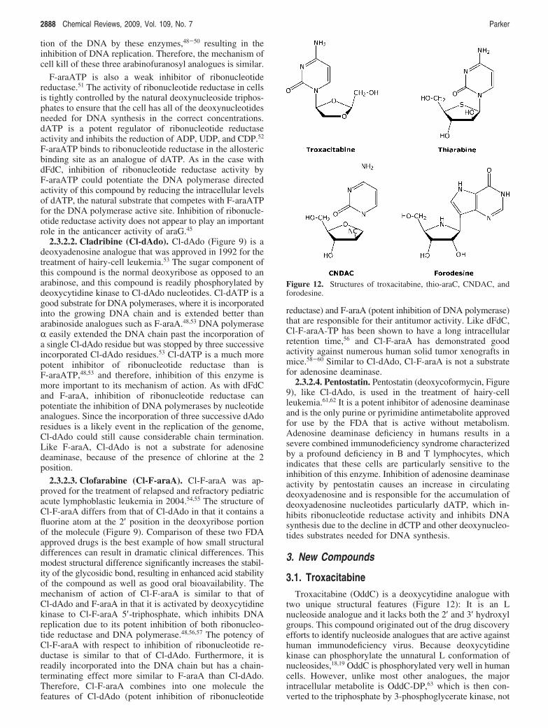
3.1. TroxacitabineTroxacitabine (OddC) is a deoxycytidine analogue with
two unique structural features (Figure 12): It is an Lnucleoside analogue and it lacks both the 2′ and 3′ hydroxylgroups. This compound originated out of the drug discoveryefforts to identify nucleoside analogues that are active againsthuman immunodeficiency virus. Because deoxycytidinekinase can phosphorylate the unnatural L conformation ofnucleosides,18,19 OddC is phosphorylated very well in humancells. However, unlike most other analogues, the majorintracellular metabolite is OddC-DP,63 which is then con-verted to the triphosphate by 3-phosphoglycerate kinase, not
Figure 12. Structures of troxacitabine, thio-araC, CNDAC, andforodesine.
2888 Chemical Reviews, 2009, Vol. 109, No. 7 Parker
nucleoside diphosphate kinase.64,65 Unlike most other dideoxy-nucleotides, OddC-TP is a good substrate for DNA poly-merase R and is incorporated into the DNA chain where itis an absolute DNA chain terminator because of its lack ofa 3-OH group.66 Because of the chiral preference for 3′-5′proof-reading exonucleases associated with DNA poly-merase, once incorporated into DNA, OddC is not easilyremoved from the DNA chain,67 although OddC is recog-nized by apurinic/apyrimidinic endonuclease.68 OddC is avery poor substrate for cytidine deaminase. OddC hasdemonstrated efficacy in both solid and hematologicalmalignancies in clinical trials.69
3.2. ThiarabineAlthough thiarabine (T-araC) is structurally similar to araC
(Figure 12), the antitumor activity of T-araC against a varietyof human tumor xenografts in mice is dramatically betterthan that of araC,70 a compound that does not demonstratesolid tumor activity in these animal models or in patients.T-araC has also demonstrated better activity than gemcitabineagainst various human tumor xenografts in mice. Althoughthe basic mechanism of action of T-araC71-75 is similar tothat of araC (both compounds are phosphorylated to theirrespective triphosphates (T-araCTP or araCTP) and inhibitDNA synthesis), there are several quantitative differencesin the metabolism and biochemical activity of these twocompounds that can explain their differences in antitumoractivity. Most importantly, the half-life of T-araCTP in solidtumor cells is approximately 10 times longer than that ofaraCTP,76 and T-araCTP is a much more potent inhibitor ofDNA synthesis than is araCTP.71 As with gemcitabine, thesetwo activities are believed to be very important to the activitydemonstrated in mice against solid tumor xenografts. Inaddition, the interaction of T-araC with numerous otherenzymes involved with the activation of deoxycytidineanalogues differs from araC, and these differences may alsocontribute to the in vivo activity of T-araC. With respect toaraC and its metabolites, T-araC is a poor substrate fordeoxycytidine kinase and deoxycytidine deaminase activities.T-araCMP is a poor substrate for dCMP deaminase activity,but it is a better substrate for CMP/UMP kinase than isaraCMP, a difference that may help explain the long half-life of T-araCTP.76 Like araC, T-araC has only a modesteffect on ribonucleotide reductase activity. T-araC has beenevaluated in two clinical trials to treat solid tumors77,78 andis currently being prepared for further clinical evaluation.T-araC demonstrated partial responses in some of the heavilypretreated patients with relapsed solid tumors in these trials.
3.3. Sapacitabine1-[2-C-cyano-2-deoxy-�-D-arabinofuranosyl]-cytosine (CN-
DAC) is a deoxycytidine analogue with a structure that issimilar to araC (Figure 12). However, instead of a 2′-hydroxygroup, CNDAC has a 2′-cyano group. Similar to araC,CNDAC is phosphorylated via deoxycytidine kinase toCNDAC-TP, which is a good substrate for DNA polymerasesinvolved in DNA replication. Once incorporated into theDNA chain, CNDAC is a powerful chain terminator.79 Chainelongation by DNA polymerase R was severely inhibited bythe incorporation of CNDAC into the 3′-terminus, which wasgreater than that observed with either araC and gemcitabine.If CNDAC is incorporated into the internal DNA linkages,it has a secondary affect on DNA integrity. Once the DNA
chain is extended after the incorporation of CNDAC, the3′-phosphodiester link between CNDAC and the nextnucleotide is not stable and the DNA chain is spontaneouslycleaved through a � elimination reaction that generates aDNA chain that is terminated with 2′-C-cyano-2′,3′-didehy-dro-2′,3′-dideoxycytidine. Therefore, incorporation ofCNDAC into DNA chains can result in single strand breaksin the DNA. This mechanistic consideration contributed tothe design of this molecule, and the dideoxy analogue hasbeen detected in the DNA of cells treated with CNDAC.81,82
Like araC, treatment with CNDAC does not inhibit ribo-nucleotide reductase activity. An N4 palmitoyl derivative ofCNDAC (Sapacitabine) is being evaluated in the clinic forantitumor activity.83
3.4. ForodesinePeople born with a deficiency of purine nucleoside
phosphorylase (PNP) are healthy except that they do notproduce T-cells, which results in a severe immunodeficiencydisease that usually causes death early in life.84,85 Thiscondition suggests that inhibitors of PNP would haveselective activity against T-cell malignancies. PNP is animportant enzyme in the salvage of purine nucleosides, andin its absence, intracellular deoxyguanosine is not cleavedto guanine but is instead converted to deoxyguanosine 5′-triphosphate (dGTP), which is a feedback inhibitor ofribonucleotide reductase activity. Therefore, the expandeddGTP pool in T-cells results in the inhibition of ribonucleo-tide reductase activity and depletion of intracellular deoxy-nucleotides that are required for DNA synthesis. Thesensitivity of T cells to PNP inhibition is believed to be dueto relatively high levels of nucleoside kinase activity andlow levels of nucleotidase activity in these cells. Forodesine(Figure 12) is a potent inhibitor of PNP activity with a Ki of72 pM.86,87 The affinity of this compound for the enzyme isapproximately 1 million times that for inosine, the naturalsubstrate. Forodesine was potent enough to result in aprofound inhibition of PNP activity in intact animals andhas demonstrated excellent activity against human peripheralblood lymphocytes engrafted into SCID mice. Forodesineis similar to pentostatin in that it is active without metabo-lism. The FDA granted orphan drug status to forodesine inFebruary of 2004, and it is being evaluated in human clinicaltrials for the treatment of cutaneous T-cell lymphoma andchronic lymphocytic leukemia.88-90
3.5. Suicide Gene Therapy of Cancer UsingPurine and Pyrimidine Analogues
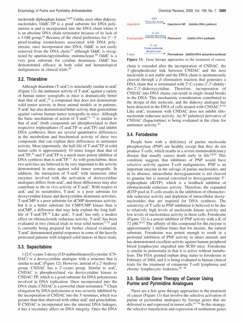
There are a few gene therapy approaches to the treatmentof cancer (Figure 13) that involve the selective activation ofpurine or pyrimidine analogues by foreign genes that aredelivered to and expressed in tumor cells.91-94 In this strategy,the selective transfection and expression of nonhuman genes
Figure 13. Gene therapy approaches to the treatment of cancer.
Enzymology of Purine and Pyrimidine Antimetabolites Chemical Reviews, 2009, Vol. 109, No. 7 2889
in tumor cells creates a difference in the tumor cells thatcan be exploited to selectively kill the tumor cells. Theoreti-cally, this approach to the treatment of cancer should killcancer cells with much less toxicity than is seen withconventional therapy. The genes for these enzymes are firstdelivered to tumor cells by various viral or bacterial vectorsthat have been engineered for this purpose, and then thepatient is treated systemically with prodrugs that are activatedto cytotoxic compounds by the enzymes expressed from thegenes.
The gene that has received the most attention is the herpessimplex virus (HSV) thymidine kinase (TK), and numerousclinical trials have been conducted to evaluate this approachwithout much success. The expression of this gene in cellsinfected with HSV is the basis of the selective antiviraltherapy for the treatment of HSV infections, and its expres-sion in tumor cells has been used to activate ganciclovir, apurine nucleoside analogue used in the treatment of CMV,to cytotoxic nucleotide metabolites. The HSV-TK strategy,however, has a limited ability to kill neighboring tumor cellsthat do not express the gene (bystander cells), because theproduct of the reaction of HSV-TK with ganciclovir isganciclovir-5′-monophosphate, which does not easily diffuseout of the cell in which it was formed. The bystander activityseen with the HSV-TK approach is dependent upon gapjunctions and requires cell-to-cell contact. Since currenttechnology is not able to deliver foreign genes to the majorityof the tumor cells, the limited bystander activity of ganci-clovir monophosphate is a major limiting factor of the HSV-TK approach in the treatment of cancer.
In addition, ganciclovir-TP kills tumor cells by inhibitingDNA polymerases involved in DNA replication, much likeconventional nucleoside analogues. Therefore, ganciclovirprimarily targets proliferating cells. Since solid tumors oftenhave a low growth fraction, the lack of activity of thisapproach against nonproliferating tumor cells is anotherdeficiency of this approach to the treatment of solid tumors.
5-Fluorocytosine (F-Cyt) is approved for the treatment offungal diseases because of its selective deamination in fungalcells to FUra and has also been evaluated in gene therapystrategies in which E. coli or yeast cytosine deaminase isexpressed in tumor cells. Human cells do not express cytosinedeaminase, and F-Cyt is well tolerated in people. Deliveryof cytosine deaminase to tumor cells has been shown tosensitize them to F-Cyt, and a few clinical trials are underwayto evaluate this gene therapy strategy.
E. coli purine nucleoside phosphorylase (PNP), unlikehuman PNP, accepts adenosine as a substrate and cleavesthe glycosidic bond to produce adenine and ribose-1-phosphate. This difference in substrate specificity betweenthese two enzymes has been exploited to create a genetherapy strategy to activate deoxyadenosine analogues to veryactive adenine analogues in tumor cells. The adenineanalogues produced from E. coli PNP can readily diffuse toand kill surrounding tumor cells that do not express E. coliPNP, which is an important attribute for gene therapyapproaches to the treatment of cancer due to the difficultyof delivering genes to tumor cells. Because human cellscontain nucleoside and nucleobase transporters in theirmembranes that facilitate the diffusion of purines acrossmembranes in either direction, the bystander activity forpurine and pyrimidine bases is not dependent upon gapjunctions and does not require cell-to-cell contact, as is thecase with ganciclovir nucleotides. Excellent antitumor activ-
ity has been observed with F-araAMP against human tumorxenografts in mice, even when only 2.5% of the tumor cellsexpress E. coli PNP.95 Two clinical trials are scheduled tobegin in 2009 to evaluate the safety and efficacy of the useof E. coli PNP with F-araAMP in treatment of solid tumors.
In addition to the high bystander activity of the E. coliPNP approach, the toxic adenine analogue formed fromF-araAMP and E. coli PNP (2-fluoroadenine) has a uniquemechanism of action that results in the killing of bothproliferating and nonproliferating tumor cells.96 2-Fluoro-adenine is converted to an ATP analogue, that inhibits RNAand/or protein synthesis. This mechanism of cell kill isdifferent from that of all currently used anticancer agentsand would not be tolerated if the agent was administeredsystemically. Fluoroadenine has been evaluated in mousemodels of cancers and has not demonstrated selectiveantitumor activity. The activity of this antitumor strategyagainst nonproliferating cells is of particular importance tothe treatment of solid tumors, which often have a very lowgrowth fraction. The ability to kill nonproliferating tumorcells is a major characteristic of the E. coli PNP approachthat distinguishes it from both the cytosine deaminase andthe thymidine kinase approach.
The use of gene therapy to deliver genes to tumor cellssolves the problem associated with the lack of selectivity ofthe current chemotherapy, but it introduces another difficultproblem to solve, i.e., selective delivery of genes to tumorcells with sufficient enzyme expression. The vectors availablein 2009 do not express enough enzyme activity in enoughtumor cells after systemic administration to activate enoughprodrug. Therefore, gene therapy approaches in the clinichave been limited to tumors that can be injected with thevector. It is hoped that, with continued study, new vectorswill be developed that will be able to selectively deliversufficient amount of genes to tumors after systemic admin-istration, allowing for activity against metastatic disease.However, because of the difficulty of delivering vectors totumor cells throughout the body, gene therapy may onlyprove to be useful for the treatment of localized tumors.
4. Drug Design ConsiderationsThere is clearly an important role for nucleosides in the
treatment of cancer, and the design of new agents withinthis class of compounds is still warranted. However, design,synthesis, and evaluation of new analogues as potentialanticancer agents is not currently a major emphasis in thedrug development community. The reasons for this lack ofactivity include (a) concerns about the toxicity of nucleosideanalogues and the important goal of designing new drugswith less toxicity than the classical agents and (b) concernsthat perhaps new nucleoside analogues would not be suf-ficiently different from those already known and approvedfor human use, and therefore no further advances were likely.Although toxicity is still a problem and is an issue that ishard to circumvent with antimetabolites (or other classicalcytotoxic agents), the information provided in the precedingpages clearly indicates that small structural changes can haveprofound effects on the biological activity of nucleosideanalogues and suggests that new agents with useful activitiescan still be identified.
An important aspect of the design of purine and pyrimidineantimetabolites is that the drug design process is largelyempirical in nature. Compounds are designed that arestructurally similar to existing agents based on a thorough
2890 Chemical Reviews, 2009, Vol. 109, No. 7 Parker
understanding of the past work in this field, and they aretested in various biological assays. As indicated in thisreview, a considerable amount of structure activity relation-ship (SAR) data is available from the many years of workwith this class of compounds that helps guide the design ofnew compounds. Although this review has focused on thesuccess stories, there are many more examples of antime-tabolites that have been designed and synthesized that havenot been successful, and a thorough understanding of boththe successes and failures is critical to the rational develop-ment of new agents of this class. The analysis of the existingFDA-approved anticancer nucleosides indicates a clear andsimple guideline that should be considered in the design ofnew agents in this class. The new compounds should includestructural changes that are as small as possible, and as fewchanges as possible should be made to the natural molecule,with 1-3 changes being the most desirable number.
Because all of the purine and pyrimidine analogues usedin the treatment of cancer are prodrugs (except pentostatin),their mechanism of action is very complex and involvesinteraction with many different anabolic and catabolicenzymes. Therefore, it will not be easy to replace thisempirical process with a more “rational” drug design process.Although the empirical approach used in the design of newnucleosides is also a “rational” way to design new drugs,the newer “rational” drug design concepts optimally involvethe use of the three-dimensional structure of the protein targetcoupled with biochemical results and in silico modelingmethodology. This approach is most useful when a drug isenvisioned to act largely by affecting a single enzymatictarget. Although structural information with the variousenzymes (particularly deoxycytidine kinase) is increasinglybeing utilized to aid in the design of new antimetabolites,the design of new antimetabolites is not driven by the desireto interact with just one enzyme. For example, it would notbe helpful to design a potent nucleotide inhibitor of DNApolymerase based on structural information, if the nucleosidecomponent of the designed nucleotide inhibitor cannot readilypenetrate the cell and be converted to the triphosphate.Nucleotide analogues do not make good drugs, because theydo not easily penetrate cell membranes and the phosphatesare rapidly removed by plasma phosphatases, although inrecent years phosphonate nucleotides have proven to beuseful in the treatment of certain viral diseases97 and someresearch groups are developing strategies to deliver nucleo-tides to tumor cells.98
Another complicating factor in the “rational” design ofnew analogues is that the known active agents often inhibitmore than one intracellular target. This attribute can beviewed as a strength of this class of compounds and is oneof the reasons that antimetabolites have been so successfulin the clinic. The multiple points of mechanistic action,however, bring with them serious challenges in terms of“rational” drug design. As difficult as it is to develop a newdrug that inhibits only one intracellular target, it is muchmore than twice as difficult to design one compound thatcan inhibit two or more enzymes.
The process of evaluating a new analogue for antitumoractivity is fairly simple. Once a new antimetabolite has beendesigned and synthesized, the first and most importantexperiment is to determine whether or not the compoundcan kill cancer cells in in vitro assays. A positive resultindicates that the compound is able to interact with themetabolic enzymes of either the purine or pyrimidine
pathway to create a metabolite that inhibits an enzymeimportant to DNA replication. Because of the considerableknowledge around this class of compounds, the biochemicaldetails of the mechanism of action can be sketched out witha fair degree of accuracy based on knowledge of the structureof the new agent. However, biochemical studies must stillbe done to determine how the new agent differs fromstructurally similar compounds and whether the new agenthas characteristics that may be beneficial.
Because of the similarity of structure and mechanism ofaction of nucleoside analogues, there is no in vitro assaythat can predict whether or not any new agent will havesufficient selectivity to be useful in the clinic. An analoguemust be able to kill cells in vitro, but to determine whetherit will have the appropriate antitumor selectivity, thecompound must be evaluated in in vivo studies againstvarious mouse models of cancer. Since selectivity for tumorcells is the most important aspect of a new antitumor agentand in vitro studies cannot predict for selectivity, newanalogues must be evaluated in in vivo tumor models as soonas possible. Of course there are serious issues with the abilityof the currently used in vivo mouse models to predict forclinical activity, but they are currently the best method todetermine whether an agent has antitumor activity at dosesthat are tolerated in an intact animal. Because large amountsof compound are needed for in vivo studies, compoundavailability is often a significant hurdle to conducting thesestudies. Regardless, the importance of in vivo studies cannotbe overstated and they should be initiated as soon assufficient compound is available to proceed. Through manyyears of experience in the drug discovery process at SouthernResearch Institute, we have learned that activity against onetype of human tumor xenograft (for instance breast tumors)does not predict for activity against that particular tumor typein human disease. The current best predictor for clinicalactivity for any compound against any tumor type is thedemonstration of robust antitumor activity (ability to causeregressions and cures) in numerous human tumor xenograftsin mice. If an agent demonstrates robust antitumor activityin mouse models, then clinical trials are necessary todetermine which tumor types, if any, are sensitive to theagent in people.
5. SummaryPurine and pyrimidine analogues remain an important class
of drugs in the treatment of cancer. Although these agentsshare many structural and biochemical characteristics, eachcompound has unique activities that make it a useful drug.The basis of selectivity of these agents is not clearly defined(because the molecular targets exist in both tumor cells andnormal host tissues) but is believed to be primarily due todifferences in metabolism and proliferative states betweentumor cells and normal cells. For instance, the greaterexpression of deoxycytidine kinase in leukemias and lym-phomas is believed to contribute to the sensitivity of thesemalignancies to nucleoside analogues that are activated bythis enzyme. Furthermore, most normal cells in a patient arequiescent and, therefore, are not sensitive to these agents.However, the selectivity of antimetabolites is still poor andbetter agents are needed with fewer toxicities.
Analysis of the existing agents identifies three primarycharacteristics of antimetabolites that are important to theirability to kill tumor cells: sufficient metabolism to activemetabolite; long retention of active metabolite; and potent
Enzymology of Purine and Pyrimidine Antimetabolites Chemical Reviews, 2009, Vol. 109, No. 7 2891
and sustained inhibition of DNA replication or function. Theanalogue should be a reasonable substrate for the activatingenzymes, although clearly this aspect of the activity of ananalogue can be affected by the potency of the activemetabolite against the enzymatic target. For instance, ananalogue that produces a very potent active metabolite wouldnot be as dependent on activation. In addition to all theanabolic enzymes involved in the activation of nucleosideanalogues, there are numerous catabolic enzymes that interactwith these compounds, and these enzymes can also haveprofound impact on their biological activity and are importantin theactivityofallof thepurineandpyrimidineantimetabolites.
The compound should be a good selective inhibitor ofDNA replication and have minimal effects on RNA andprotein synthesis, as inhibition of these activities leads totoxicity. The primary intracellular targets of the existingpurine and pyrimidine antimetabolites are DNA polymerases,thymidylate synthetase, and ribonucleotide reductase. Al-though some of the currently approved agents (FUra,mercaptopurine, thioguanine, and aza-Cyd) are converted toribonucleotide metabolites and are extensively incorporatedinto RNA, the primary activity of these compounds thatresults in their antitumor activity is their inhibition of DNAsynthesis or disruption of DNA function. Unless there isselective activation in tumor cells, nucleoside analogues thattarget RNA synthesis or function should be extremelycytotoxic, since all cells require RNA for vitality.
As with most other classical antitumor agents, the inhibi-tion of DNA replication is the most important action ofpurine and pyrimidine metabolites responsible for theirantitumor activity. Disruption of de novo purine biosynthesisor RNA effects are secondary to activities that disrupt DNAreplication or cause DNA damage. However, inhibition ofDNA synthesis is not sufficient to kill a tumor cell. Forexample, an agent such as aphidicolin, which is a potentinhibitor of DNA replication, is a good cell synchronizer,because it only inhibits DNA synthesis and, unlike nucleosideanalogues, it does not cause any lasting inhibition. Once itis removed from the cell, DNA synthesis readily resumeswithout lasting toxicity. Nucleoside analogues have twoattributes that result in a lasting inhibition of DNA replicationafter removal of the drug by natural processes within thebody. First, the active metabolites of these agents arenucleotide analogues, which do not readily penetrate cellmembranes and, therefore, are retained in the cell after thedrug has been removed, which is an attribute that is uniqueto this class of antitumor agents. The half-life for the removalof the triphosphates from cells can be quite long, which leadsto continued use by the polymerases and, thus, continuedinhibition of DNA replication. The intracellular retention timeof the active metabolites (nucleoside triphosphate) can varyconsiderably between the various analogues, and this canhave an important effect on the activity of an agent againstsolid tumor cells. The much longer half-life of dFdC-TP thanaraCTP is believed to be a primary contributing factor tothe solid tumor activity of gemcitabine and the lack of solidtumor activity of araC. Second, nucleosides are incorporatedinto DNA, resulting in a DNA molecule that is not easilyextended and must be repaired before synthesis can resume.Therefore, an agent that causes DNA damage that is poorlyor slowly repaired will result in prolonged damage to theDNA, which will lead to the induction of apoptosis.
In conclusion, purine and pyrimidine antimetabolites arean important class of drugs used in the treatment of cancer
and viral diseases. Although the toxicity of these compoundscan limit their usefulness, the antimetabolites will continueto play an important role in the treatment of cancer for theforeseeable future. It is likely that some of the new nucleosideanalogues that are currently in the pipeline will be approvedfor use in the coming years. Although drug discovery is beingpursued of new anticancer agents that target enzyme activitiesmore closely associated with the cancer phenotype, theunpredicted toxicity of these new agents could still be a majorissue of these agents as well. The design, synthesis, andevaluation of new purine and pyrimidine analogues is still aproductive area for discovering new drugs for the treatmentof cancer, since many years of knowledge with respect totheir potential actions and toxicity has accumulated. Novelnucleoside analogues with unique actions are continuouslybeing identified, and the information provided in this reviewindicates that small structural modifications of nucleosideanalogues can have profound effects on their chemicalstability and spectrum of biological activity.
6. References(1) Zhang, J.; Visser, F.; King, K. M.; Baldwin, S. A.; Young, J. D.; Cass,
C. E. Cancer Metastasis ReV. 2007, 26, 85.(2) Sampath, D.; Rao, V. A.; Plunkett, W. Oncogene 2003, 22, 9063.(3) Plunkett, W.; Gandhi, V. Cancer Chemother. Biol. Response Modif.
2001, 19, 21.(4) Arner, E. S. J.; Eriksson, S. Pharmacol. Ther. 1995, 67, 155.(5) Johansson, N. G.; Eriksson, S. Acta Biochim. Pol. 1996, 43, 143.(6) Eriksson, S.; Munch-Peterson, B.; Johansson, K.; Eklund, H. Cell.
Mol. Life Sci. 2002, 59, 1327.(7) Rompay, A. R. V.; Johansson, M.; Karlsson, A. Pharmacol. Ther.
2000, 87, 189.(8) Sabini, E.; Ort, S.; Monnerjahn, C.; Konrad, M.; Lavie, A. Nat. Struct.
Biol. 2003, 10, 513.(9) Shcherbakova, P. V.; Bebenek, K.; Kunkel, T. A. Sci. Aging Knowledge
EnViron. 2003, 2003, 1.(10) Matherly, L. H.; Schuetz, J. D.; Westin, E.; Goldman, I. D. Anal.
Biochem. 1989, 182, 338.(11) Elion, G. B. Science 1989, 244, 41.(12) Nelson, J. A.; Carpenter, J. W.; Rose, L. M.; Adamson, D. J. Cancer
Res. 1975, 35, 2872.(13) Karran, P. Br. Med. Bull. 2007, 79, 153.(14) Yoshida, S.; Yamada, M.; Masaki, S.; Saneyoshi, M. Cancer Res. 1979,
39, 3955.(15) Ling, Y. H.; Nelson, J. A.; Cheng, Y. C.; Anderson, R. S.; Beattie,
K. L. Mol. Pharmacol. 1991, 40, 508.(16) Aarbakke, J.; Janka-Schaub, G.; Elion, G. B. Trends Pharmacol. Sci.
1997, 18, 3.(17) Tomalik-Scharte, D.; Lazar, A.; Fuhr, U.; Kirchheiner, J. Pharmaco-
genomics J. 2008, 8, 4.(18) Heidelberger, C.; Chaudhuri, N. K.; Danenberg, P.; Mooren, D.;
Griesbach, L.; Duschinsky, R.; Schnitzer, R. J.; Pleven, E.; Scheiner,J. Nature 1957, 179, 663.
(19) Danenberg, P. V. Biochim. Biophys. Acta 1977, 473, 73.(20) Myers, C. E. Pharmacol. ReV. 1981, 33, 1.(21) Parker, W. B.; Cheng, Y. C. Pharmacol. Ther. 1990, 48, 381.(22) Walko, C. M.; Lindley, C. Clin. Ther. 2005, 27, 23.(23) Homsi, J.; Garrett, C. R. Cancer Control 2006, 13, 42.(24) Grant, S. AdV. Cancer Res. 1998, 72, 197.(25) Townsend, A. J.; Cheng, Y. C. Mol. Pharmacol. 1987, 32, 330.(26) Huang, P.; Chubb, S.; Hertel, L. W.; Grindey, G. B.; Plunkett, W.
Cancer Res. 1991, 51, 6110.(27) Ostruszka, L. J.; Shewach, D. S. Cancer Chemother. Pharmacol. 2003,
52, 325.(28) Gandhi, V.; Legha, J.; Chen, F.; Hertel, L. W.; Plunkett, W. Cancer
Res. 1996, 56, 4453.(29) Plunkett, W.; Huang, P.; Searcy, C. E.; Gandhi, V. Semin. Oncol. 1996,
23 (Suppl 10), 3.(30) Stresemann, C.; Lyko, F. Int. J. Cancer 2008, 123, 8.(31) Cihak, A.; Vesely, J.; Skoda, J. AdV. Enzyme Regul. 1985, 24, 335.(32) Momparler, R. L. Pharmacol. Ther. 1985, 30, 287.(33) Oki, Y.; Aoki, E.; Issa, J. J. Crit. ReV. Oncol./Hematol. 2007, 61,
140.(34) Bouchard, J.; Momparler, R. L. Mol. Pharmacol. 1983, 24, 109.(35) Link, P. A.; Baer, M. R.; James, S. R.; Jones, D. A.; Karpf, A. R.
Cancer Res. 2008, 68, 9358.
2892 Chemical Reviews, 2009, Vol. 109, No. 7 Parker
(36) Vesely, J.; Cihak, A.; Sorm, F. Collect. Czech. Chem. Commun. 1969,34, 901.
(37) Newman, E. M.; Santi, D. V. Proc. Natl. Acad. Sci. U.S.A. 1982, 79,6419.
(38) Plunkett, W.; Huang, P.; Gandhi, V. Semin. Oncol. 1990, 17, 3.(39) Gandhi, V.; Plunkett, W. Drug Dispos. 2002, 41, 93.(40) Chilson, O. P.; Fisher, J. R. Arch. Biochem. Biophys. 1963, 102, 77.(41) Frederickson, S. Arch. Biochem. Biophys. 1966, 113, 383.(42) Buie, L. W.; Epstein, S. S.; Lindley, C. M. Clin. Ther. 2007, 29, 1887.(43) Reist, E. J.; Goodman, L. Biochemistry 1964, 3, 15.(44) Cohen, A.; Lee, J. W.; Gelfand, E. W. Blood 1983, 61, 660.(45) Shewach, D. S.; Daddona, P. E.; Ashcraft, E.; Mitchell, B. S. Cancer
Res. 1985, 45, 1008.(46) Rodriguez, C. O., Jr; Stellrecht, C. M.; Gandhi, V. Blood 2003, 102,
1842.(47) Rodriguez, C. O., Jr; Mitchell, B. S.; Ayres, M.; Eriksson, S.; Gandhi,
V. Cancer Res. 2002, 62, 3100.(48) Parker, W. B.; Bapat, A. R.; Shen, J. X.; Townsend, A. J.; Cheng,
Y. C. Mol. Pharmacol. 1988, 34, 485.(49) Huang, P.; Chubb, S.; Plunkett, W. J. Biol. Chem. 1990, 265, 16617.(50) Gandhi, V.; Mineishi, S.; Huang, P.; Chapman, A. J.; Yang, Y.; Chen,
F.; Nowak, B.; Chubb, S.; Hertel, L. W.; Plunkett, W. Cancer Res.1995, 55, 1517.
(51) Nutter, L. M.; Cheng, Y. C. Pharmacol. Ther. 1984, 26, 191.(52) Goodman, G. R.; Beutler, E.; Saven, A. Best Pract. Res., Clin.
Haematol. 2003, 16, 101.(53) Parker, W. B.; Shaddix, S. C.; Chang, C. H.; White, E. L.; Rose, L. M.;
Brockman, R. W.; Shortnancy, A. T.; Montgomery, J. A.; Secrist, J. A.,III; Bennett, L. L. Jr Cancer Res. 1991, 51, 2386.
(54) Bonate, P. L.; Arthaud, L., Jr; Stephenson, K.; Secrist, J. A., III;Weitman, S. Nat. ReV. Drug DiscoVery 2006, 5, 855.
(55) Faderl, S.; Gandhi, V.; Keating, M. J.; Jeha, S.; Plunkett, W.;Kantarjian, H. M. Cancer 2005, 103, 1985.
(56) Xie, C.; Plunkett, W. Cancer Res. 1995, 55, 2847.(57) Xie, K. C.; Plunkett, W. Cancer Res. 1996, 56, 3030.(58) Waud, W. R.; Schmid, S. M.; Montgomery, J. A.; Secrist, J. A., III
Nucleosides Nucleotides Nucleic Acids 2000, 19, 447.(59) Takahashi, T.; Shimizu, M.; Akinaga, S. Cancer Chemother. Phar-
macol. 2002, 50, 193.(60) Carson, D. A.; Wasson, D. B.; Esparza, L. M.; Carrera, C. J.; Kipps,
T. J.; Cottam, H. B. Proc. Natl. Acad. Sci. U.S.A. 1992, 89, 2970.(61) Cheson, B. Semin. Oncol. 1992, 19, 695.(62) Tallman, M. S.; Hakimian, D. Blood 1995, 86, 2463.(63) Grove, K. L.; Guo, X.; Liu, S. H.; Gao, Z.; Chu, C. K.; Cheng, Y. C.
Cancer Res. 1995, 55, 3008.(64) Krishnan, P.; Fu, Q.; Lam, W.; Liou, J. Y.; Dutschman, G.; Cheng,
Y. C. J. Biol. Chem. 2002, 277, 5453.(65) Krishnan, P.; Gullen, E. A.; Lam, W.; Dutschman, G. E.; Grill, S. P.;
Cheng, Y. C. J. Biol. Chem. 2003, 278, 36726.(66) Kukhanova, M.; Liu, S. H.; Mozzherin, D.; Lin, T. S.; Chu, C. K.;
Cheng, Y. C. J. Biol. Chem. 1995, 270, 23055.(67) Grove, K. L.; Cheng, Y. C. Cancer Res. 1996, 56, 4187.(68) Chou, K. M.; Kukhanova, M.; Cheng, Y. C. J. Biol. Chem. 2000,
275, 31009.(69) Swords, R.; Giles, F. Hematology 2007, 12, 219.(70) Waud, W. R.; Gilbert, K. S.; Shepherd, R. V.; Montgomery, J. A.;
Secrist, J. A., III Cancer Chemother. Pharmacol. 2003, 51, 422.(71) Parker, W. B.; Shaddix, S. C.; Rose, L. M.; Waud, W. R.; Shewach,
D. S.; Tiwari, K. N.; Secrist, J. A., III Biochem. Pharmacol. 2000,60, 1925.
(72) Richardson, F.; Black, C.; Richardson, K.; Franks, A.; Wells, E.;Karimi, S.; Sennello, G.; Hart, K.; Meyer, D.; Emerson, D.; Brown,E.; LeRay, J.; Nilsson, C.; Tomkinson, B.; Bendele, R. CancerChemother. Pharmacol. 2005, 55, 213.
(73) Clarke, M. L.; Damaraju, V. L.; Zhang, J.; Mowles, D.; Tackaberry,T.; Lang, T.; Smith, K. M.; Young, J. D.; Tomkinson, B.; Cass, C. E.Mol. Pharmacol. 2006, 70, 303.
(74) Richardson, K. A.; Vega, T. P.; Richardson, F. C.; Moore, C. L.;Rohloff, J. C.; Tomkinson, B.; Bendele, R. A.; Kuchta, R. D. Biochem.Pharmacol. 2004, 68, 2337.
(75) Someya, H.; Shaddix, S. C.; Tiwari, K. N.; Secrist, J. A., III; Parker,W. B. J. Pharmacol. Exp. Ther. 2003, 304, 1314.
(76) Someya, H.; Waud, W. R.; Parker, W. B. Cancer Chemother.Pharmacol. 2006, 57, 772.
(77) Goss, G.; Siu, L. L.; Gauthier, I.; Chen, E. X.; Oza, A. M.; Goel, R.;Maroun, J.; Powers, J.; Walsh, W.; Maclean, M.; Drolet, D. W.; Rusk,J.; Seymour, L. K. Cancer Chemother. Pharmacol. 2006, 58, 703.
(78) Lee, C. P.; de Jonge, M. J.; O’Donnell, A. E.; Schothorst, K. L.;Hanwell, J.; Chick, J. B.; Brooimans, R. A.; Adams, L. M.; Drolet,D. W.; de Bono, J. S; Kaye, S. B.; Judson, I. R.; Verweij, J. Clin.Cancer Res. 2006, 12, 2841.
(79) Azuma, A.; Huang, P.; Matsuda, A.; Plunkett, W. Biochem. Pharmacol.2001, 61, 1497.
(80) Matsuda, A.; Nakajima, Y.; Azuma, A.; Tanaka, M.; Sasaki, T. J. Med.Chem. 1991, 34, 2917.
(81) Hanaoka, K.; Suzuki, M.; Kobayashi, T.; Tanzawa, F.; Tanaka, K.;Shibayama, T.; Miura, S.; Ikeda, T.; Iwabuchi, H.; Nakagawa, A.;Mitsuhashi, Y.; Hisaoka, M.; Kaneko, M.; Tomida, A.; Wataya, Y.;Nomura, T.; Sasaki, T.; Matsuda, A.; Tsuruo, T.; Kurakata, S. Int. J.Cancer 1999, 82, 226.
(82) Wang, Y.; Liu, X.; Matsuda, A.; Plunkett, W. Cancer Res. 2008, 68,3881.
(83) Gilbert, J.; Carducci, M. A.; Baker, S. D.; Dees, E. C.; Donehower,R. InVest. New Drugs 2006, 24, 499.
(84) Markert, M. L. Immunodefic. ReV. 1991, 3, 45.(85) Montgomery, J. A. Med. Res. ReV. 1993, 13, 209.(86) Schramm, V. L. Biochem. Biophys. Acta 2002, 1587, 107.(87) Gandhi, V.; Balakrishnan, K. Semin. Oncol. 2007, 34 (suppl 5), S8.(88) Larson, R. A. Semin. Oncol. 2007, 34 (suppl 5), S13.(89) Furman, R. R.; Hoelzer, D. Semin. Oncol. 2007, 34 (suppl 5), S29.(90) Gore, L.; Stelljes, M.; Quinones, R. Semin. Oncol. 2007, 34 (suppl
5), S35.(91) Springer, C. J.; Niculescu, I. J. Clin. InVest. 2000, 105, 1161.(92) Denny, W. A. J. Biomed. Biotechnol. 2003, 2003, 48.(93) Dachs, G. U.; Tupper, J.; Tozer, G. M. Anti-Cancer Drugs 2005, 16,
349.(94) Altaner, C. Cancer Let. 2008, 270, 191.(95) Hong, J. S.; Waud, W. R.; Levasseur, D. N.; Townes, T. M.; Wen,
H.; McPherson, S. A.; Moore, B. A.; Bebok, Z.; Allan, P. W.; Secrist,J. A., III; Parker, W. B.; Sorscher, E. J. Cancer Res. 2004, 64, 6610.
(96) Parker, W. B.; Allan, P. W.; Shaddix, S. C.; Rose, L. M.; Speegle,H. F.; Gillespie, G. Y.; Bennett, L. L., Jr. Biochem. Pharmacol. 1998,55, 1673.
(97) De Clercq, E. AntiViral Res. 2007, 75, 1.(98) Boyer, S. H.; Sun, Z.; Jiang, H.; Esterbrook, J.; Gomez-Galeno, J. E.;
Craigo, W.; Reddy, K. R.; Ugarkar, B. G.; MacKenna, D. A.; Erion,M. D. J. Med. Chem. 2006, 49, 7711.
CR900028P
Enzymology of Purine and Pyrimidine Antimetabolites Chemical Reviews, 2009, Vol. 109, No. 7 2893