Embed Size (px)
Citation preview

Seediscussions,stats,andauthorprofilesforthispublicationat:https://www.researchgate.net/publication/229339112
Enzymologyandstructureofcatalases
ArticleinAdvancesinInorganicChemistry·January2000
DOI:10.1016/S0898-8838(00)51001-0
CITATIONS
147
READS
717
3authors,including:
PeterNicholls
UniversityofEssex
218PUBLICATIONS5,065CITATIONS
SEEPROFILE
IgnacioFita
UniversityofBarcelona
209PUBLICATIONS6,127CITATIONS
SEEPROFILE
Availablefrom:PeterNicholls
Retrievedon:21November2016

P1: FMW/FYE P2: FDJAdvances in Inorganic Chemistry PS006-02 August 11, 2000 14:10 Stylefile version:April 24, 2000
ADVANCES IN INORGANIC CHEMISTRY, VOL. 51
ENZYMOLOGY AND STRUCTURE OF CATALASES
PETER NICHOLLS,* IGNACIO FITA,† and PETER C. LOEWEN‡
*Department of Biological Sciences, Central Campus, University of Essex, Wivenhoe Park,Colchester, CO4 3SQ, United Kingdom;
†CID–CSIC, 08034 Barcelona, Spain; and‡Department of Microbiology, University of Manitoba, Winnipeg, Manitoba R3T 2N2
I. IntroductionII. Categorization
A. Type A: Monofunctional CatalasesB. Type B: Catalase-PeroxidasesC. Type C: Nonheme CatalasesD. Type D: Minor Catalases
III. PhysiologyA. FunctionB. Regulation of Expression
IV. KineticsA. The Catalatic PathwayB. The Peroxidatic Activity of CatalasesC. Compound I and the Pathways via Compound ID. Compound II and the Pathways via Compound IIE. Control by NADPH: The “Extra” PathwayF. The Catalatic Activity of Catalase-Peroxidases
V. Structure of Type A CatalasesA. Subunit StructureB. Quaternary Structure and InterweavingC. Heme Composition and LocationD. Channels and CavitiesE. NADPH BindingF. ComplexesG. Unusual Modifications
VI. Structure of Type B Catalase-PeroxidasesVII. Structure of Chloroperoxidase
VIII. Mechanism of the Catalatic ReactionA. Compound I FormationB. Compound I Reduction
IX. SummaryReferences
51
Copyright C© 2001 by Academic Press.All rights of reproduction in any form reserved.
0898-8838/01 $35.00
P1: FMW/FYE P2: FDJAdvances in Inorganic Chemistry PS006-02 August 11, 2000 14:10 Stylefile version:April 24, 2000
52 NICHOLLS, FITA, AND LOEWEN
I. Introduction
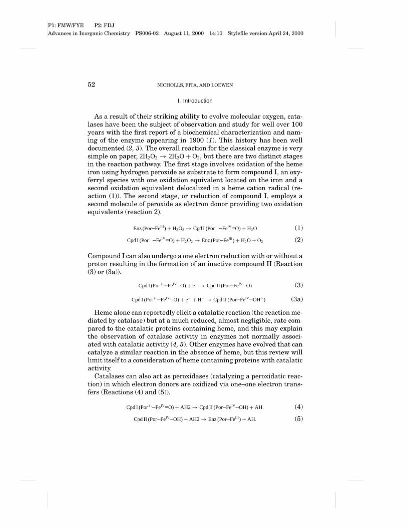
As a result of their striking ability to evolve molecular oxygen, cata-lases have been the subject of observation and study for well over 100years with the first report of a biochemical characterization and nam-ing of the enzyme appearing in 1900 (1). This history has been welldocumented (2, 3). The overall reaction for the classical enzyme is verysimple on paper, 2H2O2→ 2H2O+O2, but there are two distinct stagesin the reaction pathway. The first stage involves oxidation of the hemeiron using hydrogen peroxide as substrate to form compound I, an oxy-ferryl species with one oxidation equivalent located on the iron and asecond oxidation equivalent delocalized in a heme cation radical (re-action (1)). The second stage, or reduction of compound I, employs asecond molecule of peroxide as electron donor providing two oxidationequivalents (reaction 2).
Enz (Por FeIII )+ H2O2→ Cpd I (Por+. FeIV O)+ H2O (1)
Cpd I (Por+. FeIV O)+ H2O2→ Enz (Por FeIII )+ H2O+O2 (2)
Compound I can also undergo a one electron reduction with or without aproton resulting in the formation of an inactive compound II (Reaction(3) or (3a)).
Cpd I (Por+. FeIV O)+ e− → Cpd II (Por FeIV O) (3)
Cpd I (Por+. FeIV O)+ e− + H+ → Cpd II (Por FeIV OH+) (3a)
Heme alone can reportedly elicit a catalatic reaction (the reaction me-diated by catalase) but at a much reduced, almost negligible, rate com-pared to the catalatic proteins containing heme, and this may explainthe observation of catalase activity in enzymes not normally associ-ated with catalatic activity (4, 5). Other enzymes have evolved that cancatalyze a similar reaction in the absence of heme, but this review willlimit itself to a consideration of heme containing proteins with catalaticactivity.
Catalases can also act as peroxidases (catalyzing a peroxidatic reac-tion) in which electron donors are oxidized via one–one electron trans-fers (Reactions (4) and (5)).
Cpd I (Por+. FeIV O)+ AH2→ Cpd II (Por FeIV OH)+ AH. (4)
Cpd II (Por FeIV OH)+ AH2→ Enz (Por FeIII )+ AH. (5)
P1: FMW/FYE P2: FDJAdvances in Inorganic Chemistry PS006-02 August 11, 2000 14:10 Stylefile version:April 24, 2000
ENZYMOLOGY AND STRUCTURE OF CATALASES 53
Generally, the peroxidatic reaction of true catalases is weak in compar-ison to actual peroxidases, but can be an important reaction in the classknown as catalase-peroxidases (Section II,B).
II. Categorization
The diversity among catalases, evident in the variety of subunit sizes,the number of quaternary structures, the different heme prostheticgroups, and the variety of sequence groups, enables them to be orga-nized in four main groups: the “classic” monofunctional enzymes (typeA), the catalase-peroxidases (type B), the nonheme catalases (type C),and miscellaneous proteins with minor catalatic activities (type D).
A. TYPE A: MONOFUNCTIONAL CATALASES
The largest and most extensively studied group of catalases is com-posed of what are effectively monofunctional enzymes. The dismuta-tion of hydrogen peroxide is their predominant activity and any per-oxidatic activity is minor and restricted to small substrates. The mostconvenient way of subcategorizing this group is based on subunit sizewith an accompanying attention to heme content. This gives rise to twosubgroups, one containing small subunit enzymes (55 to 69 kDa) withheme b associated, and one containing large subunit enzymes (75 to84 kDa) with heme d associated. The monofunctional catalases char-acterized in greatest detail have all proved to be active as tetramers,although dimeric, heterotrimeric, and hexameric enzymes have beenreported, but never conclusively characterized. Indeed, the commonal-ity of tetrameric structures (see below), even between the small andlarge subunit classes of enzymes, demands the presentation of exten-sive and convincing evidence to confirm any structure that is purportedto be other than tetrameric.
A phylogenetic analysis of 70 monofunctional catalase sequences (6),now extended to include 113 sequences, has revealed a subdivision intothree distinct groups or clades, a distinct grouping of sequences arisingfrom a phylogenetic analysis. Clade I contains the plant enzymes andone branch of bacterial catalases. Clade II contains only large subunitcatalases with bacterial and fungal origins. Clade III contains a thirdgroup of bacterial enzymes as well as fungal and animal enzymes andone enzyme with an archaebacterial origin. The main groupings aresupported at very high confidence levels at the main nodes as shown

P1: FMW/FYE P2: FDJAdvances in Inorganic Chemistry PS006-02 August 11, 2000 14:10 Stylefile version:April 24, 2000
54 NICHOLLS, FITA, AND LOEWEN
FIG. 1. Unrooted phylogenetic tree based on the core amino acid sequences of 113catalases. The numbers at the three main nodes represent the proportion (out of 100)of bootstrap sampling that supports the topology. The three main clades are circled forclarity.
in Fig. 1. The inference from such a tree is that the main clades arosefrom a progenitor catalase through a minimum of at least two geneduplication events. Whether the progenitor enzyme was a large subunitor a small subunit enzyme remains the subject of discussion.
B. TYPE B: CATALASE-PEROXIDASES
The next largest group of catalases are the catalase-peroxidases, sonamed because they exhibit a significant peroxidatic activity in addi-tion to the catalatic activity. They have been characterized in both fungiand bacteria and resemble certain (type I) plant and fungal peroxidases

P1: FMW/FYE P2: FDJAdvances in Inorganic Chemistry PS006-02 August 11, 2000 14:10 Stylefile version:April 24, 2000
ENZYMOLOGY AND STRUCTURE OF CATALASES 55
in sequence. There is more uniformity in sequence within this group ofcatalases, which contain heme b, have subunits larger than 80 kDa(with a few exceptions), and are active as either dimers or tetramers. Ithas been hypothesized that the catalase-peroxidases may have arisenthrough a duplication and fusion event giving rise to two domains withsimilar sequences in the same subunit (7). One of the domains hasretained activity and a greater sequence similarity to other catalase-peroxidases, while the second has evolved with greater sequence devi-ation into an inactive form without bound heme.
A phylogenetic analysis of the catalase-peroxidase sequences (2) nowextended to 20 available sequences, does not reveal any major subgroup-ings comparable to those in the catalase family. Whether this is becauseof the small number of sequences or because of the homogeneity of theenzymes will become evident as further sequences come available. Asa result we will, for the time being, refer to the catalase-peroxidases asa single group of enzymes.
C. TYPE C: NONHEME CATALASES
Currently the smallest group, there are only three nonheme cata-lases so far characterized and an equal number sequenced, all of bac-terial origin (Lactobacillus plantarum, Thermoleophilum album, andThermus thermophilus). The active site of each of the three enzymes(8–10) contains a manganese-rich reaction center rather than a hemegroup, and it was this lack of a heme that led to them originally be-ing called “pseudo-catalases.” Crystal structures have been determinedfor the Lactobacillus plantarum and Thermus thermophilus enzymes(11) and have confirmed the active site as containing a bridged binu-clear manganese cluster. Its mechanism of catalytic action is currentlyunder discussion. Until more sequences are available, a phylogeneticanalysis is not warranted.
D. TYPE D: MINOR CATALASES
Several heme-containing proteins, including most peroxidases (12),have been observed to exhibit a low level of catalatic activity, with thechloroperoxidase from Caldariomyces fumago exhibiting the greatestreactivity as a catalase (13–15). Despite the fact that there is as yetonly one such example to consider, it provides an alternate mechanismfor the catalatic reaction and is addressed in this review. It was firstcharacterized for its ability to chlorinate organic substrates in the pres-ence of chloride and hydrogen peroxide at acid pH, but was later found

P1: FMW/FYE P2: FDJAdvances in Inorganic Chemistry PS006-02 August 11, 2000 14:10 Stylefile version:April 24, 2000
56 NICHOLLS, FITA, AND LOEWEN
to have peroxidatic and catalatic properties above pH 4 in the absenceof chloride ion or chloride ion and organic substrate, respectively. Theenzyme has a molecular weight of 42 kDa and is active as a monomer.There are three additional classes of haloperoxidases, two of which arenonheme enzymes and are not considered here; the third, includingthe heme-containing bromoperoxidase from Streptomyces violaceus, isconsidered as a Type A catalase based on its sequence (16).
Other proteins such as methemoglobin and metmyoglobin have beenobserved to produce molecular oxygen in the presence of hydrogen per-oxide, but at a very low rate (5). This may simply be a property of theheme, which can promote a low-level catalatic reaction in the absence ofprotein. Consequently, it is possible that all heme-containing proteinsmay exhibit catalatic reactions if assayed carefully, but such minor,largely nonquantifiable activities are not considered here.
III. Physiology
A. FUNCTION
What is the role of catalase in organisms? The obvious is that it pro-tects the organism against reactive oxygen species, particularly thosederived from hydrogen peroxide. The existence of so many prokaryoticcatalases, as well as their occasional inducibility, suggests that a se-lective advantage is maintained by the ability to produce and to usecatalase intermittently when any organism is liable to experience sud-den increases in environmental or internally generated peroxide levels.Cultures of Escherichia coli subjected to long periods of aeration die offmore rapidly if they lack catalase HPII (hydroperoxidase II) than if it ispresent (17), and Ma and Eaton (18) demonstrated a protective role forcatalase (HPII or HPI) in E. coli cultures. In the latter report, the pro-tection was more evident in dense than in dilute bacterial suspensions,and they speculated that a form of “group protection” against oxidativestress could have been one of the selective forces leading to the evolu-tion of multicellular organisms. Although many populations of E. colimay indeed be clonal in character and thus capable of exhibiting groupselection characteristics according to conventional Darwinian theory,interpretation of such results in terms of advantage to the individualcell is still possible. The individual cell, even among prokaryotes, ismore likely to be at risk from internally generated than externally pro-duced H2O2. Internal H2O2 may be dissipated either by catalatic activityor by diffusion out of the cell. The amount of catalase expressed in cells

P1: FMW/FYE P2: FDJAdvances in Inorganic Chemistry PS006-02 August 11, 2000 14:10 Stylefile version:April 24, 2000
ENZYMOLOGY AND STRUCTURE OF CATALASES 57
under most conditions will represent the minimum amount required tokeep the maximum peroxide concentration during a pulse of productionbelow 0.1 or 0.2 mM (19, 20). This can be achieved in E. coli by keepingabout 0.1% of its total cell protein in the form of catalase. In other mi-croorganisms much higher catalase levels may be needed to preserve alow level of peroxide under all conditions. Rhodobacter spheroides canreportedly synthesize up to 25% of its protein in the form of catalase(21).
In the case of higher eukaryotes, including humans, Nicholls andSchonbaum (22) had suggested that catalase might be a “fossil enzyme,”present but without a functional role. This was in part based uponthe finding by Aebi et al. (23, 24) of healthy acatalasics among SwissArmy recruits. One of us (P.N.) remembers the striking photograph ofsuch a soldier, about the same age as himself, and obviously physicallyfar stronger and fitter. Unfortunately, no longitudinal study of theseacatalasics seems to have been carried out after Hugo Aebi’s death.In the last 35 years we have learned much more about the roles ofperoxide-generated free radicals in disease, DNA damage, and aging.P.N. therefore wonders what the Swiss acatalasics of his age look likenow.
Enzymes with an intermittent role may be much more important thanwe thought in 1963. This was perhaps first clearly emphasized by Deis-seroth and Dounce in their catalase review of 1970 (25). These authorsalso pointed out the likelihood that the specific location of most eukary-otic catalase in the peroxisomes represents a functional response to theneed to decompose hydrogen peroxide generated by the aerobic oxidasespresent in these same organelles, including hydroxy-acid oxidases andD-amino-acid oxidases.
B. REGULATION OF EXPRESSION
The physiology of catalase expression and its control in bacteria,yeast, and plants has been reviewed elsewhere (2, 26, 27). The follow-ing precis is presented so that a summary of physiological informationrelevant to the detailed biochemistry is readily available.
1. Prokaryotes
The early work on catalase expression was carried out largely inE. coli and revealed two main response mechanisms. One or the otheror both responses have been identified in most other bacteria express-ing a catalase. The expected and most obvious response is to oxidativestress. Addition of hydrogen peroxide directly or of ascorbate, which

P1: FMW/FYE P2: FDJAdvances in Inorganic Chemistry PS006-02 August 11, 2000 14:10 Stylefile version:April 24, 2000
58 NICHOLLS, FITA, AND LOEWEN
reacts with oxygen to produce hydrogen peroxide, to the medium ofexponential phase cells causes a 10- to 20-fold increase in HPI levels(28). This is the result of activation of OxyR, which controls the expres-sion of eight or nine genes encoding enzymes such as HPI and alkylhy-droperoxidase (29). As cells grow normally through exponential phaseinto stationary phase, the level of HPI rises about twofold and thenfalls slightly, a phenomenon that has been attributed, although notwithout some controversy, to a response to increasing levels of the alter-nate sigma factor RpoS in stationary phase (30–32). Other reagents thatimpose oxidative stress, such as paraquat, cause a similar response.
A less expected response is the 10- to 20-fold increase in HPII levels ascells grow into stationary phase (28). The explanation for this responseis that the enzyme serves a protective role during periods of slow orno growth. Indeed, the mutation of katE results in strains that die offmore rapidly during extended incubation in stationary phase (17). Theincrease in HPII is the result of increasing levels of the alternate sigmafactor RpoS, which is a central control element for a generalized stressresponse, including starvation, acid shock, and hypertonic shift (see re-view in 33). The involvement of another transcription factor controllingkatE expression has never been demonstrated. The levels of RpoS andits influence on transcription are regulated by a complex interplay offactors working at the levels of transcrition, translation, and enzymestability. Response to oxidative stress and response to other stresses arethe two main themes found throughout the prokaryotes, with any vari-ations presumably arising from environmental demands arising fromunique habitats.
2. Eukaryotes
Regulation of catalase expression in eukaryotes takes place as part ofa generalized response mechanism. In yeast, promoter elements of theperoxisomal catalase CTA-1 respond to glucose repression and activa-tion by fatty acids as part of organelle synthesis. The cytosolic catalaseCTT-1 responds as part of a generalized stress response to starvation,heat, high osmolarity, and H2O2, and there is even evidence of transla-tional control mediated by heme availability (26).
Expression of the multiple catalases in plants (e.g., three in maizeand four in mustard) are developmentally controlled, giving rise tocomplex response patterns. The picture is further complicated by over-lapping responses to environmental stresses such as pathogenesis, ra-diation, hormones, temperature extremes, oxygen extremes, and H2O2
(27).

P1: FMW/FYE P2: FDJAdvances in Inorganic Chemistry PS006-02 August 11, 2000 14:10 Stylefile version:April 24, 2000
ENZYMOLOGY AND STRUCTURE OF CATALASES 59
IV. Kinetics
A. THE CATALATIC PATHWAY
The kinetic behavior of classical catalases remains widely misunder-stood, as evidenced by the frequent quoting of Km and kcat/Km valuesfor catalases without any rider explaining that these parameters donot have the meaning they possess for standard Michaelis–Menten en-zymes, and this continues despite the fact that the matter was effec-tively clarified more than 50 years ago by Bonnichsen, Chance, andTheorell (34). As already described, catalases react with hydrogen per-oxide in a two-stage process. In reaction (1), the ferric enzyme combineswith hydrogen peroxide to generate water and compound I, the effectiveenzyme–substrate intermediate or ES, and the rate constant for this re-action is designated k1. The reverse reaction with a rate constant of k−1 isnegligible and will not be considered further. In reaction (2), compoundI combines with a second molecule of hydrogen peroxide to regeneratethe ferric enzyme, molecular oxygen, and water. The rate constant forthis reaction is designated k2, and that for the reverse reaction, which isalso negligible and not considered further, k−2. The combined reactionsare summarized in Fig. 2A. As both reactions are peroxide-dependent,the simplest model of enzyme activity, that of Bonnichsen, Chance, andTheorell (34), predicts that the enzyme is never saturated with its sub-strate and that the turnover of substrate increases indefinitely as theperoxide concentration increases. This will be referred to as the BCTmechanism.
The velocities of reactions (1) and (2), v1 and v2, can be expressed interms of the total enzyme concentration (or total heme groups) [E] andthe concentration of enzyme–substrate complex [ES], as
v1 = k1[H2O2]([E] − [ES]) (6)
and
v2 = k2[H2O2][ES]. (7)
At steady state these two rates are equal, and we have
k1[H2O2]([E] − [ES])= k2[H2O2][ES], (8)
which can be simplified to
k1([E] − [ES])= k2[ES]. (9)

P1: FMW/FYE P2: FDJAdvances in Inorganic Chemistry PS006-02 August 11, 2000 14:10 Stylefile version:April 24, 2000
60 NICHOLLS, FITA, AND LOEWEN
FIG. 2. The Bonnichsen, Chance, and Theorell (34) mechanism for the dismutationof hydrogen peroxide by catalase. (A) The simple ping-pong mechanism (ferric-peroxidecompound cycle) involves only the successive formation and decomposition of the com-pound I intermediate by two successive molecules of H2O2. (B) Reversible ES (Fe3+–H2O2)and ternary (compound I–H2O2]) complexes are added to the mechanism in A.
Solving for [ES] gives
[ES]= [E]k1/(k2+ k1). (10)
It follows that the [ES]/[E] ratio is a constant:
[ES]/[E] = k1/(k2+ k1) = a. (11)
Hence, the concentration of the ES complex in the steady state is
P1: FMW/FYE P2: FDJAdvances in Inorganic Chemistry PS006-02 August 11, 2000 14:10 Stylefile version:April 24, 2000
ENZYMOLOGY AND STRUCTURE OF CATALASES 61
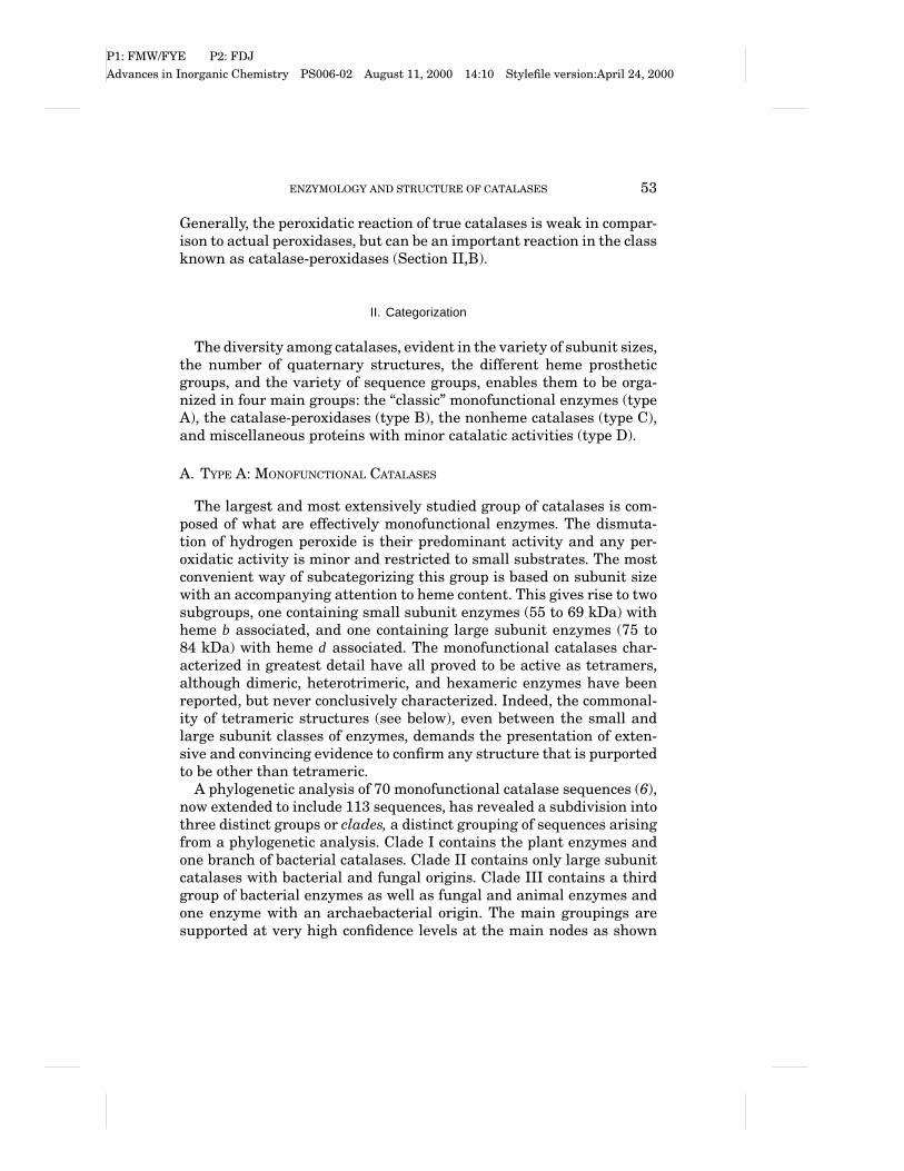
independent of [H2O2]. Reaction 2 or the decomposition of compound I isthe rate limiting step resulting in the overall velocity being described by
V = 2k1k2[H2O2][E]/(k2+ k1), (12)
where the factor 2 is present because each complete cycle involves theloss of two molecules of peroxide. Equation (12) shows that the rate ofperoxide decay is proportional to a constant,
k′ = 2k1k2/(k2+ k1), (13)
multiplied by the product of the concentrations of substrate and enzymeactive sites.
Measurement of the overall rate constant, together with a measure-ment of the steady-state proportion of enzyme that is compound I(a),always permits calculation of the values of the two intrinsic rate con-stants according to
k2 = k′/2a (14)
and
k1 = k′/2(1− a). (15)
The value of k1 may be determined directly if a trap compound is avail-able that reacts irreversibly and more rapidly with compound I thandoes a second H2O2.
Classical low Km values for the mammalian enzyme that have ap-peared in the literature are the result of enzyme inactivation by hy-drogen peroxide when measurements were carried out with peroxidelevels in excess of 10 mM over time scales of 10 minutes or longer. Therapid sampling/titration method of Bonnichsen overcame the inacti-vation problem and permitted a satisfactory correlation of the overallcatalytic measurements and Chance’s observations on the interme-diate complex (compound I). Eventually, the introduction of the UVdetector/spectrophotometer and the consequent assay based upon theUV absorbance of peroxide (35) further simplified the process by elim-inating the discontinuous titrimetric assay.
Obviously, there must be a limit to the turnover of any enzyme. Ratescannot theoretically go on increasing indefinitely with substrate con-centration. In the case of mammalian catalases, the limits appear to liein the range between a first order rate of 2× 10⁶ sec−1 and 1× 10⁷ sec−1
(36). That is, each heme active site can theoretically decompose be-tween 2 and 10 million molecules of H2O2 per second. As two molecules

P1: FMW/FYE P2: FDJAdvances in Inorganic Chemistry PS006-02 August 11, 2000 14:10 Stylefile version:April 24, 2000
62 NICHOLLS, FITA, AND LOEWEN
are decomposed per turnover, that means that the lifetime of the ac-tive Michaelis–Menten complex, or compound I, lies between 0.2 and1 microsecond.
The more complex scheme required if a true Km is involved is shownin Fig. 2B. There are two possible steps that could provide limitingunimolecular processes, governed by kA and kB, the steps involved information of compound I and the decay of the “ternary” complex, re-spectively. Nicholls and Schonbaum (22) gave reasons the latter is thepreferred limiting step for mammalian catalase. These reasons havenot changed much over the past 35 years. But the increased number ofcatalases examined, especially the catalase-peroxidases, makes reeval-uation appropriate (see below).
Several catalases, including the type B catalase-peroxidases, seem toshow true substrate saturation at much lower levels of peroxide thanoriginally observed for the mammalian enzyme (in the range of a fewmillimolar). This means that the limiting maximal turnover is less andthe lifetime of the putative Michaelis–Menten intermediate (with theredox equivalent of two molecules of peroxide bound) is much longer.The extended scheme for catalase in Fig. 2B shows that relationshipsbetween free enzyme and compound I, and the presumed rate-limitingternary complex with least stability or fastest decay in eukaryotic en-zymes of type A and greatest stability or slowest decay in prokaryotictype B enzymes.
B. THE PEROXIDATIC ACTIVITY OF CATALASES
In addition to their catalatic (peroxide dismuting) activity, catalasesalso use peroxides to oxidize secondary hydrogen and electron “donor”molecules. There are two major families of possible hydrogen donors:two-electron donors such as alcohols and one-electron donors such asphenols. The two modes of redox behavior are quite distinct. Keilinand Nicholls (37) described six donor types, classified according to theirreactivities with the two catalase peroxide compounds. Figure 3A showsthe basis for this classification. What has happened over the past 40years to modify this scheme? Firstly, the number of known catalaseshas increased immensely, but although there are significant quanti-tative differences in rates of reaction with specific donor types, mem-bers of the type A family of catalases, including bovine liver catalase(BLC) and HPII, share many characteristics, including donor specifici-ties. Less complete surveys are available for the more recently discov-ered catalase-peroxidases or type B enzymes. Secondly, a major devel-opment has been the discovery of the special donor role of NADPH

P1: FMW/FYE P2: FDJAdvances in Inorganic Chemistry PS006-02 August 11, 2000 14:10 Stylefile version:April 24, 2000
ENZYMOLOGY AND STRUCTURE OF CATALASES 63
FIG. 3. Classification of catalase hydrogen donors. (A) The initial classification pro-posed by Keilin and Nicholls in 1958 (37). D1 donors, including ascorbate and ferro-cyanide, reduce only compound I to compound II. D2 donors, including phenols and aro-matic amines, reduce compound I to II and compound II to ferric enzyme. D3 donors,including alcohols and formate, reduce only compound I to ferric enzyme. D4 donors,including nitrite, reduce compound I and compound II to ferric enzyme. D5 donors, in-cluding azide and hydroxylamine, reduce compound I to ferrous enzyme and compoundII to ferric enzyme. D6 donors, including H2O2, reduce compound I to ferric enzyme andcompound II to compound III. (B) A revision of the scheme in part A that reflects theinclusion of the new donor group D7, including NADPH and NADH, which reduces acompound II precursor (compound II’) to ferric enzyme. In addition, the D1-categorizedferrocyanide is now regarded as primarily a reducer of the compound II’ intermediate,and the D5-categorized azide may reduce Fe via an intermediate compound II.

P1: FMW/FYE P2: FDJAdvances in Inorganic Chemistry PS006-02 August 11, 2000 14:10 Stylefile version:April 24, 2000
64 NICHOLLS, FITA, AND LOEWEN
(listed as a seventh donor type below). Thirdly, it has been found thatsome type A enzymes, including both protoheme forms, such as the en-zyme from Aspergillus, and chlorin heme forms, such as E. coli HPII,appear not to form compound II at all. A fourth modification of the clas-sical scheme involves the observation that certain types of peroxide,such as t-butyl hydroperoxide and peroxynitrous acid, may form com-pound II directly by a homolytic rather than heterolytic split of the O Obond, with consequent release of a peroxidelike radical into the medium(t-butylO
.or NO
.2). In addition to these overall changes, there have been
more detailed changes to the 1958 pathways, as described later.
C. COMPOUND I AND THE PATHWAYS VIA COMPOUND I
The visible spectra of beef liver catalase (Type A) and its two activeperoxide compounds are shown in Fig. 4. The unliganded enzyme hasa Soret band at 405 nm (EmM/heme≈ 120) and a characteristic visible
FIG. 4. Visible spectra of catalase, compound I, and compound II; 5 mM (heme) beefliver catalase (Boehringer-Mannheim) in 0.1 M potassium phosphate buffer pH 7.4, 30◦C.Compound I was formed by addition of a slight excess of peroxoacetic acid. CompoundII was formed from peroxoacetic acid compound I by addition of a small excess of potas-sium ferrocyanide. Absorbance values are converted to extinction coefficients using 120mM−1 for the coefficient at 405 nm for the ferric enzyme (confirmed by alkaline pyridinehemochromogen formation). Spectra are corrected to 100% from occupancies of ≈90%compound I, 10% ferric enzyme (steady state compound I) and 88% compound II, 12%compound I (steady state compound II). The extinction coefficients for the 500 to 720 nmrange have been multiplied by 10. Unpublished experiments (P.N., 1999).

P1: FMW/FYE P2: FDJAdvances in Inorganic Chemistry PS006-02 August 11, 2000 14:10 Stylefile version:April 24, 2000
ENZYMOLOGY AND STRUCTURE OF CATALASES 65
spectrum with peaks at 500 and 622 nm and a shoulder at 540 nm,typical of ferric protoheme proteins, differing from the paradigmaticmetmyoglobin spectrum by having a long wavelength charge transfer(CT) band at a shorter wavelength and with a higher extinction co-efficient, presumably due to the proximal tyrosinate. Compound I, theporphyrin radical-ferryl state (Eq. (1)), is characterized by a much lowerSoret peak (EmM/heme≈ 65–70) and a long wavelength band at 665–670nm, of an intensity almost equal to that of the original CT band, indi-cating a disruption of the resonating p-bond system of the porphyrinring. Beyond 700 nm there are absorbances (not shown) due to the pres-ence of heme groups degraded to bile pigments, largely biliverdin withsome retention of the heme iron to form verdohemes. Commercial beefliver catalases typically contain up to 45% bile pigment. The structureof the catalase heme groups was originally described as substantiallydistorted compared to those of other hemoproteins, because these ver-doheme structures were averaged with the protoheme ones (38).
The donor types D3, D4, and D6 of Keilin and Nicholls (37) all re-duce compound I of Type A enzymes directly to the ferric state in a two-electron process without detectable intermediates. Each of these donorsis probably also able to bind in the heme pocket of the free enzyme. Al-cohols (type D3) form complexes with free ferric Type A enzymes whoseapparent affinities parallel the effectiveness of the same alcohols ascompound I donors (39). Formate (type D3) reacts with mammalianferric enzyme at a rate identical to the rate with which it reduces com-pound I to free enzyme (22). Its oxidation by compound I may thus sharean initial step analogous to its complex formation with ferric enzyme.Formate also catalyzes the reduction of compound II to ferric enzymeby “endogenous” donors in the enzyme (40, 41). Both compound I andcompound II may thus share with the free enzyme the ability to ligateformate in the heme pocket. Nitrite, which is oxidized to nitrate by atwo-electron reaction with compound I (type D4), also forms a character-istic complex with free enzyme (42). In both cases the reaction involvesthe donor in its protonated (HNO2) form.
Hydrogen peroxide itself was given a separate donor status (D6) be-cause in addition to acting like the D3 family and reducing compound Ito ferric enzyme in a single two-electron step it can also react with cata-lase compound II to give the “oxy” or protonated oxy species, compoundIII (22) according to
Cpd II (Por FeIV OH+)+ H2O2→ Cpd III (Por FeII O2H+)+ H2O. (14)
Compound III, like compound II, is an inactive form of catalase withrespect to the normal catalatic cycle, and thus may contribute to theinactivation of the enzyme at high peroxide levels (42).

P1: FMW/FYE P2: FDJAdvances in Inorganic Chemistry PS006-02 August 11, 2000 14:10 Stylefile version:April 24, 2000
66 NICHOLLS, FITA, AND LOEWEN
The classical type A enzymes show marked differences in their abili-ties to oxidize two-electron donors. All the enzymes initially examined,whether eukaryotic or prokaryotic in origin, were members of clade III.These are rather effective as two-electron peroxidases. But the oxida-tions of ethanol and formate by the paradigmatic clade II enzyme fromE. coli, HPII, are much slower. There is little information concerningthe activity of type A enzymes in clade I.
There are also substantial differences between classical type A en-zymes and the type B catalase-peroxidases. The latter enzymes, al-though they show peroxidase activity toward donors of type D2, areinactive or only weakly active toward D3 donors such as ethanol.
D. COMPOUND II AND THE PATHWAYS VIA COMPOUND II
The visible spectrum of beef liver catalase compound II is also shownin Fig. 4. The enzyme in its one-electron oxidized (ferryl) state has aSoret band at 427 nm (EmM/heme≈ 92) and a characteristic visible spec-trum with intense peaks at 533 and 567 nm (EmM/heme≈ 18), indicatinga low spin state, differing from the corresponding ferrylmetmyoglobinspectrum by having sharper peaks at a shorter wavelength and withhigher extinction coefficients, due either to redox delocalization at theproximal tyrosinate or to protonation of the ferryl species (Eq. (3a)).
The donor types D2, D4, and D5 of Keilin and Nicholls (37) all re-duce compound II to ferric enzyme in a one-electron process withoutdetectable intermediates. Donors of type D2, phenols and amines, alsoreduce compound I to compound II. Nitrite, the only member of cate-gory D4, reduces compound I in a two-electron step as described earlier.Donors of type D1 reduce compound I to compound II, but have no ap-preciable effect upon compound II itself. Reactivity of the one-electrondonors seems independent of heme pocket binding in the free enzyme.
The donor type D5 comprises the two species azide and hydroxy-lamine. These both react with the enzyme in the presence of peroxideto give rise to ferrous forms of catalase, otherwise normally inacces-sible (catalase is the only common hemoprotein that is nonreducibleby dithionite). The final inhibited form of catalase in the presence ofazide and peroxide is NO-ferrocatalase, but not every azide moleculebecomes an NO
.; only in the presence of CO is there a stoichiomet-
ric inhibition of enzyme by peroxide with formation of 1 equiv of CO-ferrocatalase for every peroxide molecule added (43). This suggested athree-electron reduction of compound I either to give ferrocatalase, N2,and NO
.(10–20% total) or to give ferrocatalase, N
., and N2O (80–90%
total). However, Kalyanaraman et al. (45) have demonstrated the for-mation of the azidyl (N N N
.) radical in the reaction, and Lardinois

P1: FMW/FYE P2: FDJAdvances in Inorganic Chemistry PS006-02 August 11, 2000 14:10 Stylefile version:April 24, 2000
ENZYMOLOGY AND STRUCTURE OF CATALASES 67
and Rouxhet (46) proposed a role for compound II formation in catalaseinhibition by azide. Nicholls and Chance (unpublished data) had in factidentified a precursor species closely similar to compound II before theappearance of ferroenzyme. The latter cannot be the result of a secondreaction of azide with compound II, as the latter reaction gives rise toferric enzyme. The simplest hypothesis involves the secondary reactionof the azidyl radical with compound II to give the ferroenzyme witheither of two possible breakdown modes of the azidyl radical shown inFig. 5. The alternative pathway in which the azidyl radical itself reactswith oxygen to give NO and N2O (45) is too slow and does not seem to
FIG. 5. (A) The peroxidatic reaction proceeding via compound II (the D2 family). (B)A catalytic cycle involving azide proceeding via compound II and ferroenzyme (the D5family).

P1: FMW/FYE P2: FDJAdvances in Inorganic Chemistry PS006-02 August 11, 2000 14:10 Stylefile version:April 24, 2000
68 NICHOLLS, FITA, AND LOEWEN
generate a reductant capable of reducing ferric catalase or compoundII to the ferro form.
Both classical type A enzymes (clade III) and the heme d family (cladeII) show a comparatively high sensitivity to azide inhibition and arereduced to ferrous forms in the presence of peroxide and azide (47). Incontrast, the catalase-peroxidase (type B) enzymes (see below) are onlyweakly azide-sensitive.
E. CONTROL BY NADPH: THE “EXTRA” PATHWAY
Kirkman and Gaetani (48) discovered the seventh catalase donortype, D7, unknown to Keilin and Nicholls (37). In his calculations ofin vivo or at least “in erythro” rates, Nicholls (49) had already ponderedthe possibility of some unusual regeneration pathway for blood catalase,but had vaguely wondered about a role for glutathione, following hisoriginal studies on cysteine and glutathione interactions with catalase(37). Following the identification of tightly bound NADP+/NADPH byconventional chromatographic methods, Fita and Rossmann (50) wentback to their data and found that a piece of what previously had beenthought to be disordered peptide actually fitted NADPH quite well.
Kirkman and Gaetani (48) were able to show not only that tightlybound NADP+ or NADPH was present in mammalian catalases, butalso that the presence of the reduced nucleotide decreased compound IIformation. The kinetics were, however, anomalous. Of the original fam-ilies of donors, two types could decrease compound II accumulation: thetwo-electron D3 category, exemplified by ethanol, which remove com-pound I before compound II can be formed spontaneously, and the one-electron category (D2), which reduce compound II itself. NADPH fallsinto neither category. Although it prevents compound II formation fromcompound I, it does not reduce compound I directly to free enzyme (thatis, it is not a conventional two-electron donor), and although it preventscompound II formation, it does not reduce compound II to ferric enzymeonce the former species has been produced in its absence. That is, it alsocannot act as a one-electron donor. We are presented with an apparentparadox. Hillar and Nicholls (47) attempted to resolve the paradox bypostulating an intermediate between the compound I and compoundII states possessing an unique reactivity with bound NADPH, as in-dicated in Fig. 6. The initial (slow) step in formation of compound IIfrom compound I lies in the migration of an oxidizing equivalent to anearby protein residue P (as occurs rapidly and stably, for example,in cytochrome c peroxidase). In a second (and relatively rapid) event,the “endogenous” donor centers in the molecule (remote tyrosines, etc.)

P1: FMW/FYE P2: FDJAdvances in Inorganic Chemistry PS006-02 August 11, 2000 14:10 Stylefile version:April 24, 2000
ENZYMOLOGY AND STRUCTURE OF CATALASES 69
FIG. 6. The catalytic cycle involving a postulated compound II’ and NADPH.
reduce the radical P.
back to its stable PH state. What is PH/P.? One
candidate is the proximal tyrosine, oxidized to the tyrO.
form with re-moval of the charged p-electron radical. A second possibility is one ofthe other nearby tyrosines such as Y235 or Y214 (bovine catalase num-bering). A third possibility, suggested by Bicout et al. (51) (see alsoRef. 3), is that the conserved serine S196 may also act as a radical site.Little seems to be known about serine radicals in any other enzymes,and the likely thermodynamics are therefore hard to estimate.
Two current alternative views are available as to how remotely boundNADPH may work. One sees its action as involving two successive one-electron oxidations (52, 53). The effectiveness of NADPH in preventingcompound II formation is then due to the high reactivity of the NADP
.
intermediate as reductant of the compound II generated in the first one-electron step. The other model (47) prefers to see NADPH as a hydridedonor responsible for the almost simultaneous reduction of the ferryliron and the protein radical species.
Kirkman and Gaetani (54) have reexamined the kinetics of NADPHoxidation and compound II formation by mammalian catalase. Undersome experimental conditions the rate of NADPH oxidation is substan-tially higher than the rate of compound II formation in the absence ofNADPH. This finding may be accommodated in a scheme such as thatof Fig. 6 if the rate of formation of compound II’ is greater than the rateof compound II formation. This can arise either if there is more than oneroute for compound II’ decay, only one of which proceeds via compoundII, or if the formation of compound II’ from compound I is reversible,as indicated in the scheme (Fig. 6). Such reversibility is highly prob-able in view of the redox potentials of the species involved. Althoughdirect measurements have not been carried out with catalases, the cor-responding metmyoglobin and horse radish peroxidase peroxide com-pounds (55, 56) have potentials (E′o values) of +880 mV (horse radish
P1: FMW/FYE P2: FDJAdvances in Inorganic Chemistry PS006-02 August 11, 2000 14:10 Stylefile version:April 24, 2000
70 NICHOLLS, FITA, AND LOEWEN
peroxidase or HRP comp. I), +900 mV (HRP comp. II), and +890 mV(ferrylmetmyoglobin). Corresponding E′o values for the trpH/trp. andtyrOH/tyrO
.pairs are +1050and +940mV, respectively (57). Although
these potentials may vary in particular proteins, it seems likely thatthe redox gap between compound I and compound II′ (Fig. 6) will beabout 60 mV in favor of compound I. Under the conditions described byKirkman and Gaetani (54), the rate of formation of compound II is ap-proximately 25% the rate of NADPH oxidation. This is consistent withthe proposed mechanism provided that the rate of reaction of NADPH(k5) is fast and the compound I ←→ compound II′ step is an equilibrium.
The role of NADPH as “protective” donor (58, 59) seems to correlatewell with the tendency of the different categories of catalases to formthe inactive compound II. Only those classical catalases that are vulner-able to the latter inactivation step show NADP+ and NADPH binding.The scheme shown in Fig. 3B summarizes the minimal revisions of the1958 scheme (Fig. 3A) needed to accommodate the newer findings.
F. THE CATALATIC ACTIVITY OF CATALASE-PEROXIDASES
Members of the catalase-peroxidase family were discovered sepa-rately in different microorganisms by several groups of workers in thelate 1970s and 1980s (60–62). It soon became evident that these en-zymes are homologous to the eukaryotic type I plant peroxidases ofWelinder (63). Although these enzymes show both catalase andperoxidase activities and thus obtained the name catalase-peroxidase(CatPx), it is not clear whether their peroxidatic activities are propor-tionately greater than similar activities shown by classical (type A)enzymes (64). What is clear is that the catalatic activity displays sev-eral unique distinguishing characteristics. Hochman and Shemesh (65)showed that the Rhodopseudomonas capsulata enzyme is characterizedby instability in presence of the substrate H2O2, saturation kinetics(Km ≈ 4 mM) for the catalatic reaction, and a relative insensitivity toazide and hydroxylamine.
Follow-up experiments with the similar Klebsiella pneumoniae en-zyme (66, 67) also showed that the pH profiles for CatPx are quitedifferent from those for classical catalases. The latter’s catalatic activ-ities are essentially pH-independent from pH 5 to 10 (68); CatPx ofKlebsiella showed a sharp pH optimum between pH 6 and 7 (66). Asimilar eukaryotic fungal CatPx (69) was also characterized by a sharppH sensitivity and saturation kinetics (Km ≈ 3.4 mM) and, like the bac-terial enzymes, was sensitive to incubation with peroxide but not to theclassical catalase inhibitors azide (except weakly) and aminotriazole.

P1: FMW/FYE P2: FDJAdvances in Inorganic Chemistry PS006-02 August 11, 2000 14:10 Stylefile version:April 24, 2000
ENZYMOLOGY AND STRUCTURE OF CATALASES 71
The low peroxidatic activities were less pH sensitive than the catalaticactivity.
Most recently, the study of catalase-peroxidases has been extended tothe cyanobacteria, which, unlike the eubacteria, seem often to be char-acterized by possession of only a CatPx (type B) and no type A catalase.Obinger et al. (70) showed that the Anacystis nidulans enzyme hada Km for H2O2 of 4.3 mM and a maximal turnover of over 7000 sec−1.At low peroxide levels the activity was about 20% that of beef liverenzyme. Peroxoacetic acid produced a typical compound I species. Ananalogous enzyme is found in Synechocystis 6803 (70, 71). This enzyme,like the eubacterial examples, shows a narrow pH optimum betweenpH 6 and pH 7 (72) with apparent pK values close to 7 and between 5.5and 6.0. The pH profiles for the peroxidatic activity were different fromthose for catalatic activity. Again peroxoacetate produces a characteris-tic compound I. However, the reaction of compound I with one-electronreductants produces a high-spin intermediate different from typicalcompound II. Regelsberger et al. (71) also find that the one-electron re-duction product obtained by adding ascorbate to compound I is a highspin species with Soret band closely similar to that of free enzyme andvisible spectrum with a peak at 626 nm compared to 631 nm for ferricenzyme.
Moreover, the steady-state spectrum in the presence of hydrogen per-oxide showed no sign of the presence of compound I (identified from theperoxoacetate spectrum), and compound I produced by peroxoacetatewas unaffected by addition of hydrogen peroxide. Regelsberger et al.(71) and Jakopitsch et al. (72) nevertheless interpret their results interms of the classical “BCT” catalatic cycle (Fig. 3), attributing the dif-ferences to substantial differences in the kinetic constants for formationand decomposition of compound I, k1 and k2 (Eqs. (4) and (5)). An alter-native analysis is represented in Fig. 7. Only the peroxidatic activity ofthe Type B enzymes may proceed via the usual porphyrin–cation radicalcompound I. The heme configuration, analogous to that of cytochromec peroxidase of yeast, may permit an alternative doubly oxidized stateinvolving a protein radical. If this is the pH-sensitive intermediate, thefailure to detect appreciable amounts of compound I during the steadystate and the inactivity of peroxoacetate-derived compound I towardH2O2 may all be explicable. In addition, the unusual spectrum of com-pound II of this enzyme may also indicate a more stable protein radicalstate than the usual ferryl state, even for this intermediate. Electronparamagnetic studies of CatPx enzymes to detect radical states are ex-pected to be undertaken soon that may confirm, refute, or modify thisscheme.

P1: FMW/FYE P2: FDJAdvances in Inorganic Chemistry PS006-02 August 11, 2000 14:10 Stylefile version:April 24, 2000
72 NICHOLLS, FITA, AND LOEWEN
FIG. 7. Catalatic and peroxidatic reactions of type B enzymes. This represents a mod-ification of the schemes of Figs. 2 and 5A and is proposed to account for the characteristicfeatures of catalase-peroxidases. Compound I is drawn as Fe5+ O and can representeither a p-cation radical or alternative radical structure. The precise nature remainsundefined (see Section IV,F).
V. Structure of Type A Catalases
Detailed structural information about Type A catalases is availablefrom the crystal structures of seven monofunctional catalases that havebeen solved. These include representatives from small-subunit clade IIIenzymes of animal (bovine liver catalase or BLC (73, 74) and humanerythrocyte (T. P. Ko, submitted to Protein Data Base as file 1qqw)), fun-gal (Saccharomyces cerevisiae SCC-A) (75, 76), and bacterial (Proteusmirabilis PMC and Micrococcus lysodeikticus MLC) (77, 78) origins, andlarge subunit clade II enzymes of fungal (Penicillium vitale PVC) (79,80) and bacterial (E. coli HPII) (81, 82) origins. Despite the differencesin size, all six enzymes share a number of common features that appearto be characteristic of catalases, including a homo tetrameric quater-nary structure with the heme group deeply buried in a beta-barrel corestructure in each subunit.
Within this common core structure, modifications have been identi-fied that provide catalases with further unique properties. The large-subunit enzymes have extensions at both the amino and carboxyl ends,the latter having a flavodoxinlike structure, a unique His–Tyr bond,a protected cysteine, and a modified heme. NADPH binding and anoxidized methionine are found in small subunit enzymes. Identifica-tion and assignment of roles to channels providing access to and egressfrom the deeply buried heme have recently become the focus of study.Analysis of the structure of catalase HPII of E. coli has been facilitatedby the construction of more than 75 mutants (Table I).

P1: FMW/FYE P2: FDJAdvances in Inorganic Chemistry PS006-02 August 11, 2000 14:10 Stylefile version:April 24, 2000
ENZYMOLOGY AND STRUCTURE OF CATALASES 73
TABLE I
CHARACTERIZATION OF THE HPII MUTANT VARIANTS
Specific activitya
Mutation (units/mg) Hemeb His392–Tyr415c
Wild type 14,322 d yGln3Arg nd (active)Gln3Arg/Glu21Ala nd (active)Gln3Arg/Gly74Ser/Ser75Ala nd (active)1(Gln3-Ser20) np1(Gln3-Gly34) np1(Gln3-Thr50) np1(Gln3-Gly74) np(Gln3-Gly74)/Val303Ser/Phe317Ser/
Phe518Ser/Leu571Stop npGlu21Ala nd (active)Gly34Ser/Ser35Ala ndThr50Ser ndGly74Ser/Ser75Ala nd (active)His128Ala <0.1 b nHis128Asn <0.1 b nHis128Glu npHis128Gln npHis128Asn/Asn201His <0.1 bSer167Thr 1,100 d + bSer167Ala 100 bSer167Cys npSer167Asn npVal169Ala 3,788 d yVal169Ser 3,703 d yVal169Cys 16 b nVal169Cys/Cys438Ser/Cys669Ser 10 bAsp197Ala 14,354 d yAsp197Ser 14,721 d yAsp197Ser/His395Gln 15,473 d yAsn201His 100 b nAsn201Asp 1,700 dAsn201Ala 1,300 dAsn201Gln 50 dAsn201Arg npArg260Ala 35,891 dArg260Ala/Lys294Ala 12,880 dd
Glu270Asp 16,137 d + bGlu270Asp/Glu362His 16,859 dIle274Ser 3,416 dIle274Ser/Pro356Leu npIle274Ser/Leu407Met npIle274Ser/Pro356Leu/Leu407Met npLys294Ala 18,625 d + b
(continued )

P1: FMW/FYE P2: FDJAdvances in Inorganic Chemistry PS006-02 August 11, 2000 14:10 Stylefile version:April 24, 2000
74 NICHOLLS, FITA, AND LOEWEN
TABLE I (Continued )
Specific activity a
Mutation (units/mg) Hemeb His392–Tyr415c
Val303Ser ndPhe317Ser ndPro356Leu 13,220 d + bPro356Leu/Leu407Met 12,437 d + bGlu362His 18,325 d + bHis392Ala 8,875 b nHis392Gln 7,368 b nHis392Glu 4,507 b+ dd nHis392Asp 2,857 b nHis395Ala 9,821 d yHis395Gln 9,224 d yLeu407Met 12,679 d + bSer414Ala 5,511 b+ dd yTyr415Phe npTyr415His npGln419Ala 7,317 dd yGln419His 11,494 dd yCys438Ser 7,350 dCys438Ala 6,100 dCys438Ser/Cys669Ser 8,050 dCys438Ala/Cysgg9Ala 12,870 dPhe518Ser npLeu571Stop npIle593Stop npVal603Stop npCys669Ser 10,840 d + bCys669Ala 7,800 dTrp742Stop 2,624 dArg744Stop 8,600 dArg744Ala 8,786 dArg744Lys 5,832 dArg744Ala/Ile745Stop 6,929 dArg744Lys/Ile745Stop 9,284 dIle745Stop 13,753 dPro746Stop 15,022 dLys747Stop 15,349 dLys750Stop 15,067 d
a nd, not determined; np, insufficient protein accumulated in cells for purification;nd(active), near wild type levels of catalase were noted in crude extracts but enzymewas not purified.
b d, heme d; b, heme b. Heme composition was determined by spectral and HPLCanalyses. In all cases, the enzymes contained one heme per subunit. Where no indica-tion is given, no determination was made.
c Presence of the His392–Tyr415 (y, yes; n, no) covalent bond was determined byMALDI-MS analysis of trypic digest mixtures. Where no indication is given, no deter-mination was made.
d There was an increased percentage of the trans isomer in these variants.

P1: FMW/FYE P2: FDJAdvances in Inorganic Chemistry PS006-02 August 11, 2000 14:10 Stylefile version:April 24, 2000
ENZYMOLOGY AND STRUCTURE OF CATALASES 75
A. SUBUNIT STRUCTURE
The sequences of all catalases exhibit extensive similarity in the coreregion defined by the b-barrel and active site to the extent that thereare 15 invariant residues among 110 catalase sequences, with the vari-ations in an additional 3 probably arising from incorrect sequencing.These include the essential histidine situated in the active site and theessential tyrosine residue that forms the fifth ligand with the heme.Many other residues are highly conserved, varying in only two or threeof the catalases. Such extensive similarity speaks to a strong drive forconservation and is suggestive that the three-dimensional structureimposes limitations on the changes that are possible with retention ofactivity.
The sequence conservation is reflected in a highly conserved sec-ondary and tertiary structure that is most clearly illustrated in thethree-dimensional superposition of Ca atoms. Ignoring the C-terminaldomains of PVC and HPII, the deviation of Ca atoms in a superpositionof HPII with PVC, BLC, PMC, and MLC results in root mean square de-viations of 1.1, 1.5, 1.6, and 1.5 A for 525, 477, 471, and 465 equivalentcenters, respectively (83). In other words, there is very little differencein the tertiary structure of the subunits over almost the complete lengthof the protein. The large and small subunits are shown in Fig. 8 forcomparison.
The tertiary structure of small subunit enzymes can be subdividedinto four distinct regions, and the C-terminal or flavodoxin domain ofthe large subunit enzymes becomes a fifth region. These are indicatedin Fig. 8 for clarity. The first region is the amino terminal arm (Fig.8), which extends 50 or more residues from the amino terminus almostto the essential histidine residue (to residue 53 in PMC, 60 in PVC,73 in BLC, and 127 in HPII). There is very little structural similar-ity in the N-terminal region and, in the case of HPII, the structure ofthe terminal 27 residues is not even defined and they do not appearin the crystal structure. Within the N-terminal arm is a 20-residue he-lix, helix a2 in HPII, which is the first secondary structure elementcommon to all catalases. The presence of helix a1 varies among cata-lases, and there is no sequence or location equivalence even when it ispresent.
The second region is the antiparallel b-barrel (Fig. 8) forming thecore of the subunit. It includes about 250 residues from the essentialhistidine toward the C-terminus. The first four strands (b1–4) are con-tiguous and are separated from the second four strands (b5–6) by threehelices (a3–5). The first four strands form the distal side of the hemepocket and portions of the second four strands participate in bindingNADPH in small subunit enzymes.

P1: FMW/FYE P2: FDJAdvances in Inorganic Chemistry PS006-02 August 11, 2000 14:10 Stylefile version:April 24, 2000
76 NICHOLLS, FITA, AND LOEWEN
FIG. 8. Comparison of the subunit structures of a small subunit catalase (BLC) (A) anda large subunit catalase (HPII) (B). Segments including the N-terminal domain, the betabarrel core, the wrapping domain, the alpha helical domain, and the C-terminal domain(HPII) are indicated and are described in Section V,A.

P1: FMW/FYE P2: FDJAdvances in Inorganic Chemistry PS006-02 August 11, 2000 14:10 Stylefile version:April 24, 2000
ENZYMOLOGY AND STRUCTURE OF CATALASES 77
The third region is the wrapping domain (Fig. 8), which includes 110residues in an extended structure linking the b-barrel and the a-helicalsection. There is little secondary structure in this region, althoughhelix a9 contains many of the residues that form the proximal side ofthe heme pocket, including the essential fifth ligand tyrosine. A largesurface area for the formation of subunit-subunit interactions is createdin this extended structure.
The fourth region is the a-helical domain (Fig. 8) containing about60 residues organized in four contiguous a helices (a10–13) that form aclose association with a-helices 3 to 5 in the b-barrel region to stabilizethe structure. This region contains the C-terminus of small-subunitenzymes and, in large-subunit enzymes, may have a critical role in thefolding pathway.
In large subunit enzymes (PVC and HPII), a short segment of about30 residues links the a-helical domain to the C-terminal domain (Fig.8). The latter segment is a conspicuous addition to the small subunitcontaining about 150 residues folded into a structure that resemblesflavodoxin. For example, there is a root mean square deviation of 3.0 Abetween flavodoxin and approximately 100 residues of the C-terminaldomains of either HPII or PVC. This can be compared to the 1.8 Aroot mean square deviation for 134 centers between the C-terminal do-mains of HPII and PVC. Unlike the N-terminal end, the final C-terminalresidue Ala753 is visible in the structure of HPII. The C-terminal do-main contains extensive secondary structure in the form of four a-helices (a15–18) and eight b-strands (b9–16). Despite the obvious struc-tural similarity to flavodoxin, there is no evidence of nucleotide bindingin the domain and its function remains a mystery.
Attempts to generate a “small-subunit enzyme” by removing the C-terminal domain of HPII by site-directed mutagenesis were unsuccess-ful (84). Truncation of the subunit at residues 571, 593, or 603 resultedin no protein accumulating in the cell. The lack of protein accumula-tion was shown to be the result of proteolytic degradation of the proteinbefore it could fold into a protease-resistant form. Even when threehydrophobic residues that would have been exposed by removal of thedomain were changed to hydrophilic residues, truncated HPII-like pro-tein did not accumulate. Progressive truncation from the carboxy endrevealed that shortening the protein past Arg744 had the same effect asremoval of the complete C-terminal domain. The side chain of Arg744extends into the C-terminal domain and is involved in a number of hy-drogen bonds that are clearly important in stabilizing the structure ofthe domain. These results also show that a properly folded C-terminaldomain is essential for the efficient folding of the whole protein into a

P1: FMW/FYE P2: FDJAdvances in Inorganic Chemistry PS006-02 August 11, 2000 14:10 Stylefile version:April 24, 2000
78 NICHOLLS, FITA, AND LOEWEN
stable, protease-resistant structure. Any interference with the foldingof the domain interferes with the folding of the whole protein.
B. QUATERNARY STRUCTURE AND INTERWEAVING
All seven catalases, which have had their crystal structures solved,exist as homotetramers. Even HPII, which was originally character-ized biochemically as a hexamer (85), was found to be tetrameric uponsolution of the crystal structure. The reason for the biochemical de-termination of HPII as a hexamer was subsequently traced to the factthat the gel filtration elution volume varies with salt concentration suchthat elution at low salt results in a larger apparent molecular weight.The consistency in quaternary structure among both large- and small-subunit catalases, that of a tetramer, suggests that variations from thetetrameric structure among heme containing catalases will be rare.
The globular shape of the tetrameric small-subunit enzymes resem-bles a “dumbbell” in the R–Q orientation (Fig. 9) having dimensionsof 90 by 70 by 105 A along the P, Q, and R axes, respectively, and a“waist” dimension of 50 A in the R= 0 plane. The large subunit enzymehas similar dimensions along the P and Q axes, although with a lessobvious “waist,” but is 140 A long along the R axis (Fig. 9). The asso-ciation of subunits gives rise to an unusual six-stranded antiparallelstructure involving all four subunits. Many elements of the quaternaryassociation have been extensively reviewed (3, 83).
Perhaps the most unusual feature, and certainly the one that has thegreatest implications with regard to the folding pathway and stabilityof the complex, is the interweaving of adjacent subunits that resultsin two tightly associated dimers per tetramer. The N-terminal arm ofeach subunit is folded underneath the wrapping domain of an adjacentsubunit and the subunits are oriented such that each “Q-related” dimerpair contains two overlapped interactions (Fig. 10). In the case of small-subunit enzymes such as BLC, about 25 residues of the amino terminusextend beyond the overlap region, and there is very little interaction ofthose residues with the remainder of the subunit. The length of theoverlapped segment is greater in large subunit enzymes such as HPIIwhere 80 residues are “trapped.” Furthermore, this longer section formsan extensive network of interactions with the core of the subunit. Themore extensive interweaving and N-terminal interactions of HPII pre-dict a more stable structure, and this was corroborated in a study ofthe denaturation patterns of HPII and BLC (86). HPII was activatedslightly at temperatures up to 75◦C and lost activity in concert with

P1: FMW/FYE P2: FDJAdvances in Inorganic Chemistry PS006-02 August 11, 2000 14:10 Stylefile version:April 24, 2000
ENZYMOLOGY AND STRUCTURE OF CATALASES 79
FIG. 9. Comparison of the dimensions of small-subunit (BLC) and large-subunit (HPII)catalases. BLC is shown in panels A and B, in the R–P and R–Q orientations, respectively;HPII is shown in C and D, also in the R–P and R–Q orientations, respectively.
a transition in secondary structure having a Tm of 82◦C. BLC, by com-parison, exhibited a similar secondary structure transition and loss ofactivity, but with a Tm of 56◦C (86). The most striking illustration of theenhanced stability provided by the interwoven structure is that factthat the dimer structure does not dissociate in potassium phosphatebuffer even at 95◦C or in 7 M urea, 1% SDS at 60◦C (Fig. 11). For com-parison, BLC dimers, with the shorter overlapped segment, dissociateat room temperature in the urea–SDS solution.
Bergdoll et al. (87) have proposed that some proteins, including cata-lase, exhibit “arm exchange” or an interaction of one subunit withan adjacent subunit to stabilize quaternary structure, and that this

P1: FMW/FYE P2: FDJAdvances in Inorganic Chemistry PS006-02 August 11, 2000 14:10 Stylefile version:April 24, 2000
80 NICHOLLS, FITA, AND LOEWEN
FIG. 10. Illustration of the interweaving of two subunits of the small-subunit BLC (A)and the large-subunit HPII (B). The residues in the immediate proximity of the overlapsare shown as spheres. A schematic representation of the interwoven structures are shownin C for BLC and D for HPII. Note the longer N-terminal region of HPII (80 residues inHPII as compared to 25 in BLC) that extend through the loop on the opposite subunit.
interaction is stabilized or enhanced by one or more proline residues.In the case of catalase, Pro69 of BLC, which is highly conserved amongall catalases (115 of 117), was identified as the key residue in this in-teraction. However, the importance of prolines in catalase structuremay be far more profound for two reasons. First, the interweaving ofsubunits in catalases is a more significant interaction than the “armexchange” interactions or associations identified in other proteins, and,second, far more than one proline are involved when the two proline-

P1: FMW/FYE P2: FDJAdvances in Inorganic Chemistry PS006-02 August 11, 2000 14:10 Stylefile version:April 24, 2000
ENZYMOLOGY AND STRUCTURE OF CATALASES 81
FIG. 11. Conversion of HPII dimers to monomers. In panel A, HPII was incubatedfor 10 min at 70, 75, 80, 85, 90, 95, and 100◦C in 50 mM potassium phosphate buffer(pH 7). In panel B, HPII was incubated for 10 min at 50, 55, 60, 65, 70, 75, and 100◦Cin 50 mM potassium phosphate (pH 7) and 5.6 M urea. The temperatures of incubationare indicated above each lane. In both panels A and B, samples were removed, cooled toroom temperature, and added to SDS-urea loading buffer. Samples were loaded and run,without further heating, on an 8% polyacrylamide gel. Reprinted with permission fromSwitala et al. (86). Copyright 1999 American Chemical Society.
rich regions surrounding the interwoven regions are considered. Thisis most evident in HPII, where 10 of 52 residues between 426 and 477,the overlapping segment of the wrapping domain, are proline, and 8 of28 residues between 31 and 58, in the trapped N-terminal segment, are

P1: FMW/FYE P2: FDJAdvances in Inorganic Chemistry PS006-02 August 11, 2000 14:10 Stylefile version:April 24, 2000
82 NICHOLLS, FITA, AND LOEWEN
proline. There is no equivalent of the N-terminal proline-rich segmentin small-subunit enzymes because they lack the N-terminal extension,and this may be another explanation for the enhanced resistance todenaturation of HPII dimers as compared to small-subunit enzymes.These proline-rich segments are in addition to the highly conservedproline identified as Pro69 in BLC (Pro123 in HPII), which may be onemore component of this proline network. The prolines would contributeto enhanced rigidity in the overlapped region of the protein and therebystabilize the quaternary structure. In addition, they may influence thefolding pathway as suggested by the folding problems encountered byproteins in which the N-terminal proline-rich region up to residue 50was deleted (84).
The interweaving of subunits necessitates a very complex folding pro-cess in which subunit–subunit association must occur before subunitfolding is complete. In other words, the formation of secondary, tertiary,and quaternary structural elements are intermixed with quaternary in-teractions between partially folded subunits being formed before finaltertiary and possibly even secondary structural elements can be intro-duced. A survey of the properties of BLC (73, 88), yeast catalase (89),and HPII (84) provides some insight into the folding process of cata-lases. For example, dimers formed by BLC have dimensions that wouldbe expected of R-related dimers (88). Yeast accumulates large quan-tities of hemeless catalase monomer, and formation of the tetramericform occurs only after heme is bound by the subunit (89). By contrast,HPII requires all portions of its structure to be present for correct fold-ing into an active enzyme (84).
A possible mechanism based on these observations is described inFig. 12. There is an initial folding of domains within the apocatalase,followed by association with the heme, leaving the amino terminal arm,wrapping domain, and alpha helical domain (plus carboxyl domain inlarge-subunit enzymes) unassociated with the core (A to B in Fig. 12).The requirement for heme early in the folding process may explainwhy catalases have such a high heme occupancy as compared to thecatalase-peroxidases. Following complex formation with the heme, two“R-related” subunits will associate (B to C in Fig. 12). The next step in-volves the association of “Q-related” subunits (C to D in Fig. 12). Onlyone pair of “Q-related” subunits is shown in Fig. 12, but there would betwo dimers associating simultaneously. During the “Q-related” subunitassociation, the amino terminal arms fold against the subunit, followedby the wrapping such that the a-helical domains (and C-terminal por-tion) fold overtop of the N-terminal segment to “trap” it (D to E in Fig.12). In this way, each “Q-related” subunit pair will have two interwoven
P1: FMW/FYE P2: FDJAdvances in Inorganic Chemistry PS006-02 August 11, 2000 14:10 Stylefile version:April 24, 2000
ENZYMOLOGY AND STRUCTURE OF CATALASES 83
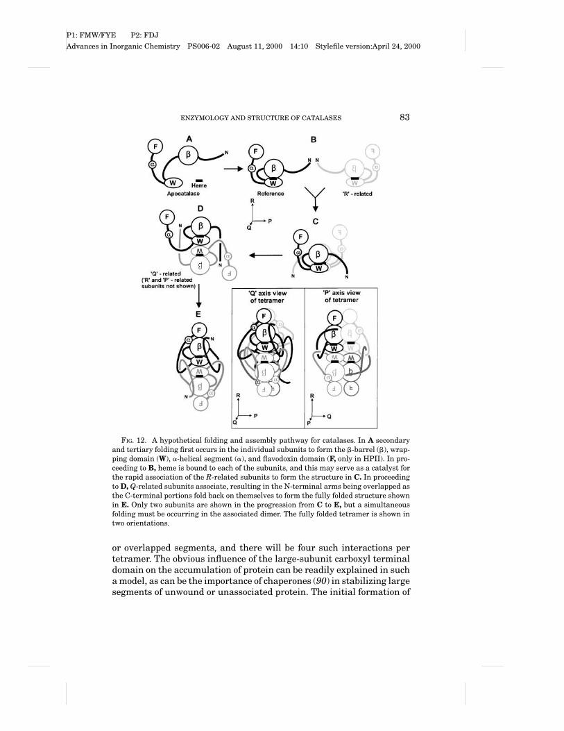
FIG. 12. A hypothetical folding and assembly pathway for catalases. In A secondaryand tertiary folding first occurs in the individual subunits to form the b-barrel (b), wrap-ping domain (W), a-helical segment (a), and flavodoxin domain (F, only in HPII). In pro-ceeding to B, heme is bound to each of the subunits, and this may serve as a catalyst forthe rapid association of the R-related subunits to form the structure in C. In proceedingto D, Q-related subunits associate, resulting in the N-terminal arms being overlapped asthe C-terminal portions fold back on themselves to form the fully folded structure shownin E. Only two subunits are shown in the progression from C to E, but a simultaneousfolding must be occurring in the associated dimer. The fully folded tetramer is shown intwo orientations.
or overlapped segments, and there will be four such interactions pertetramer. The obvious influence of the large-subunit carboxyl terminaldomain on the accumulation of protein can be readily explained in sucha model, as can be the importance of chaperones (90) in stabilizing largesegments of unwound or unassociated protein. The initial formation of
P1: FMW/FYE P2: FDJAdvances in Inorganic Chemistry PS006-02 August 11, 2000 14:10 Stylefile version:April 24, 2000
84 NICHOLLS, FITA, AND LOEWEN
the R-related dimer shown in Fig. 6 is suggested by the dimensionsof the BLC dimer, but the extreme stability of the Q-related dimer ofHPII suggests that this alternate dimer might form. If this were thecase, it would be necessary to change the order in Fig. 12 to B to D to Eto tetramer. This is a relatively minor change to the pathway, however,and the main point, that the formation of tertiary and quaternary inter-actions are intermixed, remains the same. Cartoons of the structuresof tetrameric HPII from two different orientations to further illustratethe interactions are also shown in Fig. 12.
C. HEME COMPOSITION AND LOCATION
The heme of catalases is deeply buried within the core of the cata-lase subunit. Protoheme IX or heme b is found in all small-subunitcatalases so far characterized. The two large-subunit enzymes HPIIand PVC have been characterized biochemically, spectrally, and struc-turally (91) as containing heme d in which ring III is oxidized to a cis-hydroxyspirolactone. Heme b is initially bound to both enzymes duringassembly, and it is subsequently oxidized by the catalase itself duringthe early rounds of catalysis (92).
Another significant difference between the large- and small-subunitenzymes lies in the fact that the heme d of HPII and PVC is flipped 180◦
relative to the heme b moiety of BLC, MLC, SCC-A, and PMC (Fig. 13).This is clearly a function of the residues that form the heme pocket,although attempts to force a change in heme orientation in HPII bymutating residues that interact with the heme were unsuccessful. Theheme is situated in the b-barrel and has interactions with the wrappingdomain and with the amino-terminal arm of the R-related subunit. Thedimensions of the pocket demand that heme bind in its final conforma-tion and that flipping once inside the pocket not be possible.
The flipped orientation of the heme in HPII and PVC results in theoxidized ring being sufficiently well removed (7 A) from the essentialhistidine (His128 in HPII) and the presumed peroxide binding site tocomplicate an explanation of the reaction mechanism. The explanationis further complicated by the cis-stereospecificity of the reaction thatresults in both oxygens being situated on the proximal side of the hemeaway from what is considered to be the normal reaction center on thedistal side. This stereochemistry dictates that the hydroxyl group on theheme d have originated on the proximal side of the heme, and a mech-anism has been proposed to explain the reaction in both PVC and HPII(93). The mechanism assumes that compound I is formed as a first step

P1: FMW/FYE P2: FDJAdvances in Inorganic Chemistry PS006-02 August 11, 2000 14:10 Stylefile version:April 24, 2000
ENZYMOLOGY AND STRUCTURE OF CATALASES 85
FIG. 13. Active site residues in a small-subunit catalase BLC (A) and a large-subunitcatalase HPII (B). The active site residues are labeled, and hydrogen bonds are shownbetween the serine (113 in BLC and 167 in HPII) and the essential histidine (74 inBLC and 128 in HPII). A single water is shown hydrogen bonded to the histidine. Theequivalent water in BLC is located by analogy to the position of the water in HPII.The unusual covalent bond between the Nd of His392 and the Cb of Tyr415 in HPII isevident on the proximal side of the heme in B. The flipped orientations of the hemes areevident in a comparison of the two structures, as is the cis-hydroxyspirolactone structureof heme d in B.
in the heme modification, after which it reacts with a water molecule onthe proximal side that acts as an electron donor to the porphyrin cationradical (Fig. 14). Cyclization to form the spirolactone follows complet-ing the reduction of compound I. Water molecules are present on theproximal side, and a potential channel has been identified that wouldallow access for water. The two residues Ser414 and Gln 419 ensure thecis stereochemistry such that changing either residue results in moreof the trans isomer being formed (94).
The mechanism in Fig. 14 applies equally well to both PVC and HPII.However, an unusual bond between the imidazole ring of His392 andthe b-carbon of Tyr415 on the proximal side of the HPII heme has beenidentified (Fig. 13) (93), and subsequently its presence was correlatedwith heme oxidation. The apparent correlation between heme oxidationand His–Tyr bond formation suggested a mechanistic linkage betweenthe two modifications and an alternate mechanism unique to HPII wasproposed (Fig. 15). As with the first mechanism, the reaction assumesthe formation of compound I that is available for reduction. Formation ofthe His–Tyr bond, involving a base catalyzed proton extraction from the

P1: FMW/FYE P2: FDJAdvances in Inorganic Chemistry PS006-02 August 11, 2000 14:10 Stylefile version:April 24, 2000
86 NICHOLLS, FITA, AND LOEWEN
FIG. 14. A mechanism to explain heme modification in the P. vitale catalase and pos-sibly E. coli HPII. For simplicity, the phenyl ring of Tyr415 is not shown, and only ringIII of the heme and the heme iron are shown. Compound I is an oxyferryl species formed,along with water, in the reaction of one H2O2 with the heme. The iron is in a formal FeV
oxidation state, but one oxidation equivalent is delocalized on the heme to create the oxo-FeIV -heme cation, shown as the starting species, compound I. A water on the proximalside of the heme is added to the heme cation species of compound I shown in A to generatea radical ion in B. The electron flow toward the oxo-iron would generate the cation shownin (C), leading to the spirolactone product shown in D. In E, an alternate mechanism forthe His–Tyr bond formation in HPII is presented that could occur independently of theheme modification reaction. Reprinted with permission of Cambridge University Pressfrom Bravo et al. (93).

P1: FMW/FYE P2: FDJAdvances in Inorganic Chemistry PS006-02 August 11, 2000 14:10 Stylefile version:April 24, 2000
ENZYMOLOGY AND STRUCTURE OF CATALASES 87
FIG. 15. A proposed mechanism coupling the formation of the His–Tyr bond to theoxidation of ring III of the heme in HPII. The mechanism begins with the formationof compound I shown in A. A concerted series of reactions, possibly triggered by eitherAsp197/His395 or by a putative anionic species bound to compound I, results in thetransfer of a hydroxyl to the heme from the H2O2 shown in C, which would facilitatespirolactone cyclization to form the final product containing the His–Tyr bond and themodified heme shown in D. Reprinted with permission of Cambridge University Pressfrom Bravo et al. (93).
imidazole ring of His392 by a still unidentified species, initiates a con-certed reaction that results in peroxide serving as a hydroxide donor to,and consequent reducer of, the porphyrin cation radical (Fig. 15). Subse-quent spirolactone formation completes the reduction of the compoundI. Supporting the existence of such a mechanism is the observation
P1: FMW/FYE P2: FDJAdvances in Inorganic Chemistry PS006-02 August 11, 2000 14:10 Stylefile version:April 24, 2000
88 NICHOLLS, FITA, AND LOEWEN
that changing His392 to Gln precludes the His–Tyr bond formation andalso prevents heme oxidation. Similarly, all inactive variants of HPIIin which the heme is not oxidized do not contain the His–Tyr linkage.One exception to this generalization is the His392Glu variant in whichsome heme oxidation takes place despite the absence of His–Tyr bondformation (93). However, even here there appears to be an aberrant re-action pathway because a trans hydroxy spirolactone predominates asthe oxidized heme. Surprisingly, the P. vitale catalase has a Gln in thesimilar position to the reactive His392 of HPII, but still oxidizes theheme. The conclusion seems to be that despite similar heme structuresin HPII and PVC, the mechanisms leading to heme d may be differentin the two enzymes.
One significant conclusion arising from the characterization of theHis392Gln variant of HPII (93), which retained near-wild-type levelsof activity despite containing heme b, is that heme d is not required forcatalytic activity in the large-subunit enzymes. It is unreasonable toassume that such a modification would have evolved without a reason,but an unambiguous explanation still has not been found. One possi-bility is that the oxidized form imparts a greater resistance to hemedamage and subsequent enzyme inactivation in the presence of highconcentrations of hydrogen peroxide. BLC is known to have a signif-icant population of damaged heme (73), and it is rapidly inactivatedby peroxide concentrations above 300 mM (95). By comparison, HPIIhas nearly 100% occupancy of heme d and retains activity in the pres-ence of up to 3 M hydrogen peroxide. The observation that the hemeb–containing His392Gln variant is no more sensitive to high concentra-tions of peroxide than the wild-type enzyme would argue against thisconclusion, but the possibility of differences in heme damage betweenthe two enzymes after such a treatment has not been determined.
There are a number of relatively invariant residues among the cata-lases, but four that have been identified over the years as being essen-tial for catalatic activity and integrity of the enzyme are a His (128 inHPII or 74 in BLC), ), a Ser (167 in HPII and 113 in BLC), an Asn(201 in HPII or 147 in BLC) and a Tyr (415 in HPII or 357 in BLC),the last forming the fifth ligand with the heme iron. Changing His128in HPII produced variants that had no detectable activity, confirmingthat the active site His truly is an “essential” residue for the catalaticreaction. On the other hand, replacements of either Ser167 or Asn 201resulted in mutant variants with low levels of activity, revealing thatthe residues facilitated the catalatic reaction but were not “essential.”Mutations in Tyr415 abolished the accumulation of any protein, indi-cating that it was essential for the efficient folding of the protein into a

P1: FMW/FYE P2: FDJAdvances in Inorganic Chemistry PS006-02 August 11, 2000 14:10 Stylefile version:April 24, 2000
ENZYMOLOGY AND STRUCTURE OF CATALASES 89
protease resistant form. Presumably, heme binding is abolished and, inthe absence of heme, correct folding of the subunit core does not occur(92).
D. CHANNELS AND CAVITIES
The cavity structure of catalases has been extensively analyzed forBLC (38), PMC (77), PVC (80), HPII (82), and SCC-A (76), revealing anumber of large cavities that do not seem to have any role in catalysis.By contrast, smaller pockets exist on the distal side of each of the hemesdeeply buried inside each of the subunits that are the active sites of theenzyme. With such deeply buried active site cavities, it is necessary todefine access routes that will allow the substrate hydrogen peroxide topenetrate almost 30 A into the protein. Furthermore, the rapid turnoverrate of up to 106 per second strongly suggests that there must be sepa-rate inlet and outlet channels to allow the rapidly evolving oxygen to beremoved without interfering with incoming substrate. The structuresof all six catalases reveal two obvious channels leading from the molec-ular surface to the active site cavity that can fulfill this role. They havebeen termed major or perpendicular and minor or lateral in the vari-ous enzymes. This review will use the terminology “perpendicular” and“lateral” because they clearly describe the orientation of the channelsrelative to the plane of the heme (Fig. 16). Additional routes for access-ing the vicinity of the active site include one leading to the molecularcenter from the region of the heme propionates and a second leading tothe heme proximal side.
The perpendicular channel has long been considered the principalroute by which substrate hydrogen peroxide accesses the active site.Two studies of the channel by site-directed mutagenesis have revealedthat changes to the largely hydrophobic residues in the lower part ofthe channel just above the heme do affect enzyme activity in both yeast(3, 96) and bacterial catalases (94). Enlarging the channel either bychanging a valine immediately above the active site histidine to ala-nine or by replacing phenylalanines with smaller but bulkier aliphaticgroups generally causes a decrease in catalatic activity and an increasein peroxidatic activity. These results provide convincing evidence thatthe channel is used to access the active site. They also reveal that thesize of the channel is critical for optimum catalatic activity becauseenlarging it, which would theoretically increase substrate accessibil-ity, results in a reduction in catalatic activity. On the other hand, theenlarged channel was effective in increasing the peroxidatic activity.

P1: FMW/FYE P2: FDJAdvances in Inorganic Chemistry PS006-02 August 11, 2000 14:10 Stylefile version:April 24, 2000
90 NICHOLLS, FITA, AND LOEWEN
FIG. 16. A cartoon showing the putative channels that provide access to the activesite of a large-subunit catalase in A and a small-subunit catalase in B. The main orperpendicular channel is labeled “P” and the minor or lateral channel, which is bifurcated,is labeled “L.” A potential channel leading to the proximal side of the heme is shown witha dashed line.
The lateral channel was referred to as the minor channel in part be-cause it was blocked by NADPH in small-subunit enzymes, which cre-ated confusion as to whether or not it could act as an efficient channelfor substrate or product movement. Furthermore, the channel is reallytwo channels both originating at the site of NADPH binding on the sur-face of the enzyme. The situation is less complicated in HPII, whereNADPH is not bound and the lateral channel provides clear access tothe molecular surface. The location in HPII equivalent to the NADPHbinding site of small subunit enzymes differs in having Arg260 formingan ionic bond with Glu270, resulting in partial occlusion of the upperbranch of the lateral channel. Mutation of Arg260 to Ala significantlyenlarges that portion of the channel and results in a threefold increasein specific activity and turnover rate (95). Furthermore, alkylated hy-droxylamines and sulf hydryl reagents are more effective inhibitors ofthe Ala260 variant than of the wild-type HPII. It is clear that the in-hibitors can access the active site through the lateral channel, but doesthis mean that the lateral channel also provides access for the sub-strate hydrogen peroxide to the active site? The answer is not clear.

P1: FMW/FYE P2: FDJAdvances in Inorganic Chemistry PS006-02 August 11, 2000 14:10 Stylefile version:April 24, 2000
ENZYMOLOGY AND STRUCTURE OF CATALASES 91
The enhanced catalatic activity could arise from more facile exhaustof products just as easily as from enhanced substrate accessibility. Theeffect of inhibitors is a largely static process that is complete once theinhibitor has become bound in the active site. The catalatic process, onthe other hand, requires a constant influx of substrate peroxide andefflux of product oxygen and water. As a result, the inlet channels forinhibitors and substrate may be different.
At this time, the proposal of additional access channels is quite con-jectural. It seems likely that there is a channel or access route to theproximal side of the heme in order to provide access for the hydrogenperoxide or water needed for heme oxidation and His–Tyr bond forma-tion. Furthermore, the electron density of compound I from PMC (97)reveals the presence of an anionic species that is not present in thenative enzyme. However, the rapid influx–efflux rates up to 106 per secneeded for such a species to be a component of compound I would poseinteresting constraints on a channel, and there does not seem to be alikely candidate in the region. Similarly, the potential channel leadingto the cavity at the molecular center is not an ideal candidate for sub-strate or product movement because of its relationship to the active siteresidues. However, if the lateral channel is truly blocked by NADPH insmall-subunit enzymes, this route may provide an alternative access orexhaust route. Both of these latter two channels require further inves-tigation before a clear role can be ascribed to them.
E. NADPH BINDING
NADPH seems to be a common component of small-subunit cata-lases, being present in bacterial (PMC and MLC), yeast (SCC-A andSCC-T), and mammalian (BLC) enzymes. The order of affinity isNADPH>NADH>NADP+>NAD+, with one nucleotide bound per mo-nomer. The nucleotide is not a compulsory cofactor and PMC has beenisolated and crystallized without the nucleotide bound. This has pro-vided an opportunity to observe that the structural adjustments re-quired for nucleotide binding are minor. The nucleotide binds at a siteabout 20 A from the heme iron in an environment that is highly con-served among the small-subunit enzymes. The cavity filled by NADPHin small-subunit enzymes is partially filled in large-subunit enzymes,such as HPII, by a segment (residues 509–595) of the a-helical andlinking regions.
The role of the NADPH has not been unequivocally determined. Aninactive form of small-subunit enzymes, compound II, can be formed at

P1: FMW/FYE P2: FDJAdvances in Inorganic Chemistry PS006-02 August 11, 2000 14:10 Stylefile version:April 24, 2000
92 NICHOLLS, FITA, AND LOEWEN
low peroxide concentrations as a result of a one-electron reduction ofcompound I. It has been proposed that NADPH serves as an electronsource either to prevent the formation of compound II or to convertthe inactive compound II back to the FeIII state, thereby circumventinginactivation (see Section IV,B and IV,E). In the case of large-subunit en-zymes, the fact that compound II has never been characterized providesan explanation for why NADPH may not be bound.
NADPH bound to most proteins has an extended conformation thatpresumably facilitates its role as an electron donor (98). However, incatalase NADPH is folded into a much more compact structure, result-ing in the adenine and nicotinamide rings being only 3.8 A apart, al-though not quite in a stacked conformation. Only the NADPH in flavinreductase P exists in a more compact structure with stacked adenineand nicotinamide rings 3.6 A apart (99). The compact structure in cata-lase may allow more effective electron donation from the nicotinamidewhile still allowing the adenine ring to serve as an anchor for the nu-cleotide on the enzyme surface. The pathway of electron transfer fromNADPH to the heme has been the subject of considerable conjecture,including the proposal of protein radical intermediates and of Pro toSer or Phe to Gly to Ser channeling (51). The definitive experimentsto characterize the transfer pathway, possibly through the mutation ofresidues, have yet to be carried out.
The location of NADPH in the lateral channel of small-subunit en-zymes blocks the channel such that it cannot realistically be consid-ered to have either an inlet or exhaust function in the presence of thenucleotide. For the channel to have a role in these enzymes, it is nec-essary to predict that NADPH binds preferentially to the enzyme inits resting state or to inactive forms of the enzyme, such that NADPHis not bound when the enzyme is actively degrading H2O2. Thus, thevariation in NADPH occupancy among enzymes might reflect the re-active history of the enzyme immediately prior to isolation, as wellas the Ka for NADPH binding and the NADPH concentration. For ex-ample, enzyme isolated from a culture with high peroxide levels mayhave a lower amounts of bound NADPH because the enzyme is moreactive.
F. COMPLEXES
Catalases bind or react with a number of molecules that can be eithersubstrates (hydrogen peroxide and some small alcohols) or inhibitors(cyanide, azide, etc.) (see Section IV,B). Several such intermediates have

P1: FMW/FYE P2: FDJAdvances in Inorganic Chemistry PS006-02 August 11, 2000 14:10 Stylefile version:April 24, 2000
ENZYMOLOGY AND STRUCTURE OF CATALASES 93
been trapped in crystalline form and subjected to structural analysis.Arguably the most interesting is that of the reaction intermediates com-pound I and compound II of PMC (97). Preformed crystals were perfusedwith peroxoacetic acid, which can oxidize the enzyme to form compoundI but cannot reduce it back to the ground state. Subsequent reductionwith dithiothreitol converted compound I to compound II.
As would be expected in an enzyme with a turnover rate that canbe as high as 106 per second, there were no significant rearrangementsin the C-alpha backbone structures of compound I and II compared tothe ground state enzyme. However, there were two very significant ad-ditions of electron density to the structures. One corresponded to theexpected ferryl oxygen on the distal side of the heme iron. The secondcorresponded to the presence of a presumed anionic species replacinga water molecule in a proximal side cavity about 18 A from the hemeiron. The presumption of negative charge was based on the basicity ofthe cavity and the possibility that it may serve to neutralize the posi-tive charge on the compound I heme. This ion has been implicated asa possible catalyst in the mechanism of His–Tyr bond formation andheme oxidation in HPII (90). However, as noted earlier, the concept ofions flowing in and out of the enzyme with each turnover of compound Iimposes the requirement of an easily accessible channel. Alternatively,the ion may remain bound while the enzyme is active and dissociateonly when activity subsides. One subtle change in the catalytic cen-ter involves movement of the heme iron from 0.1 A below the plane to0.3 A above the plane of the heme (97).
The only major difference between compounds I and II is the loss ofthe anion from the proximal side cavity during the formation of com-pound II. A subtle change in the location of the heme iron from 0.3 Aabove the plane to being in the plane of the heme also occurs (97).Whether the loss of the putative anion is simply a reflection of the re-duced positive charge on the heme in compound II or the explanationfor the inactivation of compound II relative to compound I remains un-determined.
The adduct formed between PVC and the inhibitor aminotriazolehas been crystallized and the structure determined (G. N. Murshudov,personal communication), providing unequivocal evidence for themechanism of inhibition. There is a covalent linkage between the essen-tial distal side histidine and the aminotriazole, which remains parallelto the heme forming a hydrogen bond with the active site asparagine.The formation of such a covalent linkage requires that the imidazolering be oxidized by hydrogen peroxide either directly or indirectly,

P1: FMW/FYE P2: FDJAdvances in Inorganic Chemistry PS006-02 August 11, 2000 14:10 Stylefile version:April 24, 2000
94 NICHOLLS, FITA, AND LOEWEN
possibly involving compound I. No interaction between the heme ironand the aminotriazole is evident. By contrast, the complex of HPII withazide reveals a coordination of the azide molecule between the activesite histidine and the heme iron (83). No other significant changes inthe structure as compared to the wild-type enzyme were noted in eithercase.
G. UNUSUAL MODIFICATIONS
Catalases have proven to be a treasure trove of unusual modifica-tions. The first noted modification was the oxidation of Met53 of PMCto a methionine sulfone (77). Met53 is situated in the distal side activesite adjacent to the essential His54 in a location where oxidation bya molecule of peroxide would not be unexpected. Among the catalaseswhose structures have been solved, PMC is unique in having the sulfonebecause valine is the more common replacement in other catalases. Thesulfone does not seem to have a role in the catalytic mechanism and isclearly generated as a posttranslational modification. A small numberof catalases from other sources, principally bacteria, have Met in thesame location as PMC, and it is a reasonable prediction that the sameoxidation occurs in those enzymes as well, although this has not beendemonstrated.
The subunit of HPII contains two cysteines, 438 and 669, of whichCys438 is situated in the core of the tetramer and Cys669 is situatedon the surface of the C-terminal domain. Replacement of the cysteines,either individually or together, causes only small reductions in specificactivity of the enzyme, indicating that neither is essential for cataly-sis or enzyme folding. Analysis of free sulf hydryl groups revealed thatCys448 was blocked and attempts to remove the blocking group wereunsuccessful with anything but alkali (100). This and other analyseseliminated a number of possible modifications such as acylation, disul-fide bonding, oxidation to sulfinic or sulfonic acids, and carbamoylation.Analysis of CNBr digests of HPII and its mutant variants by MALDImass spectrometry revealed that the peptide containing Cys438 has amass that is 43 Da larger than expected. As a control, the same pep-tide from the Cys438Ser variant exhibited the expected mass (allowingfor the mutation). An unambiguous identification of the blocking grouphas not yet been achieved, and the working hypothesis at the momentis that it might involve a hemithioacetal linkage with acetaldehyde.The role of the modification also remains undetermined, although, likethe heme oxidation and methionine oxidation in PMC, it may be thatthe modification increases the enzyme’s resistance to inactivation by

P1: FMW/FYE P2: FDJAdvances in Inorganic Chemistry PS006-02 August 11, 2000 14:10 Stylefile version:April 24, 2000
ENZYMOLOGY AND STRUCTURE OF CATALASES 95
hydrogen peroxide. However, the remoteness of the residue from theactive site may make this a questionable assertion.
The modification of heme b to heme d (91, 92) observed in the large-subunit enzymes HPII and PVC has already been discussed in detail.Whether all large-subunit enzymes will be found to contain such a modi-fied heme remains to be seen, and the fact that HPII variants containingheme b retain activity suggests that naturally occurring large subunitcatalases with heme b may be found.
The covalent bond linking the Nd of the imidazole ring of His392 tothe Cb of the essential Tyr415 of HPII (93) has also been described (seeSection V,C and Fig. 13). It is found in HPII but not in the closely relatedlarge-subunit enzyme, PVC, where the residue equivalent to His392 is aGln making such a bond impossible. The formation of this unusual bondseems to be linked to heme oxidation, which suggests that its role, likethat of heme oxidation, may be to enhance resistance to peroxide, andis therefore another mechanism for stabilizing the enzyme. However,the absence of the covalent bond does not lead to easier denaturation,so it does not contribute measurably to thermal stability. On the otherhand, the enzyme has a very rapid turnover rate at high peroxide con-centrations, and the added rigidity on the proximal side may help theenzyme to maintain an active conformation in the face of high peroxideconcentration or during rapid product formation.
Another unusual covalent bond has been observed in a mutant vari-ant of HPII in which Val169 situated immediately above the essentialHis128 is changed to a Cys (94). The purified variant enzyme exhibitsless than 0.1% of wild-type activity, contains heme b, and does not con-tain the His–Tyr bond. The reasons for the lack of activity were clarifiedonly when the crystal structure was solved, revealing a covalent bondbetween the Cys-S and the Cd of His128. The planar nature of the imida-zole ring including the S is suggestive of a retention of sp2 character orunsaturation in the imidazole ring. The imidazole ring is rotated about30◦, removing it from a being stacked over the heme, thereby interferingwith its participation in the catalytic reaction. No other example of sucha Cys–His bond has been reported, but the potential exists on the dis-tal side of the heme in the KatE protein from Xanthomonas campestris,and this is being investigated.
VI. Structure of Type B Catalase-Peroxidases
The isolation of crystalline catalase-peroxidase has been an elusivegoal that so far has not been achieved. The fact that there is no accurate

P1: FMW/FYE P2: FDJAdvances in Inorganic Chemistry PS006-02 August 11, 2000 14:10 Stylefile version:April 24, 2000
96 NICHOLLS, FITA, AND LOEWEN
view of the active sites of the enzyme has complicated a detailed studyof the roles of individual residues in catalysis. Fortunately, catalase-peroxidases are similar in sequence to type I plant and fungal peroxi-dases, particularly in the vicinity of the heme active site. The fact thatthe structures of a number of peroxidases have been solved has al-lowed certain assumptions to be made about the identity and locationof a number of highly conserved active site residues in the catalase-peroxidases based on analogy with the peroxidase structures.
Heme-containing peroxidases all contain heme b with a histidine im-idazole as the proximal ligand. On the distal side, a histidine (#52 incytochrome c peroxidase or CCP) and arginine (#48 in CCP) are highlyconserved among both peroxidases and catalase-peroxidases. A thirdposition on the distal side of the heme is also highly conserved as either atryptophan in type I peroxidases (including yeast CCP residue #51) or asphenylalanine in all type II and III peroxidases (including horse radishperoxidase, among others). The orientation of these residues in peroxi-dases, using CCP as a model, is shown in Fig. 17. The relative positionsof the residues in other peroxidases are virtually identical even to theextent that the phenyl and indole rings, respectively, of the phenylala-nine and tryptophan options at Trp51 are coplanar when superimposed.The high conservation of these key residues, in three-dimensional ori-entation among the peroxidases and in sequence among the peroxidasesand catalase-peroxidases, provides strong support for the existence ofa similar orientation of residues in the catalase-peroxidases includingKatG. Furthermore, a recent analysis of the role of these residues inE. coli KatG by site-directed mutagenesis of the key Arg, His, and Trp
FIG. 17. Active site residues in yeast CCP (A) and chloroperoxidase (B). The activesite residues are labeled, and a single water identified in the electron density situatedover the heme iron is shown.

P1: FMW/FYE P2: FDJAdvances in Inorganic Chemistry PS006-02 August 11, 2000 14:10 Stylefile version:April 24, 2000
ENZYMOLOGY AND STRUCTURE OF CATALASES 97
residues has confirmed that they are involved in the catalytic mech-anism (101). The role of these residues in the catalatic mechanism isdiscussed later.
VII. Structure of Chloroperoxidase
The crystal structure of the monomeric chloroperoxidase from C. fu-mago has been determined (14), revealing a gross structure and activesite that are very different from those of other catalases and peroxi-dases. The enzyme exists as a 42-kDa monomer containing one heme bgroup bound in an eight-helical-segment array. Within the active siteand from a mechanistic standpoint, the most significant differences liein the proximal side fifth ligand to the heme iron, which is a cysteine,rather than histidine or tryptophan, and in the presence of a single cat-alytic residue on the distal side of the heme, glutamic acid, rather thanhistidine in combination with asparagine or arginine as in catalases orcatalase-peroxidases, respectively. There is a histidine on the distal sideof the heme, but it is situated above the propionate side chains of theheme, seemingly too far away from the heme iron to participate directlyin the formation or reduction of compound I (Fig. 17). In addition, thereis a MnII associated with the heme propionates and His105, but its roleis unknown.
VIII. Mechanism of the Catalatic Reaction
A. COMPOUND I FORMATION
The first mechanism for the formation of compound I by either a per-oxidase or a catalase was proposed in 1980 by Poulos and Kraut (102)based on the then newly determined structure of cytochrome c peroxi-dase. This has been reviewed recently (103, 104). It remains the opera-tive mechanism for peroxidases with similar active sites and has beenadapted to explain the catalatic reaction. In CCP, Arg48 and His52 onthe heme distal side were identified as the catalytic determinants thatbind and polarize the hydrogen peroxide molecule in concert with theheme iron. The imidazole ring of His52, which is oriented perpendicularto the plane of and about 6 A above the heme, acts as a proton acceptorfrom the hydrogen peroxide, while the Arg side chain, which is situatedlaterally across the heme from the imidazole, stabilizes the charged in-termediates. The resulting oxyferryl compound I has the iron oxidized
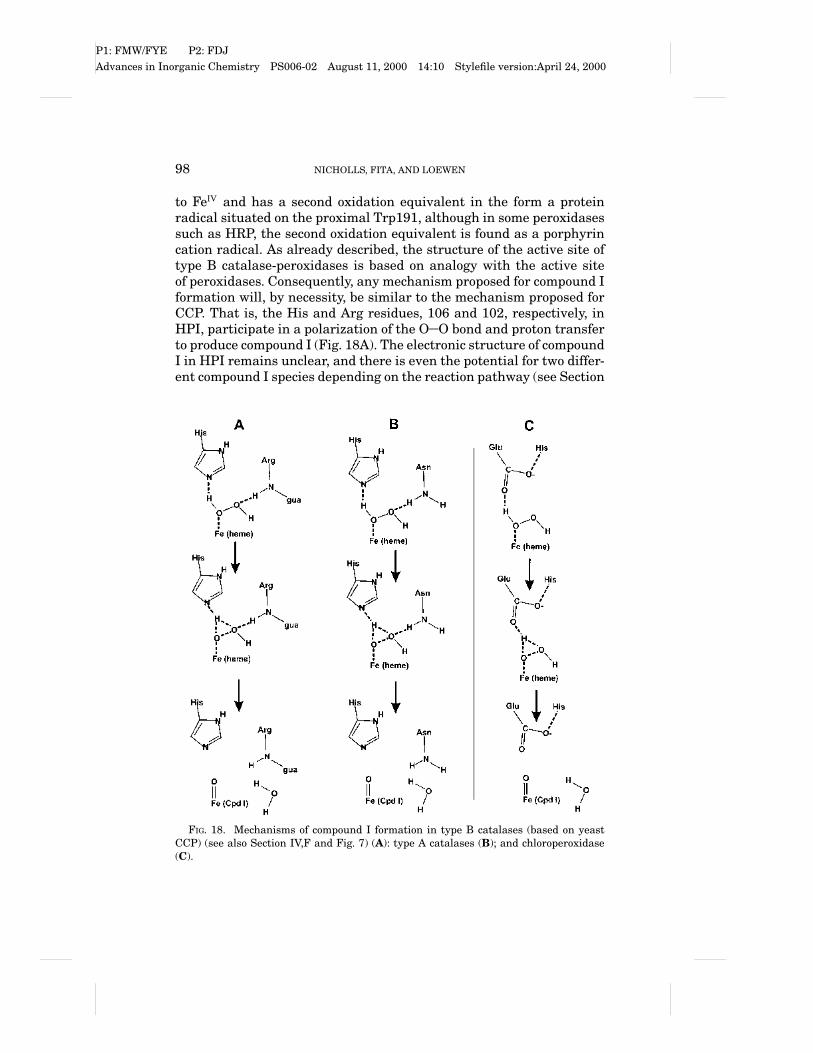
P1: FMW/FYE P2: FDJAdvances in Inorganic Chemistry PS006-02 August 11, 2000 14:10 Stylefile version:April 24, 2000
98 NICHOLLS, FITA, AND LOEWEN
to FeIV and has a second oxidation equivalent in the form a proteinradical situated on the proximal Trp191, although in some peroxidasessuch as HRP, the second oxidation equivalent is found as a porphyrincation radical. As already described, the structure of the active site oftype B catalase-peroxidases is based on analogy with the active siteof peroxidases. Consequently, any mechanism proposed for compound Iformation will, by necessity, be similar to the mechanism proposed forCCP. That is, the His and Arg residues, 106 and 102, respectively, inHPI, participate in a polarization of the O O bond and proton transferto produce compound I (Fig. 18A). The electronic structure of compoundI in HPI remains unclear, and there is even the potential for two differ-ent compound I species depending on the reaction pathway (see Section
FIG. 18. Mechanisms of compound I formation in type B catalases (based on yeastCCP) (see also Section IV,F and Fig. 7) (A): type A catalases (B); and chloroperoxidase(C).

P1: FMW/FYE P2: FDJAdvances in Inorganic Chemistry PS006-02 August 11, 2000 14:10 Stylefile version:April 24, 2000
ENZYMOLOGY AND STRUCTURE OF CATALASES 99
IV, F and Fig. 7). In catalatic mode, HPI exhibits no significant spectralchange, suggesting that compound I has an oxidation equivalent in theform of a protein radical rather than a porphyrin radical. On the otherhand, the W105F variant of HPI, which operates only in peroxidaticmode, has a porphyrin radical clearly evident in the absorbance andEPR spectra (101).
The mechanism for compound I formation in catalases is very similarto that just described for peroxidases. The active site of a type A cata-lase also has a histidine, which is essential for catalytic activity, and anasparagine, which, although not essential, greatly enhances catalysis(92). Despite the orientations of the active site residues of catalase, par-ticularly the histidine, differing significantly from the orientations ofthe equivalent residues in peroxidases, the histidines have similar rolesin both enzymes while the asparagine of catalase plays a role similarto the arginine of peroxidase (Fig. 18B). In contrast to its orientationin peroxidases, the imidazole of the essential histidine in catalases iscoplanar with the heme, effectively stacked, about 3.5 A above ring III ofheme b in BLC or ring IV of heme d in HPII. Like the arginine in peroxi-dases, the asparagine residue is situated laterally across the heme fromthe histidine in a position where it can hydrogen bond with the hydrogenperoxide during catalysis. The presence of similar residues allowed thecatalatic mechanism to be modeled on the peroxidatic mechanism (38).Interaction of the hydrogen peroxide with both the heme FeIII and theimidazole ring of the histidine weakens and stretches the O H bond,allowing the second oxygen of the peroxide to simultaneously form ahydrogen bond with the imidazole. The proton on the O that is inter-acting with the Fe is then transferred, via the imidazole, to the secondoxygen, giving rise to water and compound I. The asparagine residueparticipates through hydrogen bonding with the peroxide and stabilizesthe polarized intermediates.
For chloroperoxidase, a type D catalase, the mechanism for compoundI formation must differ significantly from those just described becausethe only active site residue sufficiently close to the heme iron to par-ticipate directly in the reaction is glutamate183. The glutamate is hy-drogen bonded with His105, but the latter is too distant to participatedirectly in the reaction with hydrogen peroxide. Therefore, the hydro-gen peroxide must initially associate in the active site with the hemeFeIII and the glutamate side chain. As the reaction progresses, a secondhydrogen bond may form with the highly electronegative glutamate sidechain to facilitate proton transfer from the Fe O H to the second O,producing water and compound I (Fig. 18C). Even making allowancesfor possible differences arising from assays in different laboratories, the

P1: FMW/FYE P2: FDJAdvances in Inorganic Chemistry PS006-02 August 11, 2000 14:10 Stylefile version:April 24, 2000
100 NICHOLLS, FITA, AND LOEWEN
catalatic activity of the chloroperoxidase is not more than 2% that oftype A or B catalases, suggesting that the active site of the chloroperox-idase is not optimized for the catalatic reaction, consistent with it beinga side reaction.
The reaction of other minor or type D catalases such as methemoglo-bin and metmyoglobin is not treated in detail here, because they areminor activities, significantly lower than even that of chloroperoxidase.The orientation of residues on the distal side of the heme is not op-timized for the catalatic reaction to the extent that there is even asixth ligand of the heme, a histidine, that would preclude a close as-sociation of the heme with hydrogen peroxide without a significantside-chain movement. It is only after an extended treatment with H2O2
and oxidation of the Fe that a low level of catalatic activity becomesevident.
B. COMPOUND I REDUCTION
The mechanism for the reduction of compound I by catalases mustdiffer significantly from the mechanism in peroxidases because of theinvolvement of hydrogen peroxide as the two-electron donor ratherthan sequential organic substrates acting as one-electron donors. Basedagain on the peroxidatic reaction, the catalase-peroxidase or type Bcatalases were initially thought to employ Arg102 and His106 (HPIInumbering) as catalytic residues in the reduction stage. However, re-cent evidence suggests that the active site tryptophan, Trp105 in HPI,plays an important role. Removal of Trp105, while not affecting com-pound I formation to the extent that compound I could be identifiedby absorbance and EPR spectrometry, significantly inhibits its subse-quent reduction by hydrogen peroxide (101). Because it is not possiblefor hydrogen peroxide to bind simultaneously to all three of His106,Trp105, and Arg102, a modified second stage is proposed in which theindole ring of Trp105 has a role analogous to that of the active siteAsn in type A catalases. Along with the imidazole of His106, it bindsthe second hydrogen peroxide and facilitates hydrogen transfer to theoxyferryl oxygen and to the imidazole ring of His106. The final transferof the second reducing equivalent, from the imidazole of His106 to theFe O H, completes the cycle, producing water and native enzyme(Fig. 19A). This mechanism is independent of the protein and porphyrinradical structure of compound I (see Section IV,F and Fig. 7).
In the type A catalases, there are only two active site residues inlocations where they can influence the reaction, a histidine and anasparagine. A mechanism for compound I reduction in catalases was

P1: FMW/FYE P2: FDJAdvances in Inorganic Chemistry PS006-02 August 11, 2000 14:10 Stylefile version:April 24, 2000
ENZYMOLOGY AND STRUCTURE OF CATALASES 101
propsed by Fita and Rossmann (38) in BLC involving these two residues.The hydrogen peroxide binds to compound I, forming hydrogen bondswith the oxyferryl oxygen, the His imidazole, and the Asn side chain.This structure allows the transfer of one hydrogen to the oxyferryl oxy-gen and a second to the imidazole, resulting in formation of oxygen. Thehydrogen and second reducing equivalent on the imidazole ring couldthen be transferred to the FeIV−OH to produce water and regeneratethe enzyme in its native state (Fig. 19B). The role of the Asn in thisstage might be to stabilize the nascent oxygen during the reaction. Di-rect evidence for this portion of the mechanism is lacking, although theAsn201His variant of HPII exhibits spectral changes consistent witha modification of the heme environment despite a very low catalaticactivity and no oxidation of the heme (92).
FIG. 19. Mechanisms of compound I reduction in type B catalases (based on yeast CCP)(see also Section IV,F and Fig. 7) (A); type A catalases (B); and chloroperoxidase (C).

P1: FMW/FYE P2: FDJAdvances in Inorganic Chemistry PS006-02 August 11, 2000 14:10 Stylefile version:April 24, 2000
102 NICHOLLS, FITA, AND LOEWEN
The reduction of compound I in chloroperoxidase differs from bothof the mechanisms in type A and B catalases because of there beingonly a single glutamic acid residue to catalyze the reaction. The re-ducing hydrogen peroxide must initially bind with the glutamate sidechain and the oxyferryl oxygen. The highly electronegative glutamatewill easily extract a proton from the peroxide, but it is more difficult toconceive of a hydrogen or reducing equivalent being transferred to it.Two options might be considered. In the first, the hydrogen peroxide ishighly polarized by the glutamate, resulting in a hydride transfer to theoxyferryl oxygen and simultaneous proton transfer to the glutamate.The proton on the glutamate would ultimately be transferred to theFeII O H− to produce water and native enzyme. In the second option,a single reducing equivalent, a hydrogen, is transferred to the oxy-ferryl oxygen. The second reducing equivalent and proton are trans-ferred to the glutamate and delocalized in the intricate complex involv-ing Glu183, His105, the propionate chains, and the MnII cation. Thisequivalent is then transferred back to the FeIV O H to complete thereaction (Fig. 19C). Obviously, the presence of glutamate alone on thedistal side does not present the optimal catalytic site for reaction withhydrogen peroxide, explaining the low reactivity.
IX. Summary
Catalases continue to present a challenge and are an object of interestto the biochemist despite more than 100 years of study. More than 120sequences, seven crystal structures, and a wealth of kinetic and phys-iological data are currently available, from which considerable insightinto the catalytic mechanism has been gained. Indeed, even the crystalstructures of some of the presumed reaction intermediates are avail-able. This body of information continues to accumulate almost daily.
Have we exhausted catalases as a source of information about proteinstructure and the catalatic mechanisms? The answer is clearly no. Witheach structure reported comes new information, often including struc-tural modifications seemingly unique to catalases and with roles thatremain to be explained. Despite a deeply buried active site, catalasesexhibit one of the fastest turnover rates determined. This presents theas yet unanswered question of how substrate can access the active sitewhile products are simultaneously exhausted with a potential turnoverrate of up to 106 per second. The complex folding pathway that producesthe intricate interwoven arrangement of subunits also remains to befully clarified.

P1: FMW/FYE P2: FDJAdvances in Inorganic Chemistry PS006-02 August 11, 2000 14:10 Stylefile version:April 24, 2000
ENZYMOLOGY AND STRUCTURE OF CATALASES 103
The catalase-peroxidases present other challenges. More than 20 se-quences are available, and interest in the enzyme arising from its in-volvement in the process of antibiotic sensitivity in tuberculosis-causingbacteria has resulted in a considerable body of kinetic and physiologicalinformation. Unfortunately, the determination of crystallization condi-tions and crystals remain an elusive goal, precluding the determinationof a crystal structure. Furthermore, the presence of two possible reac-tion pathways, peroxidatic and catalatic, has complicated a definitionof the reaction mechanisms and the identity of catalytic intermediates.There is work here to occupy biochemists for many more years.
ACKNOWLEDGMENTS
This work was supported by operating grants from the Natural Sciences and Engineer-ing Research Council of Canada (NSERC) to PCL and PN. The assistance of J. Switala,B. Tattrie, and M. Maj over the years is appreciated. Receipt of preprints in advance oftheir publication from C. Obinger is also appreciated.
REFERENCES
1. Loew, O. U.S. Dept of Agri. Repts. 1900, 65, 5.2. Loewen, P. C. In “Oxidative Stress and the Molecular Biology of Antioxidant De-
fenses”; Scandalios, J. G., ed.; pp. 273–308. Cold Spring Harbor Laboratory Press:Cold Spring Harbor, NY, 1997.
3. Zamocky, M.; Koller, F. Prog. Biophys. Molec. Biol. 1999, 72, 19.4. Kremer, M. L. Trans. Faraday Soc. 1965, 61, 1453.5. Keilin, D.; Hartree, E. F. Nature 1950, 160, 513.6. Klotz, M. G.; Klassen, G. R.; Loewen, P. C. Mol. Biol Evol. 1997, 14, 951.7. Welinder, K. Curr. Opin. Struc. Biol. 1992, 2, 388.8. Kono, Y.; Fridovich, I. J. Biol. Chem. 1983, 258, 6015.9. Allgood, G. S.; Perry, J. J. J. Bacteriol. 1986, 168, 563.
10. Waldo, G. S.; Fronko, R. M.; Penner-Hahn, J. E. Biochemistry 1991, 30, 10486.11. Whittaker, M. M.; Barynin, V. V.; Antonyuk, S. V.; Whittaker, J. W. Biochemistry
1999, 38, 9126.12. Arnao, M. B.; Acosta, M.; del Rio, J. A.; Varn, R.; Garcia-Canovas, F. Biochim. Bio-
phys. Acta 1990, 1041, 43.13. Thomas, J. A.; Morris, D. R.; Hager, L. P. J. Biol. Chem. 1970, 245, 3129.14. Sundaramoorthy, M.; Terner, J.; Poulos, T. L. Structure 1995, 3, 1367.15. Sun, W.; Kadima, T. A.; Pickard, M. A.; Dunford, H. B. Biochem. Cell. Biol. 1994, 72,
321.16. Facey, S.; Gross, F.; Vining, L. C.; Yang, K.; van Pee, K. H. Microbiology 1996, 142,
657.17. Mulvey, M. R.; Switala, J.; Borys, A.; Loewen, P. C. J. Bacteriol. 1990, 172, 6713.18. Ma, M.; Eaton, J. W. Proc. Nat. Acad. Sci. USA 1992, 89, 7924.

P1: FMW/FYE P2: FDJAdvances in Inorganic Chemistry PS006-02 August 11, 2000 14:10 Stylefile version:April 24, 2000
104 NICHOLLS, FITA, AND LOEWEN
19. Nicholls, P. Biochim. Biophys. Acta. 1965, 99, 286.20. Nicholls, P. Biochim. Biophys. Acta 1972, 279, 306.21. Clayton, R. K. Biochim. Biophys. Acta. 1960, 36, 35.22. Nicholls, P.; Schonbaum, G. R. In “The Enzymes”; P. Boyer; H. Lardy; K. Myrback,
eds.; Vol. VIII, 2nd ed., pp. 147–225; Academic Press: New York, 1963.23. Aebi, H.; Heiniger, J.-P.; Suter, H. Experientia 1962, 18, 129.24. Aebi, H.; Cantz, M.; Suter, H. Experientia 1965, 21, 713.25. Deisseroth, A.; Dounce, A. L. Physiol. Rev. 1970, 50, 319.26. Ruis, H.; Koller, F. In “Oxidative Stress and the Molecular Biology of Antioxidant
Defenses”; Scandalios, J. G., ed.; pp. 309–342. Cold Spring Harbor Laboratory Press:Cold Spring Harbor, NY, 1997.
27. Scandalios, J. G., Guan, L.; Polidoros, A. N. In “Oxidative Stress and the MolecularBiology of Antioxidant Defenses”; Scandalios, J. G., ed.; pp. 343–406. Cold SpringHarbor Laboratory Press: Cold Spring Harbor, NY, 1997.
28. Loewen, P. C.; Switala, J.; Triggs-Raine, B. L. Arch. Biochem. Biophys. 1985, 243,144.
29. Christman, M. F.; Storz, G.; Ames, B. N. Proc. Natl. Acad. Sci. USA 1989, 86, 3484.30. Ivanova, A.; Miller, C.; Glinsky, G.; Eisenstark, A. Molec. Microbiol. 1994, 12, 571.31. Mukhopadhyay, S.; Schellhorn, H. E. J. Bacteriol. 1994, 176, 2300.32. Visick, J. E.; Clark, S. J. Bacteriol. 1997, 179, 3158.33. Loewen, P. C.; Hennge-Aronis, R. Ann. Rev. Microbiol. 1994, 48, 53.34. Bonnichsen, R. K.; Chance, B.; Theorell, H. Acta Chem. Scand. 1947, 1, 685.35. Beers, R. F.; Sizer, I. W. J. Biol. Chem. 1952, 195, 133.36. Ogura, Y. Arch. Biochem. Biophys. 1955, 57, 288.37. Keilin, D.; Nicholls, P. Biochim. Biophys. Acta 1958, 29, 302.38. Fita, I.; Rossmann, M. G. J. Mol. Biol. 1985, 185, 21.39. Brill, A. S.; Castleman, B. W.; McKnight, M. E. Biochemistry 1976, 15, 2209.40. Nicholls, P. Biochem. J. 1961, 81, 365.41. Maj, M.; Loewen, P. C.; Nicholls, P. Biochim. Biophys. Acta 1998, 1384, 209.42. Young, L. J.; Siegel, L. M. Biochemistry 1988, 27, 2790.43. Nicholls, P. Biochem. J. 1964, 90, 331.44. Nicholls, P. Trans. Farad. Soc. 1964, 60, 137.45. Kalynaraman, B.; Janzen, E. G.; Mason, R. P. J. Biol. Chem. 1985, 260, 4003.46. Lardinois, O.; Rouxhet, P. G. Biochim. Biophys. Acta 1996, 1298, 180.47. Hillar, A.; Nicholls, P. FEBS Lett. 1992, 314, 179.48. Kirkman, H. N.; Gaetani, G. F. Proc. Natl. Acad. Sci. USA 1984, 81, 4343.49. Nicholls, P. Biochim. Biophys. Acta. 1964, 81, 479.50. Fita, I.; Rossmann, M.G. Proc. Natl. Acad. Sci. USA 1985, 82, 1604.51. Bicout, D. J.; Field, M. J.; Gouet, P.; Jouve, H.-M. Biochim. Biophys. Acta 1995, 1252,
172.52. Olson, L. P.; Bruice, T. C. Biochemistry 1995, 34, 7335.53. Almarsson, O.; Sinha, A.; Gopinath, E.; Bruice, T. C. J. Amer. Chem. Soc. 1993, 115,
7093.54. Kirkman, H. N.; Rolfo, M.; Ferraris, A. M.; Gaetani, G. F. J. Biol. Chem. 1999, 274,
13908.55. Hayashi, Y.; Yamazaki, I. J. Biol Chem. 1979, 254, 9101.56. He, B.; Sinclair, R.; Copeland, B. R.; Makino, R.; Powers, L. S.; Yamazaki, I. Bio-
chemistry 1996, 35, 2413.57. DeFelippis, M. R.; Murthy, C. P.; Faraggi, M.; Klapper, M. H. Biochemistry 1989, 28,
4847.

P1: FMW/FYE P2: FDJAdvances in Inorganic Chemistry PS006-02 August 11, 2000 14:10 Stylefile version:April 24, 2000
ENZYMOLOGY AND STRUCTURE OF CATALASES 105
58. Hillar, A.; Nicholls, P.; Switala, J. Loewen, P. C. Biochem. J. 1994, 300, 531.59. Jouve, H. M.; Pelmont, J.; Gaillard, J. Arch. Biochem. Biophys. 1986, 248, 71.60. Claiborne, A.; Malinowski, D. P.; Fridovich, I. J. Biol. Chem. 1979, 254, 11664.61. Hochman, A.; Shemesh, A. J. Biol. Chem. 1987, 262, 6871.62. Loewen, P. C.; Stauffer, G. V. Mol. Gen. Genet. 1990, 224, 147.63. Welinder, K. Biochim. Biophys. Acta 1991, 1080, 215.64. Percy, M. E. Can. J. Biochem. Cell. Biol. 1984, 62, 1006.65. Hochman, A.; Shemesh, A. J. Biol. Chem. 1987, 262, 6871.66. Goldberg, I.; Hochman, A. Biochim. Biophys. Acta 1989, 991, 330.67. Hochman, A.; Goldberg, I. Biochim. Biophys. Acta 1991, 1077, 299.68. Goldberg, I.; Hochman, A. Arch. Biochim. Biophys. 1989, 268, 124.69. Levy, E.; Eyal, Z.; Hochman, A. Arch. Biochim. Biophys. 1992, 296, 321.70. Obinger, C.; Regelsberger, G.; Strasser, G.; Burner, U.; Peschek, G. A. Biochem. Bio-
phys. Res. Commun. 1997, 235, 545.71. Regelsberger, G.; Jakopitsch, C.; Engleder, M.; Ruker, F.; Peschek, G. A.; Obinger, C.
Biochemistry 1999, 38, 10480.72. Jakopitsch, C.; Ruker, F.; Regelsberger, G.; Tockal, M.; Peschek, G.; Obinger, C. Biol.
Chem. 1999, 380, in press.73. Murthy, M. R. N.; Reid, T. J.; Sicignano, A.; Tanaka, N.; Rossmann, M. G. J. Mol.
Biol. 1981, 152, 465.74. Fita, I.; Silva, A. M.; Murthy, M. R. N.; Rossmann, M. G. Acta Cryst. B 1986, 42, 497.75. Berthet, S.; Nykyri, L. M.; Bravo, J.; Berthet-Colominas, C.; Alzari, P.; Koller, F.;
Fita, I. Protein Sci. 1997, 6, 481.76. Mate, M. J.; Zamocky, M.; Nykyri, L. M.; Herzog, C.; Alzari, P. M.; Betzel, C.; Koller,
F.; Fita, I. J. Mol. Biol. 1999, 286, 135.77. Gouet, P.; Jouve, H. M.; Dideberg, O. J. Mol. Biol. 1995, 249, 933.78. Murshudov, G. N.; Melik-Adamyan, W. R.; Grebenko, A. I.; Barynin, V. V.; Vagin, A.
A.; Vainshtein, B.K.; Dauter, Z.; Wilson, K. FEBS Lett. 1992, 312, 127.79. Vainshtein, B. K.; Melik-Adamyan, W. R.; Barynin, V. V.; Vagin, A. A.; Grebenko, A.
I. Nature 1981, 293, 1981, 411.80. Vainshtein, B. K.; Melik-Adamyan, W. R.; Barynin, V. V.; Vagin, A. A.; Grebenko,
A. I.; Borisov, V. V.; Bartels, K. S.; Fita, I.; Rossmann, M. G. J. Mol. Biol. 1986,188, 49.
81. Bravo, J.; Verdaguer, N.; Tormo, J.; Betzel, C.; Switala, J.; Loewen, P. C.; Fita, I.Structure 1995, 3, 491.
82. Bravo, J.; Mate, M. J.; Schneider, T.; Switala, J.; Wilson, K.; Loewen, P. C.; Fita, I.Proteins 1999, 34, 155.
83. Bravo, J.; Fita, I.; Gouet, P.; Jouve, H. M.; Melik-Adamyan, W.; Murshudov, G. N. In“Oxidative Stress and the Molecular Biology of Antioxidant Defenses”; Scandalios,J. G., ed.; pp. 407–445. Cold Spring Harbor Laboratory Press, Cold Spring Harbor,NY, 1997.
84. Sevinc, M. S.; Switala, J.; Bravo, J.; Fita, I.; Loewen, P. C. Prot. Eng. 1998, 11, 549.85. Loewen, P. C.; Switala, J. Biochem. Cell Biol. 1986, 64, 638.86. Switala, J.; O´ Neil, J.; Loewen, P. C. Biochemistry 1999, 38, 3895.87. Bergdoll, M.; Remy, M. H.; Cagon, C.; Masson, J. M.; Dumas, P. Structure, 1997, 4,
391.88. Tanford, C.; Lovrien, R. J. Am. Chem. Soc. 1962, 84, 1892.89. Ruis, H. Can. J. Biochem. 1979, 57, 1122.90. Hook, D. W. A.; Harding, J. J. Eur. J. Biochem. 1997, 247, 380.91. Murshudov, G. N.; Grebenko, A. I.; Barynin, V.; Dauter, Z.; Wilson, K.; Vainshtein,

P1: FMW/FYE P2: FDJAdvances in Inorganic Chemistry PS006-02 August 11, 2000 14:10 Stylefile version:April 24, 2000
106 NICHOLLS, FITA, AND LOEWEN
B. K.; Melik-Adamyan, W.; Bravo, J.; Ferran, J. M.; Switala, J.; Loewen, P. C.; Fita,I. J. Biol. Chem. 1996, 271, 8863.
92. Loewen, P. C.; Switala, J.; von Ossowski, I.; Hillar, A.; Christie, A.; Tattrie, B.;Nicholls, P. Biochemistry 1993, 32, 10159.
93. Bravo, J.; Fita, I.; Ferrer, J. C.; Ens, W.; Hillar, A.; Switala, J.; Loewen, P. C. ProteinSci., 1997, 6, 1016.
94. Mate, M. J.; Sevinc, M. S.; Hu, B.; Bujons, J.; Bravo, J.; Switala, J.; Ens, W.; Loewen,P. C.; Fita, I. J. Biol. Chem. 1999, 274, 27717.
95. Sevinc, M. S.; Mate, M. J.; Switala, J.; Fita, I.; Loewen; P. C. Protein Sci. 1999, 8,490.
96. Zamocky, M.; Herzog, C.; Nykyri, L. M.; Koller, F. FEBS Lett. 1995, 367, 241.97. Gouet, P.; Jouve, H. M.; Williams, P. A.; Andreoletti, P.; Nussaume, L.; Hadju, J.
Nature Struct. Biol. 1996, 3, 951.98. Bell, C. E.; Yeates, T. O.; Eisenberg, D. Protein Sci. 1997, 6, 2084.99. Tanner, J. J.; Tu, S. C.; Barbour, L. J.; Barnes, C. L.; Krause, K. L. Protein Sci. 1999,
8, 1725.100. Sevinc, S.; Ens, W.; Loewen, P. C. Eur. J. Biochem 1995, 230, 127.101. Hillar, A. Ph.D. Thesis, 1999, University of Manitoba, Winnipeg, MB, Canada.102. Poulos, T. L.; Kraut, J. J. Biol. Chem. 1980, 255, 8199.103. English, A. M.; Tsaprailis, G. Adv. Inorg. Chem. 1996, 43, 79.104. Dunford, H. B. In “Heme Peroxidases.” J. Wiley & Sons: New York, 1999.


![Structure of the monofunctional heme catalase DR1998 from ...cmromao/Articles-pdf/2014_FebsJ_KatDR19… · Penicillium vitale (PDB 4CAT) catalases [11–14]. The three catalases from](https://img.pdfslide.us/doc/110x75/5fd1b364c2d0642f3051de92/structure-of-the-monofunctional-heme-catalase-dr1998-from-cmromaoarticles-pdf2014febsjkatdr19.jpg)


![Enzymology [Compatibility Mode]](https://img.pdfslide.us/doc/110x75/577d1ec81a28ab4e1e8f3d6e/enzymology-compatibility-mode.jpg)