Embed Size (px)
Citation preview

Aalto-D
D 4
5/2
016 V
TT SC
IEN
CE
124
9HSTFMG*aggjji+
ISBN 978-952-60-6699-8 (printed) ISBN 978-952-60-6700-1 (pdf) ISSN-L 1799-4934 ISSN 1799-4934 (printed) ISSN 1799-4942 (pdf)
978-951-38-8404-8 (printed) 978-951-38-8403-1 (pdf) 2242-119X 2242-119X (printed) 2242-1203 (pdf)
Aalto University School of Chemical Technology Department of Biotechnology and Chemical Technology www.aalto.fi
BUSINESS + ECONOMY ART + DESIGN + ARCHITECTURE SCIENCE + TECHNOLOGY CROSSOVER DOCTORAL DISSERTATIONS
Piritta N
iemi
Enzym
atic fractionation of brewer's spent grain and bioconversion of lignin-rich fractions in a colon m
odel in vitro
Aalto
Unive
rsity
2016
Department of Biotechnology and Chemical Technology
Enzymatic fractionation of brewer's spent grain and bioconversion of lignin-rich fractions in a colon model in vitro
Piritta Niemi
DOCTORAL DISSERTATIONS


i
Acknowledgements
The work presented in this thesis was carried out at VTT Technical Research Centre of Fin-land during the years 2010-2015. The Academy of Finland is gratefully acknowledged for funding most of the work (project: Lignin fibre 133091). In addition, VTT and the Graduate Shool of Biomass Refining (BIOREGS) provided part of the funding. Funding for travelling was kindly provided by COST Actions FP0602 and FP0901.
I am grateful to my supervisors Prof. Kaisa Poutanen and Prof. Johanna Buchert for providing me with this interesting project and supervising the work. In addition, Dr. Tarja Tamminen, Dr. Annukka Aura and Prof. Craig Faulds have had an equally important role as co-supervisors. I warmly thank them all for sharing their knowledge and for their con-stant support throughout the project.
I express my sincere gratitude to the pre-examiners Prof. Stefan Willför and Associate Prof. Jean-Paul Vincken for reviewing the manuscript of this thesis and for suggesting revi-sions that clearly improved it. I also thank Prof. Markus Linder for supervising the work on behalf of Aalto University.
I am thankful to my former team leader Dr. Pekka Lehtinen and Dr. Annika Wilhelmson for hiring me as a PhD student at VTT and introducing me to the world of cereals and brewing. I also thank Dr. Ulla Holopainen-Mantila for teaching me about microscopy and Dr. Juhani Sibakov for helping me with the milling trials. I thank MSc. Matti Siika-aho for all the enzyme-related advice. I wish to acknowledge my current and former team leaders Dr. Tiina Liitiä, Dr. Juha Leppävuori and Dr. Anna-Stiina Jääskeläinen for their support during the thesis work. Also Dr. Anu Kaukovirta-Norja, Dr. Tuulamari Helaja and Dr. Raija Lantto are thanked for providing excellent research facilities at VTT.
I warmly thank my co-authors Dr. Klaus Niemelä, Dr. Annika Smeds, MSc. Ismo Mattila, Dr. Kaarina Viljanen, MSc. Taina Ohra-aho, Dr. Johanna Maukonen and MSc. Duarte Mar-tins for their invaluable contributions with the project and publications. I am grateful to Prof. Marco Orlandi for hosting my short term research visit at the University of Milano-Bicocca. Directors of BIOREGS, Prof. Liisa Viikari and Prof. Maija Tenkanen are thanked for organising the inspiring seminars and events (Vuosaari, Naantali, Hyytiälä etc.) to meet and network with other young researchers in the field.
I wish to acknowledge the skilled and helpful technical staff of laboratory 251, microbiol-ogy lab and brewing lab, who have taught me many analytical methods and also helped out with experimental work of this thesis. I am also greatful to my current and former team members for creating a pleasant and co-operative working atmosphere at the office. Dr. Arja Laitila and Dr. Brian Gibson are thanked for interesting discussions related to malting and brewing as well as providing great company on conference trips (with and without ash clouds). Daily lunches with our lunch group Ronny Wahlström, Jenni Rahikainen, Saara Lähtevänoja, Katariina Kemppainen, Katariina Rommi, Outi Mattila and Kirsi Kiiveri among many others have always been pleasant moments in often busy days. I am also

ii
greateful to my colleague Dr. Irina Tsitko for ‘menthal support’ ever since my Master’s the-sis work.
During the thesis work, I have been surrounded by the support of friends and family. I warmly thank them all for sharing the ups and downs in life with me. My Mother, who is a solid believer in higher education, has never missed out an opportunity to remind me that time spent for education is never wasted time. Finally, my deepest thanks go to Mika and little Leo for reminding me everyday of what is truly important in life.
Vantaa, February 2016 Piritta Niemi

iii
Contents
Acknowledgements ................................................................................................... i
List of abbreviations ................................................................................................ v
List of publications ................................................................................................. vi
Author’s contribution ............................................................................................ vii
1. Introduction .................................................................................................. 1
1.1 From barley to beer and spent grains ....................................................... 1
1.1.1 Grain structure .......................................................................................... 1
1.1.2 Malting and mashing ................................................................................ 2
1.1.3 Production and current uses of BSG ......................................................... 4
1.1.4 Properties and chemical composition of BSG .......................................... 4
1.2 Fractionation of BSG................................................................................. 8
1.2.1 Chemical fractionation .............................................................................. 9
1.2.2 Enzymatic fractionation ........................................................................... 11
1.2.3 Pre-treatment methods to improve enzymatic fractionation ................. 13
1.3 Applications of BSG and its fractions ..................................................... 13
1.3.1 Current use of BSG.................................................................................. 13
1.3.2 Potential applications and functionalities as food ingredient ................ 14
1.3.3 Potential non-food applications ............................................................. 14
1.4 Dietary fibre .............................................................................................15
1.4.1 Physiological functionalities of dietary fibre ...........................................15
1.4.2 Lignin as part of dietary fibre ................................................................. 16
2. Aims and hypotheses of the study .............................................................. 19
3. Materials and methods ............................................................................... 20
3.1 Materials ................................................................................................. 20
3.2 Analytical methods ................................................................................. 20
3.2.1 Composition analysis .............................................................................. 20
3.2.2 Particle size distribution analysis ........................................................... 21
3.2.3 Microscopy imaging ................................................................................ 21
3.2.4 Analyses of colon model metabolites ...................................................... 22
3.3 Experimental ........................................................................................... 23
3.3.1 Milling experiments ................................................................................ 23
3.3.2 Effect of milling on enzymatic carbohydrate hydrolysis ........................ 23
3.3.3 Effect of pH on protein and total solubilisation ..................................... 24
3.3.4 Enzymatic preparation of lignin-rich fractions ...................................... 24

iv
3.3.5 Preparation of deferuloylated fraction of BSG ....................................... 24
3.3.6 Colon model fermentation ...................................................................... 25
3.3.7 Lignin-rich fraction as growth substrate for lactobacilli and bifidobacteria 26
4. Results ......................................................................................................... 27
4.1 BSG properties and composition ............................................................ 27
4.2 Milling of BSG ........................................................................................ 28
4.2.1 Effect of milling on particle size ............................................................. 28
4.3 Effect of carbohydrase treatment and pH on BSG solubility .................. 31
4.4 Preparation of lignin-rich fractions for the colon model study .............. 33
4.4.1 Three-step enzymatic hydrolysis of BSG ................................................. 33
4.5 Interactions of lignin-rich fractions with colon microbiota in vitro ....... 35
4.6 Effects of lignin on lactobacilli and bifidobacteria ................................. 40
5. Discussion .................................................................................................. 42
5.1 Preparation of lignin-rich fractions from BSG ....................................... 42
5.1.1 Limited enzymatic solubilisation of cell wall carbohydrates ................. 42
5.1.2 Effect of pH and protease on BSG and protein solubility ...................... 44
5.1.3 Co-solubilisation of protein and lignin in alkaline conditions ................ 45
5.1.4 Insoluble lignin-carbohydrate residues ................................................. 46
5.2 Metabolism of lignin-rich fractions in a colon model ............................. 47
5.2.1 Potential lignin metabolites .................................................................... 47
5.2.2 Extent of lignin degradation by gut microbiota ..................................... 49
5.2.3 Phenolic metabolites derived from non-lignin sources ......................... 50
5.2.4 Potential bioactivities of lignin and phenolic metabolites ...................... 51
5.3 Effects of lignin on microbial carbohydrate fermentation and growth .. 52
5.3.1 Formation of SCFA from the lignin-rich fractions .................................. 52
5.3.2 Lignin-rich fraction as a growth substrate for lactobacilli and bifidobacteria 53
5.4 Methodological considerations ............................................................... 55
5.4.1 Analysis of lignin content in biomass ...................................................... 55
5.4.2 Use of the in vitro colon model ............................................................... 55
6. Conclusions and future prospects ............................................................... 57
References .............................................................................................................. 61
APPENDICES : Publications I‒V

v
List of abbreviations
BSA bovine serum albumin
BSG brewer’s spent grain
CFU colony forming units
CHP combined heat and power
DEFE deferuloylated BSG fraction
DFRC derivatisation followed by reductive cleavage
DF dietary fibre
DM dry matter
DP degree of polymerisation
EFSA European Food Safety Authority
FC fold change
FPU filter paper units
GC/FID gas chromatography coupled with flame ionisation detector
GCxGC-TOFMS two-dimensional gas chromatography coupled with time-of-flight mass detector
GMD GOLM Metabolome Database
HPLC high performance liquid chromatography
INS insoluble residue (after three-step enzymatic hydrolysis)
MRS de Man-Rogosa-Sharpe (medium)
NMR nuclear magnetic resonance
P-AEF protease-alkaline extracted fraction
Py-GC/MS analytical pyrolysis coupled with gas chromatography and mass detector
RCM reinforced clostridial medium
RT room temperature
SCFA short chain fatty acid
UV ultraviolet

vi
List of publications
This doctoral dissertation consists of a summary and of the following publications which are referred to in the text by their numerals. I Niemi P., Faulds C.B., Sibakov J., Holopainen U., Poutanen K., Buchert J. (2012)
Effect of a milling pre-treatment on the enzymatic hydrolysis of carbohydrates in brewer's spent grain. Bioresour. Technol. 116, 155-160. DOI: 10.1016/j.biortech.2012.04.043.
II Niemi P., Martins D., Buchert J., Faulds C.B. (2013) Pre-hydrolysis with carbohy-drases facilitates the release of protein from brewer's spent grain. Bioresour. Technol. 136, 529-534. DOI: 10.1016/j.biortech.2013.03.076.
III Niemi P., Tamminen T., Smeds A., Viljanen K., Ohra-aho T., Holopainen-Mantila U., Faulds C.B., Poutanen K., Buchert J. (2012) Characterization of lipids and lignans in brewer’s spent grain. J. Agric. Food Chem. 60, 9910–9917. DOI: 10.1021/jf302684x.
IV Niemi P., Aura A.-M., Maukonen J., Smeds A., Mattila I., Niemelä K., Tamminen T., Faulds C.B., Buchert J., Poutanen K. (2013) Interactions of a lignin-rich fraction from brewer’s spent grain with gut microbiota in vitro. J. Agric. Food Chem. 61, 6754‒6762. DOI: 10.1021/jf401738x.
V Aura A.-M., Niemi P., Mattila I., Niemelä K., Smeds A., Tamminen T., Faulds C.B., Buchert J., Poutanen K. (2013) Release of small phenolic compounds from brewer’s spent grain and its lignin fractions by human intestinal microbiota in vitro. J. Agric. Food Chem. 61, 9744‒9753. DOI: 10.1021/jf4024195.

vii
Author’s contribution
Publication I: Piritta Niemi had the main responsibility for preparing and writing the article and she is the corresponding author. She planned the study together with co-authors. From the experimental work she conducted enzymatic hydrolyses, wet millings in co-operation with technical staff, particle size analyses of the wet milled samples, and anal-ysis of reducing groups as a function of hydrolysis time. Composition analysis, dry millings and the respective particle size measurements, HPLC analyses, microscopy (supporting material) and enzymatic activity assays were carried out by co-authors and technical staff. Piritta Niemi was mainly responsible for calculating the results and she carried out the interpretation of results with the aid of co-authors.
Publication II: Piritta Niemi had the main responsibility for preparing and writing the article. The study was designed together by the authors, and Duarte Martins conducted the experimental work as part of his Master's Thesis work. He was supervised by Piritta Niemi and the co-authors. The interpretation of results was carried out as co-operation by all au-thors.
Publication III: Piritta Niemi had the main responsibility for preparing and writing the article, and she is the corresponding author. The study was planned together by the au-thors. Piritta Niemi carried out the enzymatic fractionation of BSG. Lipid and lignan anal-yses were conducted by co-authors. Composition analyses and microscopy imaging were carried out by technical staff. Interpretation of the results was carried out jointly by Piritta Niemi and the co-authors.
Publication IV: Piritta Niemi had the main responsibility for preparing and writing the article, and she is the corresponding author. The study was planned jointly by the authors. Piritta Niemi prepared the studied sample and participated in the experimental work of the colon model experiment led by Dr. Aura. Lignan analysis as well as GCxGC-TOFMS analy-sis including identification and determination of chemical structures of metabolites were carried out by co-authors. Piritta Niemi and Dr. Aura prepared the heat map. Experiments with bacterial strains were conducted by Dr. Maukonen. Interpretation of the results was carried out jointly by the authors.
Publication V: The study was planned together by the authors. Piritta Niemi prepared the studied samples and participated in the experimental work of the colon model experi-ment led by Dr. Aura. Lignan analyses and GCxGC-TOFMS analysis including identifica-tion and determination of chemical structures of metabolites were carried out by co-authors. Piritta Niemi and Dr. Aura prepared the heat maps. The results were interpreted together with the co-authors.

viii

1
1. Introduction
Brewing is an age-old technique and science. In fact, it is often said that brewing beer and baking are the world’s oldest food technologies. As reviewed by Boulton and Quain (2006), brewing with malts was probably started in the Middle East sometime after the birth of agriculture in 6000 BC. Most likely the early fermentations were spontaneous reactions, where a natural source of sugar was contaminated with yeast in the presence of a sufficient amount of water. Furthermore, the discovery of malting is also believed to have occurred by accident. In ancient times, alcoholic beverages were not just part of human diet but pre-sumably were associated with religion and rituals as well, due to the physiological effects of alcohol. Drinking beer instead of water also had its benefits in times when diseases, such as cholera, spread in contaminated drinking water. As described by Boulton and Quain (2006), the skill of brewing was further developed in Europe, where it was an important part of everyday life already in the medieval times, and taken to a larger scale production by the abbeys at least in the UK and Belgium. In addition to beer, brewing produces also spent grains, which are the insoluble residues of the malts containing a high amount of protein and cell wall polysaccharides.
1.1 From barley to beer and spent grains
1.1.1 Grain structure
Cereal grains are composed of the starchy endosperm, embryo (or germ) and several layers around them having different functions, such as protein storage and enzyme secretion (al-eurone) and protection (testa, pericarp and husk). The different components and layers are presented in Figure 1 and a more detailed structure of the barley grain is shown in Figure 2.
Husk and pericarp are the outermost, protective parts of the grain, shown as yellow in Figure 2. The colour is due to autofluorescence caused by phenolic components in the cell walls. Nearly all of cellulose in barley (96 %) is located in the husk (Duffus and Cochrane 1993). Below the pericarp, there is the testa or seed coat, which is a thin protective layer rich in hydrophobic cutin. The aleurone layer in barley usually consists of three layers of cells, which contain most of the storage proteins in the grain, as visualized by the red color in Figure 2. Together, pericarp, testa and aleurone layer are referred to as bran, which is rich in dietary fibre and several other nutrients. Bran is often removed in the milling of grains to flour. However, in malting and mashing husk and bran are not separated but they as well enter the process. Under the protective layers there is the endosperm, which is the largest part of the grain. Endosperm cell walls are composed of β-glucan (70 %) and arabi-noxylan (20 %), whereas in the aleurone layer the ratio is almost the opposite: 26 % of β-glucan and 67 % of arabinoxylan (Duffus and Cochrane 1993). Endosperm stores energy in

Introduction
2
the form of starch, which can be hydrolysed to sugars during germination providing energy for the growing plant. Also the embryo, where the new plant grows from, stores lipids and proteins to initiate growth.
Figure 1. Structure of a barley grain. Courtesy of Oili Lappalainen, VTT.
1.1.2 Malting and mashing
The first step in the making of beer is malting of grains. The majority of beer is brewed from barley malts but other cereals, such as wheat, buckwheat or rice can be used as well (Depraetere et al. 2004, Nic Phiarais et al. 2010, Teramoto et al. 2002). This thesis is, however, focused on barley and other raw materials are not further discussed. In malting, the grains are first steeped in cold water to increase the moisture content of the grains to above 40 % (Bamforth and Barclay 1993). This initiates the germination process and in-duces the production of endogenous enzymes. The enzymes are synthesised from the pro-tein storages in the aleurone layer and secreted to endosperm, where they start to hydro-lyse carbohydrates and proteins to provide nutrients for the embryo. Amylases, β-glucanases and proteases are the most important enzymes considering the following mash-ing process. In addition to the endogenous enzymes a diverse microbial community origi-nating from the field, storage and post-harvest processing is present in barley grains. Mi-crobial interactions with the grains may affect safety, technological, nutritional and or-ganoleptic properties of malts and beer (Laitila 2007). Germination process is stopped by kilning, which preserves the produced enzymes although they are inactivated. The enzymes are needed in mashing to further digest the endosperm cell walls and starch.
In mashing, ground malts are mixed with water, and the water temperature is raised stepwise to allow different hydrolytic enzymes to become activated and hydrolyse their substrate polymers (Briggs et al. 2004a). Mashing times and temperatures vary from brewery to brewery, but usually mashing is finished in approximately 2 h. Examples on mashing schedules and temperatures are given by Briggs et al. (2004a). β-glucanases are heat-labile and active at temperatures 30‒40 ºC. Proteolysis occurs at approximately 35‒60 ºC. Amylases are more heat-stable and become activated around 50 ºC, and saccharifi-cation of starch occurs up to 70ºC.
aleuronehusk pericarp
testa
embryo
endosperm

Introduction
3
Figure 2. a) A microscopy image of a cross-section of a barley grain, b) a close-up of the different layers of the grain (H=husk, P=pericarp, T=testa, A=aleurone, E=endosperm), and c) spent grains recovered after mashing. In all images protein appears as red and aleurone and endosperm cell walls blue. Husk and per-icarp cell walls show yellow and green autofluorescence due to their phenolic components. Images a and b are courtesy of Ulla Holopainen-Mantila, VTT. Image c is adapted from Paper I (supplementary material) and reprinted with permission of Elsevier B.V.
β-glucanases are the first enzymes to become activated in mashing and they are respon-sible for releasing the starch granules from the endosperm, as majority of endosperm cell wall is composed of β-glucan (Briggs et al. 2004a). This can be seen also from the intensive blue colour in Figure 2, as the dye Calcofluor stains β-glucan blue. In malting, a part of proteins is degraded to amino acids and peptides by proteases, and the proteolysis contin-ues in mashing. Starch gelatinization of malted barley occurs at 64–67 °C (Briggs et al. 2004b), and after the mashing temperature reaches this level, amylases start degrading starch to fermentable sugars, mainly maltose. Starch is the most abundant component in the grain and accounts for 60–64 % of the total weight (Jadhav et al. 1998). The endo-sperm is almost completely solubilised as the result of malting and mashing, and the starch granules, which appear as black particles in Figure 2b, are no longer visible in the insoluble residues of the malts, i.e. brewer’s spent grain (BSG) (Figure 2c). The applied temperatures and duration of mashing define the characteristics of the final wort, which is the sugar-rich liquid phase after mashing. After mashing, the wort containing all the solubilised compo-
a
c
b
E
A
PT
H

Introduction
4
nents is separated and BSG remains in the mash kettle (Figure 3). Hops are added to the wort, which is then boiled and fermented to beer.
Figure 3. The steps from barley to beer and BSG.
1.1.3 Production and current uses of BSG
The brewing industry produces annually 1.9 billion hectolitres of beer from barley, world-wide (FAOSTAT 2013). Asia produces most of the beer (35 %), followed by the Americas (30 %) and Europe (27 %) (FAOSTAT 2013). Brewing generates also side-streams, such as BSG, spent hops and spent yeast, of which BSG is the most abundant. Currently, the side-streams are mainly utilised as cattle feed. Approximately 15–20 kg of BSG is generated per every hectolitre of beer, which converts to an annual production of ca. 33 million tons of BSG worldwide. The commercial value of BSG is low, and if a brewery produces more BSG than the local feed companies and farmers are able to buy, it may have to pay for the dis-posal of BSG. However, as BSG is a food grade material rich in protein and dietary fibre, it would have potential for more valuable applications as well, such as a food ingredient, if suitable processing methods are developed.
1.1.4 Properties and chemical composition of BSG
BSG is composed mainly of the husks and outer layers of the grain, which are not solubil-ised in mashing. These parts consist mainly of cell wall polysaccharides and lignin, which are arranged in the cell walls in a complex matrix with cross-links between the polymers. An illustration of the plant cell wall structure has been proposed by Sticklen (2008). Part of BSG protein is located inside aleurone cells. In addition to the outer layers, remnants of endosperm remain in BSG. The heterogenous nature of BSG is visible in Figure 2c. The chemical composition of BSG is presented in Table 1. As BSG has a water content of up to
Cultivation of barley
Mashing of malts
Malting of grains
Fermentationto beer
Spent grains
Filtration of wort

Introduction
5
80 %, it is not microbiologically stable without drying or chemical preservation (Robertson et al. 2010a), which hinders the storing of BSG.
Table 1. Chemical composition of BSG from barley. Nd = not determined.
Carbohydrates The most abundant carbohydrates in BSG are arabinoxylan and cellulose (Table 1), which are cell wall polysaccharides. Cellulose is present as bundles or microfibrils, which are formed from linearly arranged cellulose chains and held together by inter- and intramolec-ular hydrogen bonds. The numerous hydrogen bonds make cellulose water-insoluble and resistant to enzymatic action. Cellulose microfibrils are surrounded by arabinoxylan, which is a branched polymer, composed of xylan backbone and substituted by arabinose and ace-tyl residues. Arabinoxylan is less rigid than cellulose, and its susceptibility to enzymatic digestion depends on the level of substitution. It is known for wheat bran that arabinoxylan in aleurone is less substituted than in pericarp, which makes aleurone more easily digesti-ble for xylanases (Benamrouche et al. 2002). The arabinose residues may be further substi-tuted with ferulic acid via ester-linkage (Ishii 1997). Ferulic acids may form dimers and cross-link arabinoxylan chains together (Bunzel et al. 2001). In the pericarp and husk, where lignin is present, ferulates can also cross-link arabinoxylan to lignin (Bunzel et al. 2004). These types of cross-links construct physical barriers for enzymes and thus protect the grain from the attacks of pathogens.
Starch is the main form of energy storage in barley grains. It is the most abundant poly-saccharide in the grains but in mashing it is almost completely solubilised into wort. Some starch (2–13 %) and residual amounts of mixed linked β-glucan (0.5–1.1 %) remain in BSG as well (Robertson et al. 2010b). The starch content is usually lower in BSG generated in lager beer production (2–8 %) than in ale production (7–13 %) (Robertson et al. 2010b).
Lignin Lignin is an important constituent of plant cell walls. It acts as a structural component providing rigidity to the cell walls, it is an important factor in water transportation of plants due to its hydrophobic nature, and it helps protect the plant from the attacks of mi-cro-organisms, as lignified cell walls are resistant to enzymatic attacks (Sarkanen and Ludwig 1971). After cellulose it is the second most abundant polymer in nature. In plants lignin is present in highest amounts in wood, but it is also present in lower amounts in many foods that belong to our everyday diet, such as cereals, fruit and vegetables (Bunzel et al. 2005, Bunzel et al. 2006). The lignin content of foods is usually not very high; for instance, in whole grain wheat, kale and pear lignin contents have been reported to be 5 %, 7 % and 16 % of the dietary fibre fraction, respectively (Bunzel et al. 2011).
In barley, lignin is present in the husk and pericarp, as indicated in Figure 2a and b by the yellow autofluorescence. Other phenolics may also influence the autofluorescence, but being the most abundant phenolic compound, lignin contributes the most. Lignin content
Arabinoxylan Glucan1 Lignin Protein Lipids Ash Ref.28.4 16.8 27.8 15.3 5.8 4.6 Mussatto et al. 200523.9 14.4 18.9 26.9 6.8 3.4 Forssell et al. 201125.4 21.8 11.9 24.0 10.6 2.4 Kanauchi et al. 199726.5 19.4 20.1 17.6 5.2 Nd Faulds et al. 2008
22–29 14–24 13–17 10–24 Nd Nd Robertson et al. 2010b1Glucan in BSG is mainly cellulose but residual amounts of starch and β-glucan are also present.

Introduction
6
is enriched in mashing and represents about 12–28 % of BSG (measured as Klason lignin) (Table 1). Lignin is formed from phenolic units or monolignols, p-coumaryl alcohol, co-niferyl alcohol and sinapyl alcohol (Figure 4a), which in lignin form p-hydroxyphenyl, guaiacyl and syringyl units, respectively. During the synthesis of plant cell walls, monolig-nols are polymerised by radical coupling reactions (Boerjan et al. 2003), and therefore lignin structure is irregular and consists of different units and different types of linkages between them, as the bond formation can occur at various sites of the monolignol mole-cules. Depending on the plant origin, lignins vary in the ratio of the monomers. Recently, BSG lignin has also been characterised using analytical pyrolysis coupled with gas chroma-tography and mass detector (Py-GC/MS), NMR spectroscopy and DFRC (derivatisation followed by reductive cleavage) (Rencoret et al. 2015). Lignins from enzymatically treated BSG fractions have also been studied using Py-GC/MS (Ohra-aho et al. 2016) and the re-sults were in agreement with the data of Rencoret et al. (2015). BSG lignin is predominant-ly composed of guaiacyl units, with ratio of syringyls to guaiacyls being 0.4‒0.5, and it is associated with ferulic and p-coumaric acids. The major intramolecular substructures are β-O-4 aryl ethers (77‒79 %) and β-5 phenyl coumarans (11‒13 %) (Rencoret et al. 2015) (Figure 4b). Ferulic acids are mostly etherified to lignin whereas p-coumaric acids are es-terified in γ position (Rencoret et al. 2015, Ohra-aho et al. 2016).
Figure 4. a) Monolignols, from which lignin is formed, and b) the most common substructures found in BSG lignin: β-O-4 aryl ether linkage and β-5 phenyl coumaran structure.
In the literature there are currently no published images of lignin in cereal grains. Wheat straw lignin is the most studied lignin related to cereals and a proposed structure of it is presented in Figure 5. However, based on the most recent findings by Rencoret et al. (2015) there are some inconsistencies between the proposed wheat straw lignin and BSG lignin. In Figure 5 α-O-4 ethers are abundant, whereas in BSG β-O-4 ethers are dominant (almost 80 %) and α-O-4 ethers were not detected. In addition, in BSG the 5-5 linked structure would be in the dibenzodioxocin form instead of the biphenyl form.
a)
b)

Introduction
7
Lignin is usually assumed to have no nutritional value. Although it may not act as an en-ergy source, lignin may induce other effects in the human gastrointestinal tract, such as adsorption of carcinogenic compounds (Funk et al. 2007), antioxidative and radical scav-enging activity in the lumen (Dizhibite et al. 2004) and being a precursor of the mammali-an lignan enterolactone (Begum et al. 2004). Lignin is known to be degraded by certain fungi and some insects, such as termites, but the digestion of lignin in humans and animals is not well known. There is some evidence of lignin digestion in humans (Kelsay et al. 1981) but more research is needed to enable better understanding of lignin metabolism.
Figure 5. Proposed structure of wheat straw lignin. Modified from Sun et al. (1997), reproduced with permis-sion from Elsevier B.V. The coloured markings signifying different components of lignin are added to the original figure. H=p-hydroxyphenyl unit, G=guaiacyl unit, S=syringyl unit, FA=ferulic acid, pCA=p-coumaric acid. The orange dotted circle points out a cross-link of lignin to carbohydrates via ferulic acid.
Proteins The protein content of barley varies from 8‒15 % (Shewry 1993). During malting, barley proteins are partially degraded to amino acids and small peptides by the endogenous bar-ley peptidases (Jones and Budde 2005). However, most of malt proteins are not dissolved in mashing but 74‒78 % of protein remains insoluble in the spent grains (Jones and Budde 2005, Celus et al. 2006). Due to the extensive endosperm solubilisation, the protein con-tent is increased in mashing and may be up to 27 % in BSG (Table 1). In barley, there are four different types of proteins, which have been classified according to their sequential extractabilities by the procedure developed by Osborne (1909). Albumins, which are main-ly enzymes, are water-soluble. Globulins may be enzymes or storage proteins and are ex-tractable by salt-solutions. Hordeins are the main storage proteins in barley. Mashing causes disulphide bridge formation in hordeins, and therefore their extraction requires a reducing agent in addition to a high alcohol content in the extraction solvent (Celus et al. 2006). Glutelins, which are structural proteins, may be extracted with dilute acid or alkali or with detergents in the presence of a reducing agent (Celus et al., 2006). The proteins in
S
G
G
S
GG
G
G
G
SG
S
H
pCA
FA

Introduction
8
BSG are mainly hordeins and glutelins, and albumins and globulins constitute only ap-proximately 10 % of BSG proteins (Celus et al. 2006).
The amino acid composition of BSG proteins has been determined by Treimo et al. (2008). BSG is rich in glutamic acid/glutamine and proline, which constitute 30 % of all amino acids. This is explained by the fact that hordeins are especially rich in these amino acids (Shewry 1993). The hordein content in barley is 35–55 % of all proteins (Shewry 1993) and is not significantly altered in mashing (43 % in BSG) (Celus et al. 2006).
Lipids Most of lipids in barley are located in the endosperm and embryo, as their role is to provide nutrients and energy for the new seedling. The lipid content of unmalted barley varies from 2.0 to 4.6 % (Morrison 1993), and linoleic acid is the main fatty acid (55 %) followed by palmitic (22 %) and oleic acids (13 %) (Kaukovirta-Norja et al. 1993). During malting the amount of lipids is reduced by approximately 20 %, but the fatty acid composition is not significantly altered (Kaukovirta-Norja et al. 1993). During mashing the lipid content is increased due to the solubilisation of other compounds, and the lipid content in BSG varies from 5.2 up to 11 % (Table 1). The main lipid class in barley and malt is triglycerides (69 %) followed by polar lipids, but in mashing triglycerides are partially de-esterified by lipase activity releasing free fatty acids (Kaukovirta-Norja et al. 1993). A more detailed character-isation of BSG lipids and lipophilic extractives has been conducted by del Río et al. (2013).
1.2 Fractionation of BSG
BSG is a food-grade material rich in nutrients, such as protein, dietary fibre and antioxi-dant phenolic compounds. Therefore it would have potential for more valuable applica-tions than cattle feed, but this would require development of suitable fractionation and processing techniques. One possible option would be to integrate the fractionation directly into the brewery, and thus save time and energy on transportation. In addition, no preser-vation of BSG would be required, if it was processed immediately at the site of production.
The fractionation methods studied for BSG include mainly wet fractionation. Separation of different components from BSG is not always straightforward. For instance, the cell wall polysaccharides contain both 5 and 6 carbon sugars, and after enzymatic or acidic hydroly-sis of carbohydrates their efficient separation to different fractions may be difficult, unless they all can be utilised for the same end-use. In addition to the lignocellulosic cell walls, there is a considerable amount of protein and lipids. The more easily digestible parts of the barley grain have already been dissolved in mashing and the recalcitrant parts remain in BSG, which contains the husks and outer layers or the grain and only residual amounts of endosperm. The role of the outer layers is to protect the grain and its nutrients from other organisms, and thus they are designed to withstand the attacks of e.g. fungal enzymes. However, the different components in BSG might have the highest value as separate com-ponents, and several studies on fractionation methods have been published (Mussatto et al. 2005, Carvalheiro et al. 2004b, Mussatto et al. 2006a, Forssell et al. 2011, Treimo et al. 2008). Nevertheless, no large-scale applications have resulted from these studies and ru-minant feed is still the main use of BSG.

Introduction
9
1.2.1 Chemical fractionation
Biomass components can be separated from each other with the aid of chemical, mechani-cal or biotechnical treatments. Chemicals may be less expensive and require shorter reac-tion times than enzymes, but on the other hand they are less specific and generation of unwanted side-products is possible. In addition, the suitability of chemicals for processing of food material has to be carefully considered, but for other end-uses than food and feed they may well be suitable methods. Acidic and alkaline treatments applied for BSG frac-tionation and the main results are summarised in Table 2, and the treatments are further discussed below.
Acid hydrolysis For BSG, the most commonly applied chemicals are acids and bases. At elevated tempera-tures dilute acids depolymerise hemicelluloses without significant damage to cellulose. The main hemicellulose in BSG is arabinoxylan, and more than 90 % of arabinoxylan can be converted to monomeric sugars by a dilute sulphuric acid treatment (Carvalheiro et al. 2004a, Mussatto et al. 2005). The resulting sugar-rich hydrolysates contained some sugar degradation products (furfural, hydroxymethylfurfural, formic, acetic and levulinic acid), which were formed during the acid hydrolysis and could inhibit subsequent microbial fer-mentation of the sugars. Nevertheless, the amounts of the side-products were found low enough not to cause inhibition of yeasts (Carvalheiro et al. 2004a). After the acid treat-ment, cellulose is more accessible for further treatments, for example for enzymatic or acidic hydrolysis (Mussatto et al. 2008b).
A milder acidic treatment called autohydrolysis is based on acetic acid released from arabinoxylan. The arabinoxylan of BSG is substituted with acetyl groups, and when ex-posed to hot water or steam, these acetyl groups are released and form acetic acid, which partially depolymerizes xylan (Kabel et al. 2002, Carvalheiro et al. 2004b). In autohydroly-sis, the sugars were mainly released as oligomers having a degree of polymerization above 9, but with prolonged reaction times the amount of smaller oligomers increases (Carval-heiro et al. 2004b). Due to the mild conditions, the generation of sugar degradation prod-ucts is lower than with dilute acid treatments but on the other hand sugar yields are also lower (Carvalheiro et al. 2004b).
Alkaline extraction Alkaline treatments, which are known to dissolve hemicelluloses and lignin, have been studied for the production of cellulose pulp from BSG (Mussatto et al. 2006a). For soda pulping process, BSG was first pre-treated with dilute sulphuric acid to remove most of arabinoxylan. The removal of hemicelluloses prior to pulping makes the material more accessible for pulping chemicals and thus reduces the amount of chemicals needed (Mus-satto et al. 2006a). Under optimized conditions 90 % of lignin can be removed with negli-gible cellulose losses. The pulping process can be applied on untreated BSG as well, but the quality of the resulting pulp is not as good as after the acid pre-treatment (Mussatto et al. 2008a). Pulps can be further bleached with hydrogen peroxide to remove the residual lig-nin (Mussatto et al. 2008a). In addition to lignin, smaller phenolic compounds, namely ferulic and p-coumaric acids are also extractable in alkaline conditions (Mussatto et al. 2007a). Delignification enhances enzymatic hydrolysis of BSG cellulose due to modifica-tions in BSG structure, which makes the cellulose fibres more susceptible to enzymatic attack (Mussatto et al. 2008b).

Introduction
10
Arabinoxylan from BSG is also extractable with alkali (Mandalari et al. 2005). A sequen-tial extraction with increasing alkali strength resulted in the solubilisation of 90 % of arab-inoxylan and over 60 % of BSG. Mild alkali dissolved arabinoxylan with high molar mass, whereas strong alkali caused cleavage of the polysaccharides resulting in lower molar mass.
In addition to lignin and hemicellulose, proteins and lipids are also affected by alkalinity. BSG proteins are mainly water-insoluble hordeins and glutelins (Celus et al. 2006), and 40 % of BSG proteins are extractable in alkaline conditions (Celus et al. 2007). The ester-linkages of triglycerides may be broken by alkali resulting in saponified free fatty acids. When extracting with alkali to dissolve proteins Celus et al. (2007) reported that a notable amount of BSG lipids were released in the same conditions. However, studies on BSG li-pids and their extractability are limited.
Table 2. Summary of chemical treatments of BSG.
Extraction of antioxidant compounds Organic solvents have been studied to extract antioxidant compounds such as phenols and flavonoids from BSG. Acetone, ethanol and methanol and their mixtures with water were
Chemical used and reaction conditions Main results Reference
Dilute acid hydrolysis
100 mg of H2SO4/g of DM, 120 °C, 17 min
93 % of hemicellulose was extracted.
Mussatto et al. 2005
Hot water 150 °C, 120 min 49 % of xylose and 47 % of arabinose were solubilised.
Kabel et al. 2002
Hot water 190 °C, 5 min61 % of arabinoxylan was degraded to oligosaccharides, 70 % of which had DP ≥ 7.
Carvalheiro et al. 2004
PulpingPre-treatment with 100 mg of H2SO4 /g of DM, pulping with 2 % NaOH
Resulting pulp contained 72 % of cellulose and 10 % of residual lignin. The cellulose content was increased to 90 % with hydrogen peroxide bleaching.
Mussatto et al. 2006a, Mussatto et al. 2008a
Alkaline hydrolysis
2 % NaOH, 120 °C, 90 min
90 % of lignin was solubilised, and also ferulic and p -coumaric acid dissolved well in the applied conditions.
Mussatto et al. 2007a
Sequential alkaline extraction
Different concentrations of Na2CO3+NaBH4 and KOH, 2 h, 4 °C or RT
Over 60 % of BSG dissolved. Ferulic acid and dimers present in the solubilised fractions. Strong alkali resulted in cleavage of arabinoxylan.
Mandalari et al. 2005
Alkaline protein extraction
Extraction with 0.1 M NaOH, 60 °C, 60 min
17 % of BSG dissolved and the solubilised fraction contained 41 % of proteins present in the starting material, and also a notable amount of fat.
Celus et al. 2007
Various solvents, such as: methanol, ethanol, acetone, hexane, ethyl acetate, acetone-water
Acetone:water 60:40 (v/v) mixture was efficient in releasing compounds with good antioxidant properties
Meneses et al. 2013
Supercritical CO2 extraction
Extract yield was 5.5 g/100 g. Antioxidativity of the extracted residue was higher than that of the extract.
Kitryte et al. 2015
Autohydrolysis
Extraction of antioxidant compounds

Introduction
11
found to produce extracts with higher phenolic concentrations and antioxidant potential than less polar solvents such as hexane and ethyl acetate (Meneses et al. 2013). Extraction of lipophilic antioxidants with supercritical CO2 was found to be ineffective (Kitryte et al. 2015). The antioxidant capacity of the extract was significantly lower than that of the origi-nal BSG suggesting that the compounds with highest antioxidant capacity were not ex-tractable with CO2 but remained in the residue. On the other hand, unextracted BSG seemed to be a potential antioxidant material as such, without any treatments.
1.2.2 Enzymatic fractionation
Enzymatic methods, which can be carried out in mild conditions and are often suitable for food processing, have been studied for BSG protein and carbohydrate solubilisation (Table 3). The benefit of enzymes is their specificity and that they can function in moderate tem-perature and pH. The downsides of enzymatic treatments are longer reaction times and a need for a high amount of water in the process, which further requires large reactors and concentration of the product solutions. The cost of enzymes may be a limiting factor for bulk products such as bioethanol (Kumar and Murthy 2011), but for more valuable applica-tions the enzyme cost can be compensated in the price of the final product. Efficient recy-cling of enzymes, if possible, would significantly reduce the costs.
It has been shown that BSG proteins can be solubilized to a large extent with proteases without any pre-treatment (Treimo et al. 2009). Alcalase 2.4, which is an alkaline subtil-isin protease from Bacillus licheniformis, has been found the most effective for BSG pro-tein solubilisation (Treimo et al. 2008). However, the optimal pH for Alcalase 2.4 is 9‒10 (Faulds et al. 2008), which likely affects the solubility of other non-proteinaceous compo-nents as well, resulting in a mixture of solubilised compounds instead of a pure peptide solution. The use of proteases significantly decreases the molecular size of proteins to pep-tides (Treimo et al. 2008) and in certain applications, such as food ingredients, this may create problems such as bitter taste. Therefore non-enzymatic fractionation methods might suit some applications better than hydrolytic approaches.
BSG carbohydrates are not easily hydrolysed with commercial enzyme preparations. Sev-eral commercial carbohydrase preparations including Depol740, Depol686, Econase and Celluclast, have been studied for hydrolysis of BSG carbohydrates (Treimo et al. 2009, Forssell et al. 2008, Mussatto et al. 2008b). Nevertheless, without any pre-treatments, only about 30% of the carbohydrates can be enzymatically removed (Treimo et al. 2009). This is not, however, surprising considering that most of BSG carbohydrates are part of the outer grain layers, whose function is to protect the grain.
It has been shown on wheat bran that xylan in the outer bran is so highly substituted with arabinose residues (xylose to arabinose ratio 0.98) that it is resistant to xylanase action, although the same enzyme was able to release 80 % of carbohydrates in the aleurone and 50 % in the inner bran (Benamrouche et al. 2002). The improved solubility of arabinoxylan from the aleurone and inner bran was strongly related to a lower degree of substitution in those tissues, but according to the authors other factors such as the presence of diferulate cross-links and cutin in the bran, are likely to also contribute to the enzyme resistance. Indeed, the cross-linking of cell wall polymers with ferulates has been shown to hinder enzymatic and microbial cell wall degradation (Grabber et al. 1998a, Grabber et al. 1998b, Grabber et al. 2009). Ferulic and p-coumaric acids can be released from the cell walls us-ing esterases (Faulds et al. 2002, Bartolomé and Gómez-Cordovés 1999), which could im-

Introduction
12
prove cell wall digestibility. In addition, another study on wheat bran showed that the pore sizes in the bran are too small for a xylanase to diffuse in without first disassembling the cell wall (Beaugrand et al. 2005). As barley is assumed to have a similar bran structure than wheat, these observations support the previous findings by several authors that BSG is a recalcitrant material and its carbohydrates are not easily solubilised with enzymes.
Lignin is another important factor limiting enzymatic cell wall degradation. It constitutes a physical barrier for enzymes preventing them from accessing their substrate. Lignin also adsorbs enzymes by hydrophobic interactions (Ooshima et al. 1990, Palonen et al. 2004). It has been demonstrated that conversion of cellulose from delignified BSG was four times higher than from untreated BSG (Mussatto et al. 2008b). The same study showed that hemicelluloses as well hinder cellulose hydrolysis, but to a lower degree compared to lig-nin. It is generally acknowledged that a pre-treatment of some kind is required for lignocel-lulosic biomasses to obtain the highest carbohydrate solubilities (Agbor et al. 2011).
Table 3. Summary of enzymatic treatments of BSG.
Pre-treatment Enzymes used Main results Reference
Total solubilisation of BSG
None
Hydrolysis with carbohydrases and proteases over a wide pH range
Alkaline pH improved total solubility. Solubility was highest (36 %) at pH 9 by Depol740.
Faulds et al. 2008
Coarse millingVarious carbohydrase preparations
Maximal solubility of carbohydrates was 26-28 % corresponding to 13-14 % of total BSG.
Forssell et al. 2008
Untreated Dilute acid Dilute acid+alkali
Celluclast1.5, 96 h 45 FPU/g
Cellulose conversion was improved from 22 % to 78 % after acid treatment and to 92 % and after acid+alkali treatment.
Mussatto et al. 2008b
Protein hydrolysis
Coarse milling Various protease preparations
Alcalase was the most effective solubilising 30 % of BSG and 77 % of protein at pH 6.8
Treimo et al. 2008
Coarse milling
Various carbohydrase and protease preparations
Sequential treatments with Depol740 and Alcalase2.4 resulted in solubilisation of 83 % of protein, 39 % of carbohydrates and 42 % of total BSG.
Treimo et al. 2009
NoneEconase and Alcalase2.4
First step with carbohydrase solubilised 14 % and second step with protease solubilised 36 % of total BSG.
Forssell et al. 2011
Solubilisation of phenolic acids
Extraction with hot ethanol
Ultraflo from Humicola insolens
80 % of ferulic 9 % of p -coumaric acid released, dimeric phenolic acids remained mostly bound to carbohydrates.
Faulds et al. 2004
Sequential treatments with carbohydrases and proteases
Carbohydrate hydrolysis

Introduction
13
1.2.3 Pre-treatment methods to improve enzymatic fractionation
One way to improve enzymatic digestibility of BSG carbohydrates would be to use a pre-treatment step. Pre-treatments that decrease particle size, open up the cell wall structures and reduce cellulose crystallinity improve enzymatic digestibility by making the material more accessible for enzymes (Hendriks and Zeeman 2009). Pre-treatments can be physi-cal, such as milling and grinding, chemical such as acid, alkali or organic solvent pre-treatment or physico-chemical, such as steam explosion and liquid hot water pre-treatment, as reviewed by Agbor et al. (2011). Biological pre-treatments with fungi are also possible, but the required treatment times are usually too long for industrial purposes (Agbor et al. 2011).
The reported pre-treatment methods for enzymatic BSG fractionation include coarse milling using a 0.5 mm (Forssell et al. 2008, Beldman et al. 1987) or 1 mm (Treimo et al. 2009) sieve. However, these techniques are not sufficient to affect micrometre scale cell wall structures or crystallinity (Beldman et al. 1987). Other types of pre-treatments that have been studied are extrusion, homogenisation with an Ultra Turrax (Beldman et al. 1987, Macheiner et al. 2003), autoclaving, microwave radiation (Macheiner et al. 2003) and thermo-mechanical pre-treatment using high pressure and temperature (Pierre et al. 2011). Nevertheless, only minor improvements for the subsequent enzymatic hydrolysis were detected except for the thermo-mechanical pre-treatment, which significantly en-hanced the hydrolysis of BSG cellulose. In addition, a hot water pre-treatment called auto-hydrolysis has been applied for the removal of hemicelluloses from BSG (Carvalheiro et al. 2004b). However, the conditions of autohydrolysis cause conversion of some of pentoses to furfural (Carvalheiro et al., 2004b), which can be undesirable in certain processes.
1.3 Applications of BSG and its fractions
1.3.1 Current use of BSG
Currently, BSG is mainly utilized as cattle feed. Since it contains a high amount of cellu-lose, it is best digested by ruminants, but can also be fed to other animals as part of their diet, as reviewed by Westendorf and Wohlt (2002). BSG cannot be fed to animals as such due to its low energy content, but should be added only as a supplement. For instance, at a level of 15 % of the diet, BSG increased the production of colonic short chain fatty acids and had beneficial effects on the intestinal mucosa in piglets (Martins et al. 2010). Basically, there is no reason, why BSG could not be used as feed, but currently it is not bringing much money to the breweries. In addition, if there are not enough cattle in the proximity to the brewery, in the worst case the produced BSG could end up as landfill. BSG is also suitable for human consumption, which is a more valuable application than feed. In addition, BSG has been suggested as raw material for chemicals, materials and energy. With the aid of different refining techniques, such as mechanical, enzymatic of chemical treatments, new applications for BSG could be developed making it a more profitable side-stream for brew-eries.
A challenge with storing of BSG is that due to its high water content (70–80 %) it will start to deteriorate within a week at +4 °C or within two days at room temperature (Rob-ertson et al. 2010a). Therefore it should be consumed almost immediately after produc-tion, if no preservatives are added. Traditional hot-air drying of BSG is costly because of the high amount of energy needed (Tang et al. 2005), and is nowadays rarely used

Introduction
14
(Westendorf and Wohlt 2002). Drying of BSG with superheated steam is significantly more energy-efficient, and has also other advantages, such as a reduced risk of fire and explo-sion, sterilization, deodorization and faster drying rates (Stroem et al. 2009). The nutri-tional value of BSG is mostly not affected by drying, but the residual starch may be altered by the drying process due to formation of amylose–lipid complexes or resistant starch (Tang et al. 2005).
1.3.2 Potential applications and functionalities as food ingredient
As BSG is rich in proteins and dietary fibre, several studies on its utilisation in foods have been reported. For instance, dried and milled BSG has been added as dietary fibre supple-ment to frankfurtes (Özvural et al. 2009), and baked (Ktenioudaki et al. 2013) or extruded snacks (Stojceska et al. 2008) without affecting the sensory parameters or physico-chemical properties too much. The nutritional value of both products was improved by the high protein and dietary fibre content of the added BSG. In addition to scientific research some practical applications of BSG food uses have been described including a protein-rich food material (Kishi et al. 1991, Gannon 1993), dietary fibre additives (Erasmus 2009, Chambers 1994) and separation of bran from BSG for an additive in breadmaking (Dreese and Hoseney 1983). Xylitol, which is a commonly used sweetener, can also be produced from BSG xylan (Mussatto et al. 2007).
Peptides prepared with controlled enzymatic proteolysis have shown potential as emul-sion-forming, foam-forming and foam-stabilizing agents (Celus et al. 2007), which could be used in foods or other products such as cosmetics. The hydroxycinnamic acids in BSG, namely ferulic and p-coumaric acid possess antioxidant properties (Meneses et al. 2013), and these compounds could be used in variable applications, such as in drinks or cosmetics to improve their antioxidant capacity (Gupta et al. 2013, Mathew and Abraham 2004).
Food additives derived from BSG have been demonstrated to possess health-promoting functionalities. For instance, a fraction with increased protein content (46 %) obtained from BSG by milling and sieving was shown to relieve constipation and colonic inflamma-tion in rats (Kanauchi and Agata 1997, Kanauchi et al. 2003). Peptides from BSG have also been claimed to possess bioactivities, such as lowering glycemic response (Li et al. 2012). In vitro studies on xylo-oligosaccharides isolated from BSG showed indications of prebiotic properties by enhancing the growth of lactobacilli and bifidobacteria in vitro (Moura et al. 2008). In addition, enzymatically extracted insoluble dietary fibre from BSG promoted binding of bile salts in vitro, which may have an effect on lowering cholesterol levels in blood (Fu et al. 2010).
1.3.3 Potential non-food applications
Utilisation of BSG in several non-food applications has also been studied. These applica-tions include adding BSG to bricks to increase porosity and strength and to lower the den-sity (Russ et al. 2005), using BSG as a biofilter medium for groundwater denitrification (Benyoucef et al. 2013), producing lactic acid from BSG carbohydrates (Mussatto et al. 2005), using BSG as a growth substrate in the production of mushrooms (Wang et al. 2001) and making activated carbon for adsorbent materials from BSG lignin (Mussatto et al. 2010). Fermentation of BSG carbohydrates to bioethanol has also been described (Xiros and Christakopoulos 2009, Birkmire et al. 2012). More extensive reviews on the possible
Introduction
15
applications for BSG have been written by Xiros and Christakopoulos (2012) and Mussatto et al. (2006b).
One alternative for BSG use is to generate energy and heat from it, and examples of com-bustion and gasification processes for BSG have been described (Larson et al. 2013, Kep-plinger et al. 2001). Furthermore, two CHP (combined heat and power) plants burning a mixture of BSG and forest residues have been built for Scottish and Newcastle breweries in Manchester and Tadcaster (UK) by MW Power (Power-Technology 2015, Greenpeace 2007). Due to the high amount of water, a drying step using a belt press is first needed to reduce the water content of BSG from 80 to 60 %. The power plants provide heat and en-ergy for the breweries, and in addition, the excess energy is sold to the local electricity net-work.
1.4 Dietary fibre
Dietary fibre (DF) consists of the indigestible parts of plant-based foods and is important to the health and wellfare of humans. According to the European Food Safety Authority (EFSA) DF includes all non-digestible carbohydrates (EFSA 2010). This contains non-starch polysaccharides, resistant starch, resistant oligosaccharides with three or more monomeric units and other non-digestible, but quantitatively minor components that are associated with the DF polysaccharides, especially lignin. Lignin is considered as part of DF when associated with carbohydrate polymers of plant origin but not as an isolated compound added to food (Commission Directive 2008). Consumption of foods rich in DF is essential for normal gastrointestinal function and health (Schneeman 1998). Whole grain cereal foods are an important source of DF but also of energy and nutrients, such as protein, vitamins and minerals (Slavin 2003, Slavin 2004). The daily dosage for DF intake recommended by the National Nutrition Council of Finland is 25‒35 g (VRN 2014).
1.4.1 Physiological functionalities of dietary fibre
DF has several physiological functionalities in the digestive tract, and their roles in protec-tion of health are under extensive investigation. DF can be classified as soluble or insoluble in water, for example β-glucan and pectins form viscous gels in water whereas cellulose remains insoluble. The hydration properties of DF such as water-holding capacity and swelling properties may have many functionalities including increasing and prolonging satiety, lowering post-prandial glycemic response and preventing absorption of potentially harmful compounds, for example cholesterol, from ingested food (Schneeman 1998, Ra-ninen et al. 2011). As DF is non-digestible, it passes through the small intestine but is par-tially fermented by intestinal microbiota in the large intestine, increasing faecal mass. Wa-ter holding capacity and increased faecal mass contribute to bulking effect, which induces bowel movement and thus shortens the transit time (Raninen et al. 2011).
Sufficient intake of DF has been shown to protect from several chronic diseases, such as obesity, cardiovascular diseases and type 2 diabetes (Smith and Tucker 2011, WHO 2003). DF is also of vital importance for the well-being of the gut and gut microbiota. In the fer-mentation of DF polysaccharides by gut microbiota the main metabolites are short chain fatty acids (SCFA) such as acetic, propionic and butyric acid, but also hydrogen and carbon dioxide are formed (Cummings and MacFarlene 1997). The formation of SCFA in the gut is important, as they are likely to provide several positive effects. For instance, butyric acid is

Introduction
16
the major energy source for colon epithelial cells, and the formation of butyric acid in the colon enables the proliferation of these cells, which can help protect from colon cancer (Comalada et al. 2006). There are also signs that SCFA can be used to treat diseases, such as ulcerative colitis (Vernia et al. 1995). In addition, SCFA provide a source of energy for the host. The amount of energy obtained from SCFA may account for up to 10 % of a per-son’s total energy need (McNeil 1984).
1.4.2 Lignin as part of dietary fibre
Currently, the effects of lignin as part of DF or interactions between lignin and human co-lon microbiota are not well characterised. Lignin is generally assumed to be an inert part of DF and resistant to microbial degradation. However, there is also some contradictory evi-dence demonstrating partial lignin digestion in human and animal in vivo studies (Kelsay et al. 1981, Williams et al. 1936, Silanikove and Brosh 1989). Polymeric lignin is not ab-sorbed but remains in the gut lumen, and could thus interact with other components of food. For example, lignin-enriched DF can adsorb carcinogenic compounds in the condi-tions of upper intestine and colon (Funk et al. 2006, Funk et al. 2007). The adsorption of carcinogens by lignin may prevent their absorption from the gut into circulation and thus reduce the risk of cancer. Phenolic compounds within the insoluble DF are able to quench soluble radicals formed in the gastrointestinal tract (Vitaglione et al. 2008), and due to its polyphenolic structure also lignin possesses such antioxidative and radical scavenging ac-tivity (Dizhbite et al. 2004, Lu et al. 1998) in the lumen.
Alternatively, lignin could have effects on gut microbiota or its conversion activities, as has been demonstrated for isolated, condensed apple and grape tannins (Bazzocco et al. 2008, Aura et al. 2013). Tannins are also polymeric polyphenolic compounds and in isolat-ed form they have been found to suppress carbohydrate fermentation to SCFA (Bazzocco et al. 2008, Aura et al. 2013). Tannins have been shown to bind proteins and thus inhibit enzymes (Scalbert 1991), which may explain the suppression of SCFA formation.
Although research on lignin degradation and metabolism in humans or other animals is limited, some studies can be found in the literature. As a component of DF lignin has been demonstrated to inhibit microbial carbohydrate fermentation in a ruminal model (Grabber et al. 2009). This would indicate that lignin may suppress microbial conversions instead of being degraded along with other cell wall components. The results of human studies have provided variable results. According to Holloway et al. (1978) lignin is not degraded in the human digestive tract. In fact, in this study the amount of lignin in faeces was more than in the ingested food indicating difficulties in lignin analytics. Opposite results were obtained by Williams and Olmsted (1936) and Kelsay et al. (1981), who detected lignin degradation in humans. However, none of the aforementioned studies analysed degradation products from lignin but the measurements were based only on lignin quantitated as an acid-insoluble residue.
Animal studies with both ruminants and monogastrics have demonstrated lignin degra-dation. In goats, lignin isolated from wheat straw was metabolised based on gravimetric analysis and a notable increase in hippuric and benzoic acid concentrations in urine (Si-lanikove and Brosh 1989). Due to the insufficient amount of other possible phenolic pre-cursors in the lignin fractions Silanikove and Brosh (1989) concluded that the origin of the detected aromatic acids was lignin. Similarly, Csonka et al. (1929) observed an increase in the urinary hippuric acid concentration in cows and dogs. In addition, they measured the
Introduction
17
content of methoxyl groups in lignin before and after digestion. Based on the loss of lignin methoxyls occurring in the digestive tract and the increased production of hippuric acid, they as well described that lignin was metabolised. A more recent study showed lignin deg-radation in rats (Begum et al. 2004). Rats were fed isotope-labelled synthetic lignin and formation of labelled degradation products was monitored. Dimeric units i.e. dilignols were cleaved from lignin and converted to enterolactone by the rats’ intestinal microbiota. In several studies, higher concentrations of enterolignans have been associated with a low-er risk of cancers (Vanharanta et al. 1999, Ingram et al. 1997, Adlercreutz 2002) indicating that lignin-related metabolites could have beneficial effects.
Preliminary results of lignin degradation and metabolism in vivo have been demonstrat-ed. However, the analytical methods vary in each study and especially the gravimetric quantitation of lignin from faecal material may not be totally accurate. More research is required to better understand the interactions of lignin and gut microbiota and to identify the metabolites originating from lignin.


19
2. Aims and hypotheses of the study
The objectives of this doctoral study were to produce lignin-rich fractions from brewer’s spent grain and to investigate their interactions with colon microbiota in vitro. The specific aims were as follows:
To study enzyme-aided fractionation of brewer’s spent grain as a means to separate lignin-rich fractions To evaluate the efficiency of milling pre-treatments in enhancing enzymatic
hydrolysis of cell wall polysaccharides To produce different types of lignin-rich fractions for in vitro studies with co-
lon microbiota
To study interactions of lignin with colon microbiota in vitro To assess, if lignin is degraded and metabolised in a metabolic colon model To study, whether lignin suppresses colon microbial conversions
The hypotheses of the study were as follows:
Brewer’s spent grain can be fractionated using enzymatic methods to produce lig-nin-enriched fractions
Lignin is at least partially metabolised by human intestinal microbiota

20
3. Materials and methods
3.1 Materials
BSG was kindly donated by the Sinebrychoff brewery (Kerava, Finland). The BSG used in the present study came from an all-malt lager mashing, where no adjuncts (additional en-zymes or other starch sources) had been added. BSG was taken directly from the process after filtering the wort away with a Meura filter, and it was stored frozen at -20 °C until used. This BSG did not contain any spent yeast or precipitated protein from wort boiling, which are often mixed into BSG that goes to feed. BSG from two different mashings were used.
The carbohydrase enzymes used were Depol740L from Humicola insolens (Biocatalysts Ltd., Cefn Coed, Wales, U.K.), Celluclast1.5L from Trichoderma reesei (Novozymes, Bagsvaerd, Denmark), Novozym188 from Aspergillus niger (Novozymes, Bagsvaerd, Denmark). The activities of the enzymatic preparations are presented in Paper I. Proteases used were Alcalase 2.4L from Bacillus licheniformis, (Novozymes, Bagsvaerd, Denmark), Promod 144GL from papaya fruit (Carica papaya) (Biocatalysts Limited, Cefn Coed, Wales, U.K.), and Acid Protease A from Aspergillus niger (Amano Enzyme USA, Elgin, IL, USA).
3.2 Analytical methods
3.2.1 Composition analysis
For composition analyses, BSG was hot-air-dried overnight at 60 °C. After drying, BSG was milled with a 0.3 mm sieve (Hosokawa Alpine AG, Augsburg, Germany). The principles of the composition analysis methods are briefly described below and more detailed infor-mation and parameters are given in Papers I–III. The analysis methods for BSG fractions were the same as for BSG. Two replicate analyses were carried out for each component and the average of the two was calculated.
The content of lipophilic extractives was measured gravimetrically after extracting the dried and milled BSG in a Soxhlet apparatus with heptane for 5 h. The carbohydrate con-tent of BSG was measured from the heptane-extracted BSG by high performance liquid chromatography (HPLC) after acid hydrolysis. The material was first incubated in 70 % sulphuric acid at 30 °C for 1 h, after which the acid content was diluted to 4 % with water, and the sample was autoclaved at 121 °C for 50 min. Remaining solids were separated from the liquid by filtration. Monosaccharides were analysed from the filtrate using Dionex Car-boPac PA-1 column in a Dionex ICS-3000 system (Dionex Corp., Sunnyvale, CA) with elec-trochemical detection, and Klason lignin was measured gravimetrically from the dried ac-id-insoluble residue. Acid-soluble lignin was measured from the filtrate based on UV ab-

21
sorbance at 203 nm and calculated using absorptivity of 128 l/g (Paper I), or at 215 and 280 nm and calculated according to Goldschmid (1971) (Papers II and III).
Nitrogen was measured from dried and milled BSG with the Kjeldahl method (Paper I and II) or by total nitrogen analysis at Analytische Laboratorien Prof. Dr. H. Malissa and G. Reuter GmbH (Lindlar, Germany) with a standard method ASTM D-5291 (Paper III). In the Kjeldahl method, proteins were degraded with 98 % sulphuric acid and 30 % hydrogen peroxide in the presence of a catalyst. The mixture was burned at 420 °C for 35 min and the formed ammonium sulphate was converted to ammonium hydroxide with NaOH in a Kjeltec 2300 system (Foss Tecator, Höganäs, Sweden). The ammonium hydroxide was distilled and reacted with boric acid to form ammonia, which was titrated using 0.1 M HCl. The nitrogen content was calculated from the amount of HCl consumed. With both analy-sis methods, the nitrogen content was converted to protein by multiplying with a factor of 6.25.
The content of inorganic material was measured gravimetrically after burning of all or-ganic material of the sample in a muffle furnace at 550 °C overnight.
Starch and β-glucan contents were determined with Megazyme kits Total starch (am-yloglucosidase/α-amylase method) and Mixed-linked β-glucan according to the manufac-turer’s (Megazyme, Bray, Ireland) instructions.
Phenolic acids were extracted from BSG with 2 M NaOH. The samples were then acidi-fied with HCl and extracted with ethyl acetate. The organic phase was collected, and the solvent was evaporated. The dried residue was dissolved in a 50:50 mixture of methanol and water. Phenolic acids were analysed with HPLC and UV detection at 324 nm. Quanti-tation was based on external standard (p-coumaric acid).
3.2.2 Particle size distribution analysis
Particle size distributions of the milled BSG materials were measured using Coulter LS230 (Beckman Coulter, Miami, FL, USA), which was able to measure both dry and wet samples. The average of two measurements was calculated.
3.2.3 Microscopy imaging
Epifluorescence microscopy was carried out as described previously (Van Craeyveld et al. 2009). In brief, samples were embedded in hydroxyethyl methylacrylate matrix from which 2 μm thick sections were cut. Prior to imaging with a microscope, the sections were treated with chemical dyes Calcofluor and Acid Fuchsin to enable visualisation of different components of BSG. Acid Fuchsin stains protein red, and Calcofluor stains β-glucan blue. The autofluorescence of lignin and other phenolics are seen as yellow and green. The exci-tation and emission wavelengths used were 400−410 nm and >455 nm, respectively. It should be noted that although specific dyes were used the method is not accurately quanti-tative, but provides information on the structural characteristics and locations of different components in the studied material. Samples were also imaged with UV light without staining. For this purpose the excitation and emission wavelengths used were 330−385 nm and >420 nm, respectively.
Stereomicroscopy was used to image dried but otherwise untreated BSG. The colour and surface features of the samples were examined with Zeiss SteREO Discovery.V8 stereomi-croscope (Carl Zeiss MicroImaging GmbH, Göttingen, Germany) and imaged using an

Materials and methods
22
Olympus DP-25 single chip colour CCD camera (Olympus Life Science Europa GmbH, Hamburg, Germany) and the Cell^P imaging software (Olympus).
3.2.4 Analyses of colon model metabolites
Short chain fatty acids (quantitative analysis) For the short chain fatty acid analysis (Papers IV and V) fermented samples were extracted with diethylether as described previously (Schooley et al. 1985). The diethylether extracts were analysed with gas chromatography with flame ionisation detector (GC/FID) (Agilent 6890 Series, Palo Alto, CA). Helium was used as the carrier gas. Both the injector and FID were kept at 250 °C. The temperature program started at 50 °C with 3 min holding time, then increased 25 °C/min up to 100 °C, finally increasing 10 °C/min to the final tempera-ture 240 °C where kept at for 10 min. Compounds were quantitated with corresponding standards.
Metabolomics (quantitative and non-targeted analysis) Phenolic metabolites were extracted from the colon model samples with ethyl acetate. 1 mL of 2 % NaCl solution was first added to the thawed fermentation samples to break the emulsion formed, especially in the P-AEF fermentation samples due to the high lipid con-tent. 50 μL of 6 M HCl was then added to lower the pH near to 1. Trans-2-hydroxycinnamic acid (Aldrich St. Louis, USA) was used as the internal standard and 15 μL of it (123 mg/L in MeOH) was added to the fermented samples. The samples were extract-ed twice with 3 mL of ethylacetate. The organic phases were collected and combined, and evaporated under nitrogen. The dried samples were stored under a nitrogen atmosphere at -20 °C until analysed.
The analysis was performed using a two-dimensional gas chromatography coupled with time-of-flight mass detector (GCxGC-TOFMS). Sample derivatisation was done automati-cally by Gerstel MPS autosampler and Maestro software. External standards (listed in Pa-pers IV and V) were used to quantitate certain phenolic metabolites. N-Methyl-N-trimethylsilyl-trifluoracetamide (Sigma, St. Louis, MO) and methoxyamine (Thermo Scien-tific, Bellefonte, PA) were used as the derivatisation reagents.
The data processing of GCxGC-TOFMS responses has been described by Aura et al. (2013). Briefly, the peaks were identified by ChromaTOF software, which matches decon-voluted spectra against NIST05 mass spectral library. The compounds in different data sets were aligned and normalised using an in-house developed software Guineu (Castillo et al. 2011) for further analyses. Alignment of the data was performed on the basis of retention indices, second dimension retention times and spectra. Metabolites were filtered according to the difference in responses between the fraction and the faecal control, i.e. the fold change (FC). FC was calculated as a ratio of the response of lignin sample to the faecal con-trol at the maximally responding time point. Metabolites with FC value >2 and relevant structure were selected for further identification.
The identity of each selected, relevant metabolite (with FC>2) was checked by comparing the recorded mass spectra with those found in the GOLM Metabolome Database (GMD) (GMD 2012), NIST05 library, in-house database, and relevant literature (e.g. Niemelä and Sjöström 1986, Niemelä 1990). This way, a number of lignin-related metabolites and other aromatic compounds could be either fully identified or partially characterised. Several dilignol-type compounds were, however, only partially characterised due to the lack of ref-

23
erence spectra. Also, in many cases their molecular weights could not be reliably con-firmed.
The visualisation was performed by calculating 2-based logarithmic fold changes of the relative peak areas from GCxGC-TOFMS analysis against the faecal control. The profile of an individual metabolite was visualised as colour intensities (red as over-expression and blue as under-expression) and the time point specific significances (t-test p-values) as as-terisks against the corresponding control. The non-targeted metabolite profiling was semi-quantitative. Clustering of the metabolites was performed according to the similarity of the time profiles.
3.3 Experimental
3.3.1 Milling experiments
Both dry and wet milling techniques were studied (Paper I). The mills used in the dry mill-ings were a pin disc mill, a TurboRotor and a ball mill. For the wet millings a Masuko grinder and a microfluidizer were used. In pin disc milling the dried material was milled at a rotor speed of 17 800 rpm using a Hosokawa Alpine 100 UPZ-lb Fine impact mill with stainless steel pin disc grinders (Hosokawa Alpine AG, Augsburg, Germany). TurboRotor treated material was provided by Mahltechnik Görgens GmbH (Dormagen, Germany) and the milling was carried out using TurboRotor type G-55 with a rotor speed of 113 m/s. The air flow was set to 1200 m3/h and the gap between the rotor and the inner liner was 3 mm. For ball milling, the dried BSG was first coarse milled with the pin disc mill as above. 33 g of sample was milled with 500 g of stainless steel balls and rotation speed of 300 rpm in a Fritsch Pulverisette 5 planetary ball mill (Fritsch GmbH, Idar-Oberstein, Germany) under argon atmosphere.
For the wet milling experiments, the mills used were Masuko Supermasscolloider MKZA10-15J, (Masuko Sangyo Co. Ltd., Kawaguchi, Japan) and Microfluidizer Processor M-110Y (Microfluidics, Newton, MA, USA). BSG was suspended in tap water with 0.02 % sodium azide to ensure microbiological stability. In Masuko milling a 6 % BSG suspension was passed through MKGA10-80 grinding stones 7 times with a grinding speed of 1500 rpm. For microfluidizer experiments, the BSG suspension was first pre-treated with the Masuko grinder. The pre-ground suspension was passed through the microfluidizer 6 times using 1000 bar pressure and chamber sizes 100 and 200 μm.
3.3.2 Effect of milling on enzymatic carbohydrate hydrolysis
Enzymatic hydrolyses of the milled BSG samples were performed in test tubes using 3 % solids content (w/w) at 50 °C and pH 5.0 for 5 h (Paper I). Three replicate hydrolyses were performed for each sample. Depol740 (dosage of 5000 nkat of xylanase activity per 1 g of substrate) was used to study the effects of milling on enzyme action. For cellulase addition experiments, the dosages were 50 FPU/g of substrate for Celluclast1.5 and 500 nkat of β-glucosidase activity per 1 g of substrate for Novozym188. Reactions were stopped by boil-ing the samples for 10 min. After centrifugation the amount of solubilised sugars in the supernatant was determined with HPLC (see 3.2.1 for details). Prior to the analysis, all sol-uble carbohydrates in the supernatant were hydrolysed to monomers with acid hydrolysis

Materials and methods
24
by adding 200 μL of 70 % sulphuric acid into 4 mL of diluted (sample/water 1:3) sample solution and autoclaving the samples for 50 min at 121 °C.
3.3.3 Effect of pH on protein and total solubilisation
The effect of pH on protein and total biomass solubilisation from BSG was studied using both undigested and carbohydrase treated BSG as substrates (Paper II). BSG was dried and coarse milled using 0.3 mm sieve (Hosokawa Alpine AG, Augsburg, Germany). The carbo-hydrase pre-treatment was carried out with Depol740 (1700 nkat of xylanase per g of BSG) using 10 % solids content at 50 °C for 5 h. After the hydrolysis, the solids and liquid were separated by centrifugation and the solids were washed with distilled water and used as substrate to study the effect of pH on non-enzymatic solubilisation and the enzymatic pro-tein degradation.
The following buffers (100 mM) were used to study the effect of pH on non-enzymatic solubilisation of BSG: sodium citrate (pH 3.5 and 4.0), McIlvaine’s buffer (pH 5.0‒6.5), sodium phosphate (pH 7.0‒8.0), Tris–HCl (8.5), and sodium carbonate (pH 9.0‒10.0). After the reaction (5 h, 50 °C), solids were separated by centrifugation, washed with dis-tilled water and lyophilised. After lyophilisation the amount of remaining solids were measured to determine the total solubilisation. In the protease experiments the hydrolysis was carried out at the optimal pH of the enzymes (3.5, 6.5 or 9.5), at 50 °C for 5 h. The pro-tease dosage (170 nkat/g of DM) was based on casein substrate. The amount of solubilised protein was determined from the supernatants with the Lowry method using the Bio-Rad DC Protein Assay. Bovine serum albumin (BSA) was used as the standard protein.
3.3.4 Enzymatic preparation of lignin-rich fractions
BSG was suspended in tap water and a 6% BSG suspension was milled with Masuko. After milling a portion of the water was removed by centrifugation. Enzymatic hydrolysis con-sisted of three steps (Figure 6) (Papers III and IV) and was carried out in a 10 L reactor using a 10 % (w/w) solids content. The amount of BSG in the first hydrolysis step was 1000 g and the enzymes used were Depol740 (5000 nkat of xylanase activity per g of BSG) and Celluclast 1.5 (50 FPU per g of BSG). The hydrolysis was carried out at 50 °C for 5 h. After the reaction supernatant was separated by centrifugation, and the solid residue was washed with water. The second step was carried out with Alcalase 2.4 (200 nkat/g) at 60 °C and pH 10 for 4 h. The decrease in pH during the hydrolysis was adjusted back to 10 with 10 M NaOH. The residue was again separated by centrifugation and washed before the third step. The final hydrolysis was a repetition of the first one with Depol740 and Cel-luclast 1.5.
Precipitation of the soluble fraction from the second hydrolytic step was carried out by lowering pH to 2.5 with 5 M hydrochloric acid. The precipitated material was separated by centrifugation and washed twice with acidic water (pH 2.5). The precipitate was referred to as protease-alkaline extracted fraction (P-AEF). All the insoluble fractions and samples taken between different hydrolytic steps were freeze-dried for further analyses.
3.3.5 Preparation of deferuloylated fraction of BSG
To release ester-linked hydroxycinnamic acids (namely ferulic and p-coumaric acids and ferulic acid dimers) BSG was incubated in 2 M NaOH at room temperature for 2 h with

25
continuous stirring (Paper V). After the incubation, solids were separated by centrifuga-tion, washed thoroughly with distilled water and neutralized with HCl. Finally the solids were lyophilized, and the dried material was denoted as deferuloylated BSG (DEFE).
Figure 6. The scheme of the three-step enzymatic hydrolysis of BSG.
3.3.6 Colon model fermentation
Lignin-rich fractions from BSG were subjected to an in vitro fermentation by human faecal microbiota (Papers IV and V). To obtain the microbiota, human faeces were collected from 5 healthy volunteers. Freshly passed faeces were immediately placed in an anaerobic chamber, pooled and homogenised. The slurry was diluted to 20.8 % (w/v) by adding the culture medium, filtered through a 1-mm sieve, placed on ice and used immediately as an inoculum in the experiment. Each sample (200 mg) was weighed into bottles (50 mL) and suspended with 2 mL of the culture medium 1 day before incubation to reduce the lag in fermentation rate due to the hydration of the samples. Pre-hydrated samples were inocu-lated with 8 mL of faecal suspension and a 16.7 % (w/v) final concentration of fresh faecal matter was obtained. Bottles were tightly closed and incubated in a water bath at 37 °C with continuous stirring (250 rev/min) for 0, 2, 4, 6, 8 and 24 h. Faecal controls were pre-pared similarly but they did not contain any of studied fractions. Fermentations were car-ried out in triplicate. After incubation, the samples were rapidly cooled using an ice water bath and the pH was measured. The samples were then rapidly frozen using liquid nitrogen and stored at -20 °C for further analyses. Averages and standard deviations were calculated from triplicate measurements at each time-point.
For statistical analyses of the phenolic metabolites and short chain fatty acids, Two-Way ANOVA was used to test significance between samples. The statistics were performed using MatLab Version R2008b. Significantly different response levels between lignin fraction
Masuko milledBSG
50°C 5 h pH 5
Solublecarbohydrates
Lignin, peptides, lipids
60°C 4 h pH 10
Insolubleresidue (INS)
Protease-alkalineextracted fraction
(P-AEF)
Solublepeptides,
lignin, lipids
Precipitationwith HCl, pH 2.5
Carbohydratedigestion:
Depol740+ Celluclast
Proteindigestion: Alcalase
Carbohydratedigestion:
Depol740+ Celluclast
50°C 5 h pH 5
Solublecarbohydrates

Materials and methods
26
and the faecal background within a time point were indicated with asterisks (* p<0.05, ** p<0.01, *** p<0.001).
3.3.7 Lignin-rich fraction as growth substrate for lactobacilli and bifidobacteria
Four lactobacilli strains (Lactobacillus rhamnosus VTT E-97800, L. rhamnosus VTT E-97948, L. paracasei VTT E-97949 and L. salivarius VTT E-981006) and three bifidobacte-rial strains (Bifidobacterium adolescentis VTT E-981074, B. breve VTT E-981075, B. longum VTT E-96664) were used to study the effects of a lignin-rich fraction on bacterial growth (Paper IV). The fraction used in the experiment was the protease-alkaline extracted fraction (P-AEF), which was extracted with heptane to remove lipids. Lactobacilli were grown on the following culture media: 1) unmodified MRS medium (de Man-Rogosa-Sharpe medium, Oxoid, Basingstoke, UK) as a positive control medium; 2) MRS medium without carbohydrates and P-AEF as a negative control medium; 3) MRS medium without carbohydrates as a basal medium with 1.0 % (w/w) addition of P-AEF. Bifidobacteria were grown on 1) unmodified Bifidobacterium medium (DSMZ medium 58) as a positive control medium; 2) Bifidobacterium medium without carbohydrates and P-AEF as a negative con-trol medium; 3) Bifidobacterium medium without carbohydrates as a basal medium with 1.0 % (w/w) addition of the fraction as test media for bifidobacteria. Incubation was per-formed in anaerobic conditions at 37 ºC and bacterial growth was monitored by serially diluting and plating on MRS agar (lactobacilli) or RCM (Reinforced Clostridial medium) agar (bifidobacteria). The plates were incubated in Anoxomat WS8000 anaerobic jars (Mart Microbiology, Lichtenvoorde, Holland) containing 10:5:85 H2:CO2:N2 for 2 d (lacto-bacilli) and 3 and 7 d (bifidobacteria) at 37 °C.
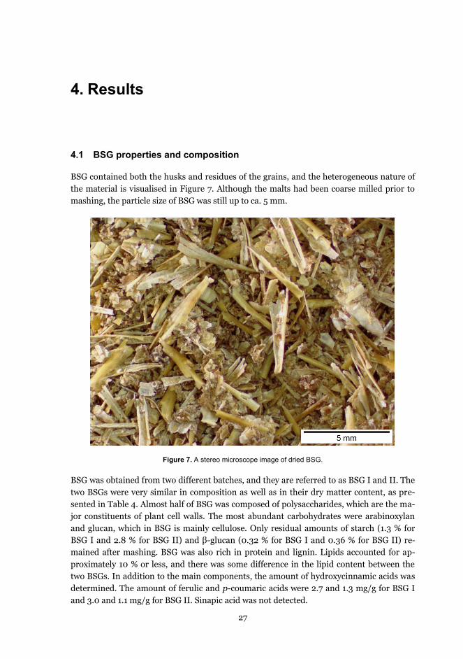
27
4. Results
4.1 BSG properties and composition
BSG contained both the husks and residues of the grains, and the heterogeneous nature of the material is visualised in Figure 7. Although the malts had been coarse milled prior to mashing, the particle size of BSG was still up to ca. 5 mm.
Figure 7. A stereo microscope image of dried BSG.
BSG was obtained from two different batches, and they are referred to as BSG I and II. The two BSGs were very similar in composition as well as in their dry matter content, as pre-sented in Table 4. Almost half of BSG was composed of polysaccharides, which are the ma-jor constituents of plant cell walls. The most abundant carbohydrates were arabinoxylan and glucan, which in BSG is mainly cellulose. Only residual amounts of starch (1.3 % for BSG I and 2.8 % for BSG II) and β-glucan (0.32 % for BSG I and 0.36 % for BSG II) re-mained after mashing. BSG was also rich in protein and lignin. Lipids accounted for ap-proximately 10 % or less, and there was some difference in the lipid content between the two BSGs. In addition to the main components, the amount of hydroxycinnamic acids was determined. The amount of ferulic and p-coumaric acids were 2.7 and 1.3 mg/g for BSG I and 3.0 and 1.1 mg/g for BSG II. Sinapic acid was not detected.

Results
28
Table 4. Compositions of the two BSGs used in the present study. BSG I was used in Paper I and BSG II in Papers II-V. *Dry matter contents of BSG I and II were 32.0 % and 31.6 %, respectively.
4.2 Milling of BSG
4.2.1 Effect of milling on particle size
The original BSG contained particles up to ca. 5 mm in size, but with milling the particle size was decreased down to μm scale (Figures 8 and 9). As shown in the microscopy imag-es, after pin disc milling and TurboRotor treatment there were still large particles present in the material, whereas Masuko and microfluidizer treated samples appeared very uni-form in size. After one hour of ball milling, there were both large cell fragments and fine particles, but after 24 h ball milling BSG appeared as fine dust. In the 24 h ball-milled sample colours seemed to blend together forming an overall brown colour. Aggregate for-mation was also detectable in the microscopy image of the 24 h ball-milled sample.
Pin disc milling, TurboRotor milling and ball milling were performed using dry BSG, whereas Masuko grinding and microfluidization were performed at over 90 % water con-tent. Ball milling produced the smallest particles but the size distribution was not very uni-form, ranging from only 1 μm to 50 μm (Figure 9). Pin disc milling was the least effective of the studied milling methods, and most of the particles produced were in the range of 100 to 1000 μm. The size distribution of TurboRotor milled BSG was wide ranging from 3 to 200 μm, but the majority of the particles were 10-100 μm in size. Both wet milling techniques produced distributions with a peak at 17 μm but the size distribution of Masuko milled BSG was somewhat wider than the that of microfluidized BSG. The distributions of the wet-milled BSGs were more uniform than those of the dry samples. In Figure 9, the distribu-tions present the percentage of the total sample volume.
It was observed that the original, undried BSG sedimented rapidly when it was diluted with water prior to the milling with the wet milling techniques. However, only after one pass through the Masuko grinder the suspension became notably more homogenous and sedimentation was remarkably slowed down. The wet-milled samples produced a homoge-nous slurry in the course of the milling process, whereas the dry milled samples sediment-ed more quickly when resuspended in water.
Component (% of dry weight*) BSG I BSG IICarbohydrates 46.7 42.2 Arabinoxylan 22.9 22.2 Glucans 20.9 17.1Lignin 19.4 19.3Protein 23.3 22.8Lipids 7.8 11.0Ash 4.9 4.7

Results
29
Figure 8. Microscopy images of BSG milled with different types of mills. BSG treated with a) pin disc mill, b) TurboRotor, c) Masuko, d) microfluidizer, e) ball mill for 1 h and f) ball mill for 24 h. Protein appears red (Acid Fuchsin), aleurone and subaleurone cell walls blue (Calcofluor) and lignified cell walls yellow and green (autofluorescence). Modified from Paper I (supplementary material) with permission of Elsevier B.V.
Particle size reduction by the different kinds of millings significantly improved the enzy-matic hydrolysis of BSG carbohydrates (Figure 10). When a milling pre-treatment was ap-plied, carbohydrate solubilisation increased from 23 up to 45 % at best. The highest solu-bility was obtained with 24 h of ball milling followed by the wet milling techniques (35‒37 %) and TurboRotor, which was almost equally efficient (34 %). Pin disc milling, which was also used as a pre-treatment for ball milling, was the least effective (28 %). The yield of hydrolysed carbohydrates correlated with the particle size (Figure 9): the smaller the parti-cle size, the better the hydrolysis yield. In addition, milling affected the amount of water-soluble carbohydrates as a significant increase in yield was detected also in the reference samples without the Depol740 enzyme preparation.
a b
c d
fe

Results
30
Figure 9. Particle size distributions of milled BSG samples. Data obtained from Paper I.
As Depol740 contained only low cellulase activity (Paper I, Table 1), the addition of en-zyme preparations high in cellulase and β-glucosidase activities, Celluclast1.5 and Novo-zym188 respectively, was studied (Figure 11). Although ball milling produced the smallest particle size and highest amount of soluble carbohydrates, it was not chosen as the pre-treatment for further studies. Instead BSG milled with Masuko was used because Masuko enabled the processing of significantly higher amounts of sample than the ball mill, and when using Masuko BSG did not need drying but could be milled in high water content.
Figure 10. Solubilisation of different carbohydrates from BSG by Depol740 enzyme preparation after millings. N = reference, no enzyme; E = with enzyme. Data obtained from Paper I.
0
2
4
6
8
10
1 10 100 1000
Volu
me
(%)
Particle size (μm)
Pin disc
Turborotor
Masuko
Microfluidizer
Ball mill 24 h
0
10
20
30
40
50
N E N E N E N E N E N E N E
Unmilled Pin disc TurboRotor Masuko Microfluidizer BallMill1 BallMill24
% o
f car
bohy
drat
es s
olub
ilised
glucosexylosearabinosemannosegalactose

Results
31
After the Masuko pre-treatment, 8 % of BSG carbohydrates had become water-soluble and Depol740 was able to release 36 % of the carbohydrates (Figure 11). The addition of Cellu-clast1.5 increased the solubilisation to 48 %, and it was not improved by a further supple-mentation with Novozym188. Compared to Depol740, Celluclast was almost equally effi-cient in xylose and arabinose hydrolysis and more efficient in cellulose hydrolysis.
Figure 11. Solubilisation of different carbohydrates from Masuko-milled BSG by Depol740 (Dep), Celluclast1.5 (Cel) and Novozym188 (Nov). Ref = no enzyme. Data modified from Paper I.
4.3 Effect of carbohydrase treatment and pH on BSG solubility
pH is an important factor in biomass solubilisation, and therefore the impact of pH on sol-ubility was studied with both undigested and carbohydrase treated BSG without adding any enzymes. After the carbohydrate hydrolysis with Depol740 BSG solubility was im-proved 2‒3 fold (Figure 12). In addition, an alkaline pH had a significant impact in increas-ing BSG solubility after the carbohydrate digestion, as 15 % of BSG was dissolved at pH 10 by the effect of alkalinity alone compared to the 5 % dissolved at pH 3.5. The increase in solubility was not as pronounced with unhydrolysed BSG, although some improvement, from 1.8 to 5.5 % was detected there as well.
BSG contained a significant amount of water-insoluble protein, and therefore the effects of pH and proteases on BSG protein solubility were also investigated (Figure 13). Three proteases with different pH optima were selected and their capability to release protein from carbohydrase-treated BSG was studied. Protein solubility was the highest with the alkaline protease (Alcalase 2.4) reaching 88 % at pH 9.5. The alkalinity had a significant effect on protein solubility as also the non-enzymatic solubility (pH 9.5 control) was 31 %. Acid Protease released 39 % and Promod144 24 % of the total protein.
0
10
20
30
40
50
60
Ref Dep Dep+Nov
Cel Cel+Nov
Dep+Cel
Dep+Cel+Nov
% o
f car
bohy
drat
es s
olub
ilise
d
glucosexylosearabinosemannosegalactose

Results
32
Figure 12. Effect of a carbohydrase treatment and pH on solubility of BSG (without enzymes). Data obtained from Paper II.
Figure 13. Effect of pH and protease on BSG protein solubility after a carbohydrate digestion. Previously un-published data.
The effect of pH on protein solubility was also investigated using optical microscopy (Figure 14). The undigested BSG was rich in protein as visualised by the red colour (Figure 14a). After it had been treated with the carbohydrate-degrading enzymes (Depol740), there was still a significant amount of protein present (Figure 14b). However, some changes in the proteinaceous material had occurred, as the red colour was somewhat less bright com-pared to the original BSG (Figure 14a). After the protease treatment at pH 9.5, protein had almost completely disappeared (Figure 14c), whereas after the non-enzymatic treatment at alkaline pH a notable amount of protein still remained in the residue (Figure 14d).
0
4
8
12
16
3.5 4 5 5.5 6 6.5 7 7.5 8 8.5 9 9.5 10
Tota
l sol
ubiliz
atio
n (%
)
pH
BSG
BSG after carbohydrate digestion
0
20
40
60
80
100
AcidProtease
Control Promod144 Control Alcalase2.4 Control
pH 3.5 pH 6.5 pH 9.5
% o
f BSG
pro
tein
sol
ubilis
ed

Results
33
Figure 14. Optical microscopy images of a) Alpine-milled BSG, b) BSG after carbohydrase treatment, c) BSG after carbohydrase and Alcalase 2.4 (pH 9.5) treatments and d) BSG after carbohydrase and non-enzymatic alkaline (pH 9.5) treatments. Reprinted from Paper II (supplementary material) with permission of Elsevier B.V.
4.4 Preparation of lignin-rich fractions for the colon model study
4.4.1 Three-step enzymatic hydrolysis of BSG
To enrich the lignin content of Masuko-milled BSG, carbohydrates and proteins were hy-drolysed by sequential enzymatic treatments (Figure 6). The compositions of the obtained fractions and the amounts of material solubilised are presented in Tables 5 and 6. In the first step 26 % of BSG was dissolved. The solubilised material was mainly carbohydrates, and their content was decreased from 42 to 22 % in the residue after the first hydrolytic step. In addition, 6 % of protein was released. The contents of lignin, protein and lipids increased in the residue as a consequence of the carbohydrate digestion.
In the second step, which was a protease treatment in alkaline conditions, most of the protein was solubilised, but in addition a significant amount of lipids and lignin were re-leased. After the second hydrolysis, 93 % of protein, 48 % of lignin and 87 % of lipids had been solubilised. Practically no carbohydrates dissolved in the alkaline proteolytic step.
In the third hydrolytic step, the first carbohydrase treatment was repeated. This time, not only carbohydrates were solubilised but part of the remaining lignin (19 %) and lipids (7 %) were released as well (percentages are from the total amount in original BSG). The compo-sition of the insoluble residue after the third hydrolysis (INS) is shown in Table 5. The
a
dc
b

Results
34
amount of the recovered residue was 19 % of original BSG. The INS fraction consisted mainly of the recalcitrant carbohydrates and lignin.
As lignin is soluble in alkaline pH but precipitates in acidic conditions, it was precipitated from the liquid phase with hydrochloric acid after the alkaline proteolysis. The obtained precipitate was denoted as protease-alkaline extracted fraction (P-AEF) and its composi-tion is also shown in Table 5. However, it was not only lignin that precipitated but lipids and peptides as well. Furthermore, it appeared that only 30 % of the lignin released in the second hydrolysis precipitated and the rest remained in the liquid phase. The amount of recovered precipitate (P-AEF) was 11 % of the original BSG and the amount of lignin in the precipitate was 14 % of the lignin present in the original BSG.
Table 5. Compositions of the fractions obtained in the three-step enzymatic hydrolysis of BSG and after chemi-cal extractions. Data modified from paper III, IV and V.
Table 6. The amount of material solubilised by the three enzymatic hydrolysis steps. The solubilities are given for each step and as a total solubilisation after all steps. Data modified from Paper III and V.
To visualise the phenolic compounds in INS and P-AEF by their autofluorescence, the samples were imaged under UV light (Figure 15c and d) and also using the chemical dyes Calcofluor and Acid Fuchsin (Figure 15 a and b). Some blue colour was detectable in INS indicating presence of aleurone cell walls even after extensive treatments with hydrolytic enzymes (Figure 15a). For P-AEF the dyes showed that protein seemed to cover the precipi-tated lignin, as only very fine green particles were visible from the red mass (Figure 15b). (Please note that the green particles may not be clearly visible in the printed image due to the reduced quality of the reprint compared to the original microscopy image). In P-AEF the particles seemed to have formed aggregates in the precipitation, as mostly very large entities up to hundreds of μm were seen. The UV image confirmed the presence of phenolic material in INS (Figure 15c). Interestingly, for P-AEF hardly any autofluorescence was seen (Figure 15d). This finding supports the idea of protein covering lignin.
1st 2nd
Carbohydrates 42.2 22.2 49.4 39.2 4.0 6.7 53.4 Arabinoxylan 22.2 15.9 31.2 25.7 2.3 3.8 20.3 Glucan 17.1 4.5 15.9 11.2 0.7 1.2 30.9 Lignin 19.3 29.1 33.5 40.3 24.4 40.7 21.9 Protein 22.8 28.6 4.8 6.6 27.2 45.2 7.6 Lipids 11 15.5 4.1 3.1 39.9 0 2.9 Ash 4.7 4.6 8.1 8.7 4.5 7.4 6.0
DEFEINS Component Initial BSG
Hydrolysis residue P-AEF Extract. P-AEF
FirstDepol740,
Celluclast1.5 26.2 26.2
Second Alcalase2.4 54.5 66.4
ThirdDepol740,
Celluclast1.5 47.3 81.3
Hydrolysis step
Enzymes used in the hydrolysis
Solubility in the step %
Cumulative solubilisation %

Results
35
Figure 15. INS and P-AEF imaged with optical microscopy using chemical dyes (a: INS, b: P-AEF) and with autofluorescence due to UV light (c: INS, d: P-AEF). Previously unpublished figures.
4.5 Interactions of lignin-rich fractions with colon microbiota in vitro
In the in vitro colon model, a wide range of metabolites was detected for each studied frac-tion: 1800 for P-AEF, 2400 for INS and 2500 for BSG and DEFE. The metabolites with FC<2 were excluded. For each fraction there were several hundred metabolites with FC>2, and from those compounds the ones with structural similarity to lignin were selected. For some compounds it was not possible to identify the exact chemical structure, but they were indicated with a group-specific name, e.g. dilignols. The heat maps showing the profiles of aromatic metabolites are presented in Figure 16. Blue colour represents under-expression and red colour over-expression of the metabolite compared to the faecal background. The asterisks denote statistical difference between the sample and the control. The clustering on the left side of the heat maps groups similar time course profiles of different metabo-lites.
Eight mono- and dimeric phenolic and aromatic metabolites were formed from BSG (Figure 16a) during the in vitro colon model fermentation, and out them three compounds, 4-methylcatechol, a dilignol (compound 2) and ferulic acid, showed significant difference from the faecal background at several time points. Another dilignol (compound 6) with statistical difference from the background also appeared at the end of the fermentation. An unidentified guaiacyl compound and 4-hydroxymethylcatechol showed a higher response in the beginning of the fermentation, after which their responses notably decreased.
a
dc
b

Results
36
0h 2h 4h 6h 8h
Day
8355 - Benzeneacetic acid, trimethylsilyl ester
413 - *4-Aminobenzoic acid, 2TMS
8308 - Benzeneacetic acid, ?-[(trimethylsilyl)oxy]-, trimethylsilyl ester
774 - 3,4-Dihydroxybenzyl alcohol,tris(trimethylsilyl)-
909 - (3-Hydroxy-4-methoxyphenyl)ethylene glycol tris(trimethylsilyl) ether
16 - Ferulic acid, 2TMS
719 - *Enterolactone, 2TMS
1 - 3,4-Dihydroxytoluene, 2TMS
*
*
* *
* *
** ** ** * *
** * *
** ** ** * 1. 4-Methylcatechol (2.8)2. Dilignol (5.5)3. Ferulic acid (3.9)4. Guaiacyl compound (7.0)5. 4-Hydroxymethylcatechol (2.2)6. Dilignol (2.7)7. 4-Aminobenzoic acid (2.1)8. Benzeneacetic acid (2.9)
0h 2h 4h 6h 8h 24h
*
***
*
***
*
*
**
*
**
** ****
**
**A)
-1 -0.5 0 0.5 1 1.5 2Value
Color Key
-1 1-0.5 0 0.5 1.5 2
Color key
0h
2h
4h
6h
8h
Day
678 - Benzoic acid, 2,5-bis[(tert-butyldimethylsilyl)oxy]-, tert-butyldimethylsilyl ester
396 - Benzeneacetic acid, trimethylsilyl ester
664 - Benzenepropanoic acid, 3-methoxy-4[(trimethylsilyl)oxy]-, trimethylsilyl ester
209 - 2-(4-Hydroxy-3-methoxyphenyl)ethanol bis(trimethylsilyl) ether
242 - 2-Hydroxymandelic acid, ethyl ester, di-TMS
672 - 3,4-Dihydroxycinnamyl alcohol,tris(O-trimethylsilyl)-
586 - Cinnamic acid, 3,5-dimethoxy-4-(trimethylsiloxy)-, methyl ester
660 - Benzenepropanoic acid, 3-methoxy-à,4-bis[(trimethylsilyl)oxy]-, trimethylsilyl ester
580 - *3,4-Dihydroxyethylbenzene, 2TMS
700 - 4-(Trimethylsilyloxy)-3-methoxycinnamaldehyde
199 - *Homovanillic acid, 2TMS
622 - *Ferulic acid, 2TMS
476 - Benzaldehyde, 3-methoxy-4-[(trimethylsilyl)oxy]-, O-methyloxime
496 - (3-Hydroxy-4-methoxyphenyl)ethylene glycol tris(trimethylsilyl) ether
393 - *Ferulic acid, 2TMS
601 - (4-Hydroxy-3-methoxyphenyl)ethylene glycol tris(trimethylsilyl) ether
615 - (3-Hydroxy-4-methoxyphenyl)ethylene glycol tris(trimethylsilyl) ether
342 - 3-Hydroxy picolinic TMS 2
583 - 2-Hydroxymandelic acid, ethyl ester, di-TMS
497 - *3,4-Dihydroxyethylbenzene, 2TMS
* **
**
* * *
*
*
** ** *
*
*
* *
**
* 1. Catechol derivative (11.9)2. Dilignol (17.3)3. Dilignol (11.2)4. Guaiacyl compound (48.0)5. Dilignol (52.5)6. cis-Ferulic acid (31.5)7. Guaiacyl compound (45.9)8. Vanillin (5.4)9. Phenolic acid (4.2)10. Homovanillic acid (34.9)11. Coniferyl aldehyde (2.4)12. Catechol derivative (5.1)13. Dilignol (5.9)14. Homovanillin (2.3)15. Aromatic compound (5.4)16. 2-Hydroxymandelic acid (2.5)17. Guaiacyl compound (3.0)18. 3-Syringylpropanoic acid (2.3)19. Benzeneacetic acid (7.3)20. Aromatic acid (2.3)
****
*
*
***
***
*
*
**
**
*
*
**
0h 2h 4h 6h 8h 24h
B)

Results
37
Figure 16. Heat maps of the aromatic metabolites found in the metabolomes of the in vitro colon fermenta-tions, A) BSG, B) P-AEF, C) INS and D) DEFE. The number in parentheses after the name is the fold change (FC) of the compound. The colour key is the 2-log value of the FC. A blue colour indicates an un-der-expression and red colour represents an over-expression of the metabolite compared to the faecal control. The asteriks denote statistical difference between the sample and the control. Data obtained from Papers IV and V.
0h 2h 4h 6h 8h
Day
5706 - Vanillylpropionic acid, bis(trimethylsilyl)-
835 - 3,4-Dihydroxybenzyl alcohol,tris(trimethylsilyl)-
548 - Benzenepropanoic acid, ?,4-bis[(trimethylsilyl)oxy]-, trimethylsilyl ester
3502 - Benzeneacetic acid, trimethylsilyl ester
530 - *3,5-Dihydroxyphenylacetic acid, 3TMS
445 - 3-(3-Hydroxyphenyl)-3-hydroxypropionic acid, tris-(O-trimethylsilyl)-
399 - *Enterolactone, 2TMS
1 - 3,4-Dihydroxyphenylacetic acid, 3TMS
9594 - Benzeneacetic acid, ?-[(trimethylsilyl)oxy]-, trimethylsilyl ester
7728 - Benzeneacetic acid, ?-[(trimethylsilyl)oxy]-, trimethylsilyl ester
181 - *Enterodiol, 4TMS
1028 - Benzenepropanoic acid, 3-methoxy-?,4-bis[(trimethylsilyl)oxy]-, trimethylsilyl ester
418 - Hydrocinnamic acid, 3,4-bis(trimethylsiloxy)-, trimethylsilyl ester
3683 - 3,4-Dihydroxybenzyl alcohol,tris(trimethylsilyl)-
160 - *3,4-Dihydroxyphenylacetic acid, 3TMS
577 - *3,4-Dihydroxyphenylacetic acid, 3TMS
377 - *Homovanillic acid, 2TMS
5 - 3,4-Dihydroxytoluene, 2TMS
3659 - 3-(3,4-Dihydroxyphenyl)-2-isothiocyanatopropionic acid, ethyl ester, di-TMS
798 - Ferulic acid, trimethylsiloxy, trimethylsilyl ester
4 - Vanillic acid, 2TMS
42 - Vanillylpropionic acid, bis(trimethylsilyl)-
639 - (3-Hydroxyphenyl)pentanoic acid, bis(O-trimethylsilyl)-
13 - Ferulic acid, 2TMS
723 - Benzaldehyde, 3-methoxy-4-[(trimethylsilyl)oxy]-, O-methyloxime
624 - 1,3-Bis(trimethylsiloxy)benzene
*
* *
** * * **
* *
*
*
*
*
*
** * *
* ** * *
*
*** ** *** *
*
*** **
* * * ** **
** **
** ** *** ** * *
* ** * * **
** ** *** ** * 1. Methoxybenzenediol (8.6)2. Vanillin (10.3)3. trans-Ferulic acid (3.1)4. 4-Hydroxyphenylpentanoic acid (2.7)5. 3-Guaiacylpropanoic acid (6.0)6. Vanillic acid (2.3)7. cis-Ferulic acid (3.0)8. Dilignol (3.5)9. 4-Methylcatechol (3.6)10. Homovanillic acid (8.4)11. 2-Hydroxyphenylacetic acid (2.6)12. Dihydroxyphenylacetic acid (2.8)13. Dilignol (3.2)14. Dilignol (4.7)15. Dilignol (2.1)16. Dilignol 2.4)17. Dilignol (2.6)18. Dilignol (13.0)19. 3,4-Dihydroxyphenylacetic acid (2.0)20. Dilignol (2.1)21. Dihydroxyphenylpropanoic acid (2.3)22. Dihydroxyphenylacetic acid (2.1)23. Benzeneacetic acid (6.4)24. 3-(4-Hydroxyphenyl)lactic acid (2.8)25. 4-Hydroxymethylcatechol (3.0)26. 3-Syringylpropanoic acid (2.9)
** ** ***
0h 2h 4h 6h 8h 24h
*
**
*
*
***
**
*
**
**
**
***
*
*
**
*
*
*
**
*
**
*
*
*
*
**
**
****
**
**
***
**
****
*** ***
**
**
****
**
C)
0h 2h 4h 6h 8h
Day
3516 - 2,5-Dihydroxybenzyl alcohol, tris(O-trimethylsilyl)-
3544 - *Enterodiol, 4TMS
423 - Benzeneacetic acid, 2,5-bis[(trimethylsilyl)oxy]-, trimethylsilyl ester
5737 - *Enterodiol, 4TMS
766 - Ferulic acid, trimethylsiloxy, trimethylsilyl ester
700 - 3,4-Dihydroxybenzyl alcohol,tris(trimethylsilyl)-
3546 - Benzoic acid, 2-[(trimethylsilyl)amino]-, trimethylsilyl ester
9815 - *3,4-Dihydroxytoluene, 2TMS
3572 - Benzeneacetic acid, 3-methoxy-?-[(trimethylsilyl)oxy]-, trimethylsilyl ester
3532 - Benzeneacetic acid, ?-[(trimethylsilyl)oxy]-, trimethylsilyl ester
*
* *
*
**
* *** 1. Dilignol (11.8)2. Dilignol (2.2)3. 4-Methylcatechol (2.5)4. 2-Aminobenzoic acid (3.0)5. 4-Hydroxymethylcatechol (3.3)6. cis-Ferulic acid (2.6)7. Dilignol (4.8)8. 2,5-Dihydroxyphenylacetic acid (3.3)9. Dilignol (2.1)10. Phenolic acid (2.0)
0h 2h 4h 6h 8h 24h
***
*
**
*
*
*
*
D)
-1 -0.5 0 0.5 1 1.5 2Value
Color Key
-1 1-0.5 0 0.5 1.5 2
Color key

Results
38
The lignin content in P-AEF (Figure 16b) was only 5 % units higher than in BSG but a no-
tably greater amount of aromatic metabolites was released from P-AEF (20) compared to BSG (8). The P-AEF metabolic profile contained several lignin-type compounds with guai-acyl structure (compounds 8, 10, 11 and 14) and one with syringyl structure (compound 18). Two unidentified guaiacyl compounds (compounds 4 and 7) had especially high FCs (48 and 46), and responses of both increased towards the end of the fermentation. For-mation of four dimeric metabolites (dilignols) was also detected. It seemed that most of the aromatic metabolites were formed at 6 h or later.
Lignin content of INS was the highest of all studied samples (Table 6), which also corre-lated with the number of low molecular weight aromatic metabolites (26) (Figure 16c). Both mono- and dimeric compounds were detected including catechols, phenolic acids and dilignols, and several compounds were statistically different from the faecal background at more than one time point. Compounds 2, 5, 6, 10 and 26 contained guaiacyl or syringyl structure, which are characteristic of lignin.
For DEFE 10 low molecular weight aromatic metabolites were found in the metabolome (Figure 16d), of which dilignols and catechols could possibly originate from lignin. Cis-ferulic acid (compound 6) was still detected, although the sample had been treated with strong alkali to release ester-linked hydroxycinnamic acids. 2-Aminobenzoic acid (com-pound 4) was unlikely to be formed from lignin. Similarly as with P-AEF (Figure 16b), ar-omatic metabolites mostly started to form at 6 h.
Certain phenolic metabolites were also analysed quantitatively using authentic stand-ards. The most important of these are presented in Figure 17. Ferulic acid, which was pre-sent in P-AEF at a high concentration, was almost completely metabolised to other prod-ucts during the fermentation. The content of ferulic acid in the other studied samples was significantly lower. Some ferulic acid was released from BSG due to microbial activities but in INS and especially in DEFE there were no notable changes in the ferulic acid concentra-tions during the 24 h incubation period (see Table 7 for the statistical significances).
4-Methylcatechol was formed from P-AEF, BSG and INS at significantly higher levels than from the faecal control, and its formation started at the very beginning of the fermen-tation suggesting that it was a primary metabolite. 3,4-Dihydroxyphenylacetic acid, which is a metabolite from dehydrodiferulic acid, was formed at the highest concentration from P-AEF, but also in BSG and INS the levels were significantly above the faecal background.
Short chain fatty acid (SCFA) formation was measured during the fermentation in order to study possible inhibitory effects of lignin-rich samples on microbial conversions (Figure 18). The initial formation rate (0–2 h) was the fastest for P-AEF (17.7 mM/h) followed by DEFE (13.0 mM/h). The initial SCFA formation rate was on a similar level in BSG (8.2 mM/h) and the faecal control (10.3 mM/h in the case of P-AEF and 8.2 mM/h for the oth-ers, as fermentations were carried out at different times and with different inocula). The lowest rate (7.3 mM/h) was observed for INS. There was no indication that lignin in the samples suppressed SCFA formation, as the levels of the studied samples were above those of the faecal control and the carbohydrates originating from the faeces and BSG fractions were partially fermented. It should be pointed out though, that in first two hours the SCFA levels of BSG were lower than for the faecal control. However, this does not seem to be due to inhibition, as the SCFA levels quickly rose after the first two hours.

Results
39
Figure 17. Concentrations of quantitated metabolites formed in the colon model fermentation. Left: metabolites formed from the fermentation of protease-alkaline extracted fraction (P-AEF), the asterisks denote statisti-cal difference between the sample and the faecal control. Right: metabolites formed from BSG, insoluble hydrolysis residue (INS) and deferuloylated BSG (DEFE). The statistical differences between the samples and faecal control (FC) are presented in Table 7. Data obtained from Papers IV and V.
Table 7. Statistically significant differences between quantified phenolic metabolites from BSG, INS, DEFE. Statistical difference (p<0.05) is represented with small letters (a, b, c, d). Data obtained from Paper V.
0
2
4
6
8
10
12
0 5 10 15 20 25
Con
cent
ratio
n (μ
mol
/L)
Time (h)
Ferulic acid
INSBSGDEFEFC
0
1
2
3
4
5
0 5 10 15 20 25
Con
cent
ratio
n (μ
mol
/L)
Time (h)
3,4-Dihydroxyphenylacetic acid
INSBSGDEFEFC
0
1
2
3
4
5
0 5 10 15 20 25C
once
ntra
tion
(μm
ol/L
)
Time (h)
4-Methylcatechol
INS
BSG
DEFE
FC
0
2
4
6
8
0 5 10 15 20 25
Con
cent
ratio
n (μ
mol
/L)
Time (h)
4-Methylcatechol
P-AEFFecal control
********
0
5
10
15
0 5 10 15 20 25
Con
cent
ratio
n (μ
mol
/L)
Time (h)
3,4-Dihydroxyphenylacetic acid
P-AEFFecal control
**
*
****
0
50
100
150
200
250
0 5 10 15 20 25
Con
cent
ratio
n (μ
mol
/L)
Time (h)
Ferulic acid
P-AEF
Fecal control
***
******
********
Time (h) BSG INS DEFE Faecal control Time (h) BSG INS DEFE Faecal control0 ab a ab b 0 – – – –2 a b bc c 2 b a c c4 a b bc c 4 a a b b6 a b bc c 6 b a c c8 a ab bc c 8 a a b b
24 b a ab b 24 ab a bc c
Time (h) BSG INS DEFE Faecal control0 – – – –2 ab a b b4 a a b b6 a a b b8 – – – –
24 – – – –
Ferulic acid 4-Methylcatechol
3,4-Dihydroxyphenylacetic acid

Results
40
Figure 18. Short chain fatty acid formation in the in vitro colon model. BSG, INS and DEFE are presented in the chart above (a) and P-AEF in the chart below (b). Data obtained from papers IV and V.
4.6 Effects of lignin on lactobacilli and bifidobacteria
No inhibitory effects on the growth were observed after two days of incubation of different strains of Lactobacillus with P-AEF (lipids removed by extraction, Table 5) as the growth substrate (Figure 19). Both strains of L. rhamnosus were able to grow on P-AEF, as their colony counts (108 CFU/mL) were nearly equal to the positive controls (0 % P-AEF + glu-cose). For L. paracasei the positive control was 0.5 log units and for L. salivarius 1.5 log units higher than P-AEF. The colony counts of all strains grown on P-AEF were above the negative control (no added glucose or P-AEF).
No inhibition was seen with the three Bifidobacterium species either. After three days the growth was low (103 CFU/mL) in the positive controls of B. adolescentis and B. longum, as most of the cells were no longer culturable. With P-AEF as the growth substrate, the colony counts were above the negative control also for bifidobacteria. Moreover, after seven days of incubation, there was still notable growth (106-107 CFU/mL) in the presence of P-AEF
0
20
40
60
80
100
120
P-AE
F
FC
P-AE
F
FC
P-AE
F
FC
P-AE
F
FC
P-AE
F
FC
P-AE
F
FC
0 h 2 h 4 h 6 h 8 h 24 h
Con
cent
ratio
n (m
mol
/L) Acetic acid Propionic acid Butyric acid
0
20
40
60
80
100
120
BS
GD
EFE IN
SFC
BS
GD
EFE IN
SFC
BS
GD
EFE IN
SFC
BS
GD
EFE IN
SFC
BS
GD
EFE IN
SFC
BS
GD
EFE IN
SFC
0 h 2 h 4 h 6 h 8 h 24 h
Con
cent
ratio
n (m
mol
/L) Acetic acid Propionic acid Butyric acid
a)
b)

Results
41
and some growth (105 CFU/mL) was also observed in the negative control. In the positive controls most of the cells were not culturable after 7 days.
Figure 19. Figure 19. Bacterial counts of lactobacilli and bifidobacteria grown in the presence and absence of glucose (positive and negative control) or in the presence of the protease-alkaline extraction (P-AEF, lipids removed). a) Lactobacilli strains after 2 days, b) bifidobacterial strains after 3 days and c) bifidobacterial strains after 7 days of incubation. The asterisks denote too long an incubation time, as most of the cells were already dead. Previously unpublished figure.
0
2
4
6
8
B. adolescentis (E1074) B. breve (E1075) B. longum (E664)
Log 1
0C
FU/m
l
0% + gluc. 0% P-AEF 1% P-AEF
0
2
4
6
8
B. adolescentis (E1074) B. breve (E1075) B. longum (E664)
Log 1
0C
FU/m
l
0% + gluc. 0% P-AEF 1% P-AEF
0
2
4
6
8
L. rhamnosus (E800) L. rhamnosus (E948) L. paracasei (E949) L. salivarius (E1006)
Log 1
0 C
FU/m
l
0% + gluc. 0% P-AEF 1% P-AEF
a)
b)
c)
*
** *
*
L. rhamnosus (E800) L. rhamnosus (E948) L. paracasei (E949) L. salivarius (E1006)
B. adolescentis (E1074) B. breve (E1075) B. longum (E664)
B. adolescentis (E1074) B. breve (E1075) B. longum (E664)

42
5. Discussion
BSG consists mainly of the cell wall materials of husks, pericarp and aleurone, and the re-sidual protein inside the aleurone cells. In mashing, malts have passed an extensive hydro-lytic treatment by a variety of enzymes resulting in the solubilisation of the starchy endo-sperm, and the resilient lignin-carbohydrate matrix along with the insoluble protein re-main in the solid residue, i.e. BSG. The results of this thesis showed that the enrichment of BSG lignin by enzymatic fractionation was limited by the recalcitrant cell wall matrix even when the material was pre-treated by milling. The separation of lignin was further compli-cated by co-solubilisation of lignin with protein. Several low molecular weight phenolic metabolites were formed from the lignin-rich fractions by the activities of faecal microbiota in the in vitro colon model indicating at least partial degradation of lignin. Lignin did not suppress microbial carbohydrate fermentation in the colon model or inhibit growth of lac-tobacilli and bifidobacteria, which is contradictory to the common conception that lignin is an antimicrobial compound.
5.1 Preparation of lignin-rich fractions from BSG
5.1.1 Limited enzymatic solubilisation of cell wall carbohydrates
It has been noted previously that BSG carbohydrates are poorly hydrolysed by commercial enzyme preparations, if no pre-treatments to open up the cell wall structures are applied (Forssell et al. 2008). Therefore, a milling pre-treatment to improve the hydrolysis yield was studied. Milling is a mechanical treatment, which does not affect the food grade status of BSG, but enhances the hydrolysis by reducing the particle size and degrading cell wall structures and therefore increasing the surface area accessible for enzymes.
The yield of enzymatically solubilised sugars from BSG was low (23 %), when no pre-treatment was applied (Figure 10). Milling improved the yield up to 45 %. Ball milling was the most effective followed by the wet milling techniques. However, even with the ball mill-ing pre-treatment more than half of the carbohydrates still remained resistant to enzymes suggesting that other factors restricting the hydrolysis still remained. When looking at Fig-ure 2b, it can be observed that the cell wall thickness in BSG is in the low micrometre range. As hardly any submicron particles were detected (Figure 9), it would suggest that cell walls remained at least partially intact even after the most effective of the studied mill-ing techniques. In other words, although milling markedly reduced particle sizes, the cell wall structures were not completely broken down and the particles were not entirely di-gestible by the enzymes. Cell wall degradation might have been enhanced by prolonging the milling, but this was not further studied. The effect of a prolonged hydrolysis time was shown to provide only minor improvements (Paper I, Figure 3).

Discussion
43
The monosaccharide profile of solubilised sugars (Figure 10) showed that hydrolysis of unmilled BSG by Depol740 resulted in solubilisation of mainly arabinoxylan and a smaller amount of cellulose-derived carbohydrates. Arabinoxylan is expected to be derived mostly from the aleurone cell walls as they contain 67 % of arabinoxylan and 26 % of β-glucan (Duffus and Cochrane 1993). The changes in BSG after the enzymatic treatment were also visualised by epifluorescence microscopy (Figure 14a). After the hydrolysis with Depol740 aleurone cell walls (shown as blue) had almost completely disappeared and only the ligni-fied cell walls remained. After mashing the β-glucan content of BSG was very low (<1 %) and thus the liberated gluco-oligosaccharides were expected to arise from cellulose. Milling increased the solubilisation of glucose and glucose-based oligosaccharides, whereas a smaller increase was observed with arabinoxylan. This implies that milling affected the cellulose-rich husk and pericarp more than the arabinoxylan-rich aleurone. This was seen for both enzymatic and non-enzymatic (control) solubilisation.
Previously ball-milling has been shown to be such a powerful treatment that it markedly increased the amount of water-soluble arabinoxylan in wheat bran by breaking covalent bonds inside arabinoxylan (Van Craeyveld et al. 2009). Moreover, the authors observed that ball-milling affected the solubility of pericarp-enriched (thus cellulose-enriched) bran more than the solubility of the original bran. It has also been reported that wheat bran is fractured more rapidly by milling than isolated aleurone layers, because pericarp is much more friable than aleurone (Antoine et al. 2004). These observations support the results of the present study, which showed that cellulose rich parts of BSG, i.e. the husk and pericarp, were more susceptible to milling. This effect was especially pronounced in the reference samples without the addition of Depol740. However, the presence of residual enzymatic activity remaining from malting and mashing cannot be ruled out, and this could have in-creased the carbohydrate solubility especially in the reference samples.
The enzyme dosage used in the Depol740 treatment was very high (Paper I, Table 1), so increasing that would most likely not further improve the yield. However, Depol740 is low in cellulase activity, and therefore the enzyme cocktail was supplemented with additional cellulases. Masuko-milled BSG was used as the raw material in this experiment, as Masuko was found to be a more feasible technique compared to ball milling; large quantities of sample could be handled and the milling could be performed in high water content thus avoiding the drying of BSG. Supplementing the enzyme mixture with cellulases increased the yield only moderately, from 36 to 48 % (Figure 11), indicating that the limited carbohy-drate solubilisation had not been due to the low cellulase activity in Depol740 but was more dependent on the physical restrictions of the material. Thus, irrespective of the en-zyme mixture used after the milling only half of the cell wall polysaccharides could be hy-drolysed.
The cell wall pore sizes in wheat kernels have been reported to be too small (<10 nm) to allow free penetration of cell wall degrading enzymes (Chesson et al. 1997). Beaugrand et al. (2005) demonstrated with an inactivated xylanase that cell wall deconstruction is a pre-requisite for enzymes to penetrate testa or pericarp, but accessibility to aleurone is not as restricted. According to Chesson et al. (1997) the differences in the pore sizes between dif-ferent plant species are likely to be small, so it can be assumed that the small pore size causes steric hindrance for enzymes also in the hydrolysis of BSG. Another factor impeding the hydrolysis is the presence of lignin, as lignin is known to adsorb and inhibit cell wall degrading enzymes (Ooshima et al. 1990, Palonen et al. 2004). Lignin is also cross-linked to arabinoxylan through ferulic acid (Bunzel et al. 2004, Ishii 1997) further restricting en-

Discussion
44
zymatic accessibility. Similarly, diferulate cross-links between arabinoxylan molecules (Bunzel et al. 2001, Hatfield et al. 1999) negatively affect cell wall degradability (Grabber et al. 1998b). A lower degree of ferulate cross-linking has been shown to result in improved hydrolysis of maize cell walls, and the impact was more pronounced for xylan than cellu-lose (Grabber et al. 1998a). Although the extent of arabinoxylan cross-linking is lower in barley compared to maize (Bunzel et al. 2001), it can still be assumed to contribute to the limited carbohydrate hydrolysis observed in this study. Furthermore, hydrophobic cuticu-larised layers in testa and husk may act as barriers for enzyme penetration (Fincher and Stone 1993).
The outer layers of cereal grains provide a protective shield keeping other organisms apart from the starch and other nutrients inside the grain, and the low extent of enzymatic hydrolysis of BSG carbohydrates is likely due to the resilient nature of the outer layers. Although the enzyme mixture contained the most relevant cell wall degrading activities, cellulases, xylanase and arabinosidase, the hydrolysis was restricted by physical barriers or the lack of some important enzymatic activity, or both. As ferulate cross-links are known to be a key factor limiting cell wall hydrolysis (Grabber et al. 1998b, Grabber et al. 2009), feruloyl esterases should enhance the hydrolysis. Depol740 is a preparation from the fun-gus Humicola insolens, which has been reported to produce feruloyl esterases acting on BSG ferulic acid (Faulds et al. 2002). However, in the present study this activity was obvi-ously not capable to effectively cleave the cross-links or the dosage was significantly lower than would have been needed. The observations discussed above as well as the results of the milling pre-treatment study suggest that either an improved enzyme mixture or anoth-er kind of pre-treatment, e.g. chemical or physico-chemical, or a combination of these, would be required for a more complete enzymatic hydrolysis of BSG carbohydrates.
5.1.2 Effect of pH and protease on BSG and protein solubility
Depending on the source of BSG, protein makes up 10‒27 % of BSG (Table 1). Previously it has been shown that even without any pre-treatment alkalinity improves the total solubili-sation of BSG (Faulds et al. 2008). In the present study the effect of alkaline conditions on BSG solubility was shown to be even more pronounced after a carbohydrate digestion (Fig-ure 12). As aleurone cells are rich in protein and their non-lignified cell walls are not as resistant to enzymatic digestion as pericarp and husk cell walls, it seems likely that the De-pol740 treatment made proteins in aleurone more susceptible to the following alkaline treatment. Alkaline conditions can also partially dissolve lignin, which in part contributes to the total solubilisation.
Three different proteases with different pH optima were compared for the hydrolysis of protein from carbohydrase treated BSG in acidic, neutral and alkaline conditions (Figure 13). Protein solubility was the highest with Alcalase 2.4 at pH 9.5 as almost 90 % of protein was hydrolysed after the two enzymatic treatments. Alcalase 2.4 has previously been found to effectively release proteins from BSG at pH 6.8‒9 (Treimo et al. 2008, Treimo et al. 2009), although the relative activity of Alcalase 2.4 is only approximately 30 % of the max-imum at pH 9, and reaches maximum between pH 9.5 and 10 (Faulds et al. 2008). Almost one third of the protein was released by the alkali alone (control) suggesting that the high pH was also an important factor in protein solubility. The acidic and neutral proteases were not as effective in protein release, and especially the non-enzymatic solubilisations at pH 3.5 and 6.5 were very low.

Discussion
45
The results are in line with the observation that water-solubility of barley proteins is the lowest from pH 4 to 6 and the highest from pH 10 to 11 (Yalçın and Çelik, 2007). Based on the findings of Yalçın and Çelik (2007) it would seem likely that pIs of BSG proteins are in the range of pH 4‒6, which makes them less water-soluble in this pH range. Furthermore, presumably the increased net charge of proteins at pH 9.5 enhanced both enzymatic and non-enzymatic protein release from BSG. However, the effects may have been different for hordeins and glutelins, which are the two classes of water-insoluble proteins in BSG. Most likely, alkalinity affected glutelins more as they are known to be soluble in mild alkali (Linko et al. 1989), whereas hordein solubilisation is enhanced by a reducing agent (Os-borne 1909).
5.1.3 Co-solubilisation of protein and lignin in alkaline conditions
A three-step enzymatic hydrolysis of BSG was carried out to enrich the lignin content of the residue by the removal of proteins and carbohydrates (Figure 6). First, a carbohydrase treatment solubilised 26 % of Masuko-milled BSG (Table 6), and this was for the most part carbohydrates, although a minor amount of protein was co-solubilised. Secondly, a proteo-lytic treatment resulted in the release of almost 90 % of proteins and almost half of lignin. Finally, a carbohydrase treatment similar to the first step was repeated. In total, 81 % of BSG was dissolved by the three hydrolyses, and two different types of lignin-rich fractions were obtained.
40 % of BSG was released in the proteolytic treatment due to both the alkaline conditions and the presence of a protease. As part of the lignin was known to dissolve in the alkaline proteolysis, the aim was to separate the released lignin from the peptide solution by precip-itation with acid (precipitate denoted as P-AEF). This type of lignin would be almost free of carbohydrates and thus more accessible for the gut microbiota, which would make it an interesting substrate for the further in vitro studies. As a result of the protease treatment, it was assumed that protein would have been degraded to such small peptides that they would not co-precipitate with lignin. According to previous research, peptides obtained from BSG using Alcalase 2.4 are mostly less than 1 kDa in size (Treimo et al. 2009). How-ever, the precipitation did not occur as planned, as only one third of lignin precipitated and the rest remained in the soluble phase. Furthermore, despite the anticipated low molecular weight of the peptides, part of the soluble proteinaceous material (17 %) co-precipitated with lignin (Table 5). Most of the peptides still remained water-soluble, as was expected. The improved water-solubility of lignin and the co-precipitation of proteinaceous material raise the question of interactions between lignin and proteins, as lignin is normally water-insoluble at low pHs. Such interactions, e.g. hydrophobic, electrostatic and/or hydrogen bonding could possibly contribute to the water-solubility of lignin released by the alkaline protease treatment and thus explain the low amount of precipitated lignin.
Thus far, there is only a limited amount of studies showing protein-lignin interactions, and the interactions are not yet fully understood. In research related to nutrition, polyphe-nol-protein interactions are somewhat more studied, but they are usually limited to tan-nins (Bennick 2002) or flavonoids (Papadopoulou and Frazier 2004), which both are known to bind to proteins resulting in soluble or insoluble complexes. Several studies con-cerning enzymatic hydrolysis of lignocellulose have shown that lignin interacts with cellu-lase enzymes and adsorbs them on its surface (Ooshima et al. 1990, Eriksson et al. 2002, Palonen et al. 2004, Rahikainen et al. 2013). The adsorption can be reduced by pre-

Discussion
46
treating lignin with another protein, which acts as a “lignin-blocker” (Yang and Wyman, 2006). When analysing lignin isolated from BSG with three well-known methods (milled wood lignin, dioxane lignin and cellulolytic lignin), Rencoret et al. (2013) discovered that all three lignin preparations contained a significant amount of protein (20‒30 %), implicat-ing that protein-lignin interactions likely occur in BSG as well. This was also supported in the present study by the microscopy image of the precipitate (P-AEF) showing only a pro-tein-rich surface and hardly any autofluorescence (Figures 15b and d). Salas et al. (2013) described strong, nonspecific interactions between soy proteins and lignin model films. Furthermore, interactions with the soy proteins increased the hydrophilicity of the lignin films. This would be in line with the results of the present study, according to which lignin was made more water-soluble by the presence of a high concentration of peptides. The lig-nin-protein/peptide interactions were not, however, studied in more detail in this work.
In addition to lignin and protein, the precipitate contained a significant amount of fat. The ester-linkages of triglycerides were probably hydrolysed by the alkali, as the amount of free fatty acids had increased in the P-AEF compared to BSG (Paper III, Figure 3b). A part of the fat co-precipitated with lignin and peptides, whereas a part of it formed a layer on top of the liquid phase when separating the precipitate by centrifugation, and they were thus excluded from the P-AEF.
5.1.4 Insoluble lignin-carbohydrate residues
The third hydrolysis step was similar to the first one with the aim to further enrich the lig-nin content of the insoluble residue after the protein removal. The outcome was somewhat different to the first carbohydrate hydrolysis as this time not only carbohydrates were sol-ubilised. It seemed that the previous alkaline treatment had opened up cell wall structures and enabled the release of some of the remaining lignin and a small amount of lipids as well. The residue (INS) contained 40 % of lignin (Table 5), which was twice the content of the starting material. The amount of lignin in the residue corresponded to 39 % of the ini-tial lignin in BSG. The rest of the material was composed of the most resistant cell wall polysaccharides that remained unhydrolysed even after the extensive hydrolytic treat-ments. As INS still contained some ferulic acids (Figure 17), it can be assumed that lignin in INS was cross-linked to carbohydrates making it insoluble in the alkaline conditions of the proteolytic treatment.
In addition to the enzymatic fractionation, a separate alkaline extraction was carried out for BSG. Its purpose was to remove the ester-bound ferulic acids, and thus reduce the pres-ence of ferulic acid metabolites in the further in vitro colon model study, as they may be confused with lignin metabolites. Ferulic acid and its dimers, although minor components in BSG (ca. 0.3 %) (Hernanz et al. 2001), are among the most abundant of phenolic com-pounds of BSG. Strong alkali dissolved a significant amount (62 %) of the whole BSG leav-ing an insoluble residue rich in cell wall polysaccharides and lignin (denoted as deferuloy-lated BSG or DEFE) (Table 5). It should be noted that the alkaline conditions of the reac-tion (pH 14) were more severe than in the proteolytic treatment (pH 10). As BSG carbohydrates turned out to be difficult to be removed enzymatically when using only a mechanical milling pre-treatment, and the protein-lignin interactions further com-plicated the enriching of lignin, it was apparent that samples with very high lignin content could not be obtained from BSG with the applied methods. However, lignin content of die-

Discussion
47
tary fibre in common fibre-rich foods, such as kale and whole grain wheat, is around 5‒7 % (Bunzel et al. 2011). This converts to approximately 1‒2 % of lignin in the whole food mate-rial. Therefore, the lignin contents of BSG and its fractions could be considered lignin-rich compared to common foods. Three different lignin-rich preparations were obtained from BSG by the enzymatic and chemical methods. In the protease-alkaline extracted fraction, lignin was together with proteinaceous material but almost free of carbohydrates. The in-soluble residue after the three hydrolyses contained the most recalcitrant lignin-carbohydrate matrix. Finally, the fraction deferuloylated by alkali, which also consisted of lignin-carbohydrate matrix, had a low content of ferulic acid, which ideally would facilitate the distinguishing between ferulate and lignin metabolites in further metabolic study.
5.2 Metabolism of lignin-rich fractions in a colon model
Lignin is the major phenolic component in BSG (Rencoret et al. 2015). The nature of lignin of the BSG fractions produced in the present study has been characterised by Py-GC/MS (Ohra-aho et al. 2016) and the results were in agreement with the previous findings (Ren-coret et al. 2015). The interactions between lignin and human faecal microbiota were stud-ied in order to assess if faecal microbiota is able to degrade and convert lignin in the colon. The main focus was on detecting phenolic metabolites, which could originate from lignin. The analysis was divided to quantitative, targeted analysis using authentic standards and non-targeted, semiquantitative analysis, in which metabolites were identified using com-pound libraries and comparison of mass spectra with relevant literature.
A variety of mono- and dimeric phenolic metabolites were detected and identified. In Figure 20 the chemical structures are presented for the most potential lignin metabolites, which could be fully identified. BSG and DEFE, which had the lowest lignin contents, also gave rise to the least amount of phenolic metabolites, although it is unlikely that the lignin content alone affected the metabolite profile, but lignin accessibility and presence of other compounds contributed as well. The lower amount of phenolic metabolites formed from BSG was apparently due to the abundance of more easily digestible material and poor ac-cessibility of the microbiota to lignin, as no treatments to open up the cell wall matrix had been carried out. In DEFE, the content of ferulic acid was very low (Figure 17) due to the strong alkaline extraction. The alkaline conditions most likely released most of the easily fermentable material, but on the other hand the cleavage of ferulate cross-links could have facilitated the decomposing of the remaining cell walls. The amounts of phenolic metabo-lites originating from P-AEF and INS were notably higher compared to BSG and DEFE. This was probably affected by the increased amount and availability of lignin as a result of cell wall digestions and the dissolving effects of alkaline conditions. In INS lignin remained together with carbohydrates, whereas in P-AEF it appeared to be covered by proteinaceous material (Figure 15).
5.2.1 Potential lignin metabolites
Catechol compounds were formed from all of the studied fractions. 4-methylcatechol was the only one of the quantified metabolites (Figure 17), which was not a known ferulic acid metabolite (Russell et al. 2008, Braune et al. 2009). Its amount increased more rapidly in the beginning of the fermentation but slowed down towards the end, suggesting that it was a preliminary metabolite. Moreover, there was no apparent connection between the con-
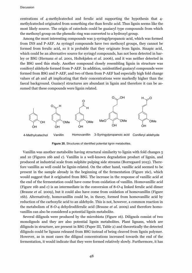
Discussion
48
centrations of 4-methylcatechol and ferulic acid supporting the hypothesis that 4-methylcatechol originated from something else than ferulic acid. Thus lignin seems like the most likely source. The origin of catechols could be guaiacyl type compounds from which the methoxyl group on the phenolic ring was converted to a hydroxyl group.
Among the most interesting compounds was 3-syringylpropanoic acid, which was formed from INS and P-AEF. As syringyl compounds have two methoxyl groups, they cannot be formed from ferulic acid, so it is probable that they originate from lignin. Sinapic acid, which could be an alternative source for syringyl compounds, has not been detected in bar-ley or BSG (Hernanz et al. 2001, Holtekjølen et al. 2006), and it was neither detected in the BSG used this study. Another compound closely resembling lignin in structure was coniferyl aldehyde formed from P-AEF. In addition, unidentified guaiacyl compounds were formed from BSG and P-AEF, and two of them from P-AEF had especially high fold change values of 46 and 48 implicating that their concentrations were markedly higher than the faecal background. Guaiacyl structures are abundant in lignin and therefore it can be as-sumed that these compounds were lignin related.
Figure 20. Structures of identified potential lignin metabolites.
Vanillin was another metabolite having structural similarity to lignin with fold changes 5 and 10 (Figures 16b and c). Vanillin is a well-known degradation product of lignin, and produced at industrial scale from sulphite pulping side streams (Borregaard 2015). There-fore vanillin as well could be lignin-related. On the other hand, vanillic acid seemed to be present in the sample already in the beginning of the fermentation (Figure 16c), which would suggest that it originated from BSG. The increase in the response of vanillic acid at the end of the fermentation could have come from oxidation of vanillin. Homovanillic acid (Figure 16b and c) is an intermediate in the conversion of 8-O-4 linked ferulic acid dimer (Braune et al. 2009), but it could also have come from oxidation of homovanillin (Figure 16b). Alternatively, homovanillin could be, in theory, formed from homovanillic acid by reduction of the carboxylic acid to an aldehyde. This is not, however, a common reaction in the metabolism of 8-O-4 dehydrodiferulic acid (Braune et al. 2009) and therefore homo-vanillin can also be considered a potential lignin metabolite.
Several dilignols were produced by the microbiota (Figure 16). Dilignols consist of two monolignols and they are also potential lignin metabolites. Plant lignans, which are dilignols in structure, are present in BSG (Paper III, Table 2) and theoretically the detected dilignols could be lignans released from BSG instead of being cleaved from lignin polymer. However, as in most cases the dilignol concentrations increased towards the end of the fermentation, it would indicate that they were formed relatively slowly. Furthermore, it has

Discussion
49
been demonstrated that lignans (or dilignols) can be obtained from lignin through the ac-tivities of gut microbiota (Begum et al. 2004). It has been claimed that lignin is not at all degraded in the human gastrointestinal tract (Holloway et al. 1978). However, in the in vivo study conducted by Holloway et al. (1978) the amount of lignin excreted was markedly higher than the amount of lignin consumed, which should not be theoretically possible, suggesting significant inaccuracies in the analy-sis method. On the contrary, Williams and Olmsted (1936) proposed that notable lignin degradation occurs in humans. However, the analytics involved quantitation of lignin as an acid-insoluble residue, which unlikely yielded reliable results from faecal mass. Instead of the gravimetric lignin quantitation, some studies based on analysis of aromatic metabolites have also been carried out. Silanikove and Brosh (1989) measured lignin degradation in goats based on urinary hippuric and benzoic acids concentrations. Notable increases in the acid concentrations were detected indicating lignin degradation. Csonka et al. (1929) measured the amount of lignin methoxyl groups and urinary aromatic acids to determine lignin digestion in cows and dogs. They concluded that based on the loss of methoxyl groups and increase in aromatic acids in urine, lignin was degraded in both animals. How-ever, in the view of current knowledge (Russell et al. 2008, Braune et al. 2009) and the abundance of catechols as metabolites in the present study, it should be reconsidered whether or not the disappearance of methoxyls can be regarded as evidence of lignin deg-radation, as demethylation of the phenolic ring substituents appears to be a common reac-tion by faecal microbiota resulting in the loss of a methoxyl group by conversion to a hy-droxyl group. It does not seem impossible that such a reaction could also occur on poly-meric lignin, without necessarily involving a further fragmentation of the polymer. On the other hand, the increased amounts of aromatic metabolites support their conclusions.
Although the studies discussed above date back more than several decades, standardised or generally accepted methods for quantitatively detecting lignin degradation by faecal microbiota do not exist. This is likely to be one of the main factors limiting the research of this particular topic. A more reliable way of demonstrating lignin degradation, could be, for instance, using isotope labelled lignin. This has been performed by Begum et al. (2004), who prepared deuterated synthetic lignin and followed its conversion in rats. Deuterated enterolactone was detected from the urine of rats unambiguously confirming the degrada-tion of lignin by gut microbiota in vivo. However, with the methods applied in the present study, the origin of any metabolite cannot be verified with 100 % certainty, although links to lignin seem very likely with several metabolites. With further identification of the me-tabolites, their conversion reactions and particularly the converting enzymes the metabo-lite pathways could also be studied. Knowing the pathways would confirm the origin of the metabolites and provide new information on the activities of gut microbiota.
5.2.2 Extent of lignin degradation by gut microbiota
Regarding the extent of lignin degradation by human faecal microbiota no definite conclu-sions can be drawn, as the analysis of metabolites was only semiquantitative (except for 4-methylcatechol), and instead of absolute concentrations it provides information on a) how much higher the concentration of a given compound is relative to the faecal background (fold change) and b) whether the concentration of the compound is increasing or decreas-ing over time. As the faecal background most likely contained some lignin as well, the con-

Discussion
50
centrations of metabolites originating from the background affected the fold change. The higher the background the lower the amount of a given metabolite is relative to it. Alterna-tively, if the concentration of a certain metabolite is very low in the background, even a smaller amount of it produced from the studied sample may result in a relatively large fold change. Therefore, the fold changes should only be considered as indicative.
Some measure of lignin degradation can be drawn from the amount of 4-methylcatechol, which was quantitated. The amount of 4-methylcatechol produced during the fermenta-tions corresponded to approximately 40‒80 ppm of the lignin present in the samples in the beginning of the fermentation. As the amounts of other metabolites were not quantitated, the extent of lignin degradation cannot be measured from one metabolite, but it provides some idea of the level of degradation. Also the rat study by Begum et al. (2004) gives some indication. The amount of deuterated enterolactone produced was 655 nmol/g of synthetic lignin, which corresponds to approximately 0.2 mg of enterolactone per g of lignin also indicating a low degree of lignin conversion. However, the amount of only two metabolites, enterolactone and enterodiol (not detected), were measured, and there is a possibility that other deuterated conversion products were also formed, which would suggest a more ex-tensive conversion. Regarding the rat study, it should be noted though that rat and human microbiota are different from each other and may have different capabilities to degrade lignin.
The degradation of lignin could be limited also by inability of microbiota to cleave car-bon-carbon bonds between phenolic units. As seen with diferulates, ether-linked dimers are cleaved to monomers, whereas carbon-carbon linked dimers are not (Braune et al. 2009, Russell et al. 2008, Schendel et al. 2015). However, the β-O-4 aryl ethers are the most abundant linkages in BSG lignin (Rencoret et al. 2015) suggesting that despite the possible limitations sites for cleavage are still numerous. Nevertheless, based on the re-search showing more limited fermentation of carbohydrates in lignin-containing dietary fibre (Grabber et al. 2009) it can be assumed that lignin is not an easy substrate for micro-biota. Thus, it would seem reasonable that human microbiota is able to convert only a small part of it, but according to the present study it seems more than likely that lignin is not completely inert.
5.2.3 Phenolic metabolites derived from non-lignin sources
In addition to lignin, ferulic acid and its dimers are present in BSG (Hernanz et al. 2001), and thus they were another source of phenolic metabolites. The amount of ferulic acid in the colon model was quantitated. Its content was highest in P-AEF and it was metabolised for the most part during the fermentation (Figure 17). BSG, INS and DEFE contained markedly less ferulic acid, and in INS and DEFE its concentration did not change notably over time. Moderate changes were observed for BSG as ferulic acid was rapidly metabo-lised upon release from the matrix. For INS and DEFE the formation of 3,4-dihydroxyphenylacetic acid was on a similar level compared to ferulic acid, suggesting that it was a major metabolite of ferulic acid, but for P-AEF the concentrations of these two compounds did not correlate similarly.
It should be pointed out that there are some differences in the naming of diferulates and dilignols (Figure 21). In both the phenolic ring carbons are denoted as carbons 1‒6 starting from the carbon containing the side chain. However, in monolignols the propenyl side chain carbons are denoted as α, β and γ, whereas in ferulic acid (and other hydroxycinnam-

Discussion
51
ic acids) the equivalent carbons are simply numbered 7, 8 and 9. Thus, for instance β-O-4 and 8-O-4 are similar aryl ether bonds but the first one links two monolignols and the lat-ter ferulic acids.
Figure 21. Naming of carbon atoms in monolignols and hydroxycinnamic acids.
Ferulic acid is the component, which in this study interferes most with the interpretation of the lignin metabolites. The conversions of ferulic and diferulic acids by faecal microbiota have been studied and their metabolites characterised, and thus the obvious ferulic acid metabolites could be excluded from the potential lignin metabolites. The metabolites origi-nating from ferulic acid include p- and m-coumaric acid, cinnamic acid and phenylpropa-noic acid (mono-, di- and non-hydroxylated) (Russell et al. 2008). In addition to these 3,4-dihydroxyphenylacetic acid, 3-(3,4-dihydroxyphenyl)lactic acid and homovanillic acid are formed from 8-O-4 linked dehydrodiferulic acid (Braune et al. 2009). The ether bond in 8-O-4 dehydrodiferulic acid can be cleaved by faecal microbiota, whereas in 8-5 coupled de-hydrodiferulic acid the carbon-carbon bond between the two ferulic acid units is not cleaved resulting in only dimeric metabolites (Schendel et al. 2015). For 5-5 linked diferu-lic acid only one metabolite has been identified, a dimeric compound with hydrogenated aliphatic side chains but no alterations in the methoxyl groups (Russell et al. 2008). Many of the ferulic acid and 8-O-4 dehydrodiferulic acid metabolites were produced from the BSG fractions, and although they have structural similarities with lignin as well, due to the presence of ferulic acid in the studied fractions, the origin of these metabolites was proba-bly not lignin.
Metabolites that probably originated from sources other than lignin and ferulic acid in-clude aminobenzoic acids and 4-hydroxyphenylpentanoic acid. 4-Aminobenzoic acid is formed in the synthesis of aromatic amino acids in certain bacteria including Escherichia coli (Herrmann 1995). In 4-hydroxyphenylpentanoic acid (5 carbons) the aliphatic chain is too long to be formed from lignin structure, as the aliphatic chains in lignin contain 3 car-bons.
5.2.4 Potential bioactivities of lignin and phenolic metabolites
Lignin and other polyphenolics may induce beneficial properties in humans upon inges-tion. These include acting as an antioxidant and radical scavenger (Lu et al. 1998, Dizhbite et al. 2004) due to phenolic hydroxyl groups (Barclay et al. 1997), and controlling post-prandial glycemic response (Hanhineva et al. 2010). Lignin has been shown to adsorb car-cinogenic compounds in conditions mimicking the small intestine and colon (Funk et al. 2006, Funk et al. 2007), which might help protect from cancer. Lignin may also exert an-
1 12 2
3 34 45 5
6 6
αβ
γ
7 89
R1=R2=H: p-coumaryl alcoholR1=H, R2=OMe: coniferyl alcoholR1=R2=OMe: sinapyl alcohol
R=H: p-coumaric acidR=OMe: ferulic acid

Discussion
52
timicrobial activity towards certain intestinal pathogens, such as E. coli (Baurhoo et al. 2007a). Moreover, the part of lignin that remains in polymeric form may further contrib-ute to bulking effect and bowel movement (Raninen et al. 2011) improving gut health. In addition to lignin, the effects of low molecular weight phenolic metabolites on human health should also be further elucidated.
Phenolic metabolites may induce bioactive effects upon release and conversion by colon microbiota, including antioxidative (Castelluccio et al. 1995, Vinson et al. 2003), anti-microbial (Barber et al. 2000), anti-inflammatory (Karlsson et al. 2005) and anticarcino-genic (Stich and Rosin 1984) properties. For example, ferulic acid alone possesses all the aforementioned characteristics (reviewed by Ou and Kwok 2004). Other phenolic metabo-lites, such as 4-methylcatechol and 3,4-dihydroxyphenylacetic acid have also been found good antioxidants in vitro (Gläßer et al. 2002). Plant lignans, are known to be bioactive in many ways; they are antioxidant (Eklund et al. 2005, Willför et al. 2003) and antitumor-igenic (Thompson et al. 1996, Hausott et al. 2003), and when converted to mammalian lignans enterodiol and enterolactone may protect from hormonal cancers, such as breast cancer (Adlercreutz 2002). The exact structures of the dilignol metabolites detected in the present study could not be confirmed, but characteristics such as dimeric form with guai-acyl structures were identified. Due to their structural similarities with lignans, it can be hypothesized that these dilignols as well could possess similar properties. Thus it appears that lignin-related metabolites may potentially induce bioactive effects in humans, but their impact is not known and needs to be characterised before any conclusions can be drawn. It seems likely that lignin is an undervalued compound of dietary fibre, and its role in diet and digestion by intestinal microbiota requires more attention in the future.
5.3 Effects of lignin on microbial carbohydrate fermentation and growth
5.3.1 Formation of SCFA from the lignin-rich fractions
Carbohydrates that are resistant to digestion in the stomach and small intestine are trans-ferred to the colon where they are partially fermented to short chain fatty acids (SCFA) (Cummings and MacFarlene 1997). The most important SCFA include acetic, propionic and butyric acids, which have several physiological functionalities, such as being the major source of energy of colon epithelial cells (Comalada et al. 2006). In the present study the concentrations of SCFA were measured in the colon model to see if their formation was suppressed, which could indicate that lignin inhibits enzymatic activities of the microbiota. No significant suppression was detected (Figure 18). Although in the beginning of the fer-mentation SCFA levels were slightly lower for BSG compared to the faecal background, they exceeded them at the end, and thus it would seem that carbohydrate fermentation was slowed down but not completely suppressed. Previously suppression of SCFA formation and phenolic metabolism have been noticed to occur in the presence of condensed tannins, or proanthocyanidins isolated from apples and Syrah grapes (Bazzocco et al. 2008, Aura et al. 2013). These types of tannins are mostly composed of epicatechin units and although they differ in their chemical structure compared to lignin, it was important to assess if lig-nin has similar effects towards microbial enzymes.
The untreated BSG was probably a slowly fermentable substrate, as no enzymatic break-down of the cell walls had been carried out prior to the in vitro fermentation. Thus more time for the microbiota to release sugars and ferment them to SCFA was needed. In the

Discussion
53
case of INS the more easily digestible carbohydrates had been removed by enzymatic treatments, and only the most resistant carbohydrates most tightly bound to each other and lignin remained. In DEFE the cleavage of (di)ferulate cross-links should have facilitat-ed the degradation of cell wall polysaccharides, but the amount of SCFA was not higher compared to the other fractions. It is possible that the strong alkaline treatment also dis-solved the easily digestible material and thus DEFE was not notably more fermentable de-spite the loss of cross-links. P-AEF was very low in carbohydrates (4 %), but it was still fermented at significantly higher rate and extent compared to the faecal background sug-gesting that these few carbohydrates were accessible to the microbiota. Therefore, it would seem probable that slow SCFA production was not as strongly affected by the presence of lignin in the material but was rather dependent on the resilient nature of the carbohydrate matrix.
The lignin contents of the samples were not very high, in the range of 20‒40 %, and the original idea was to obtain BSG fractions with higher lignin purity. However, if lignin isola-tion had been more comprehensive, most likely the results would have been completely different. As lignin also has antimicrobial characteristics (Baurhoo et al. 2007a), purer lignin could have had detrimental effects on the faecal microbiota, and therefore the me-tabolism of the samples and SCFA formation might have been significantly more limited. As lignin is ingested as part of dietary fibre i.e. inside the plant cell wall matrix, it is not present as a pure component in the colon, and therefore the impurities present in this study may actually have given a more realistic result. Furthermore, the carbohydrates and proteins in the studied fractions provided nutrients and energy to the microbiota and likely enabled them to survive longer in the colon model experiment.
5.3.2 Lignin-rich fraction as a growth substrate for lactobacilli and bifidobacteria
The capability of beneficial colonic bacteria to grow on the protease-alkaline extracted frac-tion (P-AEF) was studied in order to detect potential inhibition of growth by a lignin-rich substrate. For this purpose lipids were extracted from P-AEF increasing its lignin content to 41 %. The bacteria were grown on P-AEF, and positive and negative controls were also prepared. Glucose was used as growth substrate in the positive control, and the negative control contained only the basal medium without added glucose or P-AEF. Four lactobacilli and three bifidobacterial strains, which can be commonly found in the human intestinal microbiota, were selected for the study, with the exception of L. salivarius, which is more often present in saliva than colon (Goldstein et al. 2015). Lactobacilli and bifidobacteria are considered beneficial for human health as they regulate the colonisation of pathogenic bacteria in the colon, help recover from diarrhoea and relief irritable bowel syndrome among many other things (Gibson and Wang 1994, Ouwehand et al. 2002, Gomes and Malcata 1999).
Using a lignin-rich substrate (10 g/L) did not inhibit the growth of lactobacilli and bifidobacteria, but instead the fraction enabled a longer growth of bifidobacteria than glu-cose (Figure 19). The colony counts of both strains of L. rhamnosus were almost equal in the presence of P-AEF and in the controls. The MRS medium of lactobacilli did not contain carbohydrates, but apparently peptides and amino acids in the medium (15 g/L) enabled growth also in the negative control. In the absence of glucose the growth was somewhat lower for L. paracasei and L. salivarius, indicating that they were more sensitive to the lower amount (in P-AEF sample) or complete lack (in the negative control) of fermentable

Discussion
54
sugars than L. rhamnosus. However, the colony counts of bacteria grown on P-AEF were always above the negative control demonstrating that no inhibitory effects were induced by the lignin-rich growth substrate.
In the positive controls of B. adolescentis and B. longum the amount of viable cells was surprisingly low already after 3 days, but in the presence of P-AEF there was remarkable growth (107−108 CFU/mL). Also in the negative controls bifidobacteria grew almost equally well (107 CFU/mL). After 7 days of incubation, there was still significant growth (106−107 CFU/mL) in bifidobacterial cultures with P-AEF, whereas in the positive controls cultura-ble cell counts were in the level of 102 CFU/mL. A similar protective effect on bifidobacteria has been previously observed with fermented oat bran preparations, but no single growth factor was identified in that study (Kontula et al. 1998). In the negative control the growth was in the level of 105 CFU/mL.
After lipid removal, P-AEF was low in carbohydrates (7 %), but especially rich in protein-aceous material (45 %) as the result of the partial precipitation of peptides after the proteo-lytic step. Peptide preparations, such as dairy protein hydrolysates and yeast extract, are known to stimulate growth of bifidobacteria (Gomes and Malcata 1999). Although the ef-fects of cereal proteins on bifidobacteria are not as well known, presumably the peptides in P-AEF were the factor, which enabled the prolonged growth. The Bifidobacterium medium was free of carbohydrates but contained 25 g/L of peptides from different sources, which explains the growth in the negative control. However, the growth was very limited in the positive controls, except for B. breve at 3 d. This can be explained by a very rapid growth during the first incubation days induced by the easily available glucose in the medium. The high numbers of cells resulting from this exponential growth quickly depleted the nutrients from the medium, and therefore after 3 days there were no nutrients left and the culturable cell counts were thus very low. The bacteria growing on P-AEF or the basal medium with-out carbohydrates (negative control) grew more slowly due to the low amount or lack of easily available sugars, and thus the amount of nutrients originating from peptones in the medium was sufficient to maintain growth even after 7 days.
When dosed at low levels (1.25 %), lignin has been shown to increase the numbers of lac-tobacilli and bifidobacteria in broiler gut and to reduce the amount of E. coli in the faeces (Baurhoo et al. 2007b). A higher lignin dose (2.5 %) was highly efficient in preventing pop-ulating of E. coli in the cecum of E. coli challenged broilers, but on the other hand the numbers of lactobacilli and bifidobacteria were also reduced (Baurhoo et al. 2007a). It seems that lignin can have both growth-inducing and inhibitory effects on lactobacilli and bifidobacteria depending on the dosage. It is possible that at a higher dosage BSG lignin could have exerted antimicrobial properties as well. Another factor affecting the reactivity of lignin is the isolation method. In the case of the broilers lignin was isolated using the Alcell process, which is an organosolv process producing hydrophobic low-molecular weight lignin with higher purity and most probably with a higher content of free phenolic hydroxyl groups compared to the BSG fractions of the present study (Lora and Glasser 2002). Similarly, condensed tannins when isolated from fruits were inhibitory to the con-versions of faecal microbiota but did not exert such effects in their natural state inside the fruits (Bazzocco et al. 2008, Aura et al. 2013). It is possible that the proteinaceous material in P-AEF, which seemed to be associated with lignin, could have acted as a barrier reducing the antimicrobial effects of lignin. Therefore, it seems likely that not only the lignin content but also the isolation method and association of lignin with cell wall polysaccharides or

Discussion
55
proteinaceous material influence the potentially antimicrobial and inhibitory properties of lignin.
5.4 Methodological considerations
5.4.1 Analysis of lignin content in biomass
Accurately measuring the lignin content of non-wood biomass is not straightforward, and therefore BSG lignin contents reported in the literature (measured as Klason lignin) are likely to be exaggerated. Klason lignin is the insoluble residue of a biomass after acid hy-drolysis, and its amount is measured gravimetrically. As demonstrated by Bunzel et al. (2011), Klason lignin from non-wood biomasses may contain protein, ash, cutin, suberin, fats and waxes, which all interfere with the analysis. However, measuring the contents of all the aforementioned components to correct the lignin content, would require much more work, and therefore it is not routinely performed. The Klason method is better suited for wood material as the contents of protein, ash and extractives in wood are sufficiently low so they do not induce a significant error in the Klason lignin determination
Hatfield and Fukushima (2005) have discussed the issue of accurately measuring lignin. They concluded that “there is no single method that is rapid, noninvasive, handles large sample numbers, and provides accurate measure of cell wall lignin contents”. Instead, they recommend that one should always use the same method enabling comparison between samples rather than using different methods for different samples. In the present study, the Klason method (including acid-soluble lignin) was found to give exaggerated values for some samples (data not shown). In order to counteract this, lignin content was estimated by subtracting other major components (carbohydrates, protein, extractives and ash) from 100 %, as the analytical methods for those were believed to be more accurate than the Kla-son method. Furthermore, in addition to carbohydrates and lignin, faeces contain also oth-er types of insoluble components, such as waxes, cutin and a considerable amount of bacte-rial mass, and thus the Klason lignin analysis would most likely yield unreliable results. Therefore, the amount of lignin degraded during the fermentation was not quantitated.
5.4.2 Use of the in vitro colon model
The in vitro colon metabolic model is a batch model, in which microbial conversions occur in anaerobic conditions for defined periods of time. Fresh human faeces are used as the inoculum to obtain a diversified microbial population. The transit time in colon is approx-imately 28 h (Arhan et al. 1981) and the fermentation time in the present study was set accordingly (24 h). Although the conditions of the model, such as temperature, pH, anaer-obic atmosphere and the microbiota simulate the conditions of the proximal part of the colon, incubation in a batch model still provides different dynamics as compared to the continuous system of the colon.
In humans, before entering the colon food has been digested in the stomach and the small intestine leaving only the dietary fibre and other indigestible components to be fer-mented by the colonic microbiota. Therefore to obtain the most realistic results, samples should be similarly pre-digested with alimentary enzymes to remove the digestible food components before introducing the residue to the colon model (Aura et al. 1999). However, this was not performed in the present study. BSG has been exposed to hydrolytic enzymes

Discussion
56
in mashing and contains only a residual amount of starch (ca. 1 %). The rest is mostly die-tary fibre and protein (Table 1). A major part of the protein in BSG is encapsulated in aleu-rone cells (Figure 14a) and is thus not available to peptidases without decomposition of the cell walls. Moreover, the protein content of cereal materials after a pre-digestion with an upper intestine model can still be as high as 25 %, as demonstrated with wholemeal rye bread (Aura et al. 1999), suggesting that BSG protein level was not especially high for the fermentation. The protein (peptide) content was highest and most accessible in P-AEF. However, aromatic protein metabolites, such as indoles, could be distinguished from those of phenolic origin, and thus protein was not considered to interfere with the analysis of lignin metabolites. P-AEF was also rich in lipids but they were easily excluded from the phenolic metabolic profile.
Statistical significance of the responses of the metabolites compared to the faecal back-ground was estimated using two-way ANOVA and denoted with asterisks. It is worth point-ing out, that many metabolites with high FC values, such as guaiacyl compounds derived from P-AEF (Figure 16b) had only one time point statistically different from the faecal background, whereas some metabolites with low FCs had several of these points (e.g. 4-methylcatechol from BSG (Figure 16a)). The statistical significance is partially affected by the repeatability of the parallel measurements. As lignin is presumed to be a resilient sub-strate for the microbiota, it is not surprising that the repeatability between replicates was not very high. Another factor affecting the lack of statistical difference for the metabolites is most likely their low levels and origination of the same compounds also from the faecal background. If the background levels are high, the difference between the sample and the background is relatively smaller than if the same amount of a given metabolite was pro-duced and compared against a low level in the background.

57
6. Conclusions and future prospects
The objectives of this thesis were to produce lignin-rich fractions from BSG and to study the interactions of the lignin in these fractions with colon microbiota in vitro. As BSG con-tains the outer parts of the grain, which are the protective layers, it is resistant to hydroly-sis by cell wall degrading enzymes, if no pre-treatment is applied. The first specific aim was to evaluate the effectiveness of milling as a pre-treatment to enhance the enzymatic hy-drolysis of carbohydrates. Of the different types of milling pre-treatments ball-milling was the most efficient increasing carbohydrate solubilisation from 23 to 45 % and thus notably improving the enzymatic carbohydrate digestibility. However, it was not effective enough to enable total solubilisation of BSG carbohydrates. Masuko milling was somewhat less effective than ball milling enabling 35 % enzymatic carbohydrate solubilisation, but on the other it was more convenient, as a larger amount of material could be processed and no drying was required. Enzymatic hydrolysis of carbohydrates was most probably restricted by several factors: steric hindrance, presence of lignin, cross-linking of arabinoxylans and lignin by (di)ferulates and possibly the absence of feruloyl esterases or other enzymatic activities. It is evident that a more efficient pre-treatment is required to obtain high sugar yields from BSG resulting in purer lignin in the residue.
The second specific aim regarding the fractionation was to produce different types of lig-nin-enriched fractions from BSG. Despite the limited extent of carbohydrate solubilisation and complications arisen from the co-solubilisation and co-precipitation of lignin and pro-tein, two lignin-rich fractions were obtained by a three-step enzymatic hydrolysis. In addi-tion, a separate alkaline extraction provided BSG-derived material with low ferulic acid content. Lignin contents in the fractions were 20‒40 %, which can be considered high compared to common foods and thus sufficient for the colon model studies, although in only one of the fractions the lignin content was notably enriched (from 19 % in BSG to 40 %).
The third and fourth specific aims were to study the interactions of colon microbiota with lignin in vitro. More specifically, the aims were to assess if lignin is degraded and metabo-lised by colon microbiota and if lignin suppresses microbial conversions in the colon. A number of phenolic metabolites structurally similar to lignin were formed from BSG and the fractions by microbial conversions. Although the origin of the metabolites could not be determined exactly, it seemed very likely that several of the metabolites, including 3-syringylpropanoic acid, 4-methylcatechol, coniferyl aldehyde and dilignols originated from lignin. Based on these results it seems that lignin is not inert but is degraded and metabo-lised to a limited extent in the colon. Most likely microbiota is able to cleave the edges of lignin but a major part of the macromolecule remains intact. No notable suppression was detected based on the formation of short chain fatty acids from carbohydrates. Also, the presence of lignin did not inhibit the growth of lactobacilli and bifidobacteria, but instead the bacteria seemed to be capable of using the carbohydrates and peptides of the fraction

Conclusions and future prospects
58
as carbon and energy sources. Thus it appears that lignin does not suppress microbial con-versions in the colon and it seems not to be inhibitory to bacterial growth. However, this is likely to depend on the state of lignin as an isolated compound or bound in a matrix. Asso-ciation of lignin with carbohydrates or proteinaceous material may reduce the possible antimicrobial effects of lignin, as free phenolic hydroxyl groups are believed to have a key role in the antimicrobial activities of lignin.
The hypotheses of this thesis were that lignin-rich fractions can be produced from BSG using enzymatic methods, and that lignin is at least partially metabolised by human intes-tinal microbiota. It can be stated that three different types of lignin-rich fractions were ob-tained from BSG (although one with chemical and not enzymatic methods) and that lignin-related metabolites were shown to be formed from all fractions and from the original BSG upon digestion by human gut microbiota. Although the analytical methods used did not allow quantitation and in some cases full identification of the metabolites, and the results provide qualitative rather than quantitative information on lignin degradation, it can be considered that both hypotheses were correct.
The results of the present study provide new information on the significance of lignin as part of dietary fibre. Based on the findings of the present study it appears that instead of being completely inert and resistant to digestion lignin would be a source of phenolic me-tabolites and thus an undervalued component of dietary fibre. As in foods lignin is part of the dietary fibre complex and most likely degraded only to a limited extent, it does not seem probable that consuming lignin as part of plant matrix even at high dosages would result in harmful effects. In order to find applications for BSG outside of feed industry in the future, its fractionation should be further developed and optimised. Pre-treatment methods such as steam-explosion could improve the hydrolysis of carbohydrates e.g. for fermentable sugars. Fur-thermore, understanding the interactions observed between lignin and proteins could help find more efficient ways to recover these two components separately. Utilisation of dietary fibre and (undegraded) protein from BSG as food additives is another alternative to the hydrolytic fractionation approach.
Lignin is generally considered to be resistant to bioconversion by human colon microbio-ta, and thus its digestibility has been the attention of only a limited amount of studies, many of which date back several decades and have been carried out with methods that may not be the most accurate. The results of the present study suggest that more research is needed to further elucidate the role of lignin as a source of low molecular weight phenolic compounds, which potentially possess bioactive properties upon being absorbed in the colon. For instance, lignans have been shown to be released from lignin in vivo, and a number of studies provide information on their physiological functionalities.
Expensive materials, such as labelled synthetic lignin may be required to obtain indis-putable results. In addition, as in gastrointestinal tract lignin remains as part of the cell wall matrix, a synthetic lignin material not bound to polysaccharides may not be the most representative sample. More information on how the surrounding cell wall polysaccharides affect the antimicrobial properties and the accessibility of lignin to intestinal microbiota is needed. The key metabolites derived from lignin should be identified in order to quantitate them, which would enable the determination of the extent of lignin degradation. Determin-ing the bioactivities of the metabolites is also crucial to confirm the safety and potential beneficial impacts of lignin as part of food. An accurate method for measuring lignin con-

Conclusions and future prospects
59
tent in food and faecal materials is an essential tool and requires adaptation of current lig-nin analytics to suit versatile biomaterials, such as BSG, which consist of a variety of com-pounds, many of which may interfere with the traditional Klason lignin analysis.
In conclusion, potential evidence of lignin metabolism by colon microbiota was provided in the present study. Furthermore, no suppression of carbohydrate fermentation or inhibi-tory effects on bacterial growth was observed. However, due to limitations in analyses and presence of interfering phenolic compounds in the fermentation, these results are prelimi-nary and need to be confirmed with optimised methods. Further research to develop quan-titative analysis methods for microbial lignin degradation and identification of the key me-tabolites would enable a better understanding of the effects of lignin as a component of dietary fibre.


61
References
Adlercreutz H. (2002) Phytoestrogens and breast cancer. J. Steroid Biochem. Mol. Biol. 83, 113-118.
Antoine C., Lullien-Pellerin V., Abecassis J., Rouau X. (2004) Effect of wheat bran ball-milling on fragmentation and marker extractability of the aleurone layer. J. Cereal Sci. 40, 275-282.
Arhan P., Devroede G., Jehannin B., Lanza M., Faverdin C., Dornic C., Persoz B., Tétreault L., Perey B., Pellerin D. (1981) Segmental colonic transit time. Dis. Colon Rectum 24, 625-629.
Aura A.-M., Härkönen H., Fabritius M., Poutanen K. (1999) Development of an in vitro enzymic digestion method for removal of starch and protein and assessment of its performance using rye and wheat breads. J. Cereal Sci. 29, 139-152.
Aura A.-M., Mattila I., Hyötyläinen T., Gopalacharyulu P., Cheynier V., Souquet J.-M., Bes M., Le Bourvellec C., Guyot S., Orešic M. (2013) Characterization of microbial metabolism of Syrah grape products in an in vitro colon model using targeted and non-targeted analytical ap-proaches. Eur. J. Nutr. 52, 833-846.
Barclay L.R.C., Xi F., Norris J.Q. (1997) Antioxidant properties of phenolic lignin model com-pounds. J. Wood Chem. Technol. 17, 73-90.
Bamforth C.W., Barclay A.H.P. (1993) Malting technology and the uses of malt. In Barley: Chemis-try and technology. MacGregor A.W. and Bhatty R.S. Eds., American Association of Cereal Chemists Inc., St. Paul, MN, pp. 297-354.
Bartolomé B., Gómez-Cordovés C. (1999) Barley spent grain: release of hydroxycinnamic acids (fer-ulic and p-coumaric acids) by commercial enzyme preparations. J. Sci. Food Agric. 79, 435-439.
Baurhoo B., Letellier A., Zhao X., Ruiz-Feria C.A. (2007a) Cecal populations of lactobacilli and bifidobacteria and Escherichia coli populations after in vivo Escherichia coli challenge in birds fed diets with purified lignin or mannanoligosaccharides. Poult. Sci. 86, 2509-2515.
Baurhoo B., Phillip L., Ruiz-Feria C.A. (2007b) Effects of purified lignin and mannan oligosaccha-rides on intestinal integrity and microbial populations in the ceca and litter of broiler chick-ens. Poult. Sci. 86, 1070-1078.
Bazzocco S., Mattila I., Guyot S., Renard C., Aura A. (2008) Factors affecting the conversion of apple polyphenols to phenolic acids and fruit matrix to short-chain fatty acids by human faecal mi-crobiota in vitro. Eur. J. Nutr. 47, 442-452.
Beaugrand J., Paës G., Reis D., Takahashi M., Debeire P., O’Donohue M., Chabbert B. (2005) Prob-ing the cell wall heterogeneity of micro-dissected wheat caryopsis using both active and inac-tive forms of a GH11 xylanase. Planta 222, 246-257.
Begum A.N., Nicolle C., Mila I., Lapierre C., Nagano K., Fukushima K., Heinonen S., Adlercreutz H., Rémésy C., Scalbert A. (2004) Dietary lignins are precursors of mammalian lignans in rats. J. Nutr. 134, 120-127.
Beldman G., Hennekam J., Voragen A.G.J. (1987) Enzymatic hydrolysis of beer brewers' spent grain and the influence of pretreatments. Biotechnol. Bioeng. 30, 668-671.

References
62
Bennick A. (2002) Interaction of plant polyphenols with salivary proteins. Crit. Rev. Oral Biol. Med. 13, 184-196.
Benyoucef N., Cheikh A., Drouiche N., Lounici H., Mameri N., Abdi N. (2013) Denitrification of groundwater using brewer's spent grain as biofilter media. Ecol. Eng. 52, 70-74.
Birkmire S., Lindeman C., Duff B., Spooner J., Yancey M. (2012) Conversion of biomass into etha-nol. US Patent 20100196979.
Boerjan W., Ralph J., Baucher M. (2003) Lignin biosynthesis. Annu. Rev. Plant Biol. 54, 519-546.
Borregaard (2015) Vanillin house by Borrgeaard ‒ The home of sustainable vanillin http://www.vanillin.com/ Accessed 11/04/2015.
Boulton C., Quain D. (2006) Beer and brewing. In Brewing yeast & fermentation. Blackwell Sci-ence, Oxford, UK, pp. 5-14.
Braune A., Bunzel M., Yonekura R., Blaut M. (2009) Conversion of dehydrodiferulic acids by human intestinal microbiota. J. Agric. Food Chem. 57, 3356-3362.
Briggs D.E., Boulton C.A., Brookes P.A., Stevens R. (2004a) The science of malting. In Brewing science and practice. CRC Press LLC, Boca Raton, FL, USA, pp. 85-170.
Briggs D.E., Boulton C.A., Brookes P.A., Stevens R. (2004b) Malts, addjuncts and supplementary enzymes. In Brewing science and practice. CRC Press LLC, Boca Raton, FL, USA, pp. 11-51.
Brown L., Rosner B., Willett W.W., Sacks F.M. (1999) Cholesterol-lowering effects of dietary fiber: a meta-analysis. Am. J. Clin. Nutr. 69, 30-42.
Bunzel M., Ralph J., Marita J.M., Hatfield R.D., Steinhart H. (2001) Diferulates as structural com-ponents in soluble and insoluble cereal dietary fibre. J. Sci. Food Agric. 81, 653-660.
Bunzel M., Ralph J., Lu F., Hatfield R.D., Steinhart H. (2004) Lignins and ferulate–coniferyl alcohol cross-coupling products in cereal grains. J. Agric. Food Chem. 52, 6496-6502.
Bunzel M., Seiler A., Steinhart H. (2005) Characterization of dietary fiber lignins from fruits and vegetables using the DFRC method. J. Agric. Food Chem. 53, 9553-9559.
Bunzel M., Ralph J. (2006) NMR characterization of lignins isolated from fruit and vegetable insol-uble dietary fiber. J. Agric. Food Chem. 54, 8352-8361.
Bunzel M., Schüßler A., Tchetseubu S.G. (2011) Chemical characterization of Klason lignin prepara-tions from plant-based foods. J. Agric. Food Chem. 59, 12506-12513.
Carvalheiro F., Duarte R.C., Medeiros R., Gírio F.M. (2004a) Optimization of brewery’s spent grain dilute-acid hydrolysis for the production of pentose-rich culture media. Appl. Biochem. Bio-technol. 115, 1059-1072.
Carvalheiro F., Esteves M.P., Parajó J.C., Pereira H., Gírio F.M. (2004b) Production of oligosaccha-rides by autohydrolysis of brewery’s spent grain. Bioresour. Technol. 91, 93-100.
Castelluccio C., Paganga G., Melikian N., Paul Bolwell G., Pridham J., Sampson J., Rice-Evans C. (1995) Antioxidant potential of intermediates in phenylpropanoid metabolism in higher plants. FEBS Lett. 368, 188-192.
Castillo S., Mattila I., Miettinen J., Orešič M., Hyötyläinen T. (2011) Data analysis tool for compre-hensive two-dimensional gas chromatography/time-of-flight mass spectrometry. Anal. Chem. 83, 3058-3067.
Celus I., Brijs K., Delcour J.A. (2006) The effects of malting and mashing on barley protein ex-tractability. J. Cereal Sci. 44, 203-211.

References
63
Celus I., Brijs K., Delcour J.A. (2007) Enzymatic hydrolysis of brewers' spent grain proteins and technofunctional properties of the resulting hydrolysates. J. Agric. Food Chem. 55, 8703-8710.
Chambers M.J. (1994) Dietary fibre from barley spent grain and process for preparing the same. European Patent 0581741 A2.
Chesson A., Gardner P.T., Wood T.J. (1997) Cell wall porosity and available surface area of wheat straw and wheat grain fractions. J. Sci. Food Agric. 75, 289-295.
Commission Directive (2008) Commission Directive 2008/100/EC. http://eur-lex.europa.eu/ LexUriServ/LexUriServ.do?uri=OJ:L:2008:285:0009:0012:EN:PDF. Accessed 11/01/2015.
Csonka F.A., Phillips M., Jones D.B. (1929) Studies on lignin metabolism. J. Biol. Chem. 85, 65-75.
Cummings J.H., Macfarlane G.T. (1997) Colonic microflora: Nutrition and health. Nutrition 13, 476-478.
del Río J.C., Prinsen P., Gutiérrez A. (2013) Chemical composition of lipids in brewer's spent grain: A promising source of valuable phytochemicals. J. Cereal Sci. 58, 248-254.
Depraetere S.A., Delvaux F., Coghe S., Delvaux F.R. (2004) Wheat variety and barley malt proper-ties: influence on haze intensity and foam stability of wheat beer. J. Inst. Brew. 110, 200-206.
Dizhbite T., Telysheva G., Jurkjane V., Viesturs U. (2004) Characterization of the radical scavenging activity of lignins – natural antioxidants. Bioresour. Technol. 95, 309-317.
Dreese P.C., Hoseney R.C. (1983) Method of removing hulls from brewer’s spent grain. US Patent 4377601 A.
Duffus C.M., Cochrane M.P. (1993) Formation of the barley grain ‒ Morphology, physiology and biochemistry. In Barley: Chemistry and technology. MacGregor A.W. and Bhatty R.S., Eds., American Association of Cereal Chemists Inc., St. Paul, MN, pp. 31-72.
EFSA (2010) Scientific opinion on dietary reference values for carbohydrates and dietary fibre. EF-SA J. 8, (3) 1-77.
Eklund P.C., Långvik O.K., Wärnå J.P., Salmi T.O., Willför S.M., Sjöholm R.E. (2005) Chemical studies on antioxidant mechanisms and free radical scavenging properties of lignans. Org. Biomol. Chem. 3, 3336-3347.
Erasmus C. (2009) Dietary fibres. European patent 2041257 A2.
Eriksson T., Börjesson J., Tjerneld, F. (2002) Mechanism of surfactant effect in enzymatic hydroly-sis of lignocellulose. Enzyme Microb. Technol. 31, 353-364.
FAOSTAT (2013) Crops processed: beer of barley. http://faostat3.fao.org/browse/Q/QD/E. Ac-cessed 10/10/2015.
Faulds C.B., Mandalari G., LoCurto R., Bisignano G., Waldron K.W. (2004) Arabinoxylan and mono- and dimeric ferulic acid release from brewer’s grain and wheat bran by feruloyl ester-ases and glycosyl hydrolases from Humicola insolens. Appl. Microbiol. Biotechnol. 64, 644-650.
Faulds C.B., Robertson J.A., Waldron K.W. (2008) Effect of pH on the solubilization of brewers' spent grain by microbial carbohydrases and proteases. J. Agric. Food Chem. 56, 7038-7043.
Fincher G.B., Stone B.A. (1993) Physiology and biochemistry of germination in barley. In Barley: Chemistry and technology. MacGregor A.W. and Bhatty R.S., Eds., American Association of Cereal Chemists Inc., St. Paul, MN, pp. 247-295.

References
64
Forssell P., Kontkanen H., Schols H.A., Hinz S., Eijsink V.G.H., Treimo J., Robertson J.A., Waldron K.W., Faulds C.B., Buchert J. (2008) Hydrolysis of brewers’ spent grain by carbohydrate de-grading enzymes. J. Inst. Brew. 114, 306-314.
Forssell P., Treimo J., Eijsink V.G.H., Faulds C.B., Collins S., Schols H.A., Hinz S.W.A., Myllymäki O., Tamminen T., Zoldners J., Viljanen K., Waldron K.W., Buchert J. (2011) Enzyme-aided fractionation of brewer's spent grains in pilot scale. J. Am. Soc. Brew. Chem. 69, 91-99.
Fu Q., Li L., Yu X., Li B. (2010) In vitro binding of bile salts by different insoluble dietary fiber ex-tracted from brewers’ spent grain. Int. J. Food Eng. 6, 1-13.
Funk C., Weber P., Thilker J., Grabber J.H., Steinhart H., Bunzel M. (2006) Influence of lignifica-tion and feruloylation of maize cell walls on the adsorption of heterocyclic aromatic amines. J. Agric. Food Chem. 54, 1860-1867.
Funk C., Braune A., Grabber J.H., Steinhart H., Bunzel M. (2007) Model studies of lignified fiber fermentation by human fecal microbiota and its impact on heterocyclic aromatic amine ad-sorption. Mutat. Res. 624, 41-48.
Gannon J.J. (1993) Husk-free protein from spent grains of barley. US Patent 5260092 A.
Gibson G.R., Wang X. (1994) Regulatory effects of bifidobacteria on the growth of other colonic bacteria. J. Appl. Bacteriol. 77, 412-420.
Gläßer G., Graefe E.U., Struck F., Veit M., Gebhardt R. (2002) Comparison of antioxidative capaci-ties and inhibitory effects on cholesterol biosynthesis of quercetin and potential metabolites. Phytomedicine 9, 33-40.
Goldschmid O. (1971) Ultraviolet spectra. In Lignins: Occurrence, formation, structure and reac-tions. Sarkanen K.V., Ludwig C.H., Eds., John Wiley & Sons, New York, NY, pp. 241-298.
Goldstein E.J.C., Tyrrell K.L., Citron D.M. (2015) Lactobacillus species: Taxonomic complexity and controversial susceptibilities. Clin. Infect. Dis. 60, S98-S107.
Golm metabolome database. http://gmd.mpimp-golm.mpg.de/search.aspx. Accessed 05/30/2013.
Gomes A.M.P., Malcata F.X. (1999) Bifidobacterium spp. and Lactobacillus acidophilus: Biological, biochemical, technological and therapeutical properties relevant for use as probiotics. Trends Food Sci. Technol. 10, 139-157.
Grabber J.H., Ralph J., Hatfield R.D. (1998a) Ferulate cross-links limit the enzymatic degradation of synthetically lignified primary walls of maize. J. Agric. Food Chem. 46, 2609-2614.
Grabber J.H., Hatfield R.D., Ralph J. (1998b) Diferulate cross-links impede the enzymatic degrada-tion of non-ligninfied maize walls. J. Sci. Food Agric. 77, 193-200.
Grabber J.H., Mertens D.R., Kim H., Funk C., Lu F., Ralph J. (2009) Cell wall fermentation kinetics are impacted more by lignin content and ferulate cross-linking than by lignin composition. J. Sci. Food Agric. 89, 122-129.
Greenpeace (2007) Case study: Scottis & Newcastle Royal Brewery, Mancherster. http://www.greenpeace.org.uk/climate/case-study-scottish-newcastle-royal-brewery-manchester. Accessed 10/14/2015.
Gupta S., Jaiswal A.K., Abu-Ghannam N. (2013) Optimization of fermentation conditions for the utilization of brewing waste to develop a nutraceutical rich liquid product. Ind. Crops Prod. 44, 272-282.
Hausott B., Greger H., Marian B. (2003) Naturally occurring lignans efficiently induce apoptosis in colorectal tumor cells. J. Cancer Res. Clin. Oncol. 129, 569-576.

References
65
Hatfield R.D., Ralph J., Grabber J.H. (1999) Cell wall cross-linking by ferulates and diferulates in grasses. J. Sci. Food Agric. 79, 403-407.
Hatfield R., Fukushima R.S. (2005) Can lignin be accurately measured? Crop Sci. 45, 832-839.
Hendriks A.T.W.M., Zeeman G. (2009) Pretreatments to enhance the digestibility of lignocellulosic biomass. Bioresour. Technol. 100, 10-18.
Hernanz D., Nuñez V., Sancho A.I., Faulds C.B., Williamson G., Bartolomé B., Gómez-Cordovés C. (2001) Hydroxycinnamic acids and ferulic acid dehydrodimers in barley and processed bar-ley. J. Agric. Food Chem. 49, 4884-4888.
Herrmann K.M. (1995) The shikimate pathway as an entry to aromatic secondary metabolism. Plant Physiol. 107, 7-12.
Holloway W.D., Tasman-Jones C., Lee S.P. (1978) Digestion of certain fractions of dietary fiber in humans. Am. J. Clin. Nutr. 31, 927-930.
Holtekjølen A.K., Kinitz C., Knutsen S.H. (2006) Flavanol and bound phenolic acid contents in dif-ferent barley varieties. J. Agric. Food Chem. 54, 2253-2260.
Ingram D., Sanders K., Kolybaba M., Lopez D. (1997) Case-control study of phyto-oestrogens and breast cancer. Lancet 350, 990-994.
Ishii T. (1997) Structure and functions of feruloylated polysaccharides. Plant Sci. 127, 111-127.
Jadhav S.J., Lutz S.E., Ghorpade V.M., Salunkhe D.K. (1998) Barley: Chemistry and value-added processing. Crit. Rev. Food Sci. Nutr. 38, 123-171.
Jones B.L., Budde A.D. (2005) How various malt endoproteinase classes affect wort soluble protein levels. J. Cereal Sci. 41, 95-106.
Kabel M.A., Carvalheiro F., Garrote G., Avgerinos E., Koukios E., Parajó J.C., Gírio F.M., Schols H.A., Voragen, A.G.J. (2002) Hydrothermally treated xylan rich by-products yield different classes of xylo-oligosaccharides. Carbohydr. Polym. 50, 47-56.
Kanauchi O., Agata K. (1997) Protein, and dietary fiber-rich new foodstuff from brewer's spent grain increased excretion of feces and jejunum mucosal protein content in rats. Biosci. Biotech. Bi-ochem. 61, 29-33.
Kanauchi O., Serizawa I., Araki Y., Suzuki A., Andoh A., Fujiyama Y., Mitsuyama K., Takaki K., Toyonaga A., Sata M., Bamba T. (2003) Germinated barley foodstuff, a prebiotic product, ameliorates inflammation of colitis through modulation of the enteric environment. J. Gas-troenterol. 38, 134-141.
Karlsson P.C., Huss U., Jenner A., Halliwell B., Bohlin L., Rafter J.J. (2005) Human fecal water inhibits COX-2 in colonic HT-29 cells: Role of phenolic compounds. J. Nutr. 135, 2343-2349.
Kaukovirta-Norja A., Laakso S., Reinikainen P., Olkku J. (1993) Lipolytic and oxidative changes of barley lipids during malting and mashing. J. Inst. Brew. 99, 395-403.
Kelsay J.L., Goering H.K., Behall K.M., Prather E.S. (1981) Effect of fiber from fruits and vegetables on metabolic responses of human subjects: fiber intakes, fecal excretions, and apparent di-gestibilities. Am. J. Clin. Nutr. 34, 1849-1852.
Kepplinger L.W., Pelz D., Zanker G. (2001) Process for thermally utilizing spent grains. US Patent 6167636 B1.
Kishi S., Kimura T., Minami T., Kobayashi H. (1991) Protein-rich products of brewer’s spent grain origin. US Patent 5156877 A.

References
66
Kitrytė V., Šaduikis A., Venskutonis P.R. (2015) Assessment of antioxidant capacity of brewer’s spent grain and its supercritical carbon dioxide extract as sources of valuable dietary ingredi-ents. J. Food Eng. 167, 18-24.
Kontula P., Jaskari J., Nollet L., De Smet I., von Wright A., Poutanen K., Mattila-Sandholm T. (1998) The colonization of a simulator of the human intestinal microbial ecosystem by a pro-biotic strain fed on a fermented oat bran product: effects on the gastrointestinal microbiota. Appl. Microbiol. Biotechnol. 50, 246-252.
Ktenioudaki A., Crofton E., Scannell A.G.M., Hannon J.A., Kilcawley K.N., Gallagher E. (2013) Sen-sory properties and aromatic composition of baked snacks containing brewer's spent grain. J. Cereal Sci. 57, 384-390.
Kumar D., Murthy G. S. (2011) Impact of pretreatment and downstream processing technologies on economics and energy in cellulosic ethanol production. Biotechnol. Biofuels. 4, (27) 1-19.
Laitila A. (2007) Microbes in the tailoring of barley malt properties. VTT Publications. 645, 1-107.
Larson G., Holmes C., Smith B., Wilson D.D. (2013) Spent grain fuel product and process. US Patent 20130055622 A1.
Li L., Li B., Tian Y., Chen L., Zhu L., Liu G., Su J., Li X., (2012) Protein hydrolysate, polypeptide solution and polypeptide, preparation method and use thereof. US Patent 20120302731 A1.
Linko R., Lapveteläinen A., Laakso P., Kallio H. (1989) Protein composition of a high-protein barley flour and barley grain. Cereal Chem. 66, 478-482.
Lora J.H., Glasser W.G. (2002) Recent industrial applications of lignin: A sustainable alternative to nonrenewable materials. J. Polym. Environ. 10, 39-48.
Lu F., Chu L., Gau R. (1998) Free radical scavenging properties of lignin. Nutr. Cancer 30, 31-38.
Macheiner D., Adamitsch B.F., Karner F., Hampel W.A. (2003) Pretreatment and hydrolysis of brewer's spent grains. Eng. Life Sci. 3, 401-405.
Mandalari G., Faulds C.B., Sancho A.I., Saija A., Bisignano G., LoCurto R., Waldron K.W. (2005) Fractionation and characterisation of arabinoxylans from brewers' spent grain and wheat bran. J. Cereal Sci. 42, 205-212.
Martins C., Pinho M., Lordelo M.M., Cunha L.F., Carvalho J., Freire J.P.B. (2010) Effect of brewers grain on intestinal microbial activity and mucosa morphology of weaned piglets. Livest. Sci. 133, 132-134.
Mathew S., Abraham T.E. (2004) Ferulic Acid: An antioxidant found naturally in plant cell walls and feruloyl esterases tnvolved in its release and their applications. Crit. Rev. Biotechnol. 24, 59-83.
McNeil N.I. (1984) The contribution of the large intestine to energy supplies in man. Am. J. Clin. Nutr. 39, 338-342.
Meneses N.G.T., Martins S., Teixeira J.A., Mussatto S.I. (2013) Influence of extraction solvents on the recovery of antioxidant phenolic compounds from brewer’s spent grains. Sep. Purif. Technol. 108, 152-158.
Morrison. W.R. (1993) Barley lipids. In Barley: Chemistry and technology. MacGregor A.W. and Bhatty R.S., Eds., American Association of Cereal Chemists Inc., St. Paul, MN, USA, pp. 199-246.
Moura P., Cabanas S., Lourenço P., Gírio F., Loureiro-Dias M.C., Esteves M.P. (2008) In vitro fer-mentation of selected xylo-oligosaccharides by piglet intestinal microbiota. LWT Food Sci. Technol. 41, 1952-1961.

References
67
Mussatto S.I., Roberto I.C. (2005) Acid hydrolysis and fermentation of brewer's spent grain to pro-duce xylitol. J. Sci. Food Agric. 85, 2453-2460.
Mussatto S.I., Dragone G., Rocha G.J.M., Roberto I.C. (2006a) Optimum operating conditions for brewer's spent grain soda pulping. Carbohydr. Polym. 64, 22-28.
Mussatto S.I., Dragone G., Roberto I.C. (2006b) Brewers' spent grain: generation, characteristics and potential applications. J. Cereal Sci. 43, 1-14.
Mussatto S.I., Dragone G., Roberto I.C. (2007a) Ferulic and p-coumaric acids extraction by alkaline hydrolysis of brewer's spent grain. Ind. Crops Prod. 25, 231-237.
Mussatto S., Fernandes M., Dragone G., Mancilha I., Roberto I. (2007b) Brewer’s spent grain as raw material for lactic acid production by Lactobacillus delbrueckii. Biotechnol. Lett. 29, 1973-1976.
Mussatto S., Rocha G., Roberto I. (2008a) Hydrogen peroxide bleaching of cellulose pulps obtained from brewer’s spent grain. Cellulose 15, 641-649.
Mussatto S.I., Fernandes M., Milagres A.M.F., Roberto I.C. (2008b) Effect of hemicellulose and lignin on enzymatic hydrolysis of cellulose from brewer's spent grain. Enzyme Microb. Tech-nol. 43, 124-129.
Mussatto S.I., Fernandes M., Rocha G.J.M., Orfao J.J.M., Teixeira J.A., Roberto I.C. (2010) Produc-tion, characterization and application of activated carbon from brewer's spent grain lignin. Bioresour. Technol. 101, 2450-2457.
Nic Phiarais B.P., Mauch A., Schehl B.D., Zarnkow M., Gastl M., Herrmann M., Zannini E., Arendt E.K. (2010) Processing of a top fermented beer brewed from 100% buckwheat malt with sen-sory and analytical characterisation. J. Inst. Brew. 116, 265-274.
Niemelä K. (1990) Low-molecular-weight organic compounds in birch kraft black liquor. Ann. Acad. Sci. Fenn., Ser. A II, Chem. 229, 1-142.
Niemelä K., Sjöström E. (1986) Simultaneous identification of aromatic and aliphatic low molecular weight compounds from alkaline pulping liquor by capillary gas-liquid chromatoraphy−mass spectrometry. Holzforschung 40, 361-368.
Ohra-aho T., Niemi P., Aura A.-M., Orlandi M., Poutanen K., Buchert J., Tamminen T. (2016) Struc-ture of brewer’s grain lignin and its interactions with gut microbiota in vitro. J. Agric. Food Chem. 64, 812-810.
Ooshima H., Burns D.S., Converse A.O. (1990) Adsorption of cellulase from Trichoderma reesei on cellulose and lignacious residue in wood pretreated by dilute sulfuric acid with explosive de-compression. Biotechnol. Bioeng. 36, 446-452.
Osborne, T.B. (1909) The vegetable proteins. Longmans, Green and Co., London, UK, pp. 125.
Ou S., Kwok K.-C. (2004) Ferulic acid: Pharmaceutical functions, preparation and applications in foods. J. Sci. Food Agric. 84, 1261-1269.
Palonen H., Tjerneld F., Zacchi G., Tenkanen M. (2004) Adsorption of Trichoderma reesei CBH I and EG II and their catalytic domains on steam pretreated softwood and isolated lignin. J. Bi-otechnol. 107, 65-72.
Papadopoulou A., Frazier R.A. (2004) Characterization of protein-polyphenol interactions. Trends Food Sci. Technol. 15, 186-190.
Pierre G., Sannier F., Goude R., Nouviaire A., Maache-Rezzoug Z., Rezzoug S., Maugard T. (2011) Evaluation of thermomechanical pretreatment for enzymatic hydrolysis of pure microcrystal-line cellulose and cellulose from brewers’ spent grain. J. Cereal Sci. 54, 305-310.

References
68
Power-Technology (2015) Scottish & Newcastle biomass-fuelled power plant, United Kingdom. http://www.power-technology.com/projects/scottishandnewcastle/. Accessed 10/14/2015.
Rahikainen J.L., Martin-Sampedro R., Heikkinen H., Rovio S., Marjamaa K., Tamminen T., Rojas O.J., Kruus K. (2013) Inhibitory effect of lignin during cellulose bioconversion: The effect of lignin chemistry on non-productive enzyme adsorption. Bioresour. Technol. 133, 270-278.
Ralph J., Grabber J.H., Hatfield R.D. (1995) Lignin-ferulate cross-links in grasses: active incorpora-tion of ferulate polysaccharide esters into ryegrass lignins. Carbohydr. Res. 275, 167-178.
Raninen K., Lappi J., Mykkänen H., Poutanen K. (2011) Dietary fibre type reflects physiological functionality: comparison of grain fiber, inulin, and polydextrose. Nutr. Rev. 69, 9-21.
Rencoret J., Prinsen P., Gutiérrez A., Martínez Á.T., del Río J.C. (2015) Isolation and structural characterization of the milled wood lignin, dioxane lignin, and cellulolytic lignin preparations from brewer’s spent grain. J. Agric. Food Chem. 63, 603-613.
Robertson J.A., I'Anson K.J.A., Brocklehurst T.F., Faulds C.B., Waldron K.W. (2010a) Effect of stor-age conditions on the microbial ecology and biochemical stability of cell wall components in brewers' spent grain. J. Agric. Food Chem. 58, 7266-7272.
Robertson J.A., I'Anson K.J.A., Treimo J., Faulds C.B., Brocklehurst T.F., Eijsink V.G.H., Waldron K.W. (2010b) Profiling brewers' spent grain for composition and microbial ecology at the site of production. LWT Food Sci. Technol. 43, 890-896.
Russ W., Mörtel H., Meyer-Pittroff R. (2005) Application of spent grains to increase porosity in bricks. Constr. Build. Mater. 19, 117-126.
Russell W.R., Scobbie L., Chesson A., Richardson A.J., Stewart C.S., Duncan S.H., Drew J.E., Duthie G.G. (2008) Anti-inflammatory implications of the microbial transformation of dietary phe-nolic compounds. Nutr. Cancer 60, 636-642.
Salas C., Rojas O.J., Lucia L.A., Hubbe M.A., Genzer J. (2013) On the surface interactions of pro-teins with lignin. ACS Appl. Mater. Interfaces 5, 199-206.
Sarkanen K.V., Ludwig C.H. (1971) Definition and nomenclature. In Lignins Occurrence, formation, structure and reactions. Sarkanen K.V., Ludwig C.H., Eds., John Wiley & Sons Inc., New York, NY, USA, pp. 1-18.
Scalbert A. (1991) Antimicrobial properties of tannins. Phytochem. 30, 3875-3883.
Schendel R.R., Karrer C., Bunzel D., Huch M., Hildebrand A.A., Kulling S.E., Bunzel M. (2015) Structural transformation of 8-5-coupled dehydrodiferulates by human intestinal microbiota. J. Agric. Food Chem. 63, 7975-7985.
Schneeman B.O. (1998) Dietary fiber and gastrointestinal function. Nutr. Res. 18, 626-632.
Schooley D.L., Kubiak F.M., Evans J.V. (1985) Capillary gas chromatographic analysis of volatile and non-volatile organic acids from biological samples as the t-butyldimethylsilyl derivatives. J. Chromatogr. Sci. 23, 385-390.
Shewry P.R. (1993) Barley seed proteins. In Barley: Chemistry and technology. MacGregor A.W. and Bhatty R.S., Eds., American Association of Cereal Chemists Inc., St. Paul, MN, pp. 131-197.
Silanikove N., Brosh A. (1989) Lignocellulose degradation and subsequent metabolism of lignin fermentation products by the desert black Bedouin goat fed on wheat straw as a single-component diet. Br. J. Nutr. 62, 509-520.
Slavin J. (2003) Why whole grains are protective: biological mechanisms. Proc. Nutr. Soc. 62, 129-134.

References
69
Slavin J. (2004) Whole grains and human health. Nutr. Res. Rev. 17, 99-110.
Smith C.E., Tucker K.L. (2011) Health benefits of cereal fibre: a review of clinical trials. Nutr. Res. Rev. 24, 118-131.
Stich H.F., Rosin M.P. (1984) Naturally occurring phenolics as antimutagenic and anticarcinogenic agents. Adv. Exp. Med. Biol. 177, 1-29.
Sticklen M.B. (2008) Plant genetic engineering for biofuel production: towards affordable cellulosic ethanol. Nat. Rev. Genet. 9, 433-443.
Stojceska V., Ainsworth P., Plunkett A., İbanoğlu S. (2008) The recycling of brewer's processing by-product into ready-to-eat snacks using extrusion technology. J. Cereal Sci. 47, 469-479.
Stroem L.K., Desai D.K., Hoadley A.F.A. (2009) Superheated steam drying of brewer’s spent grain in a rotary drum. Adv. Powder Technol. 20, 240-244.
Sun R., Lawther J.M., Banks W.B. (1997) A tentative chemical structure of wheat straw lignin. Ind. Crops Prod. 6, 1-8.
Tang Z., Cenkowski S., Izydorczyk M. (2005) Thin-layer drying of spent grains in superheated steam. J. Food Eng. 67, 457-465.
Teramoto Y., Yoshida S., Ueda S. (2002) Characteristics of a rice beer (zutho) and yeast isolated from the fermented product in Nagaland, India. World J. Microbiol. Biotechnol. 18, 813-816.
Thompson L.U., Seidl M.M., Rickard S.E., Orcheson L.J., Fong H.H.S. (1996) Antitumorigenic effect of a mammalian lignan precursor from flaxseed. Nutr. Cancer 26, 159-165.
Treimo J., Aspmo S.I., Eijsink V.G.H., Horn S.J. (2008) Enzymatic solubilization of proteins in brewer's spent grain. J. Agric. Food Chem. 56, 5359-5365.
Treimo J., Westereng B., Horn S.J., Forssell P., Robertson J.A., Faulds C.B., Waldron K.W., Buchert J., Eijsink V.G.H. (2009) Enzymatic solubilization of brewers' spent grain by combined action of carbohydrases and peptidases. J. Agric. Food Chem. 57, 3316-3324.
Van Craeyveld V., Holopainen U., Selinheimo E., Poutanen K., Delcour J.A., Courtin C.M. (2009) Extensive dry ball milling of wheat and rye bran leads to in situ production of arabinoxylan oligosaccharides through nanoscale fragmentation. J. Agric. Food Chem. 57, 8467-8473.
Vanharanta M., Voutilainen S., Lakka T.A., van der Lee M., Adlercreutz H., Salonen J.T. (1999) Risk of acute coronary events according to serum concentrations of enterolactone: a prospective population-based case-control study. Lancet 354, 2112-2115.
Vernia P., Marcheggiano A., Caprilli R., Frieri G., Corrao G., Valpiani D., Paolo M.C.D., Paoluzi P., Torsoli A. (1995) Short-chain fatty acid topical treatment in distal ulcerative colitis. Aliment. Pharmacol. Ther. 9, 309-313.
Vinson J.A., Mandarano M., Hirst M., Trevithick J.R., Bose P. (2003) Phenol antioxidant quantity and quality in food: Beers and the effect of types of beer on an animal model of atherosclero-sis. J. Agric. Food Chem. 51, 5528-5533.
Vitaglione P., Napolitano A., Fogliano V. (2008) Cereal dietary fibre: a natural functional ingredient to deliver phenolic compounds into the gut. Trends Food Sci. Technol. 19, 451-463.
VRN (2014) Suomalaiset ravitsemussuositukset 2014 (Finnish nutritional recommendations 2014). http://www.ravitsemusneuvottelukunta.fi/files/attachments/fi/vrn/ravitsemussuositukset_2014_fi_web.3.pdf, p. 25.
Wang D., Sakoda A., Suzuki M. (2001) Biological efficiency and nutritional value of Pleurotus os-treatus cultivated on spent beer grain. Bioresour. Technol. 78, 293-300.

References
70
Westcott N., Muir A. (2003) Flax seed lignan in disease prevention and health promotion. Phyto-chem. Rev. 2, 401-417.
Westendorf M.L., Wohlt J.E. (2002) Brewing by-products: their use as animal feeds. Vet. Clin. Food Anim. 18, 233-252.
WHO (2003) Diet, nutrition and the prevention of chronic diseases. WHO technical report series 916, pp. 1-149.
Willför S.M., Ahotupa M.O., Hemming J.E., Reunanen M.H.T., Eklund P.C., Sjöholm R.E., Ecker-man C.S.E., Pohjamo S.P., Holmbom B.R. (2003) Antioxidant activity of knotwood extrac-tives and phenolic compounds of selected tree species. J. Agric. Food Chem. 51, 7600-7606.
Williams R.D., Olmsted W.H., Hamann C.H., Fiorito J.A., Duckles D. (1936) The effect of cellulose, hemicellulose and lignin on the weight of the stool: A contribution to the study of laxation in man. J. Nutr. 11, 433-449.
Xiros C., Christakopoulos P. (2009) Enhanced ethanol production from brewer's spent grain by a Fusarium oxysporum consolidated system. Biotechnol. Biofuels 2, (4) 1-12.
Yalçın E., Çelik S. (2007) Solubility properties of barley flour, protein isolates and hydrolysates. Food Chem. 104, 1641-1647.
Yang B., Wyman C.E. (2006) BSA treatment to enhance enzymatic hydrolysis of cellulose in lignin containing substrates. Biotechnol. Bioeng. 94, 611-617.
Özvural E.B., Vural H., Gökbulut İ., Özboy-Özbaş Ö. (2009) Utilization of brewer’s spent grain in the production of Frankfurters. Int. J. Food Sci. Tech. 44, 1093-1099.

Aalto-D
D 4
5/2
016 V
TT SC
IEN
CE
124
9HSTFMG*aggjji+
ISBN 978-952-60-6699-8 (printed) ISBN 978-952-60-6700-1 (pdf) ISSN-L 1799-4934 ISSN 1799-4934 (printed) ISSN 1799-4942 (pdf)
978-951-38-8404-8 (printed) 978-951-38-8403-1 (pdf) 2242-119X 2242-119X (printed) 2242-1203 (pdf)
Aalto University School of Chemical Technology Department of Biotechnology and Chemical Technology www.aalto.fi
BUSINESS + ECONOMY ART + DESIGN + ARCHITECTURE SCIENCE + TECHNOLOGY CROSSOVER DOCTORAL DISSERTATIONS
Piritta N
iemi
Enzym
atic fractionation of brewer's spent grain and bioconversion of lignin-rich fractions in a colon m
odel in vitro
Aalto
Unive
rsity
2016
Department of Biotechnology and Chemical Technology
Enzymatic fractionation of brewer's spent grain and bioconversion of lignin-rich fractions in a colon model in vitro
Piritta Niemi
DOCTORAL DISSERTATIONS