Embed Size (px)
Citation preview

Enzymatic Cross-Linking versus Radical Polymerization in thePreparation of Gelatin PolyHIPEs and Their Performance as
Scaffolds in the Culture of Hepatocytes
Andrea Barbetta,*,† Mara Massimi,*,‡ Laura Conti Devirgiliis,‡ and Mariella Dentini†
Department of Chemistry, University of Rome “La Sapienza”, P.le A. Moro 5, 00185 Rome, Italy, andDepartment of Basic and Applied Biology, University of L’Aquila, via Vetoio, 67010 L’Aquila, Italy
Received June 5, 2006; Revised Manuscript Received July 31, 2006
Highly open porous biodegradable scaffolds, based on gelatin A3, were fabricated with the aim of using them fortissue-engineering applications. The fabrication process is based on an emulsion-templating technique. In thepreparation of gelatin scaffolds two different cross-linking procedures were adopted: (I) radical polymerizationof the methacrylate functionalities, previously introduced onto the gelatin chains and (II) formation of isopeptidebridges among the gelatin chains promoted by the enzyme microbial transglutaminase. The method of cross-linking exerts a pronounced effect on the morphology of the porous biomaterials: radical polymerization ofmethacrylated gelatin allowed the production of scaffolds with a better defined porous structure, while theenzymatically cross-linked scaffolds were characterized by a thinner skeletal framework. A suitable sample ofeach kind of the differently cross-linked porous biomaterials was tested for the culture of hepatocytes. The scaffoldobtained by radical polymerization possessed a morphology characterized by relatively large voids and interconnects,and as a consequence, it was more suitable for hepatocytes colonization. On the other hand, the enzymaticallycross-linked scaffold resulted in less cytotoxicity and the cultured hepatocytes expressed a better differentiatedphenotype, as evidenced by a greater expression and more correct localization of key adhesion proteins.
1. Introduction
Hydrogels are materials becoming increasingly important inmany biomedical applications such as skin substitutes, adhesives,matrixes for drug delivery, and scaffolds for tissue engineering.
State of the art experience dictates that parameters determin-ing hydrogel scaffold performance should be the chemical chaincomposition, the nature of the “junction zones” (physical gels)or of the bridging units (chemical gels), the capacity to interactand hold water molecules, and last but not least, the porosity.Cell scaffolds must in fact ensure favorable cell-chains contacts,diffusion of cells and nutrients inside (metabolites outside) thenetwork, possible cells clustering if required (as in the case ofhepatocytes) so that biological performance of the whole systemmay be satisfactory and long lasting.
As part of our ongoing research project focused on theproduction of novel scaffolds that mimic the in vivo cellularmicroenvironment, we used naturally occurring biopolymers ascomponents of the scaffolds together with a high internal phaseemulsion (HIPE) polymerization route as the fabrication process.This method allows the synthesis of solid foams characterizedby a highly porous and fully interconnected morphology. Sofar, different kinds of polymers have been used as buildingcomponents of the solid foams: poly(styrene-co-divinylben-zene),1-3 poly(caprolactone-co-styrene),4 poly(lactic acid-co-styrene),5 and more recently dextran, pullulan, and gelatin.6,7
The last-mentioned biopolymers have a well-establishedrecord of applications in the biomedical field,8 and the derived
scaffolds are expected to show biocompatibility towards theseeded cells.
However, in the approach we have previously developed,these biomacromolecules were subjected to chemical derivati-zation with methacrylate groups in order to be able to undergocross-linking by radical polymerization and to meet the demandsof long-term clinical use.
It is recently becoming more recognized that the context inwhich cells are grown is fundamental, and by changing theirenvironment, cells can radically alter their behavior. Wewondered whether a cross-linking method which avoids theintroduction of foreign chemical functionalities on the biopoly-meric chains might contribute in improving the biocompatibilityof the scaffolds. In this respect, an enzymatic cross-linkingprocess will be of obvious advantage because it would allowthe production of porous chemical gels endowed with at leastthe same biocompatibility as the starting biopolymers.9 There-fore, the goal of this work was to prepare gelatin A3 polyHIPEscaffolds using two different cross-linking procedures: a“conventional” one,6,7 involving radical polymerization of themethacrylated derivative of gelatin, and a novel one, involvingthe formation of isopeptide bonds between theγ-carbonyl groupof a glutamine residue and theε-amino group of a lysine residuecatalyzed by microbial transglutaminase (MTGase).10 Thisenzyme has been used mainly in the food industry, but recentlyit has found application in the biomedical field as an in situself-cross-linking agent forming biodegradable gelatin11 ormixed gelatin-chitosan,12 fibrin,13 and collagen14 scaffolds. Toour knowledge, this is the first time that MTGase was employedin the preparation of porous biomaterials using the HIPEmethodology.
The two differently cross-linked types of gelatin-basedbiomaterials were morphologically characterized, and theirperformances as scaffolds in the culture of hepatocytes were
* Corresponding authors. Phone:+39-06-49913633 (A.B.);+39-0862-433290 (M.M.). Fax:+39-06-4457112 (A.B.);+39-0862-433273 (M.M.).E-mail address: [email protected] (A.B.); [email protected] (M.M.).
† University of Rome “La Sapienza”.‡ University of L’Aquila.
3059Biomacromolecules 2006,7, 3059-3068
10.1021/bm060533l CCC: $33.50 © 2006 American Chemical SocietyPublished on Web 10/11/2006

compared. Primary rat hepatocytes were used as the mainexperimental cell system, because they represent a physiologicalmodel. We also used HepG2 cells, which are considered asuitable human model, as they are able to express most typicaland differentiated human hepatocyte functions.15
Hepatocytes are widely used as a model to investigatephysiopathological processes and offer a wide range of potentialapplications, including pharmacological and toxicological test-ing, cellular transplants, and bioartificial liver supports.16
Experimental evidence has shown that the use of three-dimensional supports, such as the two-layer collagen gels,17
woven capillary network,18 nonwoven fabrics,19 sponges, orfoams,20 may improve long-term hepatocyte functions. Althoughimportant progress has been made, there still remains a majorchallenge in designing an optimized 3D scaffold. Such a scaffoldshould provide an environment very similar to that foundnaturally in the liver lobule and should be able to guarantee aprolonged differentiated state of hepatocytes and, thus, enhancedliver-specific functions.
2. Experimental Section
2.1. Materials. Gelatin A3 (extracted from porcine skin and with aBloom number equal to 300) and trinitrobenzenesulfonic acid (TBNS),5% w/v solution in water, were supplied by Sigma-Aldrich. Thesurfactant Triton X-405 (70% w/v solution in water), methacrylicanhydride (MA), 2,2′-azoisobutyronitrile (AIBN), sodium chloride,dimethyl sulfoxide (DMSO), and toluene were purchased from Aldrichand used without further purification. Ca2+-independent transglutami-nase (MTGase) derived from the microorganismStreptoVerticillum waskindly supplied by Chance and Hunt Food Ingredients, U.K. Accordingto information from the supplier, the enzyme concentration in thepreparation accounts for 1% w/w of the total material, the reminderbeing maltodextrin (99%).
2.2. PolyHIPE Synthesis.Gelatin A3 purification and the synthesisof its methacrylated derivative has been previously described in detail.6,7
The procedure for the synthesis of gelatin A3 based polyHIPEs (cross-linked either by radical polymerization or enzymatically) is reportedschematically in Figure 1a. The apparatus for the HIPEs preparation
consists of a three-necked round-bottom flask fitted with a D-shapedpaddle driven by an overhead stirrer and a condenser connected to athermostat. The flask is partially submerged in a water bath maintainedat 50 °C by a thermocouple, the temperature at which concentratedsolutions of gelatin (typically, 20% w/v, based on the volume of thecontinuous phase) are fluid and processable into a HIPE. The organicphase (toluene) is added dropwise under stirring (300 rpm) from thethermostated (50°C) condenser provided with a valve (Figure 1a). Thisguarantees that no heterogeneities arise locally within the emulsion asa consequence of the addition of drops of the dispersed phase at a lowertemperature. A representative example of polyHIPE preparation isgiven: a solution of 0.5 g of gelatin (20% w/v) and 0.25 g of TritonX-405 (10% w/v, gross, with respect to the aqueous phase) weredissolved in 2.5 mL of PBS buffer, pH) 7.0. The solution was placedin the round-bottom flask, and the dispersed phase was added dropwisefrom the condenser under stirring. After completing the addition oftoluene, MTGase dissolved in PBS, MTGase/gelatin) 1:1000 w/wwas added to the emulsion and stirring was continued for 5 min toproduce a homogeneous emulsion. An analogous procedure wasfollowed in the case gelatin-methacrylate. After completing theaddition of toluene, a radical initiator (AIBN, azobisisobutyrronitrile,1% w/v with respect to the volume of the dispersed phase) dissolvedin the minimum volume of toluene was added to the emulsion andstirring was continued for 10 min. The resulting HIPEs were transferredinto a polyethylene bottle and placed in an oven set at 60°C, in thecase of gelatin-methacrylate, or at 50°C, in the case of gelatin beingcross-linked with MTGase, for 24 h. Foams with a nominal pore volume(PV ) percentage of the volume of the oil discontinuous phase in theemulsion used to produce the matrix) of 85%, 90%, 92% were preparedusing for the calculation of the volume of toluene to be used the formulareported below:
where VO and VA are the volume of the oil and aqueous phases,respectively.
Two different purification procedures were followed according tothe cross-linking typology of the scaffolds. In the case of gelatin-poly(methacrylate), the solid monolith was soaked in DMSO, which
Figure 1. Schematic representation of HIPE synthesis (a) and polymerization of the external phase leading to a polyHIPE (b). In step c removalof the internal phase and purification from impurities (surfactant, MTGAse, additives) is performed by exchanging toluene with DMSO/ethanoland subsequent Soxhlet extraction with water/critical point drying. Figures shown in (d) and (e) refer to light and SEM micrographs of an HIPEand a polyHIPE, respectively.
PV% )VO
VO + VA× 100 (1)
3060 Biomacromolecules, Vol. 7, No. 11, 2006 Barbetta et al.

was changed regularly (typically three times a day) for 1 week. Thesolid foam was then Soxhlet extracted with water for 1 day and finallyfreeze-dried. In the case of enzymatically cross-linked gelatin, the solidfoams were Soxhlet extracted with ethanol in order to displace bothwater and toluene and finally critical point dried with liquid CO2.
In the following, gelatin methacrylated or gelatin cross-linked withMTGase scaffolds will be designated as GMAX or GMTGaseX,respectively, where X represents the pore volume calculated by meansof eq 1. Solid foams obtained by employing additives in the emulsionformulation will be distinguished from the others by the presence intheir coding names of the suffixadd (e.g., GMAXadd).
2.3. Determination of Pore and Interconnect Size Distributions.The internal structure, cavities (hereafter defined as voids), and porethroat size (hereafter defined as interconnects) of the polyHIPEs werestudied using scanning electron microscopy (SEM) (LEO 1450VP)operating at 10 or 20 kV. Prior to observation, fractured samples weremounted on aluminum stubs using adhesive carbon disks to increasethe conductivity. All samples were sputtered with a thin layer of gold(∼10 nm) in argon atmosphere using a SEM coating unit 953, AgarScientific, to ensure conductivity. The micrographs of all porousmatrixes were used to determine the void and interconnect size of thepolyHIPEs. In practice, several micrographs taken at a magnificationthat allowed us to view about 50 voids (or interconnects) at a timewere collected from different areas chosen at random within the exposedsurface of the specimen. Images of each sample were imported intothe program SEMAfore, and the diameters of all the voids (D) andinterconnects (d) (from a few to several hundreds) were measured andused to determine the number distributions of voids and interconnectsas well as their average values (⟨D⟩ and⟨d⟩). To check reproducibilityand to assign an error to⟨D⟩ and⟨d⟩, each polyHIPE was prepared inthree different batches and diameter measurements were carried outon each of them. Errors are expressed as the standard deviations amongthe independent set of values.
2.4. Chemical Determination of Free Amine Groups in MTGaseGelatin Scaffolds.Freeze-dried gelatin gels cross-linked with MTGase(2-4 mg) were incubated in 2 mL of a solution of TNBS (0.01 M) insodium hydrogen carbonate (pH 8.2, 4% w/v%) for 2 h at 40°C. Then,hydrochloric acid (6 M, 3 mL) was added to the solution to hydrolyzethe gelatin gels in 1.5 h at 60°C. After cooling to room temperature,deionized water (5 mL) was added to the solution, and the absorbanceat 345 nm was measured against a TNBS solution without gelatin, whichhad been treated in exactly the same way as the cross-linked gelatinsamples. With the use of the adsorption coefficient of 2,4,6-trinitro-aniline-derivatized (hydroxyl)lysine residues (ε ) 14 600 L/(mol cm),21
the amount of free amine groups per 1000 amino acids was calculated,assuming a molecular weight for a gelatin chain of 1000 amino acidsof 105 g/mol. The experiments were performed in triplicate.
2.5. Cell Culture. 2.5.1. Cell Isolation and Culture.Male Wistarrats (150-180 g) kept on a standard ad libitum diet with free access totap water were used, according to principles of laboratory animal care.Before surgery, the animals were anesthetized with an intraperitonealinjection of Farmotal (10 mg/100 g body weight; Farmitalia, Italy).Cells were isolated by perfusion of livers with collagenase (Sigma)according to the method of Moldeus et al.22 Cell viability was assessedby Trypan-blue exclusion and was at least 90%. Highly differentiatedHepG2 cells were obtained from the American Type Culture Collection(ATCC, Rockville, Maryland). Primary rat hepatocytes and HepG2 cellswere suspended in Dulbecco’s modified Eagle’s medium/F12 (Gibco/BRL, MD) containing 10% fetal bovine serum, 2 mML-glutamine,100 µg/mL streptomycin and 100 U/mL penicillin (Sigma, St. Louis,MO), plated in 24-well culture plates containing the UV-sterilizedbiomaterials and incubated at 37°C in a humidified atmosphere of 5%CO2. The medium was changed 4 h after plating and subsequently every24 h.
2.5.2. Cell-Loading Efficiency.For a general setting of our three-dimensional cell culture systems, primary hepatocytes at different initialdensities ranging from 1.5 to 6.0× 10⁵ per well were seeded in 24-
well culture plates containing the biomaterials of interest, carefully cutinto 1 mm thick disks (Ø 12 mm). Four hours after seeding, unattachedsuspended hepatocytes were removed and carefully counted. Attachedhepatocytes were estimated by subtracting this number from the initialseeding number, and the cell-loading efficiency was then calculated(percent of adherent cells in relation to the initial seeding density).23
Statistical analysis was performed using Student’st-test. Differenceswith a p value<0.05 were considered significant.
2.5.3. Proliferation/Cytotoxicity Assay.The viability of the primaryhepatocytes in the two different types of scaffolds after 24, 48, 72,and 96 h of culture was determined by means of MTS assay. Six smalldisks (Ø 0.5 cm, approximately 30 mm3 wet volume) were randomlycut from each cell-seeded scaffold and placed in 96-well plates; 20µLof Cell Titer 96 AQueous One Reagent (Promega, Madison, WI) wasthen added to each well and left in contact with the cells for 3 h at 37°C. MTS [3-(4,5-dimethylthiazol-2-yl)-5-(3-carboxymethoxyphenyl)-2-(4-sulfophenyl)-2H-tetrazolium] is bioreduced by metabolically activecells into a colored formazan product that is soluble in tissue culturemedium.24 For detection, the absorbance was measured at 490 nm.Values obtained in the absence of cells were considered as background.Statistical analysis was performed using Student’st-test. Differenceswith a p value<0.05 were considered significant.
2.5.4. Scanning and Confocal Laser Microscopy.The morphologyof entrapped primary and HepG2 hepatocytes was examined byscanning electron microscopy (SEM). After fixation in 2.5% glutar-aldeyde in pH 7.5 phosphate buffer, followed by postfixation in 1%osmium, cell-seeded scaffolds were dehydrated in a graded ethanolsolution (from 30% to 100%). Samples were then critical point driedusing liquid CO2 and coated with 5 nm of vacuum-evaporated goldbefore examination under an XL 30 CP (Philips) scanning electronmicroscope.
For visualization of the junctional proteins E-cadherin andâ-catenin,a standard protocol was followed25 with minor modifications. Briefly,the entrapped cells were fixed in methanol at-20 °C for 15 min,washed with phosphate buffer saline (PBS), and incubated for 30 minin a blocking solution containing 1% bovine serum albumin and 5%normal goat serum, before being stained for 1 h with monoclonalantibodies to E-cadherin (1:80) orâ-catenin (1:100) (Zymed Labora-tories, CA). The specimens were then incubated with an antimouseAF455- or AF488-conjugate (1:200) (Molecular Probes Inc., Eugene,OR) and, following extensive washings, mounted on glass slides andcoverslips, using an aqueous medium containing antifade to reduce thequenching of fluorescence. Similarly, some specimens of primaryhepatocytes cultured under conventional two-dimensional conditionswere treated for visualization of junctional proteins. All specimens wereexamined with a SARASTRO 2000 (Molecular Dynamics) confocalscanning laser microscope, equipped with an argon ion laser (25 mW),giving an excitation wavelength in the region of 458-514 nm, as thelight source. Negative controls were performed by exposing specimensunder similar conditions, while omitting the primary antibody.
3. Results and Discussion
3.1. Gelatin Scaffolds by Radical Polymerization.Highinternal phase emulsions, defined as having an internal dropletphase exceeding 74 vol % of the total volume of the emulsion,are highly viscous but liquid formulations. The high viscosityis a result of compressed droplets, which are polyhedral in shapebut with relatively uniform droplet size distribution.26 In Figure1d a light micrograph illustrates the typical appearance of anHIPE. During the polymerization/cross-linking reaction of thecontinuous phase (Figure 1b) of HIPEs the structure of theemulsion acts as a template for the resulting porous intercon-nected polymer solid foam. The SEM micrograph of Figure 1eshows the characteristic morphology of the ensuing polyHIPEconsisting of approximately spherical cavities (voids) intercon-nected by a plurality of window holes (interconnects).
Preparation and Performance of Gelatin PolyHIPEs Biomacromolecules, Vol. 7, No. 11, 2006 3061
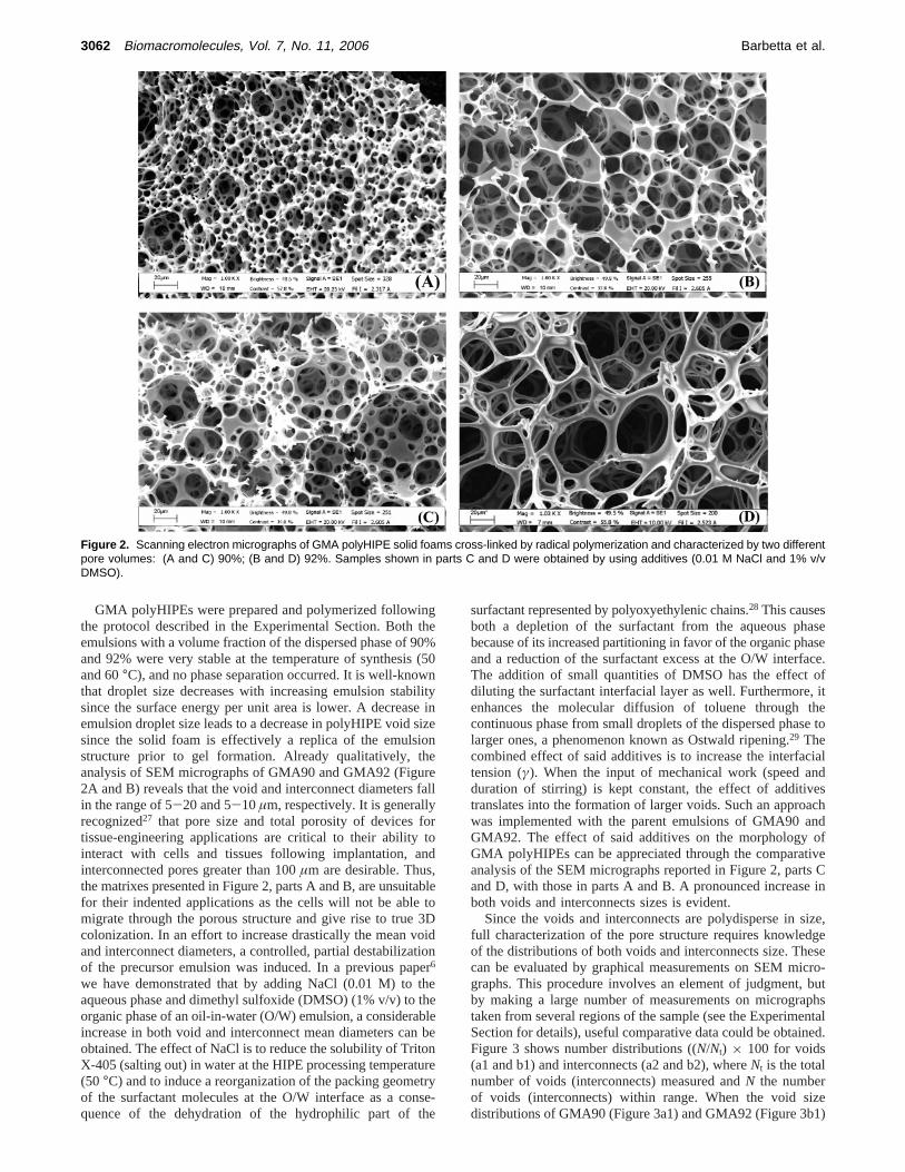
GMA polyHIPEs were prepared and polymerized followingthe protocol described in the Experimental Section. Both theemulsions with a volume fraction of the dispersed phase of 90%and 92% were very stable at the temperature of synthesis (50and 60°C), and no phase separation occurred. It is well-knownthat droplet size decreases with increasing emulsion stabilitysince the surface energy per unit area is lower. A decrease inemulsion droplet size leads to a decrease in polyHIPE void sizesince the solid foam is effectively a replica of the emulsionstructure prior to gel formation. Already qualitatively, theanalysis of SEM micrographs of GMA90 and GMA92 (Figure2A and B) reveals that the void and interconnect diameters fallin the range of 5-20 and 5-10µm, respectively. It is generallyrecognized27 that pore size and total porosity of devices fortissue-engineering applications are critical to their ability tointeract with cells and tissues following implantation, andinterconnected pores greater than 100µm are desirable. Thus,the matrixes presented in Figure 2, parts A and B, are unsuitablefor their indented applications as the cells will not be able tomigrate through the porous structure and give rise to true 3Dcolonization. In an effort to increase drastically the mean voidand interconnect diameters, a controlled, partial destabilizationof the precursor emulsion was induced. In a previous paper6
we have demonstrated that by adding NaCl (0.01 M) to theaqueous phase and dimethyl sulfoxide (DMSO) (1% v/v) to theorganic phase of an oil-in-water (O/W) emulsion, a considerableincrease in both void and interconnect mean diameters can beobtained. The effect of NaCl is to reduce the solubility of TritonX-405 (salting out) in water at the HIPE processing temperature(50 °C) and to induce a reorganization of the packing geometryof the surfactant molecules at the O/W interface as a conse-quence of the dehydration of the hydrophilic part of the
surfactant represented by polyoxyethylenic chains.28 This causesboth a depletion of the surfactant from the aqueous phasebecause of its increased partitioning in favor of the organic phaseand a reduction of the surfactant excess at the O/W interface.The addition of small quantities of DMSO has the effect ofdiluting the surfactant interfacial layer as well. Furthermore, itenhances the molecular diffusion of toluene through thecontinuous phase from small droplets of the dispersed phase tolarger ones, a phenomenon known as Ostwald ripening.29 Thecombined effect of said additives is to increase the interfacialtension (γ). When the input of mechanical work (speed andduration of stirring) is kept constant, the effect of additivestranslates into the formation of larger voids. Such an approachwas implemented with the parent emulsions of GMA90 andGMA92. The effect of said additives on the morphology ofGMA polyHIPEs can be appreciated through the comparativeanalysis of the SEM micrographs reported in Figure 2, parts Cand D, with those in parts A and B. A pronounced increase inboth voids and interconnects sizes is evident.
Since the voids and interconnects are polydisperse in size,full characterization of the pore structure requires knowledgeof the distributions of both voids and interconnects size. Thesecan be evaluated by graphical measurements on SEM micro-graphs. This procedure involves an element of judgment, butby making a large number of measurements on micrographstaken from several regions of the sample (see the ExperimentalSection for details), useful comparative data could be obtained.Figure 3 shows number distributions ((N/Nt) × 100 for voids(a1 and b1) and interconnects (a2 and b2), whereNt is the totalnumber of voids (interconnects) measured andN the numberof voids (interconnects) within range. When the void sizedistributions of GMA90 (Figure 3a1) and GMA92 (Figure 3b1)
Figure 2. Scanning electron micrographs of GMA polyHIPE solid foams cross-linked by radical polymerization and characterized by two differentpore volumes: (A and C) 90%; (B and D) 92%. Samples shown in parts C and D were obtained by using additives (0.01 M NaCl and 1% v/vDMSO).
3062 Biomacromolecules, Vol. 7, No. 11, 2006 Barbetta et al.

solid foams obtained without (blue line) and with additives (redline) are compared, it can be seen that in the latter case there isa marked shift toward the large diameter side and a broadeningof the distributions. Figure 3, parts a2 and b2, shows thecorresponding number distributions of interconnect size. As withthe void size, there is a general shift and broadening of thedistributions even though less pronounced.
Table 1 summarizes results obtained from voids and intercon-nects diameters measurement on GMA polyHIPEs in terms oftheir average values (⟨D⟩, ⟨d⟩ ) plus associated uncertainnessand the percentages of voids with diameters exceeding either50 or 100µm and interconnects diametersg 20µm. As alreadypointed out qualitatively, the solid foams obtained without theemployment of additives (GMA90 and GMA92) have no voidwith D g 50 µm and no interconnect withd g 20 µm. On thecontrary, in the case of GMA90add and GMA92add solidfoams, a high percentage of voids diameters exceeds 50µm,with GMA92adddisplaying a significant contribution of voidswith D g 100 µm andd g 20 µm. This is in agreement withthe presence in both GMA92add void and interconnect diam-eters distributions (Figure 3, parts b1 and b2, red lines) of along tail extending in the high-diameter range and with largevalues of⟨D⟩ and⟨d⟩ (Table 1). When the⟨D⟩ values of couplesof solid foams characterized by the same PV (90% or 92%)
but differing by the absence or presence of additives in theirHIPE formulations are compared, it can be seen that thepercentage increment in⟨D⟩ from GMA90 to GMA90add isabout 350% and 500% from GMA92 to GMA92add. The samekind of analysis carried for⟨d⟩ allows us to establish that apercentage increase of 80% and 280% was obtained for PV 90%and 92% solid foams, respectively.
The effect of destabilizing additives used in the present workon the HIPEs here examined is similar to that observed by otherauthors in the case of water-in-oil (W/ O) HIPEs.2,3
3.2. Gelatin Scaffolds by MTGase Cross-Linking.As a firstexperimental run, we tried to synthesize a foam characterizedby a PV of 85% and with a concentration of gelatin A3 of 20%w/v dissolved in PBS buffer using a MTGase/gelatin ratio of1:200 w/w. We discovered that the concentration and time ofaddition of MTGase to the emulsion is crucial. If the MTGasewas present already in the aqueous phase of the HIPE from thevery beginning of emulsion formation, cross-linking startedduring the addition of the organic phase. This prevented theformation of the HIPE. We were then forced to add MTGasedissolved in PBS at the end of emulsion formation in order tominimize the contact time with gelatin during the addition ofthe internal phase. Also the amount of MTGase added to theHIPE is important. If the MTGase/gelatin ratio exceeded 1:500,cross-linking started almost instantaneously, while still stirringthe emulsion in order to disperse MTGase homogeneouslywithin the entire volume of the HIPE. This led to a polyHIPEwith a highly distorted morphology. We found that the scaffoldspreserved the characteristic morphology of polyHIPEs when aMTGase/gelatin ratio of 1:1000 was used and stirring wasstopped after 5 min after addition of the enzyme.
In Figure 4, parts a and b, SEM micrographs show themorphology of the solid foams characterized by PV 85% and90% by following the preparation scheme outlined above. Theskeletal framework of the solid foam is characterized by thinnerwalls with respect to GMA-type solid foams and, as a result,by weaker mechanical properties (from a qualitative point ofview). We found also that the morphologies of the obtainedGMTGase scaffolds were influenced by the method of purifica-
Figure 3. Number distributions of voids (a1, b1) and interconnects (a2, b2) size of GMA polyHIPE solid foams obtained without (blue line) andwith (red line) additives (0.01 M NaCl and 1% v/v DMSO).
Table 1. Average Values of Voids and Interconnects of PolyHIPESolid Foams and Number Percentage of Voids and Interconnectswith Diameters Exceeding 50, 100, and 20 µm, respectively
sample ⟨D⟩ µm ⟨d⟩ µm% D
g 50 µm% D
g 100 µm% d
g 20 µm
GMA90 14 ( 4 7 ( 2 0 0 0GMA92 14 ( 4 8 ( 2 0 0 0GMA90add 61 ( 5 12 ( 3 83 ( 4 6 ( 3 6.5 ( 2GMA92add 84 ( 8 28 ( 6 85 ( 4 25 ( 5 78 ( 3
GMTGase85 19 ( 6 10 ( 3 0 0 0GMTGase90 34 ( 6 12 ( 3 2 ( 1 0 0GMTGase85add 26 ( 8 12 ( 6 2 ( 1 0 0GMTGase90add 60 ( 10 18 ( 7 83 ( 6 34 ( 7 34 ( 5
Preparation and Performance of Gelatin PolyHIPEs Biomacromolecules, Vol. 7, No. 11, 2006 3063

tion and drying. If freeze-drying was chosen as the dryingprocedure, the skeletal framework of the porous gel underwenta partial collapse. On the contrary, the drying procedure basedon critical point drying by supercritical CO2 has the advantageof leaving the porous texture quite intact. The average dimen-sions of voids and interconnects determined from analysis ofSEM micrographs of GMTGase85 and GMTGase90 solid foams(Figure 4, parts a and b) are reported in Table 1. These values,again, are not adequate for the viable colonization of thescaffolds from seeded cells. The strategy devised for increasingboth ⟨D⟩ and⟨d⟩ in the case of GMA polyHIPEs was adoptedalso in the case of enzymatically cross-linked gelatin polyHIPEs.The same concentrations of NaCl and DMSO were employedin the emulsion formulation, and the outcome of such anapproach can be observed in the micrographs reported in Figure4, parts c and d. Again, the effect of additives resolves in ashift and broadening of the diameter distributions (Figure 5).Table 1 data shows that only GMTGase90add possesses arelevant percentage of voids and interconnects with diametersg 100 and 20µm, respectively.
The percentage increment in⟨D⟩ and⟨d⟩ was 36% and 18%for GMTGase85add and 76% and 33% for GMTGase90addwith respect to GMTGase85 and GMTGase90, respectively.
We wondered what could be the origin of the fibrous-likeappearance of the GMTGase polyHIPEs.
Some hypothesis are listed below.3.2.1. Low Cross-Linking Density.From the amino acid
composition of gelatin A313 it turns out that the theoretical cross-linking degree is dictated by the percentage of lysine andhydroxy lysine residues which bear primary amine groups. Thecarboxylic acid content in gelatin A3 is much higher than aminegroups (80 vs 30 with respect to 1000 amino acids). Further-
more, the actual degree of cross-linking may be lower than thislimit. The two-phases nature of the emulsion may have aninfluence on the activity of MTGase. Unpredictable smallamounts of toluene are present in the aqueous phase due bothto partitioning and molecular diffusion (Ostwald ripening).30 Theamount of organic solvents dissolved in the aqueous phaseshould be higher when DMSO is used as an additive because itwill partition between the organic and aqueous phases and itwill enhance both partitioning and diffusion of toluene throughthe continuous phase. Exposure of MTGase to small amountsof organic solvent, surfactant, and NaCl (which causes anincrease in the ionic strength of the aqueous phase) may partiallyimpair its activity giving rise to lower degrees of cross-linking.
This hypothesis was checked through the determination ofthe percentage of the residual free amino groups in substratessuch as the “conventional gels” obtained by cross-linking withMTGase a gelatin solution at the same concentration of theaqueous phase of an HIPE (20% w/v), a polyHIPE obtainedwithout using any additives, and a polyHIPE synthesized byemploying additives. Such a comparison was based on aspectrophotometric method using trinitrobenzensulfonic acid21
which allows the determination of the content of free aminegroups.
If organic solvents and salt dissolved in the aqueous phasedepress MTGase activity, a higher percentage of free aminogroups should be detected in the polyHIPEs (without and/orwith additives) with respect to the conventional gel.
As can be seen (Table 2), the percentage of residual freeamine groups is higher in the polyHIPEs (independently fromthe presence of additives) than in the conventional gel. Thismeans that the actual degree of cross-linking is lower in the
Figure 4. Scanning electron micrographs of gelatin A3 polyHIPE solid foams cross-linked with MTGase characterized by two different porevolumes: (a and c) 85%; (b and d) 90%. Samples shown in parts c and d were obtained by using additives (0.01 M NaCl and 1% v/v DMSO).
3064 Biomacromolecules, Vol. 7, No. 11, 2006 Barbetta et al.

polyHIPE than in the conventional gel. Thus, the two liquidphases of the emulsion do exert an influence on MTGaseactivity.
3.2.2. Nature of the Bridging Units. A structural featureinfluencing the different morphologies recorded by employingthe two different modes of cross-linking resides on the differentkind of bridges established among gelatin chains. In radicalpolymerization of the methacrylic moieties, a poly(methacrylate)chain develops among many interlinked gelatin chains. Inenzymatic cross-linking an isopeptide junction joins two gelatinchains at a time, and as a consequence, the overall extent ofnetworking is less efficient. On the other hand, Giraudier etal.31 demonstrated that gelatin chains even when cross-linkedby MTGase are able to undergo to some extent triple-helixformation on cooling at room temperature and after a maturationtime, thus creating additional physical junction zones amonggelatin chains (chemical-physical gel). This should aid inconferring to gelatin gels improved mechanical properties.Nevertheless, in the present case the very low density ofchemical cross-linking and kind of chemical junctions are onthe overall at the basis of the relatively poor mechanicalproperties of GMTGase polyHIPEs as compared to those ofthe GMA type.
This explains why GMTGase polyHIPEs when exposed tohigh vacuum, during freeze-drying, undergo a partial collapseof the porous structure.
3.3. Cell Studies.When hepatocytes are removed from thearchitectural liver, formation of cell aggregates in the hydrogelscaffold is a requisite for enhancing liver-specific functions.Thus, in liver tissue engineering, the scaffolds sustaining theself-assembling of hepatocytes into aggregates must haveappropriate dimension of pores in order to host such aggregates.As a consequence, in the context of the present study, the mostpromising candidates of the two differently cross-linked typesof polyHIPEs that were tested for three-dimensional culturesof primary rat and HepG2 hepatocytes are GMA92add andGMTGase90add (Table 1).
We first tried to optimize the system by selecting theappropriate number of primary hepatocytes to be seeded in eachwell. As expected, the number of cells that could be entrappedin the matrixes increased with the seeding density, reaching amaximum value that depended on the affinity of the materialfor that kind of cell. A maximum of 3.9× 10⁵ cells entrappedper well was found in GMTGase scaffolds, while the numberof cells attached was appreciably lower in GMA92addscaffold(3.2 × 10⁵). A density of 3.0× 10⁵ per well (24-well plate)was then chosen for all the experiments with primary hepato-cytes. By calculating the cell-loading efficiency, we found thatenzymatic reticulation was helpful for initial cell attachment(Figure 6). Initial cell density was found to be less critical forHepG2 cells (data not shown); however, a seeding density of1.5 × 105 per well was considered appropriate for theseproliferating cells.
From a morphological point of view, the scaffold moresuitable for hepatocyte colonization should be GMA92addsince,as is evident from data reported in Table 1, it is the onecharacterized by larger voids and interconnects in comparisonto GMTGase90add. Nevertheless, cell-loading efficiency data,Figure 6, are higher for the second type of scaffold, indicatingthat not only are the differences in porosity between the twotypes of scaffolds not relevant but that hepatocytes seem todisplay a better affinity for the GMTGase90add matrix.
The viability of the primary hepatocytes in the scaffolds and,indirectly, the cytotoxicity of polymers were analyzed usingMTS, a compound that can only be metabolized by healthy cells.
Figure 5. Number distributions of voids (a1, b1) and interconnects (a2, b2) size of gelatin A3 cross-linked with MTGase polyHIPE solid foamsobtained without (blue line) and with (red line) additives (0.01 M NaCl and 1% v/v DMSO).
Table 2. Number of Amino Acids Residues Bearing Side ChainAmine Groups in Gelatin A3 and Percentages of Residual FreeAmine Groups in Conventional Gel and in PolyHIPEsCross-Linked with MTGase
residual % of amine groupsa
no. ofamino acidsbearing NH2
functionalitiesin gelatin A3b
conventionalgel PV % GMTGase GMTGase(add)
30 46 85 79 8190 80 78
a Determined by the method described in ref 21. b Number of free aminegroups per 1000 amino acids.
Preparation and Performance of Gelatin PolyHIPEs Biomacromolecules, Vol. 7, No. 11, 2006 3065

Cell viability was satisfactory during the first 72 h of culture,without significant differences, on both GMA92add andGMTGase90add scaffolds. At 96 h, the absorbance, and thusthe number of viable cells, was significantly higher for theGMTGase90add scaffold (Figure 7). These results were com-parable to that observed in two-dimensional cultures until 48h. However, at longer time the number of viable hepatocytesdecreased of about 30% under conventional culture conditions,suggesting that our matrixes are more suitable for preservingcell integrity and viability for long-term maintenance.
The ultrastructural analysis, performed by SEM, showed thatboth primary hepatocytes (Figure 8, parts a and c) and HepG2cells (Figure 10, parts a and d) adhered on both the polymerstested, with a homogeneous colonization of the scaffolds. Thiswas particularly consistent with scaffolds obtained with radicalpolymerization (GMA92add), which are characterized by agreater stability and a better defined morphology. Nevertheless,on the GMTGase90add scaffold, cell aggregates were largerand better organized in the three-dimensional space than thoseon the GMA92addscaffold. In addition, both GMTGase90add-seeded hepatocytes and HepG2 cells displayed an improvedmorphology and a better quality of their cell surfaces, withabundant and regular microvilli. They also appear more closelyconnected, suggesting the presence of better organized celljunctions (Figure 8, parts b and d; Figure 10, parts a and d).On the other hand, the GMTGase90addscaffold resulted in morefragility and less resistance in longer-term cultures.
The expression of a differentiated phenotype is strictlydependent on cell-cell adhesion, which is highly critical forall epithelial cells, including hepatocytes. An impaired expres-sion of cell-junctional molecules, and of E-cadherin andâ-catenin in particular, is often correlated with cell dedifferen-tiation and/or transformation.32
A certain degree of dedifferentiation is always observed inprimary hepatocytes under standard conventional monolayercultures, and it has sometimes been associated with the lack ofappropriate contacts between neighboring cells.33 The reestab-lishment of optimal cell-cell interactions is thus indicative ofa functional cell recovery. By means of immunofluorescenceexperiments and confocal laser microscopy, we verified the
Figure 6. Cell-loading efficiency of primary rat hepatocytes withinGMA92add (2) and GMTGase90add (b) scaffolds. Data are ex-pressed as the mean value ( standard deviation of at least threedifferent experiments done in triplicate. Quantitative parameters werecompared using the Student’s t-test. / Significant with respect toGMA92add, P < 0.05.
Figure 7. Cell viability assay (MTS) on primary hepatocytes culturedon GMA92add (grey bars) and GMTGase90add (white bars) scaf-folds. Data are expressed as the mean 490 nm absorbance (standard deviation of at least three different experiments done inquadruplicate. Quantitative parameters were compared using theStudent’s t-test. / Significant with respect to GMA92add, P < 0.05.
Figure 8. Low (a, c) and high (b, d) magnification scanning electronmicrographs of primary rat hepatocytes cultured for 72 h on GMA92add(a, b) or GMTGase90add scaffolds (c, d).
Figure 9. Confocal laser microscopy of the junctional proteinsE-cadherin (a, c, e) and â-catenin (b, d, f) in primary rat hepatocytescultured for 72 h on GMA92add (a, b) or GMTGase90add scaffolds(c, d) or in two-dimensional cultures (e, f).
3066 Biomacromolecules, Vol. 7, No. 11, 2006 Barbetta et al.

correct expression of these two key adhesion proteins in normaland proliferating hepatocytes, cultured on both the GMA92addand GMTGase90add three-dimensional scaffolds. In the caseof primary hepatocytes, while the GMTGase90add scaffoldshowed a good expression of both E-cadherin andâ-catenin(Figure 9, parts c and d), onlyâ-catenin was appreciablyexpressed in GMA92add matrix (Figure 9b). In addition, inGMA92addscaffold (Figure 9, parts a and b) the positivity waslower and not confined to the cell membrane but also scatteredin the cytoplasm, suggesting a less adequate localization. Instandard two-dimensional conditions, both proteins appearedreduced in expression and delocalized from plasma membrane(Figure 9, parts e and f), strongly highlighting the betterinfluence of enzymatic reticulation on the localization andexpression of these adhesion molecules.
The images concerning immunolocalization experiments withHepG2 cells also agree with the primary hepatocyte results(Figure 10b,c,e,f). Also in this case, both scaffolds supportedthe expression of E-cadherin (Figure 10, parts b and e) andâ-catenin (Figure 10, parts c and f); however, in cells seededin GMTGase90add scaffold the labeling was stronger andpresent mostly at the level of the cell membranes, indicating amore correct localization (Figure 10, parts e and f). Thesefindings, when compared with data reported in the literatureconcerning E-cadherin andâ-catenin visualization in conven-tional bidimensional culture,25,34still indicate the positive effectof the three-dimensional architecture on the expression of adifferentiated phenotype. In addition, in these proliferating cells(HepG2), the fluorescence labeling also put into evidence thelarger size of the cell aggregates present on the enzymatic cross-linked biomaterial. These results were evident after 72 h ofculture but were also detectable at shorter intervals (data notshown).
Taken together, our immunofluorescence studies show thatin the GMTGase90addscaffold, E-cadherin andâ-catenin bothlocalize on the plasma membranes of normal or growinghepatocytes, and the scattered positivity distributed on wholecells completely disappears, indicating total insertion of theadherens complexes at the level of the cell contacts. This is inaccordance with the superior organization of the cell aggregatesobserved by SEM analysis, and it seems to point to an enhancedbiocompatibility of these materials.
4. Conclusion
The work presented in this paper compares the influence oftwo different cross-linking procedures on the morphology ofemulsion-templated gelatin solid foams. Radical polymerizationallows the production of foams characterized by larger voidsand interconnects and with a better wall texture than thosecharacterizing foams cross-linked with MTGase. The latter typeof solid foams on the contrary seems to provide a more friendlyenvironment for hepatocytes, presumably thanks to the absenceof any foreign chemical functionalities, thus allowing us toexploit fully the biocompatible nature of gelatin. In this matrix,hepatocytes show not only good levels of vitality but also largercell aggregates and were able to establish satisfactory cell-cell contacts, as indicated by the high and correct expressionof specific molecules, i.e., the junctional proteins E-cadherinandâ-catenin, known to be extremely important for maintenanceof the differentiated phenotype and, in turn, for hepatocytefunctionality.32
Efforts are currently being directed to envisage differentfoaming procedures and zero-bridges cross-linking methods ableto give rise to more robust foams and possessing larger voidsand interconnects. Such a cross-linking method under investiga-tion is based on the use of the systemN,N-(3-(dimethylamino)-propyl-N-ethyl carbodiimide (EDC) andN-hydroxysulfosuc-cinimide (NHSS).
Acknowledgment. The authors thank Mr. Robert Cant ofChance and Hunt Food Ingredients, U.K. for the kind supplyof microbial transglutaminase and the University of Rome(Ateneo Funds) and Consorzio Interuniversitario Biotecnologie(CIB) for funding this research. We also thank Dr. D. Ferro ofthe Department of Chemistry of the University of Rome andThe Microscopy Core Facilities of University of L’Aquila forthe invaluable help with the confocal and scanning electronmicroscopy.
References and Notes
(1) Akay, G.; Birch, M. A.; Bokhari, M. A.Biomaterials2004, 25, 3991-4000.
(2) Hayman, M. W.; Smith, K. H.; Cameron, N. R.; Przyborski, S. A.Biochem. Biophys. Res. Commun.2004, 314, 483-488.
Figure 10. Scanning (a, d) and confocal laser microscopy (b, c, e, f) of HepG2 cells cultured for 72 h on GMA92add (a, b, c) or GMTGase90addscaffolds (d, e, f). Immunofluorescence of the junctional protein E-cadherin (b, e) and â-catenin (c, f).
Preparation and Performance of Gelatin PolyHIPEs Biomacromolecules, Vol. 7, No. 11, 2006 3067

(3) Hayman, M. W.; Smith, K. H.; Cameron, N. R.; Przyborski, S. A.J.Biochem. Biophys. Methods2005, 62, 231-240.
(4) Busby, W.; Cameron, N. R.; Jahoda, J. A. B.Biomacromolecules2001, 2, 154-164.
(5) Busby, W.; Cameron, N. R.; Jahoda, J. A. B.Polym. Int.2002, 51,871-881.
(6) Barbetta, A.; Dentini, M.; Zannoni, E. M.; De Stefano, M. E.Langmuir2005, 21, 12333-12341.
(7) Barbetta, A.; Dentini, M.; De Vecchis, M. S.; Filippini, P.; Formisano,G.; Caiazza, S.AdV. Funct. Mater.2005, 15, 118-124.
(8) (a) Ferreira, L.; Gil, M. H.; Cabrita, A. M. S.; Dordick, J. S.Biomaterials2005, 26, 4707-4716. (b) Huang, X.; Nayak, B. R.;Lowe, T. L.J. Polym. Sci., Part A: Polym. Chem.2004, 42, 5054-5066. (c) Na, K.; Shin, D.; Yun, K.; Park, K.-H.; Lee, K. C.Biotechnol. Lett.2003, 25, 381-385. (d) Kim, S.; Chae, S. Y.; Na,K.; Kim, S. W.; Bae, Y. H.Biomaterials2003, 24, 4843-51. (e)Na, K.; Shin, D.; Yun, K.; Park, K.-H.; Lee, K. C.Biotechnol. Lett.2003, 25, 381-5. (f) Kim, H.-W.; Song, J.-H.; Kim, H.-E.AdV. Funct.Mater. 2005, 15, 1988-1994. (g) Chang, C.-H.; Kuo, T.-F.; Lin,C.-C.; Chou, C.-H.; Chen, K.-H.; Lin, F.-H.; Liu, H.-C.Biomaterials2006, 27, 1876-1888. (h) Young, S.; Wong, M.; Tabata, Y.; Mikos,A. G. J. Controlled Release2005, 109, 256-274. (i) Huang, Y.;Onyeri, S.; Mbonda, S.; Moshfeghian, A.; Madihally, S. V.Bioma-terials 2005, 26, 7616-7627. (j) Yang, S.-H.; Chen, P.-Q.; Chen,Y.-F.; Lin, F.-H. J. Biomed. Mater. Res.2005, 74B, 488-494.
(9) (a) Schorsch, C.; Carrie, H.; Clark, A. H.; Norton, I. T.Int. Dairy J.2000, 10, 519-528. (b) Motoki, M.; Seguro, K.Trends Food Sci.Technol.1998, 9, 204-210.
(10) Fuchsbauer, H. L.; Gerber, U.; Engelmann, J.; Seeger, T.; Sinks, C.;Hecht, T.Biomaterials1996, 17, 1481-1488.
(11) (a) Balakrishnan, B.; Jayakrishnan, A.Biomaterials2005, 26, 3941-3951. (b) Crescenzi, V.; Francescangeli, A.; Taglienti, A.Biomac-romolecules2002, 3, 1384-1391. (c) Chen, T.; Small, D. A.;McDermott, M. K.; Bentley, W. E.; Payne, G. F.Biomacromolecules2003, 4, 1558-1563.
(12) (a) Embree, H. D.; Brown, E. M.; Taylor, M. M.; Payne, G. F.Biomaterials2003, 24, 2831-2841. (b) Mi, F.-L.Biomacromolecules2005, 6, 975-987.
(13) San, Y.; Giraudier, O.; Garde, V. L.Biopolymers2005, 77, 257-263.
(14) Ray-Neng, C.; Hsiu-O, H.; Ming-Thau, S.Biomaterials2005, 26,4229-4235.
(15) (a) Kelly, J. H.; Darlington, G. J.In Vitro Cell. DeV. Biol. 1989, 25,217-222. (b) Hongo, T.; Kajikawa, M.; Ishida, S.; Ozawa, S.; Ohno,Y.; Sawada, J.; Umezawa, A.; Ishikawa, Y.; Kobayashi, T.; Honda,H. J. Biosci. Bioeng.2005, 99, 237-44.
(16) (a) McLaughlin, B. E.; Tosone, C. M.; Custer, L. M.; Mullon, C.Ann. N.Y. Acad. Sci.1999, 875, 310-325. (b) Katherine, M. K.;Vacanti, J. P.Transplant Immunol.2004, 12, 303-10. (c) Hongo,T.; Kajikawa, M.; Ishida, S.; Ozawa, S.; Ohno, Y.; Sawada, J.;Umezawa, A.; Ishikawa, Y.; Kobayashi, T.; Honda, H.J. Biosci.Bioeng.2005, 99, 237-44. (d) Vanhaecke, T.; Rogiers, V.MethodsMol. Biol. 2006, 320, 209-27.
(17) (a) Dunn, J. C. Y.; Tomkins, R. G.; Yarmush, M. L.Biotechnol.Prog. 1991, 7, 237-242. (b) Wang, Y. J.; Liu, H. L.; Guo, H. T.;Wen, H. W.; Liu, J.World J. Gastroenterol.2004, 10, 699-702.
(18) Gerlach, J.; Schnoy, N.; Smith, M. D.; Neuhaus, P.Artif. Organs1994, 18, 1-5.
(19) Catalano, G.; De Bartolo, L.; Vico, V.; Ambrosio, L.Biomaterials2001, 22, 659-65.
(20) (a) Kaufmann, P. M.; Heimrath, S.; Kim, B. S.; Mooney, D. J.CellTransplant.1997, 6, 463-468. (b) Glicklis, R.; Shapiro, L.; Agbaria,R.; Merchuk, S. C.Biotechnol. Bioeng.2000, 67, 344-353. (c) Li,J.; Pan, J.; Zhang, L.; Yu, Y.Biomaterials2003, 24, 2317-22.
(21) Kuijpers, A. J.; Engbers, G. H. M.; Feijen, J.; De Smedt, S. C.;Meyvis, T. K. L.; Demeester, J.; Krijgsveld, J.; Zaat, S. A. J.; Dankert,J. Macromolecules1999, 32, 3325-3333.
(22) Moldeus, P.; Holdberg, J.; Orrenius, S.Methods Enzymol.1978, 52C,60-71.
(23) Park T. G.J. Biomed. Mater. Res.2002, 59, 127-35.(24) Riss, T. L.; Moravec, R. A.Mol. Biol. Cell.1992, (Suppl. 3), 184a.(25) Ara, C.; Conti Devirgiliis, L.; Massimi, M.Cell Commun. Adhes.
2004, 11, 13-23.(26) (a) Lissant, K. J.J. Colloid Interface Sci.1966, 22, 462-468. (b)
Lissant, K. J.J. Colloid Interface Sci.1973, 42, 201-208. (c) Lissant,K. J.; Peace, B. W.; Wu, S. H.; Mayhan, K. G.J. Colloid InterfaceSci.1974, 47, 416-423. (d) Cameron, N. R.; Sherrington, D. C.AdV.Polym Sci.1996, 126, 163-214. (e) Cameron, N. R.Polymer2005,46, 1439-1449.
(27) (a) Agrawal, C. M.; Ray, R. B.J. Biomed. Mater. Res.2001, 55,141-150. (b) Hollinger, J. O.; Leong, K.Biomaterials1996, 17,187-194. (c) Hu, Y. H.; Grainger, D. W.; Hollinger, J. O.J. Biomed.Mater. Res.2002, 59, 563-572.
(28) (a) Schott, H.J. Colloid Interface Sci.1998, 205, 496-502. (b) Goel,S. K. J. Colloid Interface Sci.1999, 212, 604-606.
(29) Kabalnov, A. S.; Pertsov, A. V.; Shchukin, E. D.J. Colloid InterfaceSci.1987, 118, 590-597.
(30) Barbetta, A.; Cameron, N. R.Macromolecules2004, 37, 3188-3201,3202-3213.
(31) Giraudier, S.; Hellio, D.; Djabourov, M.; Larreta-Garde, V.Biomac-romolecules2004, 5, 1662-1666.
(32) (a) Kirkpatrick, C.; Peifer, M.Curr. Opin. Genet. DeV. 1995, 5, 56-65. (b) Ihara, A.; Koizumi, H.; Hashizume, R.; Uchikoshi, T.Hepatology1996, 23, 1441-1447. (c) Knudsen, K. A.; Frankowski,C.; Johnson, K. R.; Wheelock, M. J.J. Cell Biochem.1998, (Suppl.30/31), 168-176. (d) Gumbiner, B. M.J. Cell Biol.2000, 148, 399-403. (e) Wei, Y.; Fabre, M.; Branchereau, S.; Gauthier, F.; Perilongo,G.; Buendia, M. A.Oncogene2000, 19, 498-504.
(33) Yuasa, C.; Tomita, Y.; Sono, M.; Ishimura, K.; Ichihara, A.J. Cell.Physiol.1993, 156, 522-530.
(34) Lin, C. Y.; Lin, C. J.; Chen, K. H.; Wu, J. C.; Huang, S. H.; Wang,S. M. FEBS Lett.2006, 580, 3042-350.
BM060533L
3068 Biomacromolecules, Vol. 7, No. 11, 2006 Barbetta et al.


![Materials Science and Engineering C - ULisboa · In addition to the porous structure of scaffolds, great attention must be paid to their composition [5]. Gelatin is a common substrate](https://img.pdfslide.us/doc/110x75/5cbfa7bb88c993cd2c8bb6d3/materials-science-and-engineering-c-ulisboa-in-addition-to-the-porous-structure.jpg)