Embed Size (px)
Citation preview
P1: SFK/UKS P2: SFK
BLBS093-c02 Collier October 19, 2011 4:2 Trim: 244mm×172mm
Chapter 2
Physiological Basics of TemperatureRegulation in Domestic AnimalsDonald E. Spiers
Introduction
Domestic animals inhabit many different thermal environments around the world, and in everysituation they must utilize physiological mechanisms to balance heat flow into and out of thebody. In the extreme cold of polar regions, the animal’s primary challenge is to limit or reducethe outflow of metabolic heat from the body and increase the inflow of heat from the ambient.Animals that live in desert regions must reverse these processes by augmenting the environmentof metabolic heat and minimizing the influx of thermal energy from an environment with a heatcontent level that possibly surpasses that in the animal’s body. The classic example of an animalthat is able to rapidly shift between these thermal extremes is the camel (Schmidt-Nielsen et al.,1956), which utilizes simultaneous shifts in thermal and water balance to raise and lower bodyheat. The difficulty in understanding these processes begins with a basic misunderstanding ofthe key elements in the thermal environment – heat and temperature.
The Definitions of Heat and Temperature
Any material with molecular movement possesses kinetic energy in the form of heat. Everyobject in our typical environment has a temperature above absolute zero and therefore containssome level of heat. A more appropriate appreciation of the value of heat in the universe and,more importantly to living animals, is derived from a consideration of the First and SecondLaws of Thermodynamics. In summary, the First Law states that we have all the energy in theuniverse today that has ever existed. The total amount has neither increased nor decreased overtime. Therefore, energy is never created but only transferred from one form to another. Heatrepresents the bottom level of energy transformation, which leads to the Second Law. Simply
Environmental Physiology of Livestock, First Edition. Edited by R. J. Collier and J. L Collier.C© 2012 John Wiley & Sons, Inc. Published 2012 by John Wiley & Sons, Inc.
17
P1: SFK/UKS P2: SFK
BLBS093-c02 Collier October 19, 2011 4:2 Trim: 244mm×172mm
18 Environmental Physiology of Livestock
put – this law states that all order goes toward disorder. We have less organized energy in theuniverse today than we had yesterday, with the result being the ultimate release of energy inits lowest form (i.e., heat). There are reversals of this breakdown in organization, in whichenergy can be assimilated into a more organized form (e.g., growth, development). However,these periods are only temporary. Ultimately, animals age and die, with breakdown of thisshort-term organization and release of energy back to the universe.
The Second Law of Thermodynamics becomes important in defining heat flow. The state-ment that all order moves toward disorder can be rephrased as heat always flows downhillfrom hot to cold objects. This process does not occur in reverse, without adding energy tothe system. For example, opening the door to your home in the winter results in heat flowingpassively downhill along a temperature gradient from warm to cold environments. Newtonproposed in his Law of Cooling that the rate of change in the temperature of an object isdirectly proportional to the temperature difference between the object and its surroundings, orthe change in object temperature over time. Kleiber (1975) noted that Newton was unawareof heat flow and incorrectly considered temperature to be the object that flowed. He alsosuggested that it is inappropriate to use this law to explain heat loss in homeotherms sincethey are not inanimate objects that cool, but instead are constantly generating heat to maintainhomeothermy. A more appropriate expression of heat transfer in living birds and mammals isFourier’s Law of Heat Flow. This Law, which accounts for the influence of thermal gradients,states that the rate of heat flow by conduction within a system is equal to: 1) the thermalconductivity of the conducting material; 2) the area across which the heat flows and insulatorthickness; and 3) the thermal gradient between the exchange regions. Each of these phys-ical parameters affect an animal’s ability to regulate its core body temperature in differentthermal environments.
There are several unique facts about temperature that make it essential for thermoregulation.First, it determines the direction of heat flow. Both Newton’s and Fourier’s Laws have thetemperature gradient as a central physical component in the determination of cooling andheat flow, respectively. Birds utilize a large thermal gradient to regulate body temperature. Ingeneral, they have a higher internal body temperature (i.e., >40◦C) than mammals (i.e., <39◦C)under thermoneutral conditions. This elevated temperature provides them with an advantage inthe heat, in that the thermal gradient between animal and environment is increased, facilitatingheat loss from the bird and maintenance of homeothermy. Second, temperature is an indicatorof the heat intensity within a system. Animals have specific neurosensors located throughoutthe body, both peripherally and centrally, which detect temperature and not heat. Therefore,an animal’s judgment that an object or environment is “hot” or “cold” is based on temperatureand not on heat content. Likewise, there are numerous physiological and behavioral activitiesthat depend on detection of temperature and not heat. Onsets of shivering activity in the coldand panting activity in the heat are directly related to temperature of the environment and, inturn, the skin site where the thermoreceptors are located.
Thermal conductivity, which is another physical component of Fourier’s Law, also altersthe perception of temperature (i.e., hot versus cold) and potentially many of the physiologicalresponses to the thermal environment. Most people agree that humid cold air feels colderthan dry air. The question is why? Initial consideration of this question concerning thermalcomfort produces the obvious answer that an increase in the water content of air increases itsthermal conductivity and results in greater heat loss from the animal. In fact, this is incorrect.A phase shift in water from solid to liquid to gaseous states produces a large change in thermalconductivity. Water vapor has a lower thermal conductivity than dry air. Therefore, addingwater vapor to air, by using misters and foggers, slightly insulates the animal, in addition to
P1: SFK/UKS P2: SFK
BLBS093-c02 Collier October 19, 2011 4:2 Trim: 244mm×172mm
Physiological Basics of Temperature Regulation in Domestic Animals 19
reducing the vapor pressure gradient for evaporative heat loss to the environment. In reality,this alteration in thermal conductivity is very small given that there can only be about 2% waterin the air under these extremely humid conditions created by these devices. Adams (1992)answered this question by making a distinction between thermal perception and actual thermalstrain on the animal. He noted that we could feel colder, in the absence of altered thermal strain,as a result of an increase in the water content of the tissue surrounding the thermoreceptors(i.e., deep below skin surface). This region (i.e., stratum corneum) readily absorbs water underhumid conditions, which increases its thermal conductivity as a result of increased liquidcontent. With this increase, there is a drop in the temperature of the region containing thethermoreceptors toward the level of the skin surface. The receptors will signal that it is colder,even though the level of thermal stress is essentially unchanged. Exposure to a dry, coldenvironment will reduce the hydration and thermal conductivity of the stratum corneum, withless reduction in the temperature of the receptors in this region. As a result, the individual feelswarmer. The combination of skin hydration and thermal conductivity levels is very importantin determining how we perceive our environment and its temperature.
The effect of skin hydration on cold perception works in a similar manner on heat perception,albeit in the opposite direction. Under extremely hot conditions, skin surface temperature mayexceed skin thermoreceptor temperature. As in the cold, an increase in skin hydration willincrease thermal conductivity of this region and cause the temperatures of these regions toapproach each other. In this case, thermoreceptor temperature will rise to give the perceptionof an increase in heat stress. Again, actual heat stress remains essentially unchanged. However,the perception of heat stress is real. As a result, this environment is often termed “steamy” or“muggy,” and there is usually a reduction in performance.
Although the terms temperature and heat are often used to explain behavioral and physio-logical events related to thermoregulation and, as shown, are in many ways “linked” to eachother, they are distinctly different concepts that should never be used interchangeably. A sim-ilar problem arises in the use of the terms stress and strain as they refer to the impact of theenvironment on biological systems.
Stress of the Environment versus Strain on the System
The term “stress” is a part of every discussion of the immediate effects of the environmenton an animal, and adaptation to these effectors. In fact, it is often used interchangeably with“strain” in describing animal-environment interactions. A short definition of stress or stressoris any condition or agent that alters the resting state of a system (physiological or biological;Fregly, 1996) and ultimately results in an adaptive response (Curtis, 1983). The displacementfrom base level of the system due to stressor impact is defined as the “strain” on the system(Fregly, 1996). This system shift can be due to internal or external stressors, and must be ameasurable property, such as intensity, duration, frequency, or variability (Fregly, 1996).
One reason for confusion in using the terms “stress” and “strain” is that many agentscan be classified as either, depending on the system or pathway in question. Any integrativephysiological analysis faces this dilemma. For example, a change in the external thermalenvironment (i.e., stressor) may cause shifts in skin or core body temperatures (i.e., strains).However, body temperature (now the stressor) will elicit a series of physiological responsesor strains, such as redirection of blood flow, altered respiratory rate, or shift in metabolic heatproduction. This term confusion can be eliminated by simply defining the analyzed system interms of its components.
P1: SFK/UKS P2: SFK
BLBS093-c02 Collier October 19, 2011 4:2 Trim: 244mm×172mm
20 Environmental Physiology of Livestock
Potter (1971) used stressor terminology to define the ideal environment and adaptation.His proposal was that extremely low or high levels of stress result in the lowest levels ofperformance, and that there is a requirement for a certain level of stress in our daily lives toobtain the optimum level of function and survival. Potter summarized that our culture shouldprovide “systematic challenges by physical and mental tasks” at specific periods of life toinduce expression of our full genetic potential and adaptation. It is likely that similar levels ofchallenge at appropriate times would improve performance of domestic animals as well.
The British physicist Thomas Young is credited with the discovery in 1807 of an inherentproperty of any material that predicts the strain in the material that results from a knownstressor (i.e., Young’s Modulus of Elasticity; stress/strain). Fregly (1996) suggested that thisconcept might be utilized to identify an animal’s ability to adapt to a particular stressor andcharacterize different components of the acclimation process (e.g., duration, frequency, andmagnitude of response). Environmental stressors can impact an object in one of two ways. Theobject can be deformed or moved from its location. Isotonic contraction of skeletal muscle isa good example of this, where tension remains unchanged but there is a shortening of musclelength and movement (Sherwood et al., 2005). In contrast, an object could respond with anincrease in tension and no deviation in location. This would be characteristic of isometriccontraction where there is not shortening of the skeletal muscle but a rise in strain (Sherwoodet al., 2005). Fregly (1996) expands the Modulus concept beyond inanimate objects to includeanimals by explaining that its reciprocal (i.e., strain/stress) represents the adaptive complianceof an animal in response to a stressor and might be referred to as an “index of acclimation.”He notes that such an index could be used to 1) measure the degree of acclimation following aspecific period of exposure, 2) determine the completion time for acclimation, and 3) comparelevels of acclimation across species. Quantification of the stressor is relatively easy and wouldcenter on measurements of thermal input. A greater challenge is the quantification of thestrain to the animal’s cells, organs, or systems. An additional, and often neglected, factor is thetemporal component that must be incorporated into any strain/stress index of both acclimationand de-acclimation for a given situation.
Homeostasis and Maintenance of a Constant State
All living organisms are in a constant state of dynamic change. Initially, this idea might appearto be a contradiction, because the terms “constant state” and “change” are used. In fact, this isnot the case. Constant state refers to the organization that occurs within the body systems, andchange identifies the movement that must occur around this organization in order to maintain asteady-state environment. For example, core body temperature of many adult birds and mam-mals is maintained at a relatively constant level through the continuous flow of heat into andout of the body. The fact that systems within living animals continuously function to maintaina constant internal environment or minimize changes due to the external environment (i.e., lafixite du milieu interieur) was initially presented by the French physiologist Claude Bernard(1878). He stated that this constant state is a requirement for any living organism to maintainan existence that is independent of its environment. The next significant step in the evolutionof the concept of self-sustaining organization occurred when Harvard physiologist Walter B.Cannon (1932) proposed the term homeostasis. He realized that living organisms utilize com-plex coordinated physiological reactions to reduce systemic disturbances in different externalenvironments. Homeostasis united all physico-chemical interactions within a living animalunder the goal of maintaining a constant internal environment. Physiologists agree that this
P1: SFK/UKS P2: SFK
BLBS093-c02 Collier October 19, 2011 4:2 Trim: 244mm×172mm
Physiological Basics of Temperature Regulation in Domestic Animals 21
is one of the most important concepts in physiology. A common misconception, however, isthat Cannon intended to use this term to define the maintenance of constant states within thebody, along the path of Bernard’s original proposal. In fact, his aim was not to define a staticconstancy, but one that is dynamic and moving with variation around a maintained value. Morerecently there has been a tendency to forget that this fluidity was a component of the originalconcept of constant state.
Although there are a wide variety of models associated with life functions and homeostasis,most have several basic characteristics in common. Each has a central pool that is maintainedat a theoretical constant level under normal conditions by balanced input and output of energy.In the thermoregulatory system, the pool is usually body heat content that is determinedby the inflow and outflow of heat from the body. Using the concept of balance, the inflowcan occur from the environment with exposure to air temperature above skin temperature orelectromagnetic radiation that actually penetrates the body surface. Heat can also be contributedto the central pool as a result of generation within the body (e.g., shivering thermogenesis).Exchanges within the body are additional determinants of the pool size. These include theincorporation of material into other compounds, as well as storage of the pool materialthroughout the body. In the case of the thermoregulatory system, there are deposits of largequantities of heat in different regions, such as brown adipose tissue found in many neonates orskeletal muscle during shivering or exercise. On the outflow side, products in the pool can bebroken down or metabolized and released from the body. Returning to the thermoregulatorysystem, we know that heat is the lowest energy state and so cannot be converted into other forms.However, it is certainly released from the body and under normal conditions balances the inflow.
Any system in the body can be evaluated and classified by placement in one of three states:1) output greater than input (negative balance); 2) input greater than output (positive balance);and 3) input equals output (stable state or homeostasis). These states can be easily visualizedfor the thermoregulatory system if one thinks of the system as a balance, weighing heatloss against heat gain. Using this scenario, a negative balance would produce hypothermia,a positive balance would result in hyperthermia, and the balance of input and output wouldresult in homeothermy. “Homeothermy” is defined as the balance of heat production and heatloss (IUPS Thermal Commission, 2001) to maintain body-heat content relatively constantin different thermal environments. The term was used, not many years after “homeostasis”was introduced by Cannon to describe the development of thermoregulation in young rats(Brody, 1943).
The development of models around the concepts of homeostasis and maintenance of aconstant state was originally used as a simplified approach to understand complex physiologicalprocesses, such as thermoregulation and homeothermy. They were simple in that they consistedof a selected variable, such as body heat content, that served as a black box with input andoutput activities. The benefit of this approach is its simplicity and ease of visualization. Itremains to be determined if it is sufficient for evaluation of stress in production environments.Another concern is that this simplistic approach does not allow for prediction of an output,such as thermal status. In addition, it does not provide a mechanism that allows for integrationof multiple systems, which more likely approximates the true thermoregulatory system.
Control of the Thermoregulatory System
Animal models are simple constructs that do not duplicate, but approximate biological sys-tems (Horton and Bicak, 1987). These artificial creations allow researchers to study normal
P1: SFK/UKS P2: SFK
BLBS093-c02 Collier October 19, 2011 4:2 Trim: 244mm×172mm
22 Environmental Physiology of Livestock
and pathological events across different animal species and also to conceptualize complexprocesses. More importantly, these creations provide the ability to test specific componentsof a system in order to better understand physiological activities and their interactions underdifferent conditions. This is especially true for the thermoregulatory system, with its numeroustheories to explain and predict the control of body temperature. Such models run the gamutfrom the sub-cellular level to the interaction of the animal with its environment. In addition,there are old and new concepts in the literature that have been used as general models tounderstand the effects of stress on physiological systems. Initially, the idea of homeostasis wasused to describe how different physiological activities maintain important variables (e.g., coretemperature, osmotic concentration) within a constant range, even during exposure to envi-ronmental stressors. Control theory was integrated into this concept as a means to understandhow a stable state could be created and maintained through the use of feedback mechanisms.This concept has evolved over the decades to incorporate change into our view of physiolog-ical regulation and now includes the theories of rheostasis (Mrosovsky, 1990), homeorhesis(Waddington, 1940), and allostasis (Eyer, 1975; Eyer and Sterling, 1977).
Despite the wide variety of models associated with life functions and homeostasis, most haveseveral basic characteristics in common. Each has a theoretical central pool that is maintainedat a constant level under normal conditions by balanced input and output of energy. In thecase of the thermoregulatory system, the pool is usually body heat content that is determinedby the inflow and outflow of heat from the body. Using the concept of balance, the inflowis usually from the environment with exposure to air temperature above skin temperature orelectromagnetic radiation that actually penetrates the body surface. Heat can also be generatedwithin the body (e.g., shivering thermogenesis) and contribute to the central pool. Exchangeswithin the body are additional determinants of the pool size. These include the incorporationof material into other compounds, as well as, storage of the pool material throughout the body.Within the thermoregulatory system, there are deposits of large quantities of heat in differentregions, such as brown adipose tissue found in many neonates or skeletal muscle duringshivering or exercise activities. The heat deposit locations shift with the thermal environmentand status of the animal. During cold stress, the chief deposit location is the thermal core inthe center of the body. In contrast, the deposit locations are spread more evenly throughout thebody (Aschoff and Wever, 1958) during heat stress.
Interest in the use of a control system approach or engineering concepts of regulation haveexisted for most of the twentieth century. The initial idea likely emerged with Barbour’s demon-stration (1912) that the hypothalamus acted as a thermostat in temperature control. In spite ofthe early emergence of control system terminology, there was no major advancement of thisidea until mid-century, when studies using thermodes to examine the control characteristicsof the preoptic anterior hypothalamus emerged (Hammel et al., 1960). Wiener (1961) firstemphasized the use of animal/machine analogies and noted that the maintenance of home-ostasis is achieved through negative feedback control. The concept was originally borrowedfrom engineers in an attempt to compartmentalize and, in turn, simplify complex physiologicalprocesses and systems. “A control system can be viewed as a set of communication channelsinterconnecting subsystems that process information” (Houk, 1988). The advantage of usingthis approach for the analysis of a problem is that the input and output at each level of a modelcan be controlled and measured to identify components of a physiological system. An animal’spotential for maintenance of homeostasis is dependent on its ability to sense and respond tochanges in its internal and external environments.
The general model for a control system, as it relates to the thermoregulatory system, hasseveral basic components with different levels of complexity and controversy. The components
P1: SFK/UKS P2: SFK
BLBS093-c02 Collier October 19, 2011 4:2 Trim: 244mm×172mm
Physiological Basics of Temperature Regulation in Domestic Animals 23
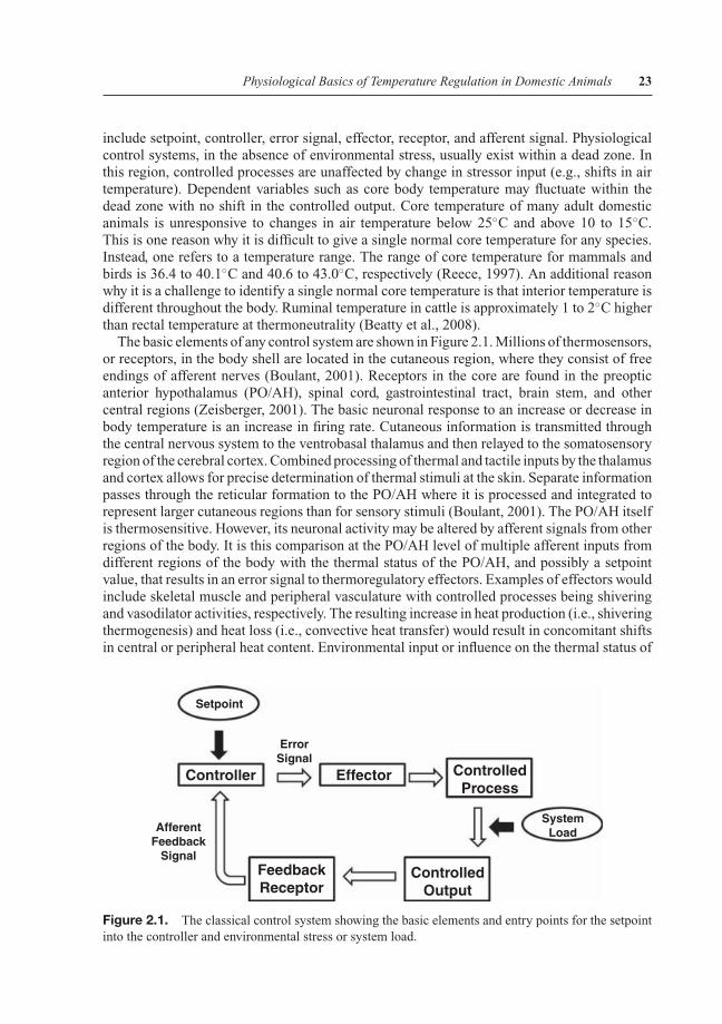
include setpoint, controller, error signal, effector, receptor, and afferent signal. Physiologicalcontrol systems, in the absence of environmental stress, usually exist within a dead zone. Inthis region, controlled processes are unaffected by change in stressor input (e.g., shifts in airtemperature). Dependent variables such as core body temperature may fluctuate within thedead zone with no shift in the controlled output. Core temperature of many adult domesticanimals is unresponsive to changes in air temperature below 25◦C and above 10 to 15◦C.This is one reason why it is difficult to give a single normal core temperature for any species.Instead, one refers to a temperature range. The range of core temperature for mammals andbirds is 36.4 to 40.1◦C and 40.6 to 43.0◦C, respectively (Reece, 1997). An additional reasonwhy it is a challenge to identify a single normal core temperature is that interior temperature isdifferent throughout the body. Ruminal temperature in cattle is approximately 1 to 2◦C higherthan rectal temperature at thermoneutrality (Beatty et al., 2008).
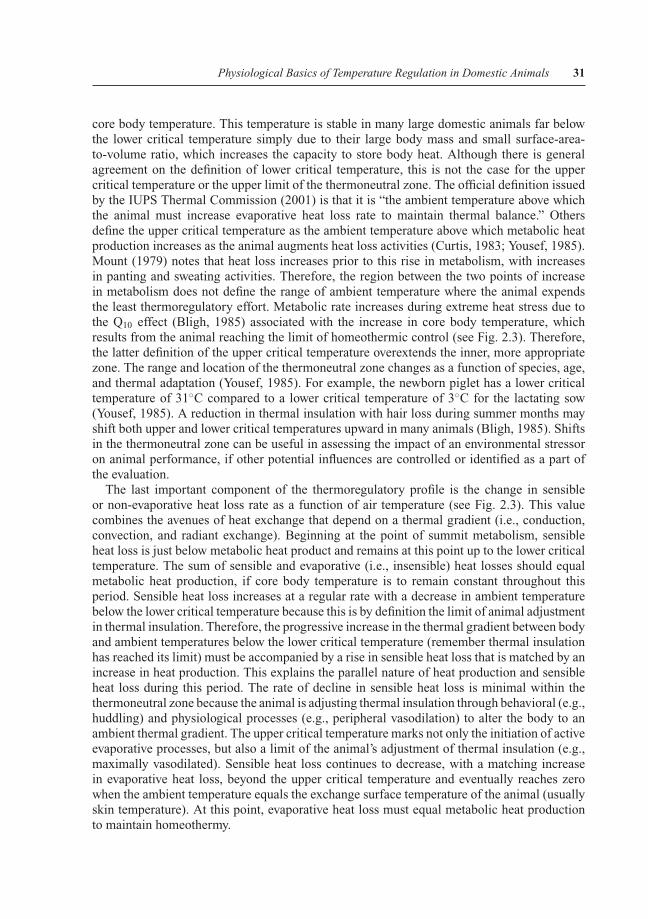
The basic elements of any control system are shown in Figure 2.1. Millions of thermosensors,or receptors, in the body shell are located in the cutaneous region, where they consist of freeendings of afferent nerves (Boulant, 2001). Receptors in the core are found in the preopticanterior hypothalamus (PO/AH), spinal cord, gastrointestinal tract, brain stem, and othercentral regions (Zeisberger, 2001). The basic neuronal response to an increase or decrease inbody temperature is an increase in firing rate. Cutaneous information is transmitted throughthe central nervous system to the ventrobasal thalamus and then relayed to the somatosensoryregion of the cerebral cortex. Combined processing of thermal and tactile inputs by the thalamusand cortex allows for precise determination of thermal stimuli at the skin. Separate informationpasses through the reticular formation to the PO/AH where it is processed and integrated torepresent larger cutaneous regions than for sensory stimuli (Boulant, 2001). The PO/AH itselfis thermosensitive. However, its neuronal activity may be altered by afferent signals from otherregions of the body. It is this comparison at the PO/AH level of multiple afferent inputs fromdifferent regions of the body with the thermal status of the PO/AH, and possibly a setpointvalue, that results in an error signal to thermoregulatory effectors. Examples of effectors wouldinclude skeletal muscle and peripheral vasculature with controlled processes being shiveringand vasodilator activities, respectively. The resulting increase in heat production (i.e., shiveringthermogenesis) and heat loss (i.e., convective heat transfer) would result in concomitant shiftsin central or peripheral heat content. Environmental input or influence on the thermal status of
Setpoint
SystemLoadAfferent
FeedbackSignal
FeedbackReceptor
ErrorSignal
Controller Effector ControlledProcess
ControlledOutput
Figure 2.1. The classical control system showing the basic elements and entry points for the setpointinto the controller and environmental stress or system load.
P1: SFK/UKS P2: SFK
BLBS093-c02 Collier October 19, 2011 4:2 Trim: 244mm×172mm
24 Environmental Physiology of Livestock
the animal occurs between the controlled processes and output event in the form of a systemload. For example, the same level of cutaneous vasodilation in mild heat would result in greaterheat loss than in severe heat due to the differences in the tissue to air thermal gradient betweenthe two environments. The lower heat content in the former example would result in a lowertissue temperature than in severe heat. Thermosensors (i.e., feedback receptors) respond tothe rate of change in local temperature with a high level of sensitivity to a few thousandths ofa degree. The result of this stimulation is an increased firing rate as the information returnsthrough a feedback signal into the central nervous system. Additional comparisons occur atthis point with system reevaluation and a new error signal.
Control systems can be positive or negative in structure. Negative systems are regulatory inthat they attempt to minimize the shifts in body temperature. The generation of an error signalin the previous example produces an effector response that moves the controlled output in theopposite direction that, in turn, diminishes the error value. This opposite response is referredto as negative action. Other control systems may be positive or feed-forward systems and arenonregulatory (Houk, 1988). In this case, a signal generated by the central nervous system inresponse to incoming afferent information (i.e., input) amplifies the output and moves the bodyforward in a certain direction. A thermoregulatory example would be the sensing of a coldburst of air by cutaneous thermoreceptors that initiate shivering activity even before there is achange in core temperature. The approaching cold is anticipated and defensive action taken topotentially reduce strain on the system.
There are two types of control that are key components of the thermoregulatory system.They are proportional and differential controls. Of the two, proportional control is the mostcommon. It is referred to as proportional because the magnitude of the output signal is di-rectly related to the magnitude of the input signal. Acute sweating and shivering intensities aretypically proportional to the thermal loads once the animal is above or below critical body tem-peratures, respectively. Differential control systems are more complex in nature. Basically, themagnitude of the output response is determined by the rate of change in the input signal. Theresponse of cutaneous thermosensors is characteristic of differential control. Rapid exposureto hot or cold ambient temperatures will produce a greater perception of these temperaturesthan slow exposure. An example of differential control and heat perception is rapid versusgradual entry into a hot bath. The bath temperature is the same in both situations, but the per-ception of heat is very different. It is thought that differential control provides a feed-forward,anticipatory response to an approaching stress and allows the animal an opportunity to preparefor strain.
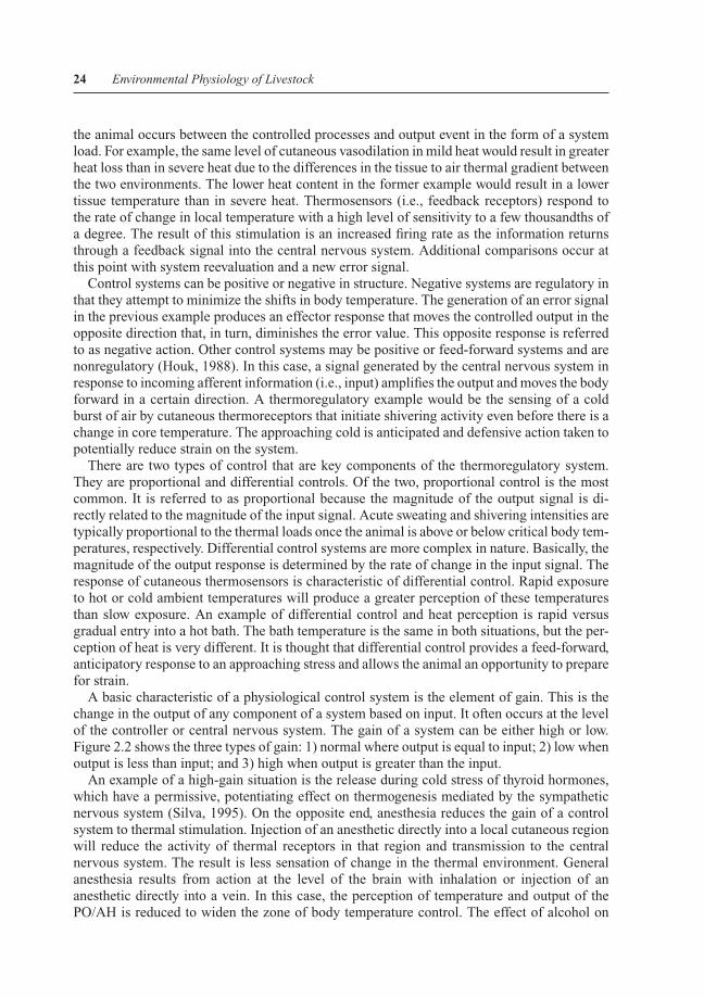
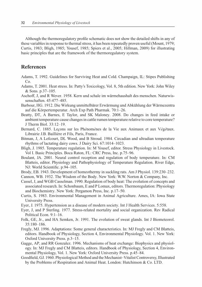
A basic characteristic of a physiological control system is the element of gain. This is thechange in the output of any component of a system based on input. It often occurs at the levelof the controller or central nervous system. The gain of a system can be either high or low.Figure 2.2 shows the three types of gain: 1) normal where output is equal to input; 2) low whenoutput is less than input; and 3) high when output is greater than the input.
An example of a high-gain situation is the release during cold stress of thyroid hormones,which have a permissive, potentiating effect on thermogenesis mediated by the sympatheticnervous system (Silva, 1995). On the opposite end, anesthesia reduces the gain of a controlsystem to thermal stimulation. Injection of an anesthetic directly into a local cutaneous regionwill reduce the activity of thermal receptors in that region and transmission to the centralnervous system. The result is less sensation of change in the thermal environment. Generalanesthesia results from action at the level of the brain with inhalation or injection of ananesthetic directly into a vein. In this case, the perception of temperature and output of thePO/AH is reduced to widen the zone of body temperature control. The effect of alcohol on
P1: SFK/UKS P2: SFK
BLBS093-c02 Collier October 19, 2011 4:2 Trim: 244mm×172mm
Physiological Basics of Temperature Regulation in Domestic Animals 25
Ou
tpu
t
10
9
8
7
6
5
4
3
2
1
1 2 3 4
I > O
I = O
I < O
5
Input
Figure 2.2. System output (O) as a function of input (I) for normal, low, and high levels of performance.
thermoregulatory ability is a good example. Alcohol intake effectively lowers the precisionof thermoregulatory control by reducing metabolic rate and heat production in the cold witha resulting drop in core temperature, and a reduction in heat loss capabilities in the hotenvironment to increase hyperthermia (Spiers, 1995). The result is an increased fluctuation incore temperature with less control.
Cassel and Casselman (1990) noted that although temperature is a measure of intensity ormagnitude of heat, it is not directly related to quantity or amount of heat. Body temperatureand heat content can change independently of each other. These differences are due primarilyto heat capacity and specific heat. In order to understand these concepts, it is important to usestandard definitions. The more recent and acceptable definitions were issued by IUPS (2001).Heat capacity (kcal/◦C) is the product of body mass and specific heat, and heat content (kcal) isthe product of body temperature, specific heat, and mass. As a result, a steer can absorb muchmore heat than a calf before there is a change in body temperature. Specific heat (kcal/kg/◦C)is defined as the amount of heat needed to raise the temperature of a unit mass of materialby 1◦C. Different materials have different specific heats. For example, the specific heat ofwater is 1 kcal/kg/◦C, whereas moist and dry animal tissues average 0.83 and 0.40 kcal/kg/◦C,respectively. The result is that hydrated tissue can absorb more than twice the heat of dehydratedtissue before there is a change in temperature. Since thermosensors detect body temperatureand not heat content, these physical characteristics become important determinants of thethermoregulatory control system performance at a location between the controlled output andreceptors. This independence of body temperature and heat content means that body heat isoften exchanged without a change of body temperature or an effect on the thermoregulatorycontrol system. Body temperature is not an accurate representation of stored heat until theupper limit of thermal capacitance is reached. These relationship differences instill in theanimal a delay and offset in the thermal response to hot and cold stress conditions. This alsomeans that the thermal core of an animal may remain stable during exposure to differentthermal environments, while there is a shift in the thermal shell temperature. During coldstress, the thermal core of a large domestic animal may be unchanged in heat content and
P1: SFK/UKS P2: SFK
BLBS093-c02 Collier October 19, 2011 4:2 Trim: 244mm×172mm
26 Environmental Physiology of Livestock
temperature. However, the heat content of the thermal shell may be reduced along with theperipheral temperature. As a result, total body heat content is decreased and the reduction intemperature of this region could affect the controlled system.
The traditional view of the thermoregulatory control system as a tight set of feedback anderror signals is inappropriate, considering there are shifts in gain and thermal capacitancethroughout the system. Regional body temperature alone is certainly not a conclusive deter-minant of body heat content. In addition, there is current controversy regarding the validityof setpoint theory and questions as to the need for a control system approach to explain ther-moregulation. Kobayashi et al. (2006) have questioned the receptor idea as presented in themodel of a thermoregulatory control system. The dogma is that temperature alters the firingrate of thermosensors as signals sent to the brain. In reality, a thermosensor alone cannot detecta temperature without decoding at the level of the brain. Likewise, thermosensors respond toother stimuli (e.g., osmotic pressure, chemicals) that could alter the firing rate in response tothermal stimulation. This would result in an inaccurate measurement that becomes an unreli-able component of the system. Instead, Kobayashi et al. (2006) proposed that thermosensorsare comparators at the level of the detection site that do not require the transfer of informa-tion directly to the brain. The suggestion is that the thermal comparators evaluate the sensedtemperature against its own reference signal. Once the comparator is activated by a thresholdsignal, it may send a signal to other regions that include the brain for thermosensation. Alongthis same line of thought, Werner (2010) proposed that neither a setpoint nor a comparison ofsignals at the level of the central nervous system is necessary to explain the thermoregulatorycontrol system. He noted that to regulate a variable within a control system model requires aninput signal, with the difference between the afferent input and the setpoint used to generatean error signal. Werner and Romanovsky (2007) argued that a setpoint is a misleading andunnecessary concept. Werner (2010) made the point that a steady-state, homeostatic condition,by definition, can only be achieved if the afferent input to the controller becomes a zero valueand there is no evidence for this. Likewise, proportional control does not acknowledge thesteady-state condition when the error signal is at zero level, but effector output is still requiredfor everyday activities. Such a condition cannot exist. Although there are current debates onthe existence of an actual setpoint or direct afferent input into the brain, the classical controlsystem concept is still a useful means to conceptualize the thermoregulatory system and testtheoretical components of the system.
Modes of Heat Exchange and the Thermoregulatory Profile
An understanding of thermoregulation, and particularly the physiological responses to heatstress, requires basic knowledge of heat production and the avenues of heat exchange. Dis-cussions of the sources of animal heat occurred as early as the time of Plato and Aristotle(Goodfield, 1960; Mendelsohn, 1964), with the current theories not developing until the 1800s.In contrast, heat exchange and heat loss to the environment did not receive serious exami-nation until the end of the 1800s and the early part of the twentieth century, with principlesborrowed from physics and engineering. Rubner (1902) concentrated on the use of calorime-try to measure heat production primarily in dogs and humans, and addressed heat exchangeby conduction, convection, evaporative, and radiant exchange. However, it was not until thecombined work of physiologists and engineers in the 1930s that major advancement occurredin partitional calorimetry (Winslow et al., 1936). There are several comprehensive reviewsof the avenues of heat exchange that have been recently published (Gagge and Gonzalez,
P1: SFK/UKS P2: SFK
BLBS093-c02 Collier October 19, 2011 4:2 Trim: 244mm×172mm
Physiological Basics of Temperature Regulation in Domestic Animals 27
1996; Werner, 1998; Adams, 2001; Hillman, 2009). Each avenue has unique differences andsimilarities that are important for their interaction within the framework of the thermoregula-tory system.
Heat Exchange
Core body temperature is considered to be the outcome of a balance between heat inflow orproduction (i.e., metabolic rate) and heat outflow. As discussed earlier, body temperature is anexpression of body heat content and therefore the traditional calculation is for the storage oftotal body heat or change in body heat content (S; kcal; kJ):
S = M + W + C + K + R − E (IUPS, 2001; Adams, 2001) (2.1)
Where:
S = storage of body heat or change in total body heat content (kcal; kJ)M = metabolic heat gain or metabolic rate (Btu/h; watts)W = work rate (positive for useful work; negative for mechanical power absorbed by body;
Btu/h; watts)C = convective heat transfer or exchange (Btu/h; watts)K = conductive heat transfer or exchange (Btu/h; watts)R = radiant heat exchange or exchange (Btu/h; watts)E = evaporative heat loss (Btu/h; watts)
An increase in total body heat storage will in turn increase core body temperature, anda decrease will reduce the core body temperature. Metabolic rate is the primary source ofheat gain for the living animal due to the Second Law of Thermodynamics, as noted earlier.The majority of energy released from biological reactions in the resting living animal is heat(Rhoades and Tanner, 1995), or a by-product of metabolism.
Heat transfer into and out of a living animal is complex and occurs by four pathways:conduction, convection, radiant exchange, and evaporation. The magnitude of heat transfer bythe first three avenues is highly dependent on the temperature differences between the exchangeobjects in the environment. Conduction occurs when two stationary objects are in contact witheach other. Other determinants of the magnitude of conductive heat transfer include thesurface area of contact, thickness of the conducting materials, and thermal conductivity ofthe conducting objects (i.e., an intrinsic property). The thermal conductivity of objects incontact with the animal is also an important component of thermal perception. For example,a metal surface with high thermal conductivity will conduct heat rapidly from the skin wherethe thermal receptors are located. In contrast, a wood surface with lower thermal conductivitywould conduct heat from the skin at a slower rate. As a result, a metal surface will feel coolerthan a wooden one even at the same temperature.
Convective heat transfer occurs when objects of different temperatures flow by each other.The dependence on the area of contact and a thermal gradient is the same as for conduction.Likewise, the convective heat transfer coefficient of the material is important. Thickness ofthe exchange materials, however, is not a determinant. The two mechanisms for convectiveheat transfer are passive and forced. Passive exchange occurs when air near the skin surfacemicroenvironment is heated to result in a reduction in air density and movement up from thesurface. This produces small currents of air that may result in significant heat loss under restingconditions. Forced convection occurs when external energy is used to move one material over
P1: SFK/UKS P2: SFK
BLBS093-c02 Collier October 19, 2011 4:2 Trim: 244mm×172mm
28 Environmental Physiology of Livestock
another. This mode of transfer is very important for internal heat movement, using blood flowand air movement in the respiratory system. Likewise, external movement of gas and fluidover the animal surface is an effective means for heat dissipation from the body when thetemperature of the medium is below skin surface temperature. However, circulation of air orwater over the animal surface at a temperature higher than skin temperature will actually addheat content and increase internal body temperature.
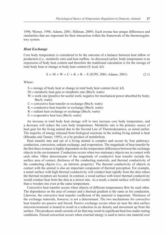
Radiant energy exchange relies on several factors that are the same for conductive andconvective exchanges. They include the temperature of the transmitting objects, the ther-mal gradient, and the exchange surface areas. The reliance of these exchange routes, whichare collectively classified as dry or sensible heat loss avenues (see Fig. 2.3), on a thermalgradient, also makes these routes less effective as environmental temperature approachesskin temperature.
There are, however, several differences between radiant exchange and the other sensibleavenues. Radiant exchange is the only heat loss process that occurs at the speed of light(299,792 km/sec) and in a vacuum. In fact, exchange by this route cannot occur if there isfluid in the space between the emitters. The rate of radiant exchange between emitters is alsodependent on the emissivity of the objects in the environment. Emissivity is defined as “theratio of the total radiant energy emitted by a full radiator at the same temperature” (IUPS,2001). A full radiator is an ideal emitter (maximum obtainable) of radiant energy from allparts of the electromagnetic spectrum. Usually this is a dull, non-shiny black surface that is
Hypothermia Hyperthermia
SummitMetabolism Summit
Evaporation
EvaporationRate
MetabolicRate
SensibleHeat Loss
Rate
HighAmbient Temperature
ThermoneutralZone
THERMOREGULATORY PROFILE
En
erg
y E
xch
ang
e R
ates
Co
reTe
mp
erat
ure
Low
LowerCritical
Temperature
UpperCritical
Temperature
CoreTemperature
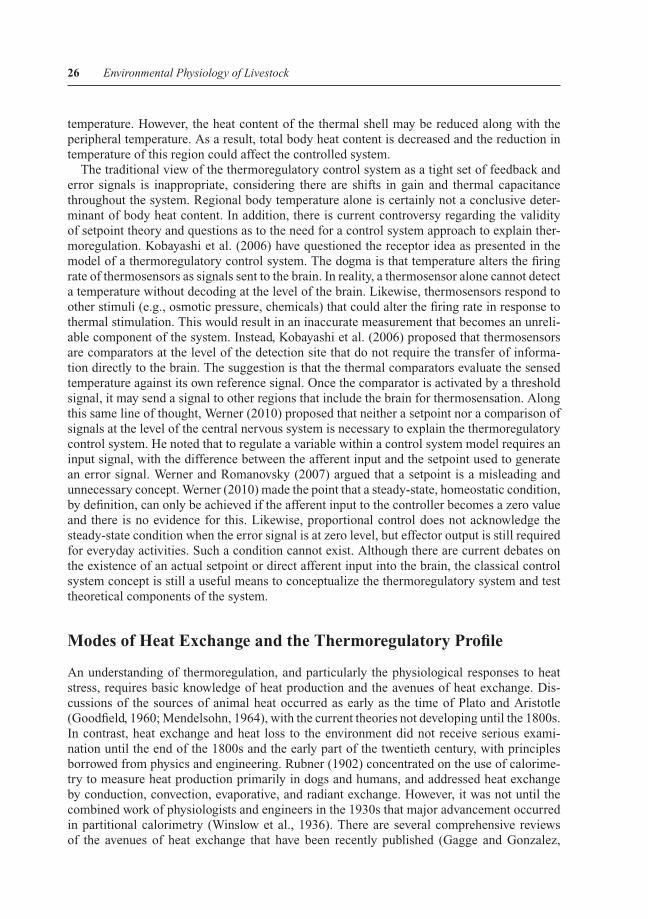
Figure 2.3. The thermoregulatory profile schematic showing both core temperature and energy ex-change routes as functions of ambient temperature. Limits of homeothermy are identified with summitmetabolic and evaporation rates. The thermoneutral zone is displayed by lower and upper critical tem-peratures (adapted from Mount, 1974).
P1: SFK/UKS P2: SFK
BLBS093-c02 Collier October 19, 2011 4:2 Trim: 244mm×172mm
Physiological Basics of Temperature Regulation in Domestic Animals 29
referred to as a black body. The emissivity of a full radiator is one, and approaches zero for ashiny surface. Therefore, reflectance of an object’s surface (shiny versus dull) is an importantdeterminant of emissivity and absorptance. Both the emissivity and temperature of a surfacedetermine the magnitude of electromagnetic energy emitted from an object and absorbed byit. In fact, this characteristic of an object is even more important than the color of an object’ssurface in affecting its capacity to absorb radiation, especially in a shaded environment orat night.
Any object with a temperature above absolute zero emits electromagnetic radiation. Onaverage, the total radiant energy from the sun at the edge of our atmosphere is approximately1.35 kW/m2, and at least half of this energy is in the infrared region of the electromagneticspectrum (Moss et al., 1982). More importantly nearly all-radiant exchange between livinganimals is in this region. The color of an object is an important factor during exposure to directsunlight, with dark objects absorbing more energy in the visible portion of the spectrum thanlight objects. However, infrared exchange is the primary avenue in the absence of sunlight,and the color of an object is not a factor (Heppner, 1970). A dark-colored object will absorband emit radiation equal to a light-colored object in the absence of visible solar radiation. Themore important factor for radiant exchange, on average, is the nature of the surface (i.e., dullor shiny; rough/smooth hair or feather coat).
Evaporation is the fourth avenue for heat loss from an animal. Adams (2001) noted that“evaporation of a liquid is not an avenue of heat exchange (gain or loss); it is only a way tolose heat.” Thermal energy is put into a liquid to produce a phase change to a gaseous state.When water evaporates from a surface, there is a loss of approximately 580 cal g−1 at a skintemperature of 30◦C (Morimoto, 2001). The important point is that the water must evaporatefrom the surface (e.g., skin, respiratory mucosa) for heat loss to occur. If it does not changeto a gaseous state due to high humidity, or if it drips off the animal, there is essentially noheat dissipation.
There are similarities and differences between evaporative loss and the other avenues of heatexchange. As noted for conduction, convection, and radiant exchange, the exposed exchangesurface area is an important factor that determines the rate of heat loss by evaporation. Inthis case, it needs to be the wet surface. Likewise, air velocity across the wet surface isa determinant, as is true for convective exchange. At this point, the similarities end withnotation of a characteristic of evaporative heat loss that makes it extremely useful in veryhot environments. It is the only avenue that does not require a thermal gradient for heatloss. There is a reduction in heat loss by conductive, convective, and radiant avenues, as thetemperature of the surrounding medium comes closer to skin temperature. This is the majorreason why animals have reduced heat loss and overheat in hot environments. In contrast,evaporative heat loss increases above the upper critical temperature (IUPS, 2001; see Fig. 2.3)with commencement of active evaporative heat loss (e.g., panting, gular fluttering, sweating).The magnitude of heat loss via this avenue, and the driving force for evaporation, is the watervapor pressure gradient between the exchange surfaces and the environment. An increase inthe water vapor pressure of the environment with a rise in humidity can severely limit heatdissipation to result in a greater level of hyperthermia.
All animals experience passive evaporative heat loss from both skin and respiratory surfaces(Hillman, 2009). Blood flows to all of these regions and hydrates the tissue at each site. Ifthere is a water vapor pressure gradient for heat loss, then this will continuously occur at thesesurfaces and exhibit a slight increase with air temperature prior to active evaporation (seeFig. 2.3). Birds actually rely on passive evaporation from the skin, since they are devoid ofsweat glands.
P1: SFK/UKS P2: SFK
BLBS093-c02 Collier October 19, 2011 4:2 Trim: 244mm×172mm
30 Environmental Physiology of Livestock
Active cutaneous evaporation occurs at the sweat glands. A comprehensive review of sweatglands (Folk and Semken, 1991), notes that there are sebaceous, apocrine (epitrichial), andeccrine (atrichial) glands in the skin. Apocrine glands are larger than eccrine and associatedwith hair follicles. They release fluid with adrenergic stimulation, while the eccrine glandsrely on cholinergic pathways (Robertshaw, 1977). In humans, the apocrine glands are situateddeep in the dermis and located in the pubic, anal, and axilla regions, with little involvement inthermoregulation (Folk and Semken, 1991). Instead, humans rely primarily on eccrine glandsfor evaporative heat loss associated with thermoregulation. These glands are located closer tothe surface, and found in largest numbers on the palms and soles of the feet. In contrast, cows,sheep, and horses have only apocrine glands and effectively use these for evaporative cooling.The distribution of these glands in cattle varies greatly with region, with larger numbers in thetrunk and neck, and fewer in the legs and ventral region (Hillman, 2009). These differencesin numbers across regions result in large differences in sweat rate during heat stress (Scharfet al., 2008). Dogs and cats have few sweat glands and they are primarily located on the paws.As a result, these animals must rely on other mechanisms for evaporative cooling, such aspanting. Pigs likewise have only a few sweat glands, and these are nonfunctional. Instead, theyspread moisture on the skin that evaporates for cooling. Birds do not have sweat glands, andmust depend on passive diffusion of moisture through the skin or use of the respiratory systemfor evaporation.
The respiratory tract is an important region for active evaporative heat loss in birds andmammals. Evaporation occurs primarily in the upper region of the tract where there are thelargest thermal and vapor pressure gradients. In every animal, there is increased frequencyof breathing above the upper critical temperature to increase heat dissipation. Mount (1979)noted that respiratory minute volume increases 10-, 12-, 15-, and 23-fold with panting in theox, sheep, rabbit, and dog, respectively. This effectively increases evaporative heat loss, butreduces tidal volume for gas exchange in the lungs (Richards, 1970). Many birds combinepanting with high frequency movement of the gular pouch to augment water evaporation duringheat stress.
Thermoregulatory Profile
The complex balance between metabolic heat production, evaporative heat loss, and sensibleheat exchange in determining core body temperature can be summarized using the thermoreg-ulatory profile diagram (see Fig. 2.3). The extreme boundaries of the profile are those pointsoutside of homeothermic maintenance, where core body temperature exhibits either a majorreduction (i.e., hypothermia) beyond summit metabolism or an increase (i.e., hyperthermia)beyond the limit of summit evaporation. Although the theoretical basis of the profile predictsno change in core body temperature prior to reaching these limitations, there are significantshifts in temperature associated with circadian rhythm (Piccione and Refinetti, 2003; Bitmanet al., 1984) and exposure to air temperature above the upper critical temperature. The upperand lower critical temperatures represent the inner, and generally more useful, borders of thethermoregulatory profile. They identify the range of the thermoneutral zone, where the animalexpends the least amount of energy for thermoregulation. This zone is, by definition, the regionwhere the animal has available the largest amount of energy for growth and production. Forthis reason, its identification is extremely important.
The lower critical temperature is the easier to define, as it is the ambient temperature belowwhich the animal must increase its metabolic heat production to maintain a relatively constant
P1: SFK/UKS P2: SFK
BLBS093-c02 Collier October 19, 2011 4:2 Trim: 244mm×172mm
Physiological Basics of Temperature Regulation in Domestic Animals 31
core body temperature. This temperature is stable in many large domestic animals far belowthe lower critical temperature simply due to their large body mass and small surface-area-to-volume ratio, which increases the capacity to store body heat. Although there is generalagreement on the definition of lower critical temperature, this is not the case for the uppercritical temperature or the upper limit of the thermoneutral zone. The official definition issuedby the IUPS Thermal Commission (2001) is that it is “the ambient temperature above whichthe animal must increase evaporative heat loss rate to maintain thermal balance.” Othersdefine the upper critical temperature as the ambient temperature above which metabolic heatproduction increases as the animal augments heat loss activities (Curtis, 1983; Yousef, 1985).Mount (1979) notes that heat loss increases prior to this rise in metabolism, with increasesin panting and sweating activities. Therefore, the region between the two points of increasein metabolism does not define the range of ambient temperature where the animal expendsthe least thermoregulatory effort. Metabolic rate increases during extreme heat stress due tothe Q10 effect (Bligh, 1985) associated with the increase in core body temperature, whichresults from the animal reaching the limit of homeothermic control (see Fig. 2.3). Therefore,the latter definition of the upper critical temperature overextends the inner, more appropriatezone. The range and location of the thermoneutral zone changes as a function of species, age,and thermal adaptation (Yousef, 1985). For example, the newborn piglet has a lower criticaltemperature of 31◦C compared to a lower critical temperature of 3◦C for the lactating sow(Yousef, 1985). A reduction in thermal insulation with hair loss during summer months mayshift both upper and lower critical temperatures upward in many animals (Bligh, 1985). Shiftsin the thermoneutral zone can be useful in assessing the impact of an environmental stressoron animal performance, if other potential influences are controlled or identified as a part ofthe evaluation.
The last important component of the thermoregulatory profile is the change in sensibleor non-evaporative heat loss rate as a function of air temperature (see Fig. 2.3). This valuecombines the avenues of heat exchange that depend on a thermal gradient (i.e., conduction,convection, and radiant exchange). Beginning at the point of summit metabolism, sensibleheat loss is just below metabolic heat product and remains at this point up to the lower criticaltemperature. The sum of sensible and evaporative (i.e., insensible) heat losses should equalmetabolic heat production, if core body temperature is to remain constant throughout thisperiod. Sensible heat loss increases at a regular rate with a decrease in ambient temperaturebelow the lower critical temperature because this is by definition the limit of animal adjustmentin thermal insulation. Therefore, the progressive increase in the thermal gradient between bodyand ambient temperatures below the lower critical temperature (remember thermal insulationhas reached its limit) must be accompanied by a rise in sensible heat loss that is matched by anincrease in heat production. This explains the parallel nature of heat production and sensibleheat loss during this period. The rate of decline in sensible heat loss is minimal within thethermoneutral zone because the animal is adjusting thermal insulation through behavioral (e.g.,huddling) and physiological processes (e.g., peripheral vasodilation) to alter the body to anambient thermal gradient. The upper critical temperature marks not only the initiation of activeevaporative processes, but also a limit of the animal’s adjustment of thermal insulation (e.g.,maximally vasodilated). Sensible heat loss continues to decrease, with a matching increasein evaporative heat loss, beyond the upper critical temperature and eventually reaches zerowhen the ambient temperature equals the exchange surface temperature of the animal (usuallyskin temperature). At this point, evaporative heat loss must equal metabolic heat productionto maintain homeothermy.
P1: SFK/UKS P2: SFK
BLBS093-c02 Collier October 19, 2011 4:2 Trim: 244mm×172mm
32 Environmental Physiology of Livestock
Although the thermoregulatory profile schematic does not show the detailed shifts in any ofthese variables in response to thermal stress, it has been repeatedly proven useful (Mount, 1979;Curtis, 1983; Bligh, 1985; Yousef, 1985; Spiers et al., 2005; Hillman, 2009) for illustratingbasic principles that are the framework of the thermoregulatory system.
References
Adams, T. 1992. Guidelines for Surviving Heat and Cold. Champaign, IL: Stipes PublishingCo.
Adams, T. 2001. Heat stress. In: Patty’s Toxicology, Vol. 8, 5th edition. New York: John Wiley& Sons. p.37–105.
Aschoff, J, and R Wever. 1958. Kern und schale im warmehaushalt des menschen. Naturwis-senschaften. 45:477–485.
Barbour, HG. 1912. Die Wirkung unmittelbarer Erwarmung und Abkuhlung der Warmezentraauf die Korpertemperatur. Arch Exp Path Pharmak. 70:1–26.
Beatty, DT, A Barnes, E Taylor, and SK Maloney. 2008. Do changes in feed intake orambient temperature cause changes in cattle rumen temperature relative to core temperature?J Therm Biol. 33:12–19.
Bernard, C. 1885. Lecons sur les Phenomenes de la Vie aux Animaux et aux Vegetaux.Librairie J.B. Bailliere et Fils, Paris, France.
Bitman, J, A Lefcourt, DL Wood, and B Stroud. 1984. Circadian and ultradian temperaturerhythms of lactating dairy cows. J Dairy Sci. 67:1014–1023.
Bligh, J. 1985. Temperature regulation. In: M Yousef, editor. Stress Physiology in Livestock.Vol I. Basic Principles. Boca Raton, FL: CRC Press, Inc. p.75–96.
Boulant, JA. 2001. Neural control reception and regulation of body temperature. In: CMBlatteis, editor. Physiology and Pathophysiology of Temperature Regulation. River Edge,NJ: World Scientific. p.94–105.
Brody, EB. 1943. Development of homeothermy in suckling rats. Am J Physiol. 139:230–232.Cannon, WB. 1932. The Wisdom of the Body. New York: W.W. Norton & Company, Inc.Cassel, J, and WGB Casselman. 1990. Regulation of body heat: The evolution of concepts and
associated research. In: Schonbaum, E and P Lomax, editors. Thermoregulation: Physiologyand Biochemistry. New York: Pergamon Press, Inc. p.17–50.
Curtis, S. 1983. Environmental Management in Animal Agriculture. Ames, IA: Iowa StateUniversity Press.
Eyer, J. 1975. Hypertension as a disease of modern society. Int J Health Services. 5:558.Eyer, J, and P Sterling. 1977. Stress-related mortality and social organization. Rev Radical
Political Econ. 9:1–16.Folk, GE, Jr., and HA Semken, Jr. 1991. The evolution of sweat glands. Int J Biometeorol.
35:180–186.Fregly, MJ. 1996. Adaptations: Some general characteristics. In: MJ Fregly and CM Blatteis,
editors. Handbook of Physiology, Section 4, Environmental Physiology. Vol. 1. New York:Oxford University Press. p.3–15.
Gagge, AP, and RR Gonzalez. 1996. Mechanisms of heat exchange: Biophysics and physiol-ogy. In: MJ Fregly and CM Blatteis, editors. Handbook of Physiology, Section 4, Environ-mental Physiology, Vol. 1. New York: Oxford University Press. p.45–84.
Goodfield, GJ. 1960. Physiological Method and the Mechanist–Vitalist Controversy, Illustratedby the Problems of Respiration and Animal Heat. London: Hutchinson & Co. LTD.
P1: SFK/UKS P2: SFK
BLBS093-c02 Collier October 19, 2011 4:2 Trim: 244mm×172mm
Physiological Basics of Temperature Regulation in Domestic Animals 33
Hammel, HT, JD Hardy, and MM Fusco. 1960. Thermoregulatory responses to hypothalamiccooling in unanesthetized dogs. Am J Physiol. 198(3):481–486.
Heppner, F. 1970. The metabolic significance of differential absorption of radiant energy byblack and white birds. The Condor. 72:50–59.
Hillman, PE. 2009. Thermoregulatory Physiology. In: JA DeShazer, editors. Livestock Ener-getics and Thermal Environmental Management. St. Joseph, MI: ASABE.
Horton, JC, and CJ Bicak. 1987. Modeling for biologists. Bioscience. 37:808–809.Houk, JC. 1988. Control strategies in physiological systems. Fed Am Soc Exp Biol. J 2:
97–107.IUPS Thermal Commission. 2001. Glossary of terms for thermal physiology. Japan J Physiol
51:245–280.Kleiber, M. 1975. The Fire of Life. An Introduction to Animal Energetics. Huntington, NY:
RE Krieger Publishing Co., Inc.Kobayashi, S, M Okazawa, A Hori, K Matsumura, and H Hosokawa. 2006. Paradigm shift in
sensory system – Animals do not have sensors. J Therm Biol. 31:19–23.Mendelsohn, E. 1964. Heat and Life. The Development of the Theory of Animal Heat.
Cambridge, MA: Harvard Univ Press.Morimoto, T. 2001. Heat Loss Mechanisms. In: Physiology and Pathophysiology of Temper-
ature Regulation. CM Blatteis, editor. River Edge, NJ: World Scientific. p.80–91.Moss, CE, RJ Ellis, WE Murray, and WH Parr. 1982. Infrared Radiation. In: MJ Suess, editor.
Nonionizing Radiation Protection WHO Regional Publications European Series No. 10,Copenhagen. p.85–115.
Mount, LE. 1974. Thermal neutrality. In: JL Monteith and LE Mount, editors. Heat Loss fromAnimals and Man: Assessment and Control. London: Butterworths. p.425–439.
Mount, LE. 1979. Adaptation to Thermal Environment: Man and His Productive Animals.Baltimore: University Park Press.
Mrosovsky, N. 1990. Rheostasis: The Physiology of Change. New York: Oxford UniversityPress.
Piccione, G, and R Refinetti. 2003. Thermal chronobiology of domestic animals. Front. Bio-science 8:258–264.
Potter, VR. 1971. How is an optimum environment defined? In: Bioethics: Bridge to the Future.Englewood Cliffs, NJ: Prentice-Hall, Inc. p.133–148.
Reece, WO. 1997. Body Heat and Temperature Regulation. In: Physiology of Domestic Ani-mals. Philadelphia: Lippincott Williams and Wilkins, p.334–343.
Rhoades, R, and G Tanner. 1995. Medical Physiology. Boston: Little, Brown, and Co.Robertshaw, D. 1977. Neuroendocrine control of sweat glands. J Invest Dermatol. 69:121–129.Romanovsky, AA. 2007. Thermoregulation: Some concepts have changed. Functional archi-
tecture of the thermoregulatory system. Am J Physiol. 292:R37–R46.Rubner, M. 1902. The Laws of Energy Consumption in Nutrition. Franz Deuticke. Leipzig
and Vienna.Scharf, B, LE Wax, GE Aiken, and DE Spiers. 2008. Regional differences in sweat rate
response of steers to short-term heat stress. Int J Biometeorol. 52:725–732.Schmidt-Nielsen, B, K Schmidt-Nielsen, TR Houpt, and SA Jarnum. 1956. Water balance of
the camel. Am J Physiol. 185:185–184.Sherwood, L, H Klandorf, and PH Yancey. 2005. Muscle physiology. In: Animal Physiology:
From Genes to Organisms. Belmont, CA: Thomson Brooks/Cole. p.314–358.Silva, JE. 1995. Thyroid hormone control of thermogenesis and energy balance. Thyroid.
5:481–492.
P1: SFK/UKS P2: SFK
BLBS093-c02 Collier October 19, 2011 4:2 Trim: 244mm×172mm
34 Environmental Physiology of Livestock
Spiers, DE. 1995. Thermoregulation and ethanol. In: RR Watson, editor. Drug and AlcoholAbuse Reviews, Vol. 6: Alcohol and Hormones. Totowa, NJ: Humana Press Inc. p.193–208.
Spiers, DE, TJ Evans, and GE Rottinghaus. 2005. Interaction between thermal stress and fescuetoxicosis: Animal models. In: CA Roberts, CP West, and DE Spiers, editors. Neotyphodiumin Cool-Season Grasses. Ames, IA: Blackwell Publishing. p.243–270.
Waddington, CH. 1940. Organisers and Genes. Cambridge, MA: Cambridge University Press.Werner, J. 2010. System properties, feedback control and effector coordination of human
temperature regulation. Eur J Appl Physiol. 109:13–25.Werner, J. 1998. Biophysics of heat exchange between body and environment. Chapter 3. In:
CM Blatteis, editor. Physiology and pathophysiology of temperature regulation. p.25–45.London: World Scientific Publishing Co. Pte. Ltd.
Wiener, N. 1961. Cybernetics: or Control and Communication in the Animal and the Machine.Cambridge, MA: MIT Press.
Winslow, CEA, LP Herrington, and AP Gagge. 1936. A new method of partitional calorimetry.Am J Physiol. 116:641–655.
Yousef, K. 1985. Thermoneutral Zone. In: M Yousef, editor. Stress Physiology in Livestock.Vol. I. Basic Principles. Boca Raton, FL: CRC Press, Inc. p.67–74.
Zeisberger, E. 2001. Cold Adaptation. In: CM Blatteis, editor. Physiology and Pathophysiologyof Temperature Regulation. River Edge, NJ: World Scientific. p.208–227.


![Global environmental challenges [and livestock]](https://img.pdfslide.us/doc/110x75/546e5489b4af9fa5268b4624/global-environmental-challenges-and-livestock.jpg)