Embed Size (px)
Citation preview
TASMANIAN INFECTION PREVENTION AND CONTROL UNIT
Department of Health and Human Services
Environmental cleaning assessment protocol Version 2 November 2013
P a g e | 1
Environmental Assessment Cleaning Protocol
Tasmanian Infection Prevention and Control Unit (TIPCU)
Department of Health and Human Services, Tasmania
Published 2013
Copyright—Department of Health and Human Services
Permission to copy is granted provided the source is acknowledged
Contact: TIPCU
GPO Box 125
Hobart, Tasmania, Australia, 7001
Email: [email protected]
Website: www.dhhs.tas.gov.au/tipcu
Authors:
Brett Mitchell, TIPCU
Anne Wells, TIPCU
Fiona Wilson, TIPCU
Alistair McGregor, TIPCU
Suggested citation: Mitchell, B., Wells, A., Wilson, F., McGregor, A. (2013). Environmental Assessment Cleaning Protocol. Department of Health and Human Services.
P a g e | 2
Contents Foreword ................................................................................................................................................................................ 4
Background ............................................................................................................................................................................. 5
Overview ................................................................................................................................................................................. 5
Value of a standardised approach ...................................................................................................................................... 6
Principles of participation .................................................................................................................................................... 6
Definitions ............................................................................................................................................................................... 7
Fluorescent gel and light assessment ................................................................................................................................ 8
Overview ....................................................................................................................................... 8
Principles ....................................................................................................................................... 8
Selection of UV gel sites .................................................................................................................. 9
Frequency and number of assessments ........................................................................................... 10
The procedure ............................................................................................................................. 11
Visual assessment ................................................................................................................................................................ 12
Overview ..................................................................................................................................... 12
Principles ..................................................................................................................................... 12
Tasmanian visual assessment .......................................................................................................... 12
Areas to be assessed and determination of cleanliness ..................................................................... 13
Areas to be assessed and determination of cleanliness ..................................................................... 14
Frequency and number of assessments – patient care areas .............................................................. 15
Frequency and number of assessments –general ward areas ............................................................. 16
The procedure ............................................................................................................................. 16
Online assessment tool ...................................................................................................................................................... 16
Assessors ............................................................................................................................................................................... 17
Reporting ............................................................................................................................................................................... 17
References ............................................................................................................................................................................ 18
Appendix A – Agreement to Participate – DHHS acute care facility...................................................................... 20

P a g e | 3
Appendix B – Agreement to Participate – non DHHS facility .................................................................................. 21
Appendix C – Agreement to Participate – DHHS Non - acute care facility ......................................................... 22
Appendix D – Example of where to apply the gel ....................................................................................................... 23
Appendix E – Examples of the online assessment tool .............................................................................................. 26
P a g e | 4
Foreword
There is evidence to demonstrate that environment plays an important part in infection prevention and control. For
this reason, we consider the evaluation of environmental cleanliness to be an integral part of any infection prevention
and control program. Numerous studies in the literature discuss novel methodologies that assess both the extent of
environmental contamination in the hospital environment and efficacy of cleaning (1-6). Methods of evaluating
environmental cleanliness can be broken down into two types: process evaluation, such as visual inspection and
fluorescent gel application; and outcome evaluation, in which cleanliness is measured using methods such as
adenosine triphosphate (ATP) or microbial cultures (7). An important fact to remember, however, is that just
because the environment may appear ‘dirty’, this does not necessarily mean it poses an increased risk of infection for
patients. Conversely, an environment that appears ‘clean’ may harbour microorganisms that are potentially harmful.
The purpose of this protocol is to outline a program of environmental cleanliness evaluation that can be carried out
in a standardised manner within Tasmanian healthcare facilities.
Brett Mitchell Dr Alistair McGregor

P a g e | 5
Background
The Tasmanian Infection Prevention and Control Unit (TIPCU) has undertaken a review of the literature and
interviewed key stakeholders in Australia to establish and evaluate current methods to assess environmental
cleanliness. This work has been published in a peer reviewed journal (7). The findings were presented to infection
control professionals and environmental services managers from across Tasmania at a 2012 forum convened by the
TIPCU. At this forum, there was consensus to develop a standardised method evaluating environmental cleanliness
in Tasmanian healthcare facilities The methods chosen to evaluate environmental cleanliness were visual inspection
and the use of fluorescent light, using a framework similar to the assessment processes described in the national
hand hygiene initiative. To that end, the TIPCU were to develop a protocol to enable implementation of a
standardised method for evaluating environmental cleanliness in Tasmanian hospitals. This protocol is the result of
this work.
Overview
Evaluating environmental cleanliness will involve two elements, which are described in detail in this protocol:
1. The use of an ultraviolet (UV) solution and fluorescent light assessment conducted quarterly in rooms that
have undergone a discharge clean.
2. A visual assessment conducted at least quarterly.
This protocol does not address the following issues:
• The product(s) used to clean and the circumstances of when certain products should be used
• How to clean or types of cleaning methods
• Frequency of cleaning
• Models of cleaning, including staffing
• Actions required to be taken as a result of the assessment
P a g e | 6
Value of a standardised approach A standardised approach to evaluating environmental cleanliness has a number of advantages, including:
• Demonstrating a collaborative commitment to tackle the issue of healthcare associated infections.
• Providing local feedback to staff to help identify and improve practices
• Evaluating the impact of changes to cleaning models and processes
• Identifying areas of high compliance to explore and share contributory processes and models.
• Benchmarking
• Assisting in meeting national accreditation standards, more specifically Standard 3 of the National Safety and Quality Health Service (8)
Principles of participation Participation in the Tasmanian environmental cleanliness of healthcare program is voluntary. To participate in this program you need to:
DHHS acute care hospitals:
• Contact TIPCU via telephone or email to notify them of your organisations desire to participate in the program
• Complete form in Appendix A and ensure sign off by THO CEO/Executive Sponsor
• When you choose to participate TIPCU will provide your organisation with access to all TIPCU developed on-line tools.
• For DHHS acute care facilities, namely Royal Hobart Hospital, Launceston General Hospital, Mersey Community Hospital and North West Regional Hospital, TIPCU will provide the site with enough UV fluorescent gel markers and UV lights for the number of sites consistent with the protocol for one year from the date they agree to participate as long as the agreement is signed prior to December 31st 2013.
Other DHHS and non-DHHS healthcare facilities:
• Contact TIPCU via telephone or email to notify them of your organisations desire to participate in the program
• Complete form in Appendix B and ensure sign off by THO CEO/Executive Sponsor
• When you choose to participate TIPCU will provide your organisation with access to all TIPCU developed on-line tools.
P a g e | 7
Definitions
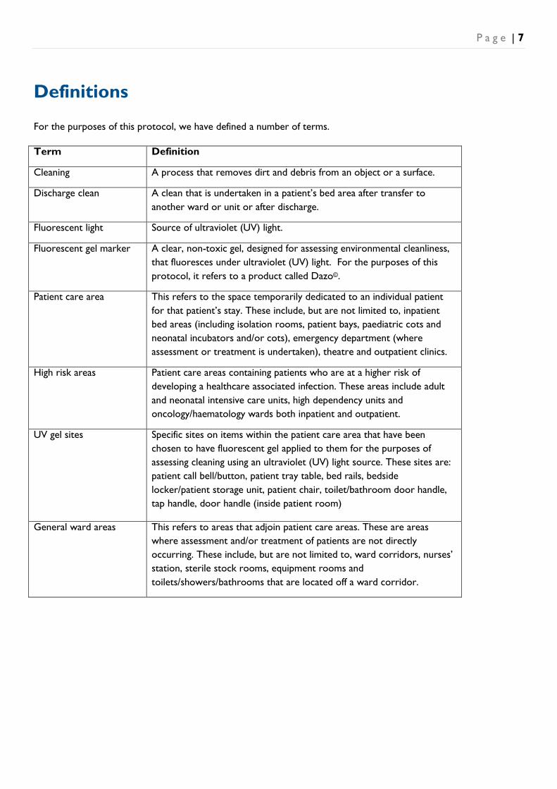
For the purposes of this protocol, we have defined a number of terms.
Term Definition
Cleaning A process that removes dirt and debris from an object or a surface.
Discharge clean A clean that is undertaken in a patient’s bed area after transfer to another ward or unit or after discharge.
Fluorescent light Source of ultraviolet (UV) light.
Fluorescent gel marker A clear, non-toxic gel, designed for assessing environmental cleanliness, that fluoresces under ultraviolet (UV) light. For the purposes of this protocol, it refers to a product called Dazo©.
Patient care area This refers to the space temporarily dedicated to an individual patient for that patient’s stay. These include, but are not limited to, inpatient bed areas (including isolation rooms, patient bays, paediatric cots and neonatal incubators and/or cots), emergency department (where assessment or treatment is undertaken), theatre and outpatient clinics.
High risk areas Patient care areas containing patients who are at a higher risk of developing a healthcare associated infection. These areas include adult and neonatal intensive care units, high dependency units and oncology/haematology wards both inpatient and outpatient.
UV gel sites Specific sites on items within the patient care area that have been chosen to have fluorescent gel applied to them for the purposes of assessing cleaning using an ultraviolet (UV) light source. These sites are: patient call bell/button, patient tray table, bed rails, bedside locker/patient storage unit, patient chair, toilet/bathroom door handle, tap handle, door handle (inside patient room)
General ward areas This refers to areas that adjoin patient care areas. These are areas where assessment and/or treatment of patients are not directly occurring. These include, but are not limited to, ward corridors, nurses’ station, sterile stock rooms, equipment rooms and toilets/showers/bathrooms that are located off a ward corridor.
P a g e | 8
Fluorescent gel and light assessment
Overview
This method uses fluorescent gel applied to surfaces in patient care areas. The solution dries on surfaces and resists
dry abrasion but is easily removed with light abrasion after wetting. The gel is visible only under UV light. Therefore,
once the gel has been applied and after a room has been cleaned, the thoroughness of cleaning can be determined by
using a fluorescent light to determine if gel remains. There are numerous studies demonstrating the usefulness of this
methods (9-13).
Principles
This protocol aims to be applicable to all Tasmanian hospitals that want to participate in the program (for example,
acute public hospitals, rural hospitals, and private hospitals), so it is not possible to explicitly document all possible
variations of how to undertake this evaluation. In order to provide consistency, there are some broad principles that
should be followed, and these are:
• Only rooms/patient care areas that are undergoing a discharge clean should be assessed. The rationale for
this is two-fold. First, evidence suggests that persons who inhabit a room following an occupant who had an
infectious agent are then more likely to acquire that infectious agent themselves (14-16). Second, the
assessment is easier to implement in rooms where the patient is no longer present.
• Only persons who are familiar with this protocol and who have received training on how to use the
fluorescent gel and light should perform the assessment. A training package is available from the TIPCU.
• The UV gel sites assessed as part of this process are only those stipulated in this protocol (see “Selection of
UV gel sites” for more detail).
• If a room/patient care area does not contain at least half of the required UV gel sites, do not perform the
UV gel audit in this room/patient care area.
• The assessment should not be intentionally undertaken in a covert manner.
• For acute public hospitals, a minimum of 25% of patient care areas assessed should be from any ‘high risk’
areas (more detail provided under ‘Frequency and number of assessments’ section).
• Only UV gel sites deemed as ‘Not Clean’ should be re cleaned prior to a new patient being admitted to that
patient care area. The entire room/patient care area does not require re-cleaning.
• The location of gel application does not indicate that only part of an object that should be cleaned.
P a g e | 9
Selection of UV gel sites
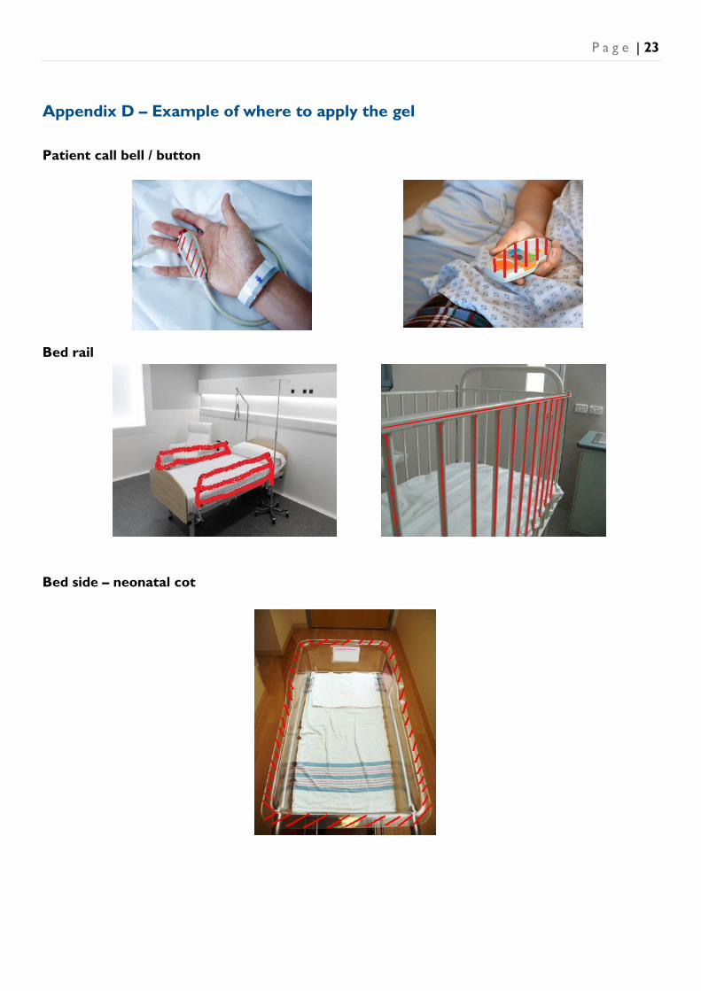
The objects in a patient care area to which the gel can be applied vary; however, there is some consistency in the literature regarding the objects chosen. The purpose of stipulating the location of gel application is to aid the assessment of whether an object has been cleaned. For the purposes of this protocol, the fluorescent gel and light should be used once on each of the following objects, if they are present. Additional details on where to apply the UV gel are also provided in Appendix C – Example of where to apply the gel.
1. Patient call bell/button
• Apply to the call bell itself, not to any attachment, e.g. cord
• If there is more than one call bell in use, apply to the one closest to the patient bed area
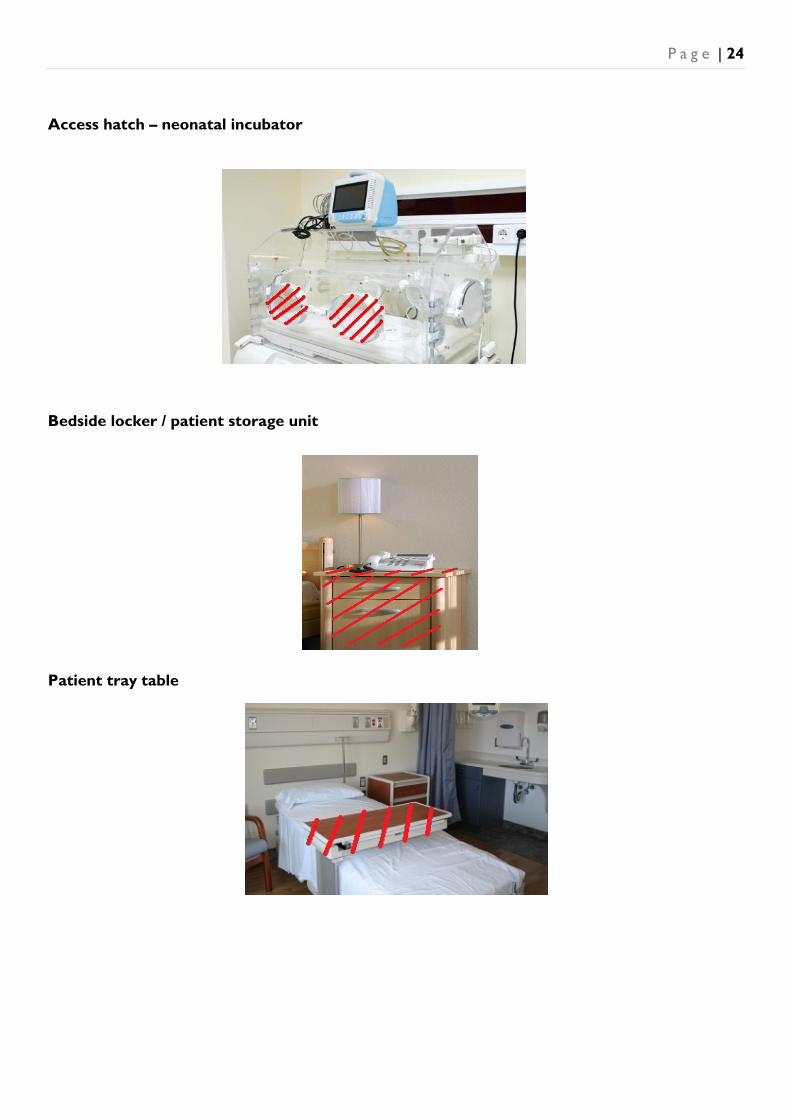
2. Patient tray table
• Apply gel to the top of the table/tray or one of its sides.
3. Bed rails
• Apply to a bed rail on the sides of patient’s bed or cot or to the access hatch on a neonatal incubator.
4. Bedside locker/patient storage unit
• Apply gel to the flat top of the locker or the top half of the sides or front.
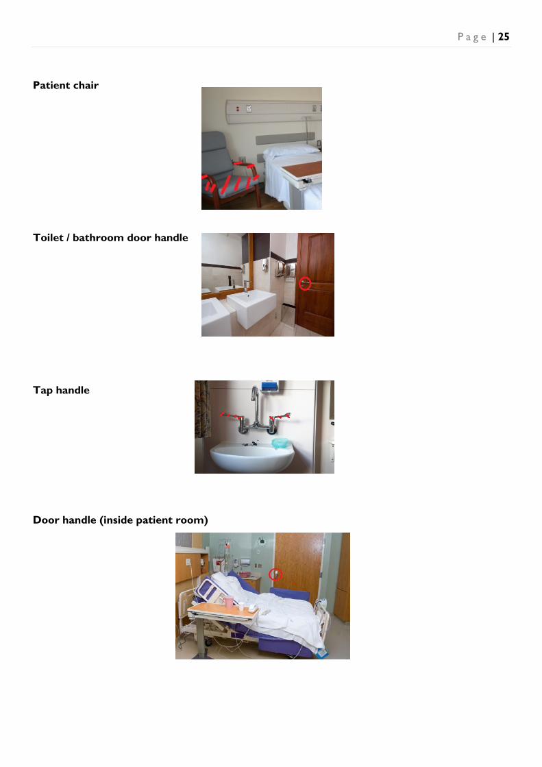
5. Patient chair
• Apply gel to one of the arm rests or seat.
6. Toilet/bathroom door handle
• This refers to a door handle in an en-suite or where the toilet/bathroom is shared by patients in the same area. This does not apply to toilets or bathrooms that are outside the patient area, for example, in a corridor.
• Apply gel to any part of the door handle.
7. Tap handle
• This refers to a sink in the patient room, patient area or en-suite that is for patient or clinical use. It does not apply to sinks that are outside the patient area, for example, in a corridor, walkway, treatment rooms, etc.
• Apply gel to one of the tap handles.
8. Door handle (inside patient room)
• This applies only to single rooms. It refers to the inside door handle of a door that is in the patient area.
• Apply gel to the inside door handle
P a g e | 10
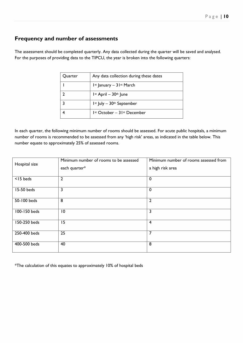
Frequency and number of assessments
The assessment should be completed quarterly. Any data collected during the quarter will be saved and analysed. For the purposes of providing data to the TIPCU, the year is broken into the following quarters:
Quarter Any data collection during these dates
1 1st January – 31st March
2 1st April – 30th June
3 1st July – 30th September
4 1st October – 31st December
In each quarter, the following minimum number of rooms should be assessed. For acute public hospitals, a minimum number of rooms is recommended to be assessed from any ‘high risk’ areas, as indicated in the table below. This number equate to approximately 25% of assessed rooms.
Hospital size Minimum number of rooms to be assessed
each quarter*
Minimum number of rooms assessed from
a high risk area
<15 beds 2 0
15-50 beds 3 0
50-100 beds 8 2
100-150 beds 10 3
150-250 beds 15 4
250-400 beds 25 7
400-500 beds 40 8
*The calculation of this equates to approximately 10% of hospital beds
P a g e | 11
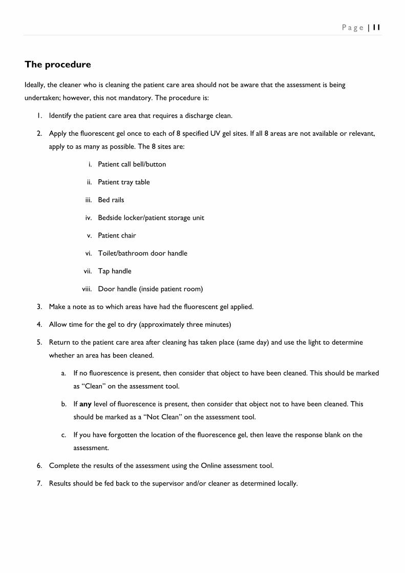
The procedure
Ideally, the cleaner who is cleaning the patient care area should not be aware that the assessment is being
undertaken; however, this not mandatory. The procedure is:
1. Identify the patient care area that requires a discharge clean.
2. Apply the fluorescent gel once to each of 8 specified UV gel sites. If all 8 areas are not available or relevant,
apply to as many as possible. The 8 sites are:
i. Patient call bell/button
ii. Patient tray table
iii. Bed rails
iv. Bedside locker/patient storage unit
v. Patient chair
vi. Toilet/bathroom door handle
vii. Tap handle
viii. Door handle (inside patient room)
3. Make a note as to which areas have had the fluorescent gel applied.
4. Allow time for the gel to dry (approximately three minutes)
5. Return to the patient care area after cleaning has taken place (same day) and use the light to determine
whether an area has been cleaned.
a. If no fluorescence is present, then consider that object to have been cleaned. This should be marked
as “Clean” on the assessment tool.
b. If any level of fluorescence is present, then consider that object not to have been cleaned. This
should be marked as a “Not Clean” on the assessment tool.
c. If you have forgotten the location of the fluorescence gel, then leave the response blank on the
assessment.
6. Complete the results of the assessment using the Online assessment tool.
7. Results should be fed back to the supervisor and/or cleaner as determined locally.
P a g e | 12
Visual assessment
Overview
The primary method for assessing the cleanliness of healthcare environments is visual inspection (17, 18).
Commonly, environmental cleanliness assessments are undertaken by environmental cleaning staff, and the
effectiveness of these is intermittently assessed by healthcare professionals such as infection control staff or trained
monitoring consultants (19, 20). While visual assessment of the cleanliness of a hospital ward, surface or item may
satisfy aesthetic obligations, it cannot reliably assess the infection risk posed to patients(17). It is for this reason that
visual assessment in isolation is not necessarily a reliable indicator for standards of healthcare cleanliness. Visual
assessments are, however, common practice in Tasmania, Australia and internationally.
Principles
As this protocol aims to be applicable to all Tasmanian hospitals that want to participate in the program (for example
acute public hospitals, rural hospitals, and private hospitals), it is not possible to explicitly document all possible
variations of how to undertake this assessment. In order to provide consistency, only persons who are familiar with
this protocol and who have received training on this assessment should undertake it.
Tasmanian visual assessment
Visual assessments are used to look at cleanliness in all areas of the hospital. For the purposes of the Tasmanian
assessment, two different visual assessment tools have been developed, to allow flexibility and additional clarity for
specific items. The two visual areas are defined by the location in which the assessment is being undertaken, namely:
1. Patient care areas.
a. This refers to the space temporarily dedicated to an individual patient for that patient’s stay.
b. This refers to the space temporarily dedicated to an individual patient for that patient’s stay. These
include, but are not limited to, inpatient bed areas including isolation rooms, patient bays, paediatric
cots and neonatal incubators and/or cots, emergency department (where assessment or treatment is
undertaken), theatre and outpatient clinics.
c. The surroundings to be included in a patient area are:
i. For single/isolation rooms, the entire room and any en-suite from which access can be
gained from the room.
ii. For shared patient areas, the entire room in which all patients are physically located and also
any shared toilet/bath/shower room that can be accessed directly from this area.
b. Assessments should be undertaken after a routine clean or a discharge clean.
P a g e | 13
2. General ward areas.
a. This refers to areas that adjoin patient care areas. These are areas where assessment or treatment
of patients does not directly occur. These include, but are not limited to, ward corridors, nurses’
station, sterile stock rooms, equipment rooms and toilets/showers/bathrooms that are located off a
ward corridor.
b. Assessments should be undertaken after a routine clean.
Areas to be assessed and determination of cleanliness
The areas to be assessed for both the Patient Care Area Assessment and the General Ward Area Assessment t are
detailed in the following table. The areas were chosen to ensure consistency with approaches taken in NSW (21),
Victoria (22) and existing practices in Tasmania. Additionally, some sites are more specific so as to allow
comparisons between the visual assessment and fluorescent gel for those sites.
An assessed area will be deemed “Clean” or “Not clean”, based on the descriptor provided in the table below. If an
area is not assessed, “Not applicable” may be selected on the assessment tool.
P a g e | 14
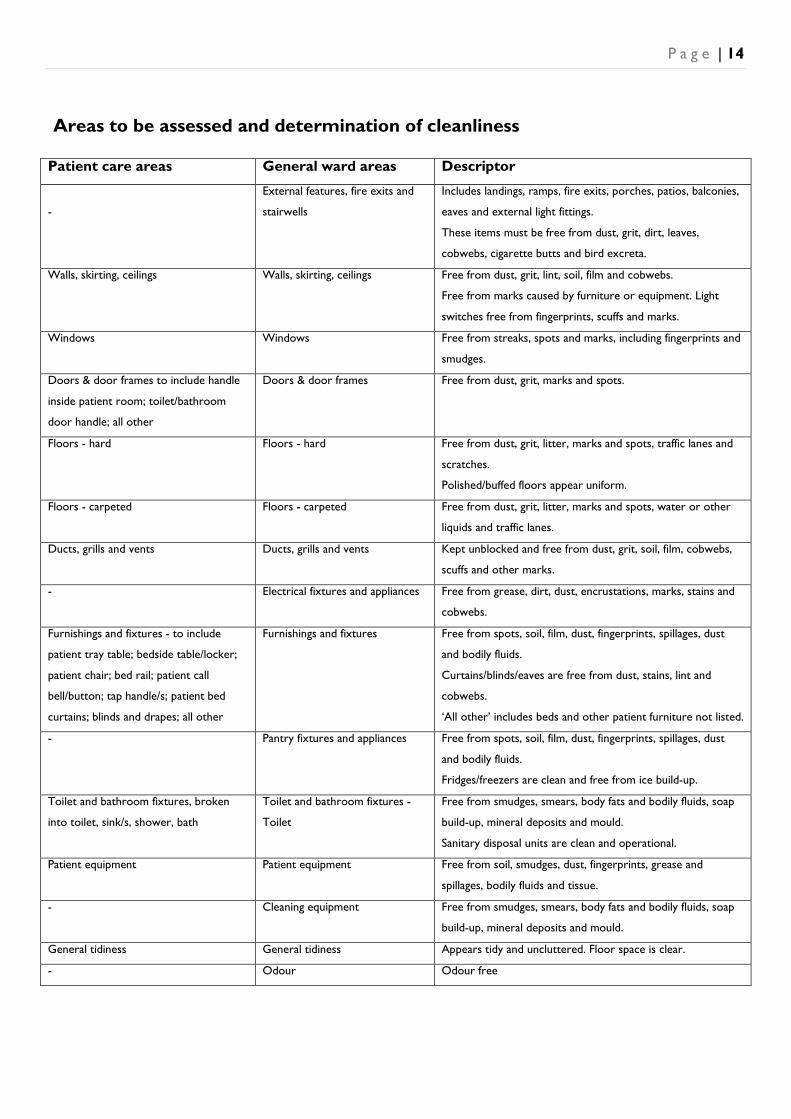
Areas to be assessed and determination of cleanliness
Patient care areas General ward areas Descriptor
-
External features, fire exits and
stairwells
Includes landings, ramps, fire exits, porches, patios, balconies,
eaves and external light fittings.
These items must be free from dust, grit, dirt, leaves,
cobwebs, cigarette butts and bird excreta.
Walls, skirting, ceilings Walls, skirting, ceilings Free from dust, grit, lint, soil, film and cobwebs.
Free from marks caused by furniture or equipment. Light
switches free from fingerprints, scuffs and marks.
Windows Windows Free from streaks, spots and marks, including fingerprints and
smudges.
Doors & door frames to include handle
inside patient room; toilet/bathroom
door handle; all other
Doors & door frames Free from dust, grit, marks and spots.
Floors - hard Floors - hard Free from dust, grit, litter, marks and spots, traffic lanes and
scratches.
Polished/buffed floors appear uniform.
Floors - carpeted Floors - carpeted Free from dust, grit, litter, marks and spots, water or other
liquids and traffic lanes.
Ducts, grills and vents Ducts, grills and vents Kept unblocked and free from dust, grit, soil, film, cobwebs,
scuffs and other marks.
- Electrical fixtures and appliances Free from grease, dirt, dust, encrustations, marks, stains and
cobwebs.
Furnishings and fixtures - to include
patient tray table; bedside table/locker;
patient chair; bed rail; patient call
bell/button; tap handle/s; patient bed
curtains; blinds and drapes; all other
Furnishings and fixtures Free from spots, soil, film, dust, fingerprints, spillages, dust
and bodily fluids.
Curtains/blinds/eaves are free from dust, stains, lint and
cobwebs.
‘All other’ includes beds and other patient furniture not listed.
- Pantry fixtures and appliances Free from spots, soil, film, dust, fingerprints, spillages, dust
and bodily fluids.
Fridges/freezers are clean and free from ice build-up.
Toilet and bathroom fixtures, broken
into toilet, sink/s, shower, bath
Toilet and bathroom fixtures -
Toilet
Free from smudges, smears, body fats and bodily fluids, soap
build-up, mineral deposits and mould.
Sanitary disposal units are clean and operational.
Patient equipment Patient equipment Free from soil, smudges, dust, fingerprints, grease and
spillages, bodily fluids and tissue.
- Cleaning equipment Free from smudges, smears, body fats and bodily fluids, soap
build-up, mineral deposits and mould.
General tidiness General tidiness Appears tidy and uncluttered. Floor space is clear.
- Odour Odour free
P a g e | 15
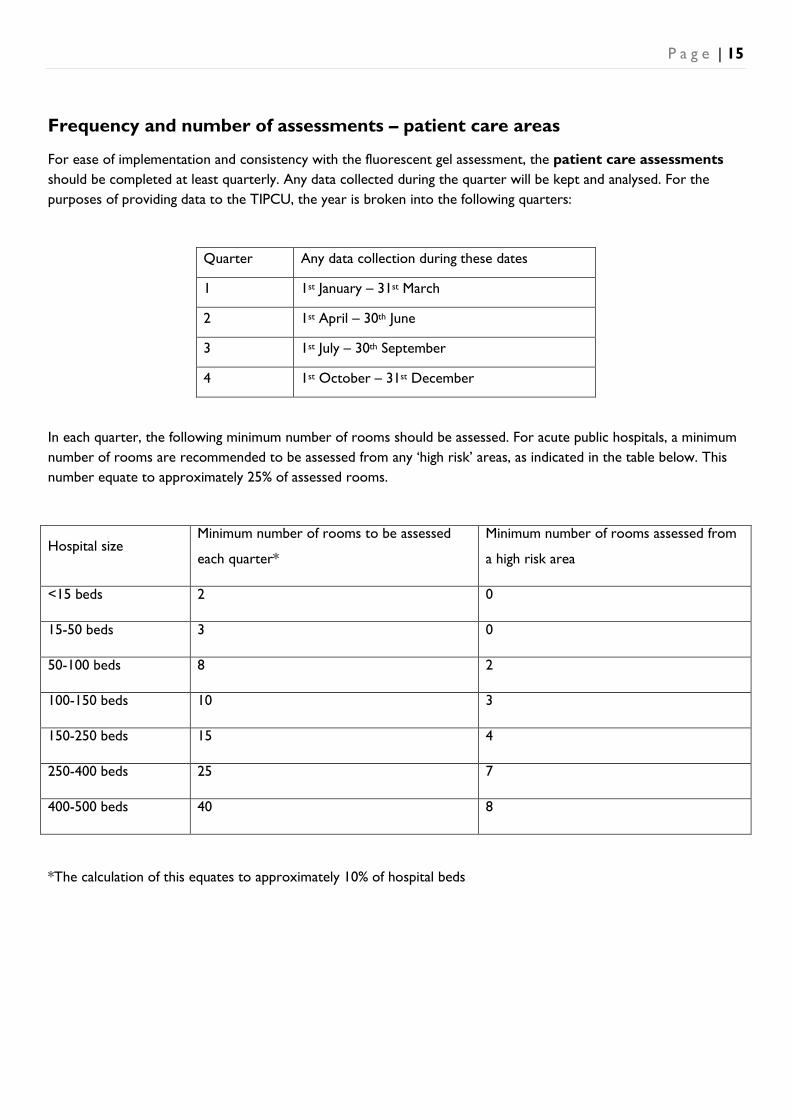
Frequency and number of assessments – patient care areas
For ease of implementation and consistency with the fluorescent gel assessment, the patient care assessments should be completed at least quarterly. Any data collected during the quarter will be kept and analysed. For the purposes of providing data to the TIPCU, the year is broken into the following quarters:
Quarter Any data collection during these dates
1 1st January – 31st March
2 1st April – 30th June
3 1st July – 30th September
4 1st October – 31st December
In each quarter, the following minimum number of rooms should be assessed. For acute public hospitals, a minimum number of rooms are recommended to be assessed from any ‘high risk’ areas, as indicated in the table below. This number equate to approximately 25% of assessed rooms.
Hospital size Minimum number of rooms to be assessed
each quarter*
Minimum number of rooms assessed from
a high risk area
<15 beds 2 0
15-50 beds 3 0
50-100 beds 8 2
100-150 beds 10 3
150-250 beds 15 4
250-400 beds 25 7
400-500 beds 40 8
*The calculation of this equates to approximately 10% of hospital beds
P a g e | 16
Frequency and number of assessments –general ward areas
The general ward assessments can be undertaken at a frequency determined locally.
The procedure
The procedure is set out below:
1. Identify a room to be assessed. This should be after the room has been cleaned, either a routine or discharge clean.
2. Complete the assessment (patient care area and/ or general ward area) using the online assessment tool.
a. For each item in the assessment tool, mark “Clean” or “Not Clean”.
b. If an item is not present or cannot be assessed, mark “Not applicable”.
3. Results should be fed back to the supervisor and/or cleaner as determined locally.
Online assessment tool
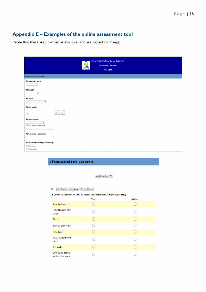
Use the online assessment tool developed by the TIPCU for completion of both the fluorescent gel assessment and visual assessment. Participating hospitals will be given their own unique web address that links directly to the tool. Information regarding access to data/reports is provided in the next section.
The TIPCU will manage the online assessment tool to ensure consistency across the State. Examples of the online assessment tool are provided in Appendix B, but are subject to change.
P a g e | 17
Assessors
Only persons who have undergone training and successfully undertaken an exam may undertake the assessments contained in this protocol. This is to ensure a high level of inter-rater reliability given the relatively subjective nature of these assessments and to have a mechanism for communicating changes and updates to the assessors. To support this process, the TIPCU have developed a PowerPoint presentation explaining this protocol and the assessment process. The TIPCU will provide initial training on the assessment tool to a limited number of hospital hotel services manager/s or supervisors and infection control staff.
The following process should occur for anyone who wants to or is required to undertake assessing:
1. Hospitals should identify individuals who will undertake this assessment.
2. Potential assessor should review the PowerPoint presentation provided by the TIPCU. If they have any questions, they should seek advice from their infection control unit and/or relevant hotel services manager/s or supervisors.
3. Once ready to undertake an exam, the infection control unit and/or hotel services manager/s or supervisor/s will provide the potential assessor with a link to an online multiple choice questionnaire exam. This exam is written in a manner that is easily understood.
4. Once the exam is completed, the TIPCU will be notified of the result.
• For persons who obtained an 80% or above result:
TIPCU will notify the assessor and supervisor1 of the result.
Their name will be added to list of assessors on the online assessment tool.
• For persons did not obtain an 80% or above result:
The TIPCU will contact their supervisor1. 1 Supervisor details will be collected as a mandatory part of the exam process
Reporting
Reports regarding both the fluorescent gel and visual a can be run locally by nominated persons in the participating hospital. For each participating hospital, the TIPCU will provide a unique password protected web link from which reports can be run. The report data can be downloaded into an Excel document for further manipulation as needed.
The Tasmanian healthcare associated advisory committee (or equivalent) will review data at a State level to examine any trends.
P a g e | 18
References 1. Dancer SJ. The role of environmental cleaning in the control of hospital-acquired infection. J Hosp
Infect. 2009;73(4):378-85.
2. Dancer SJ. Hospital cleaning in the 21st century. European Journal of Clinical Microbiology and
Infectious Disease. 2011;30(12):1473-81. Epub 2011 Apr 17.
3. Cooper RA, Griffith CJ, Malik RE, Obee P, Looker N. Monitoring the effectiveness of cleaning in
four British hospitals. Am J Infect Control. 2007;35(5):338-41.
4. Griffith C, Cooper R, Gilmore J, Davies C, Lewis M. An evaluation of hospital cleaning regimes and
standards. J Hosp Infect. 2000;45(1):19-28.
5. Carling PC, Parry MM, Rupp ME, Po JL, Dick B, Von Beheren S. Improving cleaning of the
environment surrounding patients in 36 acute care hospitals. Infect Control Hosp Epidemiol.
2008;29(11):1035-41.
6. Malik RE, Cooper RA, Griffith CJ. Use of audit tools to evaluate the efficacy of cleaning systems in
hospitals. Am J Infect Control. 2003;31(3):181-7.
7. Mitchell BG, Wilson, F., Dancer, SJ., McGregor, A. Methods to evaluate environmental cleanliness in
healthcare facilities. Healthcare Infection. 2013;18(1).
8. Australian Commission on Safety and Quality in Health Care (ACSQHC). National Safety and
Quality Health Service Standards (September 2011),. Sydney: ACSQHC; 2012.
9. Carling PC, Briggs J, Hylander D, Perkins J. An evaluation of patient area cleaning in 3 hospitals using
a novel targeting methodology. Am J Infect Control. 2006;34(8):513-9.
10. Carling PC, Briggs JL, Perkins J, Highlander D. Improved Cleaning of Patient Rooms Using a New
Targeting Method. Clin Infect Dis. 2006 February 1, 2006;42(3):385-8.
11. Carling PC, Parry MF, Bruno-Murtha LA, Dick B. Improving environmental hygiene in 27 intensive
care units to decrease multidrug-resistant bacterial transmission*. Crit Care Med. 2010;38(4):1054.
12. Goodman ER, Platt R, Bass R, Onderdonk AB, Yokoe DS, Huang SS. Impact of an environmental
cleaning intervention on the presence of methicillin-resistant Staphylococcus aureus and vancomycin-
resistant enterococci on surfaces in intensive care unit rooms. Infect Control Hosp Epidemiol.
2008;29(7):593.
13. Murphy CL, Macbeth DA, Derrington P, Gerrard J, Faloon J, Kenway K, et al. An assessment of high
touch object cleaning thoroughness using a fluorescent marker in two Australian hospitals. Healthcare
Infection. 2012;16(4):156-63.
P a g e | 19
14. Bonten MJ, Slaughter S, Ambergen AW, Hayden MK, van Voorhis J, Nathan C, et al. The role of
"colonization pressure" in the spread of vancomycin-resistant enterococci: an important infection control
variable. Arch Intern Med. 1998;158(10):1127-32.
15. Eveillard M, Lancien E, Hidri N, Barnaud G, Gaba S, Benlolo JA, et al. Estimation of methicillin-
resistant Staphylococcus aureus transmission by considering colonization pressure at the time of hospital
admission. J Hosp Infect. 2005;60(1):27-31.
16. Merrer J, Santoli F, Appere de Vecchi C, Tran B, De Jonghe B, Outin H. "Colonization pressure"
and risk of acquisition of methicillin-resistant Staphylococcus aureus in a medical intensive care unit. Infect
Control Hosp Epidemiol. 2000;21(11):718-23.
17. Dancer SJ. How do we assess hospital cleaning? A proposal for microbiological standards for
surface hygiene in hospitals. J Hosp Infect. 2004;56(1):10-5.
18. Carling PC, Bartley JM. Evaluating hygienic cleaning in health care settings: What you do not know
can harm your patients. Am J Infect Control. 2010;38(5):S41-S50.
19. National Health & Medical Research Council. Australian Guidelines for the Prevention and Control
of Infection in Healthcare.: Commonwealth of Australia.; 2010.
20. Pratt RJ, Pellowe CM, Wilson JA, Loveday HP, Harper PJ, Jones S, et al. epic2: National evidence-
based guidelines for preventing healthcare-associated infections in NHS hospitals in England. J Hosp Infect.
2007;65:S1-S59.
21. Clinical Excellence Commission. Environmental Cleaning Policy. In: NSW Ministry of Health, editor.
Sydney: NSW Ministry of Health; 2012.
22. Department of Health. Cleaning standards for Victorian health facilities 2011 In: Quality Safety and
Patient Experience Branch, editor. Melbourne: Department of Health; 2011.
P a g e | 20
Appendix A – Agreement to Participate – DHHS acute care facility
Date:
Name of facility/hospital participating:
We, the undersigned, agree to participate in the Tasmanian Environmental Assessment Program that has been designed by the Tasmanian Infection Prevention and Control Unit (TIPCU) for use in healthcare facilities.
By signing this agreement we agree to:
• Perform the environmental assessments as outlined in the TIPCU Environmental Assessment Program Protocol.
• Submit collected data via the on-line data collection tool
We acknowledge that:
• TIPCU will provide Access to the password protected on-line data collection and reporting tool
• TIPCU will supply, for the Royal Hobart Hospital, Launceston General Hospital, NW Regional Hospital and Mersey Hospital, enough UV fluorescent gel markers and UV lights for the number of sites consistent with the protocol for one year from the date on this agreement, so long as the agreement is signed prior to December 31st 2013. After this time, the supply of such equipment is discretionary.
• TIPCU may use data submitted by your hospital for the purposes of aggregating Tasmanian level data i.e. state level – not hospital level data. These state level data may be published.
Executive Sponsor Infection Control Manager/Unit Environmental Services Manager
Name Name Name
Signature Signature Signature
Date Date Date
P a g e | 21
Appendix B – Agreement to Participate – non DHHS facility
Date:
Name of facility/hospital participating:
We, the undersigned, agree to participate in the Tasmanian Environmental Assessment Program that has been designed by the Tasmanian Infection Prevention and Control Unit (TIPCU) for use in healthcare facilities.
By signing this agreement we agree to:
• Perform the environmental assessments as outlined in the TIPCU Environmental Assessment Program Protocol.
• Submit collected data via the on-line data collection tool
We acknowledge that:
• TIPCU will provide access to the password protected on-line data collection and reporting tool
• TIPCU may use data submitted by your hospital for the purposes of aggregating Tasmanian level data i.e. state level – not hospital level data. These state level data may be published.
Executive Sponsor Infection Control Manager/Unit Environmental Services Manager
Name Name Name
Signature Signature Signature
Date Date Date
P a g e | 22
Appendix C – Agreement to Participate – DHHS Non - acute care facility
Date:
Name of facility/hospital participating:
We, the undersigned, agree to participate in the Tasmanian Environmental Assessment Program that has been designed by the Tasmanian Infection Prevention and Control Unit (TIPCU) for use in healthcare facilities.
By signing this agreement we agree to:
• Perform the environmental assessments as outlined in the TIPCU Environmental Assessment Program Protocol.
• Submit collected data via the on-line data collection tool
We acknowledge that:
• TIPCU will provide access to the password protected on-line data collection and reporting tool
• TIPCU will supply data collection tools, UV fluorescent gel markers and UV lights for the number of sites consistent with the protocol for one year from the date on this agreement.
• TIPCU will evaluate the Tasmanian Environmental Assessment Program in non-acute DHHS sites after six months.
• TIPCU may use data submitted by your hospital for the purposes of aggregating Tasmanian level data i.e. state level – not hospital level data. These state level data may be published.
Executive Sponsor Infection Control Manager/Unit Environmental Services Manager
Name Name Name
Signature Signature Signature
Date Date Date
P a g e | 23
Appendix D – Example of where to apply the gel
Patient call bell / button
Bed rail
Bed side – neonatal cot
P a g e | 24
Access hatch – neonatal incubator
Bedside locker / patient storage unit
Patient tray table
P a g e | 25
Patient chair
Toilet / bathroom door handle
Tap handle
Door handle (inside patient room)
P a g e | 26
Appendix E – Examples of the online assessment tool (Note that these are provided as examples and are subject to change)

TASMANIAN INFECTION PREVENTION AND CONTROL UNIT
Population Health
Department of Health and Human Services
GPO Box 125, Hobart 7001