Embed Size (px)
Citation preview
ENV H 452/542: Environmental and Occupational
Health Microbiology II:
Detection and Control of Environmentally Transmitted
Microbial Hazards
John Scott MeschkeOffice: 4225 Roosevelt Way NE, Suite 2338
Phone: 206-221-5470
Email: [email protected]
Gwy-Am ShinOffice: 4225 Roosevelt Way NE, Suite 2339
Phone: 206-543-9026
Email: [email protected]
Course Description
• This course will review environmental detection and control of pathogenic organisms.
• The first half of the course will cover methods of sample collection, processing and target detection.
• The second half of the course will examine methods of decontamination and disinfection, as well as other engineered controls of environmentally transmitted pathogens.
Course Objectives
Students will gain a working knowledge of:• the methods used for sample collection,
processing and target detection of environmentally transmitted pathogens,
• methods of disinfection and decontamination of environmental media, and
• practices and engineered controls for the containment of pathogens and prevention of their spread.

Text and References
• Recommended Text: Environmental Microbiology (Maier, Pepper, and Gerba; Academic Press)
• Readings will typically be assigned for each class session (20-25 pages; from text or will be distributed in class or on website)
Reference Texts and Journals
Course Format
• Lectures (typically)– 5 Minutes Questions– 30-40 Minutes Lecture Material– 5-10 Minutes Follow-up Questions
• Student-Led Discussions– Division into 3 groups (based on CVs)– One group leads per session – Choose article (or series of articles) and come-up with
list of discussion questions – Due 1 week before session

Grading Opportunities
• On decimal scale:– A(3.9-4.0) Excellent work; typically >>90% of
available points– D (0.9-1.1) very poor work; typically <66% of
available points
• Expected Graduate Average 3.7 or above• Expected Undergraduate Average 3.4 or
above

Grading Opportunities
• Graduate Students– CV (5%)– Homework (10%)– Midterm (25%)– Class Participation
(5%)– Oral Exam (15%)– Review Paper (15%)– Final Exam (25%)
• Undergraduates– CV (5%)– Homework (20%)– Midterm (30%)– Class Participation
(5%)– Oral Exam (10%)– Final Exam (30%)
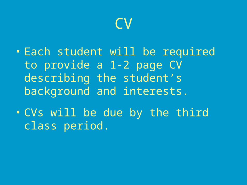
CV
• Each student will be required to provide a 1-2 page CV describing the student’s background and interests.
• CVs will be due by the third class period.

Homework
• Two (each worth 5 or 10% of overall grade for graduate and undergraduate students, respectively)
• Chance to earn points back (1/2 points lost)– Must indicate why answer given is wrong (NOT why
you missed it, i.e. I was thinking about something else) and Must indicate correct answer
– Due one week after initially returned
• Late homeworks may be penalized (10% for each class period late)
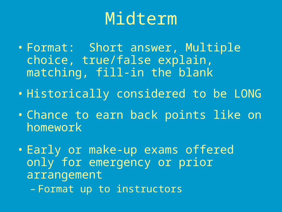
Midterm
• Format: Short answer, Multiple choice, true/false explain, matching, fill-in the blank
• Historically considered to be LONG
• Chance to earn back points like on homework
• Early or make-up exams offered only for emergency or prior arrangement– Format up to instructors
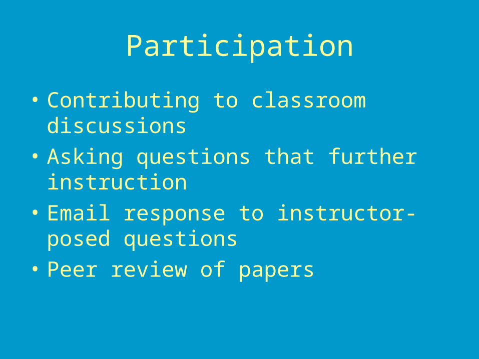
Participation
• Contributing to classroom discussions
• Asking questions that further instruction
• Email response to instructor-posed questions
• Peer review of papers

Review Paper• Graduate students ONLY; undergrads will
assist with peer review
• Topic relevant to course material
• Work in teams of two
• As long as necessary, but not to exceed 25 pages of double-spaced text; use ASM formatting
• Follow posted deadlines
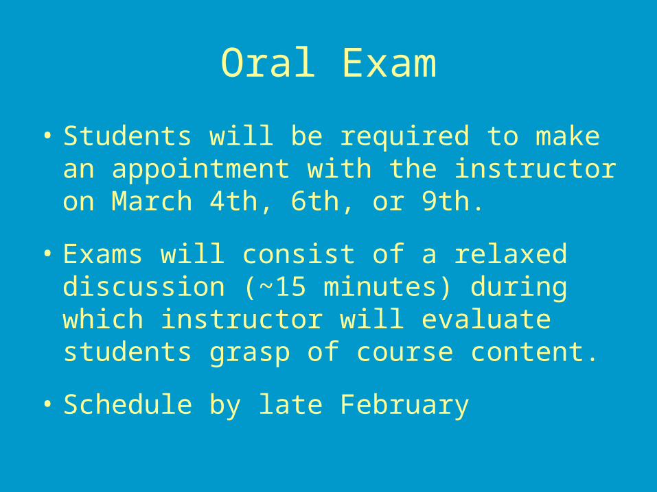
Oral Exam
• Students will be required to make an appointment with the instructor on March 4th, 6th, or 9th.
• Exams will consist of a relaxed discussion (~15 minutes) during which instructor will evaluate students grasp of course content.
• Schedule by late February

Final Exam
• March 18th, 2:30-4:20
• Similar format to midterm, may be problem solving questions
• Open note, open book
• NO opportunity to earn points back

Course Rules• Come to class, please try to let me know ahead of time
if you can not make it. • Arrive on time• Turn in assignments on time• Come to class prepared (keep up with reading)• Be courteous (No newspapers, audible cell phones,
PDAs, beepers)• Food and drinks are welcome (but keep it quiet)• Refrain from unnecessary talking• ASK QUESTIONS• Try to remain awake (at least no snoring please)• Let me know how I am doing (if I am moving too fast,
not being clear, or otherwise not getting the message across, I need to know.)
Use of Indicator Microorganisms

Microbial Indicator Concepts and Purposes
• The types of pathogens that can contaminate water, food, air and other environmental media are diverse and there are many different ones.
• Measuring all of these pathogens on a routine basis for determining presence or absence or acceptable concentration is not possible.– Methods are not available to recover and measure some
of them,– Methods are available for other pathogens, but they are
technically demanding, some are slow to produce results and their costs are high.
• The alternative is to measure something other than a pathogen that is indicative of contamination, predicts pathogen presence and estimates human health risks.
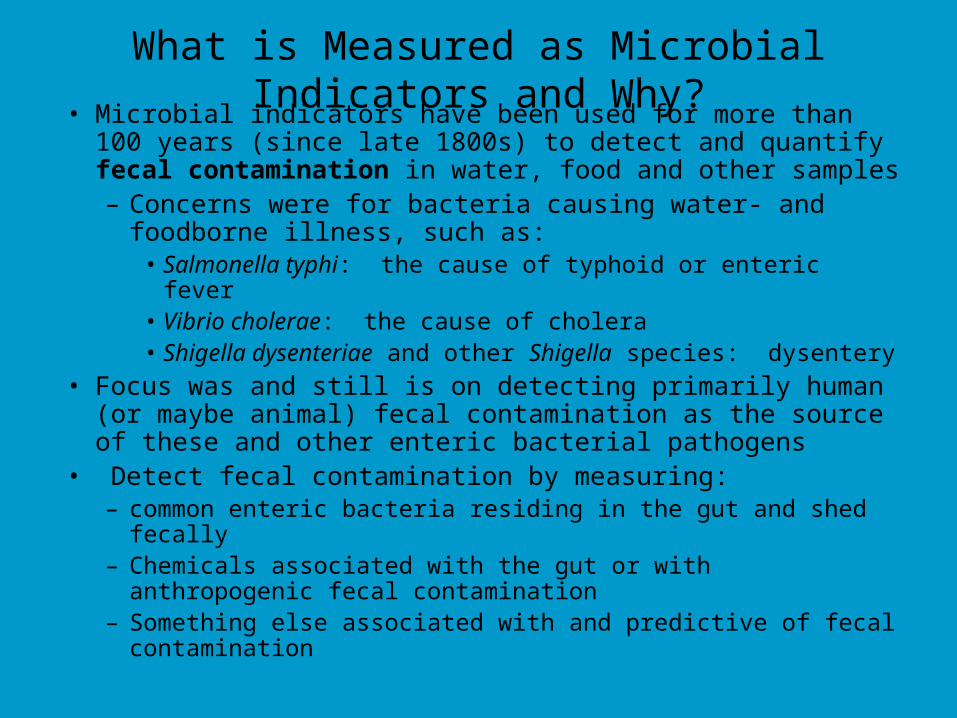
What is Measured as Microbial Indicators and Why?• Microbial indicators have been used for more than 100 years (since
late 1800s) to detect and quantify fecal contamination in water, food and other samples– Concerns were for bacteria causing water- and foodborne illness,
such as:• Salmonella typhi: the cause of typhoid or enteric fever• Vibrio cholerae: the cause of cholera• Shigella dysenteriae and other Shigella species: dysentery
• Focus was and still is on detecting primarily human (or maybe animal) fecal contamination as the source of these and other enteric bacterial pathogens
• Detect fecal contamination by measuring:– common enteric bacteria residing in the gut and shed fecally– Chemicals associated with the gut or with anthropogenic fecal
contamination– Something else associated with and predictive of fecal contamination
What is Measured as Microbial Indicators and Why?
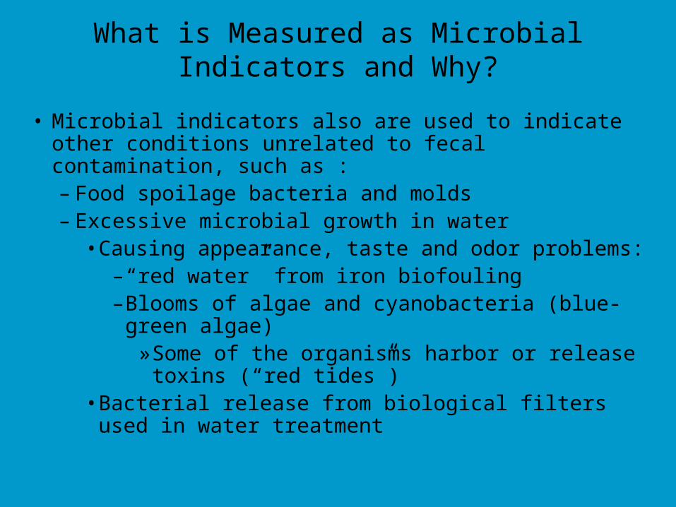
• Microbial indicators also are used to indicate other conditions unrelated to fecal contamination, such as :– Food spoilage bacteria and molds– Excessive microbial growth in water
• Causing appearance, taste and odor problems: – “red water” from iron biofouling– Blooms of algae and cyanobacteria (blue-green algae)
» Some of the organisms harbor or release toxins (“red tides”)
• Bacterial release from biological filters used in water treatment
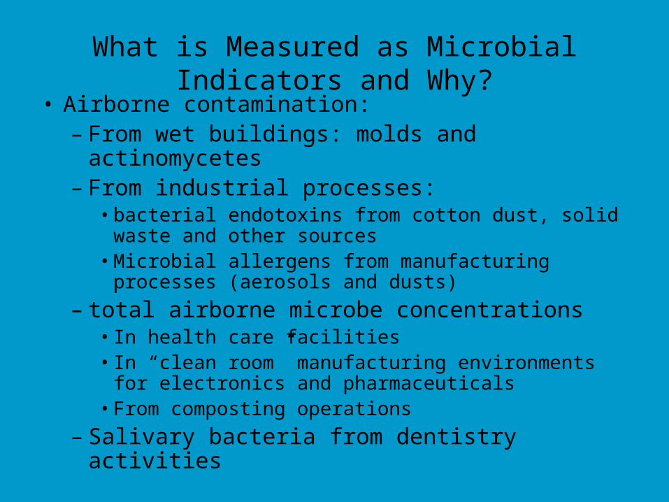
What is Measured as Microbial Indicators and Why?• Airborne contamination:
– From wet buildings: molds and actinomycetes– From industrial processes:
• bacterial endotoxins from cotton dust, solid waste and other sources
• Microbial allergens from manufacturing processes (aerosols and dusts)
– total airborne microbe concentrations• In health care facilities• In “clean room” manufacturing environments for electronics
and pharmaceuticals• From composting operations
– Salivary bacteria from dentistry activities
Pathogen Detection and Monitoring
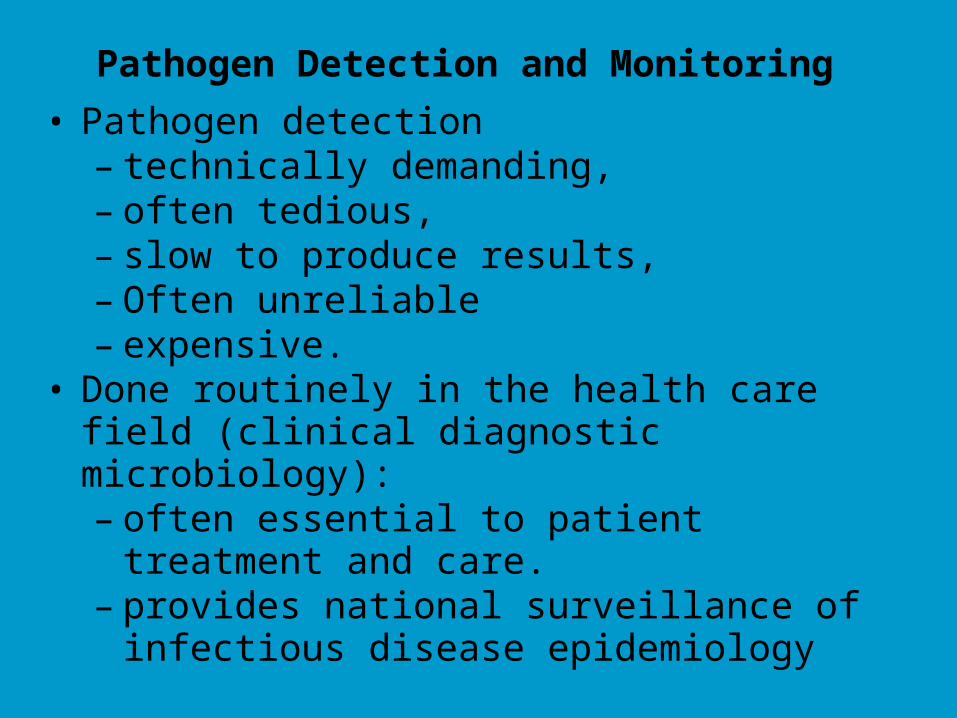
• Pathogen detection– technically demanding, – often tedious,– slow to produce results, – Often unreliable– expensive.
• Done routinely in the health care field (clinical diagnostic microbiology):– often essential to patient treatment and care.– provides national surveillance of infectious disease
epidemiology
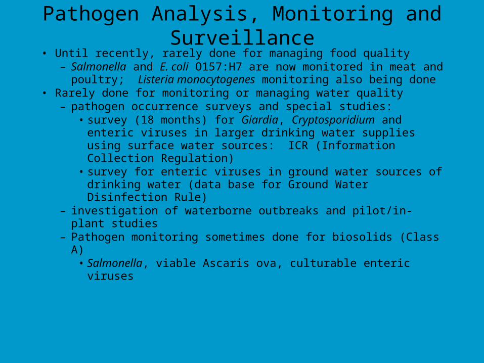
Pathogen Analysis, Monitoring and Surveillance• Until recently, rarely done for managing food quality
– Salmonella and E. coli O157:H7 are now monitored in meat and poultry; Listeria monocytogenes monitoring also being done
• Rarely done for monitoring or managing water quality– pathogen occurrence surveys and special studies:
• survey (18 months) for Giardia, Cryptosporidium and enteric viruses in larger drinking water supplies using surface water sources: ICR (Information Collection Regulation)
• survey for enteric viruses in ground water sources of drinking water (data base for Ground Water Disinfection Rule)
– investigation of waterborne outbreaks and pilot/in-plant studies– Pathogen monitoring sometimes done for biosolids (Class A)
• Salmonella, viable Ascaris ova, culturable enteric viruses
Microbial Indicators of Fecal Contamination
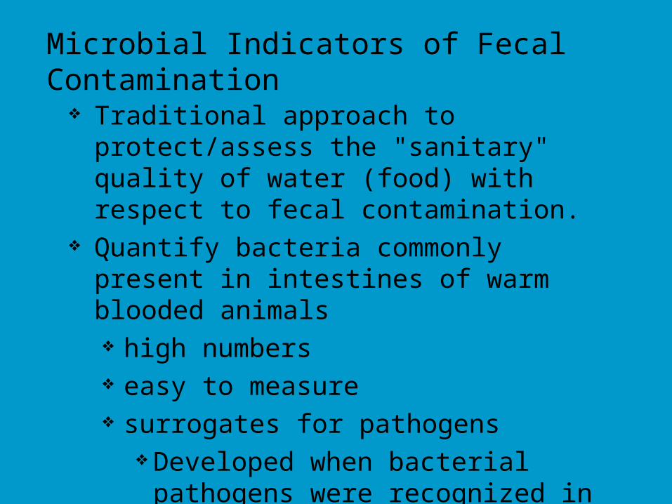
Traditional approach to protect/assess the "sanitary" quality of water (food) with respect to fecal contamination.
Quantify bacteria commonly present in intestines of warm blooded animals high numbers easy to measure surrogates for pathogens
Developed when bacterial pathogens were recognized in late 1800s and early 1900s
Salmonella, Shigella, V. chloerae, etc.
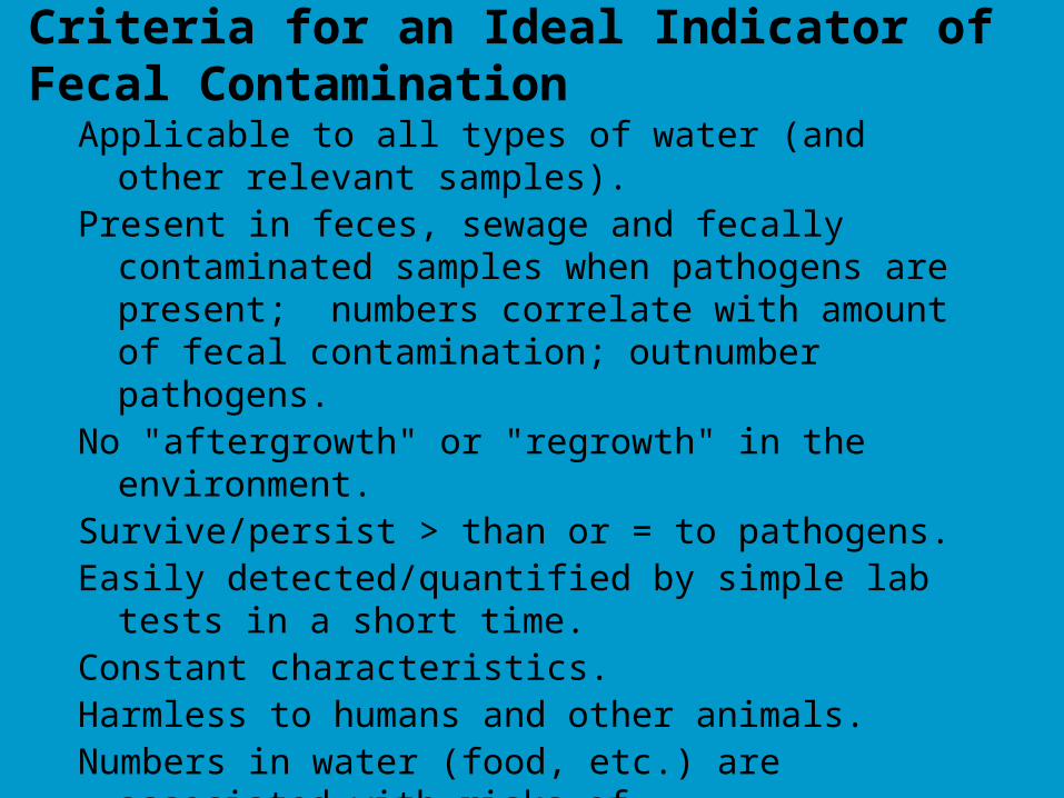
Criteria for an Ideal Indicator of Fecal Contamination
Applicable to all types of water (and other relevant samples).Present in feces, sewage and fecally contaminated samples when
pathogens are present; numbers correlate with amount of fecal contamination; outnumber pathogens.
No "aftergrowth" or "regrowth" in the environment.Survive/persist > than or = to pathogens.Easily detected/quantified by simple lab tests in a short time.Constant characteristics.Harmless to humans and other animals.Numbers in water (food, etc.) are associated with risks of
enteric illness in consumers (dose-response relationship).
Adapted from: Bonde, G.J. (1963) Bacterial Indicators of Water Pollution. Teknisk Forlag, Copenhagen; Bonde, G. J. (1966) Bacteriological Methods for Estimation of Water Pollution. Health Lab.
Sci., 3: 124.
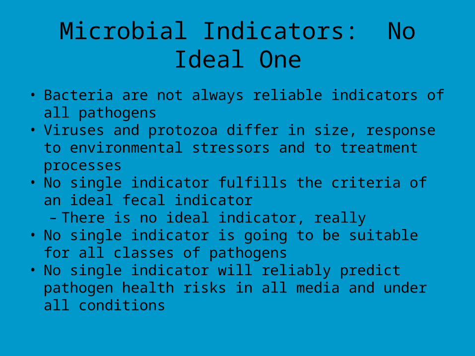
Microbial Indicators: No Ideal One
• Bacteria are not always reliable indicators of all pathogens• Viruses and protozoa differ in size, response to environmental
stressors and to treatment processes• No single indicator fulfills the criteria of an ideal fecal indicator
– There is no ideal indicator, really• No single indicator is going to be suitable for all classes of
pathogens• No single indicator will reliably predict pathogen health risks in
all media and under all conditions
Introduction to Sampling of Environmental Media
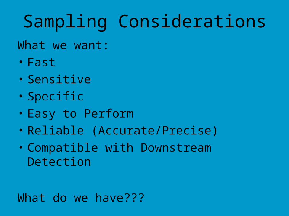
Sampling ConsiderationsWhat we want:
• Fast
• Sensitive
• Specific
• Easy to Perform
• Reliable (Accurate/Precise)
• Compatible with Downstream Detection
What do we have???
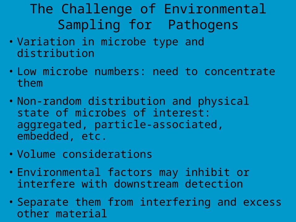
The Challenge of Environmental Sampling for Pathogens
• Variation in microbe type and distribution• Low microbe numbers: need to concentrate them• Non-random distribution and physical state of microbes
of interest: aggregated, particle-associated, embedded, etc.
• Volume considerations• Environmental factors may inhibit or interfere with
downstream detection• Separate them from interfering and excess other
material
Detection of Pathogens in The Environment
• Three main steps:
• (1) recovery and concentration,
• (2) purification and separation, and
• (3) assay and characterization.
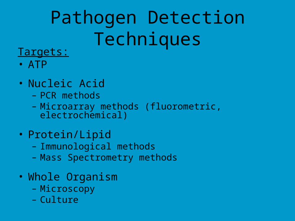
Pathogen Detection TechniquesTargets:• ATP
• Nucleic Acid– PCR methods– Microarray methods (fluorometric, electrochemical)
• Protein/Lipid– Immunological methods– Mass Spectrometry methods
• Whole Organism– Microscopy– Culture
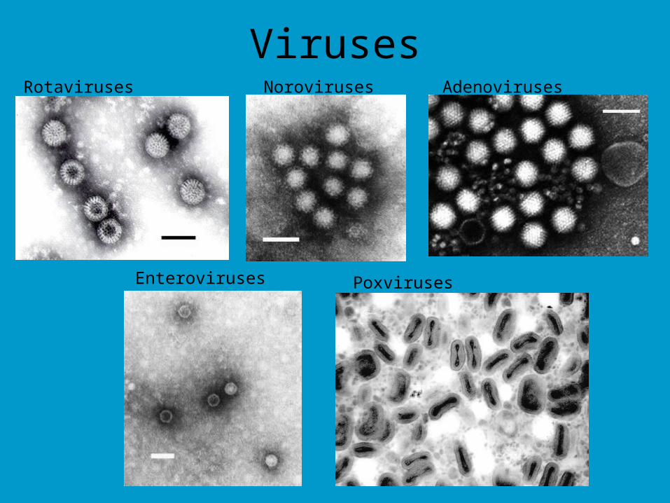
VirusesRotaviruses Noroviruses Adenoviruses
Enteroviruses Poxviruses
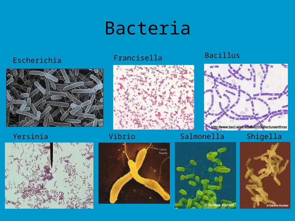
Bacteria
Escherichia Francisella Bacillus
Yersinia Vibrio Salmonella Shigella
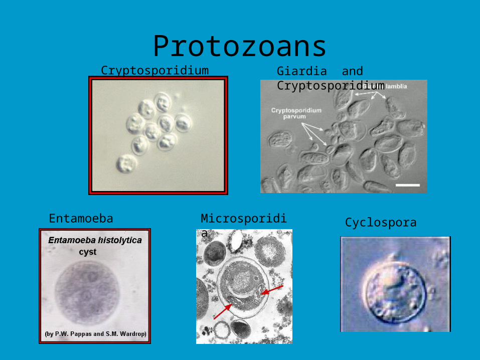
ProtozoansCryptosporidium Giardia and Cryptosporidium
Entamoeba Microsporidia Cyclospora
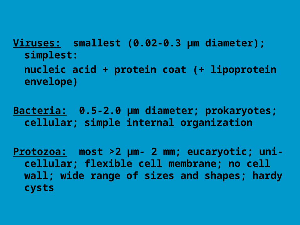
Viruses: smallest (0.02-0.3 µm diameter); simplest:
nucleic acid + protein coat (+ lipoprotein envelope)
Bacteria: 0.5-2.0 µm diameter; prokaryotes; cellular; simple internal organization
Protozoa: most >2 µm- 2 mm; eucaryotic; uni-cellular; flexible cell membrane; no cell wall; wide range of sizes and shapes; hardy cysts
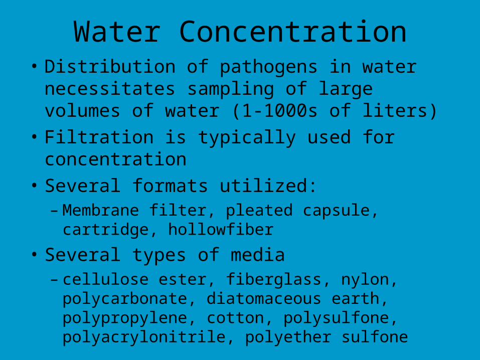
Water Concentration• Distribution of pathogens in water
necessitates sampling of large volumes of water (1-1000s of liters)
• Filtration is typically used for concentration
• Several formats utilized:– Membrane filter, pleated capsule, cartridge,
hollowfiber
• Several types of media – cellulose ester, fiberglass, nylon, polycarbonate,
diatomaceous earth, polypropylene, cotton, polysulfone, polyacrylonitrile, polyether sulfone
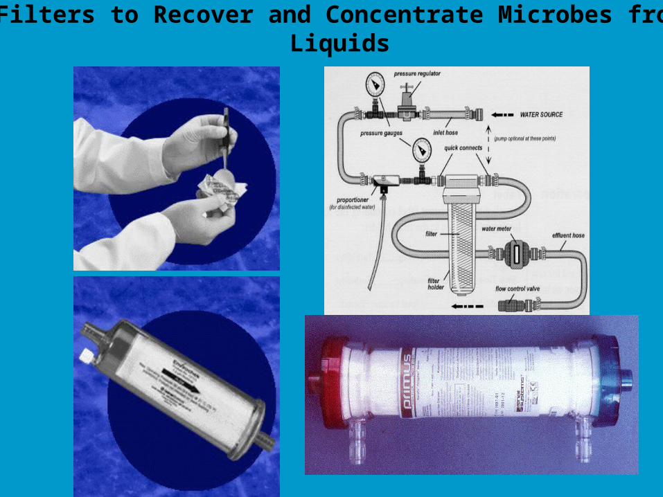
Filters to Recover and Concentrate Microbes from Liquids
Types of Filtration
• Size Exclusion/Retention
• Adsorption/Elution
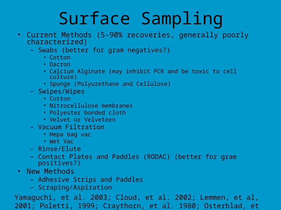
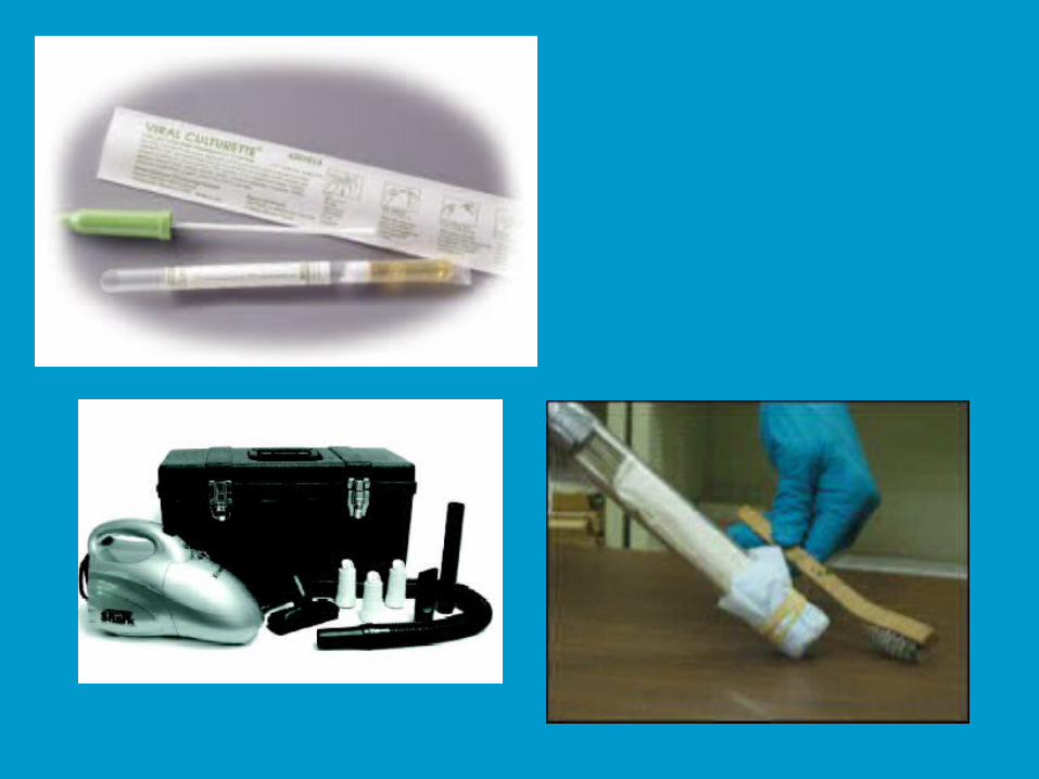
Surface Sampling• Current Methods (5-90% recoveries, generally poorly characterized)
– Swabs (better for gram negatives?)• Cotton• Dacron• Calcium Alginate (may inhibit PCR and be toxic to cell culture)• Sponge (Polyurethane and Cellulose)
– Swipes/Wipes• Cotton• Nitrocellulose membranes• Polyester bonded cloth• Velvet or Velveteen
– Vacuum Filtration• Hepa bag vac• Wet Vac
– Rinse/Elute– Contact Plates and Paddles (RODAC) (better for gram positives?)
• New Methods– Adhesive Strips and Paddles– Scraping/Aspiration
Yamaguchi, et al. 2003; Cloud, et al. 2002; Lemmen, et al, 2001; Poletti, 1999; Craythorn, et al. 1980; Osterblad, et al. 2003; Taku, et al. 2003
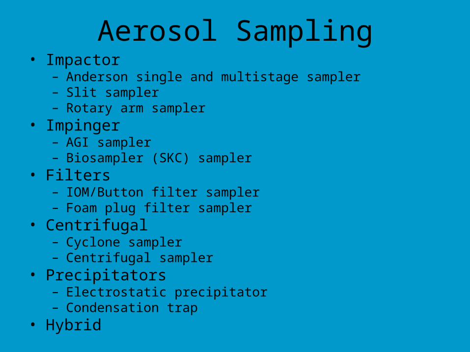
Aerosol Sampling• Impactor
– Anderson single and multistage sampler– Slit sampler – Rotary arm sampler
• Impinger– AGI sampler– Biosampler (SKC) sampler
• Filters– IOM/Button filter sampler– Foam plug filter sampler
• Centrifugal– Cyclone sampler– Centrifugal sampler
• Precipitators– Electrostatic precipitator– Condensation trap
• Hybrid
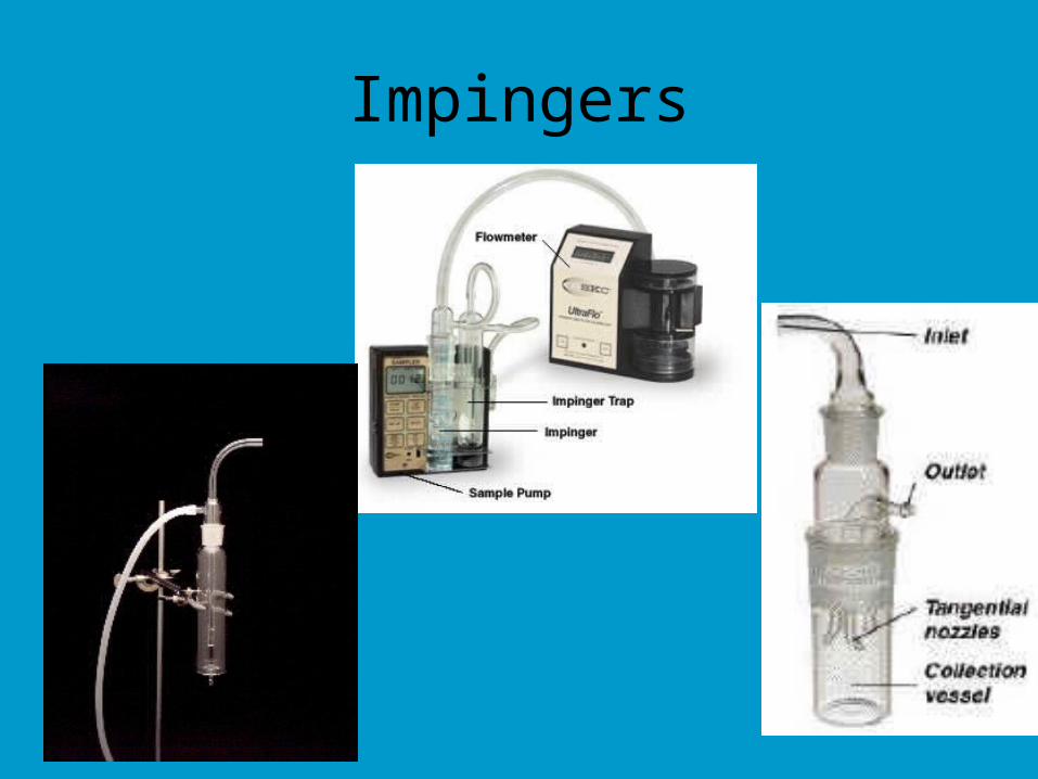
Impingers
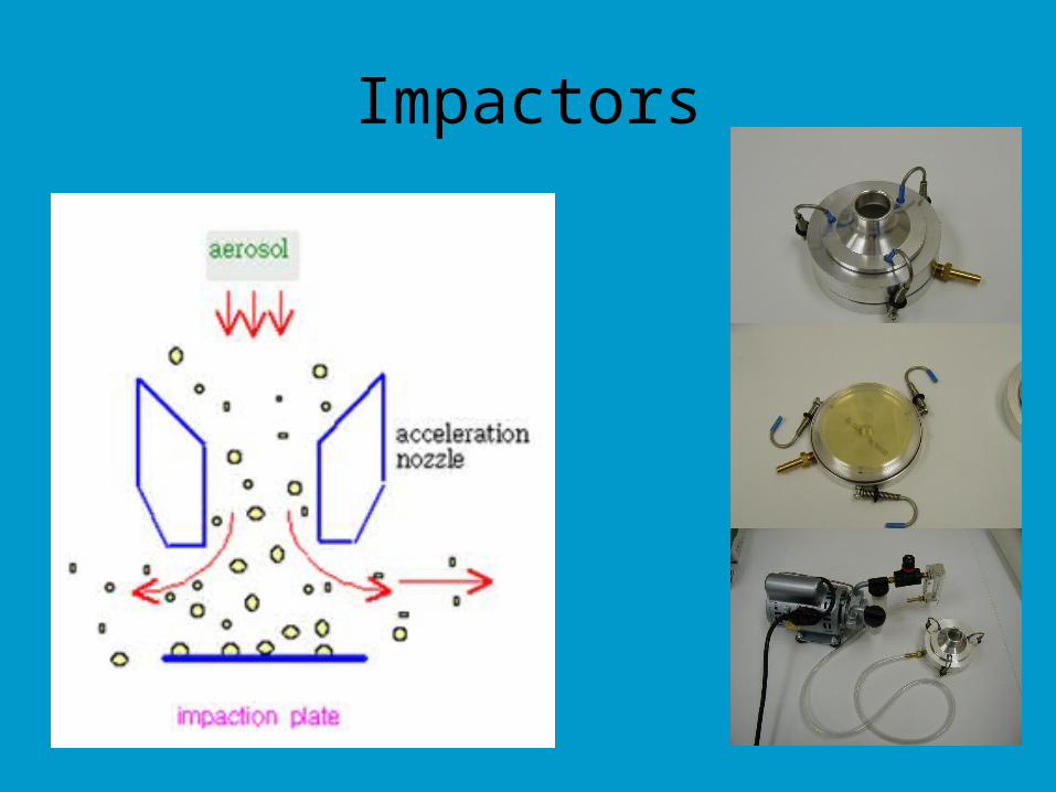
Impactors
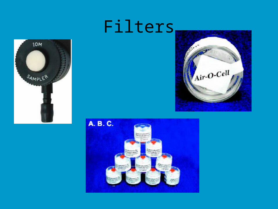
Filters
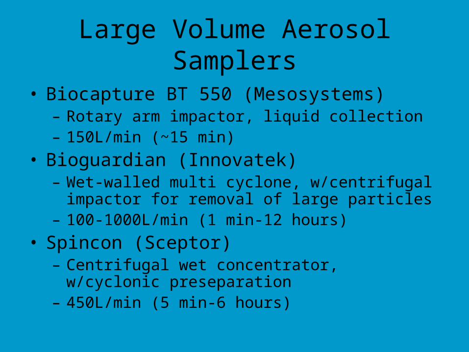
Large Volume Aerosol Samplers
• Biocapture BT 550 (Mesosystems)– Rotary arm impactor, liquid collection– 150L/min (~15 min)
• Bioguardian (Innovatek)– Wet-walled multi cyclone, w/centrifugal impactor for
removal of large particles– 100-1000L/min (1 min-12 hours)
• Spincon (Sceptor)– Centrifugal wet concentrator, w/cyclonic
preseparation– 450L/min (5 min-6 hours)
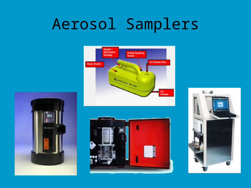
Aerosol Samplers
Separation and Purification Methods
• Purification, separation and concentration of target microbes in primary sample or sample concentrate
– Separate target microbes from other particles and from solutes
– Reduce sample size (further concentrate)
Separation/Purification Methods
• Variety of physical, chemical and immunochemical methods:– Sedimentation and flotation (primarily
parasites)– Precipitation (viruses)– Filtration (all classes)– Immunomagnetic separation or IMS (all
classes)– Flow cytometry (bacteria and parasites);
an analysis, too
Secondary Concentration and Purification
• PEG (polyethylene glycol)• Organic Flocculation • IMS (Immunomagnetic separation)• Ligand capture• BEaDs (Biodetection Enabling Device)• Capillary Electrophoresis• Microfluidics• Nucleic Acid Extraction• Spin Column Chromatography• Floatation• Sedimentation• Enrichment