Embed Size (px)
Citation preview

Entamoeba histolytica cell movement: A central rolefor self-generated chemokines and chemorepellentsMehreen Zaki, Natalie Andrew, and Robert H. Insall*
School of Biosciences, University of Birmingham, Edgbaston, Birmingham B15 2TT, United Kingdom
Edited by Kathryn V. Anderson, Sloan–Kettering Institute, New York, NY, and approved October 2, 2006 (received for review June 30, 2006)
Entamoeba histolytica cells, the cause of amoebic dysentery, arehighly motile, and this motility is an essential feature of the patho-genesis and morbidity of amoebiasis. However, the control of E.histolytica motility within the gut and during invasion is poorlyunderstood. We have used an improved chemotaxis assay to identifythe key extracellular signals mediating Entamoeba chemotaxis. Thedominant responses we observe are caused by factors generated byE. histolytica cells themselves. Medium that has been conditioned byE. histolytica growth causes both chemokinesis and negative chemo-taxis. The speed of random movement is more than doubled inconditioned compared with fresh medium, and cells move efficientlyaway from conditioned medium by negative chemotaxis. Ethanol, theproduct of Entamoeba glucose metabolism, is the principal compo-nent of the chemokinetic response. The closely related but nonpatho-genic Entamoeba dispar shows no change in motility in response toconditioned medium implying that these responses are central to E.histolytica pathogenesis.
invasion � negative chemotaxis � pathogenesis
An estimated 50 million individuals suffer the severe morbidityassociated with invasive Entamoeba histolytica infections, with
�100,000 deaths annually (1). Parasite–host interactions that de-termine the course of infection, in particular asymptomatic colo-nization vs. symptomatic invasive disease, are largely still a mystery.It is, however, accepted that amoebic adherence to and contact-dependent killing of target cells, followed by phagocytosis, are keyevents (1, 2). The ability to interact with target cell surfaces istherefore a major process underpinning amoebic invasion of thehuman intestine. Consequently, research centered on E. histolyticacell surface receptors has great clinical promise. In particular, a roleof chemotaxis seems likely. Zymosan activated C5a in humanserum, lysed red blood cells, whole bacteria, components of the ratcolon, N-acetylneuraminic acid (NANA) and NANA-containingcompounds, fibronectin and fibronectin-derived fragments, andhuman TNF have all been shown to provide chemotactic stimuli(3–7).
E. histolytica motility and chemotaxis have been studied by usingrelatively few methods, including in haemocytometers (8), tubemigration (9), and Boyden chamber assays (3). Under the first twoconditions, cells have almost no resistance to their movementexcept substrate adhesion. During invasive disease, however, amoe-bae move in more restrictive conditions, similar to metastasis andextravasation. Under-agarose (under-agar) assays provide such anenvironment and have been used to study the motility and chemo-taxis of a variety of cells, including neutrophils, macrophages (10,11), and the free-living amoeba Dictyostelium (12), an evolutionaryrelative of Entamoeba (13). Under-agar assays have the addedadvantage of allowing moving cells to be visualized and parameterssuch as cell shape to be studied in detail.
To date, there appears to have been only one attempt atestablishing an under-agar assay for studying E. histolytica motility(7). In this work, migratory distances were measured only afterfixation and staining of cells. An improved under-agar assay forstudying Dictyostelium chemotaxis under mechanically inhibitedconditions has recently been reported (14). The assay allows
high-resolution live imaging of cells, and movement is quantifiablefor both individual and whole populations of cells.
We have, therefore, adapted this assay for studying Entamoebachemotaxis. We have in the course of this study discovered thatcomponents of E. histolytica-conditioned culture medium are keydeterminants of E. histolytica cell motility. The response to condi-tioned medium (CM) includes an increase in both speed andnegative chemotaxis. Crucially, the commensal (nonpathogenic)Entamoeba dispar does not show any such behavior, implying thatnegative chemotaxis may play an important role in E. histolyticapathogenesis. These findings, therefore, have clinical implications,in particular suggesting a mechanism that drives E. histolytica cellmigration away from the intestinal lumen and mucous layer andtoward the underlying epithelium.
ResultsE. histolytica Chemotaxis Is Dominated by Cell-Derived Stimuli. Weoptimized the three-trough under-agar assay used by Dictyosteliumresearchers (14) for Entamoeba (see Supporting Text, which ispublished as supporting information on the PNAS web site, fordetails). LYI-S-2 medium and neat serum, which were found toattract Entamoeba in earlier studies (3), were included as internalcontrols. As expected, the migration distance was greatest towardLYI-S-2 (Table 1 and data not shown), which was �1.5 timesgreater than that seen with the MM-1 control (Table 1 and Fig. 1Bi).The response to serum was unexpectedly negligibly different fromthe control (Table 1 and Fig. 1Ai). LPA and cAMP, which arestrong chemoattractants for Dictyostelium, were not chemoattrac-tants for Entamoeba under any conditions (Table 1 and data notshown).
Dictyostelium cells in under-agar assays never move out of thewell unless a chemoattractant is present (ref. 14; corroborated byour observations; data not shown), but we consistently found that
Author contributions: M.Z. and R.H.I. designed research; M.Z. performed research; M.Z.,N.A., and R.H.I. analyzed data; and M.Z. and R.H.I. wrote the paper.
The authors declare no conflict of interest.
This article is a PNAS direct submission.
Abbreviations: CI, chemotactic index; FM, fresh medium; CM, conditioned medium; under-agar, under-agarose.
*To whom correspondence should be addressed. E-mail: [email protected].
© 2006 by The National Academy of Sciences of the USA
Table 1. Under-agar migratory responses of E. histolytica tosome chemoattractants using the original three-well assaytemplate
Migration distances, �m
Chemotactic stimulus I margin O margin
LYI-S-2 244.5 � 50.8 154.0 � 19.9Serum 142.8 � 12.3 146.4 � 9.8MM-1 150.8 � 16 169.0 � 7.4LPA 163.7 � 11.7 159.6 � 7.6cAMP 152.5 � 3.2 141.2 � 5.6
Numbers represent mean values of two experiments at 2 h.
www.pnas.org�cgi�doi�10.1073�pnas.0605437103 PNAS � December 5, 2006 � vol. 103 � no. 49 � 18751–18756
MIC
ROBI
OLO
GY
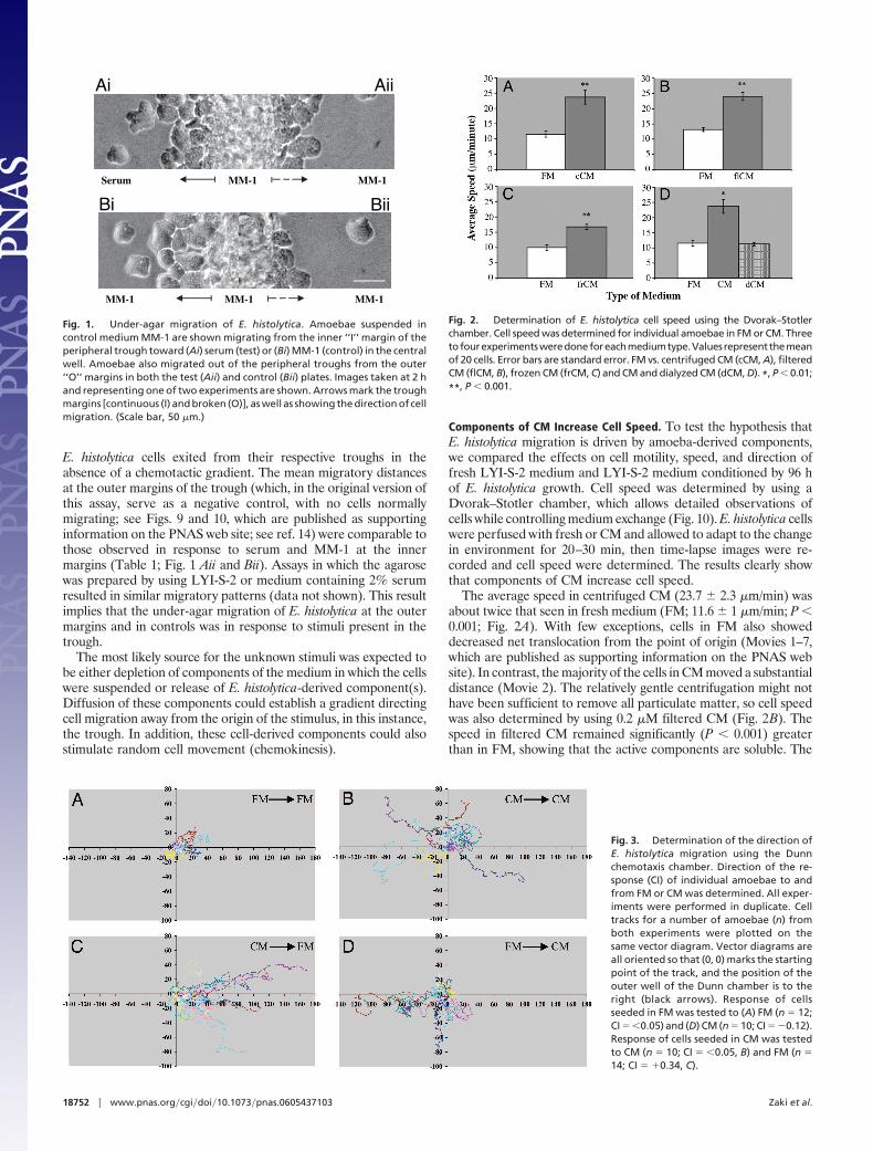
E. histolytica cells exited from their respective troughs in theabsence of a chemotactic gradient. The mean migratory distancesat the outer margins of the trough (which, in the original version ofthis assay, serve as a negative control, with no cells normallymigrating; see Figs. 9 and 10, which are published as supportinginformation on the PNAS web site; see ref. 14) were comparable tothose observed in response to serum and MM-1 at the innermargins (Table 1; Fig. 1 Aii and Bii). Assays in which the agarosewas prepared by using LYI-S-2 or medium containing 2% serumresulted in similar migratory patterns (data not shown). This resultimplies that the under-agar migration of E. histolytica at the outermargins and in controls was in response to stimuli present in thetrough.
The most likely source for the unknown stimuli was expected tobe either depletion of components of the medium in which the cellswere suspended or release of E. histolytica-derived component(s).Diffusion of these components could establish a gradient directingcell migration away from the origin of the stimulus, in this instance,the trough. In addition, these cell-derived components could alsostimulate random cell movement (chemokinesis).
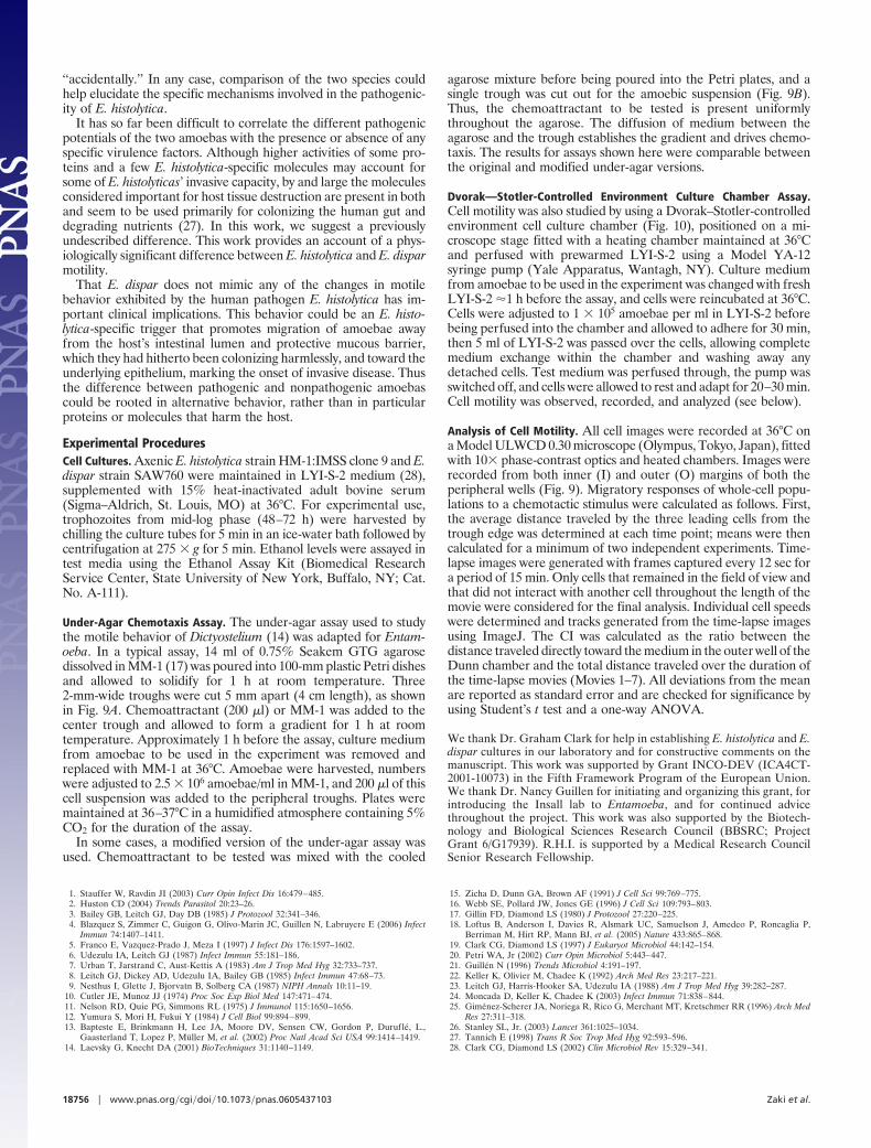
Components of CM Increase Cell Speed. To test the hypothesis thatE. histolytica migration is driven by amoeba-derived components,we compared the effects on cell motility, speed, and direction offresh LYI-S-2 medium and LYI-S-2 medium conditioned by 96 hof E. histolytica growth. Cell speed was determined by using aDvorak–Stotler chamber, which allows detailed observations ofcells while controlling medium exchange (Fig. 10). E. histolytica cellswere perfused with fresh or CM and allowed to adapt to the changein environment for 20–30 min, then time-lapse images were re-corded and cell speed were determined. The results clearly showthat components of CM increase cell speed.
The average speed in centrifuged CM (23.7 � 2.3 �m/min) wasabout twice that seen in fresh medium (FM; 11.6 � 1 �m/min; P �0.001; Fig. 2A). With few exceptions, cells in FM also showeddecreased net translocation from the point of origin (Movies 1–7,which are published as supporting information on the PNAS website). In contrast, the majority of the cells in CM moved a substantialdistance (Movie 2). The relatively gentle centrifugation might nothave been sufficient to remove all particulate matter, so cell speedwas also determined by using 0.2 �M filtered CM (Fig. 2B). Thespeed in filtered CM remained significantly (P � 0.001) greaterthan in FM, showing that the active components are soluble. The
Fig. 3. Determination of the direction ofE. histolytica migration using the Dunnchemotaxis chamber. Direction of the re-sponse (CI) of individual amoebae to andfrom FM or CM was determined. All exper-iments were performed in duplicate. Celltracks for a number of amoebae (n) fromboth experiments were plotted on thesame vector diagram. Vector diagrams areall oriented so that (0, 0) marks the startingpoint of the track, and the position of theouter well of the Dunn chamber is to theright (black arrows). Response of cellsseeded in FM was tested to (A) FM (n � 12;CI � �0.05) and (D) CM (n � 10; CI � �0.12).Response of cells seeded in CM was testedto CM (n � 10; CI � �0.05, B) and FM (n �14; CI � �0.34, C).
Ai Aii
Bi Bii
MM-1
MM-1 MM-1MM-1
MM-1Serum
Fig. 1. Under-agar migration of E. histolytica. Amoebae suspended incontrol medium MM-1 are shown migrating from the inner ‘‘I’’ margin of theperipheral trough toward (Ai) serum (test) or (Bi) MM-1 (control) in the centralwell. Amoebae also migrated out of the peripheral troughs from the outer‘‘O’’ margins in both the test (Aii) and control (Bii) plates. Images taken at 2 hand representing one of two experiments are shown. Arrows mark the troughmargins [continuous (I) and broken (O)], as well as showing the direction of cellmigration. (Scale bar, 50 �m.)
Fig. 2. Determination of E. histolytica cell speed using the Dvorak–Stotlerchamber. Cell speed was determined for individual amoebae in FM or CM. Threetofourexperimentsweredoneforeachmediumtype.Values representthemeanof 20 cells. Error bars are standard error. FM vs. centrifuged CM (cCM, A), filteredCM (flCM, B), frozen CM (frCM, C) and CM and dialyzed CM (dCM, D). *, P � 0.01;
**, P � 0.001.
18752 � www.pnas.org�cgi�doi�10.1073�pnas.0605437103 Zaki et al.

average speed in LYI-S-2, which had been incubated at 37°C for96 h without cells, was comparable to that seen with FM (data notshown).
CM Factors Are Stable and Dialyzable. To further characterize thenature of the active components of CM, aliquots of CM were frozenat �20°C for 1 week and 1 and 5 months. Aliquots were thawed atdesignated time points, and cell speed was determined in compar-ison with FM. Fig. 2C represents the increase in average cell speedwith CM that had been frozen for 5 months (P � 0.001) vs. FM. Nosignificant difference was observed between the aliquots frozen for1 week and 1 and 5 months (15.7 � 0.9, 17.8 � 1.4, and 16.7 � 0.84�m/min, respectively). However, a significant (P � 0.01) decreasewas seen with frozen CM when compared with earlier results(centrifuged and filtered). This loss in activity does not appear to beaffected by the duration of the freezing but rather by the process offreezing and/or thawing itself.
Finally, cell speed was also compared between FM, CM, anddialyzed CM (Fig. 2D). To prepare the dialyzed fraction, CM wasdialyzed against two changes of 10� volumes of FM at 4°C over a24-h period by using 12- to 14-kDa Visking dialysis tubing (MedicellInternational, London, U.K.). The average speed of E. histolytica indialyzed CM was comparable to that seen with FM, and undialyzedCM speed was significantly (P � 0.01) greater than both FM and
dialyzed CM. The active components are thus smaller than 12–14kDa in size.
CM Causes Chemotaxis as Well as Chemokinesis. We have clearlyshown that CM components cause chemokinesis. However, ourresults from the under-agar assay suggested a chemotactic response.We therefore studied amoebic responses to CM by using Dunnchambers, which allow cell direction to be analyzed under definedconditions (15, 16). As expected, cells suspended in homogenousmedium moved randomly, and cells moved more slowly in FM thanin CM (Fig. 3 A and B and Movies 3 and 4). In gradients of FM andCM, cells moved directionally, showing that CM induces chemo-taxis as well as chemokinesis.
When cells were seeded in CM, they nearly uniformly migratedtoward FM [chemotactic index (CI) � �0.34] in the outer well ofthe Dunn chamber (Fig. 3C and Movie 5). When the experimentalsetup was reversed, the amoebae seeded in FM were clearlyrepelled (CI � �0.12) by the CM in the outer well of the Dunn
Table 2. Under-agar migratory responses of E. histolytica to andfrom different media using the modified single trough assay
Chemotactic stimulusMigration
distances, �m
CM*3 FM 506.4 � 61.2FM*3 CM 80.13 � 8.4FM*3 FM 211.5 � 23.5CM*3 CM 129.7 � 12.3MM-1Gluc�1*3MM-1Gluc�1 14 � 7.3MM-1Gluc�1*3 FM 665.9 � 26.8�
CM*3MM-1Gluc�1 721.7 � 21.5�
MM-1Gluc�1*3 CM 266.6 � 26.3FM*3MM-1Gluc�1 205.3 � 47
Numbers represent mean values of three experiments at 6 h. FM (freshLYI-S-2 medium); CM (96-hr-old E. histolytica-conditioned LYI-S-2 medium);MM-1Gluc�1 (MM-1 medium without glucose). Medium in the trough is de-noted by (*),whereas the arrow points towards the medium in the agaroselayer. Statistical comparisons (�) show a significant increase in distance mi-grated; P � 0.01 (also see Fig. 5).
D
FM FM
CM CM
CM FM
FM CM
C
B
A
Fig. 4. Determination of the direction of E. histolytica migration using theunder-agar chemotaxis assay. Direction of the response of a population ofamoebae to and from FM or CM was determined. Images taken at 6 h andrepresenting one of three experiments are shown. Response of amoebae in FMwas tested to FM (A) and CM (D). Response of amoebae in CM was tested to CM(B) and FM (C). The broken arrow marks the trough margins as well as showingthe direction of cell migration. C depicts the maximum width of the visual field atthis magnification and not the furthest migrated (leading) cell(s). (Scale bar,50 �m.)
MM-1Gluc - MM-1Gluc -
FM MM-1Gluc-
MM-1Gluc- FM
CM MM-1Gluc -
MM-1Gluc- CM
A
B
C
D
E
Fig. 5. Determination of E. histolytica migration (un-der-agar) using MM-1Gluc� as the control. Direction ofthe response of a population of amoebae to FM or CMwas determined. Images taken at 6 h and representingone of three experiments are shown. Response of amoe-bae in MM-1Gluc� was tested to MM-1Gluc� (A), CM (D),and FM (E). Response of amoebae in CM was tested toMM-1Gluc� (B) and finally, the response of amoebae inFM was tested to MM-1Gluc� (C). The broken arrowmarks the trough margins as well as showing the direc-tion of cell migration.B depicts the maximum width ofthe visual field at this magnification and not the furthestmigrated (leading) cell(s). Statistical comparisons be-tween B and A and E and A show a significant increasein distance migrated; P � 0.01. (Scale bar, 50 �m.)
Zaki et al. PNAS � December 5, 2006 � vol. 103 � no. 49 � 18753
MIC
ROBI
OLO
GY

chamber (Fig. 3D and Movie 6). These results could indicate eitherpositive chemotaxis toward FM or negative chemotaxis away fromthe CM in which they were seeded.
Positive vs. Negative Chemotaxis. To distinguish whether amoebaewere in fact attracted to FM or repelled by CM, we adopted asimplified variation of the under-agar assay used earlier. Thissingle-trough assay (Fig. 9B) measures chemotactic gradientsformed between the medium in the trough and the medium in theagarose. We first tested this assay to see whether it would supportthe Dunn chamber analysis (Fig. 3). As before, cells in CM in thetrough migrated significant distances under agarose containingFM, whereas cells in FM barely moved under agarose containingCM (Table 2 and Fig. 4 C and D), when the experimental setup wasreversed. Amoebae were also able to migrate out of their respectivetroughs to different degrees when in homogenous FM and CM(Table 2 and Fig. 4 A and B). The under-agar assay therefore mimicsthe results achieved with the Dunn chamber.
We then tested both FM and CM against the MM-1 control (17)in all possible combinations by using the single-trough under-agarassay. The distance migrated was similar for all of the test settings,including the control (data not shown). This and our earlierobservation (Fig. 1 Aii and Bii) suggested that MM-1 was sufficientto support E. histolytica-CM chemotaxis. However, a modificationof MM-1 without glucose (the only ingredient unnecessary forshort-term viability, attachment and motility; ref. 17) provided asharp contrast to all of the other tested media. MM-1Gluc� causedlittle if any under-agar migration of control cells (Table 2 and Fig.5A). This shows that MM-1Gluc� neither contains chemoattrac-tants that can be depleted nor supports the generation of cell-derived chemorepellents. MM-1Gluc� is thus an ideal basis todistinguish positive and negative chemotaxis.
We tested the migration of amoebae in different combinations ofMM-1Gluc�, CM and FM in the well, and the agarose. The resultswere complex, but the clearest result was obtained with cells in CMand MM-1Gluc� agarose. Cells migrated rapidly away from thetrough into the agarose (P � 0.01; Table 2 and Fig. 5B). BecauseMM-1Gluc� itself is not a chemoattractant, this clearly demon-strates that cells in glucose-containing medium make a negativechemoattractant for E. histolytica.
Amoebae in MM-1Gluc� migrated toward either CM or FM
(Table 2 and Fig. 5 D and E), somewhat more toward the FM (P �0.01). These effects were consistently less pronounced than whencells move away from CM. We believe that the migration ofamoebae toward CM reflects a somewhat diluted response to theattractive components of LYI-S-2 culture medium, which arepresumably partially but not yet completely depleted from the CM.
Our earlier observations (Fig. 3 and 4) and those shown inprevious work (7) were therefore presumably caused by a combi-nation of repulsion from component(s) of CM and by attractiontoward FM. Fig. 5 shows that negative chemotaxis from CM has agreater effect than the positive chemotaxis toward medium seenpreviously and is thus a principal determinant of cell motility undernormal experimental conditions.
Ethanol Is a Key Component of CM. Comparison of E. histolyticaresponses to LYI-S-2, MM-1, and MM-1Gluc� strongly suggestedthat the active components of CM were being generated as aconsequence of glucose metabolism. E. histolytica lives by anaerobic
Fig. 6. Determination of E. histolytica cell speed in simulated-CM using theDvorak–Stotler chamber. Cell speed was determined for individual amoebaein FM, CM, and simulated-CM (FETOH) containing varying (10, 25, 50, and 100mM) amounts of ethanol. Three to four experiments were done for eachmedium type. Values represent the mean of 10 cells. Error bars are standarderror. Statistical comparisons between 100 mM FETOH and FM and CM and FMshow a significant increase in cell speed; P � 0.01.
Fig. 7. Determination of the direction ofE. histolytica migration in simulated CMusing the Dunn chemotaxis chamber. Di-rection of the response (CI) of individualamoebae to and from FM, CM, and simu-lated CM containing 100 mM ethanol (FE-
TOH100 mM) was determined. All experimentswere performed in triplicate. Cell tracks for10 cells from all three experiments wereplotted on the same vector diagram. Vec-tor diagrams are all oriented so that (0, 0)marks the starting point of the track, andthe position of the outer well of the Dunnchamber is to the right (black arrows). Re-sponses of cells seeded in FETOH100 mM (CI ��0.05, A) or CM (CI � �0.4, C) were testedto FM. Response of cells seeded in FM wastested to FETOH100 mM (CI � �0.05, B) and CM(CI � �0.14, D).
18754 � www.pnas.org�cgi�doi�10.1073�pnas.0605437103 Zaki et al.

carbohydrate metabolism (18), generating ethanol from glucose byglycolysis. We therefore compared the effects of ethanol on cellspeed and direction by mixing varying concentrations of ethanolwith FM. Cell speed was determined by using the Dvorak–Stotlerchamber, whereas the direction of E. histolytica response to andfrom the different test media was studied with the Dunn chamber.
We found that medium containing ethanol at physiologicalconcentrations (100 mM) causes faster movement (P � 0.01; Fig.6). The average speed of cells in medium containing 100 mMethanol is equal to that seen with CM in both Dvorak–Stotler andDunn chamber assays. CI values of close to zero in both Fig. 7 A andB suggest that the directional response is random. To confirm thatour estimate of ethanol concentrations was correct, we assayedethanol levels in CM by using a colorimetric enzymatic assay. A 96-hCM contained 128 � 5.5 mM ethanol. The chemokinetic but notthe chemotactic effect of CM can therefore be completely recreatedby physiological ethanol levels, showing that ethanol is a principaldeterminant of the CM responses.
Responses to CM Correlate with Pathogenicity. These changes in E.histolytica motility in response to accumulation of amoebic metab-olites have important clinical implications. In vivo chemokinesis andchemotaxis could be triggers that induce amoebic migration awayfrom the host’s intestinal lumen and protective mucous barriertoward the underlying epithelium. To test this hypothesis, wedecided to see whether the commensal (nonpathogenic) E. dispar,which is E. histolytica’s closest known relative (19), showed similarchemokinesis and chemotaxis. E. histolytica and E. dispar are similarin their genetic background, cell biology and host range; humans arethe only host for both, and therefore a comparison between the twoprovides an important means for identifying E. histolytica-specificvirulence and pathogenicity mechanisms.
The Dvorak–Stotler chamber was used to determine cell speedin fresh 96-h-old E. dispar-CM and simulated-CM containing 100mM ethanol. We found no significant difference in cell speedbetween the three media types tested (Fig. 8A and Movie 7). Thiswas a striking result. Both Entamoeba species are maintained underidentical in vitro culture conditions in our laboratory (see Experi-mental Procedures), and there is no reason to believe that the twoamoebae do not share the same metabolic pathway. There is noevidence of significant differences in gene content; differencesdescribed in the literature are uncommon and specific. Limitedsequence comparisons have demonstrated 95% identity in codingand 80% in noncoding regions and, where studied, even the order
of the genes on the chromosomes of the two organisms has beenfound to be identical (20).
These data imply that E. dispar is not responsive to the activecomponents of E. dispar-CM rather than that there are differencesin the metabolic pathways of E. dispar and E. histolytica. To confirmthis, E. dispar cell speed was also tested in the presence of E.histolytica CM (Fig. 8A). Again, there was no significant differencebetween the motility of cells in FM and CM.
Having observed that the CM components inducing chemoki-nesis and chemotaxis are different, we decided to look at E. disparchemotaxis by using the modified single-trough under-agar assay.Under-agar migration of E. dispar to and from FM, E. dispar-CM,and CM was studied in all possible permutations. E. dispar cells didnot migrate out of the trough in any of the tested combinations (Fig.8B and data not shown). As described earlier and in ref. 14, this isthe expected result if no chemotactic stimuli are present. Thus, CMis neither a chemokine nor a chemoattractant for E. dispar, instriking contrast to the dominant role it plays in E. histolyticamotility. It therefore seems that the extracellular signals controllingE. histolytica and E. dispar motility are distinct, and the central roleof cell-derived repellents is specific to the pathological species.Needless to say this has profound implications for the basis of E.histolytica pathogenesis.
DiscussionParasite motility plays an important role in invasive amoebiasis(21). E. histolytica motility has been analyzed by using a limitednumber of methods (3, 7–9), few of which mimic the restrictivein vivo environment that the amoebae are likely to be confrontedwith during invasive disease. For invasive disease to occur theprotective mucosal barrier must be broken allowing contactbetween E. histolytica and the underlying epithelial cells. Mech-anisms that allow this are not fully understood. It appears,however, that both physical forces and chemical signals areinvolved (22–24). To this end, we have adapted an under-agarchemotaxis assay (14) to study Entamoeba motility and migra-tion under mechanically inhibited conditions. The assay has beenshown to be reproducible, allows live imaging of cells in twodimensions and the movement is quantifiable.
We have, in the course of this analysis, shown that componentsof E. histolytica-conditioned culture medium modulate E. histo-lytica motility. E. histolytica displayed both negative chemotaxisand chemokinesis when exposed to the CM. Ethanol, a principalend product of anaerobic carbohydrate metabolism (18) is a keydeterminant of the observed responses. This is not to say that FMdid not support chemotaxis. Positive chemotaxis toward FM hadbeen expected (3, 25) and was observed by using both the Dunnand the under-agar assays. These results serve as an ideal positivecontrol as they reflect the robustness of the methods used.However, analysis of E. histolytica responses to and from FM andCM, using maintenance medium without glucose (MM-1Gluc�)as a control, presents convincing evidence that both positive andnegative chemotactic responses occur independently. Thus, re-sults in our earlier assays and presumably those reported earlier(7) are the combined effect of both these processes. However,the negative response away from CM is in our hands strongerthan the positive response toward FM.
A central result of our study is the observation that the com-mensal (nonpathogenic) E. dispar does not respond to the accu-mulation of metabolic products in the same way as the closelyrelated E. histolytica. Both species (19) can inhabit the humanintestine, but whereas the former is capable of causing life threat-ening intestinal and extraintestinal disease, the latter is not invasive(26). Biologically, the ability to invade and metastasize provides noobvious evolutionary advantage to E. histolytica. Cysts are neverformed in tissues and tissue invasion is therefore a ‘‘dead-endstreet’’ for the parasite’s life cycle. It is likely therefore that E.histolytica is an opportunistic pathogen, and that invasion occurs
Fig. 8. Determination of E. dispar cell speed and direction of migration. (A) TheDvorak–Stotler chamber was used to determine cell speed of individual amoebaein FM, E. dispar-CM (Ed-CM), simulated CM containing 100 mM ethanol (FETOH100
mM), and CM. Three to four experiments were done for each medium type. Valuesrepresent the mean of 10 cells. Error bars are standard error. Statistical compar-isons revealed no significance. (B) Under-agar response of amoebae in (i) Ed-CMand (ii) CM was tested to FM. Images taken at 6 h and representing one of threeexperiments are shown. The broken arrow marks the trough margins as well asshowing the direction of cell migration. (Scale bar, 50 �m.)
Zaki et al. PNAS � December 5, 2006 � vol. 103 � no. 49 � 18755
MIC
ROBI
OLO
GY
‘‘accidentally.’’ In any case, comparison of the two species couldhelp elucidate the specific mechanisms involved in the pathogenic-ity of E. histolytica.
It has so far been difficult to correlate the different pathogenicpotentials of the two amoebas with the presence or absence of anyspecific virulence factors. Although higher activities of some pro-teins and a few E. histolytica-specific molecules may account forsome of E. histolyticas’ invasive capacity, by and large the moleculesconsidered important for host tissue destruction are present in bothand seem to be used primarily for colonizing the human gut anddegrading nutrients (27). In this work, we suggest a previouslyundescribed difference. This work provides an account of a phys-iologically significant difference between E. histolytica and E. disparmotility.
That E. dispar does not mimic any of the changes in motilebehavior exhibited by the human pathogen E. histolytica has im-portant clinical implications. This behavior could be an E. histo-lytica-specific trigger that promotes migration of amoebae awayfrom the host’s intestinal lumen and protective mucous barrier,which they had hitherto been colonizing harmlessly, and toward theunderlying epithelium, marking the onset of invasive disease. Thusthe difference between pathogenic and nonpathogenic amoebascould be rooted in alternative behavior, rather than in particularproteins or molecules that harm the host.
Experimental ProceduresCell Cultures. Axenic E. histolytica strain HM-1:IMSS clone 9 and E.dispar strain SAW760 were maintained in LYI-S-2 medium (28),supplemented with 15% heat-inactivated adult bovine serum(Sigma–Aldrich, St. Louis, MO) at 36°C. For experimental use,trophozoites from mid-log phase (48–72 h) were harvested bychilling the culture tubes for 5 min in an ice-water bath followed bycentrifugation at 275 � g for 5 min. Ethanol levels were assayed intest media using the Ethanol Assay Kit (Biomedical ResearchService Center, State University of New York, Buffalo, NY; Cat.No. A-111).
Under-Agar Chemotaxis Assay. The under-agar assay used to studythe motile behavior of Dictyostelium (14) was adapted for Entam-oeba. In a typical assay, 14 ml of 0.75% Seakem GTG agarosedissolved in MM-1 (17) was poured into 100-mm plastic Petri dishesand allowed to solidify for 1 h at room temperature. Three2-mm-wide troughs were cut 5 mm apart (4 cm length), as shownin Fig. 9A. Chemoattractant (200 �l) or MM-1 was added to thecenter trough and allowed to form a gradient for 1 h at roomtemperature. Approximately 1 h before the assay, culture mediumfrom amoebae to be used in the experiment was removed andreplaced with MM-1 at 36°C. Amoebae were harvested, numberswere adjusted to 2.5 � 106 amoebae/ml in MM-1, and 200 �l of thiscell suspension was added to the peripheral troughs. Plates weremaintained at 36–37°C in a humidified atmosphere containing 5%CO2 for the duration of the assay.
In some cases, a modified version of the under-agar assay wasused. Chemoattractant to be tested was mixed with the cooled
agarose mixture before being poured into the Petri plates, and asingle trough was cut out for the amoebic suspension (Fig. 9B).Thus, the chemoattractant to be tested is present uniformlythroughout the agarose. The diffusion of medium between theagarose and the trough establishes the gradient and drives chemo-taxis. The results for assays shown here were comparable betweenthe original and modified under-agar versions.
Dvorak—Stotler-Controlled Environment Culture Chamber Assay.Cell motility was also studied by using a Dvorak–Stotler-controlledenvironment cell culture chamber (Fig. 10), positioned on a mi-croscope stage fitted with a heating chamber maintained at 36°Cand perfused with prewarmed LYI-S-2 using a Model YA-12syringe pump (Yale Apparatus, Wantagh, NY). Culture mediumfrom amoebae to be used in the experiment was changed with freshLYI-S-2 �1 h before the assay, and cells were reincubated at 36°C.Cells were adjusted to 1 � 105 amoebae per ml in LYI-S-2 beforebeing perfused into the chamber and allowed to adhere for 30 min,then 5 ml of LYI-S-2 was passed over the cells, allowing completemedium exchange within the chamber and washing away anydetached cells. Test medium was perfused through, the pump wasswitched off, and cells were allowed to rest and adapt for 20–30 min.Cell motility was observed, recorded, and analyzed (see below).
Analysis of Cell Motility. All cell images were recorded at 36°C ona Model ULWCD 0.30 microscope (Olympus, Tokyo, Japan), fittedwith 10� phase-contrast optics and heated chambers. Images wererecorded from both inner (I) and outer (O) margins of both theperipheral wells (Fig. 9). Migratory responses of whole-cell popu-lations to a chemotactic stimulus were calculated as follows. First,the average distance traveled by the three leading cells from thetrough edge was determined at each time point; means were thencalculated for a minimum of two independent experiments. Time-lapse images were generated with frames captured every 12 sec fora period of 15 min. Only cells that remained in the field of view andthat did not interact with another cell throughout the length of themovie were considered for the final analysis. Individual cell speedswere determined and tracks generated from the time-lapse imagesusing ImageJ. The CI was calculated as the ratio between thedistance traveled directly toward the medium in the outer well of theDunn chamber and the total distance traveled over the duration ofthe time-lapse movies (Movies 1–7). All deviations from the meanare reported as standard error and are checked for significance byusing Student’s t test and a one-way ANOVA.
We thank Dr. Graham Clark for help in establishing E. histolytica and E.dispar cultures in our laboratory and for constructive comments on themanuscript. This work was supported by Grant INCO-DEV (ICA4CT-2001-10073) in the Fifth Framework Program of the European Union.We thank Dr. Nancy Guillen for initiating and organizing this grant, forintroducing the Insall lab to Entamoeba, and for continued advicethroughout the project. This work was also supported by the Biotech-nology and Biological Sciences Research Council (BBSRC; ProjectGrant 6/G17939). R.H.I. is supported by a Medical Research CouncilSenior Research Fellowship.
1. Stauffer W, Ravdin JI (2003) Curr Opin Infect Dis 16:479–485.2. Huston CD (2004) Trends Parasitol 20:23–26.3. Bailey GB, Leitch GJ, Day DB (1985) J Protozool 32:341–346.4. Blazquez S, Zimmer C, Guigon G, Olivo-Marin JC, Guillen N, Labruyere E (2006) Infect
Immun 74:1407–1411.5. Franco E, Vazquez-Prado J, Meza I (1997) J Infect Dis 176:1597–1602.6. Udezulu IA, Leitch GJ (1987) Infect Immun 55:181–186.7. Urban T, Jarstrand C, Aust-Kettis A (1983) Am J Trop Med Hyg 32:733–737.8. Leitch GJ, Dickey AD, Udezulu IA, Bailey GB (1985) Infect Immun 47:68–73.9. Nesthus I, Glette J, Bjorvatn B, Solberg CA (1987) NIPH Annals 10:11–19.
10. Cutler JE, Munoz JJ (1974) Proc Soc Exp Biol Med 147:471–474.11. Nelson RD, Quie PG, Simmons RL (1975) J Immunol 115:1650–1656.12. Yumura S, Mori H, Fukui Y (1984) J Cell Biol 99:894–899.13. Bapteste E, Brinkmann H, Lee JA, Moore DV, Sensen CW, Gordon P, Durufle, L.,
Gaasterland T, Lopez P, Muller M, et al. (2002) Proc Natl Acad Sci USA 99:1414–1419.14. Laevsky G, Knecht DA (2001) BioTechniques 31:1140–1149.
15. Zicha D, Dunn GA, Brown AF (1991) J Cell Sci 99:769–775.16. Webb SE, Pollard JW, Jones GE (1996) J Cell Sci 109:793–803.17. Gillin FD, Diamond LS (1980) J Protozool 27:220–225.18. Loftus B, Anderson I, Davies R, Alsmark UC, Samuelson J, Amedeo P, Roncaglia P,
Berriman M, Hirt RP, Mann BJ, et al. (2005) Nature 433:865–868.19. Clark CG, Diamond LS (1997) J Eukaryot Microbiol 44:142–154.20. Petri WA, Jr (2002) Curr Opin Microbiol 5:443–447.21. Guillen N (1996) Trends Microbiol 4:191–197.22. Keller K, Olivier M, Chadee K (1992) Arch Med Res 23:217–221.23. Leitch GJ, Harris-Hooker SA, Udezulu IA (1988) Am J Trop Med Hyg 39:282–287.24. Moncada D, Keller K, Chadee K (2003) Infect Immun 71:838–844.25. Gimenez-Scherer JA, Noriega R, Rico G, Merchant MT, Kretschmer RR (1996) Arch Med
Res 27:311–318.26. Stanley SL, Jr. (2003) Lancet 361:1025–1034.27. Tannich E (1998) Trans R Soc Trop Med Hyg 92:593–596.28. Clark CG, Diamond LS (2002) Clin Microbiol Rev 15:329–341.
18756 � www.pnas.org�cgi�doi�10.1073�pnas.0605437103 Zaki et al.