Embed Size (px)
Citation preview
June 15, 2012 ◆ Volume 85, Number 12 www.aafp.org/afp American Family Physician 1191
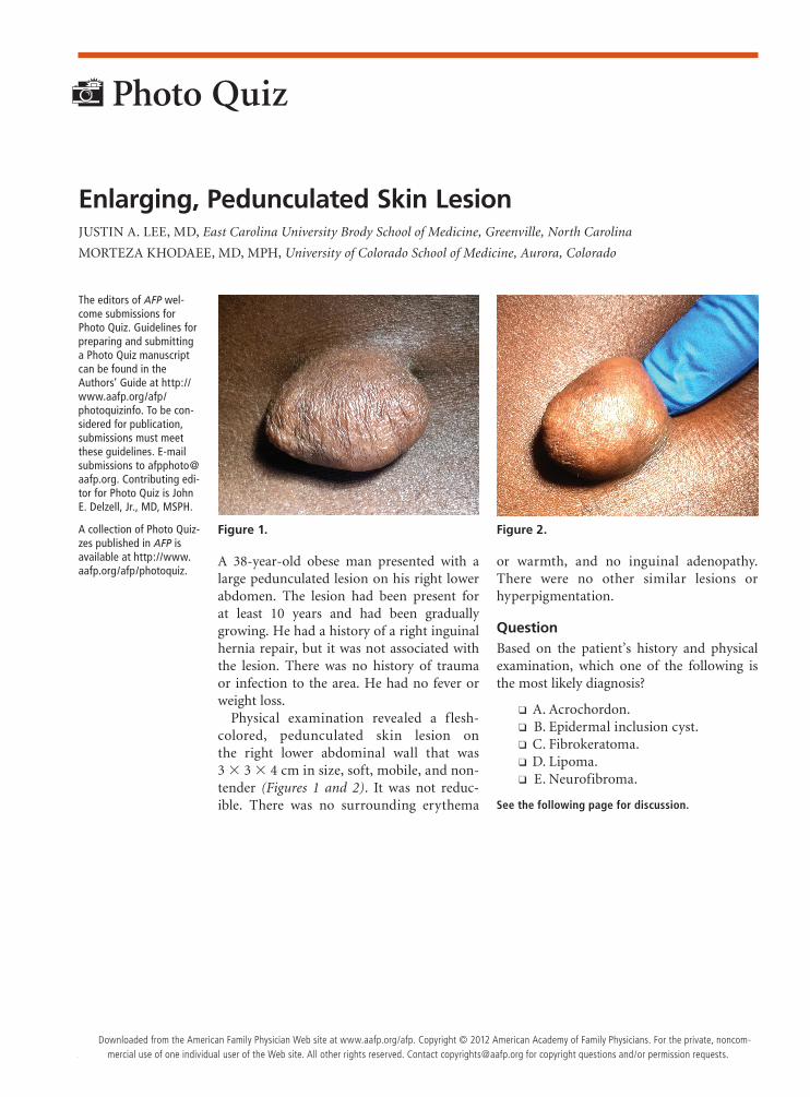
A 38-year-old obese man presented with a large pedunculated lesion on his right lower abdomen. The lesion had been present for at least 10 years and had been gradually growing. He had a history of a right inguinal hernia repair, but it was not associated with the lesion. There was no history of trauma or infection to the area. He had no fever or weight loss.
Physical examination revealed a f lesh-colored, pedunculated skin lesion on the right lower abdominal wall that was 3 × 3 × 4 cm in size, soft, mobile, and non-tender (Figures 1 and 2). It was not reduc-ible. There was no surrounding erythema
or warmth, and no inguinal adenopathy. There were no other similar lesions or hyperpigmentation.
QuestionBased on the patient’s history and physical examination, which one of the following is the most likely diagnosis?
❑ A. Acrochordon. ❑ B. Epidermal inclusion cyst. ❑ C. Fibrokeratoma. ❑ D. Lipoma. ❑ E. Neurofibroma.
See the following page for discussion.
Enlarging, Pedunculated Skin LesionJUSTIN A. LEE, MD, East Carolina University Brody School of Medicine, Greenville, North Carolina
MORTEZA KHODAEE, MD, MPH, University of Colorado School of Medicine, Aurora, Colorado
The editors of AFP wel-come submissions for Photo Quiz. Guidelines for preparing and submitting a Photo Quiz manuscript can be found in the Authors’ Guide at http://www.aafp.org/afp/ photoquizinfo. To be con-sidered for publication, submissions must meet these guidelines. E-mail submissions to [email protected]. Contributing edi-tor for Photo Quiz is John E. Delzell, Jr., MD, MSPH.
A collection of Photo Quiz-zes published in AFP is available at http://www.aafp.org/afp/photoquiz.
Photo Quiz
Figure 1. Figure 2.
Downloaded from the American Family Physician Web site at www.aafp.org/afp. Copyright © 2012 American Academy of Family Physicians. For the private, noncom-mercial use of one individual user of the Web site. All other rights reserved. Contact [email protected] for copyright questions and/or permission requests.
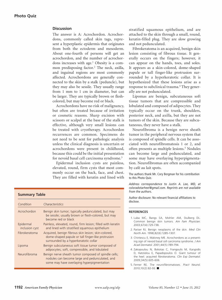
Photo Quiz
1192 American Family Physician www.aafp.org/afp Volume 85, Number 12 ◆ June 15, 2012
DiscussionThe answer is A: Acrochordon. Acrochor-dons, commonly called skin tags, repre-sent a hyperplastic epidermis that originates from both the ectoderm and mesoderm. About one-fourth of persons will get an acrochordon, and the number of acrochor-dons increases with age.1 Obesity is a com-mon predisposing factor.2 The neck, axilla, and inguinal regions are most commonly affected. Acrochordons are generally con-nected to the skin by a stalk (peduncle), but they may also be sessile. They usually range from 1 mm to 1 cm in diameter, but can be larger. They are typically brown or flesh-colored, but may become red or black.
Acrochordons have no risk of malignancy, but often are treated because of irritation or cosmetic reasons. Sharp excision with scissors or scalpel at the base of the stalk is effective, although very small lesions can be treated with cryotherapy. Acrochordon recurrences are common. Specimens do not need to be sent for pathologic analysis unless the clinical diagnosis is uncertain or acrochordons were present in childhood, because this could be the initial presentation for nevoid basal cell carcinoma syndrome.3
Epidermal inclusion cysts are painless, elevated, round, firm cysts that most com-monly occur on the back, face, and chest. They are filled with keratin and lined with
stratified squamous epithelium, and are attached to the skin through a small, round, keratin-filled plug. They are slow growing and not pedunculated.
Fibrokeratoma is an acquired, benign skin lesion consisting of fibrous tissue. It gen-erally occurs on the fingers; however, it can appear on the hands, toes, and soles. It appears as a skin-colored, dome-shaped papule or tall finger-like protrusion sur-rounded by a hyperkeratotic collar. It is hypothesized that these lesions arise as a response to subclinical trauma.4 They gener-ally are not pedunculated.
Lipomas are benign, subcutaneous soft tissue tumors that are compressible and lobulated and composed of adipocytes. They typically occur on the trunk, shoulders, posterior neck, and axilla, but they are not tumors of the skin. Because they are subcu-taneous, they never have a stalk.
Neurofibroma is a benign nerve sheath tumor in the peripheral nervous system that is composed of spindle cells. It can be asso-ciated with neurofibromatosis 1 or 2, and often presents as multiple lesions.5 Nodules can become large and pedunculated, and some may have overlaying hyperpigmenta-tion. Neurofibromas are often accompanied by café au lait spots.
The authors thank Mr. Cory Bergman for his contribution to this Photo Quiz.
Address correspondence to Justin A. Lee, MD, at [email protected]. Reprints are not available from the authors.
Author disclosure: No relevant financial affiliations to disclose.
REFERENCES
1. Luba MC, Bangs SA, Mohler AM, Stulberg DL. Common benign skin tumors. Am Fam Physician. 2003;67(4):729-738.
2. Pariser RJ. Benign neoplasms of the skin. Med Clin North Am. 1998;82(6):1285-1307.
3. Chiritescu E, Maloney ME. Acrochordons as a present-ing sign of nevoid basal cell carcinoma syndrome. J Am Acad Dermatol. 2001;44(5):789-794.
4. Zakopoulou N, Bokotas C, Frangoulis M, Karypidis D, Hatziolou E, Papadopoulos O. Giant tumour of the heel: acquired fibrokeratoma. Clin Exp Dermatol. 2009;34(5):605-606.
5. Ferner RE. The neurofibromatoses. Pract Neurol. 2010;10(2):82-93. ■
Summary Table
Condition Characteristics
Acrochordon Benign skin tumor; typically pedunculated, but may be sessile; usually brown or flesh-colored, but may become red or black
Epidermal inclusion cyst
Painless, elevated, round, firm lesion; filled with keratin and lined with stratified squamous epithelium
Fibrokeratoma Acquired, benign fibrous skin lesion; skin-colored, dome-shaped papule or tall finger-like protrusion surrounded by a hyperkeratotic collar
Lipoma Benign subcutaneous soft tissue tumor composed of adipocytes; soft, compressible, and lobulated
Neurofibroma
Benign nerve sheath tumor composed of spindle cells; nodules can become large and pedunculated, and some may have overlaying hyperpigmentation