Embed Size (px)
Citation preview

Enhancer-associated H3K4 monomethylationby Trithorax-related, the Drosophilahomolog of mammalian Mll3/Mll4
Hans-Martin Herz,1 Man Mohan,1 Alexander S. Garruss,1 Kaiwei Liang,1 Yoh-hei Takahashi,1
Kristen Mickey,1 Olaf Voets,2 C. Peter Verrijzer,2 and Ali Shilatifard1,3
1Stowers Institute for Medical Research, Kansas City, Missouri 64110, USA; 2Department of Biochemistry, Center forBiomedical Genetics, Erasmus University Medical Centre, 3000 DR Rotterdam, The Netherlands
Monomethylation of histone H3 on Lys 4 (H3K4me1) and acetylation of histone H3 on Lys 27 (H3K27ac) arehistone modifications that are highly enriched over the body of actively transcribed genes and on enhancers.Although in yeast all H3K4 methylation patterns, including H3K4me1, are implemented by Set1/COMPASS(complex of proteins associated with Set1), there are three classes of COMPASS-like complexes in Drosophilathat could carry out H3K4me1 on enhancers: dSet1, Trithorax, and Trithorax-related (Trr). Here, we reportthat Trr, the Drosophila homolog of the mammalian Mll3/4 COMPASS-like complexes, can function asa major H3K4 monomethyltransferase on enhancers in vivo. Loss of Trr results in a global decrease ofH3K4me1 and H3K27ac levels in various tissues. Assays with the cut wing margin enhancer implieda functional role for Trr in enhancer-mediated processes. A genome-wide analysis demonstrated that Trr isrequired to maintain the H3K4me1 and H3K27ac chromatin signature that resembles the histone modificationpatterns described for enhancers. Furthermore, studies in the mammalian system suggested a role for theTrr homolog Mll3 in similar processes. Since Trr and mammalian Mll3/4 complexes are distinguishedby bearing a unique subunit, the H3K27 demethylase UTX, we propose a model in which the H3K4monomethyltransferases Trr/Mll3/Mll4 and the H3K27 demethylase UTX cooperate to regulate the transitionfrom inactive/poised to active enhancers.
[Keywords: Trithorax-like; H3K4 monomethylation; COMPASS-like complexes; enhancers]
Supplemental material is available for this article.
Received July 22, 2012; revised version accepted October 17, 2012.
Gene expression by RNA polymerase II (Pol II) is regu-lated at multiple levels in order to allow for the faithfultransmission of genomic information throughout devel-opment and upon external stimuli (Cheung and Cramer2012; Zhou et al. 2012). In Drosophila, the Trithorax (Trx)and Polycomb (Pc) groups of proteins have long beenknown as crucial regulators of development and are re-quired for the maintenance of active transcription ortranscriptional repression, respectively (Poux et al. 2002;Klymenko and Muller 2004). The subsequent identifica-tion of Trx (a Trx group member) as a histone H3K4methyltransferase and Enhancer of zeste [E(z); a Pc grouprepresentative] as a H3K27 methyltransferase exemplifiesthe importance of developmental gene regulation withina chromatin context (Simon and Tamkun 2002; Simon andKingston 2009; Beisel and Paro 2011; Schuettengruberet al. 2011; Shilatifard 2012).
Our molecular understanding of the implementation ofH3K4 methylation activity was aided by the biochemicalpurification of Set1, the yeast homolog of Trx (Miller et al.2001). Set1 was found within the macromolecular com-plex COMPASS (complex of proteins associated withSet1) and was identified as the first H3K4 methylasecapable of catalyzing mono-, di- and trimethylation ofhistone H3K4 (Briggs et al. 2001; Miller et al. 2001;Roguev et al. 2001; Krogan et al. 2002; Schneider et al.2005; Shilatifard 2012). In contrast to yeast, where Set1within COMPASS mediates all of the H3K4 methylationpatterns, Drosophila contains three H3K4 methyltrans-ferases—dSet1, Trx, and Trithorax-related (Trr)—that areeach related to yeast Set1 and can be found in COMPASS-like complexes (Fig. 1A; Mohan et al. 2011). Mammalspossess two representatives for each of the three H3K4methyltransferases found in Drosophila, resulting ina total of six COMPASS-like complexes: Set1a and Set1b(related to dSet1), Mll1 and Mll2 (related to Trx), and Mll3and Mll4 (related to Trr) (Shilatifard 2012). All COMPASS-like complexes contain a set of core components: Wdr5,
3Corresponding authorE-mail [email protected] published online ahead of print. Article and publication date areonline at http://www.genesdev.org/cgi/doi/10.1101/gad.201327.112.
2604 GENES & DEVELOPMENT 26:2604–2620 � 2012 by Cold Spring Harbor Laboratory Press ISSN 0890-9369/12; www.genesdev.org
Cold Spring Harbor Laboratory Press on November 17, 2018 - Published by genesdev.cshlp.orgDownloaded from

Ash2, Rbbp5, and Dpy30 (Fig. 1A, highlighted in green).The three classes of the COMPASS family—dSet1/Set1a/Set1b, Trx/Mll1/Mll2, and Trr/Mll3/Mll4—each containadditional class-specific subunits providing functional di-versity for the complexes (Fig. 1A, highlighted in blue;Hughes et al. 2004; Yokoyama et al. 2004; JH Lee et al.2007; Lee and Skalnik 2008; Wu et al. 2008; Eissenberg andShilatifard 2010; Mohan et al. 2011, 2012; Schuettengruberet al. 2011; Shilatifard 2012).
In agreement with what had already been reported formammalian Set1a and Set1b (Wu et al. 2008), recent studieshave ascribed Drosophila Set1 within COMPASS a pre-dominant role in bulk H3K4 dimethylation (H3K4me2) andtrimethylation (H3K4me3) (Ardehali et al. 2011; Mohanet al. 2011; Hallson et al. 2012). Nonetheless, Trx/Mll1/Mll2 is required gene-specifically to implement H3K4me3at the promoters of the Hox genes (Wang et al. 2009). Sim-
ilarly, the Trr/Mll3/Mll4 COMPASS-like complexes playan important role in hormone receptor-induced signaling.Upon hormonal induction, they are recruited to targetpromoters by nuclear hormone receptors, resulting inincreased H3K4me3 and gene activation (Goo et al. 2003;Sedkov et al. 2003; Lee et al. 2006; Mo et al. 2006; Johnstonet al. 2011; Vicent et al. 2011; Chauhan et al. 2012). How-ever, RNAi-mediated knockdown of Trr, the Drosophilahomolog of mammalian Mll3/Mll4, showed only amodest reduction in H3K4me2 and H3K4me3 levels inwing imaginal discs (Mohan et al. 2011). These subtlechanges in H3K4me2 and H3K4me3 are consistent withTrr regulating the activation of a subset of genes, suchas the role of Trr in the transcriptional induction ofecdysone receptor target genes at promoter regions(Sedkov et al. 2003; Johnston et al. 2011). Dependingon the developmental context and the requirement
Figure 1. The Drosophila Mll3/Mll4 homolog Trr isa major H3K4 monomethyltransferase in vivo. (A) TheCOMPASS family of H3K4 methyltransferase com-plexes in yeast and Drosophila (Mohan et al. 2011,2012). Trr and LPT are highlighted in red, Trx is inpurple, dSet 1 is in orange, core complex subunits are ingreen, and complex-specific subunits are in blue. (B–E)RNAi-mediated knockdown of Drosophila H3K4 meth-yltransferases in the posterior compartment of the wingimaginal disc. GFP expression in green marks theposterior part (highlighted by a white arrow in theantibody-only channel) where the knockdown occurs.Knockdown of Trr (B9) or LPT (E9) results in a globalreduction of H3K4me1. No bulk changes in H3K4me1were observed when Trx (C9) or dSet1 (D9) levels werereduced by RNAi. (F,G) Flipase-catalyzed induction oftrr mutant clones (no GFP expression) with the eye-specific eyeless (ey) promoter. Wild-type tissue ismarked in green (GFP expression). Representative trr
mutant clones are outlined by white dashed lines.Mutant clones of trr1, a trr-null allele, display decreasedlevels of H3K4me1 (F9) and loss of Trr (G9). (H) Westernblot of lysates from RNAi-treated Drosophila S2 cells.(Lane 1) Control lacZ-RNAi. (Lane 2) dSet1-RNAi.(Lane 3) trx-RNAi. (Lane 4) trr-RNAi. (Lane 5) LPT-RNAi. (Panel 1) a-H3. (Panel 2) a-H3K4me1. (Panel 3)a-H3K4me2. (Panel 4) a-H3K4me3. Genotypes used wereas follows: UAS-Dcr-2/+; en-GAL4 UAS-EGFP/+; UAS-
trr-RNAi/+ (B), UAS-Dcr-2/+; en-GAL4 UAS-EGFP/UAS-trx-RNAi (C), UAS-Dcr-2/+; en-GAL4 UAS-EGFP/UAS-
dSet1-RNAi (D), UAS-Dcr-2/+; en-GAL4 UAS-EGFP/+;
UAS-LPT-RNAi/+ (E), and trr1 FRT19A/ubi-GFP FRT19A;
ey-FLP/+ (F,G).
Trr-mediated H3K4 monomethylation at enhancers
GENES & DEVELOPMENT 2605
Cold Spring Harbor Laboratory Press on November 17, 2018 - Published by genesdev.cshlp.orgDownloaded from
for Trr-mediated processes, these effects on H3K4me2and H3K4me3 might be more pronounced at certainstages of development (Sedkov et al. 2003; Chauhanet al. 2012).
Genome-wide mapping of various histone modifica-tions has provided a means to identify signatures of differ-ent chromatin states. For example, while H3K4me3 isfound to be enriched at transcription start sites (TSSs),monomethylation of H3K4 (H3K4me1) was shown to beenriched at enhancers (Heintzman et al. 2007; Wang et al.2008). Enhancers constitute promoter-distally locatedgenomic elements that, in many instances, are necessaryfor the induction and maintenance of gene expression.They are often bound by developmental transcriptionfactors, which, through looping, bring these distal regu-latory elements in close proximity to the promoter-proximal regions regulating their transcriptional activi-ties. Thus, enhancers provide an important regulatorycog for optimal transcriptional coherence to allow fortissue- and context-specific transcription of importantdevelopmental genes in a time-sensitive and optimizedmanner (Levine 2010; Bulger and Groudine 2011; Ongand Corces 2011). The ability of enhancers to work atlong distances is mediated by cohesins, which werefirst identified to function in this process through agenetic screen for factors required for communicationbetween the cut locus and its ;80-kb distally locatedwing margin enhancer (Rollins et al. 1999, 2004; Dorsettet al. 2005).
Recently, it has been shown that chromatin signaturescan be used to further classify enhancers as being in eitheractive or inactive/‘‘poised’’ states. Active enhancers aredually marked with H3K4me1 and H3K27 acetylation(H3K27ac), which allows them to be distinguished frominactive/poised enhancers that are characterized by thepresence of H3K4me1 and H3K27me3 (Heintzman et al.2009; Creyghton et al. 2010; Rada-Iglesias et al. 2011;Zentner et al. 2011; Bonn et al. 2012). H3K27ac is cat-alyzed by CBP and p300 in mammals and by a single CBP-related enzyme in Drosophila (Tie et al. 2009; Pasini et al.2010; Jin et al. 2011). As lysine residues cannot be si-multaneously modified by both methylation and acetyla-tion, it has been proposed that the histone demethylaseUTX can facilitate CBP-mediated H3K27ac through itsability to remove methyl groups from H3K27 (Tie et al.2012).
During a recent study of dUTX’s function in Notchsignaling, we observed that dUTX mutant tissue exhibitsa global decrease in H3K4me1 (Herz et al. 2010) but doesnot show significant changes in H3K4me2 or H3K4me3(Herz et al. 2010). Thus, dUTX appears to link the re-moval of H3K27me3 with the implementation of bothH3K4me1 and H3K27ac. dUTX is a complex-specificsubunit of the Drosophila Trr complex (Fig. 1A; Mohanet al. 2011), while mammalian UTX is found in the ho-mologous mammalian Mll3/Mll4 H3K4 methyltransfer-ase complexes (Cho et al. 2007; Issaeva et al. 2007; MGLee et al. 2007; Patel et al. 2007). Other complex-specificsubunits of the Trr/Mll3/Mll4 complexes include Pa1,Ptip, and Ncoa6 (Fig. 1A). One feature of the Drosophila
Trr complex that distinguishes it from mammalian Mll3/Mll4 complexes is that in Drosophila, two proteins—Trrand LPT (Lost PHDs of Trr)—constitute the functionalhomolog of either Mll3 or Mll4 in mammals. While Trrcorresponds to the C-terminal portion of Mll3/Mll4,which includes the catalytic SET domain, LPT is homol-ogous to the Mll3/Mll4 N terminus and contains severalPHD domains and an HMG domain (Mohan et al. 2011;Chauhan et al. 2012).
Here, we report a novel function for the Trr/COMPASS-like complex as a major H3K4 monomethyltransferase atenhancers. Our results suggest that in various tissues, Trror LPT loss coincides with a strong decrease in bulkH3K4me1 and a reduction in H3K27ac levels. Further-more, Trr can function in enhancer-mediated processes,as it modulates enhancer–promoter interactions at the cutlocus in wing imaginal discs. Genome-wide, Trr is requiredfor the maintenance of the H3K4me1 and H3K27ac chro-matin signature previously described for enhancers. Anal-ysis of the role of Mll3 (one of the mammalian homologs ofTrr) also suggests a role for Mll3/Mll4–COMPASS-likecomplexes in enhancer monomethylation. We proposea model in which UTX, as a complex-specific subunit ofthe Trr and Mll3/4 complexes, demethylates H3K27 atinactive/poised enhancers to prime them for H3K27acby CBP.
Results
Trr and its partner, LPT, are required for H3K4me1in vivo
In order to investigate the function of H3K4me1 inDrosophila, we used RNAi by the Gal4/UAS system inwing imaginal discs directed toward the DrosophilaH3K4 methyltransferases that might regulate this mod-ification. We found that expression of dsRNA against Trrunder the control of the engrailed promoter results ina strong decrease in global H3K4me1 in the posterior halfof the wing imaginal disc (Fig. 1B; Supplemental Fig. S1A).No bulk changes in H3K4me1 were observed with trx-RNAi (Fig. 1C; Supplemental Fig. S1B) or dSet1-RNAi(Fig. 1D; Supplemental Fig. S1C). We and others havepreviously shown that Trr is only homologous to theC-terminal portion of mammalian Mll3/Mll4 and thatthe functional equivalent of the Mll3/Mll4 N terminus isrepresented by another protein—LPT—in Drosophila(Mohan et al. 2011; Chauhan et al. 2012). Likewise, inthe absence of LPT function (Supplemental Fig. S1D),H3K4me1 is strongly reduced in wing imaginal discs(Fig. 1E), suggesting that Trr and LPT work together tomediate global H3K4me1. A role for Trr in H3K4me1was further confirmed with a trr-null allele (trr1) in eye-antenna imaginal discs, as trr mutant clones show re-duced H3K4me1 staining compared with surroundingwild-type tissue (Fig. 1F,G).
Western blot analysis in S2 cells after RNAi-mediatedknockdown of the three H3K4 methyltransferases orLPT confirmed our findings from imaginal discs (Fig.1H; Supplemental Fig. S1E–H). Whereas dSet1-RNAi
Herz et al.
2606 GENES & DEVELOPMENT
Cold Spring Harbor Laboratory Press on November 17, 2018 - Published by genesdev.cshlp.orgDownloaded from

results in almost complete bulk losses of H3K4me2(Fig. 1H, panel 3) and H3K4me3 (Fig. 1H, panel 4), aglobal reduction in H3K4me1 can only be observed withtrr-RNAi and LPT-RNAi (Fig. 1H, panel 2). Since Set1and related enzyme activities are stimulated by coreCOMPASS subunits (Dou et al. 2006; Steward et al.2006; Shilatifard 2012), we used a baculovirus systemto reconstitute the SET domains of Trr and dSet1 withthe core subunits of COMPASS and observed that inthe context of the core subunits, both SET domains of Trrand dSet1 were able to mono-, di- and trimethylate H3K4(Supplemental Fig. S2). However, Trr showed a preferencefor H4K4me1, while dSet1 showed a tendency towardH3K4me3. Taken together, these results support thein vivo data showing that Trr/LPT is a major H3K4monomethyltransferase.
Trr/COMPASS-like core and complex-specific subunitsare required for H3K4me1 in vivo
Recently, all three H3K4 methyltransferases in Drosophila—dSet1, Trx, and Trr—have been shown to exist in inde-pendent COMPASS-like complexes (Mohan et al. 2011).Each COMPASS-like complex can be further subdividedinto core members that are common to each complex andcomplex-specific subunits that are unique to only one ofthe complexes. Wds, Ash2, Rbbp5, and Dpy-30L1 consti-tute the core components of all COMPASS family mem-bers, while dUTX, Ptip, Pa1, and Ncoa6 are found onlyin the Trr/LPT complex (Fig. 1A). Hcf is another compo-nent that is often copurified with the COMPASS family(Mohan et al. 2011).
In order to identify subunit-specific requirements forbulk H3K4me1, we systematically depleted core mem-bers of the COMPASS family (Fig. 2A–C), Hcf (Fig. 2D),or Trr complex-specific subunits (Fig. 2E–G) in theposterior part of wing imaginal discs by RNAi. ash2-RNAi and Rbbp5-RNAi resulted in drastic losses ofH3K4me1 (Fig. 2A,B; Supplemental Fig. S3A,D,E),H3K4me2 (data not shown), and H3K4me3 (data notshown). The loss of H3K4me1 is not the result of Trrdestabilization, as Trr levels are unaltered when Ash2 orRbbp5 are knocked down (data not shown), thus suggest-ing a fundamental requirement for these core compo-nents in Trr-mediated H3K4me1. Other core components,such as Dpy-30L1 (Fig. 2C; Supplemental Fig. S3F) or Hcf(Fig. 2D; Supplemental Fig. S3B,G), do not seem to berequired for implementing bulk H3K4me1. Dpy-30L1-RNAi also did not affect H3K4me2 or H3K4me3 (data notshown). Hcf-RNAi did not affect H3K4me2 levels (datanot shown) but did show reduced H3K4me3 (data notshown), in accordance with a recent study (Hallson et al.2012).
Depletion of Trr complex-specific subunits such asdUTX (Fig. 2E; Supplemental Fig. S3C,H), Ptip (Fig. 2F;Supplemental Fig. S3I), or Ncoa6 (Fig. 2G; SupplementalFig. S3J) resulted in decreased global H3K4me1 levels. Insummary, our data support a model in which the Trr/LPTCOMPASS-like complex is required for the maintenanceof the majority of H3K4me1 levels.
Trr mediates stabilization of Trr/COMPASS-likecomplex-specific subunits and H3K27ac
The knockdown efficiencies of Trx, Trr, and LPT wereconfirmed for all components of the COMPASS family;namely, dSet1 (Fig. 3A, panel 1), Trx (Fig. 3A, panel 2), Trr(Fig. 3A, panel 3), and LPT (Fig. 3A, panel 4). LPT levelswere strongly reduced not only with LPT-RNAi (Fig. 3A,lane 5, panel 4), but also with trr-RNAi (Fig. 3A, lane 4,panel 4). dUTX levels were also diminished upon trr-RNAi (Fig. 3A, lane 4, panel 5), reminiscent of what hasbeen observed for Caenorhabditis elegans UTX-1 whenSET-16 (Trr homolog) is knocked down (Vandamme et al.2012). However, contrary to the findings in C. elegans,where reduction in SET-16 levels altered transcription ofutx-1, we did not observe transcriptional changes fordUTX or LPT with trr-RNAi in Drosophila (data notshown). We further tested LPT and dUTX levels in Trr-depleted wing imaginal discs either by RNAi or when trrmutant clones were induced in eye-antenna imaginaldiscs (Fig. 3B–E). Both LPT (Fig. 3B) and dUTX (Fig. 3C)were reduced in trr-RNAi wing imaginal discs. A similareffect was seen in trr mutant clones for LPT (Fig. 3D) anddUTX (Fig. 3E) levels in eye-antenna imaginal discs.Conversely, LPT-RNAi or dUTX-RNAi in wing imaginaldiscs does not show significant effects on Trr stability(data not shown). Thus, in Drosophila, Trr is required forstabilization of at least some of the Trr/COMPASS-likecomplex-specific subunits.
The destabilization of dUTX in the absence of Trr isinteresting in light of the recent finding that dUTX, bybinding to CBP, modulates H3K27ac (Tie et al. 2012). Thisfinding links the H3K27 demethylase dUTX (Smith et al.2008; Herz et al. 2010) to H3K27ac and provides a modelfor how a transition could occur from a silenced H3K27-methylated chromatin state to an activated state thatis marked by H3K27ac. Comparable with the resultsobtained with dUTX-RNAi (Tie et al. 2012), trr-RNAiin wing imaginal discs results in a global reduction ofH3K27ac (Fig. 3F), and similar effects can be observed intrr mutant clones in eye-antenna imaginal discs (Fig. 3G).In conclusion, our data suggest that Trr is required tostabilize Trr/COMPASS-like complex-specific subunitsto regulate H3K27ac. Based on genome-wide studies, ithas been proposed that the distribution pattern forH3K4me1 is very broad and particularly enriched on en-hancers and over the body of actively transcribed genes(Fig. 3H; Barski et al. 2007; Heintzman et al. 2007). Like-wise, H3K27ac has been reported to mark active en-hancers but can also be found on promoters and genebodies (Fig. 3H; Heintzman et al. 2009; Creyghton et al.2010; Rada-Iglesias et al. 2011; Zentner et al. 2011; Bonnet al. 2012).
Trr is required for enhancer function at the cut (ct)locus in wing imaginal discs
Based on our findings that Trr is globally required forproper H3K4me1 and H3K27ac, we turned to the well-characterized ct locus (Jack et al. 1991; Rollins et al. 2004;Dorsett et al. 2005) in order to functionally verify a possible
Trr-mediated H3K4 monomethylation at enhancers
GENES & DEVELOPMENT 2607
Cold Spring Harbor Laboratory Press on November 17, 2018 - Published by genesdev.cshlp.orgDownloaded from

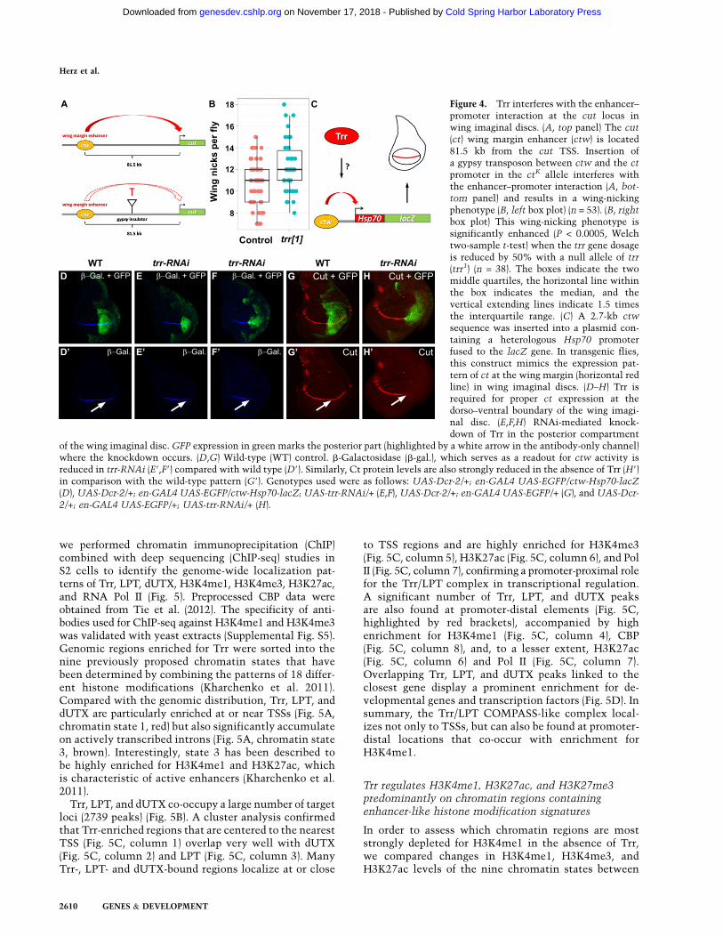
role of Trr in enhancer-mediated processes. The ct wingmargin enhancer is required for proper expression of ct atthe dorso–ventral boundary in the wing imaginal disc(Jack et al. 1991; Rollins et al. 2004; Dorsett et al. 2005). Inthe ctk allele, a gypsy transposon is inserted between thect wing margin enhancer and the ct promoter, interferingwith enhancer–promoter communication and resultingin a characteristic wing-notching phenotype (Fig. 4A;Dorsett et al. 2005). The ctk allele has been deployed forthe characterization of cohesin subunits such as Nipped-B,
which are important regulators of enhancer–promoter in-teraction at the ct gene (Rollins et al. 2004). The wing-notching phenotype of the ctk allele can be quantified bycounting the number of wing nicks per fly in homozygousctk mutants. ctk control flies average 10.6 wing nicks perfly (Fig. 4B, left box plot). Removing 50% of the trr genedosage by crossing in a null allele of trr, trr1 (Sedkov et al.1999), increases the number of wing nicks to an average of12.3 (Fig. 4B, right box plot). This represents a statisticallysignificant enhancement (P < 0.0005, Welch two-sample
Figure 2. Trr/COMPASS-like core and complex-specific subunits are required for H3K4me1. (A–G) RNAi-mediated knockdown ofcore members of COMPASS-like complexes (A–D) and Trr complex-specific subunits (E–G) in the posterior compartment of the wingimaginal disc. GFP expression in green marks the posterior part (highlighted by a white arrow in the antibody-only channel) where theknockdown occurs. ash2-RNAi (A9) and Rbbp5-RNAi (B9) result in strongly decreased H3K4me1, whereas other core members such asDpy-30L1 (C9) and Hcf (D9) are not required for bulk H3K4me1. RNAi-mediated knockdowns of the Trr complex-specific subunitsdUTX (E9), Ptip (F9), and Ncoa6 (G9) only weakly affect bulk levels of H3K4me1. (H) Cartoon representing the implementation of bulkH3K4me1 by the Trr/LPT complex. The SET domain-containing catalytic subunit Trr and LPT are highlighted in red, core complexsubunits are in green, and complex-specific subunits are in blue. Genotypes used were as follows: UAS-Dcr-2/+; en-GAL4 UAS-EGFP/
UAS-ash2-RNAi (A), UAS-Dcr-2/+; en-GAL4 UAS-EGFP/UAS-Rbbp5-RNAi (B), UAS-Dcr-2/+; en-GAL4 UAS-EGFP/+; UAS-Dpy-30L1-
RNAi/+ (C), UAS-Dcr-2/+; en-GAL4 UAS-EGFP/+; UAS-Hcf-RNAi/+ (D), UAS-Dcr-2/+; en-GAL4 UAS-EGFP/+; UAS-dUTX-RNAi/+
(E), UAS-Dcr-2/+; en-GAL4 UAS-EGFP/+; UAS-Ptip-RNAi/+ (F), and UAS-Dcr-2/+; en-GAL4 UAS-EGFP/+; UAS-Ncoa6-RNAi/+ (G).
Herz et al.
2608 GENES & DEVELOPMENT
Cold Spring Harbor Laboratory Press on November 17, 2018 - Published by genesdev.cshlp.orgDownloaded from

t-test) and supports a requirement for Trr in enhancer-mediated processes.
To further test whether Trr directly regulates the wingmargin enhancer, we used a transgenic fly line in whichthe ct wing margin enhancer (ctw) is inserted upstream ofthe Hsp70 promoter fused to the lacZ gene (Fig. 4C). Thistransgene recapitulates the endogenous ct expression pat-tern at the dorso–ventral boundary in wing imaginaldiscs (Fig. 4C,D). Trr knockdown in the posterior half ofthe wing imaginal disc results in reduced lacZ expression(Fig. 4E,F), suggesting a direct role for Trr in enhancer–promoter communication. Furthermore, upon RNAi-mediated knockdown of Trr, the dorso–ventral stripeof Ct, which can be detected across the whole antero–
posterior axis in wild-type wing imaginal discs (Fig. 4G),is now confined to the anterior portion of the disc (Fig.4H), providing further evidence for an enhancer-medi-ated function of Trr at the endogenous ct locus. Proteinlevels of other regulators of ct, such as Wingless (Wg)(Supplemental Fig. S4A,B) or Notch (N) (SupplementalFig. S4C,D), were not changed when Trr function wasremoved.
The Trr/COMPASS-like complex localizesto promoters and enhancers
In order to determine whether the Trr/LPT complex gen-erally acts as a H3K4 monomethyltransferase on enhancers,
Dpy
Figure 3. Trr stabilizes Trr complex-specific subunits and promotes H3K27 acetylation. Trr is required for LPT and dUTX stability (A–E) and proper H3K27ac (F,G). (A) Western blots of lysates from RNAi-treated Drosophila S2 cells. (Lane 1) Control lacZ-RNAi. (Lane 2)dSet1-RNAi. (Lane 3) trx-RNAi. (Lane 4) trr-RNAi. (Lane 5) LPT-RNAi. (Panel 1) a-dSet1; the arrow marks the dSet1 band, and theasterisk indicates an unspecific band. (Panel 2) a-Trx; the arrow marks the Trx band, and the asterisks indicate unspecific bands. (Panel3) a-Trr. (Panel 4) a-LPT. (Panel 5) a-dUTX. (Panel 6) aa-Tub. (B,C,F) RNAi-mediated knockdown of Trr in the posterior compartment ofthe wing imaginal disc. GFP expression in green marks the posterior part (highlighted by a white arrow in the antibody-only channel)where the knockdown occurs. LPT (B9) and dUTX (C9) are destabilized in the absence of Trr. (F9) H3K27ac levels are decreased when Trrfunction is removed. (D,E,G) Flipase-catalyzed induction of trr mutant clones (no GFP expression) with the eye-specific eyeless (ey)promoter. Wild-type tissue is marked in green (GFP expression). Representative clones are outlined by white dashed lines. Mutantclones of trr1, a trr-null allele, display decreased levels of LPT (D9), dUTX (E9), and H3K27ac (G9). (H) Possible functions for the Trr/LPTcomplex in implementing H3K4me1. The SET domain-containing catalytic subunit Trr and LPT are highlighted in red, core complexsubunits are in green, and complex-specific subunits are in blue. Genotypes used were as follows: UAS-Dcr-2/+; en-GAL4 UAS-EGFP/+;
UAS-trr-RNAi/+ (B,C,F) and trr1 FRT19A/ubi-GFP FRT19A; ey-FLP/+ (D,E,G).
Trr-mediated H3K4 monomethylation at enhancers
GENES & DEVELOPMENT 2609
Cold Spring Harbor Laboratory Press on November 17, 2018 - Published by genesdev.cshlp.orgDownloaded from
we performed chromatin immunoprecipitation (ChIP)combined with deep sequencing (ChIP-seq) studies inS2 cells to identify the genome-wide localization pat-terns of Trr, LPT, dUTX, H3K4me1, H3K4me3, H3K27ac,and RNA Pol II (Fig. 5). Preprocessed CBP data wereobtained from Tie et al. (2012). The specificity of anti-bodies used for ChIP-seq against H3K4me1 and H3K4me3was validated with yeast extracts (Supplemental Fig. S5).Genomic regions enriched for Trr were sorted into thenine previously proposed chromatin states that havebeen determined by combining the patterns of 18 differ-ent histone modifications (Kharchenko et al. 2011).Compared with the genomic distribution, Trr, LPT, anddUTX are particularly enriched at or near TSSs (Fig. 5A,chromatin state 1, red) but also significantly accumulateon actively transcribed introns (Fig. 5A, chromatin state3, brown). Interestingly, state 3 has been described tobe highly enriched for H3K4me1 and H3K27ac, whichis characteristic of active enhancers (Kharchenko et al.2011).
Trr, LPT, and dUTX co-occupy a large number of targetloci (2739 peaks) (Fig. 5B). A cluster analysis confirmedthat Trr-enriched regions that are centered to the nearestTSS (Fig. 5C, column 1) overlap very well with dUTX(Fig. 5C, column 2) and LPT (Fig. 5C, column 3). ManyTrr-, LPT- and dUTX-bound regions localize at or close
to TSS regions and are highly enriched for H3K4me3(Fig. 5C, column 5), H3K27ac (Fig. 5C, column 6), and PolII (Fig. 5C, column 7), confirming a promoter-proximal rolefor the Trr/LPT complex in transcriptional regulation.A significant number of Trr, LPT, and dUTX peaksare also found at promoter-distal elements (Fig. 5C,highlighted by red brackets), accompanied by highenrichment for H3K4me1 (Fig. 5C, column 4), CBP(Fig. 5C, column 8), and, to a lesser extent, H3K27ac(Fig. 5C, column 6) and Pol II (Fig. 5C, column 7).Overlapping Trr, LPT, and dUTX peaks linked to theclosest gene display a prominent enrichment for de-velopmental genes and transcription factors (Fig. 5D). Insummary, the Trr/LPT COMPASS-like complex local-izes not only to TSSs, but can also be found at promoter-distal locations that co-occur with enrichment forH3K4me1.
Trr regulates H3K4me1, H3K27ac, and H3K27me3predominantly on chromatin regions containingenhancer-like histone modification signatures
In order to assess which chromatin regions are moststrongly depleted for H3K4me1 in the absence of Trr,we compared changes in H3K4me1, H3K4me3, andH3K27ac levels of the nine chromatin states between
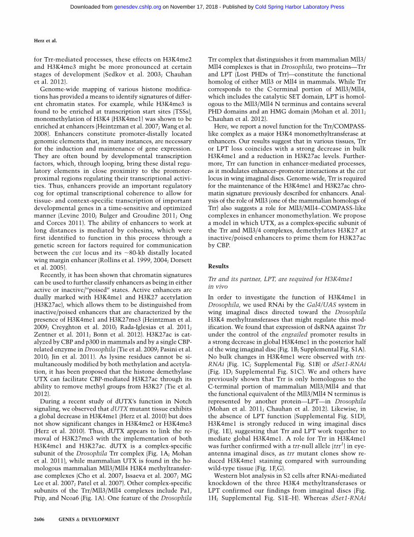
Figure 4. Trr interferes with the enhancer–promoter interaction at the cut locus inwing imaginal discs. (A, top panel) The cut(ct) wing margin enhancer (ctw) is located81.5 kb from the cut TSS. Insertion ofa gypsy transposon between ctw and the ct
promoter in the ctK allele interferes withthe enhancer–promoter interaction (A, bot-
tom panel) and results in a wing-nickingphenotype (B, left box plot) (n = 53). (B, rightbox plot) This wing-nicking phenotype issignificantly enhanced (P < 0.0005, Welchtwo-sample t-test) when the trr gene dosageis reduced by 50% with a null allele of trr(trr1) (n = 38). The boxes indicate the twomiddle quartiles, the horizontal line withinthe box indicates the median, and thevertical extending lines indicate 1.5 timesthe interquartile range. (C) A 2.7-kb ctw
sequence was inserted into a plasmid con-taining a heterologous Hsp70 promoterfused to the lacZ gene. In transgenic flies,this construct mimics the expression pat-tern of ct at the wing margin (horizontal redline) in wing imaginal discs. (D–H) Trr isrequired for proper ct expression at thedorso–ventral boundary of the wing imagi-nal disc. (E,F,H) RNAi-mediated knock-down of Trr in the posterior compartment
of the wing imaginal disc. GFP expression in green marks the posterior part (highlighted by a white arrow in the antibody-only channel)where the knockdown occurs. (D,G) Wild-type (WT) control. b-Galactosidase (b-gal.), which serves as a readout for ctw activity isreduced in trr-RNAi (E9,F9) compared with wild type (D9). Similarly, Ct protein levels are also strongly reduced in the absence of Trr (H9)in comparison with the wild-type pattern (G9). Genotypes used were as follows: UAS-Dcr-2/+; en-GAL4 UAS-EGFP/ctw-Hsp70-lacZ(D), UAS-Dcr-2/+; en-GAL4 UAS-EGFP/ctw-Hsp70-lacZ; UAS-trr-RNAi/+ (E,F), UAS-Dcr-2/+; en-GAL4 UAS-EGFP/+ (G), and UAS-Dcr-
2/+; en-GAL4 UAS-EGFP/+; UAS-trr-RNAi/+ (H).
Herz et al.
2610 GENES & DEVELOPMENT
Cold Spring Harbor Laboratory Press on November 17, 2018 - Published by genesdev.cshlp.orgDownloaded from

control and trr-RNAi S2 cells (Fig. 6A–C). Chromatinregions represented by state 7 (heterochromatin, highlyenriched for H3K9me2/3) are strongly depleted forH3K4me1, H3K4me3, and H3K27ac (Kharchenko et al.2011). Therefore, state 7 was used to normalize the sam-ples against background read levels. When Trr wasdepleted, the strongest losses in H3K4me1 were ob-served in chromatin states 3 (Fig. 6A, brown box plots)and 4 (Fig. 6A, coral box plots), which are characterizedby the highest levels of H3K4me1 enrichment andsimultaneously most closely mimic enhancer-like his-tone modification patterns. Additionally, a significantreduction in H3K4me1 was also evident in Pc-repressedregions (Fig. 6A, dark-gray box plots) and silent chroma-tin (Fig. 6A, light-gray box plots) and, to a lesser degree,on actively transcribed exons (Fig. 6 A, purple and greenbox plots). Similar changes in H3K4me1 were observedin LPT-RNAi S2 cells (Supplemental Fig. S6A). Unex-pectedly, H3K4me1 increases with trr-RNAi and LPT-RNAi on TSSs (Fig. 6A; Supplemental Fig. S6A, red boxplots), which might be the result of an indirect effect offreeing up more substrate for other H3K4 methyltrans-ferases. No comparable changes in H3K4me3 wereobserved within highly enriched H3K4me3 states suchas TSS regions (Fig. 6B, red box plots), with the exceptionof a weak reduction of H3K4me3 in state 3 (Fig. 6B, coralbox plots).
Decreases in H3K27ac levels after trr-RNAi paralleledthe losses of H3K4me1 in states 3 (Fig. 6C, brown boxplots) and 4 (Fig. 6C, coral box plots). Not surprisingly,changes in H3K27ac could not be detected in Pc-repressedregions (Fig. 6C, dark-gray box plots) or in silent chroma-tin (Fig. 6C, light-gray box plots), as they do not containsignificant levels of H3K27ac in wild-type S2 cells. Thus,the observed global decreases of H3K27ac in trr-RNAiwing imaginal discs (Fig. 3F) and trr mutant clones (Fig.3G) likely correspond to H3K27ac losses on active en-hancer-like chromatin regions. The decrease of H3K27acat enhancers when removing Trr function is consistentwith previous reports that H3K4me1 precedes H3K27ac(Bonn et al. 2012). As observed for H3K4me1, H3K27ac isalso increased at TSS regions in trr-RNAi S2 cells (Fig. 6C,red box plots), which further suggests that some indirecteffects on these histone modifications are occurring byreducing Trr levels.
dUTX was recently shown to modulate the balancebetween H3K27ac and H3K27me3 (Tie et al. 2012). Wefound that dUTX and LPT levels are most stronglyreduced on enhancer-like chromatin states 3 and 4 upontrr-RNAi (Fig. 6D,E). These are also the regions that showan increase in H3K27me3 after trr-RNAi (Fig. 6F). Simi-larly, knockdown of LPT in S2 cells shows significantincreases in H3K27me3 on states 3 and 4 (SupplementalFig. S6B). In summary, our data suggest an interdepen-
Figure 5. The Trr complex localizes topromoters and enhancers. (A) The stackedbar plot shows the genome-wide percentageof each chromatin state as defined byKharchenko et al. (2011). The top rowshows the whole-genome distribution ofthe nine chromatin states in S2 cells, thesecond row shows the distribution for Trr-enriched regions, the third row shows thedistribution for LPT-enriched regions, andthe fourth row shows the distribution fordUTX-enriched regions. (Red) State 1, pro-moter-proximal regions; (purple) state 2,transcriptional elongation; (brown) state 3,intronic regions highly enriched forH3K4me1 and H3K27ac; (coral) state 4, re-lated to state 3; (green) state 5, X chromo-some, highly enriched for H4K16ac andH3K36me3; (dark gray) state 6, Polycomb-repressed regions; (dark blue) state 7, het-erochromatin, high levels of H3K9me2/me3; (light blue) state 8, heterochromatin-like, moderate levels of H3K9me2/me3;(light gray) state 9, silent domains. (B) Venndiagram displaying co-occupancy betweenTrr (red), LPT (green), and dUTX (blue)peaks. Trr-enriched (4009 peaks), LPT-enriched (6879 peaks), and dUTX-enriched
(5239 peaks) regions overlap significantly within 100 base pairs (bp) (2739 peaks). (C) Cluster analysis of Trr-occupied regions centeredon the TSS of the nearest gene. Each column extends over 20 kb (610 kb from the TSS). (Column 1) Trr. (Column 2) dUTX. (Column 3)LPT. (Column 4) H3K4me1. (Column 5) H3K4me3. (Column 6) H3K27ac. (Column 7) Pol II. (Column 8) CBP. CBP data were obtainedfrom Tie et al. (2012). (D) Gene ontology (GO) term analysis of overlapping Trr, LPT, and dUTX peaks (from B). GO terms were ascribedbased on the 2134 closest genes to overlapping peaks. P-values shown are Benjamini-corrected.
Trr-mediated H3K4 monomethylation at enhancers
GENES & DEVELOPMENT 2611
Cold Spring Harbor Laboratory Press on November 17, 2018 - Published by genesdev.cshlp.orgDownloaded from

dency of H3K4me1, H3K27ac, and H3K27me3 levels onenhancer-like chromatin.
The mammalian homolog of Trr, Mll3, is also a majormonomethyltransferase
To verify that the mammalian homologs of Trr havea similar function in implementing H3K4me1 on en-hancers, we performed ChIP-seq studies in mouse em-bryonic fibroblasts (MEFs) (Fig. 7A,B). Many genomicregions that are located outside of genes and are enrichedfor H3K4me1 show decreased H3K4me1 levels witha concomitant increase in H3K27me3 in Mll3 knockoutMEFs (Fig. 7A, right scatter plot). In contrast, Mll1 knock-out MEFs display an increase in H3K4me1 and H3K27me3(Fig. 7A, left scatter plot). The modest shift into the upperleft quadrant, representing decreased H3K4me1 and in-creased H3K27me3, might be even greater in Mll3/Mll4double-knockout cells, as Mll3 and Mll4 are found insimilar complexes and may share some functional re-dundancy toward H3K4me1 in the mammalian system.Nonetheless, many promoter-distal regions with a con-siderable decrease in H3K4me1 can be identified inMll3 knockout MEFs. For example, the Hoxd clusteris strongly enriched for H3K4me1 in Mll3 wild-typeMEFs and is known to contain many enhancers(Fig. 7B; Montavon et al. 2011). Removal of Mll3function results in a decrease of H3K4me1 at manysites within the Hoxd cluster and a broad increase inH3K27me3 throughout this cluster (Fig. 7B). Thus, atleast the role of Mll3 as a H3K4 monomethyltransferase
appears to be functionally conserved in the mammaliansystem.
Based on these findings, we propose a model in whichthe Trr/Mll3/Mll4 COMPASS-like complexes form a hubcombining the H3K27 demethylase function of UTX withthe H3K4 monomethyltransferase activity of Trr/Mll3/Mll4 to allow for H3K27ac by CBP/p300 on enhancers(Fig. 7C). The combination of all of the histone-modifyingactivities provides an explanation for how enhancers cantransition from an inactive/poised state to an activatedstate (Fig. 7C). The transition to the active state is pro-moted by activation of Trr/Mll3/Mll4 COMPASS-likecomplexes, which are able to achieve demethylation ofH3K27me3 through UTX and implementation of H3K4me1through Trr/Mll3/Mll4. It is also possible that the recruit-ment of UTX through Trr/Mll3/Mll4 COMPASS-like com-plexes to inactive enhancers provides context dependencyfor the activation of CBP, as many of the inactive enhancerregions are not modified by H3K27me3 and H3H27ac but dohave CBP present (Fig. 7C).
Discussion
Our previous studies of the H3K27 demethylase dUTXhad shown reduced levels of H3K4me1 in dUTX mutanttissue (Herz et al. 2010). As dUTX is a subunit of the Trr/COMPASS-like complex (Mohan et al. 2011), we investi-gated Trr’s role in H3K4me1. In this study, we describea function for Trr as a major H3K4 monomethyltransfer-ase functioning at enhancer regions. Mutant trr clones in
Figure 6. Trr/COMPASS regulates H3K4me1,H3K27ac, and H3K27me3 on chromatin regionsthat resemble enhancer-like histone modificationsignatures. (A–F) Box plot diagrams displaying foldenrichment (log2) of various histone modifications(H3K4me1, H3K4me3, H3K27ac, and H3K27me3)and dUTX and LPT in wild-type (left box plots) andtrr-RNAi (right box plots) S2 cells over the ninechromatin states as defined by Kharchenko et al.(2011). Characteristics of the different chromatinstates are summarized in Figure 5A. Enrichmentfor each chromatin state is represented by the left
box plot for wild-type S2 cells and by the right boxplot in trr-RNAi S2 cells. (A) H3K4me1 enrich-ment. (B) H3K4me3 enrichment. (C) H3K27ac en-richment. (D) dUTX enrichment. (E) LPT enrichment.(F) H3K27me3 enrichment. Fold enrichments werecalculated for each region of the genome by takingthe read density per base pair of each region divided bythe median read density per base pair of the appropriatebackground regions: all state 7 regions for A–C, allstate 5 regions for D,E, and all state 1 regions for F. Theboxes indicate the two middle quartiles, the horizontalline within the box indicates the median, and thevertical extending lines indicate 1.5 times the inter-quartile range. The notches shown around the medianindicate a 95% confidence interval of the median.
Herz et al.
2612 GENES & DEVELOPMENT
Cold Spring Harbor Laboratory Press on November 17, 2018 - Published by genesdev.cshlp.orgDownloaded from

eye-antenna imaginal discs or knockdown of Trr or itspartner, LPT, in wing imaginal discs and in S2 cells showa global decrease in both H3K4me1 and the enhancer-associated mark of H3K27ac (Figs. 1–3). Assays at the cutlocus confirm a role for Trr in enhancer-mediated pro-cesses (Fig. 4). Using ChIP-seq in cells depleted of Trr byRNAi, we found that chromosomal regions most affectedfor H3K4me1 and H3K27ac share chromatin propertieswith enhancers (Fig. 6). Furthermore, the deletion of oneof the mammalian Trr homologs, the Mll3 gene, resultsin a reduction in H3K4me1 levels with a concomitantincrease in H3K27me3 (Fig. 7A). Since loss of Trr or LPT
leads to an increase of H3K27me3 at enhancer regions,we propose a role for Trr/COMPASS in regulating thetransition from inactive/poised enhancers to active en-hancers. As Trr/LPT and dUTX are conserved in mam-mals, our findings have implications for understandingthe transitions among various enhancer states during de-velopment and their misregulation in disease.
Trr/LPT and Mll3/Mll4 COMPASS-like complexes werepreviously shown to be major activators of hormone-responsive genes and are required for the establishmentof H3K4me3 at promoter regions of these genes (Sedkovet al. 2003; Lee et al. 2006; Chauhan et al. 2012). A promoter-
Figure 7. The mammalian homolog of Trr, Mll3, is one of the major monomethyltransferases in mammalian cells. (A) Manypromoter-distal elements (those H3K4me1 peaks not occurring within a gene) in Mll3 knockout (KO) MEFs exhibit a reduction in theH3K4me1 levels and an increase in H3K27me3 levels (right scatter plot), whereas Mll1 knockout MEFs display an increase in H3K4me1and H3K27me3 levels (left scatter plot). The quotient of fold enrichment (immunprecipitation/whole-cell extract) in mutant MEFsversus wild-type (WT) MEFs was plotted for both the X-axis and Y-axis for H3K4me1 and H3K27me3, respectively. The scatter plotpoint density is indicated by color. (Blue) Low; (red) medium; (yellow) high. Mll1 and Mll3 wild-type and knockout MEFs werepreviously described in Hanson et al. (1999), Hughes et al. (2004), and Wang et al. (2009). (B) Decrease of H3K4me1 and increase ofH3K27me3 over the Hoxd cluster in Mll3 knockout MEFs. Whole-cell extracts (WCE) represent the input chromatin. (C) Modeldescribing the role of Mll3/4/Trr COMPASS-like complexes in the transitioning of enhancer activation. Mll3/4 in mammals and Trr inDrosophila participate in complexes joining the H3K27 demethylase UTX with the H3K4 monomethyltransferase activity of Mll3/4and Trr to allow for H3K4me1 by the Trr/COMPASS and H3K27ac by CBP on enhancers. The transition to the active state of enhancersis promoted by activation of the Trr/Mll3/Mll4 COMPASS complex, which is able to achieve demethylation of H3K27me3 throughUTX and H3K4me1 through Trr/Mll3/Mll4. CBP is activated via its interaction with UTX.
Trr-mediated H3K4 monomethylation at enhancers
GENES & DEVELOPMENT 2613
Cold Spring Harbor Laboratory Press on November 17, 2018 - Published by genesdev.cshlp.orgDownloaded from

proximal function for Trr is in agreement with ourobservation that Trr, LPT, and dUTX localize to manyTSSs (Fig. 5A,C). However, it is somewhat surprising thatthe knockdown of Trr or LPT does not significantly af-fect H3K4me3 at active promoters in S2 cells, as manyTrr peaks localize near TSS regions (Fig. 5C). This is notspecific to S2 cells, as the loss of Trr by RNAi does notresult in global reduction in H3K4me3 levels in fly tissue(Mohan et al. 2011). Trr has been reported to be requiredfor bulk H3K4me3 in embryos and prepupae (Sedkov et al.2003; Chauhan et al. 2012); however, Trr localization atTSSs does not necessarily imply a functional role for Trrin H3K4me3 at promoters in all developmental contexts.Trr could provide a ‘‘priming’’ mechanism without func-tional consequences for H3K4me3. Additionally, dSet1and/or Trx might be able to compensate for a loss of Trrfunction at some TSSs. We did observe an increase inH3K4me1 at TSSs after Trr or LPT RNAi (Fig. 6A; Sup-plemental Fig. S6A), which may be an indication of achange in competition among the different Set1-relatedenzymes or other, as yet unknown, factors for binding toTSS regions.
Our finding that the Trr/COMPASS-like complex isresponsible for H3K4me1 at enhancers suggests a context-dependent difference in the activity of this complex. Invitro data from our group (Supplemental Fig. S2) and others(Ardehali et al. 2011) suggest that Trr/COMPASS mayhave a tendency that is skewed toward H3K4me1 com-pared with dSet1/COMPASS, which appears to be a betterH3K4 trimethyltransferase (Supplemental Fig. S2). How-ever, at the promoters of hormone-inducible genes, thebinding of the ecdysone receptor and its partner, ultra-spiracle, could target and stabilize Trr long enough toestablish a trimethylated state. Alternatively, the cobind-ing of hormone receptors with the Trr/COMPASS-likecomplex might induce structural changes to open thecatalytic pocket of Trr to transform Trr from a H3K4monomethylase to a di- and trimethylase at target pro-moters, similar to what has been proposed for yeastSet1/COMPASS and its interactions with the Cps40subunit of the complex (Takahashi et al. 2009). At en-hancers, H3K4me1 is found over very broad regions,which is in contrast to the sharp peaks of H3K4me3 seenat TSSs. Therefore, in the context of enhancers, the Trr/LPT complex could be less constrained, interacting moretransiently with nucleosomes within a certain domain.Investigating how the Trr/COMPASS complex is re-cruited and regulated at enhancers is an important areafor future investigation.
Recently, studies in differentiated and embryonic stem(ES) cells have led to the hypothesis that enhancers canexist in inactive/poised and active states (Heintzmanet al. 2009; Creyghton et al. 2010; Rada-Iglesias et al.2011; Zentner et al. 2011; Bonn et al. 2012). The inactive/poised enhancer state is marked by H3K4me1, and it isproposed that transition to dually marked H3K4me1 andH3K27ac at active enhancers can facilitate gene activa-tion. We found that the knockdown of Trr led to decreasesin H3K4me1 in several S2 cell chromatin states describedby Kharchenko et al. (2011), with the most pronounced
effects for states 3, 4, and 6, each of which is likely tocontain enhancers. State 6 (Pc-repressed) displays featuresof inactive/poised enhancers with lower enrichment ofH3K4me1 (Fig. 6A) and no H3K27ac (Fig. 6C). State 3 hascharacteristics of active enhancers with the highest levelsof H3K4me1 and high levels of H3K27ac. State 4 also hashigh levels of H3K4me1 but only moderate levels ofH3K27ac. This state could include enhancers that are inan intermediate state between the active and inactive/poised states. The H3K27 demethylase UTX (MG Leeet al. 2007; Smith et al. 2008) could play an important rolein the transition of these enhancer states, since removalof the H3K27me3 mark at enhancers would allow forH3K27ac by CBP/p300 (Heintzman et al. 2009; Tie et al.2009, 2012; Visel et al. 2009; Kim et al. 2010). Togetherwith the fact that UTX is a subunit-specific component ofthe Drosophila Trr/LPT and mammalian Mll3/Mll4 com-plexes (Cho et al. 2007; Issaeva et al. 2007; MG Lee et al.2007; Patel et al. 2007; Mohan et al. 2011), this providesan attractive model in which the transition from in-active/poised enhancers to active enhancers is controlledby a functional module that combines H3K4me1 by Trr/Mll3/Mll4 with the removal of H3K27me3 by UTX,allowing for H3K27ac by CBP/p300 (Fig. 7C). Alterna-tively, the recruitment of UTX via Trr/Mll3/Mll4 com-plexes to enhancer regions could activate CBP functionby context dependency and the presence of the interac-tion of UTX and CBP on these enhancer regions.
It is possible that the Trr/COMPASS-like complexcould function in H3K4me1 in other contexts besidesenhancers. One function previously proposed for dUTXand its mammalian homologs, UTX and JMJD3, wasfacilitating transcription elongation by maintainingH3K27 in an unmethylated state, thereby preventing Pcrepression on gene bodies (Smith et al. 2008; Seenundunet al. 2010; Chen et al. 2012). H3K4me1 and H3K27ac, inaddition to being found at enhancers, are significantlyenriched on gene bodies of actively transcribed genes.The largest reductions in these modifications that weobserved after Trr knockdown were in states 3 and 4,which Kharchenko et al. (2011) noted are chromatin statesmore likely to be found in long genes in Drosophila thanshort ones. Shorter genes are predominantly comprised ofstate 1 (TSSs, marked by H3K4me3) and state 2 (exons,marked by H3K36me3) (Kharchenko et al. 2011). Shortergenes might be less dependent on elongation factors.However, due to the compact nature of the Drosophilagenome, in which genes are closely packed, many en-hancer elements are embedded within introns. Thus,Kharchenko et al. (2011) summarized state 3 as an ‘‘ac-tively transcribed intron (enhancer)’’ largely due to theenrichment of H3K27ac and H3K4me1. Furthermore, thisstate was enriched for DNase-hypersensitive sites, a fea-ture consistent with active enhancers. Therefore, since inDrosophila enhancers are frequently located within in-trons and since the transcribed exon states 2 and 5 showsmaller changes in H3K4me1 or H3K27ac after trr-RNAi,at least some of the changes that we observed in H3K4me1and H3K27ac are likely due to enhancer functions of Trr.Another argument in favor of Trr functioning at enhancers
Herz et al.
2614 GENES & DEVELOPMENT
Cold Spring Harbor Laboratory Press on November 17, 2018 - Published by genesdev.cshlp.orgDownloaded from

is that chromatin state 6 (Pc-repressed), which has highlevels of H3K27me3 and is transcriptionally repressed, alsoshows significant losses in H3K4me1 levels after Trr andLPT knockdown (Fig. 6A; Supplemental Fig. S6A). Pc-repressed domains include developmentally regulatedgenes as well as their enhancers, known as Polycombresponse elements (PREs).
PREs enriched for H3K27me3 and H3K4me1 could bethe Drosophila equivalent of some of the inactive/poisedenhancers described in mammalian cells. An interestingquestion is how an enhancer transitions from an inactive/poised state to an active state. In mammalian ES cells,CBP/p300 is present at enhancers in the inactive/poisedstate, yet H3K27 is methylated, preventing acetylation. Asignaling event activating UTX within the Mll3/Mll4complex and/or inactivating the H3K27 methyltrans-ferases E(z) homolog 1/2 would allow H3K27 to be demeth-ylated and then acetylated. However, at this point, it isnot clear whether the H3K27 demethylase function ofUTX is generally required for this transition from aninactive/poised state to an active enhancer state. In Dro-sophila, dUTX mutants, while generally lethal, do surviveinto adulthood (Herz et al. 2010). Similarly, mouse ES cellsrequire UTX for differentiation into mesoderm but not itsdemethylase activity (Wang et al. 2012), indicating thatdilution of H3K27me3 through replication, chromatinremodeling, and histone replacement could also lead tothe transitioning from an inactive/poised state to an activeenhancer state. Studies in Drosophila, with less redun-dancy of some of these factors, and established genetics forassaying enhancer function will provide a useful system toanswer some of these questions.
Recently, UTX and MLL3/4 have emerged as majorregulators of tumorigenesis, being frequently mutated indifferent cancers (van Haaften et al. 2009; Ashktorab et al.2010; Morin et al. 2011; Parsons et al. 2011; Mar et al.2012). Some of the effects on tumorigenesis might beexerted through the direct role of UTX and MLL3/4 ingene activation by establishing H3K4me3 at promoters.However, a recent report of changes in histone modifica-tion patterns at enhancers highlights the importance ofproperly maintaining enhancer activity to prevent carci-nogenesis (Akhtar-Zaidi et al. 2012). Our findings reportedhere raise the possibility that mutations in UTX or MLL3/4might alter the enhancer landscape by misregulatingH3K4me1 and H3K27ac levels, and this could contributeto tumorigenesis through changes in enhancer activity ofgenes, including tumor suppressor genes.
Materials and methods
Antibodies
Histone antibodies Rabbit a-H3 (1791; Abcam) was used at1:50,000 (S2 cells and yeast extract) for Westerns. Rabbita-H3K4me1 (8895, Abcam) was used at 1:1000 for imaginaldisc stainings, 1:20,000 (S2 cells) and 1:50,000 (yeast extract) forWesterns, and 30 mL (10 mg) was used for ChIP-seq. Rabbita-H3K4me1 (39297, Active Motif) was used at 1:25,000 (yeastextract) for Westerns, and 25 mL was used for ChIP-seq. Rabbita-H3K4me2 (32456, Abcam) was used at 1:5000 (S2 cells) and
1:10,000 (yeast extract) for Westerns, and 25 mL was used forChIP-seq. Rabbit a-H3K4me3 (8850, Abcam) was used at 1:1000for imaginal disc stainings and 1:100,000 (yeast extract) forWesterns. Rabbit a-H3K4me3 (#528, Shilatifard laboratory)was used at 1:2000 (S2 cells) and 1:25,000 (yeast extract) forWesterns, and 25 mL was used for ChIP-seq. Rabbit a-H3K27ac(4729, Abcam) was used at 1:4500 for imaginal disc stainings,and 10 mL (10 mg) was used for ChIP-seq. Rabbit a-H3K27me3(39155, Active Motif): Twenty micrograms was used for ChIP-seq.
Other antibodies Mouse a-Cut (2B10, supernatant, Develop-mental Studies Hybridoma Bank [DSHB]) was used at 1:50 forimaginal disc stainings. Rabbit a-Hcf (a kind gift from JerryWorkman) was used at 1:400 for imaginal disc stainings. Rabbita-LPT (#470, Shilatifard laboratory) was used at 1:400 forimaginal disc stainings, and 50 mL was used for ChIP-seq. Rabbita-LPT (#471, Shilatifard laboratory) was used at 1:4000 (S2 cells)for Westerns. Rabbit a-N (C458.2H, supernatant, DSHB) wasused at 1:20 for imaginal disc stainings. Rabbit a-RBBP5 (300A-109A, Bethyl Laboratories) was used at 1:200 for imaginal discstainings (cross-reacts to some degree with Drosophila Rbbp5).Rabbit a-Rpb1 (#828+829, Shilatifard laboratory): Twenty micro-grams was used for ChIP-seq. Rabbit a-dSet1 (#868, Shilatifardlaboratory) was used at 1:500 for imaginal disc stainings and1:1000 (S2 cells) for Westerns. Mouse a-a-Tub (#E7 supernatant,DSHB) was used at 1:2000 (S2 cells) for Westerns. Rabbit a-Trr(#570, Shilatifard laboratory) was used at 1:500 for imaginal discstainings and 1:4000 (S2 cells) for Westerns, and 50 mL was usedfor ChIP-seq. Rabbit a-Trx (#567, Shilatifard laboratory) was usedat 1:500 for imaginal disc stainings. Rabbit a-Trx (#568, Shilatifardlaboratory) was used at 1:1000 (S2 cells) for Westerns. Rabbita-dUTX (#662, Shilatifard laboratory) was used at 1:500 forimaginal disc stainings and 1:4000 (S2 cells) for Westerns. Rabbita-dUTX (#663, Shilatifard laboratory): Fifty microliters was usedfor ChIP-seq. Rabbit a-Wg (4D4, supernatant; DSHB) was used at1:50 for imaginal disc stainings.
ChIP-seq
Drosophila S2 cells (one T75 flask per histone antibody andtwo T75 flasks for other antibodies) were cross-linked in 1%formaldehyde (by adding 37% formaldehyde to medium) on anutator for 15 min at room temperature. Samples were quenchedby adding 2.5 M glycine to a final glycine concentration of225 mM and incubated on a nutator for 5 min at room temper-ature. Following centrifugation at 2000g for 5 min at 4°C, thesupernatant was aspirated. The cell pellet was resuspended in5 mL of Orlando/Paro buffer (10 mM Tris HCl at pH 7.5, 10 mMEDTA, 0.5 mM EGTA, 0.25% Triton X-100, 0.5 mM DTT,protease inhibitors [complete, EDTA-free, catalog no. 05056489,Roche]) and centrifuged at 2000g for 5 min at 4°C. The superna-tant was aspirated, and the wash step with Orlando/Paro bufferwas repeated for another two times. The cell pellet (;100 mL forone T75 flask) was resuspended in 1.5 mL of RIPA buffer (25 mMTris at pH 7.5, 140 mM NaCl, 1% Triton X-100, 1 mM EDTA,0.1% SDS, 0.1% Na-deoxycholate, 0.5% N-lauroylsarcosine,0.5 mM DTT, protease inhibitors [complete, EDTA-free; cata-log no. 05056489, Roche]) per 100 mL of cell pellet, and 1.5-mLsamples were sonicated for 15 min (50% on/off cycle, high)(Bioruptor, Diagenode) in 15-mL hard plastic tubes (430055,Corning). Sonicated chromatin was centrifuged at 14,000 rpmfor 20 min at 4°C, and the supernatant was kept. Ten microlitersof the sonicated chromatin was kept for gel analysis (to checksizing pattern), and 50 mL was used as an input control. Sizingsamples were reverse-cross-linked overnight at 65°C by adding90 mL of RIPA and 3 mL of proteinase K (30 mg/mL), and input
Trr-mediated H3K4 monomethylation at enhancers
GENES & DEVELOPMENT 2615
Cold Spring Harbor Laboratory Press on November 17, 2018 - Published by genesdev.cshlp.orgDownloaded from

samples were reverse-cross-linked by adding 50 mL of RIPA and5 mL of proteinase K and processed/purified in the same way asthe ChIP samples (see below). The remaining chromatin wasdiluted twofold with RIPA buffer (without N-lauroylsarcosine)and incubated overnight at 4°C with the respective antibody ona nutator. Sixty microliters of protein A agarose (15918-014,Invitrogen) was washed in 5 mL of RIPA buffer and centrifugedat 1000 rpm for 2 min at 4°C. The supernatant was aspirated,and the chromatin sample was added and incubated for 2 h at4°C on a nutator. After centrifugation at 1000 rpm for 2 min at4°C, the protein A agarose was transferred into a 1.5-mL tube,washed with 1 mL of RIPA buffer, incubated for 5 min at roomtemperature on a nutator, and centrifuged at 2500 rpm for 2 minat 4°C. The supernatant was aspirated, and the previouswashing steps were repeated another five times. Elution wasperformed on a nutator for 20 min at room temperature with300 mL of elution buffer (0.1 M NaHCO3, 1% SDS) containingproteinase K (1 mL of elution buffer, 5 mL proteinase K [30 mg/mL]), and the sample was centrifuged at 2500 rpm for 2 min atroom temperature. The supernatant was kept, and the elutionstep was repeated. Elution fractions were pooled and reverse-cross-linked overnight at 65°C. One microliter of RNase A(R4642, Sigma) was added to reverse-cross-linked samplesfollowed by incubation for 1 h at 37°C. DNA was isolated withthe Qiagen PCR purification kit and eluted in 50 mL of H2O,and DNA concentration was determined by Pico Green assay(Invitrogen).
MEFs ChIP was performed according to a previously describedprotocol (Wang et al. 2009). Briefly, ;107 MEFs for each assaywere cross-linked with 1% formaldehyde and sonicated. Tenmicrograms of antibody and 50 mL of protein A agarose were usedin the ChIP assays.
ChIP DNA was amplified using an adapted version of theRNA TrueSeq sample prep kit (single end primers; Illumina) toallow for different barcoding of individual ChIP libraries. DNAlibraries were validated on a 2100 Bioanalyzer (Agilent Tech-nologies) before submission to sequencing. Up to six librarieswere combined per lane. Reads were generated on the HiSeq2000 using default Illumina standards for base calling and readfiltering. Reads were aligned to the fly genome, University ofCalifornia at Santa Cruz (UCSC) dm3, using Bowtie version0.12.7, allowing unique reads only and up to three mismatchesof the 50-base-pair (bp) read length. Enrichment for all ChIP-seqsamples was determined by MACS version 1.4.1 with a thresh-old of P < 1 3 10�5 and a fold change greater than five, or falsediscovery rate (FDR) <5%. Gene and transcript annotations forflies are from Ensembl 67 and were used to determine thedistance to the nearest TSSs for Trr-enriched regions (Fig. 5C).Gene ontology (GO) term analysis (Fig. 5D) was performedusing DAVID (accessed July 2012) for the unique gene identi-fiers associated with overlapping Trr, LPT, and dUTX peaks thatare nearest to any isoform TSS. The ChIP-seq profile dia-gram (Fig. 5C) displays only the canonical start site for eachgene and is sorted based on the minimum distance and ori-entation (upstream/downstream) of a Trr peak to the canonicalstart site. The analyses for Figures 5 and 6 and SupplementalFigure S6 were carried out with one replicate each. Additionalbiological replicates of H3K4me1 and H3K27ac after trr-RNAi
are available at Gene Expression Omnibus (GEO). Mousesequencing data (Fig. 7) were aligned using the same parame-ters above and the UCSC mm9 genome. Scatter plots show foldchanges of enrichment as calculated by first determining wild-type peaks of H3K4me1 at FDR <5% using MACS and thenby calculating the total read sums for the input and immuno-precipitation samples, respectively, within these areas of en-
richment. The ratio of immunoprecipitation over input forthe knockout sample was divided by the ratio of immu-noprecipitation over input for the wild-type sample, resultingin a fold change for each wild-type peak region. The samecalculation of fold changes was performed for H3K27me3levels within the H3K4me1 peak regions. The points shownare wild-type H3K4me1 peak regions occurring upstream ofor downstream from a gene with a RefSeq mRNA identifierfrom Ensembl 66 (Fig. 7A). All sequencing data are availableat the GEO accession number GSE41440
Processed ChIP–chip CBP data for the enrichment profileshown (Fig. 5C) were obtained from Tie et al. (2012). Enrichedregions were determined at a fold change >1.1.
Chromatin state analysis Regions for the nine colors of chro-matin resulting from the analysis by Kharchenko et al. (2011)were downloaded from http://intermine.modencode.org/release-30/report.do?id=69000052. In order to determine fold enrich-ment distributions for each chromatin state, a background nor-malization algorithm was applied. First, the average level ofbackground read density per base pair for each region of hetero-chromatin (state 7) was computed, where the expected level ofreal signal for H3K4me1, H3K4me3, and H3K27ac is to be thelowest (Kharchenko et al. 2011). The average read density perbase pair for each state 7 region was computed by taking the totalnumber of unprocessed reads within each state 7 region dividedby the width of the region. The median value of the resultingdistribution of average read densities for state 7 was selected asa background measurement value. Using this background value,the average read density per base pair for all state regions wascomputed by dividing each region’s average read density perbase pair by the background value. Therefore, each region ofthe genome is represented as a single fold enrichment valueover background, and each state is represented as the distribu-tion of individual fold enrichment values from the correspond-ing regions of the given state. The distribution of fold enrich-ment values was plotted in order to determine gains and lossesfor various histone modifications (H3K4me1, H3K4me2, andH3K27ac) between wild-type and RNAi-treated samples withrespect to their chromatin states. Wild-type and trr-RNAi
region analysis for each histone modification tested used itsown level of background, respectively. For H3K27me3 analysis,state 1, having the lowest levels of this modification, waschosen for normalization. For analysis of dUTX and LPT levels,state 5, having the lowest levels of these factors, was chosen fornormalization.
Fly lines
RNAi lines were as follows: UAS-ash2-RNAi (100718, ViennaDrosophila RNAi center [VDRC]), UAS-Dpy-30L1-RNAi (27626,VDRC), UAS-Hcf-RNAi (36799, Bloomington Drosophila StockCenter), UAS-LPT-RNAi (25994, Bloomington Drosophila StockCenter), UAS-Ncoa6-RNAi (34964, Bloomington Drosophila
Stock Center), UAS-Ptip-RNAi (32133R-1, National Institute ofGenetics Japan), UAS-Rbbp5-RNAi (106139, VDRC), UAS-dSet1-RNAi (40682, VDRC), UAS-trr-RNAi (29563, Blooming-ton Drosophila Stock Center), UAS-trx-RNAi (108122, VDRC),and UAS-dUTX-RNAi (34076, Bloomington Drosophila StockCenter).
All RNAi crosses were performed at 27°C to increase knock-down efficiency.
ctk was a kind gift from Dale Dorsett. trr1 FRT19A/FM7, act-GFP was a kind gift from Alexander Mazo. ubi-GFP FRT19A; ey-
FLP was a kind gift from Ishwar Hariharan.
Herz et al.
2616 GENES & DEVELOPMENT
Cold Spring Harbor Laboratory Press on November 17, 2018 - Published by genesdev.cshlp.orgDownloaded from

Histone methyltransferase assay
The SET domains of Trr (amino acids 193–421 from AAL39418)and dSet1 (amino acids 1403–1641 from NP_001015221) and full-length human WDR5, ASH2L, RBBP5, and DPY30 (WARD) wereeach cloned into N-terminally Flag-tagged pBacPAK8. Baculovi-ruses were grown according to the manufacturer’s protocol(BacPAKBaculovirus expression system, Clontech). Sf9 cells werecotransfected with a virus mixture of Trr and human WARD,dSet1 and human WARD, or human WARD followed by a-Flagpurification, respectively. Purified COMPASS-like complexeswere incubated with 0.5 mg of free histone H3 and 200 mMS-adenosylmethionine in methyltransferase buffer (50 mM Tris-HCl at pH 8.8, 20 mM KCl, 5 mM MgCl2, 0.5 mM dithiothreitol)for 2 h at 30°C. The methylation status of histone H3 wasexamined by Western blot with the specified antibodies.
Immunofluorescence labeling of imaginal discs
Antibody labeling of wing imaginal discs was performed asdescribed in Herz et al. (2012). Generally, an average of 30 discswas dissected per experiment, and fluorescence intensities werecompared between the posterior and anterior compartments orbetween wild-type and mutant clones.
Primers
Primers for RNAi in S2 cells were as follows: lacZ forward,TAATACGACTCACTATAGGGAGGAATGCTTAATCAGTGAGGCACC; lacZ reverse, TAATACGACTCACTATAGGGAGGAAAGCCATACCAAACGACGAGC; LPT 1 forward, TAATACGACTCACTATAGGGCGACGAGGAGCACTAACTCC; LPT 1 re-verse, TAATACGACTCACTATAGGGCTTCTTGAGACCTCGGTTGC; LPT 2 forward, TAATACGACTCACTATAGGGTTGTTGTGAGCATGGAGGAG; LPT 2 reverse, TAATACGACTCACTATAGGGCCGTAGCGTCCACAAAAGTT; dSet1 1 for-ward, TAATACGACTCACTATAGGGAGCGAAGAAAAGACGACGAA; dSet1 1 reverse, TAATACGACTCACTATAGGGATTTCGTCTGCAGCTATGGG; dSet1 2 forward, TTAATACGACTCACTATAGGGAGAAGAGATTCAGATTCACGTCCTCG; dSet12 reverse, TTAATACGACTCACTATAGGGAGAGCTTCATTTGGCTGATGGAGAAC; trr 1 forward, TAATACGACTCACTATAGGGCGGAGACTCGCCTGGCAGCTTCTGC; trr 1 reverse,TAATACGACTCACTATAGGGCCTGGTTGGTGACAAGCGCTACACG; trr 2 forward, TTAATACGACTCACTATAGGGAGAAAGACGGAGCTGCTTCTCGGA; trr 2 reverse, TTAATACGACTCACTATAGGGAGACATCAGCTGGGTTTTCATCTTGG; trx 1 forward, TAATACGACTCACTATAGGGGCCAGTGTGTCCAAGTGCTATGCCC; trx 1 reverse, TAATACGACTCACTATAGGGGCGCTGGCATCCACTTCCATCGTCG; trx 2forward, TAATACGACTCACTATAGGGGCAATGCAGCAGATCAAAAA; and trx 2 reverse, TAATACGACTCACTATAGGGTCGATTCATCACCAACAGGA.
Quantitative RT–PCR primers were as follows: ash2 forward,TTTATGCCGGCAGCTATTTC; ash2 reverse, GAGCACCTCGGGATACTTGA; Dpy-30L1 forward, GCCGTGGACAACAACTCCTA; Dpy-30L1 reverse, TTCACTGTTCAATTATACAACTAAGGA; Hcf forward, ATCTGCCTCTGCCCTGTTTA; Hcfreverse, ACGTCAGTTGTGCTGCTCAC; LPT forward, CGACAAGAAGCTCAGGAACC; LPT reverse, TGGGTGTGTTCTGCATCATT; Ncoa6 forward, TCCGTTCTGGATACCCTCAC;Ncoa6 reverse, GCGTCTTGCAGGAGAGATTC; Ptip forward,CACCTCATGTCATTGGATGC; Ptip reverse, ACCTGTTGGCGAAGCATTAC; Rbbp5 forward, CGGACAAAACTACCCAGAGG; Rbbp5 reverse, CCTCGTCAGAAAATCCCAGA; dSet1forward, TCCGGGTTACAATGAGGAAG; rp49 forward, CCAG
TCGGATCGATATGCTAA; rp49 reverse, GTTCGATCCGTAACCGATGT; dSet1 reverse, TCCTCGGAGTCGCTGTAAAT; trrforward, TAAGGTGCACAAGTGGTTGC; trr reverse, CTTTCCAGGATTCGCACAAT; trx forward, AAAGGATCAAAACGGTGACG; trx reverse, ACTTGCTCAAGGCTTTTCCA; dUTXforward, AATGTTGGACCCTTGACTGC; and dUTX reverse,TCCTTGCAAGATTCCAGCTT.
RNAi in S2 cells
We used 10 mg of dsRNA per 1.5 3 106 cells per 60-mm well (six-well plate) or 80 mg of dsRNA per 12 3 106 cells per T75 flask with2.5 mL of SFX medium (1% penicillin/streptomycin) per 60-mmwell and 20 mL of SFX medium (1% penicillin/streptomycin)per T75 flask. dsRNA was mixed with the corresponding volumeof S2 cells (0.6 3 106 S2 cells per milliliter) and distributed ineither 60-mm wells or T75 flasks. RNAi-mediated knockdownwas performed for 5 d.
Acknowledgments
We thank Drs. Dale Dorsett, Ishwar Hariharan, Alexander Mazo,and Jerry Workman, the Bloomington Drosophila Stock Center,and the Developmental Studies Hybridoma Bank (DSHB) forproviding antibodies and fly stocks. We thank Dr. Edwin Smithfor helpful discussions, comments, suggestions, and criticalreading of the manuscript. We also thank Jeff Johnston andSam Meier of the Zeitlinger laboratory for computational anal-ysis advice, and the Molecular Biology core facility at StowersInstitute—namely, Anoja Perera, Rhonda Egidy, and AllisonPeak—for creating and sequencing libraries for next-generationsequencing. The work was performed to fulfill, in part, require-ments for the Ph.D. thesis research of K.L. as a student registeredwith the Open University. This investigation has been aided bya grant from The Jane Coffin Childs Memorial Fund for MedicalResearch to H.-M.H., and funds provided from the NationalInstitute of Health R01CA150265 to A.S.
References
Akhtar-Zaidi B, Cowper-Sal-lari R, Corradin O, Saiakhova A,Bartels CF, Balasubramanian D, Myeroff L, Lutterbaugh J,Jarrar A, Kalady MF, et al. 2012. Epigenomic enhancerprofiling defines a signature of colon cancer. Science 336:736–739.
Ardehali MB, Mei A, Zobeck KL, Caron M, Lis JT, Kusch T.2011. Drosophila Set1 is the major histone H3 lysine 4trimethyltransferase with role in transcription. EMBO J 30:2817–2828.
Ashktorab H, Schaffer AA, Daremipouran M, Smoot DT, Lee E,Brim H. 2010. Distinct genetic alterations in colorectal cancer.PLoS ONE 5: e8879. doi: 10.1371/journal.pone.0008879.
Barski A, Cuddapah S, Cui K, Roh TY, Schones DE, Wang Z, Wei G,Chepelev I, Zhao K. 2007. High-resolution profiling of histonemethylations in the human genome. Cell 129: 823–837.
Beisel C, Paro R. 2011. Silencing chromatin: Comparing modesand mechanisms. Nat Rev Genet 12: 123–135.
Bonn S, Zinzen RP, Girardot C, Gustafson EH, Perez-GonzalezA, Delhomme N, Ghavi-Helm Y, Wilczynski B, Riddell A,Furlong EE. 2012. Tissue-specific analysis of chromatin stateidentifies temporal signatures of enhancer activity duringembryonic development. Nat Genet 44: 148–156.
Briggs SD, Bryk M, Strahl BD, Cheung WL, Davie JK, Dent SY,Winston F, Allis CD. 2001. Histone H3 lysine 4 methylationis mediated by Set1 and required for cell growth and rDNA
Trr-mediated H3K4 monomethylation at enhancers
GENES & DEVELOPMENT 2617
Cold Spring Harbor Laboratory Press on November 17, 2018 - Published by genesdev.cshlp.orgDownloaded from
silencing in Saccharomyces cerevisiae. Genes Dev 15: 3286–3295.
Bulger M, Groudine M. 2011. Functional and mechanistic di-versity of distal transcription enhancers. Cell 144: 327–339.
Chauhan C, Zraly CB, Parilla M, Diaz MO, Dingwall AK. 2012.Histone recognition and nuclear receptor co-activator func-tions of Drosophila Cara Mitad, a homolog of the N-terminalportion of mammalian MLL2 and MLL3. Development 139:1997–2008.
Chen S, Ma J, Wu F, Xiong LJ, Ma H, Xu W, Lv R, Li X, Villen J,Gygi SP, et al. 2012. The histone H3 Lys 27 demethylaseJMJD3 regulates gene expression by impacting transcrip-tional elongation. Genes Dev 26: 1364–1375.
Cheung AC, Cramer P. 2012. A movie of RNA polymerase IItranscription. Cell 149: 1431–1437.
Cho YW, Hong T, Hong S, Guo H, Yu H, Kim D, Guszczynski T,Dressler GR, Copeland TD, Kalkum M, et al. 2007. PTIPassociates with MLL3- and MLL4-containing histone H3lysine 4 methyltransferase complex. J Biol Chem 282:20395–20406.
Creyghton MP, Cheng AW, Welstead GG, Kooistra T, Carey BW,Steine EJ, Hanna J, Lodato MA, Frampton GM, Sharp PA,et al. 2010. Histone H3K27ac separates active from poisedenhancers and predicts developmental state. Proc Natl AcadSci 107: 21931–21936.
Dorsett D, Eissenberg JC, Misulovin Z, Martens A, Redding B,McKim K. 2005. Effects of sister chromatid cohesion pro-teins on cut gene expression during wing development inDrosophila. Development 132: 4743–4753.
Dou Y, Milne TA, Ruthenburg AJ, Lee S, Lee JW, Verdine GL,Allis CD, Roeder RG. 2006. Regulation of MLL1 H3K4methyltransferase activity by its core components. Nat
Struct Mol Biol 13: 713–719.Eissenberg JC, Shilatifard A. 2010. Histone H3 lysine 4 (H3K4)
methylation in development and differentiation. Dev Biol339: 240–249.
Goo YH, Sohn YC, Kim DH, Kim SW, Kang MJ, Jung DJ, KwakE, Barlev NA, Berger SL, Chow VT, et al. 2003. Activatingsignal cointegrator 2 belongs to a novel steady-state complexthat contains a subset of trithorax group proteins. Mol Cell
Biol 23: 140–149.Hallson G, Hollebakken RE, Li T, Syrzycka M, Kim I, Cotsworth
S, Fitzpatrick KA, Sinclair DA, Honda BM. 2012. dSet1 is themain H3K4 di- and tri-methyltransferase throughout Dro-
sophila development. Genetics 190: 91–100.Hanson RD, Hess JL, Yu BD, Ernst P, van Lohuizen M, Berns A,
van der Lugt NM, Shashikant CS, Ruddle FH, Seto M, et al.1999. Mammalian Trithorax and polycomb-group homo-logues are antagonistic regulators of homeotic development.Proc Natl Acad Sci 96: 14372–14377.
Heintzman ND, Stuart RK, Hon G, Fu Y, Ching CW, HawkinsRD, Barrera LO, Van Calcar S, Qu C, Ching KA, et al. 2007.Distinct and predictive chromatin signatures of transcrip-tional promoters and enhancers in the human genome. Nat
Genet 39: 311–318.Heintzman ND, Hon GC, Hawkins RD, Kheradpour P, Stark A,
Harp LF, Ye Z, Lee LK, Stuart RK, Ching CW, et al. 2009.Histone modifications at human enhancers reflect globalcell-type-specific gene expression. Nature 459: 108–112.
Herz HM, Madden LD, Chen Z, Bolduc C, Buff E, Gupta R,Davuluri R, Shilatifard A, Hariharan IK, Bergmann A. 2010.The H3K27me3 demethylase dUTX is a suppressor of Notch-and Rb-dependent tumors in Drosophila. Mol Cell Biol 30:2485–2497.
Herz HM, Mohan M, Garrett AS, Miller C, Casto D, Zhang Y,Seidel C, Haug JS, Florens L, Washburn MP, et al. 2012.
Polycomb repressive complex 2-dependent and -independentfunctions of Jarid2 in transcriptional regulation in Drosoph-ila. Mol Cell Biol 32: 1683–1693.
Hughes CM, Rozenblatt-Rosen O, Milne TA, Copeland TD,Levine SS, Lee JC, Hayes DN, Shanmugam KS, BhattacharjeeA, Biondi CA, et al. 2004. Menin associates with a trithoraxfamily histone methyltransferase complex and with thehoxc8 locus. Mol Cell 13: 587–597.
Issaeva I, Zonis Y, Rozovskaia T, Orlovsky K, Croce CM,Nakamura T, Mazo A, Eisenbach L, Canaani E. 2007.Knockdown of ALR (MLL2) reveals ALR target genes andleads to alterations in cell adhesion and growth. Mol Cell
Biol 27: 1889–1903.Jack J, Dorsett D, Delotto Y, Liu S. 1991. Expression of the cut
locus in the Drosophila wing margin is required for cell typespecification and is regulated by a distant enhancer. De-velopment 113: 735–747.
Jin Q, Yu LR, Wang L, Zhang Z, Kasper LH, Lee JE, Wang C,Brindle PK, Dent SY, Ge K. 2011. Distinct roles of GCN5/PCAF-mediated H3K9ac and CBP/p300-mediated H3K18/27ac in nuclear receptor transactivation. EMBO J 30: 249–262.
Johnston DM, Sedkov Y, Petruk S, Riley KM, Fujioka M, JaynesJB, Mazo A. 2011. Ecdysone- and NO-mediated gene regula-tion by competing EcR/Usp and E75A nuclear receptorsduring Drosophila development. Mol Cell 44: 51–61.
Kharchenko PV, Alekseyenko AA, Schwartz YB, Minoda A,Riddle NC, Ernst J, Sabo PJ, Larschan E, Gorchakov AA,Gu T, et al. 2011. Comprehensive analysis of the chroma-tin landscape in Drosophila melanogaster. Nature 471:480–485.
Kim TK, Hemberg M, Gray JM, Costa AM, Bear DM, Wu J,Harmin DA, Laptewicz M, Barbara-Haley K, Kuersten S,et al. 2010. Widespread transcription at neuronal activity-regulated enhancers. Nature 465: 182–187.
Klymenko T, Muller J. 2004. The histone methyltransferasesTrithorax and Ash1 prevent transcriptional silencing byPolycomb group proteins. EMBO Rep 5: 373–377.
Krogan NJ, Dover J, Khorrami S, Greenblatt JF, Schneider J,Johnston M, Shilatifard A. 2002. COMPASS, a histone H3(lysine 4) methyltransferase required for telomeric silencingof gene expression. J Biol Chem 277: 10753–10755.
Lee JH, Skalnik DG. 2008. Wdr82 is a C-terminal domain-binding protein that recruits the Setd1A histone H3-Lys4methyltransferase complex to transcription start sites oftranscribed human genes. Mol Cell Biol 28: 609–618.
Lee S, Lee DK, Dou Y, Lee J, Lee B, Kwak E, Kong YY, Lee SK,Roeder RG, Lee JW. 2006. Coactivator as a target genespecificity determinant for histone H3 lysine 4 methyltrans-ferases. Proc Natl Acad Sci 103: 15392–15397.
Lee JH, Tate CM, You JS, Skalnik DG. 2007. Identifica-tion and characterization of the human Set1B histoneH3-Lys4 methyltransferase complex. J Biol Chem 282:13419–13428.
Lee MG, Villa R, Trojer P, Norman J, Yan KP, Reinberg D,Di Croce L, Shiekhattar R. 2007. Demethylation of H3K27regulates polycomb recruitment and H2A ubiquitination.Science 318: 447–450.
Levine M. 2010. Transcriptional enhancers in animal develop-ment and evolution. Curr Biol 20: R754–R763. doi: 10.1016/j.cub.2010.06.070.
Mar BG, Bullinger L, Basu E, Schlis K, Silverman LB, Dohner K,Armstrong SA. 2012. Sequencing histone-modifying en-zymes identifies UTX mutations in acute lymphoblasticleukemia. Leukemia 26: 1881–1883.
Miller T, Krogan NJ, Dover J, Erdjument-Bromage H, Tempst P,Johnston M, Greenblatt JF, Shilatifard A. 2001. COMPASS:
Herz et al.
2618 GENES & DEVELOPMENT
Cold Spring Harbor Laboratory Press on November 17, 2018 - Published by genesdev.cshlp.orgDownloaded from

A complex of proteins associated with a trithorax-relatedSET domain protein. Proc Natl Acad Sci 98: 12902–12907.
Mo R, Rao SM, Zhu YJ. 2006. Identification of the MLL2complex as a coactivator for estrogen receptor a. J Biol Chem
281: 15714–15720.Mohan M, Herz HM, Smith ER, Zhang Y, Jackson J, Washburn
MP, Florens L, Eissenberg JC, Shilatifard A. 2011. TheCOMPASS family of H3K4 methylases in Drosophila. Mol
Cell Biol 31: 4310–4318.Mohan M, Herz HM, Shilatifard A. 2012. SnapShot: Histone
lysine methylase complexes. Cell 149: 498–498e1. doi:10.1016/j.cell.2012.03.025.
Montavon T, Soshnikova N, Mascrez B, Joye E, Thevenet L,Splinter E, de Laat W, Spitz F, Duboule D. 2011. A regulatoryarchipelago controls Hox genes transcription in digits. Cell
147: 1132–1145.Morin RD, Mendez-Lago M, Mungall AJ, Goya R, Mungall KL,
Corbett RD, Johnson NA, Severson TM, Chiu R, Field M,et al. 2011. Frequent mutation of histone-modifying genesin non-Hodgkin lymphoma. Nature 476: 298–303.
Ong CT, Corces VG. 2011. Enhancer function: New insightsinto the regulation of tissue-specific gene expression. Nat
Rev Genet 12: 283–293.Parsons DW, Li M, Zhang X, Jones S, Leary RJ, Lin JC, Boca SM,
Carter H, Samayoa J, Bettegowda C, et al. 2011. The geneticlandscape of the childhood cancer medulloblastoma. Science
331: 435–439.Pasini D, Malatesta M, Jung HR, Walfridsson J, Willer A, Olsson
L, Skotte J, Wutz A, Porse B, Jensen ON, et al. 2010. Char-acterization of an antagonistic switch between histone H3lysine 27 methylation and acetylation in the transcriptionalregulation of Polycomb group target genes. Nucleic Acids
Res 38: 4958–4969.Patel SR, Kim D, Levitan I, Dressler GR. 2007. The BRCT-domain
containing protein PTIP links PAX2 to a histone H3, lysine4 methyltransferase complex. Dev Cell 13: 580–592.
Poux S, Horard B, Sigrist CJ, Pirrotta V. 2002. The Drosophila
trithorax protein is a coactivator required to prevent re-establishment of polycomb silencing. Development 129:2483–2493.
Rada-Iglesias A, Bajpai R, Swigut T, Brugmann SA, Flynn RA,Wysocka J. 2011. A unique chromatin signature uncovers earlydevelopmental enhancers in humans. Nature 470: 279–283.
Roguev A, Schaft D, Shevchenko A, Pijnappel WW, Wilm M,Aasland R, Stewart AF. 2001. The Saccharomyces cerevisiae
Set1 complex includes an Ash2 homologue and methylateshistone 3 lysine 4. EMBO J 20: 7137–7148.
Rollins RA, Morcillo P, Dorsett D. 1999. Nipped-B, a Drosophila
homologue of chromosomal adherins, participates in activa-tion by remote enhancers in the cut and Ultrabithorax genes.Genetics 152: 577–593.
Rollins RA, Korom M, Aulner N, Martens A, Dorsett D. 2004.Drosophila nipped-B protein supports sister chromatid co-hesion and opposes the stromalin/Scc3 cohesion factor tofacilitate long-range activation of the cut gene. Mol Cell Biol
24: 3100–3111.Schneider J, Wood A, Lee JS, Schuster R, Dueker J, Maguire C,
Swanson SK, Florens L, Washburn MP, Shilatifard A.2005. Molecular regulation of histone H3 trimethylationby COMPASS and the regulation of gene expression. Mol
Cell 19: 849–856.Schuettengruber B, Martinez AM, Iovino N, Cavalli G. 2011.
Trithorax group proteins: Switching genes on and keepingthem active. Nat Rev Mol Cell Biol 12: 799–814.
Sedkov Y, Benes JJ, Berger JR, Riker KM, Tillib S, Jones RS, MazoA. 1999. Molecular genetic analysis of the Drosophila
trithorax-related gene which encodes a novel SET domainprotein. Mech Dev 82: 171–179.
Sedkov Y, Cho E, Petruk S, Cherbas L, Smith ST, Jones RS,Cherbas P, Canaani E, Jaynes JB, Mazo A. 2003. Methylationat lysine 4 of histone H3 in ecdysone-dependent develop-ment of Drosophila. Nature 426: 78–83.
Seenundun S, Rampalli S, Liu QC, Aziz A, Palii C, Hong S, BlaisA, Brand M, Ge K, Dilworth FJ. 2010. UTX mediates demeth-ylation of H3K27me3 at muscle-specific genes during myo-genesis. EMBO J 29: 1401–1411.
Shilatifard A. 2012. The COMPASS family of histone H3K4methylases: Mechanisms of regulation in development anddisease pathogenesis. Annu Rev Biochem 81: 65–95.
Simon JA, Kingston RE. 2009. Mechanisms of polycomb genesilencing: Knowns and unknowns. Nat Rev Mol Cell Biol 10:697–708.
Simon JA, Tamkun JW. 2002. Programming off and on states inchromatin: Mechanisms of Polycomb and trithorax groupcomplexes. Curr Opin Genet Dev 12: 210–218.
Smith ER, Lee MG, Winter B, Droz NM, Eissenberg JC,Shiekhattar R, Shilatifard A. 2008. Drosophila UTX isa histone H3 Lys27 demethylase that colocalizes with theelongating form of RNA polymerase II. Mol Cell Biol 28:1041–1046.
Steward MM, Lee JS, O’Donovan A, Wyatt M, Bernstein BE,Shilatifard A. 2006. Molecular regulation of H3K4 trimethyl-ation by ASH2L, a shared subunit of MLL complexes. Nat
Struct Mol Biol 13: 852–854.Takahashi YH, Lee JS, Swanson SK, Saraf A, Florens L,
Washburn MP, Trievel RC, Shilatifard A. 2009. Regulationof H3K4 trimethylation via Cps40 (Spp1) of COMPASS ismonoubiquitination independent: Implication for a Phe/Tyrswitch by the catalytic domain of Set1. Mol Cell Biol 29:3478–3486.
Tie F, Banerjee R, Stratton CA, Prasad-Sinha J, Stepanik V,Zlobin A, Diaz MO, Scacheri PC, Harte PJ. 2009. CBP-mediated acetylation of histone H3 lysine 27 antagonizesDrosophila Polycomb silencing. Development 136: 3131–3141.
Tie F, Banerjee R, Conrad PA, Scacheri PC, Harte PJ. 2012. Thehistone demethylase UTX and chromatin remodeler BRMbind directly to CBP and modulate acetylation of histone H3lysine 27. Mol Cell Biol 32: 2323–2334.
Vandamme J, Lettier G, Sidoli S, Di Schiavi E, NorregaardJensen O, Salcini AE. 2012. The C. elegans H3K27 demeth-ylase UTX-1 is essential for normal development, indepen-dent of its enzymatic activity. PLoS Genet 8: e1002647. doi:10.1371/journal.pgen.1002647.
van Haaften G, Dalgliesh GL, Davies H, Chen L, Bignell G,Greenman C, Edkins S, Hardy C, O’Meara S, Teague J, et al.2009. Somatic mutations of the histone H3K27 demethylasegene UTX in human cancer. Nat Genet 41: 521–523.
Vicent GP, Nacht AS, Font-Mateu J, Castellano G, Gaveglia L,Ballare C, Beato M. 2011. Four enzymes cooperate to displacehistone H1 during the first minute of hormonal geneactivation. Genes Dev 25: 845–862.
Visel A, Blow MJ, Li Z, Zhang T, Akiyama JA, Holt A, Plajzer-Frick I, Shoukry M, Wright C, Chen F, et al. 2009. ChIP-seqaccurately predicts tissue-specific activity of enhancers.Nature 457: 854–858.
Wang Z, Zang C, Rosenfeld JA, Schones DE, Barski A, CuddapahS, Cui K, Roh TY, Peng W, Zhang MQ, et al. 2008.Combinatorial patterns of histone acetylations and methyl-ations in the human genome. Nat Genet 40: 897–903.
Wang P, Lin C, Smith ER, Guo H, Sanderson BW, Wu M, GogolM, Alexander T, Seidel C, Wiedemann LM, et al. 2009.
Trr-mediated H3K4 monomethylation at enhancers
GENES & DEVELOPMENT 2619
Cold Spring Harbor Laboratory Press on November 17, 2018 - Published by genesdev.cshlp.orgDownloaded from

Global analysis of H3K4 methylation defines MLL familymember targets and points to a role for MLL1-mediatedH3K4 methylation in the regulation of transcriptionalinitiation by RNA polymerase II. Mol Cell Biol 29: 6074–6085.
Wang C, Lee JE, Cho YW, Xiao Y, Jin Q, Liu C, Ge K. 2012. UTXregulates mesoderm differentiation of embryonic stem cellsindependent of H3K27 demethylase activity. Proc Natl Acad
Sci 109: 15324–15329.Wu M, Wang PF, Lee JS, Martin-Brown S, Florens L, Washburn
M, Shilatifard A. 2008. Molecular regulation of H3K4 tri-methylation by Wdr82, a component of human Set1/COMPASS.Mol Cell Biol 28: 7337–7344.
Yokoyama A, Wang Z, Wysocka J, Sanyal M, Aufiero DJ,Kitabayashi I, Herr W, Cleary ML. 2004. Leukemia proto-oncoprotein MLL forms a SET1-like histone methyltransfer-ase complex with menin to regulate Hox gene expression.Mol Cell Biol 24: 5639–5649.
Zentner GE, Tesar PJ, Scacheri PC. 2011. Epigenetic signaturesdistinguish multiple classes of enhancers with distinctcellular functions. Genome Res 21: 1273–1283.
Zhou Q, Li T, Price DH. 2012. RNA polymerase II elongationcontrol. Annu Rev Biochem 81: 119–143.
Herz et al.
2620 GENES & DEVELOPMENT
Cold Spring Harbor Laboratory Press on November 17, 2018 - Published by genesdev.cshlp.orgDownloaded from
10.1101/gad.201327.112Access the most recent version at doi: originally published online November 19, 201226:2012, Genes Dev.
Hans-Martin Herz, Man Mohan, Alexander S. Garruss, et al.
homolog of mammalian Mll3/Mll4DrosophilaEnhancer-associated H3K4 monomethylation by Trithorax-related, the
Material
Supplemental
http://genesdev.cshlp.org/content/suppl/2012/11/12/gad.201327.112.DC1
References
http://genesdev.cshlp.org/content/26/23/2604.full.html#ref-list-1
This article cites 77 articles, 35 of which can be accessed free at:
License
ServiceEmail Alerting
click here.right corner of the article or
Receive free email alerts when new articles cite this article - sign up in the box at the top
Copyright © 2012 by Cold Spring Harbor Laboratory Press
Cold Spring Harbor Laboratory Press on November 17, 2018 - Published by genesdev.cshlp.orgDownloaded from


![ATX3, ATX4, and ATX5 Encode Putative H3K4 ...pawlowski.cit.cornell.edu/2017b.pdfATX3, ATX4,andATX5 Encode Putative H3K4 Methyltransferases and Are Critical for Plant Development1[CC-BY]](https://img.pdfslide.us/doc/110x75/610b719c5f26fd4a0b7d6423/atx3-atx4-and-atx5-encode-putative-h3k4-atx3-atx4andatx5-encode-putative.jpg)