Embed Size (px)
Citation preview
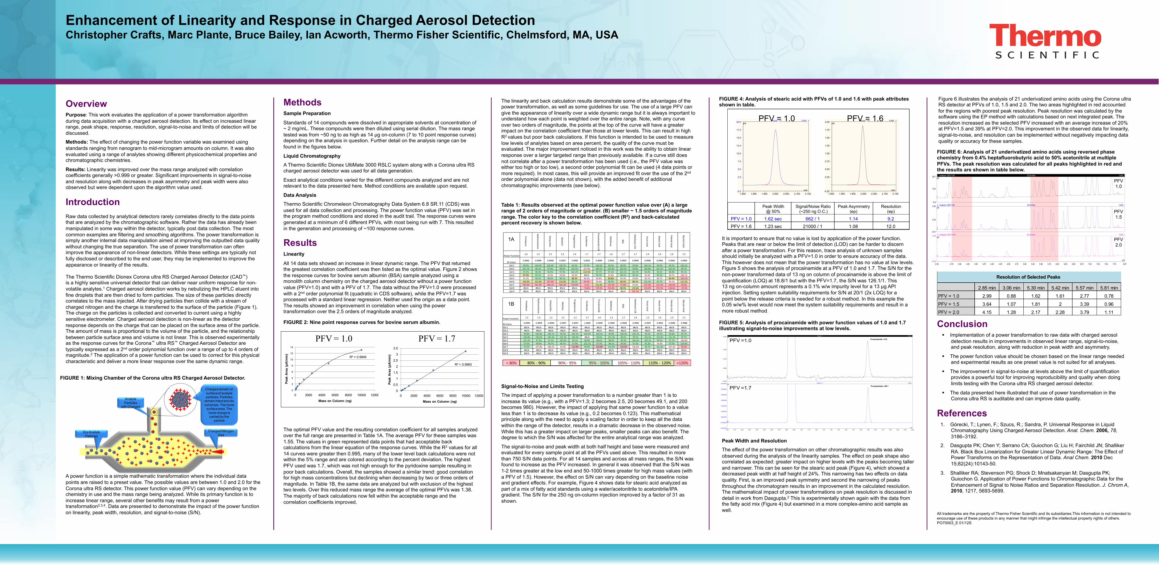
Enhancement of Linearity and Response in Charged Aerosol Detection Christopher Crafts, Marc Plante, Bruce Bailey, Ian Acworth, Thermo Fisher Scientific, Chelmsford, MA, USA
Conclusion § Implementation of a power transformation to raw data with charged aerosol
detection results in improvements in observed linear range, signal-to-noise, and peak resolution, along with reduction in peak width and asymmetry.
§ The power function value should be chosen based on the linear range needed and experimental results as one preset value is not suited for all analyses.
§ The improvement in signal-to-noise at levels above the limit of quantification provides a powerful tool for improving reproducibility and quality when doing limits testing with the Corona ultra RS charged aerosol detector.
§ The data presented here illustrated that use of power transformation in the Corona ultra RS is auditable and can improve data quality.
References 1. Górecki, T.; Lynen, F.; Szucs, R.; Sandra, P. Universal Response in Liquid
Chromatography Using Charged Aerosol Detection. Anal. Chem. 2006, 78, 3186–3192.
2. Dasgupta PK; Chen Y; Serrano CA; Guiochon G; Liu H; Fairchild JN; Shalliker RA. Black Box Linearization for Greater Linear Dynamic Range: The Effect of Power Transforms on the Representation of Data. Anal Chem. 2010 Dec 15;82(24):10143-50.
3. Shalliker RA; Stevenson PG; Shock D; Mnatsakanyan M; Dasgupta PK; Guiochon G. Application of Power Functions to Chromatographic Data for the Enhancement of Signal to Noise Ratios and Separation Resolution. J. Chrom A, 2010, 1217, 5693-5699.
Overview Purpose: This work evaluates the application of a power transformation algorithm during data acquisition with a charged aerosol detection. Its effect on increased linear range, peak shape, response, resolution, signal-to-noise and limits of detection will be discussed.
Methods: The effect of changing the power function variable was examined using standards ranging from nanogram to mid-microgram amounts on column. It was also evaluated using a range of analytes showing different physicochemical properties and chromatographic chemistries.
Results: Linearity was improved over the mass range analyzed with correlation coefficients generally >0.999 or greater. Significant improvements in signal-to-noise and resolution along with decreases in peak asymmetry and peak width were also observed but were dependent upon the algorithm value used.
Introduction Raw data collected by analytical detectors rarely correlates directly to the data points that are analyzed by the chromatographic software. Rather the data has already been manipulated in some way within the detector, typically post data collection. The most common examples are filtering and smoothing algorithms. The power transformation is simply another internal data manipulation aimed at improving the outputted data quality without changing the true separation. The use of power transformation can often improve the appearance of non-linear detectors. While these settings are typically not fully disclosed or described to the end user, they may be implemented to improve the appearance or linearity of the results. The Thermo Scientific Dionex Corona ultra RS Charged Aerosol Detector (CAD™) is a highly sensitive universal detector that can deliver near uniform response for non-volatile analytes.1 Charged aerosol detection works by nebulizing the HPLC eluent into fine droplets that are then dried to form particles. The size of these particles directly correlates to the mass injected. After drying particles then collide with a stream of charged nitrogen and the charge is transferred to the surface of the particle (Figure 1). The charge on the particles is collected and converted to current using a highly sensitive electrometer. Charged aerosol detection is non-linear as the detector response depends on the charge that can be placed on the surface area of the particle. The amount of mass is proportional to the volume of the particle, and the relationship between particle surface area and volume is not linear. This is observed experimentally as the response curves for the Corona™ ultra RS™ Charged Aerosol Detector are typically expressed as a 2nd order polynomial function over a range of up to 4 orders of magnitude.2 The application of a power function can be used to correct for this physical characteristic and deliver a more linear response over the same dynamic range. A power function is a simple mathematic transformation where the individual data points are raised to a preset value. The possible values are between 1.0 and 2.0 for the Corona ultra RS detector. This power function value (PFV) can vary depending on the chemistry in use and the mass range being analyzed. While its primary function is to increase linear range, several other benefits may result from a power transformation2,3,4. Data are presented to demonstrate the impact of the power function on linearity, peak width, resolution, and signal-to-noise (S/N).
FIGURE 1: Mixing Chamber of the Corona ultra RS Charged Aerosol Detector.
Methods
Sample Preparation
Standards of 14 compounds were dissolved in appropriate solvents at concentration of ~ 2 mg/mL. These compounds were then diluted using serial dilution. The mass range tested was from ~50 ng to as high as 14 µg on-column (7 to 10 point response curves) depending on the analysis in question. Further detail on the analysis range can be found in the figures below.
Liquid Chromatography
A Thermo Scientific Dionex UltiMate 3000 RSLC system along with a Corona ultra RS charged aerosol detector was used for all data generation.
Exact analytical conditions varied for the different compounds analyzed and are not relevant to the data presented here. Method conditions are available upon request.
Data Analysis
Thermo Scientific Chromeleon Chromatography Data System 6.8 SR.11 (CDS) was used for all data collection and processing. The power function value (PFV) was set in the program method conditions and stored in the audit trail. The response curves were generated at a minimum of 6 different PFVs, with most being run with 7. This resulted in the generation and processing of ~100 response curves.
Results
Linearity
All 14 data sets showed an increase in linear dynamic range. The PFV that returned the greatest correlation coefficient was then listed as the optimal value. Figure 2 shows the response curves for bovine serum albumin (BSA) sample analyzed using a monolith column chemistry on the charged aerosol detector without a power function value (PFV=1.0) and with a PFV of 1.7. The data without the PFV=1.0 were processed with a 2nd order polynomial fit (quadratic in CDS software), while the PFV=1.7 was processed with a standard linear regression. Neither used the origin as a data point. The results showed an improvement in correlation when using the power transformation over the 2.5 orders of magnitude analyzed.
The optimal PFV value and the resulting correlation coefficient for all samples analyzed over the full range are presented in Table 1A. The average PFV for these samples was 1.55. The values in green represented data points that had acceptable back calculations from the linear equation of the response curves. While the R2 values for all 14 curves were greater then 0.995, many of the lower level back calculations were not within the 5% range and are colored according to the percent deviation. The highest PFV used was 1.7, which was not high enough for the pyridoxine sample resulting in poor back calculations. Overall, the samples showed a similar trend: good correlation for high mass concentrations but declining when decreasing by two or three orders of magnitude. In Table 1B, the same data are analyzed but with exclusion of the highest two levels. Over this reduced mass range the average of the optimal PFVs was 1.38. The majority of back calculations now fell within the acceptable range and the correlation coefficients improved.
The linearity and back calculation results demonstrate some of the advantages of the power transformation, as well as some guidelines for use. The use of a large PFV can give the appearance of linearity over a wide dynamic range but it is always important to understand how each point is weighted over the entire range. Note, with any curve over two orders of magnitude, the points at the top of the curve will have a greater impact on the correlation coefficient than those at lower levels. This can result in high R2 values but poor back calculations. If this function is intended to be used to measure low levels of analytes based on area percent, the quality of the curve must be evaluated. The major improvement noticed in this work was the ability to obtain linear response over a larger targeted range than previously available. If a curve still does not correlate after a power transformation has been used (i.e., the PFV value was either too high or too low), a second order polynomial fit can be used (4 data points or more required). In most cases, this will provide an improved fit over the use of the 2nd order polynomial alone (data not shown), with the added benefit of additional chromatographic improvements (see below).
Signal-to-Noise and Limits Testing
The impact of applying a power transformation to a number greater than 1 is to increase its value (e.g., with a PFV=1.3; 2 becomes 2.5, 20 becomes 49.1, and 200 becomes 980). However, the impact of applying that same power function to a value less than 1 is to decrease its value (e.g., 0.2 becomes 0.123). This mathematical principle along with the need to apply a scaling factor in order to keep all the data within the range of the detector, results in a dramatic decrease in the observed noise. While this has a greater impact on larger peaks, smaller peaks can also benefit. The degree to which the S/N was affected for the entire analytical range was analyzed.
The signal-to-noise and peak width at both half height and base were measured and evaluated for every sample point at all the PFVs used above. This resulted in more than 750 S/N data points. For all 14 samples and across all mass ranges, the S/N was found to increase as the PFV increased. In general it was observed that the S/N was 1-2 times greater at the low end and 50-1000 times greater for high mass values (with a PFV of 1.5). However, the effect on S/N can vary depending on the baseline noise and gradient effects. For example, Figure 4 shows data for stearic acid analyzed as part of a mix of fatty acid standards using a water/acetonitrile to acetonitrile/IPA gradient. The S/N for the 250 ng on-column injection improved by a factor of 31 as shown.
All trademarks are the property of Thermo Fisher Scientific and its subsidiaries.This information is not intended to encourage use of these products in any manner that might infringe the intellectual property rights of others. PO70003_E 01/12S
FIGURE 2: Nine point response curves for bovine serum albumin.
Table 1: Results observed at the optimal power function value over (A) a large range of 2 orders of magnitude or greater. (B) smaller ~ 1.5 orders of magnitude range. The color key to the correlation coefficient (R2) and back-calculated percent recovery is shown below.
FIGURE 5: Analysis of procainamide with power function values of 1.0 and 1.7 illustrating signal-to-noise improvements at low levels.
FIGURE 6: Analysis of 21 underivatized amino acids using reversed phase chemistry from 0.4% heptafluorobutyric acid to 50% acetonitrile at multiple PFVs. The peak resolution was calculated for all peaks highlighted in red and the results are shown in table below.
It is important to ensure that no value is lost by application of the power function. Peaks that are near or below the limit of detection (LOD) can be harder to discern after a power transformation. For this reason, trace analysis of unknown samples should initially be analyzed with a PFV=1.0 in order to ensure accuracy of the data. This however does not mean that the power transformation has no value at low levels. Figure 5 shows the analysis of procainamide at a PFV of 1.0 and 1.7. The S/N for the non-power transformed data of 13 ng on column of procainamide is above the limit of quantification (LOQ) at 18.8/1 but with the PFV=1.7, the S/N was 126.1/1. This 13 ng on-column amount represents a 0.1% w/w impurity level for a 13 µg API injection. Setting system suitability requirements for S/N at 20/1 (2x LOQ) for a point below the release criteria is needed for a robust method. In this example the 0.05 w/w% level would now meet the system suitability requirements and result in a more robust method.
-5.6
12.5
28.7 1 - Analysis 120311 #5 22 AA M ix CAD_1pA
1
-0.61
2.50
5.94 2 - Analysis 120311 #6 22 AA M ix CAD_1pA
2
2.30 2.75 3.00 3.25 3.50 3.75 4.00 4.25 4.50 4.75 5.00 5.25 5.50 5.75 6.00 6.25 6.50 6.75 7.00 7.25 7.50 7.75 8.07-0.29
1.28 3 - Analysis 120311 #18 22 AA M ix CAD_1pA
min
3
R² = 0.9993
0
0.5
1
1.5
2
2.5
3
3.5
0 2000 4000 6000 8000 10000 12000
Peak
Are
a (p
A/m
in)
Mass on Column (ng)
R² = 0.9949
0
2
4
6
8
10
12
14
0 2000 4000 6000 8000 10000 12000
Peak
Are
a (p
A/m
in)
Mass on Column (ng)
PFV = 1.0 PFV = 1.7
FIGURE 4: Analysis of stearic acid with PFVs of 1.0 and 1.6 with peak attributes shown in table.
Figure 6 illustrates the analysis of 21 underivatized amino acids using the Corona ultra RS detector at PFVs of 1.0, 1.5 and 2.0. The two areas highlighted in red accounted for the regions with poorest peak resolution. Peak resolution was calculated by the software using the EP method with calculations based on next integrated peak. The resolution increased as the selected PFV increased with an average increase of 20% at PFV=1.5 and 39% at PFV=2.0. This improvement in the observed data for linearity, signal-to-noise, and resolution can be implemented without negatively impacting data quality or accuracy for these samples.
Peak Width and Resolution
The effect of the power transformation on other chromatographic results was also observed during the analysis of the linearity samples. The effect on peak shape also correlated as expected: greater impact on higher levels with the peaks becoming taller and narrower. This can be seen for the stearic acid peak (Figure 4), which showed a decreased peak width at half height of 24%. This narrowing has two effects on data quality. First, is an improved peak symmetry and second the narrowing of peaks throughout the chromatogram results in an improvement in the calculated resolution. The mathematical impact of power transformations on peak resolution is discussed in detail in work from Dasgupta.2 This is experimentally shown again with the data from the fatty acid mix (Figure 4) but examined in a more complex-amino acid sample as well.
-0.020
0.050
0.100
0.150
0.220 1 Power 1 CAD_1pA
1
Procainamide---13.8
0.00 0.10 0.20 0.30 0.40 0.50 0.60 0.70 0.80 0.90 1.00 1.10 1.20 1.30 1.40 1.50 1.60 1.70 1.80 1.90 2.00 2.10 2.20 2.30 2.40 2.50-0.000100
-0.000000
0.000100
0.000200
0.000300
0.000400
0.000500
0.000600
0.000700 2 Power 1.7 CAD_1pA
min
2
Procainamide---125.1
PFV =1.0
PFV =1.7
Primidone
Hydrocortisone
Ketoprofen
Warfarin
Progesterone
Pyridoxine
Procainamide C18
Diclofenac
BSA
Diclofenac C18
Erucic Acid
Stearic Acid
Palmitic Acid
Myristic Acid
Power Function1.6 1.5 1.5 1.5 1.4 1.7 1.7 1.6 1.7 1.7 1.4 1.6 1.6 1.3
R2 Value 0.9992 0.9996 0.9990 0.9997 0.9996 0.9952 0.9999 0.9991 0.9993 0.9999 0.9996 0.9996 0.9992 0.9993
Std 1 99.9% 100.6% 100.9% 100.5% 99.4% 97.3% 100.0% 99.4% 98.9% 99.8% 100.1% 99.9% 99.5% 100.2%Std 2 101.7% 98.1% 97.8% 98.9% 102.5% 107.8% 100.5% 103.9% 103.9% 99.8% 100.8% 101.5% 103.3% 98.7%Std 3 97.2% 98.3% 95.0% 97.9% 101.2% 113.5% 99.7% 97.2% 102.5% 101.9% 96.4% 96.5% 98.8% 99.1%Std 4 87.4% 95.5% 93.0% 93.8% 94.3% #N/A 96.7% 91.8% 98.1% 99.3% 93.4% 95.5% 90.7% 108.7%Std 5 91.2% 99.2% 98.0% 98.1% 88.2% 90.3% 93.8% 85.8% 94.3% 93.8% 93.7% 90.7% 84.4% 122.1%Std 6 119.2% 113.3% 127.6% 114.5% 98.1% 65.2% 94.4% 96.9% 84.5% 88.1% 101.4% 95.7% 93.1% 112.1%Std 7 192.0% 161.9% 200.1% 165.5% 134.5% 4.2% 115.2% 131.4% 77.4% 93.6% 137.3% 131.5% 133.0% 43.0%Std 8 #N/A #N/A #N/A #N/A #N/A -‐79.6% 174.3% 214.5% 88.5% 119.6% 223.0% 225.3% 234.6% -‐59.3%Std 9 #N/A #N/A #N/A #N/A #N/A #N/A #N/A #N/A #N/A 191.5% #N/A #N/A #N/A #N/A
< 80% 80% -‐ 90% 90% -‐ 95% 95% -‐ 105% 105% -‐ 110% 110% -‐ 120% >120%
Primidone
Hydrocortisone
Ketoprofen
Warfarin
Progesterone
Pyridoxine
Procainamide
Diclofenac
BSA
Diclofenac C18
Erucic Acid
Stearic Acid
Palmitic Acid
Myristic Acid
Power Function 1.2 1.3 1.2 1.2 1.2 1.7 1.6 1.3 1.7 1.6 1.2 1.4 1.3 1.5
R2 Value 0.9999 0.9999 0.9999 0.9997 0.9997 1.0000 0.9999 0.9999 0.9998 0.9999 0.9997 0.9994 0.9998 0.9988
Std 1 #N/A #N/A #N/A #N/A #N/A #N/A #N/A #N/A #N/A #N/A #N/A #N/A #N/A #N/AStd 2 #N/A #N/A #N/A #N/A #N/A #N/A #N/A #N/A #N/A #N/A #N/A #N/A #N/A #N/AStd 3 99.8% 100.0% 100.1% 99.5% 100.1% 100.0% 100.2% 99.8% 100.4% 100.1% 99.5% 99.4% 99.8% 99.7%Std 4 100.8% 100.6% 100.3% 102.3% 100.3% #N/A 99.6% 101.3% 98.5% 99.5% 102.3% 103.0% 101.3% 99.7%Std 5 101.4% 97.5% 97.2% 100.9% 96.5% 98.9% 98.6% 98.9% 99.3% 100.4% 100.1% 98.5% 97.7% 106.8%Std 6 97.0% 98.6% 99.7% 96.5% 97.8% 102.1% 97.4% 98.1% 99.0% 97.4% 95.3% 94.8% 97.0% 104.8%Std 7 97.6% 107.8% 108.2% 93.9% 113.9% 99.6% 110.9% 101.8% 110.5% 105.6% 98.7% 101.6% 107.3% 70.5%Std 8 #N/A #N/A #N/A #N/A #N/A #N/A #N/A #N/A #N/A #N/A #N/A #N/A #N/A #N/AStd 9 #N/A #N/A #N/A #N/A #N/A #N/A #N/A #N/A #N/A #N/A #N/A #N/A #N/A #N/A
1A
1B
2.85 min 3.06 min 5.30 min 5.42 min 5.57 min 5.81 min
PFV = 1.0 2.99 0.88 1.62 1.61 2.77 0.78
PFV = 1.5 3.64 1.07 1.81 2 3.39 0.96
PFV = 2.0 4.15 1.28 2.17 2.28 3.79 1.11
Resolution of Selected Peaks
Dry AnalyteParticles
Charged Nitrogen Gas
AnalyteParticles
with Charges
Charges remain on surface of analyteparticles. Particles
remain intact and do not ionize. The more
surface area. The more charge is carried by the
particle
1.850 1.900 1.950 2.000 2.050 2.100 2.150 -2.0
2.5 5.0 7.5
10.0 12.5 15.0 17.5 20.0 Power 1.0 CAD_1
pA
min 1.850 1.900 1.950 2.000 2.050 2.100 2.150 -0.20
0.25 0.50 0.75 1.00 1.25 1.50 1.75 2.00 Power 1.6 CAD_1
pA
min
Peak Width @ 50%
Signal/Noise Ratio (~250 ng O.C.)
Peak Asymmetry ( ep )
Resolution (ep)
PFV = 1.0 1.62 sec 662 / 1 1.14 9.2 PFV = 1.6 1.23 sec 21000 / 1 1.08 12.0
PFV = 1.0 PFV = 1.6
PFV 1.0
PFV 1.5
PFV 2.0