Embed Size (px)
Citation preview
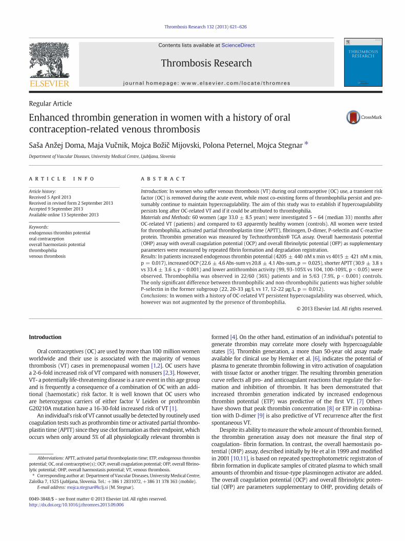
Thrombosis Research 132 (2013) 621–626
Contents lists available at ScienceDirect
Thrombosis Research
j ourna l homepage: www.e lsev ie r .com/ locate / th romres
Regular Article
Enhanced thrombin generation in women with a history of oralcontraception-related venous thrombosis
Saša Anžej Doma, Maja Vučnik, Mojca Božič Mijovski, Polona Peternel, Mojca Stegnar ⁎Department of Vascular Diseases, University Medical Centre, Ljubljana, Slovenia
Abbreviations:APTT, activated partial thromboplastin tpotential; OC, oral contraceptive(s); OCP, overall coagulatiolytic potential; OHP, overall haemostasis potential; VT, ven⁎ Corresponding author at: Department of Vascular Dise
Zaloška 7, 1525 Ljubljana, Slovenia. Tel.: +386 1 2831072E-mail address: [email protected] (M. Stegnar).
0049-3848/$ – see front matter © 2013 Elsevier Ltd. All rihttp://dx.doi.org/10.1016/j.thromres.2013.09.006
a b s t r a c t
a r t i c l e i n f oArticle history:
Received 5 April 2013Received in revised form 2 September 2013Accepted 9 September 2013Available online 13 September 2013Keywords:endogenous thrombin potentialoral contraceptionoverall haemostasis potentialthrombophiliavenous thrombosis
Introduction: In women who suffer venous thrombosis (VT) during oral contraceptive (OC) use, a transient riskfactor (OC) is removed during the acute event, while most co-existing forms of thrombophilia persist and pre-sumably continue to maintain hypercoagulability. The aim of this study was to establish if hypercoagulabilitypersists long after OC-related VT and if it could be attributed to thrombophilia.Materials and Methods: 60 women (age 33.0 ± 8.5 years) were investigated 5 – 64 (median 33) months afterOC-related VT (patients) and compared to 63 apparently healthy women (controls). All women were testedfor thrombophilia, activated partial thromboplastin time (APTT), fibrinogen, D-dimer, P-selectin and C-reactiveprotein. Thrombin generation was measured by Technothrombin® TGA assay. Overall haemostasis potential(OHP) assay with overall coagulation potential (OCP) and overall fibrinolytic potential (OFP) as supplementaryparameters were measured by repeated fibrin formation and degradation registration.Results: In patients increased endogenous thrombin potential (4205 ± 440 nM xmin vs 4015 ± 421 nM xmin,
p = 0.017), increasedOCP (22.6 ± 4.6 Abs-sumvs 20.8 ± 4.1 Abs-sum, p = 0.025), shorter APTT (30.9 ± 3.8 svs 33.4 ± 3.6 s, p b 0.001) and lower antithrombin activity (99, 93-105% vs 104, 100-109%, p b 0.05) wereobserved. Thrombophilia was observed in 22/60 (36%) patients and in 5/63 (7.9%, p b 0.001) controls.The only significant difference between thrombophilic and non-thrombophilic patients was higher solubleP-selectin in the former subgroup (22, 20-33 μg/L vs 17, 12-22 μg/L, p = 0.012).Conclusions: In women with a history of OC-related VT persistent hypercoagulability was observed, which,however was not augmented by the presence of thrombophilia.© 2013 Elsevier Ltd. All rights reserved.
Introduction
Oral contraceptives (OC) are used bymore than 100 million womenworldwide and their use is associated with the majority of venousthrombosis (VT) cases in premenopausal women [1,2]. OC users havea 2-6-fold increased risk of VT compared with nonusers [2,3]. However,VT- a potentially life-threateningdisease is a rare event in this age groupand is frequently a consequence of a combination of OC with an addi-tional (haemostatic) risk factor. It is well known that OC users whoare heterozygous carriers of either factor V Leiden or prothrombinG20210A mutation have a 16-30-fold increased risk of VT [1].
An individual’s risk of VT cannot usually be detected by routinely usedcoagulation tests such as prothrombin time or activated partial thrombo-plastin time (APTT) since they use clot formation as their endpoint,whichoccurs when only around 5% of all physiologically relevant thrombin is
ime; ETP, endogenous thrombinn potential; OFP, overall fibrino-ous thrombosis.ases, UniversityMedical Centre,, +386 31 378 363 (mobile).
ghts reserved.
formed [4]. On the other hand, estimation of an individual’s potential togenerate thrombin may correlate more closely with hypercoagulablestates [5]. Thrombin generation, a more than 50-year old assay madeavailable for clinical use by Hemker et al. [6], indicates the potential ofplasma to generate thrombin following in vitro activation of coagulationwith tissue factor or another trigger. The resulting thrombin generationcurve reflects all pro- and anticoagulant reactions that regulate the for-mation and inhibition of thrombin. It has been demonstrated thatincreased thrombin generation indicated by increased endogenousthrombin potential (ETP) was predictive of the first VT. [7] Othershave shown that peak thrombin concentration [8] or ETP in combina-tion with D-dimer [9] is also predictive of VT recurrence after the firstspontaneous VT.
Despite its ability tomeasure thewhole amount of thrombin formed,the thrombin generation assay does not measure the final step ofcoagulation- fibrin formation. In contrast, the overall haemostasis po-tential (OHP) assay, described initially by He et al in 1999 and modifiedin 2001 [10,11], is based on repeated spectrophotometric registraton offibrin formation in duplicate samples of citrated plasma to which smallamounts of thrombin and tissue-type plasminogen activator are added.The overall coagulation potential (OCP) and overall fibrinolytic poten-tial (OFP) are parameters supplementary to OHP, providing details of
622 S. Anžej Doma et al. / Thrombosis Research 132 (2013) 621–626
underlying changes in coagulation and fibrinolysis. The OHP assay wasevaluated in connection with hypercoagulability in normal pregnancyand preeclampsia [10,11], some types of thrombophilia [12,13], coro-nary heart disease [10], diabetes with microvascular complications[11], and in stroke [14].
In order to elucidate why some women using OC suffer VTwhereas the majority do not, we tested the hypothesis that womenwho suffer VT during OC use have a preexisting prothrombotictendency, associated with thrombophilic or other defects. While atransient risk factor for VT (OC) is removed during the acute event,most coexisting forms of thrombophilia persist and presumablycontinue to maintain hypecoagulability. Our aim was to establish ifhypercoagulability could be detected long after OC-related VT withglobal clotting assays (thrombin generation and OHP assay) and ifit could be attributed to thrombophilia.
Materials and Methods
Subjects
Consecutive women aged 50 years or less who had suffered VTduring OC use between the years 2005 and 2011 were included(patients). VT was confirmed by Doppler ultrasound and patients weretreated with low-molecular-weight heparin followed by oral anticoag-ulants. According to the guidelines [15] OC use was discontinued in allpatients. The exclusion criteria were malignant disease, pregnancy,puerperium, antiphospholipid syndrome or other conditions requiringprolonged anticoagulant treatment. Sixty-six patients matched theinclusion criteria. Six women refused to participate or contact wasnot possible, so 60 patients were finally included in the study. Atthe time of enrolment (5-64 months, median 33 months after acute VT)all patients had already completed anticoagulant treatment. 63healthy women of comparable age, who were not OC users or haddiscontinued OC at least 2 months before inclusion in the study,served as controls.
All participating women were without clinical signs of acute diseaseat the time of blood sampling. In all women age, body weight, bodyheight and smoking habits were recorded. Patients were asked aboutthe type and duration of the OC used and possible concomitant risk fac-tors for VT (trauma, surgery, immobilization) at the time of the disease.Body mass index (BMI) was calculated as body weight (kg)/bodyheight2 (m2). Patients and controls were not related to each other.They gave their full informed written consent to participate in thestudy, which was approved by the Slovene Ethics Committee.
Blood Sampling, Plasma and DNA Preparation
Blood was obtained on a single occasion in the morning fromthe antecubital vein with minimal venous stasis after a 20-min rest.For haemostasis assays 4.5 mL of blood were collected in each of twovacuum tubes containing 0.11 M sodium citrate (Becton Dickinson,Vacutaineer System Europe, Heidelberg, Germany), thoroughly mixedwith the anticoagulant, placed immidiately in ice water and centrifugedwithin 4 h of venepuncture at 4 °C and 2000 ×g for 30 min to obtrainplatelet-poor plasma. Before freezing plasma for detection of lupusanticoagulants was filtered through a 0.2 μm pore filter (Minisart,Sartorius Stedim Biotech GmbH, Göttingen, Germany). Aliquots ofplatelet-poor plasma were then frozen in liquid nitrogen and stored at-70 °C until analysed. For preparation of microparticle-free plasma,platelet-poor plasma was filtered through a microfilter unit (CeveronMFU 500, Technoclone, Vienna, Austria). DNA was isolated fromwhole blood or buffy coat using a commercially available kit (HighPure PCR Template Preparation Kit, Roche Diagnostica, Indianapolis,USA).
Laboratory Methods
APTT (Pathromtin SL), fibrinogen (Multifibren U), antithrombinactivity (Berichrom Antithrombin), protein C activity (BerichromProtein C, all from Dade/Behring, Marburg, Germany), lupus anticoagu-lants (LA-screen and LA-confirm, Gradipore, Sydney, Australia), activatedprotein C (APC) resistance (Coatest APC Resistance, V-S, Chromogenix,Mölndal, Sweden) and D-dimer (TriniLIA Auto-Dimer, Trinity Biotech,Bray, Ireland) were determined on an automated coagulation analyser(Behring Coagulation Timer, Dade Behring,Marburg, Germany). Free pro-tein S was determined by the immunoturbidimetric latex-agglutinationtest (STA-Liatest Free protein S, Diagnostica Stago, Asnières, France) andsoluble P-selectin by enzyme-linked immunosorbent assay (HumansP-selectin/CD62P Immunoassay, R&D Systems, Minneapolis, USA).C-reactive protein (CRP) was determined using the VITROS hs-CRPtest (Ortho-Clinical Diagnostics, Rochester, USA). Factor V Leidenand prothrombin G20210A polymorphism were determined by real-time polymerase chain reaction on ABI Prism 7000 SDS (AppliedBiosystems, Life Technologies Corp., Carlsbad, USA) using appropriateprimers and probes.
Thrombophilia was considered as described previously [14]: if anti-thrombin or protein C activity were below 0.75 or 0.70 relative tonormal plasma, respectively, if the concentration of free protein S wasbelow 0.55 relative to normal plasma, if the ratio between the screeningand the confirmatory test for lupus anticoagulantswas equal to or above1.20, if factor V Leiden or prothrombin G20210A polymorphism waspresent or if the ratio of APTT with APC versus APTT without APC wasbelow 1.93 (APC resistance).
Thrombin generation was determined using a commercial kit(Technothrombin® TGA,Technoclone, Vienna, Austria), which is basedon monitoring the fluorescence generated by thrombin cleavage of afluorigenic substrate over time on activation of the coagulation cascadeby tissue factor. The concentration of tissue factor was 5 pmol/L. Thefollowing parameters were registered: lag phase, time to peak thrombinconcentration, peak thrombin concentration and area under the curve –
endogenous thrombin potential (ETP). Amount ofmicroparticle-inducedthrombin generation was also determined by measuring thrombin gen-eration in microparticle-free (filtered using 0.2 μm vacuum filtratriondevice Ceveron®MFU-500, Technoclone, Austria) plasma versus throm-bin generation in non-filtered plasma. The amount of thrombin (peakthrombin concentration) induced by microparticles was calculated(in per cent). The source and procoagulant properties of the filteredmicroparticles have been reported elsewhere [16].
Parameters of the overall haemostasis potential assay (OHP, OCPand OFP) were determined as described by He et al. [11]. Fibrin-formation time curves were generated in microtitre plate wells andplasma samples were tested in duplicate.
For OHPmeasurement microtitre wells contained 60 μL plasma andOHP buffer (66 mmol/L Tris, 130 mmol/L NaCl, 17.0 mmol/L CaCl2,pH 7.5) with 0.04 IU/mL thrombin and 348 ng/mL recombinanttissue-type plasminogen activator. OHP curveswere generated fromau-tomated absorption measurements at 405 nm taken every minute for40 min. OCP curves were obtained in an identical way, except that theadded buffer did not contain recombinant tissue-type plasminogenactivator. Values for OCP and OHP were given by the areas under therelevant fibrin-formation time curves calculated by summation ofabsorption values (Abs-sum). The OFP values in % were calculated as[(OCP-OHP)/OCP] x 100.
Statistical Analysis
Statistical analysis was performed by the Statistica 8.0 (StatSoft Inc.,Tulsa, USA) statistical package. Distribution of variables was tested bythe Kolmogorov-Smirnov test. Normally distributed datawere expressedas means with standard deviations, while skewed data were presentedas medians with interquartile ranges. Differences between the two

Table 1Thrombin generation assay variables: lag phase, time-to-peak thrombin concentration,peak thrombin concentration and endogenous thrombin potential (ETP) in women with ahistory of oral contraceptive-related venous thrombosis (patients) and apparently healthywomen (controls). Values are presented as means ± standard deviations or medianswith interquartile ranges. The assay was performed first in platelet-poor plasma and sec-ondly after filtration of microparticles. The difference between the peak thrombin valuebefore and after filtration divided by the peak thrombin value before filtration representsthe percentage of microparticle-induced thrombin generation.
Patients (N = 60) Controls (N = 63) P
Platelet-poor plasmaLag phase (min) 7.0 (6.0 - 10.0) 7.0 (5.0 - 10.0) 0.558
623S. Anžej Doma et al. / Thrombosis Research 132 (2013) 621–626
groups studied were analysed by the χ2-test (categorical variables),the Student t-test (continuous variables with normal distribution) orMann-Whitney test (continuous variables with skewed distribution).Power calculations were performed by a statistics program StatisticalSolutions (LLC, Kansas, USA). Odds ratios were calculated by themedicalstatistics program Medcalc (Mariakerke, Belgium), which determinedodds ratios with 95% confidence intervals at given cut-offs. A p-value of0.05 or less was considered statistically significant.
Results
The majority of patients (55/60, 91.7%) suffered VT of the lowerlimbs, while 5/60 (8.3%) suffered VT of the upper extremities. At thetime of VT 35 patients were using combined OC with progestogens ofthe 3rd generation, 11 patients combined OC with drospirenone, 6 pa-tients combined OC with ciproterone acetate and 5 patients combinedOC with progestogens of the 2nd generation. All pills contained 0.02to 0.035 mg of ethinyl estradiol. Two patients used progestogen-onlyOC and one patient could not remember the type of pill. 13/60 (21.6%)patients reported injury, immobilisation or surgery before VT.
Patients and controls were comparable by age (33.5 ± 8.5 vs. 33.6 ±6.5 years, p = 0.931), bodymass index (23.8 kg/m2, 22.1- 26.4 kg/m2 vs.23.0 kg/m2, 21.7- 25.5 kg/m2, p = 0.207) and by prevalence of smokers(18/60 vs. 11/63, p = 0.102).
At least one type of thrombophilia was detected in 22/60 (36.0%)patients and in 5/63 (7.9%) controls (p b 0.001). 5 patients had twothrombophilic defects, one patient had even 3 defects. On the con-trary, in the control group all thrombophilic subjects had only onethrombophilic defect. The most prevalent thrombophilic defects werefactor V Leiden (16/60 vs. 2/63, p b 0.001) and prothrombin G20210Amutation (9/60 vs. 0/63, p = 0.001, patients vs. controls). In the patientgroup one factor V Leiden mutation and one prothrombin G20210Amutation were in a homozygous form. All subjects with APC resistancehad factor V Leidenmutation. Protein C deficiencywas observed in 2 pa-tients and 2 controls, and protein S deficiency in one patient. Athoughno antitrombin deficiency was present in either group, antithrombinactivity was significantly lower in patients (99, 93-105%) compared tocontrols (104, 100-109%, p b 0.05). Lupus anticoagulants were presentin one patient and in one control. The prevalence of thrombophiliawas significantly higher among the 20/60 (33.3%) patients who hadsuffered VT in the first year of OC use, compared to the patients whohad suffered VT after longer OC use (11/20 vs. 11/40, p = 0.037,respectively).
The thrombin generation assay revealed that ETP was significantlyhigher in patients compared to controls (Fig. 1), the peak thrombin con-centration was close to the level of statistical significance, while in the
Fig. 1. Endogenous thrombin potential (ETP) inwomenwith a history of oral contraceptive-related venous thrombosis (patients) and in apparently healthywomen (controls). Subjectswith thrombophilia are indicated by open symbols.
lag-phase and in time-to-peak no significant differences were observedbetween the two groups (Table 1). When performing the thrombingeneration assay in filtered plasma, we detected no differences in anyof the parameters of the assay, and also the percentage of microparticle-induced generated thrombin was similar in patients and controls(39 ± 11% vs. 40 ± 12%, p = 0.553, respectively).
The results for OHP, OCP and OFP are shown in Table 2. Patients hadhigher OHP (p = 0.067) and OCP (p = 0.025) values compared tocontrols.
Interestingly, in the patient group significantly shorter APTT com-pared to controls was found (30.9 ± 3.8 s vs. 33.4 ± 3.6 s, p b 0.001),while there were no significant differences in the levels of fibrinogen(3.3 ± 0.7 g/L vs. 3.1 ± 0.8 g/L, p = 0.117), D-dimer (76 μg/L,47-138 μg/L vs. 57 μg/L, 26-131 μg/L, p = 0.093), soluble P-selectin(19.6 μg/L, 12.8-25.8 μg/L vs. 17.8 μg/L, 8.0-26.4 μg/L, p = 0.226)or C-reactive protein (0.87 mg/L, 0.43-1.89 mg/L vs. 0.73 mg/L,0.24-1.59 mg/L, p = 0.218, respectively).
In order to evaluate the difference between thrombophilic andnonthrombophilic patients we tested differences between these twosubgroups of patients for all the variables studied. The only variablethat showed a significant difference was soluble P-selectin level, whichwas significantly higher in the thrombophilic group. Neither thrombingeneration nor haemostasis potential assay parameters showed anydifferences between these two subgroups of patients (Table 3).
The effects of ETP, OCP and APTT on the risk of developing VT wereevaluated by calculating odds ratios with their 95% confidence intervals(95% CI) using the cut-offs determined by the statistical programaccording to the best specificity and sensitivity (Table 4).
Discussion
We showed persistent hypercoagulability in the whole group ofwomen with a history of OC-related VT by using the thrombin genera-tion assay and the haemostasis potential assay. Since hypercoagulability
Time-to-peak (min) 10.5 (9.5 - 13.5) 11.0 (8.5 - 14.5) 0.973Peak thrombin (nM) 409.2 ± 98.4 375.5 ± 105.7 0.075ETP (nM x min) 4204.5 ± 440.4 4014.5 ± 420.5 0.017
After filtration of microparticlesLag phase (min) 8.5 (7.0 - 14.5) 9.0 (7.0 - 15.5) 0.960Time-to-peak (min) 13.5 (11.5 - 18.0) 13.5 (12.5 - 20.0) 0.281Peak thrombin (nM) 253.6 ± 82.0 229.3 ± 84.8 0.114ETP (nM x min) 3516.5 ± 628.0 3286.4 ± 747.3 0.072
Microparticles (%) 39 ± 11 40 ± 12 0.553
Table 2Haemostatic potential assay parameters: overall haemostasis potential (OHP), overallcoagulation potential (OCP) and overall fibrinolytic potential (OFP) in women with ahistory of oral contraceptive-related venous thrombosis (patients) and apparently healthywomen (controls). Values are presented as means ± standard deviations.
Patients Controls P
(N = 60) (N = 63)
OHP (Abs-sum) 12.4 ± 3.8 11.3 ± 3.3 0.067OCP (Abs-sum) 22.6 ± 4.6 20.8 ± 4.1 0.025OFP (%) 45.7 ± 8.6 46.8 ± 8.2 0.473
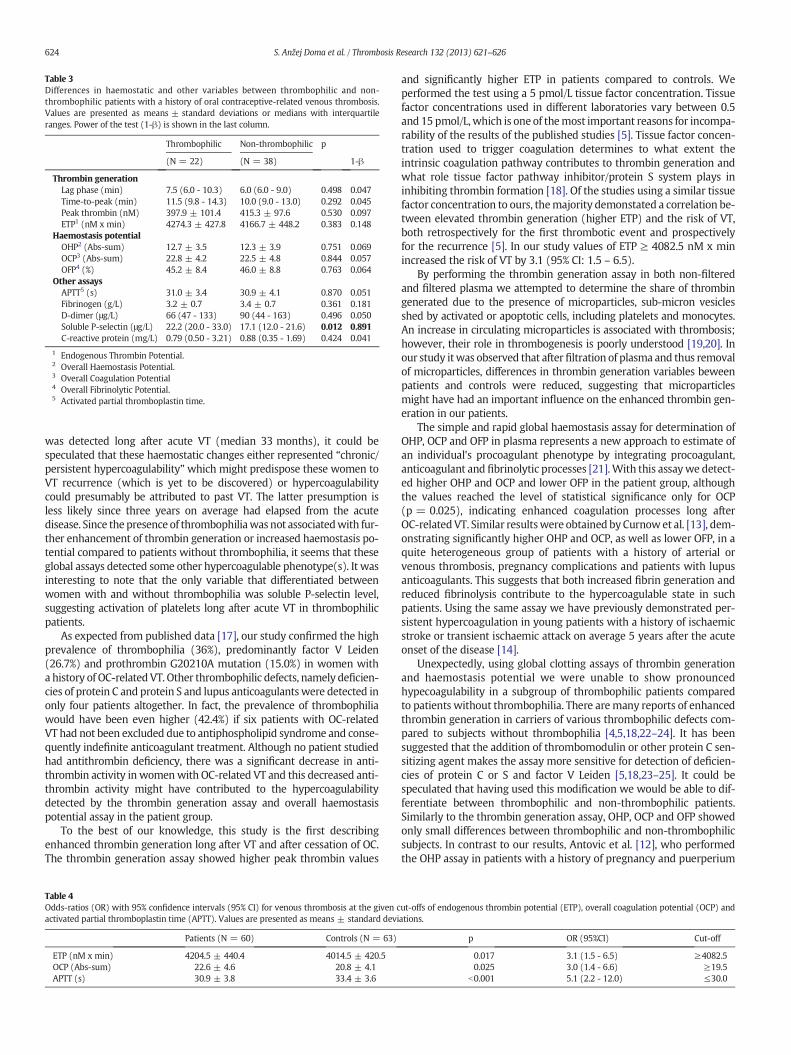
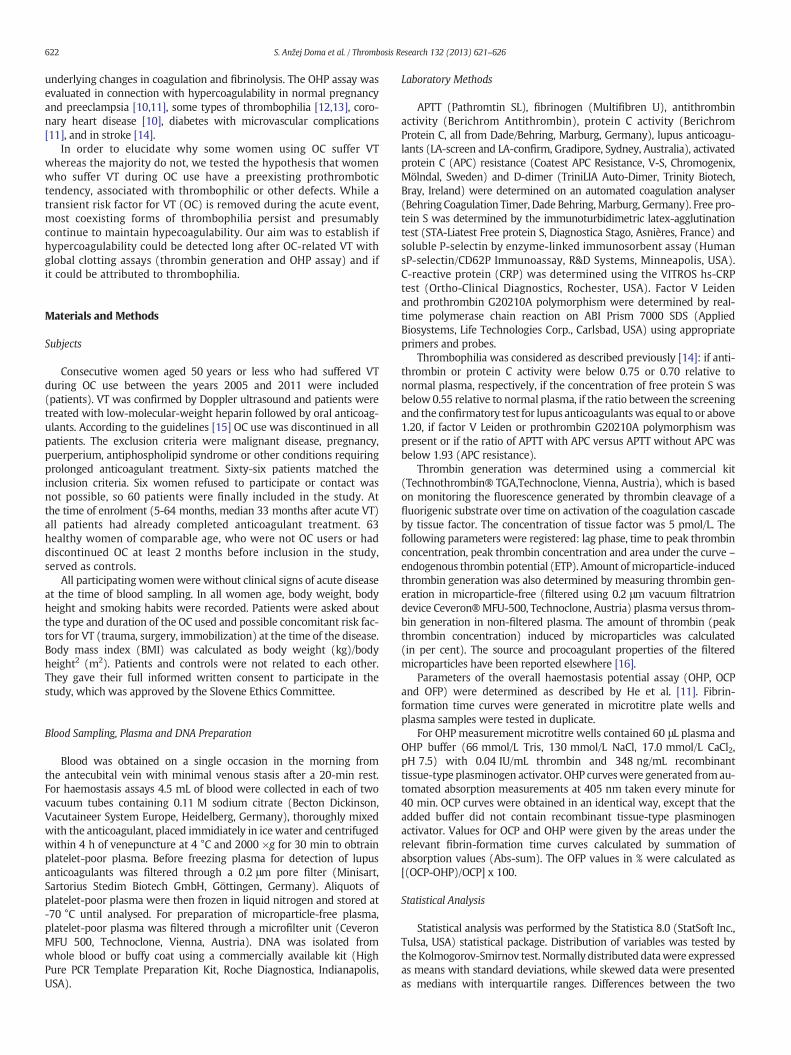
Table 3Differences in haemostatic and other variables between thrombophilic and non-thrombophilic patients with a history of oral contraceptive-related venous thrombosis.Values are presented as means ± standard deviations or medians with interquartileranges. Power of the test (1-β) is shown in the last column.
Thrombophilic Non-thrombophilic p
(N = 22) (N = 38) 1-β
Thrombin generationLag phase (min) 7.5 (6.0 - 10.3) 6.0 (6.0 - 9.0) 0.498 0.047Time-to-peak (min) 11.5 (9.8 - 14.3) 10.0 (9.0 - 13.0) 0.292 0.045Peak thrombin (nM) 397.9 ± 101.4 415.3 ± 97.6 0.530 0.097ETP1 (nM x min) 4274.3 ± 427.8 4166.7 ± 448.2 0.383 0.148
Haemostasis potentialOHP2 (Abs-sum) 12.7 ± 3.5 12.3 ± 3.9 0.751 0.069OCP3 (Abs-sum) 22.8 ± 4.2 22.5 ± 4.8 0.844 0.057OFP4 (%) 45.2 ± 8.4 46.0 ± 8.8 0.763 0.064
Other assaysAPTT5 (s) 31.0 ± 3.4 30.9 ± 4.1 0.870 0.051Fibrinogen (g/L) 3.2 ± 0.7 3.4 ± 0.7 0.361 0.181D-dimer (μg/L) 66 (47 - 133) 90 (44 - 163) 0.496 0.050Soluble P-selectin (μg/L) 22.2 (20.0 - 33.0) 17.1 (12.0 - 21.6) 0.012 0.891C-reactive protein (mg/L) 0.79 (0.50 - 3.21) 0.88 (0.35 - 1.69) 0.424 0.041
1 Endogenous Thrombin Potential.2 Overall Haemostasis Potential.3 Overall Coagulation Potential4 Overall Fibrinolytic Potential.5 Activated partial thromboplastin time.
624 S. Anžej Doma et al. / Thrombosis Research 132 (2013) 621–626
was detected long after acute VT (median 33 months), it could bespeculated that these haemostatic changes either represented “chronic/persistent hypercoagulability”which might predispose these women toVT recurrence (which is yet to be discovered) or hypercoagulabilitycould presumably be attributed to past VT. The latter presumption isless likely since three years on average had elapsed from the acutedisease. Since the presence of thrombophiliawas not associatedwith fur-ther enhancement of thrombin generation or increased haemostasis po-tential compared to patients without thrombophilia, it seems that theseglobal assays detected some other hypercoagulable phenotype(s). It wasinteresting to note that the only variable that differentiated betweenwomen with and without thrombophilia was soluble P-selectin level,suggesting activation of platelets long after acute VT in thrombophilicpatients.
As expected from published data [17], our study confirmed the highprevalence of thrombophilia (36%), predominantly factor V Leiden(26.7%) and prothrombin G20210A mutation (15.0%) in women witha history of OC-related VT. Other thrombophilic defects, namely deficien-cies of protein C and protein S and lupus anticoagulantswere detected inonly four patients altogether. In fact, the prevalence of thrombophiliawould have been even higher (42.4%) if six patients with OC-relatedVT had not been excluded due to antiphospholipid syndrome and conse-quently indefinite anticoagulant treatment. Although no patient studiedhad antithrombin deficiency, there was a significant decrease in anti-thrombin activity in womenwith OC-related VT and this decreased anti-thrombin activity might have contributed to the hypercoagulabilitydetected by the thrombin generation assay and overall haemostasispotential assay in the patient group.
To the best of our knowledge, this study is the first describingenhanced thrombin generation long after VT and after cessation of OC.The thrombin generation assay showed higher peak thrombin values
Table 4Odds-ratios (OR) with 95% confidence intervals (95% CI) for venous thrombosis at the given cactivated partial thromboplastin time (APTT). Values are presented as means ± standard devi
Patients (N = 60) Controls (N = 63)
ETP (nM x min) 4204.5 ± 440.4 4014.5 ± 420.5OCP (Abs-sum) 22.6 ± 4.6 20.8 ± 4.1APTT (s) 30.9 ± 3.8 33.4 ± 3.6
and significantly higher ETP in patients compared to controls. Weperformed the test using a 5 pmol/L tissue factor concentration. Tissuefactor concentrations used in different laboratories vary between 0.5and 15 pmol/L,which is one of themost important reasons for incompa-rability of the results of the published studies [5]. Tissue factor concen-tration used to trigger coagulation determines to what extent theintrinsic coagulation pathway contributes to thrombin generation andwhat role tissue factor pathway inhibitor/protein S system plays ininhibiting thrombin formation [18]. Of the studies using a similar tissuefactor concentration to ours, themajority demonstated a correlation be-tween elevated thrombin generation (higher ETP) and the risk of VT,both retrospectively for the first thrombotic event and prospectivelyfor the recurrence [5]. In our study values of ETP ≥ 4082.5 nM x minincreased the risk of VT by 3.1 (95% CI: 1.5 – 6.5).
By performing the thrombin generation assay in both non-filteredand filtered plasma we attempted to determine the share of thrombingenerated due to the presence of microparticles, sub-micron vesiclesshed by activated or apoptotic cells, including platelets and monocytes.An increase in circulating microparticles is associated with thrombosis;however, their role in thrombogenesis is poorly understood [19,20]. Inour study itwas observed that after filtration of plasma and thus removalof microparticles, differences in thrombin generation variables beweenpatients and controls were reduced, suggesting that microparticlesmight have had an important influence on the enhanced thrombin gen-eration in our patients.
The simple and rapid global haemostasis assay for determination ofOHP, OCP and OFP in plasma represents a new approach to estimate ofan individual's procoagulant phenotype by integrating procoagulant,anticoagulant and fibrinolytic processes [21].With this assaywe detect-ed higher OHP and OCP and lower OFP in the patient group, althoughthe values reached the level of statistical significance only for OCP(p = 0.025), indicating enhanced coagulation processes long afterOC-relatedVT. Similar resultswere obtained by Curnowet al. [13], dem-onstrating significantly higher OHP and OCP, as well as lower OFP, in aquite heterogeneous group of patients with a history of arterial orvenous thrombosis, pregnancy complications and patients with lupusanticoagulants. This suggests that both increased fibrin generation andreduced fibrinolysis contribute to the hypercoagulable state in suchpatients. Using the same assay we have previously demonstrated per-sistent hypercoagulation in young patients with a history of ischaemicstroke or transient ischaemic attack on average 5 years after the acuteonset of the disease [14].
Unexpectedly, using global clotting assays of thrombin generationand haemostasis potential we were unable to show pronouncedhypecoagulability in a subgroup of thrombophilic patients comparedto patients without thrombophilia. There aremany reports of enhancedthrombin generation in carriers of various thrombophilic defects com-pared to subjects without thrombophilia [4,5,18,22–24]. It has beensuggested that the addition of thrombomodulin or other protein C sen-sitizing agent makes the assay more sensitive for detection of deficien-cies of protein C or S and factor V Leiden [5,18,23–25]. It could bespeculated that having used this modification we would be able to dif-ferentiate between thrombophilic and non-thrombophilic patients.Similarly to the thrombin generation assay, OHP, OCP and OFP showedonly small differences between thrombophilic and non-thrombophilicsubjects. In contrast to our results, Antovic et al. [12], who performedthe OHP assay in patients with a history of pregnancy and puerperium
ut-offs of endogenous thrombin potential (ETP), overall coagulation potential (OCP) andations.
p OR (95%CI) Cut-off
0.017 3.1 (1.5 - 6.5) ≥4082.50.025 3.0 (1.4 - 6.6) ≥19.5
b0.001 5.1 (2.2 - 12.0) ≤30.0
625S. Anžej Doma et al. / Thrombosis Research 132 (2013) 621–626
related VT, demonstrated increased OHP long after the event; in caseswith APC resistance and factor V Leiden the imbalance in OHP wasmore severe than in those without. Similarly to our results, Andersenet al. [26] showed that the original overall haemostasis potential assaycannot differentiate between thrombophilic and non-thrombophilicsubjects. However, using amodification of the original assaywith Protacand pentasaccharide to enhance inhibition by activation of protein Cand by antithrombin, major thrombophilias can be detected with asensitivity of 100% and a specificity of 70-80% [21].
Women with OC-related VT differed from the controls in a signifi-cantly shorter APTT, a global test sensitive to all coagulation factorsexcept FVII and FXIII [27]. It was shown in this study that APTT equalor shorter than 30 s increased the risk of OC-related VT by 5.1 (95% CI:2.2 – 12.0), confirming a recently found association between shortenedAPTT and the risk of VT [28]. In the latter study it was demonstrated thatVT risk can be predicted by using the ratio of patient APTT to referencenormal APTT. The adjusted odds ratio for VT increased with decreasingAPTT ratio, being 2-3 for ratios lower than 0.87, independently ofinherited thrombophilic abnormalities. Additionally, Hron et al. [29]and Legnani et al. [30] demonstrated that measurement of APTT aftercompleted anticoagulant treatment allowed stratification of patientswith VT into high- and low-risk categories with regard to VT recurrence.
Apart from the most common forms of thrombophilia included in thethrombophilia screening panel, there have beenmany ‘new’ factors of thecoagulation and inflammation system described recently, which eitherindicate increased risk of VT or reflect an already ongoing pathology[31–35]. In the present study no differences either in markers of inflam-mation (fibrinogen, C-reactive protein) ormarkers of haemostasis activa-tion (D-dimer, P-selectin) were observed between patients and controls.However, when comparing thrombophilic and non-thrombophilic pa-tients an increase in soluble P-selectinwas detected in the former, indicat-ing platelet activation in this subgroup of patients.
Besides being a platelet activation marker, P-selectin is nowregarded as one of biomarkers of VT, useful especially in the diagnosisof acute VT [36]. Kyrle et al. [37] also demonstrated that P-selectin levelsafter cessation of anticoagulation in patients with first unprovoked VTpredispose these patients for VT recurrence. Furthermore, Ay et al.[38] showed that high soluble P-selectin levels independently predictVT in cancer patients. We found no data on connection of soluble P-selectin and thrombophilia, but from the mentioned studies we couldspeculate that increased levels of P-selectin in our patients withthrombophilia predispose them to VT recurrence.
In the present study more than 2/3 of patients used OC that imposea higher risk of VT, namely combined OC of the 3rd generation andOC with ciproterone acetate [3,39,40]. Progestogen-only pills which donot increase risk of VT [2,3] were used by only 2 patients. It has beenshown previously that OC use has a pronounced effect on coagulation:OC increase factor VII, factor XII, protein C and decrease antithrombinand protein S [39,41]. In women who suffer OC-related VT these effectsare even more pronounced, indicating that some women become“high haemostatic responders” and therefore most vulnerable to thethrombogenic effects of OC [42]. In a numerical simulation model andwith empirical synthetic plasma Brummel-Ziedins et al. [43] showedthat OC use creates extreme shifts in thrombin generation in both con-trol women and women with prior thrombosis, causing a subclinicalprothrombotic phenotype.With our studywe confirmed the hypothesisthat patientswith a history of OC-related VT long after acute disease andremoval of OC still expressed a prothrombotic tendency. The possibleexplanation for the fact that the majority of our patients were usingOC with the highest known risk of VT, could be that besides a possibleprothrombotic tendency upon an additional ‘stronger’ risk factor thesewomen could reach the treshold for VT more easily, compared toother patients using less thrombogenic OC.
In the present study most of the patients (40/60) investigatedsuffered VT after more than one year of OC use, which is in contrast toprevious studies describing the greatest risk during the first year, or
even during the first three months of OC use [2,3,39]. Among the 20/60patients who did suffer VT in the first year of OC use, thrombophiliawas more prevalent, suggesting these patients suffer VT not only morefrequently but also sooner than those without thrombophilic defects.
This study has some limitations, oneof thembeing the small numberof participants, especially when considering thrombophilic and non-thrombophilic patients. Secondly, the study is retrospective, so wecannot differentiate if the detected hypercoagulability existed beforeVT or was the consequence of the disease. As already noted, if we hadusedmodified thrombin generation and OHP assayswewould have pre-sumably been able to find significant differences between thrombophilicand non-thrombophilic patients.
To conclude, in this study we demonstrated that womenwith a his-tory of OC-related VT showed a hypercoagulable phenotype long afterthe event and withdrawal of the OC. The presence of thrombophilia,whichwas detected in more than a third of patients, was not associatedwith further increase of hypercoagulability detected in thewhole groupof patients by thrombin generation, haemostasis potential assay andAPTT. This could be explained by the low sensitivity of the assays, bythe insufficiency of current thrombophilia screening methods to detectall important thrombophilic defects, or by the presumption that global as-says of thrombin generation, haemostasis potential estimation and APTTdetect hypercoagulable phenotypes different from the thrombophiliadefects routinely determined in clinical practice.
Sources of Funding
The study was supported by the Slovenian Research Agency(Grant No. P3-0308).
Disclosures
None.
Conflict of Interest Statement
None.
Acknowledgements
None.
References
[1] Rosendaal FR, Helmerhorst FM, Vandenbroucke JP. Female hormones and thrombosis.Arterioscler Thromb Vasc Biol 2002;22:201–10.
[2] Gomes MP, Deitcher SR. Risk of venous thromboembolic disease associated withhormonal contraceptives and hormone replacement therapy: a clinical review.Arch Intern Med 2004;164:1965–76.
[3] Hannaford PC. Epidemiology of the contraceptive pill and venous thromboembolism.Thromb Res 2011;127(Suppl. 3):S30–4.
[4] Baglin T. The measurement and application of thrombin generation. Br J Haematol2005;130:653–61.
[5] van Veen JJ, Gatt A, MakrisM. Thrombin generation testing in routine clinical practice:are we there yet? Br J Haematol 2008;142:889–903.
[6] Hemker HC, Al Dieri R, De Smedt E, Béguin S. Thrombin generation, a function test ofthe haemostatic-thrombotic system. Thromb Haemost 2006;96:553–61.
[7] van Hylckama Vlieg A, Christiansen SC, Luddington R, Cannegieter SC, Rosendaal FR,Baglin TP. Elevated endogenous thrombin potential is associated with an increasedrisk of a first deep venous thrombosis but not with the risk of recurrence. Br JHaematol 2007;138:769–74.
[8] Hron G, Kollars M, Binder BR, Eichinger S, Kyrle PA. Identification of patients at lowrisk for recurrent venous thromboembolism by measuring thrombin generation.JAMA 2006;296:397–402.
[9] Eichinger S, Hron G, Kollars M, Kyrle PA. Prediction of recurrent venous thromboem-bolismby endogenous thrombin potential andD-dimer. Clin Chem2008;54:2042–8.
[10] He S, Bremme K, Blombäck M. A laboratory method for determination of overallhaemostatic potential in plasma. I. Method design and preliminary results. ThrombRes 1999;96:145–56.
[11] He S, Antovic A, BlombäckM. A simple and rapid laboratorymethod for determinationof haemostasis potential in plasma. II. Modifications for use in routine laboratories andresearch work. Thromb Res 2001;103:355–61.

626 S. Anžej Doma et al. / Thrombosis Research 132 (2013) 621–626
[12] Antovic A, Blombäck M, Bremme K, Van Rooijen M, He S. Increased hemostasispotential persists in women with previous thromboembolism with or without APCresistance. J Thromb Haemost 2003;1:2531–5.
[13] Curnow JL, Morel-KoppMC, Roddie C, Aboud M, Ward CM. Reduced fibrinolysis andincreased fibrin generation can be detected in hypercoagulable patients using theoverall hemostatic potential assay. J Thromb Haemost 2007;5:528–34.
[14] Anzej S, Bozic M, Antovic A, Peternel P, Gaspersic N, Rot U, et al. Evidence ofhypercoagulability and inflammation in young patients long after acute cerebralischaemia. Thromb Res 2007;120:39–46.
[15] Medical eligibility criteria for contraceptive use. 4th ed. Geneva: World HealthOrganization; 2009.
[16] Lawrie AS, Harrison P, Cardigan RA, Mackie IJ. The characterization and impact ofmicroparticles on haemostasis within fresh-frozen plasma. Vox Sang Oct2008;95(3):197–204.
[17] Stegnar M. Thrombophilia screening–at the right time, for the right patient, with agood reason. Clin Chem Lab Med 2010;48(Suppl. 1):S105–13.
[18] Castoldi E, Rosing J. Thrombin generation tests. Thromb Res 2011;127(Suppl. 3):S21–5.
[19] Berckmans RJ, Neiuwland R, Boing AN, Romijn FP, Hack CE, Sturk A. Cell-derivedmicroparticles circulate inhealthyhumans and support lowgrade thrombingeneration.Thromb Haemost 2001;85:639–46.
[20] Bucciarelli P, Martinelli I, Artoni A, Passamonti SM, Previtali E, Merati G, et al.Circulating microparticles and risk of venous thromboembolism. Thromb Res2012;129:591–7.
[21] Antovic A. The overall hemostasis potential: a laboratory tool for the investigation ofglobal hemostasis. Semin Thromb Hemost 2010;36:772–9.
[22] Eichinger S. Thrombin generation and venous thromboembolism. Hamostaseologie2008;28:37–9.
[23] Segers O, van Oerle Rv, ten Cate Ht, Rosing J, Castoldi E. Thrombin generation as anintermediate phenotype for venous thrombosis. ThrombHaemost 2010;103:114–22.
[24] Hemker HC, Al Dieri R, Béguin S. Thrombin generation assays: accruing clinicalrelevance. Curr Opin Hematol 2004;11:170–5.
[25] Tripodi A, Martinelli I, Chantarangkul V, Battaglioli T, Clerici M, Mannucci PM. Theendogenous thrombin potential and the risk of venous thromboembolism. ThrombRes 2007;121:353–9.
[26] AndresenMS, Iversen N, Abildgaard U. Overall haemostasis potential assays performedin thrombophilic plasma: the effect of preactivating protein C and antithrombin.Thromb Res 2002;108:323–8.
[27] Tripodi A,Mannucci PM.Activatedpartial thromboplastin time (APTT). New indicationsfor an old test? J Thromb Haemost 2006;4:750–1.
[28] Tripodi A, Chantarangkul V, Martinelli I, Bucciarelli P, Mannucci PM. A shortenedactivated partial thromboplastin time is associated with the risk of venous thrombo-embolism. Blood 2004;104:3631–4.
[29] Hron G, Eichinger S, Weltermann A, Quehenberger P, Halbmayer WM, Kyrle PA.Prediction of recurrent venous thromboembolism by the activated partial thrombo-plastin time. J Thromb Haemost 2006;4:752–6.
[30] Legnani C, Mattarozzi S, Cini M, Cosmi B, Favaretto E, Palareti G. Abnormally shortactivated partial thromboplastin time values are associated with increased risk ofrecurrence of venous thromboembolism after oral anticoagulation withdrawal. Br JHaematol 2006;134:227–32.
[31] Kappelmayer J, Nagy Jr B, Miszti-Blasius K, Hevessy Z, Setiadi H. The emerging valueof P-selectin as a disease marker. Clin Chem Lab Med 2004;42:475–86.
[32] Lippi G, Favaloro EJ, Montagnana M, Franchini M. C-reactive protein and venousthromboembolism: causal or casual association? Clin Chem Lab Med2010;48:1693–701.
[33] Davalos D, Akassoglou K. Fibrinogen as a key regulator of inflammation in disease.Semin Immunopathol 2012;34:43–62.
[34] Polgar J, Matuskova J, Wagner DD. The P-selectin. tissue factor. coagulation triad.J Thromb Haemost 2005;3:1590–6.
[35] Wagner DD. New links between inflammation and thrombosis. Arterioscler ThrombVasc Biol 2005;25:1321–4.
[36] Ramacciotti E, Blackburn S, Hawley AE, Vandy F, Ballard-Lipka N, Stabler C, et al.Evaluation of soluble P-selectin as a marker for the diagnosis of deep venousthrombosis. Clin Appl Thromb Hemost 2011;17:425–31.
[37] Kyrle PA, Hron G, Eichinger S,Wagner O. Circulating P-selectin and the risk of recurrentvenous thromboembolism. Thromb Haemost 2007;97:880–3.
[38] Ay C, Simanek R, Vormittag R, Dunkler D, Alguel G, Koder S, et al. High plasmalevels of soluble P-selectin are predictive of venous thromboembolism in cancerpatients: results from the Vienna Cancer and Thrombosis Study (CATS). Blood2008;112:2703–8.
[39] van Hylckama Vlieg A, Helmerhorst FM, Vandenbroucke JP, Doggen CJ, Rosendaal FR.The venous thrombotic risk of oral contraceptives, effects of oestrogen dose andprogestogen type: results of the MEGA case-control study. BMJ 2009;339:b2921.http://dx.doi.org/10.1136/bmj.b2921.
[40] Vandenbroucke JP, Rosing J, Bloemenkamp KW, Middeldorp S, Helmerhorst FM,Bouma BN, et al. Oral contraceptives and the risk of venous thrombosis. N Engl JMed 2001;344:1527–35.
[41] Rosendaal FR. Venous thrombosis: the role of genes, environment, and behavior.Hematology Am Soc Hematol Educ Program 2005:1–12.
[42] Bloemenkamp KW, Rosendaal FR, Helmerhorst FM, Koster T, Bertina RM,Vandenbroucke JP. Hemostatic effects of oral contraceptives in women whodeveloped deep-vein thrombosis while using oral contraceptives. Thromb Haemost1998;80:382–7.
[43] Brummel-Ziedins KE, Vossen CY, Butenas S, Mann KG, Rosendaal FR.Thrombin generation profiles in deep venous thrombosis. J Thromb Haemost2005;3:2497–505.