Embed Size (px)
Citation preview
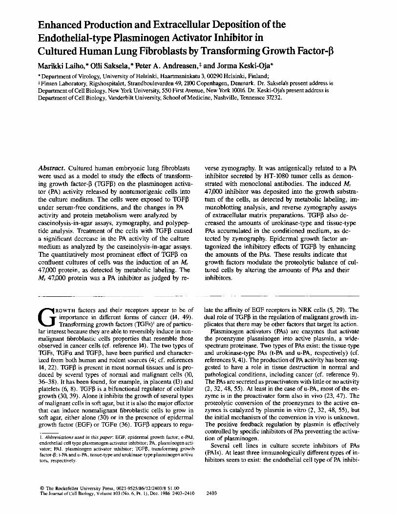
Enhanced Production and Extracellular Deposition of the Endothelial-type Plasminogen Activator Inhibitor in Cultured Human Lung Fibroblasts by Transforming Growth Factor-fl M a r i k k i Laiho,* OUi Sakse la ,* Peter A. Andreasen ,~ a n d J o r m a Keski-Oja*
* Department of Virology, University of Helsinki, Haartmaninkatu 3, 00290 Helsinki, Finland; ~Finsen Laboratory, Rigshospitalet, Strandboulevarden 49, 2100 Copenhagen, Denmark. Dr. Saksela's present address is Department of Cell Biology, New York University, 550 First Avenue, New York 10016. Dr. Keski-Oja's present address is Department of Cell Biology, Vanderbilt University, School of Medicine, Nashville, Tennessee 37232.
Abstract. Cultured human embryonic lung fibroblasts were used as a model to study the effects of transform- ing growth factor-13 (TGFI3) on the plasminogen activa- tor (PA) activity released by nontumorigenic cells into the culture medium. The cells were exposed to TGFfl under serum-free conditions, and the changes in PA activity and protein metabolism were analyzed by caseinolysis-in-agar assays, zymography, and polypep- tide analysis. Treatment of the cells with TGFI~ caused a significant decrease in the PA activity of the culture medium as analyzed by the caseinolysis-in-agar assays. The quantitatively most prominent effect of TGFI3 on confluent cultures of ceils was the induction of an Mr 47,000 protein, as detected by metabolic labeling. The Mr 47,000 protein was a PA inhibitor as judged by re-
verse zymography. It was antigenically related to a PA inhibitor secreted by HT-1080 tumor cells as demon- strated with monoclonal antibodies. The induced Mr 47,000 inhibitor was deposited into the growth substra- tum of the cells, as detected by metabolic labeling, im- munoblotting analysis, and reverse zymography assays of extracellular matrix preparations. TGFI3 also de- creased the amounts of urokinase-type and tissue-type PAs accumulated in the conditioned medium, as de- tected by zymography. Epidermal growth factor an- tagonized the inhibitory effects of TGFI3 by enhancing the amounts of the PAs. These results indicate that growth factors modulate the proteolytic balance of cul- tured ceils by altering the amounts of PAs and their inhibitors.
ROWTh factors and their receptors appear to be of importance in different forms of cancer (14, 49). Transforming growth factors (TGFs) t are of particu-
lar interest because they are able to reversibly induce in non- malignant fibroblastic cells properties that resemble those observed in cancer cells (cf. reference 14). The two types of TGFs, TGFa and TGFI3, have been purified and character- ized from both human and rodent sources (4; cf. references 14, 22). TGF~ is present in most normal tissues and is pro- duced by several types of normal and malignant cells (10, 36-38). It has been found, for example, in placenta (13) and platelets (6, 8). TGFI~ is a bifunctional regulator of cellular growth (30, 39). Alone it inhibits the growth of several types of malignant cells in soft agar, but it is also the major effector that can induce nonmalignant fibroblastic cells to grow in soft agar, either alone (30) or in the presence of epidermal growth factor (EGF) or TGFa (36). TGFI3 appears to regu-
1. Abbreviations used in this paper: EGF, epidermal growth factor; e-PAl, endothelial cell type plasminogen activator inhibitor; PA, plasminogen acti- vator; PAl, plasminogen activator inhibitor; TGFI~, transforming growth factor-13; t-PA and u-PA, tissue-type and urokinase-type plasminogen activa- tors, respectively.
late the affinity of EGF receptors in NRK cells (5, 29). The dual role of TGF~ in the regulation of malignant growth im- plicates that there may be other factors that target its action.
Plasminogen activators (PAs) are enzymes that activate the proenzyme plasminogen into active plasmin, a wide- spectrum proteinase. Two types of PAs exist: the tissue-type and urokinase-type PAs (t-PA and u-PA, respectively) (cf. references 9, 41). The production of PA activity has been sug- gested to have a role in tissue destruction in normal and pathological conditions, including cancer (cf. reference 9). The PAs are secreted as proactivators with little or no activity (2, 32, 48, 55). At least in the case of u-PA, most of the en- zyme is in the proactivator form also in vivo (23, 47). The proteolytic conversion of the proenzymes to the active en- zymes is catalyzed by plasmin in vitro (2, 32, 48, 55), but the initial mechanism of the conversion in vivo is unknown. The positive feedback regulation by plasmin is effectively controlled by specific inhibitors of PAs preventing the activa- tion of plasminogen.
Several cell lines in culture secrete inhibitors of PAs (PAls). At least three immunologically different types of in- hibitors seem to exist: the endothelial cell type of PA inhibi-
© The Rockefeller University Press, 0021-9525/86/12/2403/8 $1.00 The Journal of Cell Biology, Volume 103 (No. 6, Pt. 1), Dec. 1986 2403-2410 2403
tor (referred to here as e-PAl) (53), the protease nexin I (PN I) (44, 45), and the placental PAl (6a). Simultaneous secre- tion of PAs and PAls can easily lead to misinterpretations of the PA activity secreted by the cells, especially when PAs have been quantitated using activity measurements only (cf. reference 41). The effects of PAls have been neglected in several studies describing PA activity of cultured cells or PA activity found in tissues.
We have analyzed here the effects of TGFI3 on the PA ac- tivity of PA-producing human embryonic lung fibroblasts. These cells, upon exposure to TGFIS, produced enhanced amounts of a PAl into their medium in a time- and dose- dependent manner and also deposited it to their growth sub- stratum. TGFI5 also appeared to decrease the secretion of PA activity into the medium. Our results indicate that a major effect of TGFI5 on cultured embryonic lung fibroblasts is the enhancement of e-PAl production.
This work was presented in part at the UCLA Symposium on Growth Factors, Tumor Promoters, and Cancer Genes, Steamboat Springs, CO, April 6-13, 1986 (20).
Materials and Methods
Growth Factors and Reagents The reagents were obtained from the following sources. TGFfl was a kind gift from Drs. A. Roberts and R. Assoian (National Cancer Institute, Bethesda, MD). Its purification and properties were as described in detail (6). It stimulated the growth of NRK cells (clone 49F) and inhibited the growth of A549 lung cancer cells in soft agar as described (30, 39). EGF was purchased from Sigma Chemical Co. (St. Louis, MO).
Urokinase (60,000 IU/mg; Mr 54,000) was purchased from Calbiochem- Behring Corp. (La Jolla, CA) and plasminogen and plasmin were from Kabi Vitrum (Stockholm, Sweden). Aprotinin (Trasylol) was purchased from Bayer (Leverkusen, FRG). Antibodies to urokinase-type PA (u-PA) were produced in rabbits using the above Mr 54,000 u-PA as immunogen as de- scribed earlier (42). Tissue-type PA and rabbit polyclonal antibodies to tissue-type PA (t-PA) were a kind gift from Dr. D. Collen (University of Leuwen, Belgium) (35). Anti-u-PA and anti-t-PA antibodies did not cross- react with each other. Polyclonal and monoclonal antibodies against endo- thelial cell type PAl, secreted by dexamethasone-treated HT-1080 tumor cells, were produced as described (3, 31).
Cell Cultures Human embryonic lung fibroblasls (HEL-299 and WI-38) were obtained from American Type Culture Collection (ATCC CCL-137 and CCL-75, respectively). The two other WI-38 cell lines tested were obtained from Flow Laboratories, Inc. (Irvine, UK) and from Dr. A. Vaheri (Department of Virology, University of Helsinki, Finland). The cells were cultivated in plastic Linbro wells (16-ram diameter, Flow Laboratories, Inc.) and seeded at a density of 1 × 10⁴ cells/well in medium 199 containing 10% FCS (Gibco, Paisley, UK), 100 IU/ml penicillin, and 50 p.g/ml streptomycin. Upon confluency, 4-7 d later, the cells were washed with serum-free medium 199 and incubated under serum-free conditions at 37°C for 8 h be- fore the growth factors were added.
At the onset of the experiment the medium was changed, replaced with new serum-free medium supplemented with the growth factors as indicated, and incubated for 48 h. The medium was then collected and centrifuged at 800 g for 10 min.
Radioactive Labeling Confluent cultures of cells were washed and incubated in serum-free medium for 6-12 h before the assay. The medium was changed and the cultures were labeled with 50 laCi/ml [35S]methionine (1,390 Ci/mmol; Amersham, UK) in the presence or absence of 2 ng/ml TGFI3 at 37°C for 4-24 h. The medium was collected and clarified by centrifugation, l-ml ali- quots of the medium were precipitated using TCA (final concentration 5 %) or incubated with 50-111 aliquots of 50% (vol/vol) heparin-, concanavalin
A-, protein A-, gelatin-, or plain Sepharose (Pharmacia Fine Chemicals, Uppsala, Sweden) in a rotatory shaker at 4°C for 2 h as described in detail (21). The TCA precipitates were washed twice with ice-cold acteone, dried, and dissolved in Laemmli's sample buffer (containing 10% 2-mercaptoetha- nol) (25). The Sepharose particles were washed four times with PBS/ Tween-80 (0.01%) and the bound proteins were dissolved in Laemmli's sam- ple buffer. The samples were analyzed by 8% discontinuous SDS PAGE un- der reducing conditions (25). The radiolabeled molecular mass markers (Amersham) used were myosin (Mr 200,000), phosphorylase b (Mr 92,500), BSA (Mr 69,000), ovalbumin (Mr 46,000), carbonic anhydrase (Mr 30,000), and lysozyme (Mr 14,300).
Immunoprecipitation and Immunoblotting Analysis with Anti-e-PAI Antibodies Medium samples (l ml) from cultures incubated in the presence or absence of TGFI3 were preadsorbed with 100 t.tl 50% (vol/vol) protein A-Sepharose at 4°C for 2 h. After centrifugation the media were collected and 10 p.g of mouse monoclonal IgG for hybridoma clone 1 (31) or normal mouse IgG were added together with 50 I.tl of new protein A-Sepharose. The media were then incubated in a rotatory shaker at 4°C for 4 h. The antigen-anti- body complexes bound to protein A-Sepharose were collected by centrifu- gation and washed three times with l0 mM Tris-HCl buffer, pH 7.5, contain- ing 0.5 % Triton X-100, 0.1% SDS, and 0.5 % sodium deoxycholate, and once with PBS. The precipitated proteins were dissolved in Laemmli's sample buffer, treated at 95°C for 2 min and analyzed by 8% SDS PAGE under reducing conditions. Immunoblotting analysis of proteins was performed using immunoperoxidase staining as described (52).
Immunoaffinity Chromatography
Monoclonal anti-e-PAI IgG from hybridoma clone 1 or monoclonal IgG against the trinitrophenyl group (31) was coupled to cyanogen bromide-acti- vated Sepharose 4 B (32). l-ml columns containing ,x,1 mg of IgG were packed and equilibrated in PBS. Conditioned culture media were passed twice over the columns, and analyzed for PAl activity by reverse fibrin- agarose zymography (see below).
Analysis of Substratum-attached Material WI-38 cells were grown to confluency on plastic Nunclon tissue culture dishes (35-mm diameter, Nunc, Roskilde, Denmark). The cultures were ra- diolabeled with 50 ~Ci/ml [35S]methionine for 24 h and the medium was removed. The cell-free growth substratum-attached material was analyzed as follows. The cultures were washed rapidly with PBS followed by extrac- tion of the cells three times at 0°C for 5-min periods with 10 mM Tris-HCl buffer, pH 8.0, containing 0.5 % sodium deoxycholate and l mM phenyl- methylsulfonyl fluoride (PMSF) followed by two washes with 2 mM Tris- HC1 buffer, pH 8.0 (16). The proteins were extracted with Laemmli's sample buffer and analyzed by 8 % SDS PAGE. The procedure results in a reproduc- ible pattern of polypeptides, most of which have been identified as pericellu- lar matrix proteins (16, 19).
Caseinolysis-in-agar and Zymography Assays The assays for the demonstration of the total PA activity were performed using agarose plates containing plasminogen and casein (40). Plasminogen, when activated by PA present in the medium sample, degrades casein and forms a clear disc of caseinolysis in the gel during the sample diffusion, proportional to the PA activity of the sample and time of diffusion. The caseinolysis was measured by 24 h, during which time no background caseinolysis due to other proteinases was observed. In control, plates with- out added plasminogen were included. Human urokinase was used as a stan- dard, and PA activity was plotted in international units (from 100 to 0.01 IU/ml).
Zymography was used to identify the molecular weights of either the PAs or the PAI. To detect the molecular forms of PA, the nonreduced medium samples collected in the presence of aprotinin (200 IU/ml) were first elec- trophoresed in SDS polyacrylamide gels. SDS was removed by extensive washing (3 × 200 ml, 4 h) with PBS/Triton X-100 (2.5%), followed by casein-agarose zymography as described earlier (15). The PAls were de- tected from reduced or nonreduced medium samples using reverse casein- agarose (12) or fibrin-agarose (3) zymography. For reverse casein-agarose zymography, the samples were analyzed by 8% SDS PAGE under reducing conditions, and the polyacrylamide gels were washed and incubated in 200
The Journal of Cell Biology, Volume 103, 1986 2404
100-
10-
1-
0.1- Wl- 38~~.~__ ~ E L - 2 9 9
: 7 r i 0=.1 0 r. i I 0 0.05 5 1 5 10 TGF-B (ng/ml)
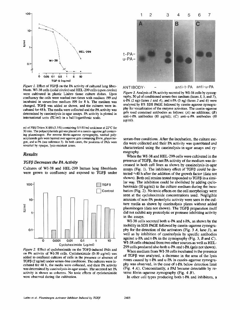
Figure 1. Effect of TGF~ on the PA activity of cultured lung fibro- blasts. WI-38 cells (solid circles) and HEL-299 cells (open circles) were cultivated in plastic Linbro tissue culture dishes. Upon confluency the cells were washed two times with medium 199 and incubated in serum-free medium 199 for 8 h. The medium was changed, TGFI3 was added as shown, and the cultures were in- cubated for 48 h. The media were collected and the PA activity was determined by caseinolysis-in-agar assays. PA activity is plotted in international units (IU/ml) in a half-logarithmic scale.
ml of PBS/Triton X-100 (2.5%) containing 0.5 IU/ml urokinase at 22°C for 30 min. The polyacrylamide gel was placed on a casein-agarose gel contain- ing plasminogen. For reverse fibrin-agarose zyrnography, washed poly- acrylamide gels were layered over agarose gels containing fibrin, plasmino- gen, and u-PA (see reference 3). In both cases, the positions of PAIs were revealed by opaque, lysis-resistant zones.
Results
TGF~ Decreases the PA Activity
Cultures of WI-38 and HEL-299 human lung fibroblasts were grown to confluency and exposed to TGFI3 under
Figure 2. Effect of cycloheximide on the TGF~induced PAls and on PA activity of WI-38 cells. Cycloheximide (0-10 I.tg/ml) was added to confluent cultures of cells in the presence or absence of TGFI3 (2 ng/ml) under serum-free conditions. The cultures were in- cubated for 48 h, the media were collected, and their PA activity was determined by caseinolysis-in-agar assays. The secreted net PA activity is shown as columns. No toxic effects of cycloheximide were observed during the cultivation.
Figure 3. Analysis of PA activity secreted by WI-38 cells by zymog- raphy, 50 Ixl of conditioned serum-free medium (lanes 3, 5, and 7), u-PA (2 ng) (lanes I and 4), and t-PA (5 ng) (lanes 2 and 6) were analyzed by 8 % SDS PAGE followed by casein-agarose zymogra- phy for visualization of the enzyme activities. The casein-agarose gels used contained antibodies as follows: (A) no additions; (B) anti-t-PA antibodies (10 ~tg/ml); (C) anti-u-PA antibodies (10 I.tg/ml).
serum-free conditions. After the incubation, the culture me- dia were collected and their PA activity was quantitated and characterized using the caseinolysis-in-agar assays and zy- mography.
When the WI-38 and HEL-299 cells were cultivated in the presence of TGF[~, the net PA activity of the medium was de- creased in both cell lines as shown by caseinolysis-in-agar assays (Fig. 1). The inhibitory effect of TGF[~ could be de- tected ~10 h after the addition of the growth factor (data not shown). Both cell strains tested responded to TGFI3 in a simi- lar way. The inhibition could be abolished by adding cyclo- heximide (10 Ixg/ml) to the culture medium during the incu- bation (Fig. 2). No toxic effects on the cell morphology were seen at the cycloheximide concentrations used. Negligible amounts of non-PA proteolytic activity were seen in the cul- ture media as shown by caseinolysis plates without added plasminogen (data not shown). The TGFI3 preparation itself did not exhibit any proteolytic or protease inhibiting activity in the assays.
WI-38 cells secreted both u-PA and t-PA, as shown by the mobility in SDS PAGE followed by casein-agarose zymogra- phy for the detection of the activators (Fig. 3 A, lane 3), as well as by inhibition of caseinolysis by specific antibodies against u-PA and t-PA in the zymography (Fig. 3, B and C). WI-38 cells obtained from two other sources as well as HEL- 299 cells produced also both u-PA and t-PA (gels not shown).
When medium from WI-38 cells incubated in the presence of TGFI3 was analyzed, a decrease in the area of the lysis zones caused by t-PA and u-PA in casein-agarose zymogra- phy was observed, in the case of t-PA below detection limit (Fig. 4 A). Concomitantly, a PAl became detectable by re- verse fibrin-agarose zymography (Fig. 4 B).
In other cell types producing both t-PA and inhibitors, a
Laiho et al. Plasminogen Activator Inhibitor Induced by TGFfl 2405
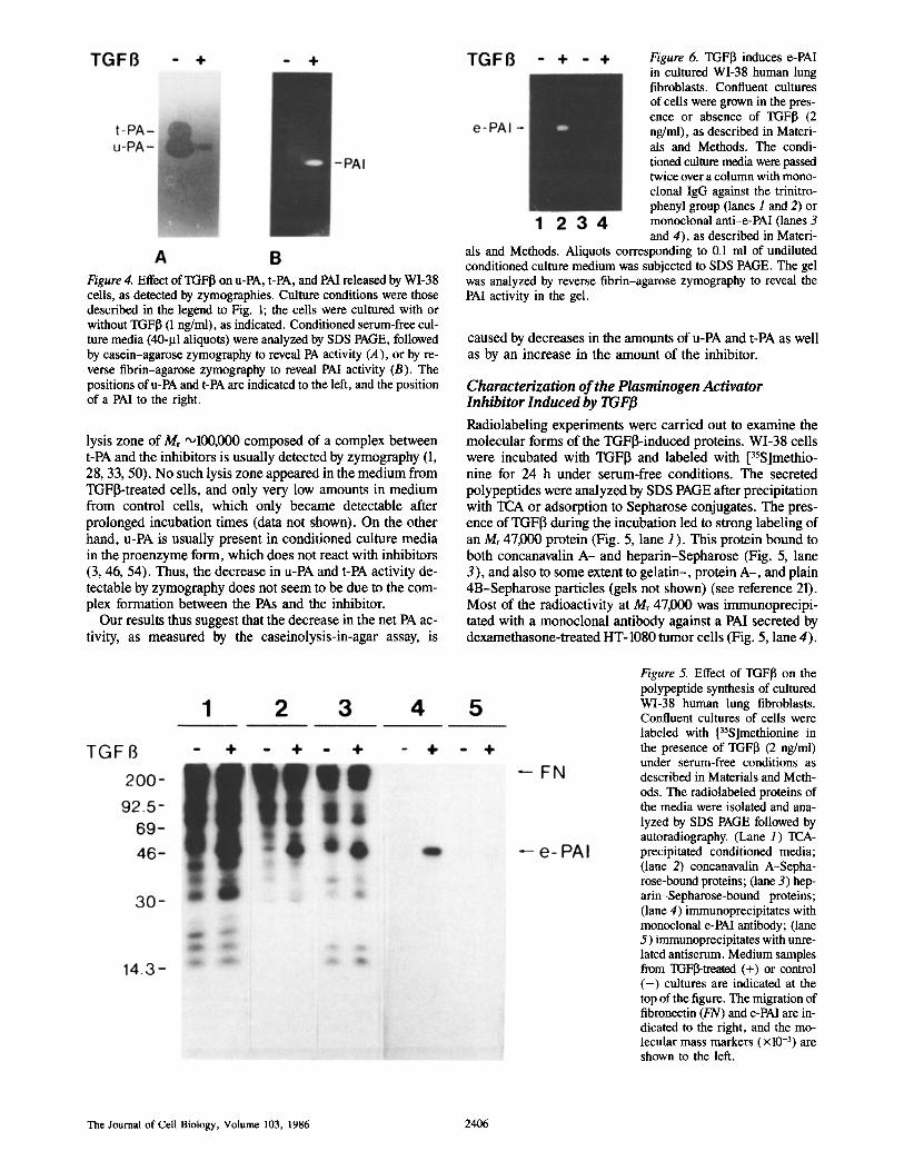
Figure 4. Effect of TGFI~ on u-PA, t-PA, and PAI released by WI-38 cells, as detected by zymographies. Culture conditions were those described in the legend to Fig. 1; the cells were cultured with or without TGFI3 (1 ng/ml), as indicated. Conditioned serum-free cul- ture media (40-I.tl aliquots) were analyzed by SDS PAGE, followed by casein-agarose zymography to reveal PA activity (A), or by re- verse fibrin-agarose zymography to reveal PAI activity (B). The positions of u-PA and t-PA are indicated to the left, and the position of a PAl to the right.
lysis zone of Mr '~100,000 composed of a complex between t-PA and the inhibitors is usually detected by zymography (1, 28, 33, 50). No such lysis zone appeared in the medium from TGFI3-treated cells, and only very low amounts in medium from control ceils, which only became detectable after prolonged incubation times (data not shown). On the other hand, u-PA is usually present in conditioned culture media in the proenzyme form, which does not react with inhibitors (3, 46, 54). Thus, the decrease in u-PA and t-PA activity de- tectable by zymography does not seem to be due to the com- plex formation between the PAs and the inhibitor.
Our results thus suggest that the decrease in the net PA ac- tivity, as measured by the caseinolysis-in-agar assay, is
Figure 6. TGFI3 induces e-PAI in cultured WI-38 human lung fibroblasts. Confluent cultures of cells were grown in the pres- ence or absence of TGFI3 (2 ng/ml), as described in Materi- als and Methods. The condi- tioned culture media were passed twice over a column with mono- clonal IgG against the trinitro- phenyl group (lanes 1 and 2) or monoclonal anti-e-PAl (lanes 3 and 4), as described in Materi-
als and Methods. Aliquots corresponding to 0.1 ml of undiluted conditioned culture medium was subjected to SDS PAGE. The gel was analyzed by reverse fibrin-agarose zymography to reveal the PAl activity in the gel.
caused by decreases in the amounts of u-PA and t-PA as well as by an increase in the amount of the inhibitor.
Characterization of the Plasminogen Activator Inhibitor Induced by TGFB
Radiolabeling experiments were carried out to examine the molecular forms of the TGFI3-induced proteins. WI-38 cells were incubated with TGFI3 and labeled with [35S]methio- nine for 24 h under serum-free conditions. The secreted polypeptides were analyzed by SDS PAGE after precipitation with TCA or adsorption to Sepharose conjugates. The pres- ence of TGFI3 during the incubation led to strong labeling of an Mr 47,000 protein (Fig. 5, lane 1). This protein bound to both concanavalin A- and heparin-Sepharose (Fig. 5, lane 3), and also to some extent to gelatin-, protein A-, and plain 4B-Sepharose particles (gels not shown) (see reference 21). Most of the radioactivity at Mr 47,000 was immunoprecipi- tated with a monoclonal antibody against a PAI secreted by dexamethasone-treated HT-1080 tumor cells (Fig. 5, lane 4).
Figure 5. Effect of TGFfl on the polypeptide synthesis of cultured WI-38 human lung fibroblasts. Confluent cultures of cells were labeled with [35S]methionine in the presence of TGFI3 (2 ng/ml) under serum-free conditions as described in Materials and Meth- ods. The radiolabeled proteins of the media were isolated and ana- lyzed by SDS PAGE followed by autoradiography. (Lane 1) TCA- precipitated conditioned media; (lane 2) concanavalin A-Sepha- rose-bound proteins; (lane 3) hep- arin-Sepharose-bound proteins; (lane 4) immunoprecipitates with monoclonal e-PAl antibody; (lane 5) immunoprecipitates with unre- lated antiserum. Medium samples from TGFl$-treated (+) or control ( - ) cultures are indicated at the top of the figure. The migration of fibronectin (FN) and e-PAl are in- dicated to the right, and the mo- lecular mass markers (xl0 -3) are shown to the left.
The Journal of Cell Biology, Volume 103, 1986 2406
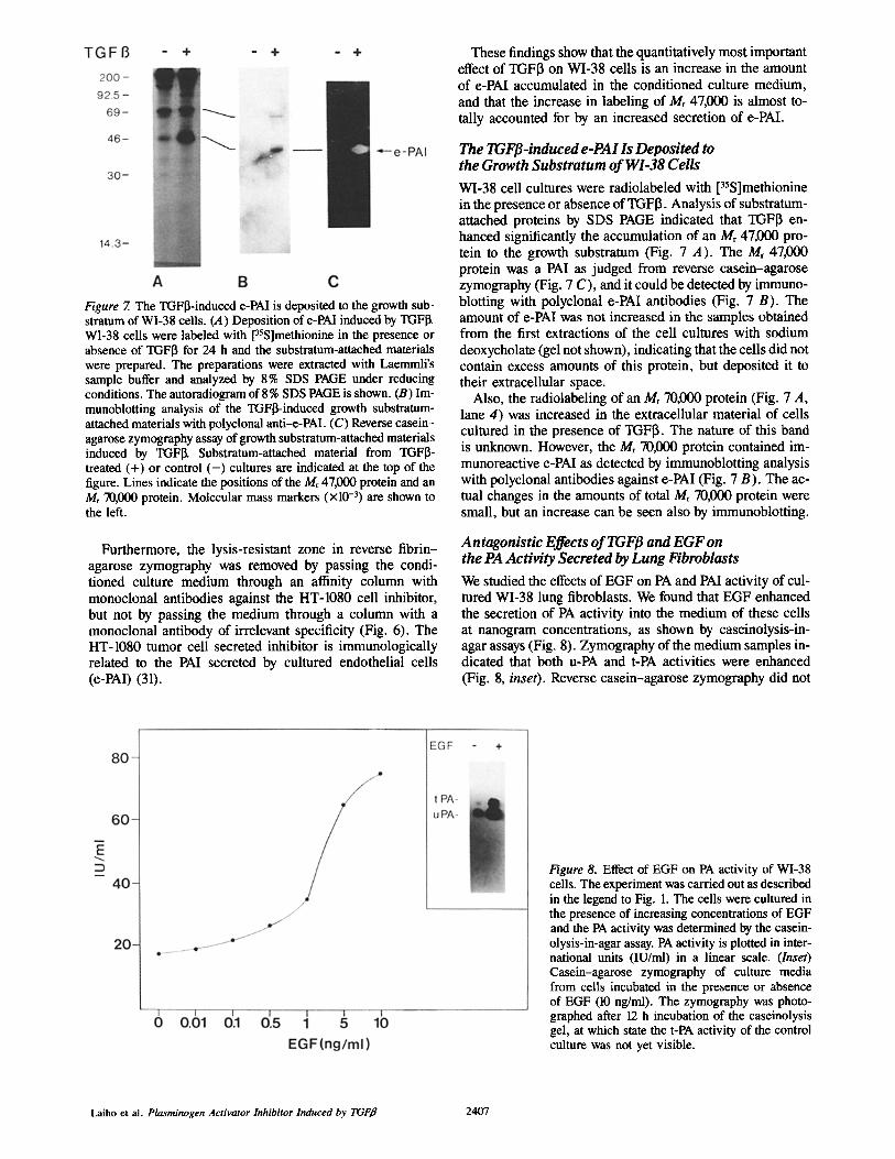
Figure 7. The TGFI3-induced e-PAl is deposited to the growth sub- stratum of WI-38 cells. (A) Deposition of e-PAI induced by TGFI~ WI-38 cells were labeled with [35S]methionine in the presence or absence of TGFI3 for 24 h and the substratum-attached materials were prepared. The preparations were extracted with Laemmli's sample buffer and analyzed by 8% SDS PAGE under reducing conditions. The autoradiogram of 8 % SDS PAGE is shown. (B) Im- munoblotting analysis of the TGFI3-induced growth substratum- attached materials with polyclonal anti-e-PAl. (C) Reverse casein- agarose zymography assay of growth substratum-attached materials induced by TGFI~ Substratum-attached material from TGFI3- treated (+) or control ( - ) cultures are indicated at the top of the figure. Lines indicate the positions of the Mr 47,000 protein and an Mr 70,000 protein. Molecular mass markers (xl0 -3) are shown to the left.
Furthermore, the lysis-resistant zone in reverse fibrin- agarose zymography was removed by passing the condi- tioned culture medium through an affinity column with monoclonal antibodies against the HT-1080 cell inhibitor, but not by passing the medium through a column with a monoclonal antibody of irrelevant specificity (Fig. 6). The HT-1080 tumor cell secreted inhibitor is immunologically related to the PAl secreted by cultured endothelial cells (e-PAl) (31).
These findings show that the quantitatively most important effect of TGFI3 on WI-38 cells is an increase in the amount of e-PAl accumulated in the conditioned culture medium, and that the increase in labeling of Mr 47,000 is almost to- tally accounted for by an increased secretion of e-PAl.
The TGF[3-induced e-PAl Is Deposited to the Growth Substratum of WI-38 Cells
WI-38 cell cultures were radiolabeled with [35S]methionine in the presence or absence of TGFI3. Analysis of substratum- attached proteins by SDS PAGE indicated that TGFI~ en- hanced significantly the accumulation of an Mr 47,000 pro- tein to the growth substratum (Fig. 7 A). The Mr 47,000 protein was a PAl as judged from reverse casein-agarose zymography (Fig. 7 C), and it could be detected by immuno- blotting with polyclonal e-PAl antibodies (Fig. 7 B). The amount of e-PAl was not increased in the samples obtained from the first extractions of the cell cultures with sodium deoxycholate (gel not shown), indicating that the cells did not contain excess amounts of this protein, but deposited it to their extracellular space.
Also, the radiolabeling of an Mr 70,000 protein (Fig. 7 A, lane 4) was increased in the extracellular material of cells cultured in the presence of TGFI3. The nature of this band is unknown. However, the Mr 70,000 protein contained im- munoreactive e-PAl as detected by immunoblotting analysis with polyclonal antibodies against e-PAl (Fig. 7 B). The ac- tual changes in the amounts of total Mr 70,000 protein were small, but an increase can be seen also by immunoblotting.
Antagonistic Effects of TGFJ3 and EGF on the PA Activity Secreted by Lung Fibroblasts
We studied the effects of EGF on PA and PAI activity of cul- tured WI-38 lung fibroblasts. We found that EGF enhanced the secretion of PA activity into the medium of these cells at nanogram concentrations, as shown by caseinolysis-in- agar assays (Fig. 8). Zymography of the medium samples in- dicated that both u-PA and t-PA activities were enhanced (Fig. 8, inset). Reverse casein-agarose zymography did not
Figure 8. Effect of EGF on PA activity of WI-38 cells. The experiment was carried out as described in the legend to Fig. 1. The cells were cultured in the presence of increasing concentrations of EGF and the PA activity was determined by the casein- olysis-in-agar assay. PA activity is plotted in inter- national units (IU/ml) in a linear scale. (Inset) Casein-agarose zymography of culture media from cells incubated in the presence or absence of EGF (10 ng/ml). The zymography was photo- graphed after 12 h incubation of the caseinolysis gel, at which state the t-PA activity of the control culture was not yet visible.
Laiho et al. Plasminogen Activator Inhibitor Induced by TGFff 2407
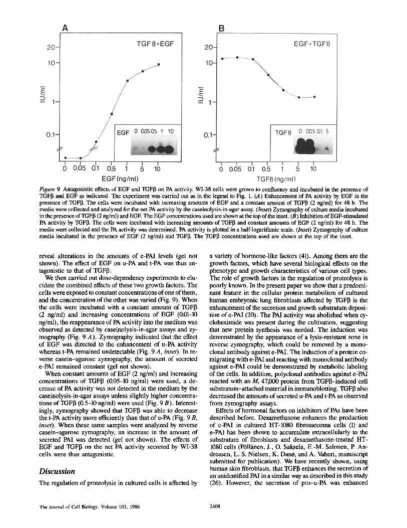
Figure 9. Antagonistic effects of EGF and TGFI3 on PA activity. WI-38 cells were grown to confluency and incubated in the presence of TGFI~ and EGF as indicated. The experiment was carried out as in the legend to Fig. 1. (A) Enhancement of PA activity by EGF in the presence of TGFI]. The cells were incubated with increasing amounts of EGF and a constant amount of TGFI3 (2 ng/ml) for 48 h. The media were collected and analyzed for the net PA activity by the caseinolysis-in-agar assay. (Inset) Zymography of culture media incubated in the presence of TGFI3 (2 ng/ml) and EGE The EGF concentrations used are shown at the top of the inset. (B) Inhibition of EGF-stimulated PA activity by TGFI]. The cells were incubated with increasing amounts of TGF[~ and constant amounts of EGF (2 ng/ml) for 48 h. The media were collected and the PA activity was determined. PA activity is plotted in a half-logarithmic scale. (Inset) Zymography of culture media incubated in the presence of EGF (2 ng/ml) and TGFI~. The TGFI3 concentrations used are shown at the top of the inset.
reveal alterations in the amounts of e-PAl levels (gel not shown). The effect of EGF on u-PA and t-PA was thus an- tagonistic to that of TGFIL
We then carried out dose-dependency experiments to elu- cidate the combined effects of these two growth factors. The cells were exposed to constant concentrations of one of them, and the concentration of the other was varied (Fig. 9). When the cells were incubated with a constant amount of TGFI3 (2 ng/ml) and increasing concentrations of EGF (0.01-10 ng/ml), the reappearance of PA activity into the medium was observed as detected by caseinolysis-in-agar assays and zy- mography (Fig. 9 A). Zymography indicated that the effect of EGF was directed to the enhancement of u-PA activity whereas t-PA remained undetectable (Fig. 9 A, inset). In re- verse casein-agarose zymography, the amount of secreted e-PAI remained constant (gel not shown).
When constant amounts of EGF (2 ng/ml) and increasing concentrations of TGFI3 (0.05-10 ng/ml) were used, a de- crease of PA activity was not detected in the medium by the caseinolysis-in-agar assays unless slightly higher concentra- tions of TGFI3 (0.5-10 ng/ml) were used (Fig. 9 B). Interest- ingly, zymography showed that TGFI3 was able to decrease the t-PA activity more efficiently than that of u-PA (Fig. 9 B, inset). When these same samples were analyzed by reverse casein-agarose zymography, an increase in the amount of secreted PAI was detected (gel not shown). The effects of EGF and TGFI3 on the net PA activity secreted by WI-38 cells were thus antagonistic.
D i s c u s s i o n
The regulation of proteolysis in cultured cells is affected by
a variety of hormone-like factors (41). Among them are the growth factors, which have several biological effects on the phenotype and growth characteristics of various cell types. The role of growth factors in the regulation of proteolysis is poorly known. In the present paper we show that a predomi- nant feature in the cellular protein metabolism of cultured human embryonic lung fibroblasts affected by TGFI3 is the enhancement of the secretion and growth substratum deposi- tion of e-PAI (20). The PAl activity was abolished when cy- cloheximide was present during the cultivation, suggesting that new protein synthesis was needed. The induction was demonstrated by the appearance of a lysis-resistant zone in reverse zymography, which could be removed by a mono- clonal antibody against e-PAI. The induction of a protein co- migrating with e-PAl and reacting with monoclonal antibody against e-PAI could be demonstrated by metabolic labeling of the cells. In addition, polyclonal antibodies against e-PAl reacted with an Mr 47,000 protein from TGFl~-induced cell substratum-attached material in immunoblotting. TGFI] also decreased the amounts of secreted u-PA and t-PA as observed from zymography assays.
Effects of hormonal factors on inhibitors of PAs have been described before. Dexamethasone enhances the production of e-PAl in cultured HT-1080 fibrosarcoma cells (1) and e-PAl has been shown to accumulate extracellularly to the substratum of fibroblasts and dexamethasone-treated HT- 1080 cells (P611~inen, J., O. Saksela, E.-M. Salonen, P. An- dreasen, L. S. Nielsen, K. Dan0, and A. Vaheri, manuscript submitted for publication). We have recently shown, using human skin fibroblasts, that TGFI~ enhances the secretion of an unidentified PAl in a similar way as described in this study (26). However, the secretion of pro-u-PA was enhanced
The Journal of Cell Biology, Volume 103, 1986 2408
simultaneously leading to an increase in the net PA activity in the two responsive cell strains examined. We have also identified polypeptide factors secreted by cultured 8387 fibrosarcoma cells, which affect cellular PA activity by en- hancing the PAl secretion (43). These sarcoma cell-derived factors resemble TGFI~, but certain differences suggest that the sarcoma factors may not be identical to it (M. Laiho, un- published results). Protease nexin has been reported to be in- duced in skin fibroblasts by phorbol esters, EGF, and throm- bin (11).
Besides enhancing e-PAI, TGFI3 also decreased the amounts of u-PA and t-PA accumulated in the media of WI- 38 cells, as judged from the decreased areas of the lysis zones caused by u-PA and t-PA; the decreases in the area of the lysis zones could not be accounted for simply by complex forma- tion with the inhibitor. Although the intracellular mechanism of this effect is presently unknown, it is likely that TGFI3 decreases the biosynthetic rate of u-PA and t-PA.
Embryonic lung fibroblasts grow as a substratum-attached cell layer in culture and secrete high amounts of PA activity into their medium. In spite of this, they attach to their sub- stratum via proteinase-sensitive fibronectin-collagen matrix (16, 19). This may be explained, at least in part, by the secre- tion of effective proteinase inhibitors. Protease nexin has been suggested to be the primary inhibitor of u-PA in fibro- blastic cells (24, 44, 45). It has been shown to inhibit extra- cellular matrix destruction of cultured HT-1080 tumor cells (7). We cannot exclude the possibility that TGFI~ affected the levels of protease nexin (or placental type PAl) in the condi- tioned culture medium of WI-38 cells. However, the induc- tion of protease nexin (or placental type PAl) is no major effect of TGFI3; most if not all of the increase in the labeling of an Mr 47,000 protein is accounted for by an increased amount of e-PAl. The data presented here show that also e-PAl binds to heparin, indicating that this characteristic, per se, cannot be used to distinguish between e-PAl and protease nexin.
Although EGF has been described to potentiate the effects of TGFI3 on the growth of normal cells in soft agar, their effects on the growth of A549 human lung carcinoma cells are antagonistic (30, 39). In addition, the receptors of TGFI~ interact with the EGF receptors at the membranes of NRK cells and evidently regulate their affinity (5, 29). Because of a possible relationship between these two receptor systems, we studied the effects of EGF on the secretion of PA activity from WI-38 cells. We found that the effect of TGFI~ on the net PA activity of embryonic lung fibroblasts was antago- nized by EGF, which alone enhanced the secretion of PA ac- tivity into the culture medium. The effect of EGF on the secretion of PA activity on this cell line is in agreement with the previous results obtained with certain other cell lines (27, 51). The cellular responses to exogenous growth factors may thus vary (18) depending on the amount and specificity of the receptors, on the interactions between the receptor systems, and on the secretion of endogenous growth factors.
Malignant transformation has in many cases been found to be associated with the production of plasminogen activators. Transformation of fibroblastic chicken cells with Rous sar- coma virus results in the production of PA (34). The mor- phological changes in these cells correlate with the PA activ- ity, and synthetic PAIs can be used to block the change (34). On the other hand, malignant transformation of cultured cells is associated with the production of different types of
growth factors (14, 49). TGFI3 is evidently responsible for the induction of soft agar growth of certain nonmalignant cells in the presence of other growth factors like platelet- derived growth factor, EGF, or TGFa (cf. reference 22). However, TGFI3 seems to have a dual role in the regulation of soft agar growth; it inhibits the growth of many epithelial and several types of malignant cells in agar and obviously does not require other growth factors to elicit this function (30, 39).
The mechanism of the inhibitory activity of TGFI3 on the growth of normal epithelial and malignant cells is unclear at present. If the stimulatory effect of TGFI3 on the secretion of PAls can be extended, it might help to explain the effects of TGFI~ on the soft agar growth of malignant cells (30, 39). Induction of inhibitors of proteolysis into the microenviron- ment of cells and their pericellular deposition may help to retain the phenotype of the cells or, alternatively, the inhibi- tors themselves could serve as attachment proteins for the cells. It has recently been reported that TGFfl enhances the production and matrix deposition of fibronectin and procol- lagen in certain cell lines (17). The enhanced deposition of the PAls into the growth substratum is a novel feature in the regulation of extracellular proteolysis and matrix degrada- tion. The proteolytic activity of cells may be regulated by TGFI~ during their growth and differentiation, and possibly also in tissue repair.
We thank Drs. R. Assoian and A. Roberts for gift of TGFI3, Drs. Klaus Hed- man and Antti Vaheri for their critical comments, and Ms. Marja Valasj~irvi for fine assistance.
This research has been supported by the Finnish Cancer Foundation, the Academy of Finland and Nordisk Insulinfond.
Received for publication 31 March 1986, and in revised form 2 September 1986.
References
I. Andreasen, P. A., T. H. Christensen, L. S. Nielsen, J.-Y. Huang, and K. Dane. 1986. Hormonal regulation of extracellular plasminogen activators and Mr 54,000 inhibitor in neoplastic cell lines, studied with monoclonal anti- bodies. Mol. Cell. Endocr. 45:137-147.
2. Andreasen, P. A., L. S. Nielsen, J. Grcndahl-Hansen, L. Skriver, J. Zeuthen, R. W. Stephens, and K. Dane. 1984. Inactive proenzyme to tissue- type plasminogen activator from human melanoma cells, identified after at~nity purification with a monoclonal antibody. EMBO (Eur. Mol. Biol. Organ.) J. 3:51-56.
3. Andreasen, P. A., L. S. Nielsen, P. Kristensen, J. GrCndahl-Hansen, L. Skriver, and K. Dane. 1986. Plasminogen activator inhibitor from human fibrosarcoma cells binds urokinase-type plasminogen activator, but not its proenzyme. J. Biol. Chem. 261:7644-7651.
4. Anzano, M. A., A. B. Roberts, J. M. Smith, M. B. Sporn, and J. E. De Larco. 1983. Sarcoma growth factor from conditioned medium of virally trans- formed cells is composed of both type tt and type [I transforming growth factors. Proc. Natl. Acad. Sci. USA. 80:6264-6268,
5. Assoian, R. K., C. A. Frolik, A. B. Roberts, D. M. Miller, and M. B. Sporn. 1984. Transforming growth factor-13 controls receptor levels for epider- mal growth factor in NRK fibroblasts. Cell. 36:35--41.
6. Assoian, R. K., C. A. Komoriya, D. M. Meyers, D. M. Miller, and M. B. Sporn. 1983. Transforming growth factor-~ in human platelets: identi- fication of a major storage site, purification and characterization. J. Biol. Chem. 258:7155-7160.
6a. /~stedt, B., I. Lecander, T. Brodin, A. Lundblad, and K. L6w. 1985. Purification of a specific placental plasminogen activator inhibitor by mono- clonal antibody and its complex formation with plasminogen activator. Thromb. Haemostasis. 53:122-125.
7. Bergman, B. L., R. W. Scott, A. Bajpai, S. Watts, and J. F. Baker. 1986. Inhibition of tumor cell-mediated extracellular matrix destruction by a fibro- blast proteinase inhibitor, protease nexin I. Proc. Natl. Acad. Sci. USA. 83:996-1000.
8. Childs, C. B., J. A. Proper, R. F. Tucker, and H. L. Moses. 1982. Serum contains a platelet-derived transforming growth factor. Proc. Natl. Acad. Sci. USA. 79:5312-6316.
9. Dane, K., P. A. Andreasen, J. GrCndahl-Hansen, P. Kristensen, L. S. Nielsen, and L. Skriver. 1985. Plasminogen activators, tissue degradation, and cancer. Adv. Cancer Res. 44:139-266.
Laiho et al. Plasminogen Activator Inhibitor Induced by TGFfl 2409
10. Derynck, R., J. A. Jarrett, E. Y. Chert, D. H. Eaton, J. R. Bell, R. K. Assoian, A. B. Roberts, M. B. Sporn, and D. V. Goeddel. 1985. Human trans- forming growth factor-beta cDNA sequence and expression in tumor cell lines. Nature (Loud.). 316:701-705.
11. Eaton, D. L., and J. B. Baker. 1983. Phorbol esters and mitogens stimu- late human fibroblasts secretion of plasmin-activatable plasminogen activator and protease nexin, an antiactivator/antiplasmin. J. Cell Biol. 97:323-328.
12. Erikson, L. A., D. A. Lawrence, and D. J. Loskutoff. 1984. Reverse fibrin autoradiography: a method to detect and partially characterize protease inhibitors after sodium dodecyl sulfate-polyacrylamide gel electrophoresis. Anal. Biochem. 137:454-463.
13. Frolik, C. A., L. L. Dart, C. A. Meyers, D. M. Smith, and M. B. Sporn. 1983. Purification and initial characterization of a type 13 transforming growth factor from human placenta. Proc. Natl. Acad. Sci. USA. 80:3676-3680.
14. Goustin, A. S., E. B. Leof, G. D. Shipley, and H. L. Moses. 1986. Growth factors and cancer. Cancer Res. 46:1015-1029.
15. Granelli-Piperno, A., and E. Reich. 1978. A study of proteases and pro- tease inhibitor complexes in biological fluids. J. Exp. Med. 146:223-234.
16. Hedman, K., M. Kurkinen, K. Alitalo, A. Vaheri, S. Johansson, and M. H/~k. 1979. Isolation of the pericellular matrix of human fibroblast cultures. J. Cell Biol. 81:83-91.
17. Ignotz, R. A., and J. Massague. 1986. Transforming growth factor-~ stimulates the expression of fibronectin and collagen and their incorporation into the extracellular matrix. J. BioL Chem. 261:4337-4345.
18. Kaplan, P. L., and B. Ozanne. 1983. Cellular responsiveness to growth factors correlates with a cell's ability to express the transformed phenotype. Cell. 33:931-938.
19. Keski-Oja, J., and A. Vaheri. 1982. The cellular target for the plasmino- gen activator, urokinase, in human fibroblasts-66,000 dalton protein. Biochim. Biophys. Acta. 720:141-146.
20. Keski-Oja, J., M. Laiho, and O. Saksela. 1986. Enhanced production of plasminogen activator inhibitors in human lung fibroblasts by transforming growth factor-13. J. Cell. Biochem.. (Suppl.)10C:181.
21. Keski-Oja, J., M. Laiho, and T. Vartio. 1986. Characterization of a novel gelatin-binding 21 kDa protein secreted by cultured adherent cells. Biochim. Biophys. Acta. 882:367-376.
22. Keski-Oja, J., E. B. Leof, R. M. Lyons, R. J. Coffey, and H. L. Moses. 1986. Transforming growth factors and control of neoplastic cell growth. J. Cell Biochem. In press.
23. Kielberg, V., P. A. Andreasen, J. Grendahl-Hansen, L. S. Nielsen, L. Skriver, and K. Dane. 1985. Proenzyme to urokinase-type plasminogen activator in the mouse in vivo. FEBS (Fed. Eur. Biochem. Soc.) Lett. 182:441--445.
24. Knauer, D. J., and D. D. Cunningham. 1984. Protease nexins: cell- secreted proteins which regulate extracellular serine proteinases. Trends Bio- chem. Sci. 9:231-233.
25. Laemmli, U. K. 1970. Cleavage of structural proteins during the assem- bly of the head of bacteriophage T 4. Nature (Loud.). 227:680-685.
26. Laiho, M., O. Saksela, and J. Keski-Oja. 1986. Transforming growth factor-13 alters plasminogen activator activity in human skin fibroblasts. Exp. Cell Res. 164:399-407.
27. Lee, L.-S., and I. B. Weinstein. 1978. Epidermal growth factor, like phorbol esters, induces plasminogen activator in HeLa ceils. Nature (Lond.). 274:696-697.
28. Levin. E. G. 1983. Latent tissue plasminogen activator produced by hu- man endothelial cells in culture: evidence for an enzyme-inhibitor complex. Proc. Natl. Acad. Sci. USA. 80:6804-6808.
29. Massagu~, J. 1985. Transforming growth factor-13 modulates the high- affinity receptors for epidermal growth factor. J. Cell Biol. 100:1508-1514.
30. Moses, H. L., R. F. Tucker, E. B. Leof, R. J. Coffey, J. Halper, and G. D. Shipley. 1985. Type-13 transforming growth factor is a growth stimulator and a growth inhibitor. Cancer Cells (Cold Spring Harbor). 3:65-71.
31. Nielsen, L. S., P. A. Andreasen, J. Grendahl-Hansen, J.-Y. Huang, P. Kristensen, and K. Dane. 1986. Monoclonal antibodies to human 54,000 molecular weight plasmiuogen activator inhibitor from fibrosarcoma cells. In- hibitor neutralization and one-step purification. Thromb. Haemostasis. 55:206- 212.
32. Nielsen, L. S., J. G. Hansen, L. Skriver, E. L. Wilson, K. Kaltoft, J. Zeuthen, and K. Dane. 1982. Purification of zymogen to plasminogen activator from human glioblastoma cells by affinity chromatography with monoclonal an- tibody. Biochemistry. 25:6410-6415.
33. Philips, M., A.-G. Juul, and S. Thorsen. 1984. Human endothelial cells
produce a plasminogen activator inhibitor and a tissue-type plasminogen activa- tor-inhibitor complex. Biochim. Biophys. Acta. 802:99-110.
34. Quigley, J. P. 1979. Phorbol ester-induced morphological changes in transformed chick fibroblasts: evidence for direct catalytic involvement of plas- minogen activator. Cell. 17:131-141.
35. Rijken, D. C., and D. Collen. 1981. Purification and characterization of the plasminogen activator secreted by human melanoma cell lines in culture. J. Biol. Chem. 256:7035-7041.
36. Roberts, A. B., M. A. Anzano, L. C. Lamb, J. M. Smith, C. A. Frolik, H. Marquardt, G. J. Todaro, and M. B. Sporn. 1982. Isolation from murine sarcoma cells of novel transforming growth factors potentiated by EGF. Nature (Lond.). 295:417-419.
37. Roberts, A. B., M. A. Anzano. L. C. Lamb, J. M. Smith, and M. B. Sporn. 1981. New class of transforming growth factors potentiated by epider- mal growth factor: isolation from non-neoplastic tissues. Proc. Natl. Acad. Sci. USA. 78:5339-5343.
38. Roberts, A. B., M. A. Anzano, C. A. Meyers, J. Wideman, R. Blacher, Y.-C.E. Pan, S. Stein, S. R. Lehrman, J. M. Smith, L. C. I.annb, and M. B. Sporn. 1983. Purification and properties of a type 13 transforming growth factor from bovine kidney. Biochemistry. 22:5692-5698.
39. Roberts, A. B., M. A. Anzano, L. M. Wakefield, N. S. Roche, D. Stern, and M. B. Sporn. 1985. Type 13 transforming growth factor: a bifunctional regu- lator of cellular growth. Proc. Natl. Acad. Sci. USA. 82:119-123.
40. Saksela, O. 1981. Radial caseinolysis in agarose: a simple method for de- tection of plasminogen activator activity in the presence of inhibitory substances and serum. Anal. Biochem. 111:276-282.
41. Saksela, O. 1985. Plasminogen activation and regulation of pericellular proteolysis. Biochim. Biophys. Acta. 823:35-65.
42. Saksela, O., T. Hovi, and A. Vaheri. 1985. Urokinase-type plasminogen activator and its inhibitor secreted by cultured human monocyte-macrophages. J. Cell. Physiol. 122:125-132.
43. Saksela, O., M. Laiho, and J. Keski-Oja. 1985. Regulation of plasmino- gen activator activity in human fibroblastic cells by fibrosarcoma cell-derived factors. Cancer Res. 45:2314-2319.
44. Scott, R. W., and J. B. Baker. 1983. Purification of human protease nexin. J. Biol. Chem. 258:10439-10444.
45. Scott, R. W., B. L. Bergman, A. Bajpai, R. T. Hersh, H. Rodriguez, B. N. Jones, C. Barreda, S. Watts, andJ. B. Baker. 1985. Protease nexin. Prop- erties and a modified purification procedure. J. Biol. Chem. 260:7029-7034.
46. Scott, R. W., D. E. Eaton, N. Durant, andJ. B. Baker. 1983. Regulation of extracellular plasminogen activator by human fibroblasts. J. Biol. Chem. 258:4397-4403.
47. Skriver, L., L.-J. Larsson, V. Kielberg, L. S. Nielsen, P. B. Andresen, P. Kristensen, and K. Dane. 1984. Immunocytochemical localization of uro- kinase-type plasminogen activator in Lewis lung carcinoma. J. Cell Biol. 99: 753-757.
48. Skriver, L., L. S. Nielsen, R. Stephens, and K. Dane. 1982. Plasmino- gen activator released as inactive proenzyme from murine cells transformed by sarcoma virus. Eur. J. Biochem. 124:409--414.
49. Sporn, M. B., and A. B. Roberts. 1985. Autocrine growth factors and cancer. Nature (Lond.). 313:745-747.
50. Sprengers, E. D., J. H. Verheijen, V. W. H. van Hiusberg, and J. J. Emeis. 1984. Evidence for the presence of two different fibrinolytic inhibitors in human endothelial cell conditioned medium. Biochim. Biophys. Acta. 801: 163-170.
51. Stoppelli, M. P., P. Verde, G. Grimaldi, E. K. Locatelli, and F. Blasi. 1986. Increase in urokinase plasminogen activator mRNA synthesis in human carcinoma cells is a primary effect of the potent tumor promoter, phorbol myristate acetate. J. Cell Biol. 102:1235-1241.
52. Towbin, H., T. Staehelin, and J. Gordon. 1979. Electrophoretic transfer of proteins from polyacrylamide gels to nitrocellulose sheets: procedure and some applications. Proc. Natl. Acad. Sci. USA. 76:4350-4353.
53. van Morik, J. A., D. A. Lawrence, and D. J. Loskutoff. 1984. Purifica- tion of an inhibitor of plasminogen activator (antiactivator) synthesized by en- dothelial cells. J. Biol. Chem. 259:14914-14921.
54. Vassalli, L-D., J.-M. Dayer, A. Wohlwend, and D. Belin. 1984. Con- comitant secretion of prourokinase and of a plasminogen activator-specific in- hibitor by cultured human monocytes-macrophages. J. Exp. Med. 159:1653- 1668.
55. Wun, T.-C., Ossowski, and E. Reich. 1982. A proenzyme form of hu- man urokinase. J. Biol. Chem. 257:7262-7268.
The Journal of Cell Biology, Volume 103, 1986 2410