Embed Size (px)
Citation preview
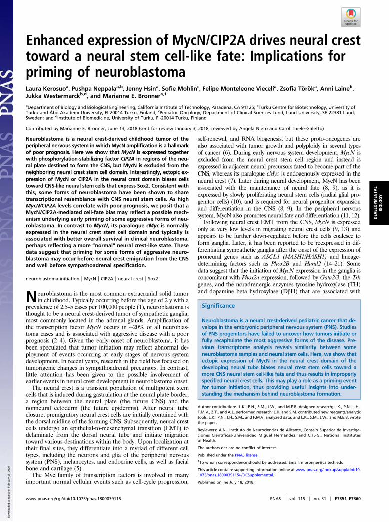
Enhanced expression of MycN/CIP2A drives neural cresttoward a neural stem cell-like fate: Implications forpriming of neuroblastomaLaura Kerosuoa, Pushpa Neppalaa,b, Jenny Hsina, Sofie Mohlinc, Felipe Monteleone Viecelia, Zsofia Töröka, Anni Laineb,Jukka Westermarckb,d, and Marianne E. Bronnera,1
aDepartment of Biology and Biological Engineering, California Institute of Technology, Pasadena, CA 91125; bTurku Centre for Biotechnology, University ofTurku and Åbo Akademi University, FI-20014 Turku, Finland; cPediatric Oncology, Department of Clinical Sciences Lund, Lund University, SE-22381 Lund,Sweden; and dInstitute of Biomedicine, University of Turku, FI-20014 Turku, Finland
Contributed by Marianne E. Bronner, June 13, 2018 (sent for review January 3, 2018; reviewed by Angela Nieto and Carol Thiele-Galetto)
Neuroblastoma is a neural crest-derived childhood tumor of theperipheral nervous system in which MycN amplification is a hallmarkof poor prognosis. Here we show that MycN is expressed togetherwith phosphorylation-stabilizing factor CIP2A in regions of the neu-ral plate destined to form the CNS, but MycN is excluded from theneighboring neural crest stem cell domain. Interestingly, ectopic ex-pression of MycN or CIP2A in the neural crest domain biases cellstoward CNS-like neural stem cells that express Sox2. Consistent withthis, some forms of neuroblastoma have been shown to sharetranscriptional resemblance with CNS neural stem cells. As highMycN/CIP2A levels correlate with poor prognosis, we posit that aMycN/CIP2A-mediated cell-fate bias may reflect a possible mech-anism underlying early priming of some aggressive forms of neu-roblastoma. In contrast to MycN, its paralogue cMyc is normallyexpressed in the neural crest stem cell domain and typically isassociated with better overall survival in clinical neuroblastoma,perhaps reflecting a more “normal” neural crest-like state. Thesedata suggest that priming for some forms of aggressive neuro-blastoma may occur before neural crest emigration from the CNSand well before sympathoadrenal specification.
neuroblastoma initiation | MycN | CIP2A | neural crest | Sox2
Neuroblastoma is the most common extracranial solid tumorin childhood. Typically occurring before the age of 2 y with a
prevalence of 2.5–5 cases per 100,000 people (1), neuroblastoma isthought to be a neural crest-derived tumor of sympathetic ganglia,most commonly located in the adrenal glands. Amplification ofthe transcription factor MycN occurs in ∼20% of all neuroblas-toma cases and is associated with aggressive disease with a poorprognosis (2–4). Given the early onset of neuroblastoma, it hasbeen speculated that tumor initiation may reflect abnormal de-ployment of events occurring at early stages of nervous systemdevelopment. In recent years, research in the field has focused ontumorigenic changes in sympathoadrenal precursors. In contrast,little attention has been given to the possible involvement ofearlier events in neural crest development in neuroblastoma onset.The neural crest is a transient population of multipotent stem
cells that is induced during gastrulation at the neural plate border,a region between the neural plate (the future CNS) and thenonneural ectoderm (the future epidermis). After neural tubeclosure, premigratory neural crest cells are initially contained withthe dorsal midline of the forming CNS. Subsequently, neural crestcells undergo an epithelial-to-mesenchymal transition (EMT) todelaminate from the dorsal neural tube and initiate migrationtoward various destinations within the body. Upon localization attheir final sites, they differentiate into a myriad of different celltypes, including the neurons and glia of the peripheral nervoussystem (PNS), melanocytes, and endocrine cells, as well as facialbone and cartilage (5).The Myc family of transcription factors is involved in many
important normal cellular events such as cell-cycle progression,
self-renewal, and RNA biogenesis, but these proto-oncogenes arealso associated with tumor growth and polyploidy in several typesof cancer (6). During early nervous system development, MycN isexcluded from the neural crest stem cell region and instead isexpressed in adjacent neural precursors fated to become part of theCNS, whereas its paralogue cMyc is endogenously expressed in theneural crest (7). Later during neural development, MycN has beenassociated with the maintenance of neural fate (8, 9), as it isexpressed by slowly proliferating neural stem cells (radial glial pro-genitor cells) (10), and is required for neural progenitor expansionand differentiation in the CNS (8, 9). In the peripheral nervoussystem, MycN also promotes neural fate and differentiation (11, 12).Following neural crest EMT from the CNS, MycN is expressed
only at very low levels in migrating neural crest cells (9, 13) andappears to be further down-regulated before the cells coalesce toform ganglia. Later, it has been reported to be reexpressed in dif-ferentiating sympathetic ganglia after the onset of the expression ofproneural genes such as ASCL1 (MASH1/HASH1) and lineage-determining factors such as Phox2B and Hand2 (14–21). Somedata suggest that the initiation of MycN expression in the ganglia isconcomitant with Phox2a expression, followed by Gata2/3, the Trkgenes, and the noradrenergic enzymes tyrosine hydroxylase (TH)and dopamine beta hydroxylase (DβH) that are associated with
Significance
Neuroblastoma is a neural crest-derived pediatric cancer that de-velops in the embryonic peripheral nervous system (PNS). Studiesof PNS progenitors have failed to uncover how tumors initiate orfully recapitulate the most aggressive forms of the disease. Pre-vious transcriptome analysis reveals similarity between someneuroblastoma samples and neural stem cells. Here, we show thatectopic expression of MycN in the neural crest domain of thedeveloping neural tube biases neural crest stem cells toward amore CNS neural stem cell-like fate and thus results in improperlyspecified neural crest cells. This may play a role as a priming eventfor tumor initiation, thus providing useful insights into under-standing the mechanism behind neuroblastoma formation.
Author contributions: L.K., P.N., S.M., J.W., and M.E.B. designed research; L.K., P.N., J.H.,F.M.V., Z.T., and A.L. performed research; L.K. and S.M. contributed new reagents/analytictools; L.K., P.N., J.H., S.M., and F.M.V. analyzed data; and L.K., S.M., J.W., and M.E.B. wrotethe paper.
Reviewers: A.N., Instituto de Neurociencias de Alicante, Consejo Superior de Investiga-ciones Científicas-Universidad Miguel Hernández; and C.T.-G., National Institutesof Health.
The authors declare no conflict of interest.
Published under the PNAS license.1To whom correspondence should be addressed. Email: [email protected].
This article contains supporting information online at www.pnas.org/lookup/suppl/doi:10.1073/pnas.1800039115/-/DCSupplemental.
Published online July 18, 2018.
www.pnas.org/cgi/doi/10.1073/pnas.1800039115 PNAS | vol. 115 | no. 31 | E7351–E7360
DEV
ELOPM
ENTA
LBIOLO
GY
Dow
nloa
ded
by g
uest
on
Feb
ruar
y 15
, 202
0
terminal differentiation and functionality of sympathetic neurons(22–24), although this remains controversial (9, 25–27). Post-natally, MycN is not expressed in the sympathetic ganglia (16).Importantly, overexpression of MycN in mouse sympathoadrenalprogenitors in vivo is not sufficient for tumor formation but in-stead results in increased neural differentiation (28). However,neuroblastoma-like tumors were reported after enforced MycNexpression in migrating neural crest cells (29), suggesting thatpremature exposure of neural crest cells to high MycN levels maybe important for neuroblastoma initiation.Like MycN, CIP2A (cancerous inhibitor of protein phosphatase
2A) is overexpressed in several cancer types (30–34) and has beenshown to play a role during CNS development as well as in thetestis (35, 36). Although CIP2A is known to stabilize cMyc byshielding it from protein phosphatase 2A (PP2A)-mediated deg-radation (37), it has not previously been associated with MycN.Here, we tackle the potential links between MycN/CIP2A func-
tion in early nervous system development and neuroblastoma. In theembryo, we find that both MycN and CIP2A are coexpressed inthe forming CNS. However, upon initiation of cMyc expression inthe neural crest stem cell domain of the neural tube (7, 13), CIP2Ashifts and is coexpressed with cMyc instead ofMycN. As in the earlyembryonic CNS, MycN and CIP2A are coexpressed in high-riskneuroblastoma. Interestingly, ectopic expression of MycN in theneural crest domain biases neural crest progenitors toward a moreCNS-like neural stem cell identity, so that MycN-expressing neuralcrest cells may lack a normal neural crest identity. Similarly, someneuroblastomas have a transcriptional resemblance to CNS neuralstem cells (38). This raises the intriguing possibility of a fate biasfrom presumptive PNS to more CNS-like cells, leading to improperdifferentiation of neural crest cells, perhaps contributing to thepriming of tumor initiation in neuroblastoma.
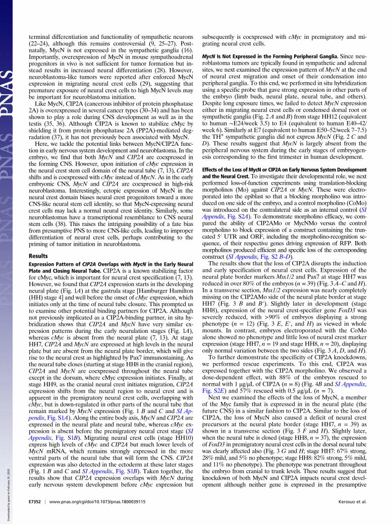
ResultsExpression Pattern of CIP2A Overlaps with MycN in the Early NeuralPlate and Closing Neural Tube. CIP2A is a known stabilizing factorfor cMyc, which is important for neural crest specification (7, 13).However, we found that CIP2A expression starts in the developingneural plate (Fig. 1A) at the gastrula stage [Hamburger Hamilton(HH) stage 4] and well before the onset of cMyc expression, whichinitiates only at the time of neural tube closure. This prompted usto examine other potential binding partners for CIP2A. Althoughnot previously implicated as a CIP2A-binding partner, in situ hy-bridization shows that CIP2A and MycN have very similar ex-pression patterns during the early neurulation stages (Fig. 1A),whereas cMyc is absent from the neural plate (7, 13). At stageHH7, CIP2A and MycN are expressed at high levels in the neuralplate but are absent from the neural plate border, which will giverise to the neural crest as highlighted by Pax7 immunostaining. Asthe neural tube closes (starting at stage HH8 in the cranial region),CIP2A and MycN are coexpressed throughout the neural tubeexcept in the dorsum, where cMyc expression initiates. Finally, atstage HH9, as the cranial neural crest initiates migration, CIP2Aexpression shifts from the neural region to neural crest and isapparent in the premigratory neural crest cells, overlapping withcMyc, but is down-regulated in other parts of the neural tube thatremain marked by MycN expression (Fig. 1 B and C and SI Ap-pendix, Fig. S1A). Along the entire body axis,MycN and CIP2A areexpressed in the neural plate and neural tube, whereas cMyc ex-pression is absent before the premigratory neural crest stage (SIAppendix, Fig. S1B). Migrating neural crest cells (stage HH10)express high levels of cMyc and CIP2A but much lower levels ofMycN mRNA, which remains strongly expressed in the moreventral parts of the neural tube that will form the CNS. CIP2Aexpression was also detected in the ectoderm at these later stages(Fig. 1 B and C and SI Appendix, Fig. S1B). Taken together, theresults show that CIP2A expression overlaps with MycN duringearly nervous system development before cMyc expression but
subsequently is coexpressed with cMyc in premigratory and mi-grating neural crest cells.
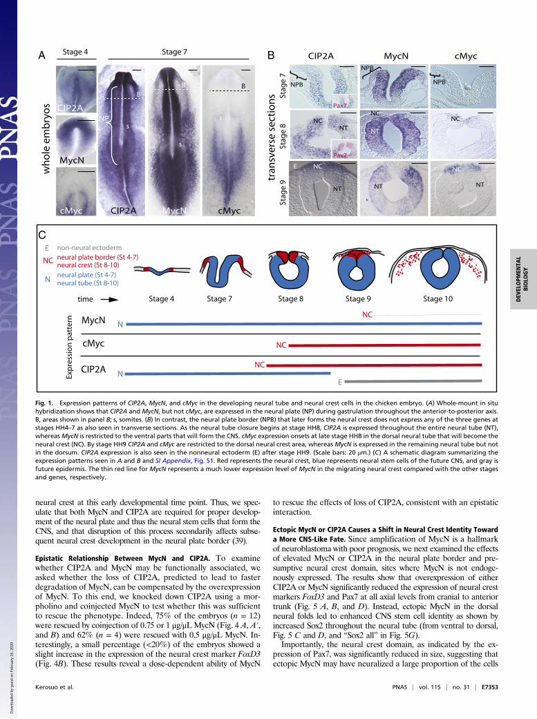
MycN Is Not Expressed in the Forming Peripheral Ganglia. Since neu-roblastoma tumors are typically found in sympathetic and adrenalsites, we next examined the expression pattern ofMycN at the endof neural crest migration and onset of their condensation intoperipheral ganglia. To this end, we performed in situ hybridizationusing a specific probe that gave strong expression in other parts ofthe embryo (limb buds, neural plate, neural tube, and others).Despite long exposure times, we failed to detect MycN expressioneither in migrating neural crest cells or condensed dorsal root orsympathetic ganglia (Fig. 2 A and B) from stage HH12 (equivalentto human ∼E24/week 3.5) to E4 (equivalent to human E40–42/week 6). Similarly at E7 (equivalent to human E50–52/week 7–7.5)the TH+ sympathetic ganglia did not express MycN (Fig. 2 C andD). These results suggest that MycN is largely absent from theperipheral nervous system during the early stages of embryogen-esis corresponding to the first trimester in human development.
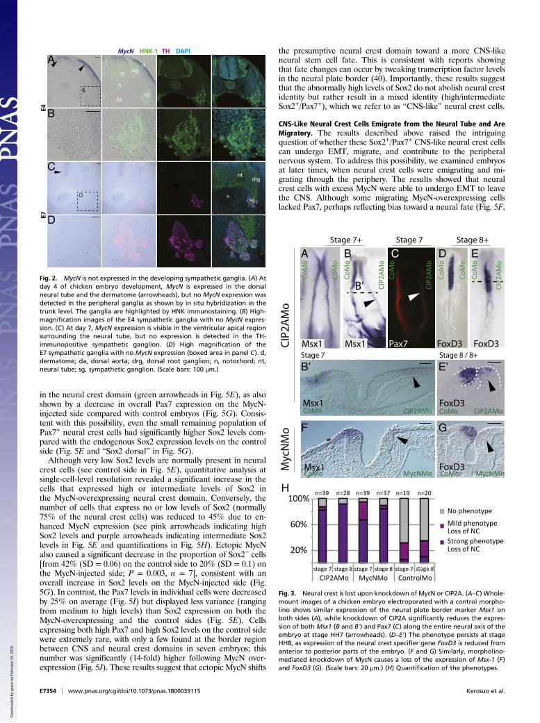
Effects of the Loss of MycN or CIP2A on Early Nervous System Developmentand the Neural Crest. To investigate their developmental role, we nextperformed loss-of-function experiments using translation-blockingmorpholinos (Mo) against CIP2A or MycN. These were electro-porated into the epiblast so that a blocking morpholino was intro-duced on one side of the embryo, and a control morpholino (CoMo)was introduced on the contralateral side as an internal control (SIAppendix, Fig. S2A). To demonstrate morpholino efficacy, we com-pared the ability of CIP2AMo or MycNMo versus the controlmorpholino to block expression of a construct containing the trun-cated 5′ UTR and ORF, including the morpholino-recognition se-quence, of their respective genes driving expression of RFP. Bothmorpholinos produced efficient and specific loss of the correspondingconstruct (SI Appendix, Fig. S2 B–D).The results show that the loss of CIP2A disrupts the induction
and early specification of neural crest cells. Expression of theneural plate border markers Msx1/2 and Pax7 at stage HH7 wasreduced in over 80% of the embryos (n = 39) (Fig. 3 A–C andH).In a transverse section, Msx1/2 expression was nearly completelymissing on the CIP2AMo side of the neural plate border at stageHH7 (Fig. 3 B and B′). Slightly later in development (stageHH8), expression of the neural crest-specifier gene FoxD3 wasseverely reduced, with >90% of embryos displaying a strongphenotype (n = 12) (Fig. 3 E, E′, and H) as viewed in wholemounts. In contrast, embryos electroporated with the CoMoalone showed no phenotype and little loss of neural crest markerexpression (stage HH7, n = 19 and stage HH8, n = 20), displayingonly normal variation between the two sides (Fig. 3 A, D, and H).To further demonstrate the specificity of CIP2A knockdowns,
we performed rescue experiments. To this end, CIP2A wasexpressed together with the CIP2A morpholino. We observed adose-dependent effect, with 88% of the embryos rescued tonormal with 1 μg/μL of CIP2A (n = 8) (Fig. 4B and SI Appendix,Fig. S2E) and 57% rescued with 0.5 μg/μL (n = 7).Next we examined the effects of the loss of MycN, a member
of the Myc family that is expressed in in the neural plate (thefuture CNS) in a similar fashion to CIP2A. Similar to the loss ofCIP2A, the loss of MycN also caused a deficit of neural crestprecursors at the neural plate border (stage HH7, n = 39) asshown in a transverse section (Fig. 3 F and H). Slightly later,when the neural tube is closed (stage HH8, n = 37), the expressionof FoxD3 in premigratory neural crest cells in the dorsal neural tubewas clearly affected also (Fig. 3 G and H; stage HH7: 67% strong,28%mild, and 5% no phenotype; stage HH8: 82% strong, 5%mild,and 11% no phenotype). The phenotype was penetrant throughoutthe embryo from cranial to trunk levels. These results suggest thatknockdown of both MycN and CIP2A impacts neural crest devel-opment although neither gene is expressed in the presumptive
E7352 | www.pnas.org/cgi/doi/10.1073/pnas.1800039115 Kerosuo et al.
Dow
nloa
ded
by g
uest
on
Feb
ruar
y 15
, 202
0
neural crest at this early developmental time point. Thus, we spec-ulate that both MycN and CIP2A are required for proper develop-ment of the neural plate and thus the neural stem cells that form theCNS, and that disruption of this process secondarily affects subse-quent neural crest development in the neural plate border (39).
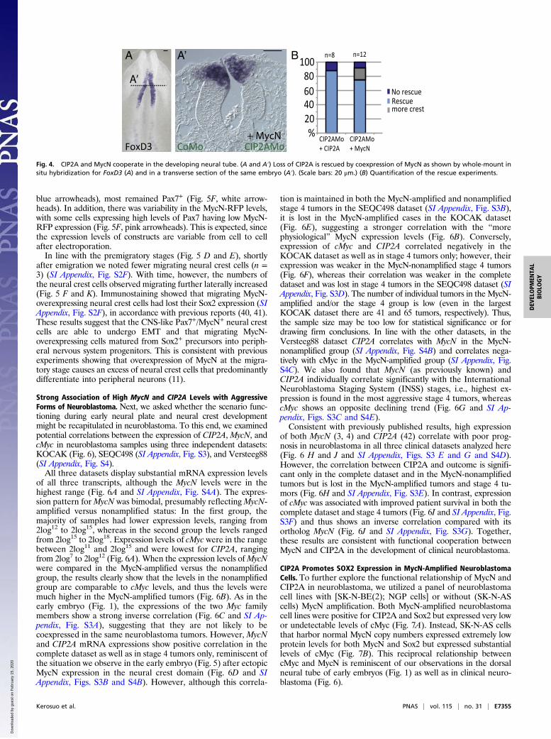
Epistatic Relationship Between MycN and CIP2A. To examinewhether CIP2A and MycN may be functionally associated, weasked whether the loss of CIP2A, predicted to lead to fasterdegradation of MycN, can be compensated by the overexpressionof MycN. To this end, we knocked down CIP2A using a mor-pholino and coinjected MycN to test whether this was sufficientto rescue the phenotype. Indeed, 75% of the embryos (n = 12)were rescued by coinjection of 0.75 or 1 μg/μL MycN (Fig. 4 A, A′,and B) and 62% (n = 4) were rescued with 0.5 μg/μL MycN. In-terestingly, a small percentage (<20%) of the embryos showed aslight increase in the expression of the neural crest marker FoxD3(Fig. 4B). These results reveal a dose-dependent ability of MycN
to rescue the effects of loss of CIP2A, consistent with an epistaticinteraction.
Ectopic MycN or CIP2A Causes a Shift in Neural Crest Identity Towarda More CNS-Like Fate. Since amplification of MycN is a hallmarkof neuroblastoma with poor prognosis, we next examined the effectsof elevated MycN or CIP2A in the neural plate border and pre-sumptive neural crest domain, sites where MycN is not endoge-nously expressed. The results show that overexpression of eitherCIP2A or MycN significantly reduced the expression of neural crestmarkers FoxD3 and Pax7 at all axial levels from cranial to anteriortrunk (Fig. 5 A, B, and D). Instead, ectopic MycN in the dorsalneural folds led to enhanced CNS stem cell identity as shown byincreased Sox2 throughout the neural tube (from ventral to dorsal,Fig. 5 C and D, and “Sox2 all” in Fig. 5G).Importantly, the neural crest domain, as indicated by the ex-
pression of Pax7, was significantly reduced in size, suggesting thatectopic MycN may have neuralized a large proportion of the cells
MycN
CIP2A
MycNcMyc CIP2A
tra
nsv
ers
e s
ect
ion
s
wh
ole
em
bry
os Pax7
Pax7
Stage 7Stage 4
Stag
e 8
Stag
e 9
Stag
e 7
MycNCIP2A cMycA B
NP
B
B NPB
NPB
NPB
NC
NC
NT NT
NCNC
NTNT
NT
NC
Stage 4 Stage 7 Stage 8 Stage 9 Stage 10
non-neural ectoderm neural plate border (St 4-7)
neural plate (St 4-7)
neural crest (St 8-10)
neural tube (St 8-10)
MycN
cMyc
CIP2A
C
N
N
NC
NC
NC
NC
N
time
Exp
ress
ion
pat
tern
E
E
E
cMyc
B
s
s
s
Fig. 1. Expression patterns of CIP2A, MycN, and cMyc in the developing neural tube and neural crest cells in the chicken embryo. (A) Whole-mount in situhybridization shows that CIP2A and MycN, but not cMyc, are expressed in the neural plate (NP) during gastrulation throughout the anterior-to-posterior axis.B, areas shown in panel B; s, somites. (B) In contrast, the neural plate border (NPB) that later forms the neural crest does not express any of the three genes atstages HH4–7 as also seen in transverse sections. As the neural tube closure begins at stage HH8, CIP2A is expressed throughout the entire neural tube (NT),whereasMycN is restricted to the ventral parts that will form the CNS. cMyc expression onsets at late stage HH8 in the dorsal neural tube that will become theneural crest (NC). By stage HH9 CIP2A and cMyc are restricted to the dorsal neural crest area, whereas MycN is expressed in the remaining neural tube but notin the dorsum. CIP2A expression is also seen in the nonneural ectoderm (E) after stage HH9. (Scale bars: 20 μm.) (C) A schematic diagram summarizing theexpression patterns seen in A and B and SI Appendix, Fig. S1. Red represents the neural crest, blue represents neural stem cells of the future CNS, and gray isfuture epidermis. The thin red line for MycN represents a much lower expression level of MycN in the migrating neural crest compared with the other stagesand genes, respectively.
Kerosuo et al. PNAS | vol. 115 | no. 31 | E7353
DEV
ELOPM
ENTA
LBIOLO
GY
Dow
nloa
ded
by g
uest
on
Feb
ruar
y 15
, 202
0
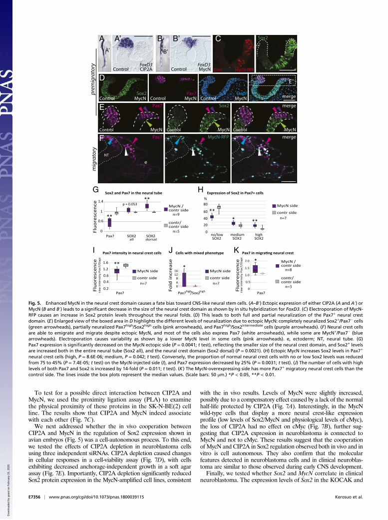
in the neural crest domain (green arrowheads in Fig. 5E), as alsoshown by a decrease in overall Pax7 expression on the MycN-injected side compared with control embryos (Fig. 5G). Consis-tent with this possibility, even the small remaining population ofPax7+ neural crest cells had significantly higher Sox2 levels com-pared with the endogenous Sox2 expression levels on the controlside (Fig. 5E and “Sox2 dorsal” in Fig. 5G).Although very low Sox2 levels are normally present in neural
crest cells (see control side in Fig. 5E), quantitative analysis atsingle-cell-level resolution revealed a significant increase in thecells that expressed high or intermediate levels of Sox2 inthe MycN-overexpressing neural crest domain. Conversely, thenumber of cells that express no or low levels of Sox2 (normally75% of the neural crest cells) was reduced to 45% due to en-hanced MycN expression (see pink arrowheads indicating highSox2 levels and purple arrowheads indicating intermediate Sox2levels in Fig. 5E and quantifications in Fig. 5H). Ectopic MycNalso caused a significant decrease in the proportion of Sox2− cells[from 42% (SD = 0.06) on the control side to 20% (SD = 0.1) onthe MycN-injected side; P = 0.003, n = 7], consistent with anoverall increase in Sox2 levels on the MycN-injected side (Fig.5G). In contrast, the Pax7 levels in individual cells were decreasedby 25% on average (Fig. 5I) but displayed less variance (rangingfrom medium to high levels) than Sox2 expression on both theMycN-overexpressing and the control sides (Fig. 5E). Cellsexpressing both high Pax7 and high Sox2 levels on the control sidewere extremely rare, with only a few found at the border regionbetween CNS and neural crest domains in seven embryos; thisnumber was significantly (14-fold) higher following MycN over-expression (Fig. 5J). These results suggest that ectopic MycN shifts
the presumptive neural crest domain toward a more CNS-likeneural stem cell fate. This is consistent with reports showingthat fate changes can occur by tweaking transcription factor levelsin the neural plate border (40). Importantly, these results suggestthat the abnormally high levels of Sox2 do not abolish neural crestidentity but rather result in a mixed identity (high/intermediateSox2+/Pax7+), which we refer to as “CNS-like” neural crest cells.
CNS-Like Neural Crest Cells Emigrate from the Neural Tube and AreMigratory. The results described above raised the intriguingquestion of whether these Sox2+/Pax7+ CNS-like neural crest cellscan undergo EMT, migrate, and contribute to the peripheralnervous system. To address this possibility, we examined embryosat later times, when neural crest cells were emigrating and mi-grating through the periphery. The results showed that neuralcrest cells with excess MycN were able to undergo EMT to leavethe CNS. Although some migrating MycN-overexpressing cellslacked Pax7, perhaps reflecting bias toward a neural fate (Fig. 5F,
20%
60%
100%
stage 7CIP2AMo
No phenotypeMild phenotype
Strong phenotypeLoss of NC
Loss of NC
stage 7 stage 7stage 8 stage 8 stage 8MycNMo ControlMo
CIP
2A
Mo
Myc
NM
o
Pax7 FoxD3
FoxD3Msx1
FoxD3Msx1
Msx1
oMA2PIC
oMoC
oMoC CoM
o oMoC
oMA2PIC
oMA2PIC
oMoC
CoMo CIP2AMo CoMo CIP2AMo
CoMo MycNMo MycNMoCoMo
Msx1Stage 7 Stage 8 / 8+
n=39 n=28 n=39 n=37 n=19 n=20
FoxD3
oMoC
Stage 7+ Stage 7 Stage 8+
A
B’
C
B’ E’
D
F G
H
B E
oMoC
Fig. 3. Neural crest is lost upon knockdown of MycN or CIP2A. (A–C) Whole-mount images of a chicken embryo electroporated with a control morpho-lino shows similar expression of the neural plate border marker Msx1 onboth sides (A), while knockdown of CIP2A significantly reduces the expres-sion of both Msx1 (B and B′) and Pax7 (C) along the entire neural axis of theembryo at stage HH7 (arrowheads). (D–E′) The phenotype persists at stageHH8, as expression of the neural crest specifier gene FoxD3 is reduced fromanterior to posterior parts of the embryo. (F and G) Similarly, morpholino-mediated knockdown of MycN causes a loss of the expression of Msx-1 (F)and FoxD3 (G). (Scale bars: 20 μm.) (H) Quantification of the phenotypes.
A
B
C
D
B
D
E4E7
MycN HNK-1 TH DAPI
ntdrg
d
sg
n
da
nt
nsg
drg
Fig. 2. MycN is not expressed in the developing sympathetic ganglia. (A) Atday 4 of chicken embryo development, MycN is expressed in the dorsalneural tube and the dermatome (arrowheads), but no MycN expression wasdetected in the peripheral ganglia as shown by in situ hybridization in thetrunk level. The ganglia are highlighted by HNK immunostaining. (B) High-magnification images of the E4 sympathetic ganglia with no MycN expres-sion. (C) At day 7, MycN expression is visible in the ventricular apical regionsurrounding the neural tube, but no expression is detected in the TH-immunopositive sympathetic ganglion. (D) High magnification of theE7 sympathetic ganglia with no MycN expression (boxed area in panel C). d,dermatome; da, dorsal aorta; drg, dorsal root ganglion; n, notochord; nt,neural tube; sg, sympathetic ganglion. (Scale bars: 100 μm.)
E7354 | www.pnas.org/cgi/doi/10.1073/pnas.1800039115 Kerosuo et al.
Dow
nloa
ded
by g
uest
on
Feb
ruar
y 15
, 202
0
blue arrowheads), most remained Pax7+ (Fig. 5F, white arrow-heads). In addition, there was variability in the MycN-RFP levels,with some cells expressing high levels of Pax7 having low MycN-RFP expression (Fig. 5F, pink arrowheads). This is expected, sincethe expression levels of constructs are variable from cell to cellafter electroporation.In line with the premigratory stages (Fig. 5 D and E), shortly
after emigration we noted fewer migrating neural crest cells (n =3) (SI Appendix, Fig. S2F). With time, however, the numbers ofthe neural crest cells observed migrating further laterally increased(Fig. 5 F and K). Immunostaining showed that migrating MycN-overexpressing neural crest cells had lost their Sox2 expression (SIAppendix, Fig. S2F), in accordance with previous reports (40, 41).These results suggest that the CNS-like Pax7+/MycN+ neural crestcells are able to undergo EMT and that migrating MycN-overexpressing cells matured from Sox2+ precursors into periph-eral nervous system progenitors. This is consistent with previousexperiments showing that overexpression of MycN at the migra-tory stage causes an excess of neural crest cells that predominantlydifferentiate into peripheral neurons (11).
Strong Association of High MycN and CIP2A Levels with AggressiveForms of Neuroblastoma. Next, we asked whether the scenario func-tioning during early neural plate and neural crest developmentmight be recapitulated in neuroblastoma. To this end, we examinedpotential correlations between the expression of CIP2A, MycN, andcMyc in neuroblastoma samples using three independent datasets:KOCAK (Fig. 6), SEQC498 (SI Appendix, Fig. S3), and Versteeg88(SI Appendix, Fig. S4).All three datasets display substantial mRNA expression levels
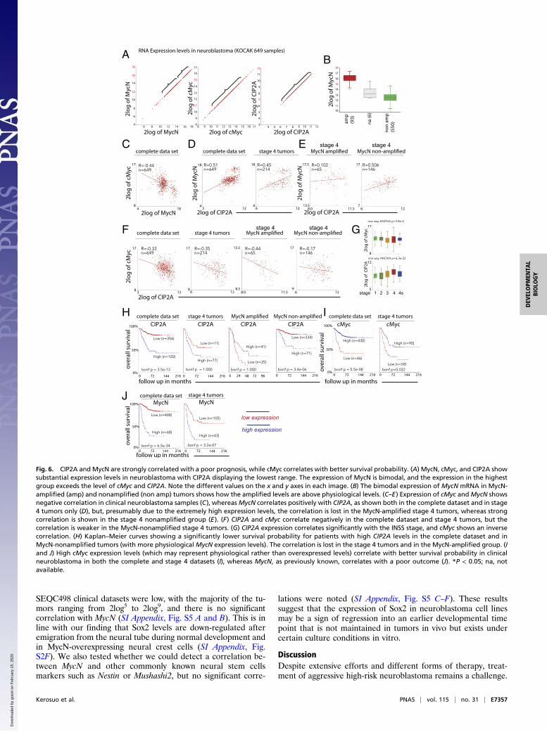
of all three transcripts, although the MycN levels were in thehighest range (Fig. 6A and SI Appendix, Fig. S4A). The expres-sion pattern forMycN was bimodal, presumably reflectingMycN-amplified versus nonamplified status: In the first group, themajority of samples had lower expression levels, ranging from2log12 to 2log15, whereas in the second group the levels rangedfrom 2log15 to 2log18. Expression levels of cMyc were in the rangebetween 2log11 and 2log15 and were lowest for CIP2A, rangingfrom 2log7 to 2log12 (Fig. 6A). When the expression levels ofMycNwere compared in the MycN-amplified versus the nonamplifiedgroup, the results clearly show that the levels in the nonamplifiedgroup are comparable to cMyc levels, and thus the levels weremuch higher in the MycN-amplified tumors (Fig. 6B). As in theearly embryo (Fig. 1), the expressions of the two Myc familymembers show a strong inverse correlation (Fig. 6C and SI Ap-pendix, Fig. S3A), suggesting that they are not likely to becoexpressed in the same neuroblastoma tumors. However, MycNand CIP2A mRNA expressions show positive correlation in thecomplete dataset as well as in stage 4 tumors only, reminiscent ofthe situation we observe in the early embryo (Fig. 5) after ectopicMycN expression in the neural crest domain (Fig. 6D and SIAppendix, Figs. S3B and S4B). However, although this correla-
tion is maintained in both the MycN-amplified and nonamplifiedstage 4 tumors in the SEQC498 dataset (SI Appendix, Fig. S3B),it is lost in the MycN-amplified cases in the KOCAK dataset(Fig. 6E), suggesting a stronger correlation with the “morephysiological” MycN expression levels (Fig. 6B). Conversely,expression of cMyc and CIP2A correlated negatively in theKOCAK dataset as well as in stage 4 tumors only; however, theirexpression was weaker in the MycN-nonamplified stage 4 tumors(Fig. 6F), whereas their correlation was weaker in the completedataset and was lost in stage 4 tumors in the SEQC498 dataset (SIAppendix, Fig. S3D). The number of individual tumors in the MycN-amplified and/or the stage 4 group is low (even in the largestKOCAK dataset there are 41 and 65 tumors, respectively). Thus,the sample size may be too low for statistical significance or fordrawing firm conclusions. In line with the other datasets, in theVersteeg88 dataset CIP2A correlates with MycN in the MycN-nonamplified group (SI Appendix, Fig. S4B) and correlates nega-tively with cMyc in the MycN-amplified group (SI Appendix, Fig.S4C). We also found that MycN (as previously known) andCIP2A individually correlate significantly with the InternationalNeuroblastoma Staging System (INSS) stages, i.e., highest ex-pression is found in the most aggressive stage 4 tumors, whereascMyc shows an opposite declining trend (Fig. 6G and SI Ap-pendix, Figs. S3C and S4E).Consistent with previously published results, high expression
of both MycN (3, 4) and CIP2A (42) correlate with poor prog-nosis in neuroblastoma in all three clinical datasets analyzed here(Fig. 6 H and J and SI Appendix, Figs. S3 E and G and S4D).However, the correlation between CIP2A and outcome is signifi-cant only in the complete dataset and in the MycN-nonamplifiedtumors but is lost in the MycN-amplified tumors and stage 4 tu-mors (Fig. 6H and SI Appendix, Fig. S3E). In contrast, expressionof cMyc was associated with improved patient survival in both thecomplete dataset and stage 4 tumors (Fig. 6I and SI Appendix, Fig.S3F) and thus shows an inverse correlation compared with itsortholog MycN (Fig. 6J and SI Appendix, Fig. S3G). Together,these results are consistent with functional cooperation betweenMycN and CIP2A in the development of clinical neuroblastoma.
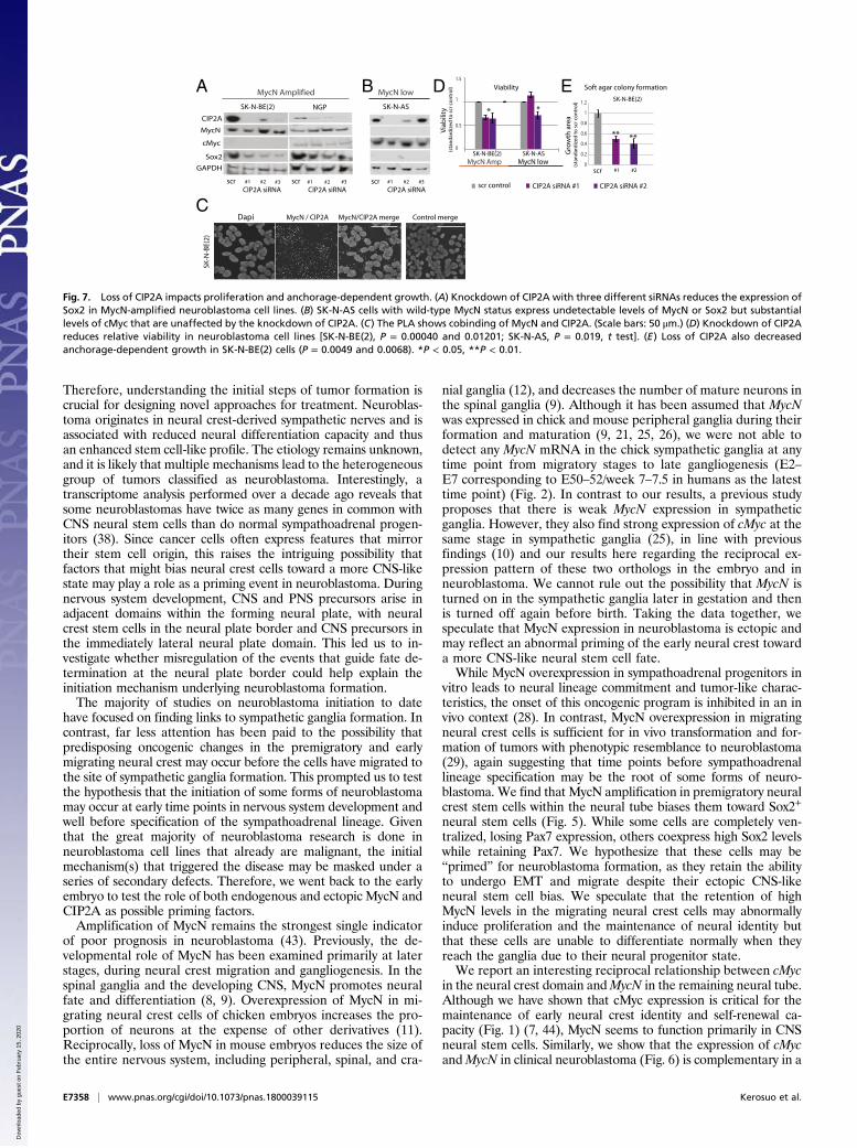
CIP2A Promotes SOX2 Expression in MycN-Amplified NeuroblastomaCells. To further explore the functional relationship of MycN andCIP2A in neuroblastoma, we utilized a panel of neuroblastomacell lines with [SK-N-BE(2); NGP cells] or without (SK-N-AScells) MycN amplification. Both MycN-amplified neuroblastomacell lines were positive for CIP2A and Sox2 but expressed very lowor undetectable levels of cMyc (Fig. 7A). Instead, SK-N-AS cellsthat harbor normal MycN copy numbers expressed extremely lowprotein levels for both MycN and Sox2 but expressed substantiallevels of cMyc (Fig. 7B). This reciprocal relationship betweencMyc and MycN is reminiscent of our observations in the dorsalneural tube of early embryos (Fig. 1) as well as in clinical neuro-blastoma (Fig. 6).
%20
CIP2AMo+ MycN
No rescue
CIP2AMo+ CIP2A
406080
100
Rescuemore crest
n=12n=8
CIP2AMoCoMo+ MycN
FoxD3
A A’
A’
B
Fig. 4. CIP2A and MycN cooperate in the developing neural tube. (A and A′) Loss of CIP2A is rescued by coexpression of MycN as shown by whole-mount insitu hybridization for FoxD3 (A) and in a transverse section of the same embryo (A′). (Scale bars: 20 μm.) (B) Quantification of the rescue experiments.
Kerosuo et al. PNAS | vol. 115 | no. 31 | E7355
DEV
ELOPM
ENTA
LBIOLO
GY
Dow
nloa
ded
by g
uest
on
Feb
ruar
y 15
, 202
0
To test for a possible direct interaction between CIP2A andMycN, we used the proximity ligation assay (PLA) to examinethe physical proximity of these proteins in the SK-N-BE(2) cellline. The results show that CIP2A and MycN indeed associatewith each other (Fig. 7C).We next addressed whether the in vivo cooperation between
CIP2A and MycN in the regulation of Sox2 expression shown inavian embryos (Fig. 5) was a cell-autonomous process. To this end,we tested the effects of CIP2A depletion in neuroblastoma cellsusing three independent siRNAs. CIP2A depletion caused changesin cellular responses in a cell-viability assay (Fig. 7D), with cellsexhibiting decreased anchorage-independent growth in a soft agarassay (Fig. 7E). Importantly, CIP2A depletion significantly reducedSox2 protein expression in the MycN-amplified cell lines, consistent
with the in vivo results. Levels of MycN were slightly increased,possibly due to a compensatory effect caused by a lack of the normalhalf-life protected by CIP2A (Fig. 7A). Interestingly, in the MycNwild-type cells that display a more neural crest-like expressionprofile (low levels of Sox2/MycN and physiological levels of cMyc),the loss of CIP2A had no effect on cMyc (Fig. 7B), further sug-gesting that CIP2A expression in neuroblastoma is connected toMycN and not to cMyc. These results suggest that the cooperationof MycN and CIP2A in Sox2 regulation observed both in vivo and invitro is cell autonomous. They also confirm that the molecularfeatures detected in neuroblastoma cells and in clinical neuroblas-toma are similar to those observed during early CNS development.Finally, we tested whether Sox2 and MycN correlate in clinical
neuroblastoma. The expression levels of Sox2 in the KOCAK and
CIP2A MycNControlControl
p = 0.053
SOX2
contr/contr side
MycN /contr side
Pax7
Flu
ore
scen
ce
1
0.6
0
1.4
SOX2all dorsal
A A’A’
B B’
B’
G
**
**
Control
n=9
n=5
yrotargimerp
yrotargim
Sox2 merge
MycN
e
0.5
1.0
0
1.5
2.0
Pax7
contr/contr side
MycN /contr side
n=8
n=3
H
E Pax7
ED
Control merge Sox2
MycN Control MycN Control MycNPax7 Dapi
Control MycN Control MycN
F Pax7 MycN-RFP merge
NT
J
FoxD3 FoxD3
Sox2C
MycN-RFPCont
Flu
ore
scen
ce
I
0
20
40
60
80%
**
**
SOX2
contr side
MycN side
n=7
SOX2 SOX2no/low medium high
*
Pax7
1.2
1.6
0.60.4
0.2Flu
ore
scen
cera
w in
ten
sity
Pax
7/D
api
contr side
MycN side
n=7
*
K
48
1216
0
Fold
incr
ease
contr side
MycN side
n=7
** *
Pax7high/Sox2high
Sox2 and Pax7 in the neural tube Expression of Sox2 in Pax7+ cells
Pax7 intensity in neural crest cells Cells with mixed phenotype
raw
inte
nsi
ty P
ax7/
Dap
i
Fig. 5. Enhanced MycN in the neural crest domain causes a fate bias toward CNS-like neural stem cells. (A–B′) Ectopic expression of either CIP2A (A and A′) orMycN (B and B′) leads to a significant decrease in the size of the neural crest domain as shown by in situ hybridization for FoxD3. (C) Electroporation of MycN-RFP causes an increase in Sox2 protein levels throughout the neural folds. (D) This leads to both full and partial neuralization of the Pax7+ neural crestdomain. (E) Enlarged view of the boxed area in D highlights the different levels of neuralization due to ectopic MycN: completely neuralized Sox2+/Pax7− cells(green arrowheads), partially neuralized Pax7high/Sox2high cells (pink arrowheads), and Pax7high/Sox2intermediate cells (purple arrowheads). (F) Neural crest cellsare able to emigrate and migrate despite ectopic MycN, and most of the cells also express Pax7 (white arrowheads), while some are MycN+/Pax7− (bluearrowheads). Electroporation causes variability as shown by a lower MycN level in some cells (pink arrowheads). e, ectoderm; NT, neural tube. (G)Pax7 expression is significantly decreased on the MycN ectopic side (P = 0.0041; t test), reflecting the smaller size of the neural crest domain, and Sox2+ levelsare increased both in the entire neural tube (Sox2 all), and the neural crest domain (Sox2 dorsal) (P = 0.0021). (H) Ectopic MycN increases Sox2 levels in Pax7+
neural crest cells (high, P = 8.6E-06; medium, P = 0.042; t test). Conversely, the proportion of normal neural crest cells with no or low Sox2 levels was reducedfrom 75 to 45% (P = 7.4E-05; t test) on the MycN-injected side (I), and Pax7 expression decreased by 25% (P = 0.0031; t test). (J) The number of cells with highlevels of both Pax7 and Sox2 is increased by 14-fold (P = 0.011; t test). (K) The MycN-overexpressing side has more Pax7+ migratory neural crest cells than thecontrol side. The lines inside the box plots represent the median values. (Scale bars: 50 μm.) *P < 0.05, **P < 0.01.
E7356 | www.pnas.org/cgi/doi/10.1073/pnas.1800039115 Kerosuo et al.
Dow
nloa
ded
by g
uest
on
Feb
ruar
y 15
, 202
0
SEQC498 clinical datasets were low, with the majority of the tu-mors ranging from 2log5 to 2log9, and there is no significantcorrelation with MycN (SI Appendix, Fig. S5 A and B). This is inline with our finding that Sox2 levels are down-regulated afteremigration from the neural tube during normal development andin MycN-overexpressing neural crest cells (SI Appendix, Fig.S2F). We also tested whether we could detect a correlation be-tween MycN and other commonly known neural stem cellsmarkers such as Nestin or Mushashi2, but no significant corre-
lations were noted (SI Appendix, Fig. S5 C–F). These resultssuggest that the expression of Sox2 in neuroblastoma cell linesmay be a sign of regression into an earlier developmental timepoint that is not maintained in tumors in vivo but exists undercertain culture conditions in vitro.
DiscussionDespite extensive efforts and different forms of therapy, treat-ment of aggressive high-risk neuroblastoma remains a challenge.
A B
C
F G
H I
J
D E
Fig. 6. CIP2A and MycN are strongly correlated with a poor prognosis, while cMyc correlates with better survival probability. (A) MycN, cMyc, and CIP2A showsubstantial expression levels in neuroblastoma with CIP2A displaying the lowest range. The expression of MycN is bimodal, and the expression in the highestgroup exceeds the level of cMyc and CIP2A. Note the different values on the x and y axes in each image. (B) The bimodal expression of MycN mRNA in MycN-amplified (amp) and nonamplified (non amp) tumors shows how the amplified levels are above physiological levels. (C–E) Expression of cMyc andMycN showsnegative correlation in clinical neuroblastoma samples (C), whereasMycN correlates positively with CIP2A, as shown both in the complete dataset and in stage4 tumors only (D), but, presumably due to the extremely high expression levels, the correlation is lost in the MycN-amplified stage 4 tumors, whereas strongcorrelation is shown in the stage 4 nonamplified group (E). (F) CIP2A and cMyc correlate negatively in the complete dataset and stage 4 tumors, but thecorrelation is weaker in the MycN-nonamplified stage 4 tumors. (G) CIP2A expression correlates significantly with the INSS stage, and cMyc shows an inversecorrelation. (H) Kaplan–Meier curves showing a significantly lower survival probability for patients with high CIP2A levels in the complete dataset and inMycN-nonamplified tumors (with more physiological MycN expression levels). The correlation is lost in the stage 4 tumors and in the MycN-amplified group. (Iand J) High cMyc expression levels (which may represent physiological rather than overexpressed levels) correlate with better survival probability in clinicalneuroblastoma in both the complete and stage 4 datasets (I), whereas MycN, as previously known, correlates with a poor outcome (J). *P < 0.05; na, notavailable.
Kerosuo et al. PNAS | vol. 115 | no. 31 | E7357
DEV
ELOPM
ENTA
LBIOLO
GY
Dow
nloa
ded
by g
uest
on
Feb
ruar
y 15
, 202
0
Therefore, understanding the initial steps of tumor formation iscrucial for designing novel approaches for treatment. Neuroblas-toma originates in neural crest-derived sympathetic nerves and isassociated with reduced neural differentiation capacity and thusan enhanced stem cell-like profile. The etiology remains unknown,and it is likely that multiple mechanisms lead to the heterogeneousgroup of tumors classified as neuroblastoma. Interestingly, atranscriptome analysis performed over a decade ago reveals thatsome neuroblastomas have twice as many genes in common withCNS neural stem cells than do normal sympathoadrenal progen-itors (38). Since cancer cells often express features that mirrortheir stem cell origin, this raises the intriguing possibility thatfactors that might bias neural crest cells toward a more CNS-likestate may play a role as a priming event in neuroblastoma. Duringnervous system development, CNS and PNS precursors arise inadjacent domains within the forming neural plate, with neuralcrest stem cells in the neural plate border and CNS precursors inthe immediately lateral neural plate domain. This led us to in-vestigate whether misregulation of the events that guide fate de-termination at the neural plate border could help explain theinitiation mechanism underlying neuroblastoma formation.The majority of studies on neuroblastoma initiation to date
have focused on finding links to sympathetic ganglia formation. Incontrast, far less attention has been paid to the possibility thatpredisposing oncogenic changes in the premigratory and earlymigrating neural crest may occur before the cells have migrated tothe site of sympathetic ganglia formation. This prompted us to testthe hypothesis that the initiation of some forms of neuroblastomamay occur at early time points in nervous system development andwell before specification of the sympathoadrenal lineage. Giventhat the great majority of neuroblastoma research is done inneuroblastoma cell lines that already are malignant, the initialmechanism(s) that triggered the disease may be masked under aseries of secondary defects. Therefore, we went back to the earlyembryo to test the role of both endogenous and ectopic MycN andCIP2A as possible priming factors.Amplification of MycN remains the strongest single indicator
of poor prognosis in neuroblastoma (43). Previously, the de-velopmental role of MycN has been examined primarily at laterstages, during neural crest migration and gangliogenesis. In thespinal ganglia and the developing CNS, MycN promotes neuralfate and differentiation (8, 9). Overexpression of MycN in mi-grating neural crest cells of chicken embryos increases the pro-portion of neurons at the expense of other derivatives (11).Reciprocally, loss of MycN in mouse embryos reduces the size ofthe entire nervous system, including peripheral, spinal, and cra-
nial ganglia (12), and decreases the number of mature neurons inthe spinal ganglia (9). Although it has been assumed that MycNwas expressed in chick and mouse peripheral ganglia during theirformation and maturation (9, 21, 25, 26), we were not able todetect any MycN mRNA in the chick sympathetic ganglia at anytime point from migratory stages to late gangliogenesis (E2–E7 corresponding to E50–52/week 7–7.5 in humans as the latesttime point) (Fig. 2). In contrast to our results, a previous studyproposes that there is weak MycN expression in sympatheticganglia. However, they also find strong expression of cMyc at thesame stage in sympathetic ganglia (25), in line with previousfindings (10) and our results here regarding the reciprocal ex-pression pattern of these two orthologs in the embryo and inneuroblastoma. We cannot rule out the possibility that MycN isturned on in the sympathetic ganglia later in gestation and thenis turned off again before birth. Taking the data together, wespeculate that MycN expression in neuroblastoma is ectopic andmay reflect an abnormal priming of the early neural crest towarda more CNS-like neural stem cell fate.While MycN overexpression in sympathoadrenal progenitors in
vitro leads to neural lineage commitment and tumor-like charac-teristics, the onset of this oncogenic program is inhibited in an invivo context (28). In contrast, MycN overexpression in migratingneural crest cells is sufficient for in vivo transformation and for-mation of tumors with phenotypic resemblance to neuroblastoma(29), again suggesting that time points before sympathoadrenallineage specification may be the root of some forms of neuro-blastoma. We find that MycN amplification in premigratory neuralcrest stem cells within the neural tube biases them toward Sox2+
neural stem cells (Fig. 5). While some cells are completely ven-tralized, losing Pax7 expression, others coexpress high Sox2 levelswhile retaining Pax7. We hypothesize that these cells may be“primed” for neuroblastoma formation, as they retain the abilityto undergo EMT and migrate despite their ectopic CNS-likeneural stem cell bias. We speculate that the retention of highMycN levels in the migrating neural crest cells may abnormallyinduce proliferation and the maintenance of neural identity butthat these cells are unable to differentiate normally when theyreach the ganglia due to their neural progenitor state.We report an interesting reciprocal relationship between cMyc
in the neural crest domain andMycN in the remaining neural tube.Although we have shown that cMyc expression is critical for themaintenance of early neural crest identity and self-renewal ca-pacity (Fig. 1) (7, 44), MycN seems to function primarily in CNSneural stem cells. Similarly, we show that the expression of cMycandMycN in clinical neuroblastoma (Fig. 6) is complementary in a
MycN / CIP2A Dapi Control mergeMycN/CIP2A merge
SK-N
-BE(
2)
1
Viability
Via
bili
ty
(sta
nd
ard
ize
d t
o s
cr c
on
tro
l)
0.5
0
1.5
scr control CIP2A siRNA #2CIP2A siRNA #1
MycN lowMycN Amp
Gro
wth
are
a
(sta
nd
ard
ize
d t
o s
cr c
on
tro
l)
Soft agar colony formation
1
0.8
0.6
0.4
0.2
0
1.2
** **
scr #1 #2
SK-N-BE(2)
SK-N-BE(2) SK-N-AS
* *
MycN
CIP2A
cMyc
Sox2
GAPDH
SK-N-BE(2) NGP
MycN Amplified MycN low
SK-N-AS
scr scr scr #2#2 #3 #2 #3#1#1#1 #3
CIP2A siRNACIP2A siRNACIP2A siRNA
C
D EA B
Fig. 7. Loss of CIP2A impacts proliferation and anchorage-dependent growth. (A) Knockdown of CIP2A with three different siRNAs reduces the expression ofSox2 in MycN-amplified neuroblastoma cell lines. (B) SK-N-AS cells with wild-type MycN status express undetectable levels of MycN or Sox2 but substantiallevels of cMyc that are unaffected by the knockdown of CIP2A. (C) The PLA shows cobinding of MycN and CIP2A. (Scale bars: 50 μm.) (D) Knockdown of CIP2Areduces relative viability in neuroblastoma cell lines [SK-N-BE(2), P = 0.00040 and 0.01201; SK-N-AS, P = 0.019, t test]. (E) Loss of CIP2A also decreasedanchorage-dependent growth in SK-N-BE(2) cells (P = 0.0049 and 0.0068). *P < 0.05, **P < 0.01.
E7358 | www.pnas.org/cgi/doi/10.1073/pnas.1800039115 Kerosuo et al.
Dow
nloa
ded
by g
uest
on
Feb
ruar
y 15
, 202
0

manner paralleling that in the embryo (Fig. 1). Our results using acombination of functional studies in the embryo (Fig. 5) togetherwith neuroblastoma cell lines (Fig. 7) and publicly available tu-mor datasets (Fig. 6 and SI Appendix, Figs. S3 and S4) suggestthat MycN overexpression (or in many cases amplification)biases their fate from neural crest toward CNS-like neural stemcells and that this bias reflects an early developmental event. Thefact that CIP2A partners with MycN only during the early neuralplate stage and not later after neural tube closure or in the mi-grating neural crest cells further strengthens our hypothesis thatsome tumors are primed at an early developmental time point. Itis intriguing to speculate that this early mechanism may reflectthe underlying cause of initiation of the most aggressive forms ofneuroblastoma. Our findings also bring neuroblastoma closer toother pediatric CNS tumors such as medulloblastoma, raising thepossibility that common mechanisms may underlie the initiationof these tumors.Many pediatric malignancies arise from embryonic cell types
that have persisted and give rise to tumors in early childhood(45). Pediatric tumors are thus unlikely to be driven by thegradual accumulation of genetic lesions but rather via oncogeniccells that are predisposed to malignant growth while carrying fewmutations (46). In Drosophila, it has been shown that in-termediate neural progenitors born during a particular timeperiod are predisposed to malignancy (47). Similarly, we specu-late here that, rather than immediately initiating rapid tumorgrowth, these early events may serve as a priming event for tu-mor susceptibility so that cells ectopically exposed to high MycNlevels early in development have the potential for metastatictumor growth later during ganglia formation.Neuroblast hyperplasia is detected in normal ganglia before
and around birth. Some of these neuroblasts progress intoneuroblastoma-like tumors upon MycN overexpression underthe TH promoter (45) but do not fully recapitulate the metastaticdisease. It is intriguing to speculate that perhaps, in addition tothe constitutive ectopic MycN expression in the developingganglia, the early priming event in the neural plate border wedescribe in this study triggers the formation of a full-blownmetastatic neuroblastoma. This also suggests that existingmouse and zebrafish neuroblastoma models that activate MycNin the peripheral ganglia may initiate the expression at a timepoint that is too late for understanding the initiation of at leastsome subgroups of the disease (45, 48–52). Finally, neuroblas-toma occasionally occurs in association with other neural crest-derived defects (also known as “neurocristopathies”) such asHirschsprung’s disease (53–56), which results from a failure ofneural crest cells to populate the most distal portions of the in-testines. This supports the idea that early onset of neuroblastomamay occur in multipotent neural crest stem cells before theirmigration into respective target tissues and perhaps may limit thepool of migrating cells.We were intrigued by the fact that CIP2A is known to stabilize
cMyc, but little was known about its interaction with MycN. Wenoted that CIP2A was expressed throughout the neural plate andneural tube at a time when cMyc is not yet expressed. Thisprompted us to study whether CIP2A might initially stabilizeMycN at early times until cMyc is turned on, and indeed, ourstudy reveals CIP2A as a binding partner of MycN (Figs. 4 and7). CIP2A has an established role as an oncogene, and itsknockdown leads to down-regulation of several oncogenic driv-ers (Akt, cMyc, and E2F1) due to PP2A dephosphorylation-mediated degradation (57). Although the loss of CIP2A doesnot compromise mouse viability, it causes defects in neural andspermatogonial progenitors (35, 36). Here we show that it is, incollaboration with MycN, also required for the correct formationof the neural stem cell characteristics of the neuroectoderm atthe neural plate but not later, after neural tube closure (Figs. 2–4).We also show that CIP2A couples with MycN in neuroblastoma
(Figs. 6 and 7), which suggests that this early developmental role ismaintained in neuroblastoma. CIP2A is overexpressed in a largefraction of all major human cancer types and, in line with previousfindings (37, 57), its inhibition leads to decreased tumor cell via-bility in neuroblastoma cell lines (Fig. 7), suggesting that CIP2Amay be a potential target for neuroblastoma therapy.cMyc is famous for its oncogenic properties and is overex-
pressed in multiple cancer types (58). Thus, it is counterintuitivethat its expression is associated with higher survival rates andgood prognosis in neuroblastoma (Fig. 6). There are severalpossible explanations for this observation. First, cMyc in neuro-blastoma may reflect a more normal multipotent neural creststem cell state that is capable of responding to cues from theenvironment to promote differentiation (7). Second, the overallexpression levels of cMyc in neuroblastoma samples are signifi-cantly lower than the expression levels of MycN in the MycN-amplified tumors (Fig. 6) and thus are likely to be similar to theendogenous physiological levels during embryonic development.This is in line with the reports on high cMyc levels as a prognosticmarker for the poor outcome in a small percentage of the un-differentiated subtype of neuroblastoma, NBUD (59, 60), in whichthe overexpression of cMyc may have triggered the highly pro-liferative oncogenic machinery that is not turned on in neural crestcells during normal development (7). In line with this, a recentstudy shows that a subset of high-risk neuroblastomas display up-regulated cMyc due to enhancer hijacking, and overexpression ofcMyc under the DβH promoter induced tumor mass growth invivo in zebrafish (61). cMyc amplification is extremely rare inneuroblastoma, but a case study reports undifferentiated mor-phology, poor survival, and low levels ofMycN expression in thesetumors (62). These studies further support our hypothesis thatphysiological rather than overexpressed cMyc levels are associatedwith the better outcome of the disease.Despite their very different endogenous roles during the de-
velopment of the nervous system, it is important to keep in mindthat all Myc family members have oncogenic properties and,upon misregulation, can trigger the onset of malignant trans-formation. In fact, in line with the reports on poor prognosis withvery high cMyc levels, transcriptional profiles of downstream tar-gets due to increased expression of any Myc member (MycN/cMyc/lMyc) are very similar in neuroblastoma and other cancertypes, and all correlate with poor capacity to differentiate (63, 64).It is thus possible that some of the reports on forced MycN/cMycoverexpression in the sympathetic ganglia reflect this general on-cogenic capacity instead of resembling the actual initiation processof neuroblastoma. Our results highlight the normal role of MycNin early neural development and raise the intriguing possibilitythat the balance of CIP2A/MycN binding at the neural plateborder can influence cell-fate decisions in early embryos in amanner that triggers priming of neuroblastoma cells.
Materials and MethodsDetailed information regarding materials and methods can be found in SIAppendix, SI Materials and Methods. Briefly, whole-mount in situ hybrid-ization and gain and loss of function experiments were performed onchicken embryos as previously described (65–67). Morpholinos were pur-chased from Gene Tools LLC (www.gene-tools.com/), immunostaining wasperformed as described (7), and Western blot lysates from the neuroblas-toma cell lines SK-N-AS and SK-N-BE(2) were made 2 d after RNAi infection;the Western blot protocol was carried out as previously described (37, 68).Fluorescence on the images was quantified by using ImageJ (NIH). The PLAwas performed according to the manufacturer’s instructions for the Duolinkkit (DUO92102; Sigma-Aldrich), and cell viability and proliferation wasmeasured using the WST-1 kit (5015944001; Roche). The statistical analyseswere performed on publicly available clinical neuroblastoma datasets(KOCAK, SEQC498, and Versteeg88) acquired from the R2 microarray anal-ysis and visualization platform (https://hgserver1.amc.nl/cgi-bin/r2/main.cgi).
Kerosuo et al. PNAS | vol. 115 | no. 31 | E7359
DEV
ELOPM
ENTA
LBIOLO
GY
Dow
nloa
ded
by g
uest
on
Feb
ruar
y 15
, 202
0
ACKNOWLEDGMENTS. We thank Dr. Marie Arsenian-Henriksson for pro-viding SK-N-BE(2) cells, Dr. Kristina Cole for providing NGP cells, Dr. RuthPalmer for providing SK-N-AS cells, and Dr. Edward K. Chan for the mousemonoclonal CIP2A antibody. This work was funded by NIH Grants HD037105
and DE024157 (to M.E.B.) and by grants from the Jane and Aatos ErkkoFoundation, the Ella and Georg Ehrnrooth Foundation, and the Väre Foun-dation (to L.K.), the American-Scandinavian Foundation (to P.N.), and theSigrid Juselius Foundation (to J.W.).
1. Matthay KK, et al. (2016) Neuroblastoma. Nat Rev Dis Primers 2:16078.2. Maris JM (2010) Recent advances in neuroblastoma. N Engl J Med 362:2202–2211.3. Brodeur GM, Seeger RC, Schwab M, Varmus HE, Bishop JM (1984) Amplification of N-
myc in untreated human neuroblastomas correlates with advanced disease stage.Science 224:1121–1124.
4. Schwab M, et al. (1984) Chromosome localization in normal human cells and neuro-blastomas of a gene related to c-myc. Nature 308:288–291.
5. Kerosuo L, Bronner-Fraser M (2012) What is bad in cancer is good in the embryo:Importance of EMT in neural crest development. Semin Cell Dev Biol 23:320–332.
6. Eilers M, Eisenman RN (2008) Myc’s broad reach. Genes Dev 22:2755–2766.7. Kerosuo L, Bronner ME (2016) cMyc regulates the size of the premigratory neural
crest stem cell pool. Cell Rep 17:2648–2659.8. Knoepfler PS, Cheng PF, Eisenman RN (2002) N-myc is essential during neurogenesis
for the rapid expansion of progenitor cell populations and the inhibition of neuronaldifferentiation. Genes Dev 16:2699–2712.
9. Sawai S, et al. (1993) Defects of embryonic organogenesis resulting from targeteddisruption of the N-myc gene in the mouse. Development 117:1445–1455.
10. Zinin N, et al. (2014) MYC proteins promote neuronal differentiation by controllingthe mode of progenitor cell division. EMBO Rep 15:383–391.
11. Wakamatsu Y, Watanabe Y, Nakamura H, Kondoh H (1997) Regulation of the neuralcrest cell fate by N-myc: Promotion of ventral migration and neuronal differentiation.Development 124:1953–1962.
12. Charron J, et al. (1992) Embryonic lethality in mice homozygous for a targeted dis-ruption of the N-myc gene. Genes Dev 6:2248–2257.
13. Khudyakov J, Bronner-Fraser M (2009) Comprehensive spatiotemporal analysis ofearly chick neural crest network genes. Dev Dyn 238:716–723.
14. Alam G, et al. (2009) MYCN promotes the expansion of Phox2B-positive neuronalprogenitors to drive neuroblastoma development. Am J Pathol 175:856–866.
15. Pei D, et al. (2013) Distinct neuroblastoma-associated alterations of PHOX2B impairsympathetic neuronal differentiation in zebrafish models. PLoS Genet 9:e1003533.
16. Ke XX, et al. (2015) Phox2B correlates with MYCN and is a prognostic marker forneuroblastoma development. Oncol Lett 9:2507–2514.
17. Hirsch MR, Tiveron MC, Guillemot F, Brunet JF, Goridis C (1998) Control of norad-renergic differentiation and Phox2a expression by MASH1 in the central and pe-ripheral nervous system. Development 125:599–608.
18. Vincentz JW, et al. (2012) A Phox2- and Hand2-dependent Hand1 cis-regulatory el-ement reveals a unique gene dosage requirement for Hand2 during sympatheticneurogenesis. J Neurosci 32:2110–2120.
19. Pattyn A, Morin X, Cremer H, Goridis C, Brunet JF (1999) The homeobox gene Phox2bis essential for the development of autonomic neural crest derivatives. Nature 399:366–370.
20. Sommer L, Shah N, Rao M, Anderson DJ (1995) The cellular function of MASH1 inautonomic neurogenesis. Neuron 15:1245–1258.
21. Wakamatsu Y, Watanabe Y, Shimono A, Kondoh H (1993) Transition of localization ofthe N-Myc protein from nucleus to cytoplasm in differentiating neurons. Neuron 10:1–9.
22. Tsarovina K, et al. (2004) Essential role of Gata transcription factors in sympatheticneuron development. Development 131:4775–4786.
23. Nakagawara A, et al. (1993) Association between high levels of expression of the TRKgene and favorable outcome in human neuroblastoma. N Engl J Med 328:847–854.
24. Ernsberger U, Reissmann E, Mason I, Rohrer H (2000) The expression of dopaminebeta-hydroxylase, tyrosine hydroxylase, and Phox2 transcription factors in sympa-thetic neurons: Evidence for common regulation during noradrenergic induction anddiverging regulation later in development. Mech Dev 92:169–177.
25. Kramer M, Ribeiro D, Arsenian-Henriksson M, Deller T, Rohrer H (2016) Proliferationand survival of embryonic sympathetic neuroblasts by MYCN and activated ALK sig-naling. J Neurosci 36:10425–10439.
26. Sawai S, Kato K, Wakamatsu Y, Kondoh H (1990) Organization and expression of thechicken N-myc gene. Mol Cell Biol 10:2017–2026.
27. Edsjö A, et al. (2004) Neuroblastoma cells with overexpressed MYCN retain their ca-pacity to undergo neuronal differentiation. Lab Invest 84:406–417.
28. Mobley BC, et al. (2015) Expression of MYCN in multipotent sympathoadrenal pro-genitors induces proliferation and neural differentiation, but is not sufficient fortumorigenesis. PLoS One 10:e0133897.
29. Olsen RR, et al. (2017) MYCN induces neuroblastoma in primary neural crest cells.Oncogene 36:5075–5082.
30. Li W, et al. (2008) CIP2A is overexpressed in gastric cancer and its depletion leads toimpaired clonogenicity, senescence, or differentiation of tumor cells. Clin Cancer Res14:3722–3728.
31. Chen KF, et al. (2010) CIP2A mediates effects of bortezomib on phospho-Akt andapoptosis in hepatocellular carcinoma cells. Oncogene 29:6257–6266.
32. Côme C, et al. (2009) CIP2A is associated with human breast cancer aggressivity. ClinCancer Res 15:5092–5100.
33. Dong QZ, et al. (2011) CIP2A is overexpressed in non-small cell lung cancer and cor-relates with poor prognosis. Ann Surg Oncol 18:857–865.
34. Khanna A, et al. (2009) MYC-dependent regulation and prognostic role of CIP2A ingastric cancer. J Natl Cancer Inst 101:793–805.
35. Kerosuo L, et al. (2010) CIP2A increases self-renewal and is linked to Myc in neuralprogenitor cells. Differentiation 80:68–77.
36. Ventelä S, et al. (2012) CIP2A promotes proliferation of spermatogonial progenitorcells and spermatogenesis in mice. PLoS One 7:e33209.
37. Junttila MR, et al. (2007) CIP2A inhibits PP2A in human malignancies. Cell 130:51–62.38. De Preter K, et al. (2006) Human fetal neuroblast and neuroblastoma transcriptome
analysis confirms neuroblast origin and highlights neuroblastoma candidate genes.Genome Biol 7:R84, and erratum (2007) 8:401.
39. Rogers CD, Jayasena CS, Nie S, Bronner ME (2012) Neural crest specification: Tissues,signals, and transcription factors. Wiley Interdiscip Rev Dev Biol 1:52–68.
40. Roellig D, Tan-Cabugao J, Esaian S, Bronner ME (2017) Dynamic transcriptional sig-nature and cell fate analysis reveals plasticity of individual neural plate border cells.eLife 6:e21620.
41. Cimadamore F, et al. (2011) Human ESC-derived neural crest model reveals a key rolefor SOX2 in sensory neurogenesis. Cell Stem Cell 8:538–551.
42. Khanna A, et al. (2013) Chk1 targeting reactivates PP2A tumor suppressor activity incancer cells. Cancer Res 73:6757–6769.
43. Cohn SL, et al.; INRG Task Force (2009) The International Neuroblastoma Risk Group(INRG) classification system: An INRG Task Force report. J Clin Oncol 27:289–297.
44. Bellmeyer A, Krase J, Lindgren J, LaBonne C (2003) The protooncogene c-myc is anessential regulator of neural crest formation in xenopus. Dev Cell 4:827–839.
45. Hansford LM, et al. (2004) Mechanisms of embryonal tumor initiation: Distinct rolesfor MycN expression and MYCN amplification. Proc Natl Acad Sci USA 101:12664–12669.
46. Chen X, Pappo A, Dyer MA (2015) Pediatric solid tumor genomics and developmentalpliancy. Oncogene 34:5207–5215.
47. Narbonne-Reveau K, et al. (2016) Neural stem cell-encoded temporal patterningdelineates an early window of malignant susceptibility in Drosophila. eLife 5:e13463.
48. Zhu S, Thomas Look A (2016) Neuroblastoma and its zebrafish model. Adv Exp MedBiol 916:451–478.
49. Corallo D, Candiani S, Ori M, Aveic S, Tonini GP (2016) The zebrafish as a model forstudying neuroblastoma. Cancer Cell Int 16:82.
50. Weiss WA, Godfrey T, Francisco C, Bishop JM (2000) Genome-wide screen for allelicimbalance in a mouse model for neuroblastoma. Cancer Res 60:2483–2487.
51. Terrile M, et al. (2011) miRNA expression profiling of the murine TH-MYCN neuro-blastoma model reveals similarities with human tumors and identifies novel candi-date miRNAs. PLoS One 6:e28356.
52. Weiss WA, Aldape K, Mohapatra G, Feuerstein BG, Bishop JM (1997) Targeted ex-pression of MYCN causes neuroblastoma in transgenic mice. EMBO J 16:2985–2995.
53. Williams P, Wegner E, Ziegler DS (2014) Outcomes in multifocal neuroblastoma aspart of the neurocristopathy syndrome. Pediatrics 134:e611–e616.
54. Roshkow JE, Haller JO, Berdon WE, Sane SM (1988) Hirschsprung’s disease, Ondine’scurse, and neuroblastoma–Manifestations of neurocristopathy. Pediatr Radiol 19:45–49.
55. Nemecek ER, Sawin RW, Park J (2003) Treatment of neuroblastoma in patients withneurocristopathy syndromes. J Pediatr Hematol Oncol 25:159–162.
56. Garavelli L, et al. (2015) Noonan syndrome-like disorder with loose anagen hair: Asecond case with neuroblastoma. Am J Med Genet A 167A:1902–1907.
57. Khanna A, Pimanda JE, Westermarck J (2013) Cancerous inhibitor of protein phos-phatase 2A, an emerging human oncoprotein and a potential cancer therapy target.Cancer Res 73:6548–6553.
58. Dang CV (2012) MYC on the path to cancer. Cell 149:22–35.59. Wang LL, et al. (2015) Augmented expression of MYC and/or MYCN protein defines
highly aggressive MYC-driven neuroblastoma: A Children’s Oncology Group study. BrJ Cancer 113:57–63.
60. Wang LL, et al. (2013) Neuroblastoma of undifferentiated subtype, prognostic sig-nificance of prominent nucleolar formation, and MYC/MYCN protein expression: Areport from the Children’s Oncology Group. Cancer 119:3718–3726.
61. Zimmerman MW, et al. (2018) MYC drives a subset of high-risk pediatric neuroblas-tomas and is activated through mechanisms including enhancer hijacking and focalenhancer amplification. Cancer Discov 8:320–335.
62. Matsuno R, et al. (January 1, 2018) Rare MYC-amplified neuroblastoma with large cellhistology. Pediatr Dev Pathol, 10.1177/1093526617749670.
63. Raetz EA, et al. (2003) Identification of genes that are regulated transcriptionally byMyc in childhood tumors. Cancer 98:841–853.
64. Fredlund E, Ringnér M, Maris JM, Påhlman S (2008) High Myc pathway activity andlow stage of neuronal differentiation associate with poor outcome in neuroblastoma.Proc Natl Acad Sci USA 105:14094–14099.
65. Acloque H, Wilkinson DG, Nieto MA (2008) In situ hybridization analysis of chickembryos in whole-mount and tissue sections. Methods Cell Biology 87:169–185.
66. Kerosuo L, Bronner ME (2014) Biphasic influence of Miz1 on neural crest developmentby regulating cell survival and apical adhesion complex formation in the developingneural tube. Mol Biol Cell 25:347–355.
67. Sauka-Spengler T, Barembaum M (2008) Gain- and loss-of-function approaches in thechick embryo. Methods Cell Biol 87:237–256.
68. Kauko O, et al. (2015) Label-free quantitative phosphoproteomics with novel pairwiseabundance normalization reveals synergistic RAS and CIP2A signaling. Sci Rep 5:13099.
E7360 | www.pnas.org/cgi/doi/10.1073/pnas.1800039115 Kerosuo et al.
Dow
nloa
ded
by g
uest
on
Feb
ruar
y 15
, 202
0