Embed Size (px)
Citation preview
Enhanced Bioactivity of Mg−Nd−Zn−Zr Alloy Achieved withNanoscale MgF2 Surface for Vascular Stent ApplicationLin Mao,†,‡,⊗ Li Shen,§,⊗ Jiahui Chen,§ Yu Wu,‡,∥ Minsuk Kwak,‡ Yao Lu,‡ Qiong Xue,‡ Jia Pei,†
Lei Zhang,† Guangyin Yuan,*,† Rong Fan,*,‡,⊥ Junbo Ge,§ and Wenjiang Ding†
†National Engineering Research Center of Light Alloys Net Forming and State Key Laboratory of Metal Matrix Composite, ShanghaiJiao Tong University, Shanghai 200240, China‡Department of Biomedical Engineering, Yale University, New Haven, Connecticut 06511, United States§Shanghai Institute of Cardiovascular Diseases, Department of Cardiology, Zhongshan Hospital, Fudan University, Shanghai 200032,China∥Department of Engineering Mechanics, Zhejiang University, Hangzhou 310027, China⊥Yale Comprehensive Cancer Center, New Haven, Connecticut 06520, United States
ABSTRACT: Magnesium (Mg) alloys have revolutionizedthe application of temporary load-bearing implants as theymeet both engineering and medical requirements. However,rapid degradation of Mg alloys under physiological conditionsremains the major obstacle hindering the wider use of Mg-based implants. Here we developed a simple method ofpreparing a nanoscale MgF2 film on Mg−Nd−Zn−Zr(denoted as JDBM) alloy, aiming to reduce the corrosionrate as well as improve the biological response. The corrosionrate of JDBM alloy exposed to artificial plasma is reduced by∼20% from 0.337 ± 0.021 to 0.269 ± 0.043 mm·y−1 due to the protective effect of the MgF2 film with a uniform and densephysical structure. The in vitro cytocompatibility test of MgF2-coated JDBM using human umbilical vein endothelial cellsindicates enhanced viability, growth, and proliferation as compared to the naked substrate, and the MgF2 film with a nanoscaleflakelike feature of ∼200−300 nm presents a much more favorable environment for endothelial cell adhesion, proliferation, andalignment. Furthermore, the animal experiment via implantation of MgF2-coated JDBM stent to rabbit abdominal aorta confirmsexcellent tissue compatibility of the well re-endothelialized stent with no sign of thrombogenesis and restenosis in the stentedvessel.
KEYWORDS: magnesium alloy, surface modification, in vitro degradation, cytocompatibility, endothelialization
■ INTRODUCTION
Coronary artery disease remains the leading cause of both deathand disability worldwide.1,2 It happens when the arteriesbecome narrowed owing to an accumulation of low-densitylipoproteins in the intima.3 Deploying stents in stenosedarteries is an effective way to expand the blocked vessel byovercoming the responses of acute elastic recoil and minimizingthe vascular trauma after transluminal angioplasty. However,the clinical application of current permanent metallic stentssuch as stainless steel4−6 and Ni−Ti shape-memory alloy7,8 ischallenged with several drawbacks, including long-term foreignbody reaction, mechanical mismatch between the stented andnonstented vessel areas, delayed re-endothelialization, and highincidence of in-stent restenosis. Biodegradable and bioabsorbedstents that maintain the vessel structure in a limited periodappear to provide an alternative approach to eliminate thecomplications associated with the long-term presence ofpermanent implants.Magnesium is the fourth most abundant element in the
human body. It is essential for the regulation of muscle
contraction and human metabolism. Most importantly, thecorrosion products of Mg alloys generated by the electro-chemical reaction Mg + 2H2O → Mg (OH)2 + H2 are notdeleterious to the surrounding tissues and can be absorbed/excreted by the human body.9−11 Previous studies have showngreat promise in using Mg alloys for biomedical applicationsince the first Mg stent was successfully implanted into apreterm baby in 2004 by Zartner et al.12 However, theuncontrolled corrosion rate of Mg alloys in physiologicalconditions limits their application as load-bearing biomedicalscaffolds.9,13 Mg alloys in chloride-containing environment aregoverned by the microgalvanic corrosion and easily suffer frompitting corrosion due to the nonequilibrium corrosion potentialbetween the second phases/precipitated phases and the α-Mgmatrix. We reported a type of Mg alloy Mg-2.5Nd-0.2Zn-0.4Zr(wt %, hereafter, denoted as JDBM) with highly homogeneous
Received: December 10, 2014Accepted: February 23, 2015Published: February 23, 2015
Research Article
www.acsami.org
© 2015 American Chemical Society 5320 DOI: 10.1021/am5086885ACS Appl. Mater. Interfaces 2015, 7, 5320−5330

nanophasic degradation for biodegradable vascular stentapplication.14,15 However, the surfaces of Mg-based stents areof high reactivity in physiological environment. The surfacephysicochemical properties of stent materials are believed to beof great importance for the initial protein deposition, surface-dependent cell behavior, and material-tissue interaction, andsupposedly for later events such as re-endothelialization andinflammatory response during vascular remodeling process.Critical advances in surface science and technology present
an alternative avenue to improve the biocompatibility andbioefficacy of Mg-based implants. Current research of Mg alloysfor biomedical applications mainly focus on elucidating theeffect of different surface modifications on the improvement ofcorrosion resistance, cytocompatibility, and hemocompatibil-ity.16,17 The importance of cell behavior, especially that ofendothelial cells, in terms of cell adhesion, proliferation, andalignment on the tissue-stent interface, is supported by theresults of several investigations demonstrating that rapid-endothelialization decreases the incidences of thrombosis andin-stent restenosis.6,18
Herein, we developed an eco-friendly and simple method ofpreparing a nanoscale MgF2 film on JDBM substrate throughchemical conversion treatment of the Mg alloy in 0.1 Mpotassium fluoride (KF) solution. The in vitro degradationbehaviors of JDBM substrate after surface modification and itscellular compatibility via direct and indirect cell assays wereinvestigated to evaluate the feasibility of the modified JDBMalloy for biodegradable stent application. Animal studies werefurther carried out in abdominal aorta of New Zealand rabbitsto evaluate the safety and efficacy of the modified JDBM stentin vivo. Results of this study indicate that the nanoscale MgF2film significantly enhances the performance of JDBM stentmaterial including degradation rate and biocompatibility invitro and in vivo and thus shows great potential forcardiovascular stent applications. To our knowledge, this isthe first report of biodegradable Mg alloy with eco-friendly KFchemical treatment for cardiovascular stent application.
■ EXPERIMENTAL SECTIONMaterials and Surface Modification. The as-cast JDBM alloy
was carried out by solution treatment (T4) at 540 °C for 10 h in argonatmosphere followed by water quench at 25 °C. After T4, the alloy wasmachined into cylindrical billets (Ø100 × 50 mm), extruded into bars(along the axial direction of the cylindrical billets, Ø20 mm) with anextrusion ratio of 25 at 350 °C, and annealed at 300 °C for 30 min.The specimen (Ø12 × 5 mm) was then ground, polished, and cleaned.JDBM alloy was chemically treated in 0.1 M KF solution for 48 h at
room temperature to allow the formation of a dense conversion film,washed using running deionized water, and dried in a stream of warmair.
Microstructure Characterizations. The surface and cross-sectional morphologies of the conversion film were identified usingfield emission scanning electron microscope (FE-SEM, SIRION 200,FEI, America). The chemical composition of the film wascharacterized by X-ray diffraction (XRD, D/MAX 2000 V, Rigaku,Japan) with a Cu Kα target and X-ray photoelectron spectroscopy(XPS, AXIS-ULTRA DLD, Kratos, Japan), respectively. The bindingenergies were calibrated using the binding energy of contaminantcarbon (C1S = 284.8 eV).
In Vitro Degradation Tests and Electrochemical Character-izations. Immersion test and electrochemical measurements wereperformed according to ASTM-G31−72 in artificial plasma (6.8 g·L−1
NaCl, 0.2 g·L−1 CaCl2, 0.4 g·L−1 KCl, 0.1 g·L−1 MgSO4, 2.2 g·L−1
NaHCO3, 0.126 g·L−1 Na2HPO4, 0.026 g·L
−1 NaH2PO4) at 37 °C andbuffered at pH = 7.4. The volume of solution used for immersion testwas calculated based on a volume-to-sample area ratio of 30 mL·cm−2.After immersion for 10 d, the samples were cleaned for removing thecorrosion products using a standard chromium trioxide (CrO3)solution, rinsed with alcohol, and dried. The corrosion rate wascalculated based on the weight loss of Mg samples. The volume ofhydrogen evolution generated by Mg alloy degradation was measuredaccording to the procedure in ref 19.
The electrochemical behavior of Mg substrate was evaluated inartificial plasma at 37 °C using a PARSTAT 2273 advancedelectrochemical system (Princeton Applied Research). A three-electrode electrochemical cell was used, with the saturated calomelelectrode as a reference electrode, a platinum electrode as a counterelectrode, and Mg substrate as a working electrode. The specimen areaexposed to the electrolyte was 1 cm2. The electrochemical impedancespectroscopy (EIS) measurement was performed from 100 mHz to100 MHz with the amplitude of alternating current of 10 mV.Potentiodynamic polarization test was carried out at a scanning rate of1 mV·s−1.
Indirect Cytotoxicity Evaluations. Human umbilical veinendothelial cells (HUVECs) were adopted to evaluate the cytotoxicityof Mg alloys by indirect cell assay. Mg alloy extract was prepared usingEBM (Lonza, Cat. No. CC-3156) serum-free medium with the samplesurface area-to-cell culture medium volume ratio of 1.25 cm2·mL−1.The apoptosis and necrosis ratios were assessed with flow cytometryby staining HUVECs with YO-PRO-1 (Life Technologies-Invitrogen,Cat. No. Y3603) and propidium iodide (PI, SIGMA-Aldrich, Cat. No.P4170). HUVECs were stained with Phalloidin (Life Technologies-Invitrogen, Cat. No. A22282) and DRAQ5 (Cell SignalingTechnology, Cat. No. 4084S) to identify cell morphologies.Proliferating HUVECs were monitored by BrdU incorporation intonuclei of dividing cells for 30 min, followed by BrdU Mouse mAbLabeling and Anti-Mouse IgG (H+L) Detection (Cell SignalingTechnology, Cat. No. 6813).
Figure 1. (a) Surface and (b) cross-sectional morphologies of the chemical conversion film on JDBM substrate treated in 0.1 M KF solution for 48 h.(inset) Detailed morphology of the conversion film with a nanoscale flakelike feature of ∼200−300 nm and a thickness of ∼0.8 μm shown in (b).
ACS Applied Materials & Interfaces Research Article
DOI: 10.1021/am5086885ACS Appl. Mater. Interfaces 2015, 7, 5320−5330
5321

Direct Cell Adhesion Experiments. HUVECs were seeded ontothe naked and coated JDBM substrates at a cell density of 2 × 10⁵ cell·mL−1 for a period of 24 and 48 h, respectively. The samples were thenrinsed gently with phosphate buffer solution (PBS) three times, andthe adherent cells were fixed, blocked, and stained with Phalloidin andDRAQ5 to evaluate the direct interaction of human endothelial cellswith Mg substrates.Animal Experiments. Animal experiments were approved by the
Animal Ethics Committee of Zhongshan Hospital and conductedunder the National Institutes of Health Guide for Care and Use ofLaboratory Animals. Sex-unlimited, 3−4 months old, healthy, andclean New Zealand white rabbits weighing 2.5−3.5 kg were broughtfrom the laboratory animal center of Zhongshan Hosptial, FudanUniversity, China.JDBM stent with an original dimension of 2 × 14 mm was
fabricated in Shanghai Jiao Tong University, China, and then treatedwith electrochemical polishing, followed by cleaning and drying. Afterthat, surface modification was carried out by immersing JDBM stent in0.1 M KF solution for 48 h to allow the conversion film formation onthe surface of stent struts.Animals were anaesthetized with a combination of 5% pentobarbital
sodium (1−2 mL·kg−1) by intravenous injection and implanted withMg stents for different periods of 2, 7, and 28 d. Serial angiography
and the follow-up in vivo angiography and intravascular ultrasound(IVUS) were performed to examine the safety and efficiency ofbiodegradable Mg alloy stents. Histologic examination of stentedvessel via hematoxylin-eosin (HE) staining and SEM was used todetermine the inflammatory response to the implant and the in-stentre-endothelialization process of biodegradable Mg alloy stent.
■ RESULTS
Surface Characterizations. The surface and cross-sec-tional morphologies of the KF chemical conversion film onJDBM alloy are presented in Figure 1. A uniform and denseconversion film is successfully obtained on JDBM substrate(Figure 1a). The inset in Figure 1a shows the more detailedmorphology of the conversion film with a nanoscale flakelikefeature of ∼200−300 nm. The conversion film with a thicknessof ∼0.8 μm bonds to the matrix strongly. The tape test wasapplied to test the adhesive ability of the conversion film toJDBM substrate. Result of this study shows that the adhesivestrength can achieve classification 4B (the area of scratch is lessthan 5% of the total area of the film) according to ASTM D
Figure 2. XRD pattern and XPS spectra for the conversion film obtained at 48 h of treatment of the JDBM alloy in KF solution. (a) XRD pattern,(b) XPS surface survey scan, (c) Mg2p, (d) F1s.
Figure 3. (a) H2 evolutions and (b) corrosion rates for immersion of JDBM and MgF2-coated JDBM substrates in artificial plasma for 10 d.
ACS Applied Materials & Interfaces Research Article
DOI: 10.1021/am5086885ACS Appl. Mater. Interfaces 2015, 7, 5320−5330
5322
3359, indicating a strong adhesion of the film to JDBMsubstrate (Figure 1b).The XRD pattern and XPS spectra for the conversion film
after 48 h of treatment on JDBM alloy are obtained, as shownin Figure 2. MgF2 phase is detected besides the α-Mg phasefrom XRD pattern (Figure 2a). The full spectrum of XPS showsthat the conversion film is mainly composed of O, F, and Mgelements (Figure 2b). The oxygen is suspected to originatefrom hydrocarbons and/or carbon oxide contamination in theenvironment. The F1s and Mg2p spectra are detected as singlepeaks (Figure 2c,d). The binding energies of 685.7 eV for Flevel and 51.2 eV for Mg level demonstrate the form of MgF2with reference to the standard database values.20 The phase ofthe chemical conversion film detected by XPS is consistent withthe analysis of XRD. Thus, it is reasonable to conclude that theKF conversion film on JDBM substrate corresponds to MgF2.Degradation Tests and Electrochemical Measure-
ments. The in vitro corrosion performances of the nakedand MgF2-coated JDBM alloys were investigated by immersiontests comprising of hydrogen (H2) evolution and weight loss inartificial plasma buffered at pH = 7.4 for 10 d at 37 °C. Thevolume of H2 generated by the degradation of MgF2-coatedJDBM alloy is much less than that generated by the nakedsubstrate during the investigated period, especially in the initialperiod (Figure 3a). This result is further supported by thedegradation rate calculation, as shown in Figure 3b. The in vitrodegradation rate of JDBM substrate measured by mass loss isreduced by ∼20% from 0.337 ± 0.021 to 0.269 ± 0.043 mm·y−1
due to the protective effect of the MgF2 film on the substratesurface. Both the tests of H2 evolution and weight loss clearlyindicate that the corrosion resistance of JDBM alloy can beimproved by the KF chemical conversion treatment.To quantify the electrochemical behavior of Mg substrate
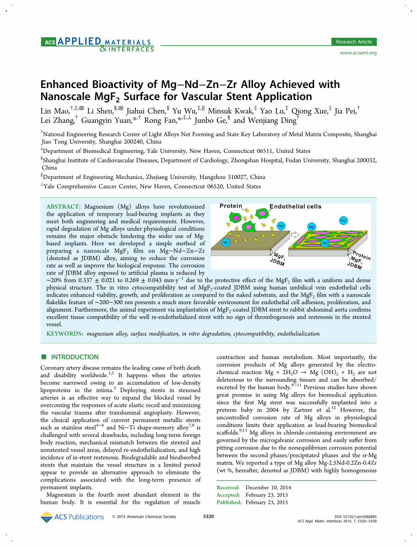
during the degradation process, we performed potentiodynamicpolarization measurement to further investigate the naked andMgF2-coated JDBM with regard to the corrosion current (icorr)and potential (Ecorr, Figure 4a). The potentiodynamic polar-ization curves of the two samples show the same trend: the icorrdecreases by ∼17% from 1.91 to 1.58 μA·cm−2 due to theprotective effect of the MgF2 film on the substrate (Table 1),indicating that JDBM alloy exhibits improved corrosionresistance after KF chemical conversion treatment. Further-more, the calculation result of the reduced corrosion current(∼17%) roughly agrees with the corrosion rate calculated bythe weight loss (∼20%) measurement. We also performed EIS
measurements to further investigate the corrosion mechanismof Mg alloy exposed to artificial plasma. Generally, the high-frequency semicircle in Nyquist plot can be attributed to thecharge transfer in combination with the corrosion products,while the low-frequency semicircle reveals the diffusion processthrough a porous layer.21,22 In comparison with the nakedsubstrate, the Nyquist plot of MgF2-coated JDBM exhibits asingle capacitive loop with an increased diameter (Figure 4b),reflecting that the MgF2 film serves as a passivating film and aneffective barrier layer with a low susceptibility to pittingcorrosion on the surface during the initial degradationprocess.23
Indirect Cell Cytotoxicity Assay. To explore the cellularresponse to the corrosion products of degradable Mg alloys, weexamined cell viability, morphology, and proliferation potentialof human endothelial cells exposed to Mg extracts. HUVECsincubated in Mg extracts for a period of 24 h were doublestained with YO-PRO-1 and propidium iodide (PI) andanalyzed with flow cytometry to qualify the viability in termsof the apoptosis and necrosis ratios. The percentage of normalhealthy cells for JDBM alloy increases from 69.5% to 89.4%after being coated with a protective MgF2 film, indicating thatthe protective film on the surface, which is well-tolerated byHUVECs, effectively inhibits the JDBM alloy from releasingexcess degradation ions via corrosion in cell culture medium(Figure 5a,b). The percentage of HUVECs in S phase (i.e.,DNA replication phase) through 24 h of incubation wasdetermined by 5-bromo-20-deoxy-uridine (BrdU) incorpora-tion, and the proliferation potential of HUVECs (96.75 ±12.14) cultured in the extract of coated JDBM is higher thanthat cultured in the naked JDBM extract (90.24 ± 9.66) andcomparable to the negative control (Figure 5c). We alsoperformed immunofluorescence characterization of HUVECmorphology by staining the cytoskeleton and nuclei. HUVECsspread and elongate in the Mg extracts and display a typicalmorphology of cobblestone, with no significant difference inmorphology as compared to the negative control cells (Figure
Figure 4. Electrochemical characterizations of JDBM and MgF2-coated JDBM degradation process. (a) Potentiodynamic polarization curves. (b)Nyquist plots.
Table 1. Corrosion Potential (Ecorr) and Current (icorr) ofJDBM and MgF2-Coated JDBM Tested by PotentiodynamicPolarization Measurement
samples Ecorr, V ICorr, ΜA·cm−2 RP, Ω·cm−2
JDBM −1.70 1.91 8.72 × 10⁴
MgF2-coated JDBM −1.66 1.58 2.21 × 10⁵
ACS Applied Materials & Interfaces Research Article
DOI: 10.1021/am5086885ACS Appl. Mater. Interfaces 2015, 7, 5320−5330
5323

5d−f). In brief, the indirect cell cytotoxicity cell assay indicatesthat primary human endothelial cells show enhanced viabilityand proliferation potential in modified JDBM extract ascompared to that incubated in the naked substrate extract.Direct Cell Adhesion Experiment. To evaluate the direct
response of human endothelial cells to the naked and MgF2-
coated JDBM alloy, HUVECs suspension at the cell density of2 × 105 cell·mL−1 was seeded onto the substrates. The two Mgalloys with different physicochemical properties give rise toquite significant differences in terms of cell adhesion, spreading,and proliferation (Figure 6a−d). A few cells are observed onthe naked JDBM surface after 24 h of culture, while the
Figure 5. In vitro cytocompatibility tests of Mg alloy extracts on human endothelial cells. The ratio of apoptotic or necrotic cells is analyzed by flowcytometry on HUVECs treated with (a) JDBM and (b) MgF2-coated JDBM extracts for 24 h. (c) Proliferation ratio of HUVECs cultured in JDBMand MgF2-coated JDBM extracts for 24 h as compared to the negative control. Immunofluorescence images showing the morphology of HUVACscultured in (d) normal cell culture medium and treated with (e) JDBM and (f) MgF2-coated JDBM extracts for 24 h. The cells were fixed,penetrated, and stained for cytoskeleton (phalloidin 532 nm: green) and nuclei (DRAQ5 635 nm: purple).
Figure 6. Cell attachment and elongation on Mg alloy substrates. HUVECs were seeded directly on (a, b) JDBM and (c, d) MgF2-coated JDBMsubstrates and cultured for 24 and 48 h. The adherent cells were fixed, penetrated, and stained for cytoskeleton (phalloidin 532 nm: green) andnuclei (DRAQ5 635 nm: purple). Statistical analysis of the adherent cells in terms of (e) cell number, (f) mean cell area, and (g) mean aspect ratio ofall the adherent HUVECs grown on the surfaces of JDBM and MgF2-coated JDBM substrates.
ACS Applied Materials & Interfaces Research Article
DOI: 10.1021/am5086885ACS Appl. Mater. Interfaces 2015, 7, 5320−5330
5324
adhesive cell density increases distinctly on the substrate ofmodified JDBM with a nanoscale flakelike MgF2 surface. We
extended the culture period to 48 h and observed obvious cellproliferation and elongated cell configuration with pseudopo-
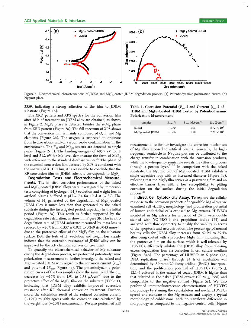
Figure 7. Four-week follow-up angiographic and the corresponding IVUS images of the abdominal aorta after (a, b) JDBM and (c, d) MgF2-coatedJDBM stent implantation. Note the longitudinal reconstruction of stented vessel and stent struts (arrow heads) at four-week implantation with anabsence of in-stent restenosis and neointimal hyperplasia.
Figure 8. HE staining images of the stented arteries showing compromised inflammation reactions and neointimal coverage on the JDBM and MgF2-coated JDBM struts in the period of (a, d) 2 d, (b, e) 7 d, and (c, f) 28 d of implantation. The white boxes (arrow heads) indicating the stent strutsapposed to the vessel wall.
ACS Applied Materials & Interfaces Research Article
DOI: 10.1021/am5086885ACS Appl. Mater. Interfaces 2015, 7, 5320−5330
5325

dium well-adhered to each other on the surface of modifiedJDBM, and the cell alignment is similar to those cultured onnormal tissue culture plate, indicating that the MgF2 film with ananoscale flakelike feature of ∼200−300 nm presents muchmore favorable surface for endothelial cell adhesion, spreading,and alignment. However, very few HUVECs adhere on thenaked JDBM substrate, and they appear to be rounded withinefficient cell spreading, indicating that rapid degradationmade the naked JDBM substrate difficult to support theadhesion and growth of primary endothelial cells. Quantitativeanalysis of cell number, surface area, and aspect ratio ofHUVECs grown on MgF2-coated JDBM was analyzed by ahigh-content image analysis program (Cell prolifer 2.0, theBroad institute of MIT, Figure 6e−g). The adhesive ability ofHUVECs is significantly improved as JDBM substrate is coatedwith MgF2 film. We also observed that the mean cell number,cell area, and aspect ratio of HUVECs increase significantlyranging from 480 ± 50 to 788 ± 77 cell·mm−2, from 1832.89 ±192.75 to 2670.32 ± 253.16 μm2, and from 1.58 ± 0.33 to 2019± 0.57, respectively, as endothelial cells were cultured on themodified JDBM substrate for 24 to 48 h of incubation. Thestatistics results confirm the morphological observation that thenanoscale flakelike MgF2 offers a more favorable surface foradhesion, spreading, and proliferation of HUVECs andpotentially aids in endothelialization of the stent into thedenuded artery.In Vivo Angiography and Intravascular Ultrasound
Findings. In a previous study, we demonstrated that the JDBMstent with excellent radial strength and compliance can becompletely expanded and well-apposed to the vessel wall, withno sign of early recoil or fracture at the initial implantation.14
Here we further performed in vivo angiography and the follow-up IVUS to investigate the biosafety and efficacy ofbiodegradable JDBM and MgF2-coated JDBM stents in rabbitabdominal aorta after implantation for 28 d (Figure 7). The
angiography images show no thrombosis and serious in-stentrestenosis in the naked and MgF2-coated JDBM stents (Figure7a,c), demonstrating that both the naked and modified stentsare safe and efficient in vivo. The follow-up IVUS was used toevaluate the expansion level, initial hyperplasia degree, and theoccurrences of thrombosis at 28 d post implantation, as shownin Figure 7b,d. Both the stents are well-apposed to the vesselwall with no evidence of strut fracture and in-stent restenosis,reflecting acceptable mechanical durability and excellent tissuecompatibility of the biodegradable stents after 28 d ofdegradation in vivo. Meanwhile, we observed a thin layer ofendothelium on the in-stent surface of the two stent struts at 28d post implantation (Figure 7b,d).
Histologic Observation and in-Stent Endothelializa-tion. We further performed HE staining to assess inflammatoryresponse to biodegradable JDBM and MgF2-coated JDBMstents in vivo (Figure 8). Histologic observation revealscompromised inflammation reactions with fewer inflammatorycell accumulation around MgF2-coated JDBM stent and a thincontinuous layer of neo-endothelium coverage on the in-stentsurface in the period of 2 d of implantation, while increasedinflammatory cells appears in the vicinity of the naked JDBMstent with partial coverage of neo-endothelium in all the threeperiods post implantation. A continuous and completeendothelium layer lines on the modified stent-supported arteryafter 28 d of implantation, indicating that MgF2 film aids in there-endothelialization process of JDBM stent into the denudedartery. The attenuation of the accumulation of inflammatorycells and the minor inflammatory reaction in the vicinity ofMgF2-coated JDBM stent can be reasonably attributed to theimproved re-endothelialization process on the in-stent surface,which has been demonstrated as the main underlyingmechanism of late stent thrombosis.24
We also investigated the distal segment of the stented arteryto identify in-stent endothelialization process at various periods
Figure 9. SEM images showing neointimal coverage of (a, b) JDBM stent and (c, d) MgF2-coated JDBM stent after 7 and 28 d of implantation.Complete neointimal coverage on the modified struts post-stenting can be finished after 28 d of implantation, and few inflammatory cells are visiblein the endothelialized stents.
ACS Applied Materials & Interfaces Research Article
DOI: 10.1021/am5086885ACS Appl. Mater. Interfaces 2015, 7, 5320−5330
5326

after implantation via SEM. A thin and noncontinuousendothelium layer covers on the surface of JDBM stent withinflammatory cell accumulation locally at 7 d post stenting,while obvious neointimal coverage on MgF2-coated JDBMstruts and fewer inflammatory cells are visible on theendothelialized stent (Figure 9a,c), reflecting that the nanoscaleflakelike MgF2 film has the potential to provide a favorablesurface suitable for the initial adhesion, proliferation, migration,and alignment of host endothelial cells. Results of this study,which correspond well with the finding of direct cell adhesionon Mg substrates (Figure 6), also indicate that completeneointimal coverage on MgF2-coated JDBM struts post-stenting can be achieved in the initial four weeks postimplantation as shown in Figure 9d. Hence, we can concludethat MgF2-coated JDBM stent causes less inflammatoryreaction in the early implantatin periods and also acceleratesthe re-endothelialization possess after stenting compared withthe naked JDBM stent. The major improvement inendothelialization process and inflammation inhibition can bereasonably attributed to the reduced degradation rate andenhanced biological activity of the MgF2 film on JDBMsubstrate surface, which demonstrates great potential of thenanoscale flakelike MgF2 coating for cardiovascular stentapplications.
■ DISCUSSION
Magnesium (Mg) alloys appear to be promising candidates astemporary structural biomaterial for orthopedic implants orcardiovascular stent applications due to the intriguingcombination properties of biocompatibility, biodegradability,and mechanical performance.12,14 Mg-based implants areexpected to be completely degraded in physiological environ-ment, and the degradation products should not pose threats tothe surrounding tissues.12 The temporary presence of Mg-basedimplants in the human body provides an avenue to overcomethe complications associated with permanent implants throughavoiding the long-term foreign body reaction and endothelialdysfunction. However, Mg alloys typically have low corrosionresistance under physiological conditions associated with arapid release of degradation ions, the formation of extensive gascavities, and early loosening of the implants, causing completefailure of the implant before the tissue has sufficientlyhealed.9,25,26
Surface modification such as surface coating/film is of greatsignificance and is a very attractive way to control the highcorrosion rate as well as improve the biocompatibility of Mgand its alloys.16,17 Chemical conversion film is an in situ growncoating that is produced by chemical or electrochemicaltreatment of a metal to produce a superficial layer that ischemically bonded to the substrate. Mg is known to be themost resistant metal to fluoride-conditioned solutions due tothe formation of a passivating protective layer on the metalsurface.27 In this study, we developed an eco-friendly surfacemodification to produce a fluoride film on the JDBM substratewith the reaction of this Mg alloy and 0.1 M KF solution for aperiod of 48 h. This conversion film is formed by the followingchemical reaction. The MgF2 film is grown directly on themetal substrate and is supposed to possess strong adhesiveability to the Mg alloy via atomic bond.
+ →+ −Mg 2F MgF22
The variation of the corrosion rates between the naked andmodified JDBM alloys is attributed to the protective effect ofMgF2 film, which shows a uniform and dense physical structure.The integrity of this barrier film enhances the corrosionresistance of JDBM alloy by preventing the substrate fromsevere eroding to release excess degradation ions inphysiological environment and, consequently, leads to enhance-ment in cellular response to the extract of MgF2-coated JDBMsubstrate (Figure 5). However, the conversion film MgF2 isinsoluble in aqueous solution but can be dissolved gradually inCl−-containing environment. The JDBM substrate begins todegrade when the electrolyte penetrates through the protectiveMgF2 film. Mg and its alloy are known to be highly reactive inchloride ion-rich solution. The standard electrochemicalpotential of Mg is −2.4 V (−1.5 V in aqueous solutions dueto the formation of a barrier film Mg(OH)2).
28 Generally, thesecond phases/precipitated phases in Mg alloys are more noblethan the α-Mg matrix, and hence, typical galvanic corrosion isthe prominent mechanism in Mg alloys due to the non-equilibrium corrosion potential between different constituentphases, resulting in severe pitting/local corrosion in Mgsubstrates. However, the main precipitated phase of extrudedJDBM alloy is Mg12Nd, and the potential difference betweenMg12Nd and the α-Mg matrix is very slight (∼25 mV),14,29
which is anticipated to slow down the galvanic corrosion andconsequently improve the homogeneous degradation of thesubstrate. Thus, the uniform corrosion behavior of the JDBMsubstrate, which can avoid local stress concentration and rapiddecrease of mechanical strength of the implants, is highlydesired for load-bearing medical device applications.The surface physicochemical properties of implanted
materials have significant effects upon production of localmicroenvironments, which profoundly influence cellularbehavior in terms of adhesion, spreading, intracellular signaling,and differentiation potential.3 Because of rapid kinetics,biomaterials and medical devices exposed to blood immediatelyacquire a layer of plasma and extracellular matrix proteins priorto interacting with host cells.30 Although the corrodibility ofMg alloys are desirable for temporary biodegradable implantapplications, the surfaces of Mg-based implants are highlyreactive in physiological medium, which may have adverseeffect on the formation of a transient provisional matrix on andaround the implants as well as the outcome of the subsequentcell colonization.31,32 Hence, further surface modifications arenecessary with respect to the improvement of surface stabilityand biological compatibility. Existing data supporting thepositive role of nanostructured metals in biological applicationsuggest that nanoscale modification of implant surface has thepotential to yield a faster and more stable integration ofbiomedical implants with greater biological activity.33 It isreported that a laser-nanostructured polystyrene surface with aperiodic feature of 200−430 nm can significantly enhanceadhesion and proliferation of mammalian cells,34 and thetopography cues down to 200 nm have adverse effect onprimary corneal and human corneal epithelial cell (SV40-HCEC) proliferation.35 In this work, we prepared a MgF2 filmwith a nanoscale flakelike feature of ∼200−300 nm on JDBMsubstrate surface with a simple chemical conversion treatmentand demonstrated that the MgF2 film favors HUVECsdeposition, proliferation, and alignment and potentially aidsin re-endothelialization of the implant into the denuded artery.Enhanced endothelial cell adhesion, proliferation, and align-ment has been suggested as a method for increasing efficacy of
ACS Applied Materials & Interfaces Research Article
DOI: 10.1021/am5086885ACS Appl. Mater. Interfaces 2015, 7, 5320−5330
5327
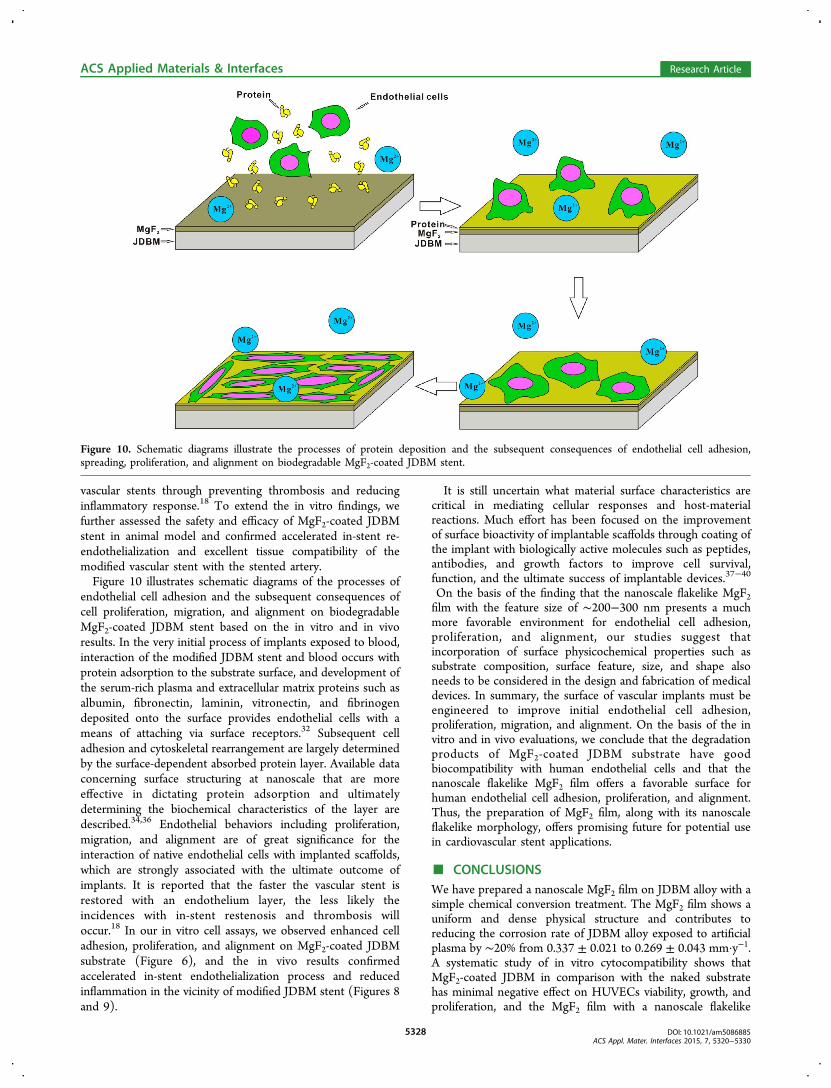
vascular stents through preventing thrombosis and reducinginflammatory response.18 To extend the in vitro findings, wefurther assessed the safety and efficacy of MgF2-coated JDBMstent in animal model and confirmed accelerated in-stent re-endothelialization and excellent tissue compatibility of themodified vascular stent with the stented artery.Figure 10 illustrates schematic diagrams of the processes of
endothelial cell adhesion and the subsequent consequences ofcell proliferation, migration, and alignment on biodegradableMgF2-coated JDBM stent based on the in vitro and in vivoresults. In the very initial process of implants exposed to blood,interaction of the modified JDBM stent and blood occurs withprotein adsorption to the substrate surface, and development ofthe serum-rich plasma and extracellular matrix proteins such asalbumin, fibronectin, laminin, vitronectin, and fibrinogendeposited onto the surface provides endothelial cells with ameans of attaching via surface receptors.32 Subsequent celladhesion and cytoskeletal rearrangement are largely determinedby the surface-dependent absorbed protein layer. Available dataconcerning surface structuring at nanoscale that are moreeffective in dictating protein adsorption and ultimatelydetermining the biochemical characteristics of the layer aredescribed.34,36 Endothelial behaviors including proliferation,migration, and alignment are of great significance for theinteraction of native endothelial cells with implanted scaffolds,which are strongly associated with the ultimate outcome ofimplants. It is reported that the faster the vascular stent isrestored with an endothelium layer, the less likely theincidences with in-stent restenosis and thrombosis willoccur.18 In our in vitro cell assays, we observed enhanced celladhesion, proliferation, and alignment on MgF2-coated JDBMsubstrate (Figure 6), and the in vivo results confirmedaccelerated in-stent endothelialization process and reducedinflammation in the vicinity of modified JDBM stent (Figures 8and 9).
It is still uncertain what material surface characteristics arecritical in mediating cellular responses and host-materialreactions. Much effort has been focused on the improvementof surface bioactivity of implantable scaffolds through coating ofthe implant with biologically active molecules such as peptides,antibodies, and growth factors to improve cell survival,function, and the ultimate success of implantable devices.37−40
On the basis of the finding that the nanoscale flakelike MgF2film with the feature size of ∼200−300 nm presents a muchmore favorable environment for endothelial cell adhesion,proliferation, and alignment, our studies suggest thatincorporation of surface physicochemical properties such assubstrate composition, surface feature, size, and shape alsoneeds to be considered in the design and fabrication of medicaldevices. In summary, the surface of vascular implants must beengineered to improve initial endothelial cell adhesion,proliferation, migration, and alignment. On the basis of the invitro and in vivo evaluations, we conclude that the degradationproducts of MgF2-coated JDBM substrate have goodbiocompatibility with human endothelial cells and that thenanoscale flakelike MgF2 film offers a favorable surface forhuman endothelial cell adhesion, proliferation, and alignment.Thus, the preparation of MgF2 film, along with its nanoscaleflakelike morphology, offers promising future for potential usein cardiovascular stent applications.
■ CONCLUSIONSWe have prepared a nanoscale MgF2 film on JDBM alloy with asimple chemical conversion treatment. The MgF2 film shows auniform and dense physical structure and contributes toreducing the corrosion rate of JDBM alloy exposed to artificialplasma by ∼20% from 0.337 ± 0.021 to 0.269 ± 0.043 mm·y−1.A systematic study of in vitro cytocompatibility shows thatMgF2-coated JDBM in comparison with the naked substratehas minimal negative effect on HUVECs viability, growth, andproliferation, and the MgF2 film with a nanoscale flakelike
Figure 10. Schematic diagrams illustrate the processes of protein deposition and the subsequent consequences of endothelial cell adhesion,spreading, proliferation, and alignment on biodegradable MgF2-coated JDBM stent.
ACS Applied Materials & Interfaces Research Article
DOI: 10.1021/am5086885ACS Appl. Mater. Interfaces 2015, 7, 5320−5330
5328

feature of ∼200−300 nm offers a much more favorable surfacefor endothelial cell adhesion, proliferation, and alignment.Furthermore, JDBM vascular stent coated with MgF2 filmimplanted in rabbit abdominal aorta confirms excellent tissuecompatibility of the well re-endothelialized stent with no sign ofthrombogenesis and restenosis in the stent-supported vessel.Therefore, the nanoscale flakelike MgF2 film exhibitssignificantly enhanced performance of JDBM alloy with regardto degradation rate and biocompatibility and shows greatpotential for cardiovascular stent applications.
■ AUTHOR INFORMATIONCorresponding Authors*E-mail: [email protected]. (G.Y.Y.)*E-mail: [email protected]. (R.F.)
Author ContributionsL.M. and G.-Y.Y. designed experiments for materials synthesis,surface modification, and degradation characterizations. L.M.and R.F. designed the in vitro cell assays. L.M., Y.W., and Q.X.performed in vitro cell assay experiments and analyzed data.L.S. and J.-H.C. performed the animal experiments, the relevantdata collection, and data analysis. All the authors contributed tothe writing of manuscript.
Author Contributions⊗L.M. and L.S. contributed equally to this work.
NotesThe authors declare no competing financial interest.
■ ACKNOWLEDGMENTSThis work was supported by the Yale University new facultystartup fund (PI: R.F.). Research at Shanghai Jiao TongUniversity was supported by the National Key TechnologyR&D Program of the Ministry of Science and Technology(2012BAI18B01), China Postdoctoral Science Foundationfunded project (2012M520892). This project was alsosupported by the National Basic Research Program (“973”Program) of China (2011CB503905) and the National NaturalScience Foundation of China (81370323 and 11402227).
■ REFERENCES(1) Go, A. S.; Mozaffarian, D.; Roger, V. L.; Benjamin, E. J.; Berry, J.D.; Blaha, M. J.; Dai, S.; Ford, E. S.; Fox, C. S.; Franco, S. HeartDisease and Stroke Statistics–2014 Update: A Report from theAmerican Heart Association. Circulation 2014, 129, 399−410.(2) Liu, M. B.; Wang, W.; Zhou, M. G. Trend Analysis on theMortality of Cardiovascular Diseases from 2004 to 2010 in China.Chin. J. Epidemiol. 2013, 34, 985−988.(3) Witztum, J. L.; Steinberg, D. Role of Oxidized Low DensityLipoprotein in Atherogenesis. J. Clin. Invest. 1991, 88, 1785.(4) Harbuzariu, A.; Dragomir-Daescu, D.; Gooden, J.; Holmes, D.;Simari, R.; Sandhu, G. New Duplex-alloy Bare Metal Stent EnablesMagnetic Capture of Endothelial Cells and Reduces NeointimalResponse to Injury. J. Am. Coll. Cardiol. 2012, 59, E59−E59.(5) Kaiser, C.; Galatius, S.; Erne, P.; Eberli, F.; Alber, H.; Rickli, H.;Pedrazzini, G.; Hornig, B.; Bertel, O.; Bonetti, P. Drug-eluting VersusBare-metal Stents in Large Coronary Arteries. New Engl. J. Med. 2010,363, 2310−2319.(6) Yang, Z.; Wang, J.; Luo, R.; Maitz, M. F.; Jing, F.; Sun, H.;Huang, N. The Covalent Immobilization of Heparin to Pulsed-plasmaPolymeric Allylamine Films on 316L Stainless Steel and the ResultingEffects on Hemocompatibility. Biomaterials 2010, 31, 2072−2083.(7) Trepanier, C.; Leung, T.; Tabrizian, M.; Yahia, L. H.; Bienvenu, J.G.; Tanguay, J. F.; Piron, D.; Bilodeau, L. Preliminary Investigation of
the Effects of Surface Treatments on Biological Response to ShapeMemory NiTi Stents. J. Biomed. Mater. Res. 1999, 48, 165−171.(8) Trepanier, C.; Tabrizian, M.; Yahia, L. H.; Bilodeau, L.; Piron, D.L. Effect of Modification of Oxide Layer on NiTi Stent CorrosionResistance. J. Biomed. Mater. Res. 1998, 43, 433−440.(9) Staiger, M. P.; Pietak, A. M.; Huadmai, J.; Dias, G. Magnesiumand its Alloys as Orthopedic Biomaterials: a Review. Biomaterials 2006,27, 1728−1734.(10) Zeng, R.; Dietzel, W.; Witte, F.; Hort, N.; Blawert, C. Progressand Challenge for Magnesium Alloys as Biomaterials. Adv. Eng. Mater.2008, 10, B3−B14.(11) Zhang, X.; Li, X.-W.; Li, J.-G.; Sun, X.-D. Preparation andCharacterizations of Bioglass Ceramic Cement/Ca−P Coating onPure Magnesium for Biomedical Applications. ACS Appl. Mater.Interfaces 2013, 6, 513−525.(12) Zartner, P.; Cesnjevar, R.; Singer, H.; Weyand, M. FirstSuccessful Implantation of a Biodegradable Metal Stent into the LeftPulmonary Artery of a Preterm Baby. Catheter. Cardiovasc. Interventions2005, 66, 590−594.(13) Witte, F.; Kaese, V.; Haferkamp, H.; Switzer, E.; Meyer-Lindenberg, A.; Wirth, C.; Windhagen, H. In vivo Corrosion of FourMagnesium Alloys and the Associated Bone Response. Biomaterials2005, 26, 3557−3563.(14) Mao, L.; Shen, L.; Niu, J.; Zhang, J.; Ding, W.; Wu, Y.; Fan, R.;Yuan, G. Nanophasic Biodegradation Enhances the Durability andBiocompatibility of Magnesium Alloys for the Next-GenerationVascular Stents. Nanoscale 2013, 5, 9517−9522.(15) Eley, E., Next-Generation Vascular Stents. Chemistry World.2013. http://www.rsc.org/chemistryworld/2013/08/biodegradable-vascular-stent-atherosclerosis.(16) Ye, S. H.; Jang, Y. S.; Yun, Y. H.; Shankarraman, V.; Woolley, J.R.; Hong, Y.; Gamble, L. J.; Ishihara, K.; Wagner, W. R. SurfaceModification of a Biodegradable Magnesium Alloy with Phosphor-ylcholine (PC) and Sulfobetaine (SB) Functional Macromolecules forReduced Thrombogenicity and Acute Corrosion Resistance. Langmuir2013, 29, 8320−8327.(17) Zomorodian, A.; Garcia, M.; Moura e Silva, T.; Fernandes, J.;Fernandes, M.; Montemor, M. Corrosion Resistance of a CompositePolymeric Coating Applied on Biodegradable AZ31 Magnesium Alloy.Acta Biomater. 2013, 9, 8660−8670.(18) Pislaru, S. V.; Harbuzariu, A.; Agarwal, G.; LATG, T. W. A. C.;Gulati, R.; Sandhu, N. P.; AA, C. M.; Kalra, M.; Simari, R. D.; Sandhu,G. S. Magnetic Forces Enable Rapid Endothelialization of SyntheticVascular Grafts. Circulation 2006, 114, I-314−I-318.(19) Song, G.; Atrens, A. Understanding Magnesium CorrosionaFramework for Improved Alloy Performance. Adv. Eng. Mater. 2003, 5,837−858.(20) Moulder, J. F.; Stickle, W. F.; Sobol, P. E.; Bomben, K. D.Handbook of X-ray Photoelectron Spectroscopy; Perkin Elmer: EdenPrairie, MN, 1992; Vol. 40.(21) Deslouis, C.; Duprat, M.; Tulet-Tournillon, C. The CathodicMass Transport Process During Zinc Corrosion in Neutral AeratedSodium Sulphate Solutions. J. Electroanal. Chem. Interfacial Electrochem.1984, 181, 119−136.(22) Park, J.; Macdonald, D. Impedance Studies of the Growth ofPorous Magnetite Films on Carbon Steel in High TemperatureAqueous Systems. Corros. Sci. 1983, 23, 295−315.(23) Brett, C.; Dias, L.; Trindade, B.; Fischer, R.; Mies, S.Characterisation by EIS of Ternary Mg Alloys Synthesised byMechanical Alloying. Electrochim. Acta 2006, 51, 1752−1760.(24) Christiansen, E. H.; Jensen, L. O.; Thayssen, P.; Tilsted, H.-H.;Krusell, L. R.; Hansen, K. N.; Kaltoft, A.; Maeng, M.; Kristensen, S. D.;Bøtker, H. E. Biolimus-Eluting Biodegradable Polymer-Coated StentVersus Durable Polymer-Coated Sirolimus-Eluting Stent in UnselectedPatients Receiving Percutaneous Coronary Intervention (SORT OUTV): a Randomised Non-Inferiority Trial. Lancet 2013, 381, 661−669.(25) Choudhary, L.; Singh Raman, R. Magnesium Alloys as BodyImplants: Fracture Mechanism under Dynamic and Static Loadings ina Physiological Environment. Acta Biomater. 2012, 8, 916−923.
ACS Applied Materials & Interfaces Research Article
DOI: 10.1021/am5086885ACS Appl. Mater. Interfaces 2015, 7, 5320−5330
5329
(26) Kannan, M. B.; Raman, R. In vitro Degradation and MechanicalIntegrity of Calcium-Containing Magnesium Alloys in Modified-Simulated Body Fluid. Biomaterials 2008, 29, 2306−2314.(27) Witte, F.; Fischer, J.; Nellesen, J.; Vogt, C.; Vogt, J.; Donath, T.;Beckmann, F. In vivo Corrosion and Corrosion Protection ofMagnesium Alloy LAE442. Acta Biomater. 2010, 6, 1792−1799.(28) Ambat, R.; Aung, N. N.; Zhou, W. Evaluation of MicrostructuralEffects on Corrosion Behaviour of AZ91D Magnesium Alloy. Corros.Sci. 2000, 42, 1433−1455.(29) Coy, A.; Viejo, F.; Skeldon, P.; Thompson, G. Susceptibility ofRare-Earth-Magnesium Alloys to Micro-galvanic Corrosion. Corros. Sci.2010, 52, 3896−3906.(30) Anderson, J. M. Biological Responses to Materials. Annu. Rev.Mater. Res. 2001, 31, 81−110.(31) Anderson, J. M.; McNally, A. K. In Biocompatibility of Implants:Lymphocyte/Macrophage Interactions, Seminars in immunopathology;Zimmerli, W., Ed.; Springer: Berlin, Germany, 2011; pp 221−233.(32) Anderson, J. M.; Patel, J. D. Biomaterial-Dependent Character-istics of the Foreign Body Response and S. epidermidis BiofilmInteractions. Biomaterials Associated Infection; Moriarty, T. F., Zaat, S.A. J., Busscher, H. J., Eds.; Springer: Berlin, Germany, 2013; pp 119−149.(33) Variola, F.; Brunski, J. B.; Orsini, G.; de Oliveira, P. T.; Wazen,R.; Nanci, A. Nanoscale Surface Modifications of Medically RelevantMetals: State-of-the Art and Perspectives. Nanoscale 2011, 3, 335−353.(34) Rebollar, E.; Frischauf, I.; Olbrich, M.; Peterbauer, T.; Hering,S.; Preiner, J.; Hinterdorfer, P.; Romanin, C.; Heitz, J. Proliferation ofAligned Mammalian Cells on Laser-Nanostructured Polystyrene.Biomaterials 2008, 29, 1796−1806.(35) Liliensiek, S. J.; Campbell, S.; Nealey, P. F.; Murphy, C. J. TheScale of Substratum Topographic Features Modulates Proliferation ofCorneal Epithelial Cells and Corneal Fibroblasts. J. Biomed. Mater. Res.,Part A 2006, 79, 185−192.(36) Mendonca̧, G.; Mendonca, D.; Aragao, F. J.; Cooper, L. F.Advancing Dental Implant Surface Technology−from Micron-toNanotopography. Biomaterials 2008, 29, 3822−3835.(37) Dean, J., 3rd; Culbertson, K. C.; D’Angelo, A. M. Fibronectinand Laminin Enhance Gingival Cell Attachment to Dental ImplantSurfaces in vitro. Int. J. Oral Maxillofac. Implants 1994, 10, 721−728.(38) Yazici, H.; Fong, H.; Wilson, B.; Oren, E.; Amos, F.; Zhang, H.;Evans, J.; Snead, M.; Sarikaya, M.; Tamerler, C. Biological Responseon a Titanium Implant-Grade Surface Functionalized with ModularPpeptides. Acta Biomater. 2013, 9, 5341−5352.(39) Liao, K.-H.; Lin, Y.-S.; Macosko, C. W.; Haynes, C. L.Cytotoxicity of Graphene Oxide and Graphene in HumanErythrocytes and Skin Fibroblasts. ACS Appl. Mater. Interfaces 2011,3, 2607−2615.(40) Yan, X.; Chen, J.; Yang, J.; Xue, Q.; Miele, P. Fabrication ofFree-Standing, Electrochemically Active, and Biocompatible GrapheneOxide−Polyaniline and Graphene−Polyaniline Hybrid Papers. ACSAppl. Mater. Interfaces 2010, 2, 2521−2529.
ACS Applied Materials & Interfaces Research Article
DOI: 10.1021/am5086885ACS Appl. Mater. Interfaces 2015, 7, 5320−5330
5330