Embed Size (px)
Citation preview
ENGL
ISH

2 3
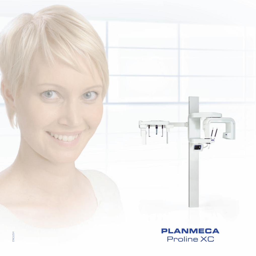
Since their introduction, the Planmeca Proline series of panoramic X-ray units sets standards for high-quality, practical, and user-friendly extraoral dental X-ray units. Innovative technical solutions, easy patient positioning, and exceptionally clear radiographs have made Planmeca Proline X-ray units incredibly popular among dental professionals. Today, there are more than 30,000 units installed all over the world.
Leader in dental X-ray units
4 5
* Standard Forms of Dentition and Mandible for Applications in Rotational Panoramic Radiography, U. Welander, P. Nummikoski, G. Tronje, W.D. McDavid, P.E. Legrell, and R.P. Langlais, Dento-Maxillofacial Radiology, 1989, Vol. 18, May
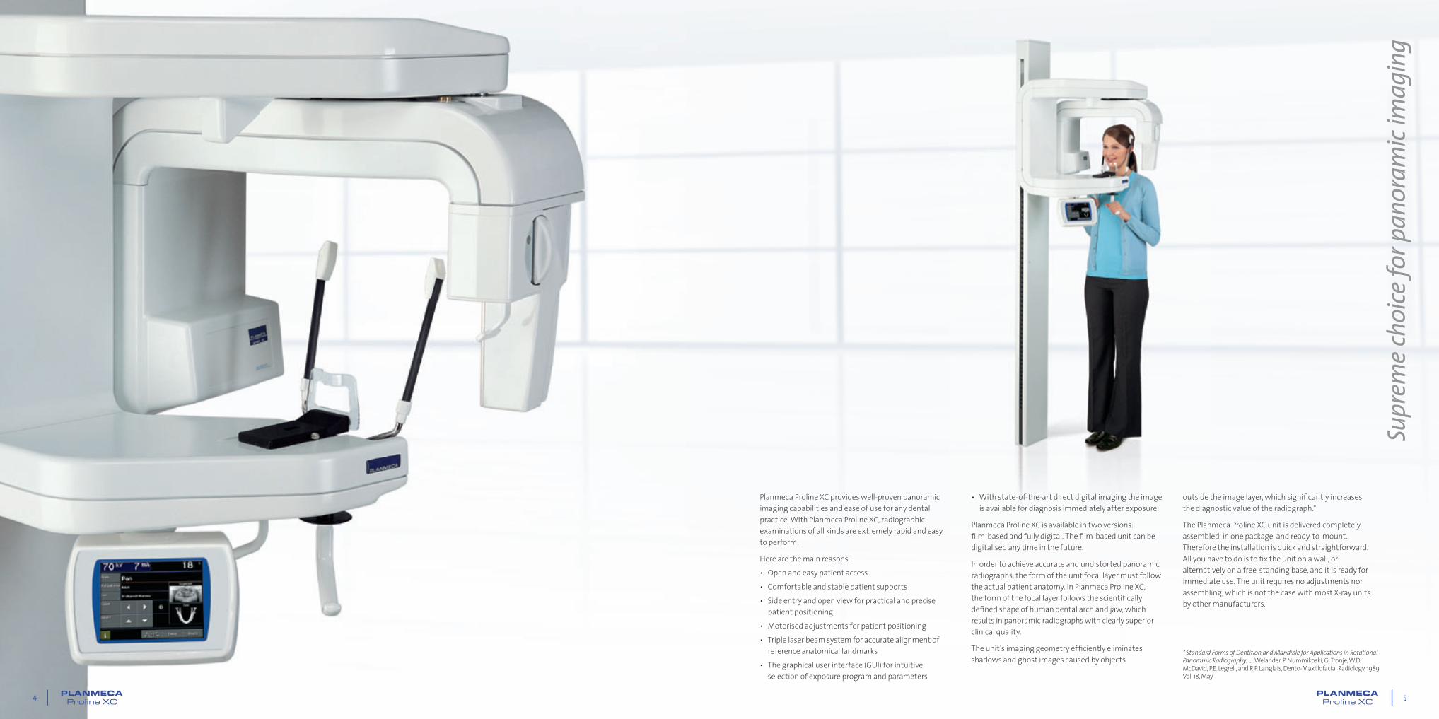
Planmeca Proline XC provides well-proven panoramic imaging capabilities and ease of use for any dental practice. With Planmeca Proline XC, radiographic examinations of all kinds are extremely rapid and easy to perform.
Here are the main reasons:• Open and easy patient access• Comfortable and stable patient supports• Side entry and open view for practical and precise
patient positioning• Motorised adjustments for patient positioning• Triple laser beam system for accurate alignment of
reference anatomical landmarks• The graphical user interface (GUI) for intuitive
selection of exposure program and parameters
• With state-of-the-art direct digital imaging the image is available for diagnosis immediately after exposure.
Planmeca Proline XC is available in two versions: fi lm-based and fully digital. The fi lm-based unit can be digitalised any time in the future.
In order to achieve accurate and undistorted panoramic radiographs, the form of the unit focal layer must follow the actual patient anatomy. In Planmeca Proline XC, the form of the focal layer follows the scientifi cally defi ned shape of human dental arch and jaw, which results in panoramic radiographs with clearly superior clinical quality.
The unit's imaging geometry effi ciently eliminates shadows and ghost images caused by objects
outside the image layer, which signifi cantly increases the diagnostic value of the radiograph.*
The Planmeca Proline XC unit is delivered completely assembled, in one package, and ready-to-mount. Therefore the installation is quick and straightforward. All you have to do is to fi x the unit on a wall, or alternatively on a free-standing base, and it is ready for immediate use. The unit requires no adjustments nor assembling, which is not the case with most X-ray units by other manufacturers.
Supr
eme c
hoice
for p
anor
amic
imag
ing

6 7
In Planmeca Proline XC, the side entry and the open positioning concept minimise errors caused by incorrect patient positioning, one of the most frequent reasons for failed radiographs. The operator can monitor the patient freely from the front, back, and side, making patient positioning quick, precise, and easy.
A triple laser beam system accurately indicates the correct anatomical positioning points. Here is how it works: • The midsagittal plane positioning beam shows
the correct sideways alignment of the patient's head. The image will be symmetric and undistorted in left-right direction.
• The Frankfort horizontal plane positioning beam shows the correct forward tilt of the patient's head. On the image, the teeth will line up straight.
• The focal layer positioning beam indicates the focal layer's position in the incisor region, helping in positioning the patient fully inside the focal layer for sharp and clear images.
Side entry allows easy access to the X-ray unit for all types of patients. The exposure can be performed with a standing patient – the recommended way for short procedures – or a seated patient.
It is also possible to take an exposure of a patient seated on a wheelchair or a hospital bed with upright lifted backrest. With Planmeca Proline XC, no mirrors are
Succ
essfu
l imag
es ev
ery s
ingl
e tim
e
needed for positioning. Instead, the patient has an open and comfortable view, so that for instance a child can see an accompanying adult throughout the procedure.
Each patient is an individual whose bone and tissue thickness varies according to his/her size, race, and age. The digital Planmeca Proline XC unit has the unique Automatic Gain Control (AGC), which optimises the sensitivity of the digital sensor to produce optimum image quality from each individual.
The Planmeca Proline XC fi lm unit can be equipped with the optional Automatic Exposure Control (AEC), which measures the patient's radiation transparency and correctly adjusts exposure values to achieve the desired fi lm darkness and contrast.
* Absorbed dose reduced by sliced exposure using sector selector system with rotational panoramic radiography, Y. Hayakawa, N. Kobayashi, Y. Kousuge, H. Fujimori, and K. Kuroyanagi, Bulletin of Tokyo Dental College, Vol. 35, No. 3, pp. 127–131, August, 1994
Planmeca Proline XC allows the selection of the correct exposure format, minimising the radiation dose for all types of patients and diagnostic purposes.
The Pediatric program automatically selects a reduced area for the exposure. This results in 20% lower patient dosage, without loss of diagnostic information.
With the Vertical Segmenting program, the exposed area can be limited only to the area of diagnostic interest. A simple selection on the main display, and the patient dose can be reduced by up to 80% compared to a full area panoramic exposure. This is highly advantageous and radiation hygienic in cases where a follow-up image is needed of a limited part of the jaw.*
In Planmeca Proline XC, the Sinus program has a specially designed image layer, which results in a radiograph with a clear view of the maxillary sinuses.
The automatic Double TMJ program produces a lateral view of open and closed temporomandibular joints on one radiograph. The imaging procedure is straightforward, and the radiograph provides easy diagnosis of the TMJ condition in one view.
The True Profi le TMJ program is an optional program that enables you to adjust optimally the TMJ imaging angles for each individual patient. It also produces specifi c perpendicular radiographic projections of the condyles.
Func
tiona
l cho
ice of
expo
sure
prog
ram
s
Standard Panoramic, segmentedSinus
True Profi le TMJStandard Panoramic
Automatic Double TMJ
Standard Pediatric
8 9
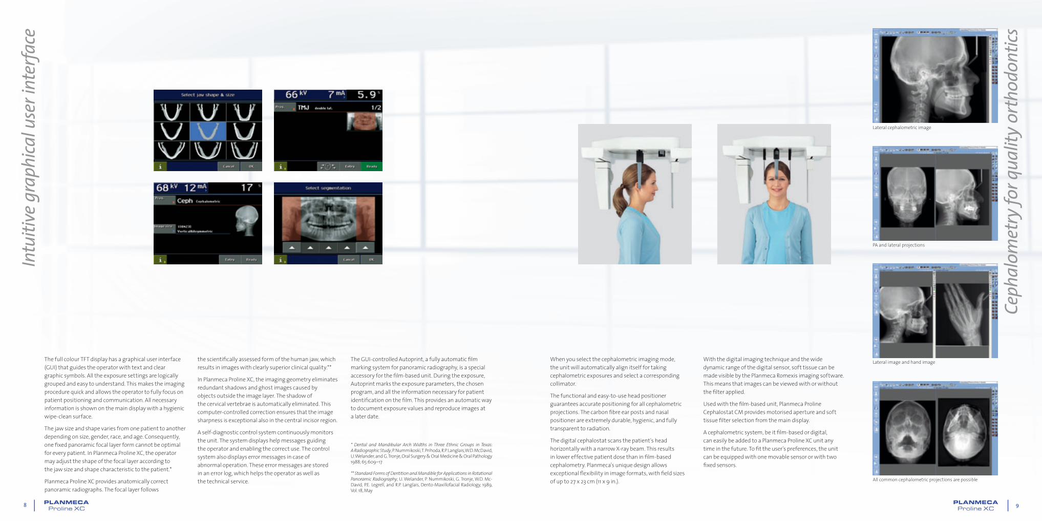
When you select the cephalometric imaging mode, the unit will automatically align itself for taking cephalometric exposures and select a corresponding collimator.
The functional and easy-to-use head positioner guarantees accurate positioning for all cephalometric projections. The carbon fi bre ear posts and nasal positioner are extremely durable, hygienic, and fully transparent to radiation.
The digital cephalostat scans the patient's head horizontally with a narrow X-ray beam. This results in lower effective patient dose than in fi lm-based cephalometry. Planmeca's unique design allows exceptional fl exibility in image formats, with fi eld sizes of up to 27 x 23 cm (11 x 9 in.).
With the digital imaging technique and the wide dynamic range of the digital sensor, soft tissue can be made visible by the Planmeca Romexis imaging software. This means that images can be viewed with or without the fi lter applied.
Used with the fi lm-based unit, Planmeca Proline Cephalostat CM provides motorised aperture and soft tissue fi lter selection from the main display.
A cephalometric system, be it fi lm-based or digital, can easily be added to a Planmeca Proline XC unit any time in the future. To fi t the user's preferences, the unit can be equipped with one movable sensor or with two fi xed sensors.
Intu
itive
grap
hica
l use
r int
erfa
ce
Ceph
alom
etry
for q
ualit
y orth
odon
tics
* Dental and Mandibular Arch Widths in Three Ethnic Groups in Texas: A Radiographic Study, P. Nummikoski, T. Prihoda, R.P. Langlais, W.D. McDavid, U. Welander, and G. Tronje, Oral Surgery & Oral Medicine & Oral Pathology 1988; 65:609–17
** Standard Forms of Dentition and Mandible for Applications in Rotational Panoramic Radiography, U. Welander, P. Nummikoski, G. Tronje, W.D. Mc-David, P.E. Legrell, and R.P. Langlais, Dento-Maxillofacial Radiology, 1989, Vol. 18, May
The full colour TFT display has a graphical user interface (GUI) that guides the operator with text and clear graphic symbols. All the exposure settings are logically grouped and easy to understand. This makes the imaging procedure quick and allows the operator to fully focus on patient positioning and communication. All necessary information is shown on the main display with a hygienic wipe-clean surface.
The jaw size and shape varies from one patient to another depending on size, gender, race, and age. Consequently, one fi xed panoramic focal layer form cannot be optimal for every patient. In Planmeca Proline XC, the operator may adjust the shape of the focal layer according to the jaw size and shape characteristic to the patient.*
Planmeca Proline XC provides anatomically correct panoramic radiographs. The focal layer follows
the scientifi cally assessed form of the human jaw, which results in images with clearly superior clinical quality.**
In Planmeca Proline XC, the imaging geometry eliminates redundant shadows and ghost images caused by objects outside the image layer. The shadow of the cervical vertebrae is automatically eliminated. This computer-controlled correction ensures that the image sharpness is exceptional also in the central incisor region.
A self-diagnostic control system continuously monitors the unit. The system displays help messages guiding the operator and enabling the correct use. The control system also displays error messages in case of abnormal operation. These error messages are stored in an error log, which helps the operator as well as the technical service.
The GUI-controlled Autoprint, a fully automatic fi lm marking system for panoramic radiography, is a special accessory for the fi lm-based unit. During the exposure, Autoprint marks the exposure parameters, the chosen program, and all the information necessary for patient identifi cation on the fi lm. This provides an automatic way to document exposure values and reproduce images at a later date.
Lateral cephalometric image
PA and lateral projections
Lateral image and hand image
All common cephalometric projections are possible

10 11
Tech
nica
l spe
cifi ca
tions
Planmeca Proline XC with fi lm-based cephalostat
Planmeca Proline XC with digital cephalostat
Physical space requirementsWeight
Width Depth Height
Planmeca Proline XC Panoramic153 cm (60 in.)
103.5 cm(40.5 in.)
220 cm(86.5 in.)
108 kg(lbs 237)
Planmeca Proline XC Panoramic with Autoprint 163 cm(64 in.)
103.5 cm(40.5 in.)
220 cm(86.5 in.)
112 kg(lbs 249)
Planmeca Proline XC with Cephalostat 223 cm(88 in.)
103.5 cm(40.5 in.)
220 cm(86.5 in.)
126 kg(lbs 278)
2200
(86
.5")
2230 (88")1140 (45")
540 (21")440
(17")
1090 (43")730 (29") 360
(14")
830 (32.5")
930
(36.
5")
2030
– 2
130
(80"
– 8
3.75
")
2200
(86
.5")
1035
(40
.5")
720
(28.
3")
930
(36.
5")
1035
(40
.5")
830 (32.5")
2210 (87") with Cephalostat1120 (44")
540 (21")
440 (17")
1090 (43")
730 (29") 360 (14")
1630 (64") with Autoprint
1530 (60") standard Panoramic
Planmeca Proline XC
Generator Constant potential, microprocessor controlled, operating frequency 80 kHz
X-ray tube D-052SB
Focal spot size 0.5 x 0.5 mm according to IEC 336
Total filtration 2.5 mm Al
Anode voltage 60–80 kV
Anode current 4–12 mA DC
Exposure time Pan 2.5–18 s
Ceph 0.2–23 s
Film size Pan 15 x 30 cm12.5 x 30 cm
Ceph 18 x 24 cm8 x 10 in.
Cassette Flat
SID Pan 480 mm (19 in.)
Ceph 163–170 cm (64–67 in.)
Magnification Pan constant 1.2
Ceph 1.08–1.13
Line voltage 100/117/220–230/240 V, 50 or 60 Hz
Regulation Automatic, ±10%
Line current 8–16 A
Colour White (RAL 9016) 620
(24.
4”)
Planmeca Romexis software
Planmeca Romexis is a complete dental imaging software, including all dental imaging modalities: intraoral, panoramic, cephalometric, 3D imaging, dental tomography as well as intraoral video and still camera imaging. With a complete set of tools for image viewing, enhancement, measurements, and annotations, Planmeca Romexis also improves the diagnostic value of radiographs. Printing, image import and export, and DICOM functionalities are also included.
Planmeca Romexis platform fully integrates digital imaging with the patient’s other clinical data. The system provides direct image capture from Planmeca’s X-ray equipment, and interfaces with 3rd party devices via TWAIN. Together with Planmeca’s X-ray equipment, Planmeca Romexis provides a unique safety feature especially useful for teaching environment: the X-ray image capture is inhibited until the supervisor has approved the student’s image capture request.
Planmeca Romexis computer recommendations
Planmeca Romexis client work station
Planmeca Romexis server
Processor 1 GHz 2 GHz
RAM 1 GB 2 GB
Hard disk space 40 GB 160 GB
Monitor 1280 x 1024 1024 x 768
Peripherals CD R/W or DVD R/W drive CD R/W or DVD R/W drive
Backup medium None necessary DAT or equivalent
Operating system Windows XPWindows 2003 ServerWindows VistaWindows 7Mac OS X
Windows XP ProWindows 2003 ServerWindows VistaWindows 7
Other Java platform (Java Virtual Machine 1.6 or later)
Java platform (Java Virtual Machine 1.6 or later)
The disk space requirements are determined by digital images. Thus the space requirements vary, but a rough estimate is in the order of 1 MB per 2D X-ray image, 7–9 MB per extraoral image, depending on a variety of image specifi c factors, and 250 MB per 3D image.
It is recommended to use the same computer as an application server and as a database server. If Planmeca Romexis server computer is also used for client activities, the hardware should meet both client and server specifi cations.
These specifi cations are recommended minimum requirements. Not meeting them may lead to degraded performance.
DICOM compatibility• Media Storage – saving images into removable DICOM media • Print – printing images on fi lm or paper with a DICOM medical printer• Storage – saving images into DICOM image archive • Query/ Retrieve – importing digital images from DICOM image archive• Worklist – importing a patient list from DICOM patient management• Storage Commitment – confi rmation of a successful image storage

10016
317
/0310
/en
Planmeca OyAsentajankatu 6 | 00880 Helsinki | Finlandtel. +358 20 7795 500 | fax +358 20 7795 555
[email protected] | www.planmeca.com
Planmeca Oy designs and manufactures a full line of high technology dental equipment, including dental care units, panoramic and intraoral X-ray units, and digital imaging products. Planmeca Oy, the parent company of the Finnish Planmeca Group,
is strongly committed to R&D, and is the largest privately held company in the field.
Images may contain optional items not included in standard delivery. Available confi gurations and features may have country or area specifi c variations.Some products displayed above may not be available in all countries or areas. Rights for changes reserved.