Embed Size (px)
Citation preview

Special Stains Handbook
en
gli
sh

2 3
Preface
Diapath S.p.A. is glad to introduce the “Special Stains Hand-book”.In this handbook are collected the components and the main staining protocols of special stains kit.Special stains are generally the non-routine stains applied to histological sections or cytological preparations, able to show specific tissue components, to differentiate cell types and de-tect the presence of any microorganisms.Special stains are, therefore, a good tool to support the re-search techniques normally used to define the diagnostic pa-tient profile.
This handbook provides practical advice for getting the most from different staining techniques and offers the best solu-tions to overcome the most common mistakes.
Before proceeding, we point out some important notes for a quick reading.In each staining protocols (and in the corresponding data sheet) is used the wording “Deparaffinize and hydrate to distilled wa-
ter” referring to the deparaffinizing steps (in xylene or substi-tutes) and hydration (ascending alcoholic scale with last step in water) of the section. In some cases the complete hydration is not necessary, but it will be mentioned, for example, the step in which start the staining.”Dehydrate, clear and mount with balsam” are the further steps in the ascending alcoholic scale and xylene (or substitutes) that allow a steady slide mounting. If it is necessary the use of a water-based mounting media, it will be specified in the staining protocol.The suggested times are an approximation and could change according to specific needs.
The special stains kits are planned for about 100 tests, considering that to cover a specimen are necessary about 10 drops. Please note that the drop number changes according to specimen size.On request, are available kits with larger packaging to perform staining in immersion.Some solutions should be prepared only shortly before their use to avoid possible degrade that could affect staining result.
To avoid undesirable specimen drying, we recommend to per-form staining keeping the coverslip in an incubation box. We suggest the use of control tests to verify that the protocol is properly performed.
The special stain kits are supplied in special packaging with practical containers which allow an application drop by drop on the specimen, ensuring a considerable material saving.The Technical Data Sheet with the staining protocol is at-tached to each package.
Here follows the necessary equipment to be used during the staining protocols (not supplied in the kit):
Incubation box with rack for horizontal staining. Alternative-ly, we suggest the use of an humidified slide box on the bot-tom with some blotting paper soaked in water
Squeeze bottle with distilled water or vertical Choplin jar filled with 50 ml of distilled water for the washing steps
Reagents for deparaffinizing: xylene or substitutes
Reagents for dehydration, hydration, clarification and mount-ing: alcoholic scale, xylene or substitutes, mounting media, coverslip
Blotting paper Additional glassware: histological Choplin jar, glass stick, rectangular jars for staining. We recommend the use of clean glassware. In case of protocols involving the use of re-agents containing silver, don’t use metallic objects.
We hope that this handbook is a valuable guide for your his-tological procedures and a valuable help to perform special staining.
Good Reading
4 5
RECEIVE IN REAL TIME ALL DIApATh SpECIAL STAINS UpDATINGS
Diapath Special Stains Handbook is a constantly updating catalogue. Visit our website www.diapath.com, register in the Reserved Area and ask to be a member of our mailing list to receive in real time all Diapath special stains updatings. Contact our Product Specialists at [email protected] to receive support, information and explanations concerning Diapath special stains protocols and methods.
The image shades reported here can change according to the employed printing process.

6 7
CLASSIfICATIoN ACCoRDING To TISSUE kIND
pIGMENTS AND MINERAL DEpoSITS codeGrimelius (argyrophil cells) 010222Fouchet-Van Gieson acc. Kutlick (bilirubin) 010220Masson Fontana (melanin) 010228Perls (ferric iron) 010236Perls-Van Gieson (ferric iron and connective tissue) 010237Hale reaction (colloidal iron) 010312Rhodamine (copper) 010248Von Kossa acc. McGee-Russel (calcium) 010241
fUNGI AND BACTERIA codeGram for histological sections (bacteria) 010221Twort’s stain (bacteria) 010310Grocott acc. Callard (fungi) 010223May Grunwald Giemsa acc. Romanowsky for tissue sections 010229Acid Fast Bacteria acc. Ziehl-Neelsen modified acc. Fite (mycobacteria) 010202Acid Fast Bacteria acc. Ziehl-Neelsen (mycobacteria) 010201Alcian yellow-Toluidine Blue (Helicobacter pylori) 010269Long Giemsa acc. Lennert (Helicobacter pylori) 010225Warthin Starry 010270
NUCLEI AND NUCLEIC ACIDS codeAgNOR 010801Feulgen and Rossenbeck 010219
LIpIDS codeOil Red O acc. Johnson 010303
STAINING foR CIToLoGY codeMay Grunwald Giemsa for smears 010802Fast Quick - M.G.G. Rapid 010253
CRYoSTAT codeFast Quick Hematoxylin Eosin 010263Oil Red O acc. Johnson (lipids) 010303Gomori’s trichrome (muscle) 010302
CoNNECTIVE TISSUE codeAzan Trichrome (renal biopsies) 010212AFOG (Acid Fuchsine Orange G) stain (renal biopsies) 010307Goldner trichrome (Masson’s trichrome with light green) 010224Silver impregnation 010211Mallory’s trichrome acc. Mc Farlane 010227Masson’s trichrome 010210Movat pentachrome stain (collagen, mucins, reticular fibers) 010247Acid Orcein (elastic fibers) 010251P.A.S.M.- Silver Methenamine acc. Callard (basal membrane) 010234Picro Mallory trichrome acc. Lendrum 010238P.T.A.H. Phosphotungstic acid hematoxylin acc. Mallory 010239Van Gieson Trichrome acc. Weigert 010240Verhoeff’s stain (elastic fibers) 010308Wiegert for elastic fibers, fast method 010242Wiegert for elastic fibers, long method 010217Weigert-Van Gieson, long method (connective tissue and elastic fibers) 010218Weigert-Van Gieson, fast method (connective tissue and elastic fibers) 010243Paraldehyde fuchsin acc. Gomori (pancreas) 010235Sirius Red for collagen 010254Gomori’s trichrome (muscle) 010302
CARBohYDRATES codeAlcian Blue pH 0.2 acc. Dorling (mucins) 010203Alcian Blue pH 0.5 acc. Dorling (mucins) 010204Alcian Blue pH 1.0 acc. Dorling (mucins) 010205Alcian Blue pH 1.5 acc. Dorling (mucins) 010206Alcian Blue pH 2.5 acc. Dorling (mucins) 010207Alcian Blue pH 3.1 acc. Dorling (mucins) 010208P.A.S. (Periodic Acid Schiff) acc. Hotchkiss-McManus (glycogen) 010231P.A.S. (Periodic Acid Schiff) acc. Morel-Maronger (glycogen) 010232P.A.S. (Periodic Acid Schiff) acc. Pearse (glycogen) 010233Alcian blue pH 2.5 – P.A.S. acc. Mowry (mucins and glycogen) 010209Mucicarmine acc. Mayer (mucins) 010245Congo Red (amyloid) 010214Sirius Red for amyloid 010306Dane trichrome (mucins and keratin) 010215Diastase Buffer (pre-treatment for P.A.S.) 010216
CENTRAL NERVoUS SYSTEM codeLuxol fast blue acc. Kluwer-Barrera 010226
WARNING: The reported protocols may require incubation time changes according to laboratory needs.
8 9
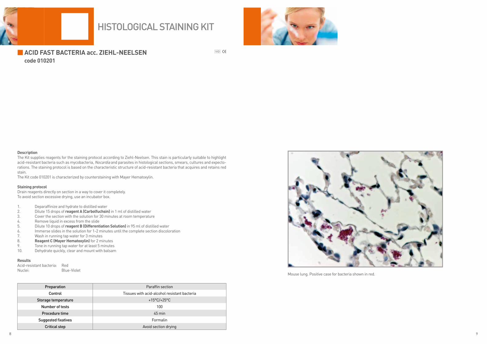
ACID fAST BACTERIA acc. ZIEhL-NEELSENcode 010201
DescriptionThe Kit supplies reagents for the staining protocol according to Ziehl-Neelsen. This stain is particularly suitable to highlight acid-resistant bacteria such as mycobacteria, Nocardia and parasites in histological sections, smears, cultures and expecto-rations. The staining protocol is based on the characteristic structure of acid-resistant bacteria that acquires and retains red stain.The Kit code 010201 is characterized by counterstaining with Mayer Hematoxylin.
Staining protocolDrain reagents directly on section in a way to cover it completely.To avoid section excessive drying, use an incubator box.
1. Deparaffinize and hydrate to distilled water2. Dilute 15 drops of reagent A (Carbolfuchsin) in 1 ml of distilled water3. Cover the section with the solution for 30 minutes at room temperature 4. Remove liquid in excess from the slide5. Dilute 10 drops of reagent B (Differentiation Solution) in 95 ml of distilled water6. Immerse slides in the solution for 1-2 minutes until the complete section discoloration7. Wash in running tap water for 3 minutes8. Reagent C (Mayer hematoxylin) for 2 minutes9. Tone in running tap water for at least 5 minutes10. Dehydrate quickly, clear and mount with balsam
ResultsAcid-resistant bacteria: RedNuclei: Blue-Violet
Preparation Paraffin section
Control Tissues with acid-alcohol resistant bacteria
Storage temperature +15°C/+25°C
Number of tests 100
Procedure time 45 min
Suggested fixatives Formalin
Critical step Avoid section drying
Mouse lung. Positive case for bacteria shown in red.
HiStologiCal StaiNiNg Kit

10 11
ACID fAST BACTERIA acc. ZIEhL-NEELSEN modified acc. fITEcode 010202
DescriptionThe Kit supplies reagents for Ziehl-Neelsen modified according Fite staining protocol. This stain is particularly suitable to highlight acid-resistant bacteria such as mycobacteria, Nocardia and parasites in histological sections, smears, cultures, expectorations and Mycobacterium Leprae (Leprosy etiological agent). The staining protocol is based on the characteristic structure of acid-resistant bacteria that acquires and retains red stain.The Kit code 010202 is characterized by counterstaining with methylene blue.
Staining protocolDrain reagents directly on section in a way to cover it completely.To avoid section excessive drying, use an incubator box.
1. Deparaffinize and hydrate to distilled water2. Reagent A (periodic Acid) for 15 minutes3. Wash in distilled water4. Reagent B (Carbolfuchsin) for 30 minutes5. Wash in distilled water6. Reagent C (Differentiation Solution) for 1 minute until the section doesn’t loose pink stain7. Wash in running tap water for 5 minutes8. Prepare countersolution: 5 drops of reagent D (Methylene blue) + 5 drops of reagent E (Basic Buffer)9. Cover the section with the solution for 30 seconds10. Wash in running tap water for 1-2 minutes11. Dehydrate quickly, clear and mount with balsam
ResultsAcid-resistant bacteria: RedBackground: Blue
Preparation Paraffin section
Control Bactery infection
Storage temperature +15°C/+25°C
Number of tests 100
Procedure time 55 min
Suggested fixatives Formalin
Critical step Avoid section drying
Mouse lung. Positive case for bacteria shown in red.
HiStologiCal StaiNiNg Kit
12 13
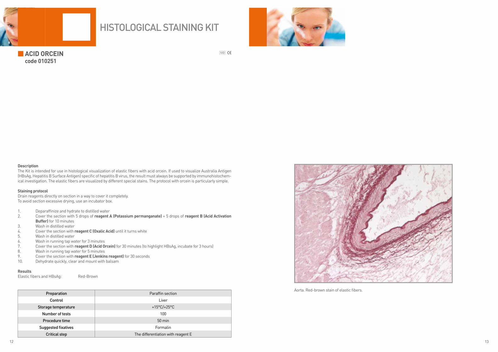
ACID oRCEINcode 010251
DescriptionThe Kit is intended for use in histological visualization of elastic fibers with acid orcein. If used to visualize Australia Antigen (HBsAg, Hepatitis B Surface Antigen) specific of hepatitis B virus, the result must always be supported by immunohistochem-ical investigation. The elastic fibers are visualized by different special stains. The protocol with orcein is particularly simple.
Staining protocolDrain reagents directly on section in a way to cover it completely.To avoid section excessive drying, use an incubator box.
1. Deparaffinize and hydrate to distilled water2. Cover the section with 5 drops of reagent A (potassium permanganate) + 5 drops of reagent B (Acid Activation
Buffer) for 10 minutes3. Wash in distilled water4. Cover the section with reagent C (oxalic Acid) until it turns white5. Wash in distilled water6. Wash in running tap water for 3 minutes7. Cover the section with reagent D (Acid orcein) for 30 minutes (to highlight HBsAg, incubate for 3 hours)8. Wash in running tap water for 5 minutes9. Cover the section with reagent E (Jenkins reagent) for 30 seconds10. Dehydrate quickly, clear and mount with balsam
ResultsElastic fibers and HBsAg: Red-Brown
Preparation Paraffin section
Control Liver
Storage temperature +15°C/+25°C
Number of tests 100
Procedure time 50 min
Suggested fixatives Formalin
Critical step The differentiation with reagent E
HiStologiCal StaiNiNg Kit
Aorta. Red-brown stain of elastic fibers.
14 15
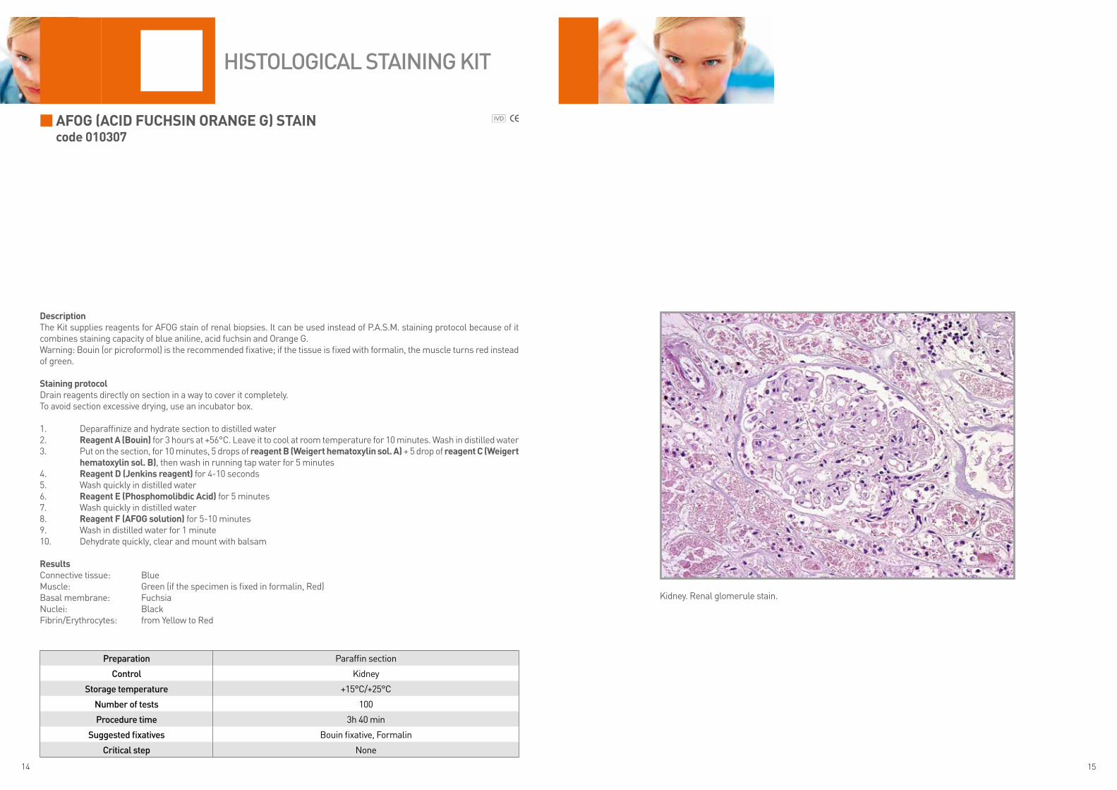
AfoG (ACID fUChSIN oRANGE G) STAINcode 010307
DescriptionThe Kit supplies reagents for AFOG stain of renal biopsies. It can be used instead of P.A.S.M. staining protocol because of it combines staining capacity of blue aniline, acid fuchsin and Orange G.Warning: Bouin (or picroformol) is the recommended fixative; if the tissue is fixed with formalin, the muscle turns red instead of green.
Staining protocolDrain reagents directly on section in a way to cover it completely.To avoid section excessive drying, use an incubator box.
1. Deparaffinize and hydrate section to distilled water2. Reagent A (Bouin) for 3 hours at +56°C. Leave it to cool at room temperature for 10 minutes. Wash in distilled water 3. Put on the section, for 10 minutes, 5 drops of reagent B (Weigert hematoxylin sol. A) + 5 drop of reagent C (Weigert
hematoxylin sol. B), then wash in running tap water for 5 minutes4. Reagent D (Jenkins reagent) for 4-10 seconds5. Wash quickly in distilled water6. Reagent E (phosphomolibdic Acid) for 5 minutes 7. Wash quickly in distilled water8. Reagent f (AfoG solution) for 5-10 minutes9. Wash in distilled water for 1 minute10. Dehydrate quickly, clear and mount with balsam
ResultsConnective tissue: BlueMuscle: Green (if the specimen is fixed in formalin, Red)Basal membrane: FuchsiaNuclei: BlackFibrin/Erythrocytes: from Yellow to Red
Preparation Paraffin section
Control Kidney
Storage temperature +15°C/+25°C
Number of tests 100
Procedure time 3h 40 min
Suggested fixatives Bouin fixative, Formalin
Critical step None
HiStologiCal StaiNiNg Kit
Kidney. Renal glomerule stain.

16 17
AGNoRcode 010801
DescriptionThe AgNOR Kit is intended for use, by silver impregnation, in histological visualization of the proteins bound to Nucleolar Organizer Region (NOR)
Staining protocolNOTE: do not use metallic objects, use only distilled water to wash the slides.The AgNOR staining protocol requires only just cut sections. Do not use polylysine or positively charged slides as they may cause background staining that interferes with preparation reading.After mounting, keep the slides in a dark place.
Drain reagents directly on section in a way to cover it completely.To avoid section excessive drying, use an incubator box.
1. Deparaffinize and hydrate section to distilled water2. Prepare the working solution: 8 ml reagent A (Gelatine) + 16 ml reagent B (Silver nitrate). Stir briefly with a glass
stick, DO NOT use metallic objects3. Immerse slides in the solution for 30 minutes at room temperature and in the darkness4. Drain slides and go to the next step5. Reagent C (fixing solution) for 1 minute6. Wash in distilled water for 1 minute7. Dehydrate, clear and mount with balsam
ResultsAgNOR: Black
Preparation Paraffin section
Control Tonsil
Storage temperature +4°C/+8°C
Number of tests 100
Procedure time 35 min
Suggested fixatives Formalin
Critical stepDo not use metallic objects. The working solution deteriorates quickly, use soon after preparation. Protect the reagent from light by covering the jar with an aluminum foil. Use only just cut sections.
HiStologiCal StaiNiNg Kit
WARNING: reagents are stored at +4°C/+8°C, we recommend to keep them at room temperature before use for at least 10 minutes. If they are used cold, the reaction speed is significantly reduced.
Mouse breast with cancer. Black stain of NOR regions.
18 19
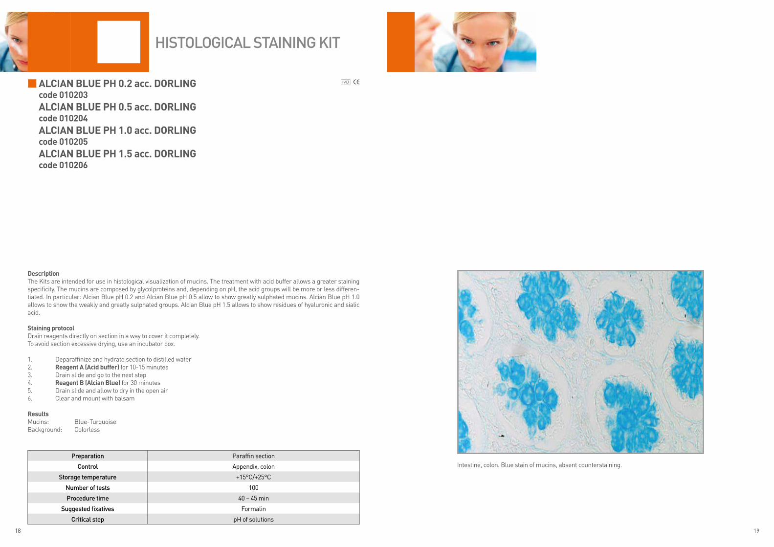
ALCIAN BLUE ph 0.2 acc. DoRLINGcode 010203ALCIAN BLUE ph 0.5 acc. DoRLINGcode 010204ALCIAN BLUE ph 1.0 acc. DoRLINGcode 010205ALCIAN BLUE ph 1.5 acc. DoRLINGcode 010206
DescriptionThe Kits are intended for use in histological visualization of mucins. The treatment with acid buffer allows a greater staining specificity. The mucins are composed by glycolproteins and, depending on pH, the acid groups will be more or less differen-tiated. In particular: Alcian Blue pH 0.2 and Alcian Blue pH 0.5 allow to show greatly sulphated mucins. Alcian Blue pH 1.0 allows to show the weakly and greatly sulphated groups. Alcian Blue pH 1.5 allows to show residues of hyaluronic and sialic acid.
Staining protocolDrain reagents directly on section in a way to cover it completely.To avoid section excessive drying, use an incubator box.
1. Deparaffinize and hydrate section to distilled water2. Reagent A (Acid buffer) for 10-15 minutes 3. Drain slide and go to the next step4. Reagent B (Alcian Blue) for 30 minutes5. Drain slide and allow to dry in the open air6. Clear and mount with balsam
ResultsMucins: Blue-TurquoiseBackground: Colorless
Preparation Paraffin section
Control Appendix, colon
Storage temperature +15°C/+25°C
Number of tests 100
Procedure time 40 – 45 min
Suggested fixatives Formalin
Critical step pH of solutions
HiStologiCal StaiNiNg Kit
Intestine, colon. Blue stain of mucins, absent counterstaining.
20 21
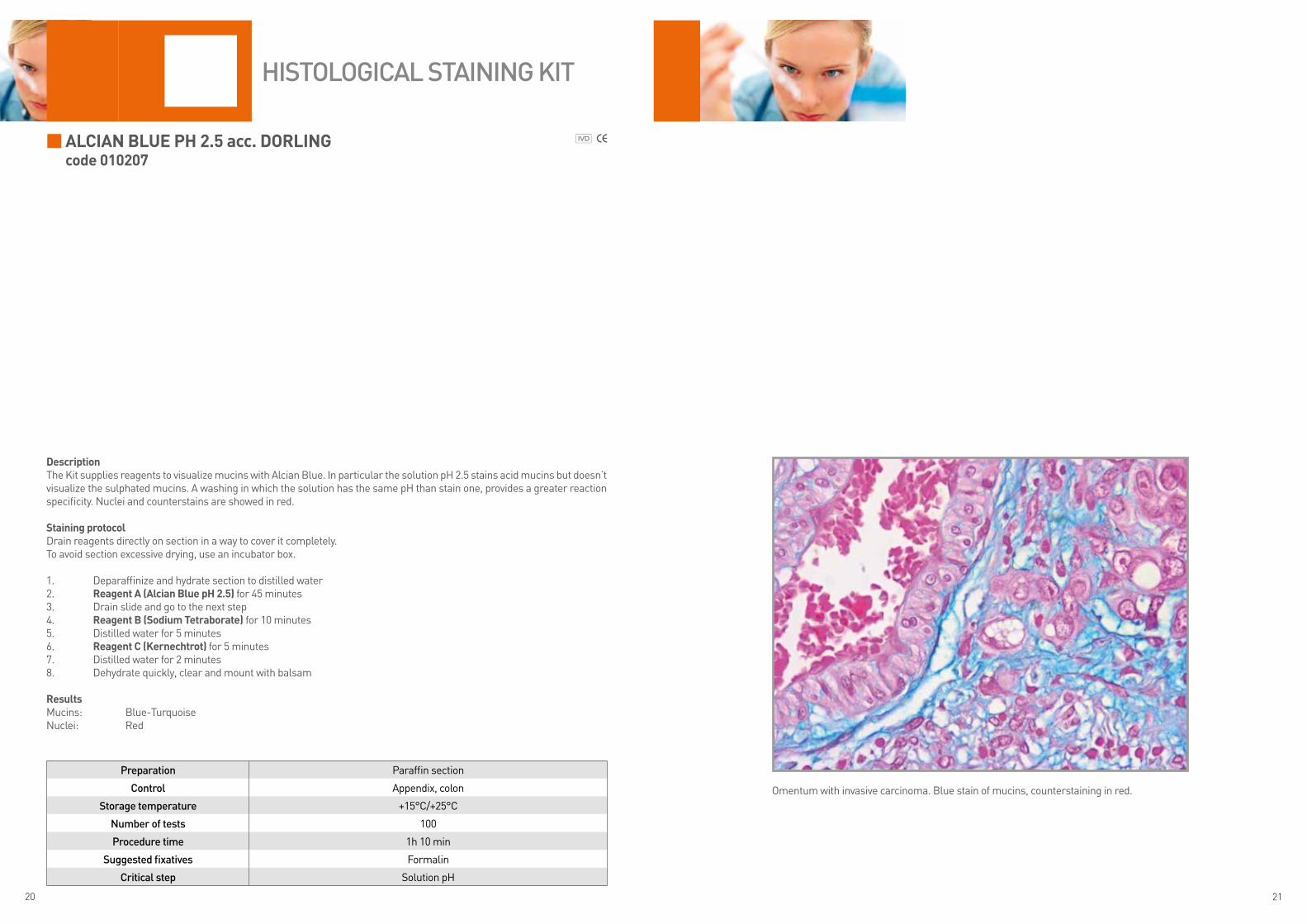
ALCIAN BLUE ph 2.5 acc. DoRLINGcode 010207
DescriptionThe Kit supplies reagents to visualize mucins with Alcian Blue. In particular the solution pH 2.5 stains acid mucins but doesn’t visualize the sulphated mucins. A washing in which the solution has the same pH than stain one, provides a greater reaction specificity. Nuclei and counterstains are showed in red.
Staining protocolDrain reagents directly on section in a way to cover it completely.To avoid section excessive drying, use an incubator box.
1. Deparaffinize and hydrate section to distilled water2. Reagent A (Alcian Blue ph 2.5) for 45 minutes 3. Drain slide and go to the next step4. Reagent B (Sodium Tetraborate) for 10 minutes5. Distilled water for 5 minutes6. Reagent C (kernechtrot) for 5 minutes 7. Distilled water for 2 minutes8. Dehydrate quickly, clear and mount with balsam
ResultsMucins: Blue-TurquoiseNuclei: Red
Preparation Paraffin section
Control Appendix, colon
Storage temperature +15°C/+25°C
Number of tests 100
Procedure time 1h 10 min
Suggested fixatives Formalin
Critical step Solution pH
HiStologiCal StaiNiNg Kit
Omentum with invasive carcinoma. Blue stain of mucins, counterstaining in red.
22 23
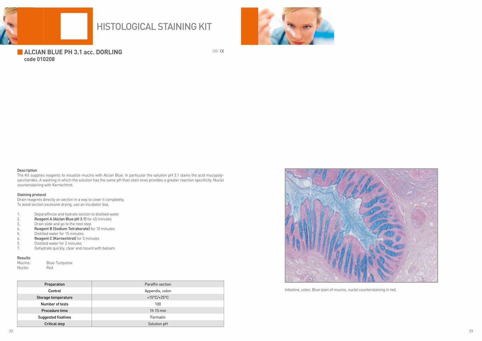
ALCIAN BLUE ph 3.1 acc. DoRLING code 010208
DescriptionThe Kit supplies reagents to visualize mucins with Alcian Blue. In particular the solution pH 3.1 stains the acid mucopoly-saccharides. A washing in which the solution has the same pH than stain ones provides a greater reaction specificity. Nuclei counterstaining with Kernechtrot.
Staining protocolDrain reagents directly on section in a way to cover it completely.To avoid section excessive drying, use an incubator box.
1. Deparaffinize and hydrate section to distilled water2. Reagent A (Alcian Blue ph 3.1) for 45 minutes 3. Drain slide and go to the next step4. Reagent B (Sodium Tetraborate) for 10 minutes5. Distilled water for 15 minutes6. Reagent C (kernechtrot) for 5 minutes 5. Distilled water for 2 minutes7. Dehydrate quickly, clear and mount with balsam
ResultsMucins: Blue-TurquoiseNuclei: Red
Preparation Paraffin section
Control Appendix, colon
Storage temperature +15°C/+25°C
Number of tests 100
Procedure time 1h 15 min
Suggested fixatives Formalin
Critical step Solution pH
HiStologiCal StaiNiNg Kit
Intestine, colon. Blue stain of mucins, nuclei counterstaining in red.
24 25
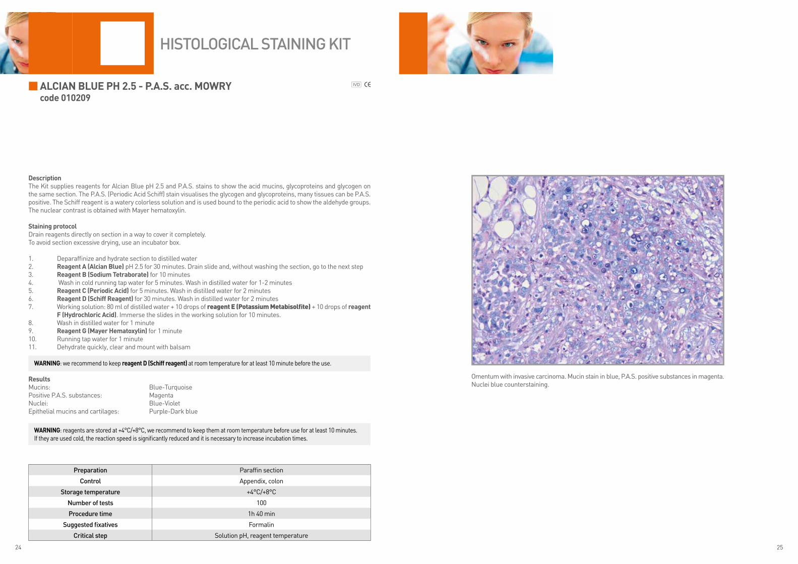
ALCIAN BLUE ph 2.5 - p.A.S. acc. MoWRYcode 010209
DescriptionThe Kit supplies reagents for Alcian Blue pH 2.5 and P.A.S. stains to show the acid mucins, glycoproteins and glycogen on the same section. The P.A.S. (Periodic Acid Schiff) stain visualises the glycogen and glycoproteins, many tissues can be P.A.S. positive. The Schiff reagent is a watery colorless solution and is used bound to the periodic acid to show the aldehyde groups. The nuclear contrast is obtained with Mayer hematoxylin.
Staining protocolDrain reagents directly on section in a way to cover it completely.To avoid section excessive drying, use an incubator box.
1. Deparaffinize and hydrate section to distilled water2. Reagent A (Alcian Blue) pH 2.5 for 30 minutes. Drain slide and, without washing the section, go to the next step3. Reagent B (Sodium Tetraborate) for 10 minutes4. Wash in cold running tap water for 5 minutes. Wash in distilled water for 1-2 minutes5. Reagent C (periodic Acid) for 5 minutes. Wash in distilled water for 2 minutes6. Reagent D (Schiff Reagent) for 30 minutes. Wash in distilled water for 2 minutes7. Working solution: 80 ml of distilled water + 10 drops of reagent E (Potassium Metabisolfite) + 10 drops of reagent
f (hydrochloric Acid). Immerse the slides in the working solution for 10 minutes.8. Wash in distilled water for 1 minute9. Reagent G (Mayer hematoxylin) for 1 minute10. Running tap water for 1 minute11. Dehydrate quickly, clear and mount with balsam
ResultsMucins: Blue-TurquoisePositive P.A.S. substances: MagentaNuclei: Blue-VioletEpithelial mucins and cartilages: Purple-Dark blue
Preparation Paraffin section
Control Appendix, colon
Storage temperature +4°C/+8°C
Number of tests 100
Procedure time 1h 40 min
Suggested fixatives Formalin
Critical step Solution pH, reagent temperature
HiStologiCal StaiNiNg Kit
WARNING: we recommend to keep reagent D (Schiff reagent) at room temperature for at least 10 minute before the use.
Omentum with invasive carcinoma. Mucin stain in blue, P.A.S. positive substances in magenta.Nuclei blue counterstaining.
WARNING: reagents are stored at +4°C/+8°C, we recommend to keep them at room temperature before use for at least 10 minutes. If they are used cold, the reaction speed is significantly reduced and it is necessary to increase incubation times.

26 27
ALCIAN YELLoW-ToLUIDINE BLUEcode 010269
DescriptionThe Kit supplies reagents to show Helicobacter pylori on gastric tissue. Alcian yellow-Toluidine blue stain can be used instead Giemsa stain because of bacteria are particularly visible on the yellow background of gastric mucins.
Staining protocolDrain reagents directly on section in a way to cover it completely.To avoid section excessive drying, use an incubator box.
1. Deparaffinize and hydrate section to distilled water2. Reagent A (periodic Acid) for 10 minutes3. Wash in running tap water4. Reagent B (potassium Metabisulfite) for 5 minutes5. Wash in running tap water6. Reagent C (Alcian Yellow) for 15 minutes 7. Wash in running tap water8. Reagent D (Toluidine Blue) for 5 minutes9. Wash in running tap water10. Dehydrate quickly, clear and mount with balsam
ResultsHelicobacter pylori: Dark blueMucins: YellowSurrounding tissue: Light blue
Preparation Paraffin sections
Control Stomach (recorded case of Helicobacter Pylori)
Storage temperature +15°C/+25°C
Number of tests 100
Procedure time 35 min
Suggested fixatives Formalin
Critical step None
HiStologiCal StaiNiNg Kit
Stomach. Helicobacter Pylori in dark blue. Nuclei and cytoplasm in blue. Yellow mucins.

28 29
AZAN TRIChRoMEcode 010212
DescriptionThe Azan trichrome is a version of Mallory trichrome for connective tissue staining. The Kit is intended for use in histological visualization of fibers, glial fibers, collagen, glomerular stroma and erythrocytes on the same sections. The staining protocol is suitable for hypophysis staining too.
Staining protocolDrain reagents directly on section in a way to cover it completely.To avoid section excessive drying, use an incubator box.
Pour reagents A, D and E in 3 vertical Choplin jars. Incubate reagent A at +56°C.
1. Deparaffinize and hydrate section to distilled water2. Incubate slides in reagent A (Azocarmine) for 45 minutes at +56°C 3. Leave it cool at room temperature for at least 10 minutes. Wash in running tap water the excess stain 4. Reagent B (Blue aniline) for 1 minute5. Drain slide and go to the next step6. Reagent C (Differentiation solution) for 1 minute. Wash quickly in distilled water 7. Immerse slides in reagent D (phosphotungstic Acid) for 60 minutes. Drain slide and go to the next step8. Immerse slide in reagent E (Mallory solution) for 60 minutes9. Quick step in ethylic alcohol 95°10. Complete dehydration and clear11. Mount with balsam
ResultsNuclei, Erythrocytes: RedMuscle: OrangeCollagen fibers: Bright light blue
hypophysisCytoplasmatic granules of hypophysis delta cells: Light blueAcidophil granules of hypophysis: Red
Preparation Paraffin section
Control Kidney
Storage temperature +15°C/+25°C
Number of tests 100
Procedure time 3h
Suggested fixatives Formalin
Critical step Steps at +56°C
HiStologiCal StaiNiNg Kit
Kidney. Blue stain of glomerules. Erythrocytes in red.
Warning: The reagent A can be used again without filtering. Reagents D and E can be used again after filtering.

30 31
CoNGo REDcode 010214
DescriptionThe Kit is intended for use in histological visualization of amyloid (insoluble protein with reduced molecular weight). The amyloid takes a particular red stain and green birefringence under polarized light.
Staining protocolDrain reagents directly on section in a way to cover it completely.To avoid section excessive drying, use an incubator box.
1. Deparaffinize and hydrate section to distilled water2. Reagent A (Congo Red) for 30 minutes 3. Drain the slide and go to the next step4. Reagent B (Lithium Carbonate) for 10 minutes5. Wash quickly in distilled water6. Reagent C (Alcoholic Differentiation Buffer) for 15-30 seconds 7. Wash in running tap water for 5 minutes8. Reagent D (Mayer hematoxylin) for 5 minutes9. Running tap water for at least 10 minutes10. Dehydrate quickly, clear and mount with balsam
ResultsAmyloid: from Pink to RedNuclei: Blue-Violet
Preparation Paraffin section
Control Positive case (ex. amyloidosis)
Storage temperature +15°C/+25°C
Number of tests 100
Procedure time 1h
Suggested fixatives Formalin
Critical step To highlight, with polarized light, cut section with thickness of at least 10 µm
HiStologiCal StaiNiNg Kit
Connective tissue. Red stain of amyloid deposits.

32 33
DANE TRIChRoMEcode 010215
DescriptionThe Kit is intended for use in histological simultaneously visualization of prekeratins, keratins and acid mucins.
Staining protocolDrain reagents directly on section in a way to cover it completely.To avoid section excessive drying, use an incubator box.
1. Deparaffinize and hydrate section to distilled water2. Reagent A (Mayer hematoxylin) for 10 minutes 3. Running tap water for 5 minutes4. Reagent B (floxin) for 3 minutes5. Running tap water until the section looses completely the stain6. Reagent C (Alcian Blue) for 5 minutes 7. Running tap water for 5 minutes8. Reagent D (orange G) for 13-15 minutes9. Distilled water for 1 minute10. Dehydrate quickly, clear and mount with balsam
ResultsMucins: BlueKeratin: from Orange to RedNuclei: Brown-Red
Preparation Paraffin section
Control Skin
Storage temperature +15°C/+25°C
Number of tests 100
Procedure time 45 – 50 min
Suggested fixatives Formalin
Critical step None
HiStologiCal StaiNiNg Kit
Intestine, colon. Blue stain of mucins.
34 35
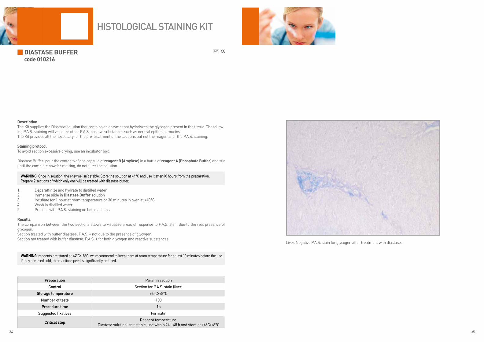
DIASTASE BUffERcode 010216
DescriptionThe Kit supplies the Diastase solution that contains an enzyme that hydrolyzes the glycogen present in the tissue. The follow-ing P.A.S. staining will visualize other P.A.S. positive substances such as neutral epithelial mucins.The Kit provides all the necessary for the pre-treatment of the sections but not the reagents for the P.A.S. staining.
Staining protocolTo avoid section excessive drying, use an incubator box.
Diastase Buffer: pour the contents of one capsule of reagent B (Amylase) in a bottle of reagent A (phosphate Buffer) and stir until the complete powder melting, do not filter the solution.
1. Deparaffinize and hydrate to distilled water2. Immerse slide in Diastase Buffer solution 3. Incubate for 1 hour at room temperature or 30 minutes in oven at +40°C4. Wash in distilled water5. Proceed with P.A.S. staining on both sections
ResultsThe comparison between the two sections allows to visualize areas of response to P.A.S. stain due to the real presence of glycogen.Section treated with buffer diastase: P.A.S. + not due to the presence of glycogen. Section not treated with buffer diastase: P.A.S. + for both glycogen and reactive substances.
Preparation Paraffin section
Control Section for P.A.S. stain (liver)
Storage temperature +4°C/+8°C
Number of tests 100
Procedure time 1h
Suggested fixatives Formalin
Critical stepReagent temperature.
Diastase solution isn’t stable, use within 24 - 48 h and store at +4°C/+8°C
HiStologiCal StaiNiNg Kit
WARNING: Once in solution, the enzyme isn’t stable. Store the solution at +4°C and use it after 48 hours from the preparation.Prepare 2 sections of which only one will be treated with diastase buffer.
WARNING: reagents are stored at +4°C/+8°C, we recommend to keep them at room temperature for at last 10 minutes before the use. If they are used cold, the reaction speed is significantly reduced.
Liver. Negative P.A.S. stain for glycogen after treatment with diastase.

36 37
fAST QUICk - M.G.G. RApIDcode 010253
DescriptionThe Kit supplies reagents for the fast stain of blood smears. The M.G.G. (May Grunwald Giemsa) stain allows to visualize the different kind of blood cells.
Staining protocol1. Dry the smear in the air 2. Immerse the slide for 5 times for 1 second each in reagent A. After immersion, wait up to complete dripping of
excess liquid 3. Immerse the slide for 5 times for 1 second each in reagent B. After immersion, wait up to complete dripping of
excess liquid4. Immerse the slide 3-5 times for 1 second each in reagent C. After immersion, wait up to complete dripping of ex-
cess liquid 5. Wash with spring water6. Dry in the air (do not use heat sources, ovens or plates)7. Clear and mount with balsam
ResultsNuclei: Violaceous Red, PinkBasophil cytoplasm: from light Blue to dark BlueAcidophil cytoplasm: from light to rosy RedPolychromatophilic cytoplasm: from Grayish to ViolaceousAcidophil granules: OrangeNeutrophil granules: Brown-dark PinkBasophil granules: Dark VioletAzurophil granules: from Purple to Purple Violet
Preparation Blood smear
Control Peripheral blood smear
Storage temperature +15°C/+25°C
Number of tests 100
Procedure time 20-25 seconds
Suggested fixatives Non-foreseen
Critical step None
CytologiCal StaiNiNg Kit
Blood smear. Stain to show the different cell components.
38 39
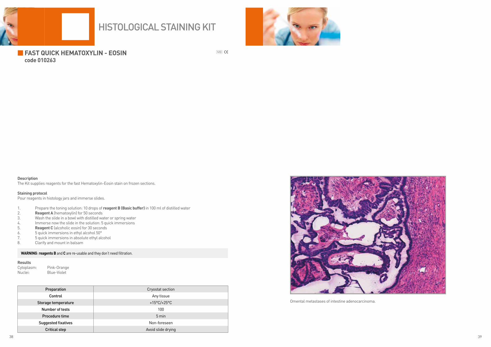
Fast Quick Hematoxylin - eosin code 010263
DescriptionThe Kit supplies reagents for the fast Hematoxylin-Eosin stain on frozen sections.
staining protocolPour reagents in histology jars and immerse slides.
1. Prepare the toning solution: 10 drops of reagent B (Basic buffer) in 100 ml of distilled water2. Reagent a (hematoxylin) for 50 seconds3. Wash the slide in a bowl with distilled water or spring water4. Immerse now the slide in the solution: 5 quick immersions5. Reagent c (alcoholic eosin) for 30 seconds6. 5 quick immersions in ethyl alcohol 50°7. 5 quick immersions in absolute ethyl alcohol 8. Clarify and mount in balsam
ResultsCytoplasm: Pink-OrangeNuclei: Blue-Violet
Preparation Cryostat section
Control Any tissue
Storage temperature +15°C/+25°C
Number of tests 100
Procedure time 5 min
Suggested fixatives Non-foreseen
Critical step Avoid slide drying
HiStologiCal StaiNiNg Kit
Omental metastases of intestine adenocarcinoma.
WaRninG: reagents B and c are re-usable and they don’t need filtration.

40 41
FeulGen anD RossenBeckcode 010219
DescriptionThe Kit supplies the reagents necessary to show DNA with Schiff reagent according to Feulgen and Rossenbeck staining protocol. The specimen is treated with hydrochloric acid which removes the purine bases and makes available the aldehyde groups to Schiff reagent. This reaction is specific for DNA.We recommend to analyse the slides the same day of the staining.
staining protocolDrain reagents directly on section in a way to cover it completely.To avoid section excessive drying, use an incubator box.
1. Deparaffinize and hydrate section to distilled water (for histological specimens) Running tap water for 5 minutes (for cytological specimens)2. Reagent a (Hydrochloric acid) for 10 minutes 3. Wash the section in distilled water4. Reagent B (schiff reagent) for at least 5 minutes (till the section turns magenta)5. Drain reagent in excess and go to the next step without washing the section6. Reagent c (metabisolphite Potassium) for 2 minutes 7. Drain reagent in excess and go to the next step without washing the section8. Reagent D (Hydrochloric acid) for 3 minutes9. Wash in running tap water10. Dehydrate quickly, clear and mount with balsam
ResultsDNA: Red magentaBackground: Colorless
Preparation Paraffin section
Control Unknown control tissue
Storage temperature +4°C/+8°C
Number of tests 100
Procedure time 25 min
Suggested fixatives Formalin
Critical step Do not use acid fixatives. Do not prolong treatment with reagent A
HiStologiCal StaiNiNg Kit
Lymphnode. Nuclei magenta staining. Absent counterstaining.
WaRninG: we recommend to not use Bouin’s fixative, as acid fixatives may interfere with hydrolysis process.If treatment with hydrochloric acid is too long, it may cause the complete DNA hydrolysis with consequent reduction of reaction sensitivity and appearance of possible false negatives.Warning: the decalcification process interferes with the staining so the bony tissue is not usually indicated for this kind of investigation.We recommend leaving the reagent D (schiff Reagent) at room temperature for at least 10 minutes before use it. If used cold, the reaction speed decreases considerably.
42 43
FoucHet-Van Gieson acc. kutlickcode 010220
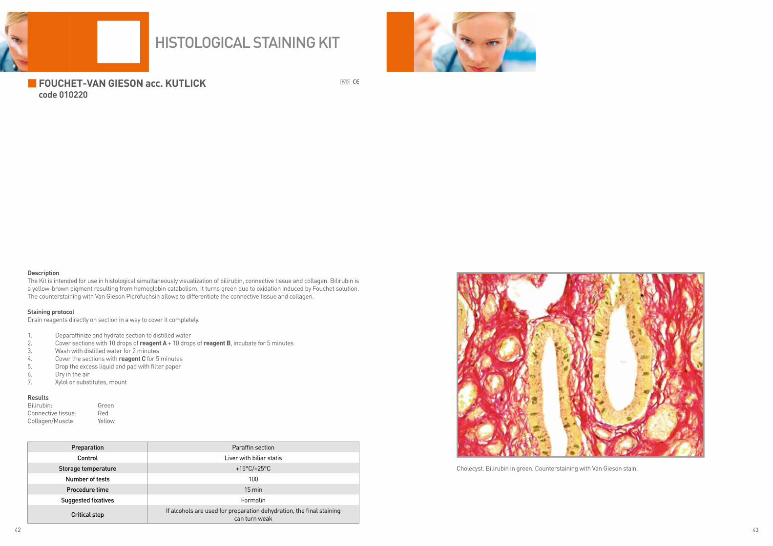
DescriptionThe Kit is intended for use in histological simultaneously visualization of bilirubin, connective tissue and collagen. Bilirubin is a yellow-brown pigment resulting from hemoglobin catabolism. It turns green due to oxidation induced by Fouchet solution. The counterstaining with Van Gieson Picrofuchsin allows to differentiate the connective tissue and collagen.
staining protocolDrain reagents directly on section in a way to cover it completely.
1. Deparaffinize and hydrate section to distilled water2. Cover sections with 10 drops of reagent a + 10 drops of reagent B, incubate for 5 minutes3. Wash with distilled water for 2 minutes4. Cover the sections with reagent c for 5 minutes5. Drop the excess liquid and pad with filter paper6. Dry in the air7. Xylol or substitutes, mount
ResultsBilirubin: GreenConnective tissue: RedCollagen/Muscle: Yellow
Preparation Paraffin section
Control Liver with biliar statis
Storage temperature +15°C/+25°C
Number of tests 100
Procedure time 15 min
Suggested fixatives Formalin
Critical stepIf alcohols are used for preparation dehydration, the final staining
can turn weak
HiStologiCal StaiNiNg Kit
Cholecyst. Bilirubin in green. Counterstaining with Van Gieson stain.
44 45
GolDneR tRicHRome (masson’s tRicHRome WitH liGHt GReen)code 010224
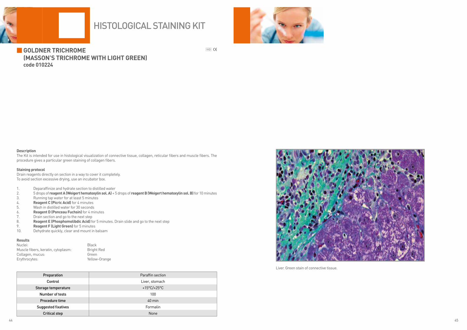
DescriptionThe Kit is intended for use in histological visualization of connective tissue, collagen, reticular fibers and muscle fibers. The procedure gives a particular green staining of collagen fibers.
staining protocolDrain reagents directly on section in a way to cover it completely.To avoid section excessive drying, use an incubator box.
1. Deparaffinize and hydrate section to distilled water2. 5 drops of reagent a (Weigert hematoxylin sol. a) + 5 drops of reagent B (Weigert hematoxylin sol. B) for 10 minutes3. Running tap water for at least 5 minutes4. Reagent c (Picric acid) for 4 minutes5. Wash in distilled water for 30 seconds6. Reagent D (Ponceau Fuchsin) for 4 minutes7. Drain section and go to the next step8. Reagent e (Phosphomolibdic acid) for 5 minutes. Drain slide and go to the next step9. Reagent F (light Green) for 5 minutes10. Dehydrate quickly, clear and mount in balsam
ResultsNuclei: BlackMuscle fibers, keratin, cytoplasm: Bright RedCollagen, mucus: GreenErythrocytes: Yellow-Orange
Preparation Paraffin section
Control Liver, stomach
Storage temperature +15°C/+25°C
Number of tests 100
Procedure time 40 min
Suggested fixatives Formalin
Critical step None
HiStologiCal StaiNiNg Kit
Liver. Green stain of connective tissue.

46 47
GomoRi’s tRicHRomecode 010302
DescriptionThe Kit allows the analysis of collagen fibers in liver and kidney tissue. Suitable to visualize the ragged red fibers present in many mitochondrial myopathies.
staining protocolDrain reagents directly on section in a way to cover it completely.To avoid section excessive drying, use an incubator box.
1. Hydrate sections up to distilled water2. Reagent a (mayer hematoxylin) for 45 seconds3. Wash in running tap water until nuclei change stain4. Wash in distilled water for 30 seconds5. Reagent B (Gomori stain) for 10 minutes6. Wash in distilled water7. Reagent c (acid Buffer) for 15 seconds8. Drain slide and go to the next step9. Reagent D (Differentiation solution) for 20 seconds10. Wash in distilled water11. Dehydrate quickly, clear and mount in balsam
ResultsMyofibrils: Green (*)Intermyofibrillar material: RedConnective tissue: Bright GreenNuclei: Blue-Violet
(*) if the tissue is fixed in formalin, the muscle turns in red
Preparation Cryostat section
Control Muscle
Storage temperature +15°C/+25°C
Number of tests 100
Procedure time 15 min
Suggested fixatives Non-foressen
Critical step Non-suitable for tissue embedded in paraffin
HiStologiCal StaiNiNg Kit
Frozen tissue. Pathological muscle.
48 49
GRam FoR HistoloGical sectionscode 010221
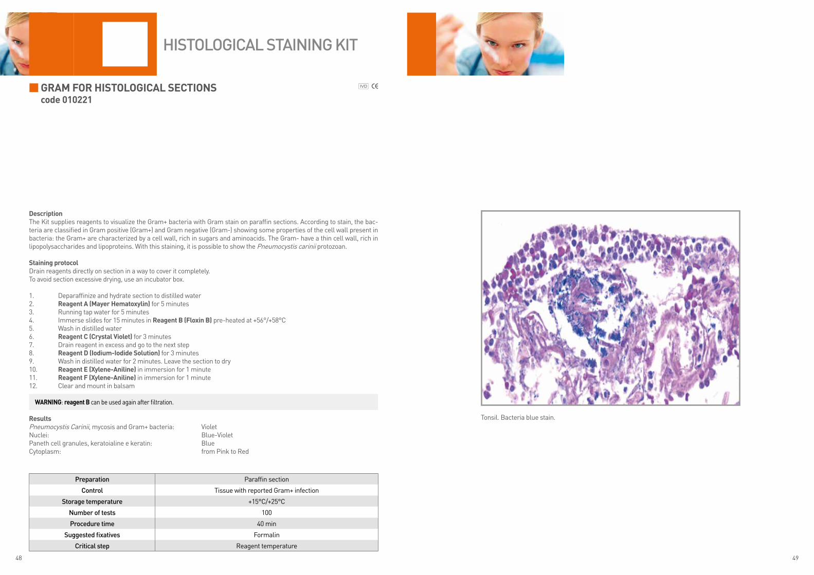
DescriptionThe Kit supplies reagents to visualize the Gram+ bacteria with Gram stain on paraffin sections. According to stain, the bac-teria are classified in Gram positive (Gram+) and Gram negative (Gram-) showing some properties of the cell wall present in bacteria: the Gram+ are characterized by a cell wall, rich in sugars and aminoacids. The Gram- have a thin cell wall, rich in lipopolysaccharides and lipoproteins. With this staining, it is possible to show the Pneumocystis carinii protozoan.
staining protocolDrain reagents directly on section in a way to cover it completely.To avoid section excessive drying, use an incubator box.
1. Deparaffinize and hydrate section to distilled water2. Reagent a (mayer Hematoxylin) for 5 minutes3. Running tap water for 5 minutes4. Immerse slides for 15 minutes in Reagent B (Floxin B) pre-heated at +56°/+58°C5. Wash in distilled water6. Reagent c (crystal Violet) for 3 minutes7. Drain reagent in excess and go to the next step8. Reagent D (iodium-iodide solution) for 3 minutes9. Wash in distilled water for 2 minutes. Leave the section to dry10. Reagent e (xylene-aniline) in immersion for 1 minute11. Reagent F (xylene-aniline) in immersion for 1 minute 12. Clear and mount in balsam
ResultsPneumocystis Carinii, mycosis and Gram+ bacteria: VioletNuclei: Blue-VioletPaneth cell granules, keratoialine e keratin: BlueCytoplasm: from Pink to Red
Preparation Paraffin section
Control Tissue with reported Gram+ infection
Storage temperature +15°C/+25°C
Number of tests 100
Procedure time 40 min
Suggested fixatives Formalin
Critical step Reagent temperature
HiStologiCal StaiNiNg Kit
Tonsil. Bacteria blue stain.
WaRninG: reagent B can be used again after filtration.

50 51
GRimeliuscode 010222
DescriptionThe Kit is intended for use in histological visualization of pancreas alpha cells. The Grimelius stain is also designed for demon-strating cells secreting argyrophilic substances such as noradrenaline, serotonin, lipofuchsin.
staining protocolDrain reagents directly on section in a way to cover it completely.To avoid section excessive drying, use an incubator box.
1. Deparaffinize and hydrate section to distilled water2. Working silver nitrate solution: 40 ml of distilled water + 2 ml reagent a (silver nitrate) + 4 ml reagent B (acetate
buffer). Preserve 1-2 ml of this solution for the second impregnation. Protect reagent from light
FiRst imPReGnation:3. Cover section with working solution of silver nitrate (STEP 2) and incubate in oven at +60°C in the darkness for 3
hours. Leave it cool at room temperature4. Reducing solution: melt reagent c (Reducing powder) in 25 ml of distilled water. Stir till the complete powder
melting. Preserve 1-2 ml of this solution for the second impregnation5. Immerse slides in the reducing solution (STEP 4) and leave in oven at +60°C in the darkness for 5 minutes. Leave it
cool at room temperature. Wash in distilled water for 3 minutes
seconD imPReGnation:6. Cover section with working solution of silver nitrate (STEP 2) at room temperature for 10 minutes7. Drain the reagent from the slide and go to the next step8. Cover section with working solution of silver nitrate (STEP 4) at room temperature for 5 minutes9. Wash in distilled water for 3 minutes10. Reagent D (Fixing solution) for 2 minutes11. Wash in distilled water12. Dehydrate quickly, clear and mount with balsam
ResultsArgyrophilic granules: from light Brown to Black
Preparation Paraffin section
Control Pancreas or intestine
Storage temperature +4°C/+8°C
Number of tests 100
Procedure time 3h 40 min
Suggested fixatives Formalin
Critical stepReagent temperature. Don not use metallic objects.
For the first impregnation, protect specimen from light covering the jar
HiStologiCal StaiNiNg Kit
Intestine. Brown-black stain of argyrophilic substances.
WaRninG: Use a oven at +60°C for the first impregnation. The second impregnation occurs at room temperature.
WaRninG: reagents are stored at +4°C/+8°C, we recommend to keep them at room temperature before use for at least 10 minutes.If they are used cold, the reaction speed is significantly reduced.
52 53
GRocott acc. callaRDcode 010223
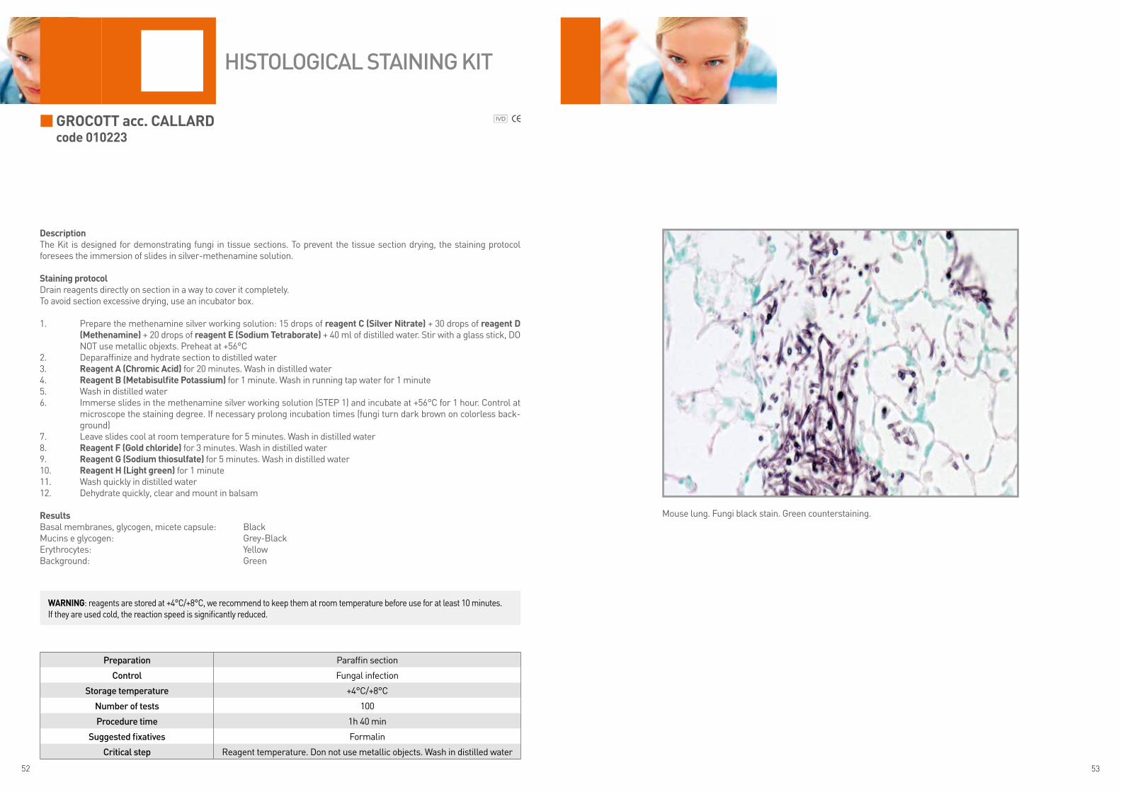
DescriptionThe Kit is designed for demonstrating fungi in tissue sections. To prevent the tissue section drying, the staining protocol foresees the immersion of slides in silver-methenamine solution.
staining protocolDrain reagents directly on section in a way to cover it completely.To avoid section excessive drying, use an incubator box.
1. Prepare the methenamine silver working solution: 15 drops of reagent c (silver nitrate) + 30 drops of reagent D (methenamine) + 20 drops of reagent e (sodium tetraborate) + 40 ml of distilled water. Stir with a glass stick, DO NOT use metallic objexts. Preheat at +56°C
2. Deparaffinize and hydrate section to distilled water3. Reagent a (chromic acid) for 20 minutes. Wash in distilled water4. Reagent B (metabisulfite Potassium) for 1 minute. Wash in running tap water for 1 minute5. Wash in distilled water6. Immerse slides in the methenamine silver working solution (STEP 1) and incubate at +56°C for 1 hour. Control at
microscope the staining degree. If necessary prolong incubation times (fungi turn dark brown on colorless back-ground)
7. Leave slides cool at room temperature for 5 minutes. Wash in distilled water8. Reagent F (Gold chloride) for 3 minutes. Wash in distilled water9. Reagent G (sodium thiosulfate) for 5 minutes. Wash in distilled water10. Reagent H (light green) for 1 minute11. Wash quickly in distilled water12. Dehydrate quickly, clear and mount in balsam
ResultsBasal membranes, glycogen, micete capsule: BlackMucins e glycogen: Grey-BlackErythrocytes: YellowBackground: Green
Preparation Paraffin section
Control Fungal infection
Storage temperature +4°C/+8°C
Number of tests 100
Procedure time 1h 40 min
Suggested fixatives Formalin
Critical step Reagent temperature. Don not use metallic objects. Wash in distilled water
HiStologiCal StaiNiNg Kit
Mouse lung. Fungi black stain. Green counterstaining.
WaRninG: reagents are stored at +4°C/+8°C, we recommend to keep them at room temperature before use for at least 10 minutes. If they are used cold, the reaction speed is significantly reduced.

54 55
Hale Reaction code 010312
DescriptionThe Kit is designed for demonstrating colloidal iron and polysaccharides acids, such as ialuronic acid in histological sections.
staining protocolDrain reagents directly on section in a way to cover it completely.To avoid section excessive drying, use an incubator box.
1. Deparaffinize and hydrate section to distilled water2. Cover section with 10 drops of reagent a (colloidal iron solution) and 10 drops of reagent B (acid solution) for 10
minutes3. Wash several times in distilled water4. Prepare 100 ml of potassium ferrocyanide solution: melt reagent c (Porassium ferro cyanide) in 80 ml of distilled
water, then add 20 ml of reagent D (chloride acid). DO NOT use metallic objects.5. Immerse slides in potassium ferrocyanide solution (STEP 4) for 10 minutes6. Wash more times in distilled water7. Cover section with reagent e (kernechtrot) for 5 minutes8. Wash quickly in distilled water9. Dehydrate quickly, clear and mount with balsam
ResultsIron: BlueAcid mucin: BlueCellular nuclei: Red
Preparation Paraffin section
Control Cartilage
Storage temperature +4°C/+8°C
Number of tests 100
Procedure time 30 min
Suggested fixatives Formalin
Critical step Use positive control. Do not use metallic objects. Use fresh reagents.
HiStologiCal StaiNiNg Kit
Intestine. Blue stain for mucins and black for iron deposits.
WaRninG: the potassium ferrocyanide solution should be fresh when used. Use again the solution could bring to false positives. We recommend to use a positive control tissue.
WaRninG: reagents are stored at +4°C/+8°C, we recommend to keep them at room temperature before use for at least 10 minutes. If they are used cold, the reaction speed is significantly reduced.
56 57
lonG Giemsa acc. lenneRtcode 010225
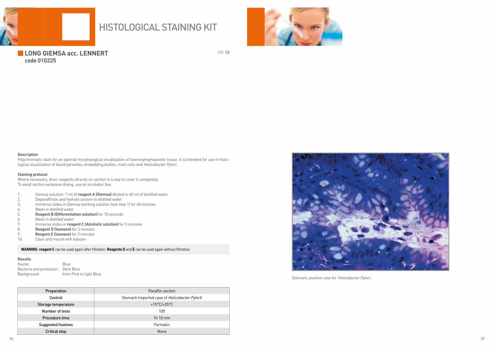
DescriptionPolychromatic stain for an optimal morphological visualization of haemolymphopoietic tissue. It is intended for use in histo-logical visualization of blood parasites, embedding bodies, mast cells and Helicobacter Pylori.
staining protocolWhere necessary, drain reagents directly on section in a way to cover it completely.To avoid section excessive drying, use an incubator box.
1. Giemsa solution: 7 ml of reagent a (Giemsa) diluted in 60 ml of distilled water2. Deparaffinize and hydrate section to distilled water3. Immerse slides in Giemsa working solution (see step 1) for 60 minutes4. Wash in distilled water5. Reagent B (Differentiation solution) for 10 seconds6. Wash in distilled water7. Immerse slides in reagent c (alcoholic solution) for 5 minutes8. Reagent D (isowave) for 2 minutes9. Reagent e (isowave) for 2 minutes10. Clear and mount with balsam
ResultsNuclei: BlueBacteria and protozoan: Dark BlueBackground: from Pink to light Blue
Preparation Paraffin section
Control Stomach (reported case of Helicobacter Pylori)
Storage temperature +15°C/+25°C
Number of tests 100
Procedure time 1h 10 min
Suggested fixatives Formalin
Critical step None
HiStologiCal StaiNiNg Kit
Stomach, positive case for Helicobacter Pylori.
WaRninG: reagent c can be used again after filtration. Reagents D and e can be used again without filtration.
58 59
luxol Fast Blue acc. kluWeR-BaRReRacode 010226
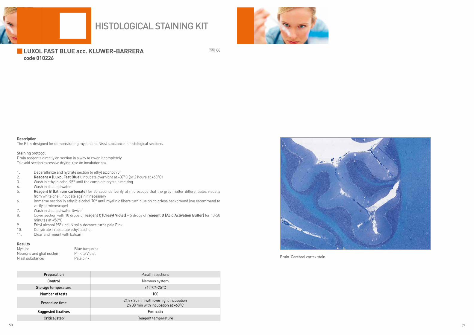
DescriptionThe Kit is designed for demonstrating myelin and Nissl substance in histological sections.
staining protocolDrain reagents directly on section in a way to cover it completely.To avoid section excessive drying, use an incubator box.
1. Deparaffinize and hydrate section to ethyl alcohol 95°2. Reagent a (luxol Fast Blue), incubate overnight at +37°C (or 2 hours at +60°C)3. Wash in ethyl alcohol 95° until the complete crystals melting 4. Wash in distilled water5. Reagent B (lithium carbonate) for 30 seconds (verify at microscope that the gray matter differentiates visually
from white one). Incubate again if necessary6. Immerse section in ethylic alcohol 70° until myelinic fibers turn blue on colorless background (we recommend to
verify at microscope)7. Wash in distilled water (twice)8. Cover section with 10 drops of reagent c (cresyl Violet) + 5 drops of reagent D (acid activation Buffer) for 10-20
minutes at +56°C9. Ethyl alcohol 95° until Nissl substance turns pale Pink10. Dehydrate in absolute ethyl alcohol11. Clear and mount with balsam
ResultsMyelin: Blue turquoiseNeurons and glial nuclei: Pink to VioletNissl substance: Pale pink
Preparation Paraffin sections
Control Nervous system
Storage temperature +15°C/+25°C
Number of tests 100
Procedure time24h + 25 min with overnight incubation
2h 30 min with incubation at +60°C
Suggested fixatives Formalin
Critical step Reagent temperature
HiStologiCal StaiNiNg Kit
Brain. Cerebral cortex stain.
60 61
malloRy tRicHRome acc. mcFaRlanecode 010227
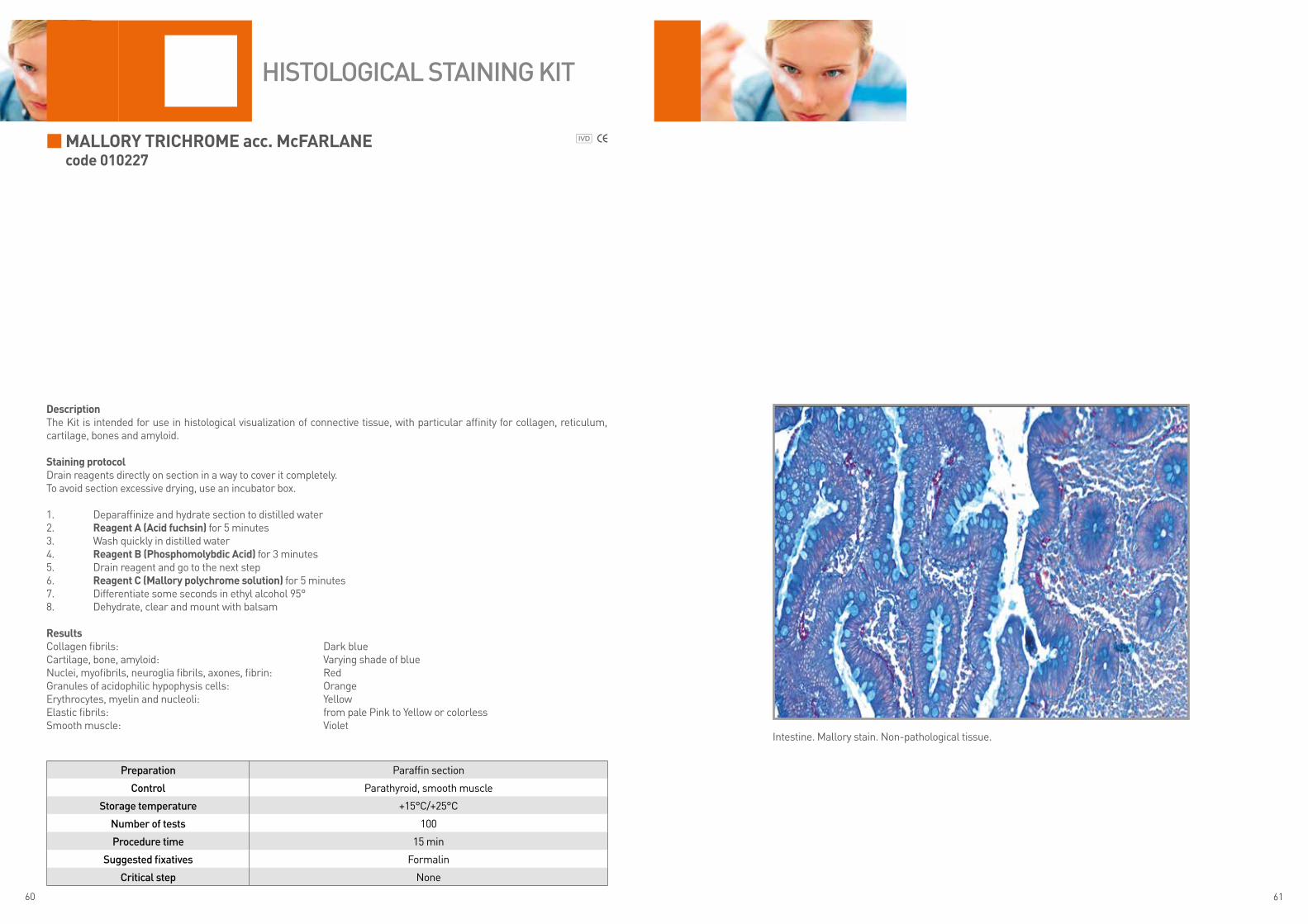
DescriptionThe Kit is intended for use in histological visualization of connective tissue, with particular affinity for collagen, reticulum, cartilage, bones and amyloid.
staining protocolDrain reagents directly on section in a way to cover it completely.To avoid section excessive drying, use an incubator box.
1. Deparaffinize and hydrate section to distilled water2. Reagent a (acid fuchsin) for 5 minutes3. Wash quickly in distilled water4. Reagent B (Phosphomolybdic acid) for 3 minutes5. Drain reagent and go to the next step6. Reagent c (mallory polychrome solution) for 5 minutes7. Differentiate some seconds in ethyl alcohol 95° 8. Dehydrate, clear and mount with balsam
ResultsCollagen fibrils: Dark blueCartilage, bone, amyloid: Varying shade of blueNuclei, myofibrils, neuroglia fibrils, axones, fibrin: RedGranules of acidophilic hypophysis cells: OrangeErythrocytes, myelin and nucleoli: YellowElastic fibrils: from pale Pink to Yellow or colorlessSmooth muscle: Violet
Preparation Paraffin section
Control Parathyroid, smooth muscle
Storage temperature +15°C/+25°C
Number of tests 100
Procedure time 15 min
Suggested fixatives Formalin
Critical step None
HiStologiCal StaiNiNg Kit
Intestine. Mallory stain. Non-pathological tissue.
62 63
masson Fontanacode 010228
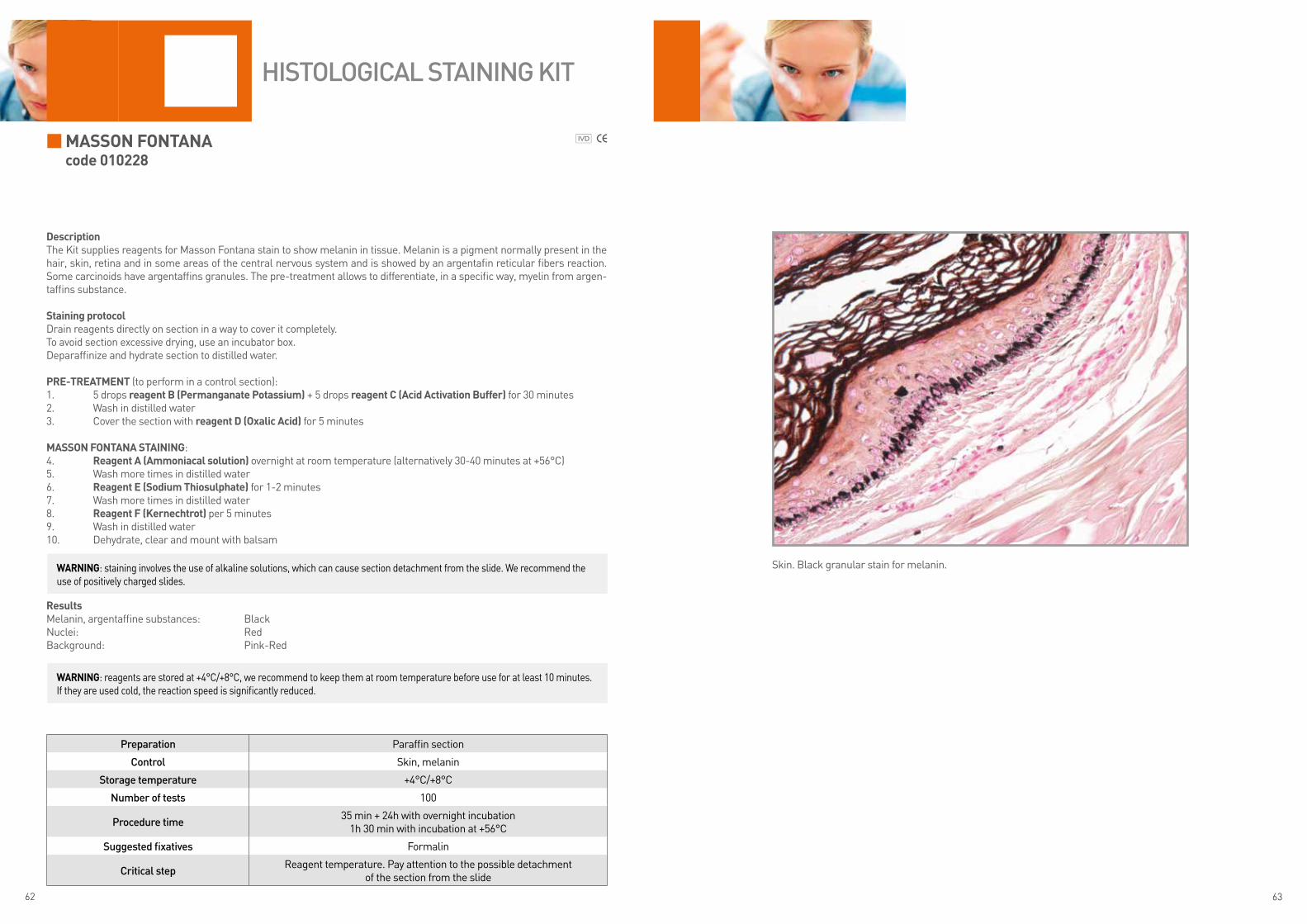
DescriptionThe Kit supplies reagents for Masson Fontana stain to show melanin in tissue. Melanin is a pigment normally present in the hair, skin, retina and in some areas of the central nervous system and is showed by an argentafin reticular fibers reaction. Some carcinoids have argentaffins granules. The pre-treatment allows to differentiate, in a specific way, myelin from argen-taffins substance.
staining protocolDrain reagents directly on section in a way to cover it completely.To avoid section excessive drying, use an incubator box.Deparaffinize and hydrate section to distilled water.
PRe-tReatment (to perform in a control section):1. 5 drops reagent B (Permanganate Potassium) + 5 drops reagent c (acid activation Buffer) for 30 minutes2. Wash in distilled water3. Cover the section with reagent D (oxalic acid) for 5 minutes
masson Fontana staininG:4. Reagent a (ammoniacal solution) overnight at room temperature (alternatively 30-40 minutes at +56°C)5. Wash more times in distilled water6. Reagent e (sodium thiosulphate) for 1-2 minutes7. Wash more times in distilled water8. Reagent F (kernechtrot) per 5 minutes9. Wash in distilled water10. Dehydrate, clear and mount with balsam
ResultsMelanin, argentaffine substances: BlackNuclei: RedBackground: Pink-Red
Preparation Paraffin section
Control Skin, melanin
Storage temperature +4°C/+8°C
Number of tests 100
Procedure time35 min + 24h with overnight incubation
1h 30 min with incubation at +56°C
Suggested fixatives Formalin
Critical stepReagent temperature. Pay attention to the possible detachment
of the section from the slide
HiStologiCal StaiNiNg Kit
Skin. Black granular stain for melanin.WaRninG: staining involves the use of alkaline solutions, which can cause section detachment from the slide. We recommend the use of positively charged slides.
WaRninG: reagents are stored at +4°C/+8°C, we recommend to keep them at room temperature before use for at least 10 minutes. If they are used cold, the reaction speed is significantly reduced.
64 65
masson tRicHRome acc. caPelli (WitH aniline Blue) code 010210
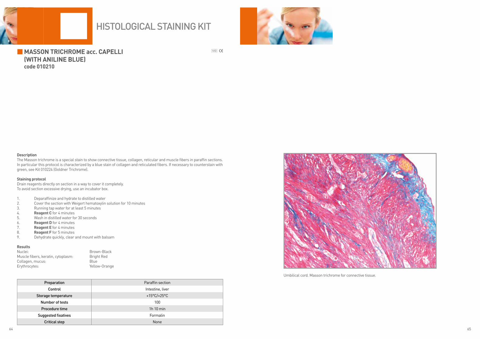
DescriptionThe Masson trichrome is a special stain to show connective tissue, collagen, reticular and muscle fibers in paraffin sections. In particular this protocol is characterized by a blue stain of collagen and reticulated fibers. If necessary to counterstain with green, see Kit 010224 (Goldner Trichrome).
staining protocolDrain reagents directly on section in a way to cover it completely.To avoid section excessive drying, use an incubator box.
1. Deparaffinize and hydrate to distilled water2. Cover the section with Weigert hematoxylin solution for 10 minutes3. Running tap water for at least 5 minutes4. Reagent c for 4 minutes5. Wash in distilled water for 30 seconds6. Reagent D for 4 minutes 7. Reagent e for 4 minutes 8. Reagent F for 5 minutes9. Dehydrate quickly, clear and mount with balsam
ResultsNuclei: Brown-BlackMuscle fibers, keratin, cytoplasm: Bright RedCollagen, mucus: BlueErythrocytes: Yellow-Orange
Preparation Paraffin section
Control Intestine, liver
Storage temperature +15°C/+25°C
Number of tests 100
Procedure time 1h 10 min
Suggested fixatives Formalin
Critical step None
HiStologiCal StaiNiNg Kit
Umbilical cord. Masson trichrome for connective tissue.
66 67
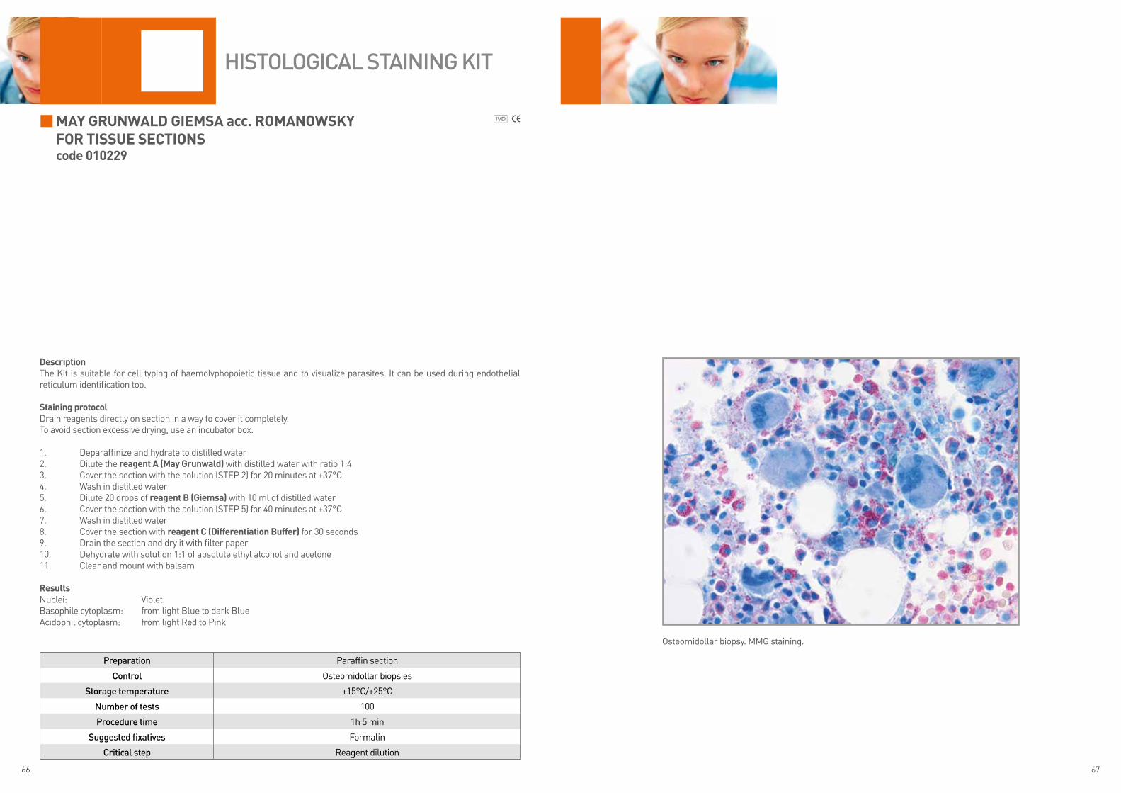
may GRunWalD Giemsa acc. RomanoWsky FoR tissue sectionscode 010229
DescriptionThe Kit is suitable for cell typing of haemolyphopoietic tissue and to visualize parasites. It can be used during endothelial reticulum identification too.
staining protocolDrain reagents directly on section in a way to cover it completely.To avoid section excessive drying, use an incubator box.
1. Deparaffinize and hydrate to distilled water2. Dilute the reagent a (may Grunwald) with distilled water with ratio 1:43. Cover the section with the solution (STEP 2) for 20 minutes at +37°C4. Wash in distilled water5. Dilute 20 drops of reagent B (Giemsa) with 10 ml of distilled water6. Cover the section with the solution (STEP 5) for 40 minutes at +37°C7. Wash in distilled water8. Cover the section with reagent c (Differentiation Buffer) for 30 seconds9. Drain the section and dry it with filter paper10. Dehydrate with solution 1:1 of absolute ethyl alcohol and acetone11. Clear and mount with balsam
ResultsNuclei: VioletBasophile cytoplasm: from light Blue to dark Blue Acidophil cytoplasm: from light Red to Pink
Preparation Paraffin section
Control Osteomidollar biopsies
Storage temperature +15°C/+25°C
Number of tests 100
Procedure time 1h 5 min
Suggested fixatives Formalin
Critical step Reagent dilution
HiStologiCal StaiNiNg Kit
Osteomidollar biopsy. MMG staining.
68 69
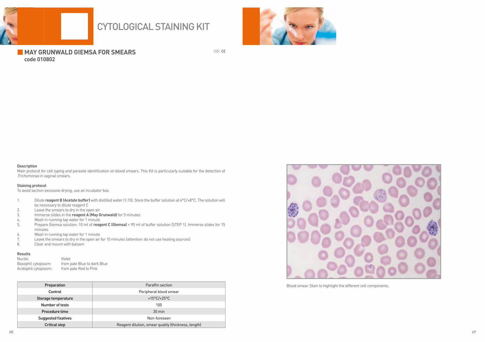
may GRunWalD Giemsa FoR smeaRscode 010802
DescriptionMain protocol for cell typing and parasite identification on blood smears. This Kit is particularly suitable for the detection of Trichomonas in vaginal smears.
staining protocolTo avoid section excessive drying, use an incubator box.
1. Dilute reagent B (acetate buffer) with distilled water (1:10). Store the buffer solution at 4°C/+8°C. The solution will be necessary to dilute reagent C
2. Leave the smears to dry in the open air3. Immerse slides in the reagent a (may Grunwald) for 5 minutes4. Wash in running tap water for 1 minute5. Prepare Giemsa solution: 10 ml of reagent c (Giemsa) + 90 ml of buffer solution (STEP 1). Immerse slides for 15
minutes6. Wash in running tap water for 1 minute7. Leave the smears to dry in the open air for 10 minutes (attention: do not use heating sources)8. Clear and mount with balsam
ResultsNuclei: VioletBasophil cytoplasm: from pale Blue to dark BlueAcidophil cytoplasm: from pale Red to Pink
Preparation Paraffin section
Control Peripheral blood smear
Storage temperature +15°C/+25°C
Number of tests 100
Procedure time 35 min
Suggested fixatives Non-foreseen
Critical step Reagent dilution, smear quality (thickness, length)
CytologiCal StaiNiNg Kit
Blood smear. Stain to highlight the different cell components.

70 71
moVat PentacHRome staincode 010247
DescriptionThe Kit is intended for use in histological visualization of: collagen, muscle tissue, reticular fibers, mucins and fibrin in par-affin sections.
staining protocolDrain reagents directly on section in a way to cover it completely.To avoid section excessive drying, use an incubator box.
1. Deparaffinize and hydrate to distilled water2. Reagent a (alcian Blue) for 20 minutes3. Wash in running tap water for 5 minutes4. Reagent B (alkaline alcoholic solution) for 60 minutes5. Wash in running tap water for 10 minutes6. Prepare reagent c adding reagents C1 and C2: c1 (Hematoxylin 1 solution) + c2 (Hematoxylin 2 solution) *7. Cover the section with the solution for 15 minutes8. Wash in distilled water9. Reagent D (Ferric chloride) until elastic fibers turn black10. Wash in distilled water11. Wash in 0.5% acetic acid solution in distilled water12. Reagent e (tiosulphate sodium) for 1 minute13. Wash in running tap water for 10 minutes14. Wash in distilled water15. Reagent F (Briebrich scarlet solution – acid Fuchsin) for 3 minutes16. Wash in 0.5% acetic acid solution in distilled water17. Reagent G (Phosphotungstic acid) for 10 minutes18. Wash in 0.5% acetic acid solution in distilled water19. Immerse slides in absolute ethyl alcohol (2 quick washings)20. Reagent H (alcoholic safranin) for 15 minutes21. Wash in absolute ethyl alcohol for 2 minutes 22. Clear and mount with balsam
*The hematoxylin solution (reagent c1+reagent c2) is stable for about 6-9 months; store at room temperature. Alternatively, add 2 drops of reagent c1 + 1 drop reagent c2 and cover the section.
ResultsNuclei and elastic fibers: Brown-Black Collagen and reticular fibers: YellowMucins: BlueFibrinoid substance, fibrin: Dark RedMuscle: Red
Preparation Paraffin section
Control Intestine, liver
Storage temperature +15°C/+25°C
Number of tests 100
Procedure time 2h 30 min
Suggested fixatives Formalin
Critical step Reagent C with limited stability
HiStologiCal StaiNiNg Kit
Intestine, colon. Polychromic stain. Non-pathological tissue.

72 73
MucicarMine acc. Mayercode 010245
DescriptionThe Kit supplies reagents to visualize acid mucins with mucicarmine stain. Counterstaining with Weigert hematoxylin and Yellow Methanil.
Staining protocolDrain reagents directly on section in a way to cover it completely.To avoid section excessive drying, use an incubator box.
1. Deparaffinize and hydrate to distilled water2. 5 drops of reagent a (Weigert hematoxylin sol. a) + 5 drops of reagent B (Weigert hematoxylin sol. B) for 2-5
minutes3. Wash in running tap water for 10 minutes4. Prepare the mucicarmine solution: 10 drops of reagent c (Mayer Mucicarmine) in 1 ml of distilled water5. Cover the section with the solution for 60 minutes at room temperature6. Wash in distilled water7. reagent D (yellow Methanil) for 1 minute8. Wash in distilled water9. Dehydrate quickly, clear and mount with balsam
resultsNuclei: Black Acid mucins: different shades of RedOther components, neutral mucins: Light Yellow
Preparation Paraffin section
Control Intestine
Storage temperature +15°C/+25°C
Number of tests 100
Procedure time 1h 25 min
Suggested fixatives Formalin
Critical step Use fresh sections; dilute reagent C before the use
HiStologiCal StaiNiNg Kit
Intestine. Pink stain of mucins.

74 75
Oil reD O acc. JOhnSOncode 010303
DescriptionThe Kit supplies reagents to show lipids with Oil Red O stain. Oil Red O is a stain with lisochromic features. Due to these fea-tures, the chromophore is soluble in lipids that turn its color.
Staining protocolDrain reagents directly on section in a way to cover it completely.To avoid section excessive drying, use an incubator box.
1. Hydrate the section to distilled water2. Working solution: 8 ml of reagent a (Oil red O) + 5 ml of reagent B (activation basic buffer). Let stand at least 10
minutes before the use*3. Cut section at cryostat4. Fix the section immersing the slide in ready to use formalin (non-equipped reagent in the Kit) for 1 minute5. Wash in running tap water6. Cover the section with the working solution (STEP 1) for 10 minutes7. Wash in running tap water8. reagent c (Mayer hematoxylin) for 3 minutes 9. Wash in running tap water for 1-2 minutes10. Mount with watery mounting media
* The diluted solution is stable up to 24 hours
resultsLipids: RedNuclei: Blue-Violet
Preparation Section at cryostat
Control Fat tissue
Storage temperature +15°C/+25°C
Number of tests 100
Procedure time 25 min
Suggested fixatives Formalin
Critical step Let stand the working solution for 10 minutes before the use
HiStologiCal StaiNiNg Kit
Frozen tissue. Lipid Red stain.

76 77
ParalDehyDe fuchSin acc. GOMOricode 010235
DescriptionThe Kit is intended for use in histological visualization of elastic fibers and pancreas endocrine components with visualization of nuclei in blue and the connective tissue in green.
Staining protocolDrain reagents directly on section in a way to cover it completely.To avoid section excessive drying, use an incubator box.
1. Deparaffinize and hydrate section to distilled water2. reagent a (Potassium Permanganate) 5 drops + reagent B (activation acid buffer) 5 drops for 10 minutes3. Wash in distilled water4. reagent c (Oxalic acid) for 5 minutes5. Wash in distilled water6. reagent D (Differentiation solution) for 5 minutes. Drain slide and go to the next step7. reagent e (Paraldehyde fuchsin) for 20 minutes. Drain slide and go to the next step8. reagent f (Differentiation solution) for 5 minutes9. Wash in distilled water10. reagent G (Mayer hematoxylin) for 5 minutes11. Wash in running tap water for 5 minutes12. reagent h (light Green) for 5 minutes13. Wash in distilled water14. Dehydrate quickly, clear and mount with balsam
resultsPancreas beta cells granules,elastic fibers, sulphated mucins, mast cells: Dark VioletNuclei: Blue-VioletConnective tissue: Green
Preparation Paraffin section
Control Pancreas
Storage temperature +15°C/+25°C
Number of tests 100
Procedure time 1h
Suggested fixatives Formalin
Critical step None
HiStologiCal StaiNiNg Kit
Pancreas. β cells granules in dark violet.

78 79
P.a.S. - PeriODic aciD Schiff acc. hOtchkiSS-McManuScode 010231
DescriptionThe Kit supplies reagents for P.A.S. (Periodic Acid Schiff) staining. The Schiff reagent is a watery, colorless solution, used in combination with periodic acid to highlight aldehyde groups. The P.A.S. stain is mainly used to visualize the presence of glycogen but also of glycoproteins. Many tissues may result P.A.S. positive, for example: the ground substance of connective tissues, cartilage, bones, mucous cells and glands, kidney structures, hypophysis basophil cells, thyroid colloid substance.
Staining protocolDrain reagents directly on section in a way to cover it completely.To avoid section excessive drying, use an incubator box.
1. Periodic acid solution: melt reagent a (Periodic acid) in 50 ml of distilled water. Stir until the complete powder melting. Store the solution at +4°C/+8°C in tightly closed container. It has validity of 12 months
2. Deparaffinize and hydrate to distilled water3. Cover the section with periodic acid solution (STEP 1) for 20-30 minutes4. Wash in distilled water5. reagent B (Schiff reagent) for 10-30 minutes (until the section turns magenta)6. Wash in distilled water7. Washing solution: 80 ml of distilled water + 10 drops of reagent c (Metabisulphite Potassium) + 10 drops of rea-
gent D (hydrochloridric acid)8. Immerse slides in washing solution (STEP 7) for 10 minutes9. Wash in distilled water10. reagent e (Mayer hematoxylin) for 2 minutes11. Wash in running tap water for 5 minutes12. Dehydrate quickly, clear and mount with balsam
resultsNuclei: Blue-VioletPositive P.A.S. substances (glycogen): Magenta
Preparation Paraffin section – cytological specimen
Control Liver
Storage temperature +15°C/+25°C
Number of tests 100
Procedure time1h 20 min for paraffin sections
30-40 min for cytological specimens
Suggested fixatives Formalin
Critical step None
HiStologiCal StaiNiNg Kit
Liver. Glycogen shown in magenta. Nuclei counterstaining in blue.

80 81
P.a.S. - PeriODic aciD Schiff acc. MOrel-MarOnGercode 010232
DescriptionThe Kit supplies reagents for P.A.S. (Periodic Acid Schiff) staining. The Schiff reagent is a watery, colorless solution, used in combination with periodic acid to visualize aldehyde groups. The P.A.S. stain is mainly used to visualize the presence of glycogen but also of glycoproteins. Many tissues may result P.A.S. positive, for example: the ground substance of connective tissues, cartilage, bones, mucous cells and glands, kidney structures, hypophysis basophil cells, thyroid colloid substance.The staining protocol acc. Morel-Maronger is characterized by staining of connective tissue with Picroindingo Carmine.
Staining protocolDrain reagents directly on section in a way to cover it completely.To avoid section excessive drying, use an incubator box.
1. Periodic acid solution: melt reagent a (Periodic acid) in 50 ml of distilled water. Stir until the complete powder melting. Store the solution at +4°C/+8°C in tightly close container. It has validity of 12 months
2. Deparaffinize and hydrate to distilled water3. Cover the section with periodic acid solution (STEP 1) for 20-30 minutes4. Wash in distilled water5. Cover the section with reagent B (Schiff reagent) for 15-30 minutes6. Wash in distilled water7. Prepare solution: 80 ml of distilled water + 10 drops of reagent c (Metabisulphite Potassium) + 10 drops of reagent
D (hydrochloridric acid)8. Immerse slides in washing solution (STEP 7) for 5 minutes9. Wash in distilled water10. Cover the section with reagent e (Picroindingo carmine) for 5 minutes11. Wash quickly in distilled water12. Dehydrate quickly, clear and mount with balsam
resultsPositive P.A.S. substances (glycogen): MagentaConnective tissue, muscle, neuroglia, erythrocytes: Yellow-Green
Preparation Paraffin section
Control Liver
Storage temperature +4°C/+8°C
Number of tests 100
Procedure time 1h 10 min
Suggested fixatives Formalin
Critical step Use reagents at room temperature
HiStologiCal StaiNiNg Kit
Liver. Glycogen shown in magenta. Tissue connective counterstaining in yellow-green.
WarninG: reagents are stored at +4°C/+8°C, we recommend to keep them at room temperature for about 10 minutes before the use. If they are used cold, the reaction speed is significantly reduced.

82 83
P.a.S. - PeriODic aciD Schiff acc. PearSecode 010233
DescriptionThe Kit supplies reagents for P.A.S. (Periodic Acid Schiff) staining. The Schiff reagent is a watery, colorless solution, used in combination with periodic acid to highlight aldehyde groups. The P.A.S. stain is mainly used to visualize the presence of glycogen but also of glycoproteins. Many tissues may result P.A.S. positive, for example: the ground substance of connective tissues, cartilage, bones, mucous cells and glands, kidney structures, hypophysis basophil cells, thyroid colloid substance.
Staining protocolDrain reagents directly on section in a way to cover it completely.To avoid section excessive drying, use an incubator box.
1. Deparaffinize and hydrate to distilled water2. reagent a (Periodic acid) for 10 minutes3. Wash in distilled water4. reagent B (Schiff reagent) for 30 minutes5. Wash in distilled water6. Working solution: 80 ml of distilled water + 10 drops of reagent c (Metabisulphite Potassium) + 10 drops of reagent
D (hydrochloridric acid)7. Immerse slides in washing solution (STEP 6) for 10 minutes8. Wash in distilled water9. Cover the section with reagent e (Mayer hematoxylin) for 2 minutes10. Wash in running tap water for 5 minutes11. Dehydrate quickly, clear and mount with balsam
resultsNuclei: Blue-VioletPositive P.A.S. substances (glycogen): Magenta
Preparation Paraffin section
Control Liver
Storage temperature +4°C/+8°C
Number of tests 100
Procedure time 1h
Suggested fixatives Formalin
Critical step Use reagents at room temperature
HiStologiCal StaiNiNg Kit
Liver. Glycogen shown in magenta. Nuclei counterstaining in blue.
WarninG: reagents are stored at +4°C/+8°C, we recommend to keep them at room temperature for about 10 minutes before the use. If they are used cold, the reaction speed is significantly reduced.

84 85
P.a.S.M. – Silver MethenaMine acc. callarDcode 010234
DescriptionThe Kit supplies reagents of P.A.S.M. (Periodic Acid Silver Methenamine) staining protocol to highlight the basement mem-branes of kidney tissue. This staining protocol is usually called Gomori’s reaction too.The P.A.S.M. stain visualizes argyrophilic elements, mucopolysaccharides, mycetes and bacteria. The treatment with periodic acid oxidizes the carbohydrates of basement membrane with aldehyde formation: these groups allow to reduce silver with consequent visualization in black of basement membranes.
Staining protocol (*)Drain reagents directly on section in a way to cover it completely.To avoid section excessive drying, use an incubator box.
1. Deparaffinize and hydrate to distilled water2. Cover the section with reagent a – 10 minutes3. Wash in distilled water – 5 minutes4. Cover the section with 10 drops of reagent B + 10 drops of reagent c + 10 drops of reagent D 5. Incubate in oven for 60 minutes at +60°C6. Verify impregnation tone at microscope. If necessary, incubate again. The section should turn tobacco7. Leave it cool at room temperature for 5 minutes8. Wash in distilled water9. Cover the section with reagent e for 5 minutes10. Wash in distilled water11. Cover the section with reagent f for 5 minutes12. Wash in distilled water13. Immerse the section in Hematoxylin solution – 2 seconds14. Running tap water – 2 seconds15. Eosin – 2 seconds16. Dehydrate quickly, xylol or substitutes. Mount with balsam
(*) Contrast suggested with hematoxylin and eosin following these steps:1. Wash in distilled water2. Immerse in hematoxylin – 2 seconds3. Running tap water – 2 seconds4. Eosin – 2 seconds5. Dehydrate quickly, xylol or substitute. Mount with balsam
resultsBasement membranes, glycogen, mycetes and bacteria capsule: Black
Preparation Paraffin section
Control Kidney
Storage temperature +4°C/+8°C
Number of tests 100
Procedure time 1h 45 min
Suggested fixatives Formalin
Critical step Reagent temperature. Do not use metallic objects
HiStologiCal StaiNiNg Kit
Kidney. Basement membrane in black.
WarninG: use excellent distilled water for washings and not use metallic objects. Verify the real oven temperature during incubation step: +60°C are mandatory for the reaction process.
WarninG: reagents are stored at +4°C/+8°C. We recommend to keep them at room temperature for about 10 minutes before the use. If they are used cold, the reaction speed is significantly reduced.
86 87
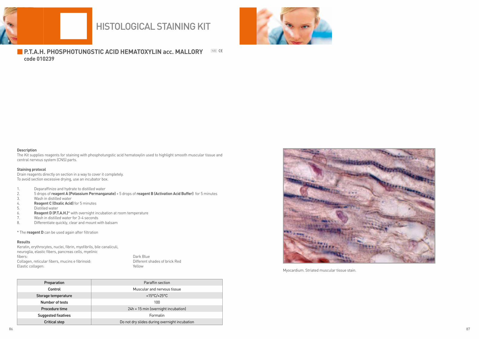
P.t.a.h. PhOSPhOtunGStic aciD heMatOxylin acc. MallOrycode 010239
DescriptionThe Kit supplies reagents for staining with phosphotungstic acid hematoxylin used to highlight smooth muscular tissue and central nervous system (CNS) parts.
Staining protocolDrain reagents directly on section in a way to cover it completely.To avoid section excessive drying, use an incubator box.
1. Deparaffinize and hydrate to distilled water2. 5 drops of reagent a (Potassium Permanganate) + 5 drops of reagent B (activation acid Buffer) for 5 minutes3. Wash in distilled water4. reagent c (Oxalic acid) for 5 minutes5. Distilled water6. reagent D (P.t.a.h.)* with overnight incubation at room temperature7. Wash in distilled water for 3-4 seconds8. Differentiate quickly, clear and mount with balsam
* The reagent D can be used again after filtration
resultsKeratin, erythrocytes, nuclei, fibrin, myofibrils, bile canaliculi,neuroglia, elastic fibers, pancreas cells, myelinicfibers: Dark BlueCollagen, reticular fibers, mucins e fibrinoid: Different shades of brick RedElastic collagen: Yellow
Preparation Paraffin section
Control Muscular and nervous tissue
Storage temperature +15°C/+25°C
Number of tests 100
Procedure time 24h + 15 min (overnight incubation)
Suggested fixatives Formalin
Critical step Do not dry slides during overnight incubation
HiStologiCal StaiNiNg Kit
Myocardium. Striated muscular tissue stain.
88 89
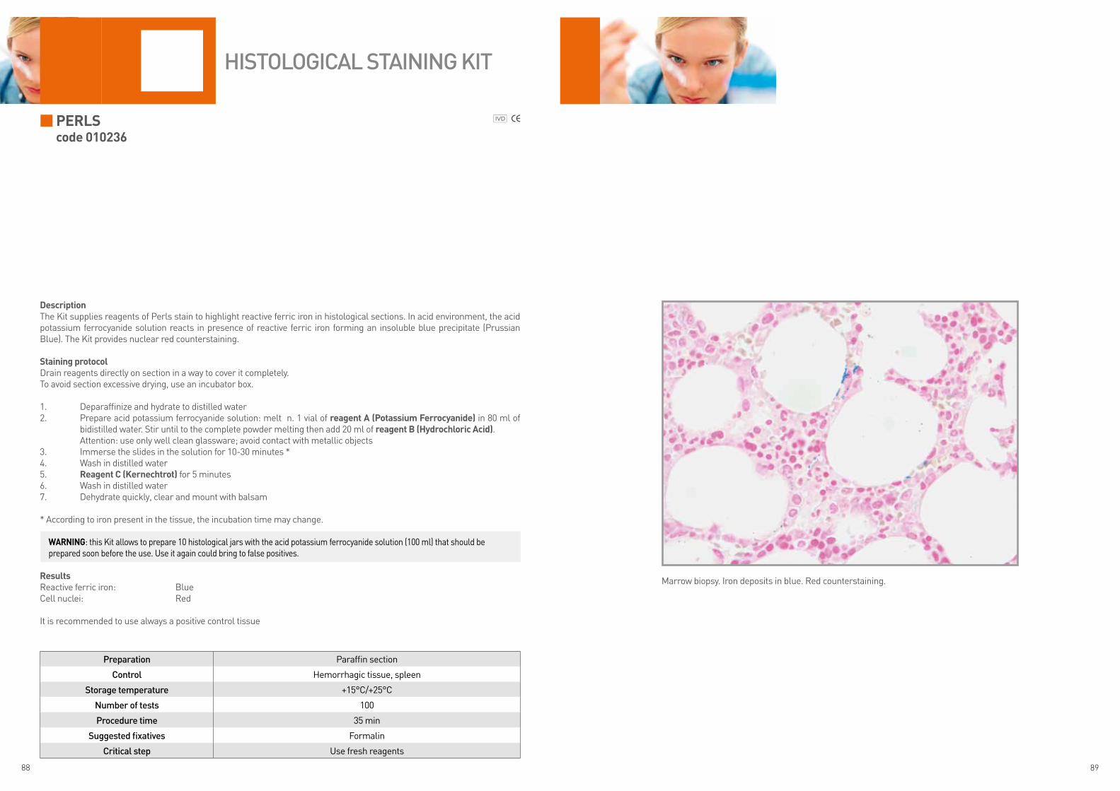
PerlS code 010236
DescriptionThe Kit supplies reagents of Perls stain to highlight reactive ferric iron in histological sections. In acid environment, the acid potassium ferrocyanide solution reacts in presence of reactive ferric iron forming an insoluble blue precipitate (Prussian Blue). The Kit provides nuclear red counterstaining.
Staining protocolDrain reagents directly on section in a way to cover it completely.To avoid section excessive drying, use an incubator box.
1. Deparaffinize and hydrate to distilled water2. Prepare acid potassium ferrocyanide solution: melt n. 1 vial of reagent a (Potassium ferrocyanide) in 80 ml of
bidistilled water. Stir until to the complete powder melting then add 20 ml of reagent B (hydrochloric acid). Attention: use only well clean glassware; avoid contact with metallic objects3. Immerse the slides in the solution for 10-30 minutes *4. Wash in distilled water5. reagent c (kernechtrot) for 5 minutes6. Wash in distilled water7. Dehydrate quickly, clear and mount with balsam
* According to iron present in the tissue, the incubation time may change.
resultsReactive ferric iron: BlueCell nuclei: Red
It is recommended to use always a positive control tissue
Preparation Paraffin section
Control Hemorrhagic tissue, spleen
Storage temperature +15°C/+25°C
Number of tests 100
Procedure time 35 min
Suggested fixatives Formalin
Critical step Use fresh reagents
HiStologiCal StaiNiNg Kit
Marrow biopsy. Iron deposits in blue. Red counterstaining.
WarninG: this Kit allows to prepare 10 histological jars with the acid potassium ferrocyanide solution (100 ml) that should be prepared soon before the use. Use it again could bring to false positives.

90 91
PerlS-van GieSOncode 010237
DescriptionThe Kit supplies reagents of Perls stain to visualize reactive ferric iron in histological sections. The acid potassium ferrocya-nide solution reacts in presence of reactive ferric iron forming an insoluble blue precipitate (Prussian Blue). The Kit supplies as counterstaining the picrofuchsin acc. Van Gieson, a solution obtained by mixing acid fuchsin with picric acid. This solution selectively stains connective tissue, collagen, muscle tissue and thickened epithelium besides glial fibrils and cytoplasm.
Staining protocolDrain reagents directly on section in a way to cover it completely.To avoid section excessive drying, use an incubator box.
1. Deparaffinize and hydrate to distilled water2. Prepare acid potassium ferrocyanide solution: melt n. 1 vial of reagent a (Potassium ferrocyanide) in 80 ml of
bidistilled water. Stir until the complete powder melting then add 20 ml of reagent B (hydrochloric acid). Attention: use only well clean glassware, avoid contact with metallic objects3. Immerse the slides in the solution for 10-30 minutes*4. Wash in distilled water5. Cover the section with reagent c (Picrofuchsin) for 5 minutes6. Wash in distilled water7. Dehydrate quickly, clear and mount with balsam
* According to iron present in the tissue, the incubation time may change
resultsReactive ferric iron: BlueCollagen: Purple RedCytoplasm, muscle, corneum stratum of the epithelium, neuroglia fibers and erythrocytes: Yellow
It is recommended to use always a positive control tissue
Preparation Paraffin section
Control Hemorrhagic tissue, spleen
Storage temperature +4°C/+8°C
Number of tests 100
Procedure time 35 min
Suggested fixatives Formalin
Critical step Use fresh reagents
HiStologiCal StaiNiNg Kit
Spleen, autopsy case. Iron deposits in blue. Counterstaining of connective tissue with Van Gieson stain.
WarninG: this Kit allows to prepare 10 histological jars with the acid potassium ferrocyanide solution (100 ml) that should be prepared soon before the use. Use it again could bring to false positives.
WarninG: reagents are stored at +4°C/+8°C, we recommend to keep them at room temperature for about 10 minutes before the use. If they are used cold, the reaction speed is significantly reduced.

92 93
PicrO MallOry trichrOMe acc. lenDruMcode 010238
DescriptionThe Kit is designed for demonstrating the different components of connective tissue.
Staining protocolDrain reagents directly on section in a way to cover it completely.To avoid section excessive drying, use an incubator box.
1. Deparaffinize and hydrate to distilled water2. Cover the section with 5 drops of reagent a (Weigert hematoxylin sol. a) and 5 drops of reagent B (Weigert hema-
toxylin sol. B) for 10 minutes3. Wash in running tap water for 10 minutes4. reagent c (Picro Mallory Orange G) for 2 minutes5. Wash in distilled water6. reagent D (fuchsin Ponceau) for 1 minute7. Wash in distilled water8. reagent e (Phosphomolybdic acid) for 15 minutes9. Wash in distilled water10. reagent f (aniline blue) for 1 minute11. Differentiate quickly in ethyl alcohol 95°12. Complete dehydration with absolute ethyl alcohol13. Clear and mount with balsam
resultsNuclei: Dark BrownCollagen fibers: Dark BlueGround substance of cartilage and bone, mucus, basophil granules of hypophysis and amyloid: different shades of BlueNeuroglia and fibroglia: RedAcidophil granules of hypophysis: OrangeMyelin and erythrocytes: YellowElastic fibers: from pale Pink to Yellow or colorless
Preparation Paraffin sections
Control Connective tissue
Storage temperature +15°C/+25°C
Number of tests 100
Procedure time 45 min
Suggested fixatives Formalin
Critical step None
HiStologiCal StaiNiNg Kit
Skin. Hair follicle stain.
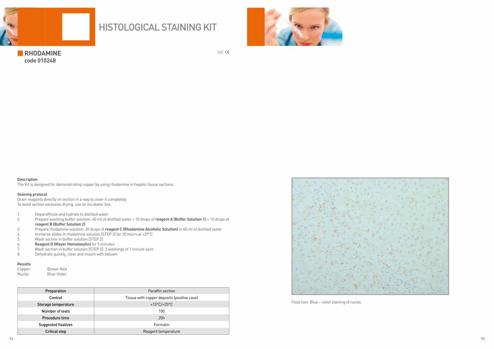
94 95
rhODaMinecode 010248
DescriptionThe Kit is designed for demonstrating copper by using rhodamine in hepatic tissue sections.
Staining protocolDrain reagents directly on section in a way to cover it completely.To avoid section excessive drying, use an incubator box.
1. Deparaffinize and hydrate to distilled water2. Prepare washing buffer solution: 40 ml of distilled water + 10 drops of reagent a (Buffer Solution 1) + 10 drops of
reagent B (Buffer Solution 2)3. Prepare rhodamine solution: 20 drops of reagent c (rhodamine alcoholic Solution) in 40 ml of distilled water4. Immerse slides in rhodamine solution (STEP 3) for 20 hours at +37°C5. Wash section in buffer solution (STEP 2)6. reagent D (Mayer hematoxylin) for 5 minutes7. Wash section in buffer solution (STEP 2): 3 washings of 1 minute each8. Dehydrate quickly, clear and mount with balsam
resultsCopper: Brown-RedNuclei: Blue-Violet
Preparation Paraffin section
Control Tissue with copper deposits (positive case)
Storage temperature +15°C/+25°C
Number of tests 100
Procedure time 20h
Suggested fixatives Formalin
Critical step Reagent temperature
HiStologiCal StaiNiNg Kit
Fetal liver. Blue – violet staining of nuclei.

96 97
Silver iMPreGnatiOncode 010211
DescriptionThe Kit is intended for use in histological visualization of connective tissue fibers. The argirophilic feature of reticular fibers is due to the capacity to bind silver salts, which reduced to metallic silver, give the typical black color.
Staining protocolDrain reagents directly on section in a way to cover it completely.To avoid section excessive drying, use an incubator box.
1. Deparaffinize and hydrate section to distilled water2. Prepare the working solution: 5 drops of reagent a (Potassium Permanganate) + 5 drops of reagent B (activation
acid Buffer). Cover the section with the solution for 5 minutes3. Wash in distilled water4. reagent c (Oxalic acid) for 3 minutes. Wash in distilled water5. reagent D (Sulphate ammonium iron) for 2 minutes. Wash in distilled water6. reagent e (ammoniacal Solution) for 2 minutes. Wash in distilled water7. reagent f (aldehyde formica in Solution) for 2 minutes. Wash in distilled water8. reagent G (Sodium thiosulfate) for 4 minutes. Wash in running tap water for 5 minutes9. Dehydrate quickly, clear and mount with balsam
resultsReticular and nervous fibers: BlackConnective tissue: BrownCollagen: Yellow
Preparation Paraffin section
Control Liver
Storage temperature +4°C/+8°C
Number of tests 100
Procedure time 25 min
Suggested fixatives Formalin
Critical stepDo not use metallic objects. Use fresh reagents. Pay attention to the possible
detachment of the section from the slide.
HiStologiCal StaiNiNg Kit
Fetal liver. Brown-black pattern of vessels.Note: the liver reticular structure isn’t fully formed as in adult tissue.
WarninG: the staining involves the use of alkaline solutions which can cause the detachment of the section from the slide. We suggest the use of positively charged slides.
WarninG: reagents are stored at +4°C/+8°C, we recommend to keep them at room temperature before use for at least 10 minutes. If they are used cold, the reaction speed is significantly reduced.

98 99
SiriuS reD fOr aMylOiDcode 010306
DescriptionThe Kit supplies reagents of Red Sirius stain to highlight amyloid, a colorless, insoluble protein with low molecular weight.
Staining protocolDrain reagents directly on section in a way to cover it completely.To avoid section excessive drying, use an incubator box.
1. Deparaffinize and hydrate to distilled water2. reagent a (Mayer hematoxylin) for 3 minutes3. Wash in running tap water for 5 minutes4. reagent B (Differentiation alcoholic Buffer) for 30 seconds5. Wash in running tap water for 1 minute6. reagent c (red Sirius) for 60 minutes7. Wash in running tap water for 10 minutes8. 2 quick steps in absolute ethyl alcohol 9. Clear and mount with balsam
resultsNuclei: Blue-VioletAmyloid: different shades of Red
Preparation Paraffin section
Control Positive case (Amyloidosis)
Storage temperature +15°C/+25°C
Number of tests 100
Procedure time 1h 25 min
Suggested fixatives Formalin
Critical step None
HiStologiCal StaiNiNg Kit
Connective tissue. Red stain for amyloid.

100 101
SiriuS reD fOr cOllaGencode 010254
DescriptionThe Kit supplies reagents of Sirius Red stain to show collagen type I and III. The collagen type I represents the 90% of the total collagen present in connective tissues, bones, skin, tendons. The collagen type III is present in reticular fibers and tissue, skin, smooth muscle.
Staining protocolDrain reagents directly on section in a way to cover it completely.To avoid section excessive drying, use an incubator box.
1. Deparaffinize and hydrate to distilled water2. reagent a (Picric red Sirius) for 60 minutes3. Wash in distilled water4. reagent B (Mayer hematoxylin) for 10 minutes5. Wash in running tap water for 10 minutes6. 2 quick steps in absolute ethyl alcohol7. Clear and mount with balsam
resultsNuclei: Blue-VioletCollagen: different shades of RedBackground, erythrocytes: Yellow
Preparation Paraffin section
Control Tissue with collagen
Storage temperature +15°C/+25°C
Number of tests 100
Procedure time 1h 20 min
Suggested fixatives Formalin
Critical step None
HiStologiCal StaiNiNg Kit
Skin. Stain of collagen vessels under derma.
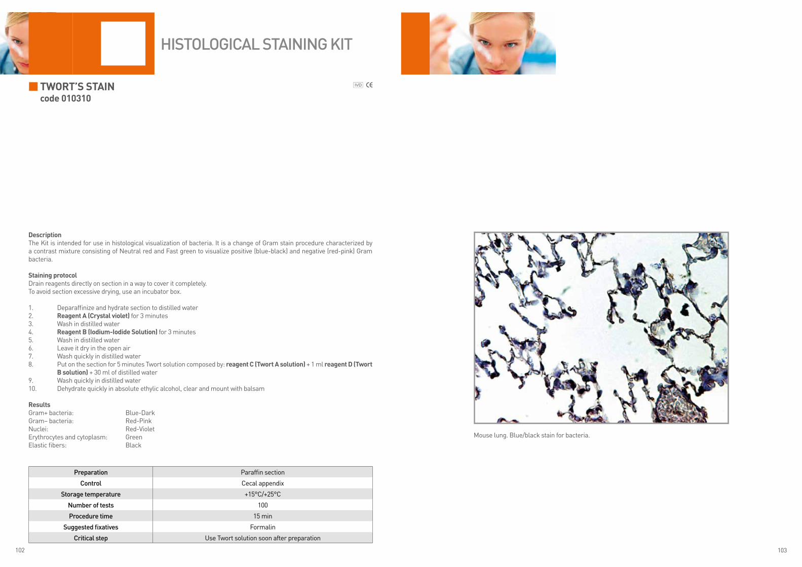
102 103
tWOrt’S Staincode 010310
DescriptionThe Kit is intended for use in histological visualization of bacteria. It is a change of Gram stain procedure characterized by a contrast mixture consisting of Neutral red and Fast green to visualize positive (blue-black) and negative (red-pink) Gram bacteria.
Staining protocolDrain reagents directly on section in a way to cover it completely.To avoid section excessive drying, use an incubator box.
1. Deparaffinize and hydrate section to distilled water2. reagent a (crystal violet) for 3 minutes 3. Wash in distilled water4. reagent B (iodium-iodide Solution) for 3 minutes5. Wash in distilled water6. Leave it dry in the open air 7. Wash quickly in distilled water8. Put on the section for 5 minutes Twort solution composed by: reagent c (twort a solution) + 1 ml reagent D (twort
B solution) + 30 ml of distilled water9. Wash quickly in distilled water10. Dehydrate quickly in absolute ethylic alcohol, clear and mount with balsam
resultsGram+ bacteria: Blue-DarkGram– bacteria: Red-PinkNuclei: Red-VioletErythrocytes and cytoplasm: GreenElastic fibers: Black
Preparation Paraffin section
Control Cecal appendix
Storage temperature +15°C/+25°C
Number of tests 100
Procedure time 15 min
Suggested fixatives Formalin
Critical step Use Twort solution soon after preparation
HiStologiCal StaiNiNg Kit
Mouse lung. Blue/black stain for bacteria.
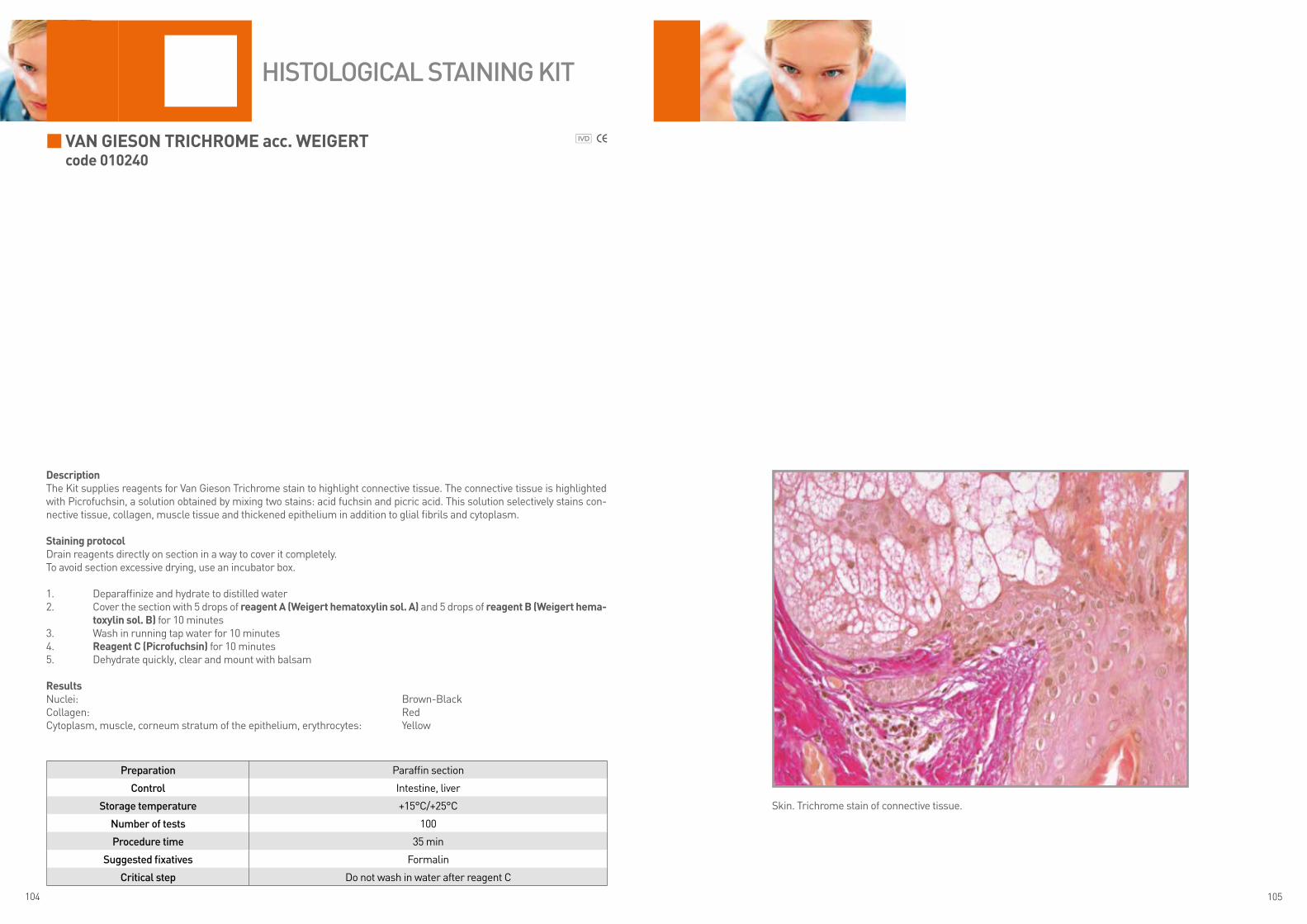
104 105
van GieSOn trichrOMe acc. WeiGertcode 010240
DescriptionThe Kit supplies reagents for Van Gieson Trichrome stain to highlight connective tissue. The connective tissue is highlighted with Picrofuchsin, a solution obtained by mixing two stains: acid fuchsin and picric acid. This solution selectively stains con-nective tissue, collagen, muscle tissue and thickened epithelium in addition to glial fibrils and cytoplasm.
Staining protocolDrain reagents directly on section in a way to cover it completely.To avoid section excessive drying, use an incubator box.
1. Deparaffinize and hydrate to distilled water2. Cover the section with 5 drops of reagent a (Weigert hematoxylin sol. a) and 5 drops of reagent B (Weigert hema-
toxylin sol. B) for 10 minutes3. Wash in running tap water for 10 minutes4. reagent c (Picrofuchsin) for 10 minutes5. Dehydrate quickly, clear and mount with balsam
resultsNuclei: Brown-Black Collagen: RedCytoplasm, muscle, corneum stratum of the epithelium, erythrocytes: Yellow
Preparation Paraffin section
Control Intestine, liver
Storage temperature +15°C/+25°C
Number of tests 100
Procedure time 35 min
Suggested fixatives Formalin
Critical step Do not wash in water after reagent C
HiStologiCal StaiNiNg Kit
Skin. Trichrome stain of connective tissue.

106 107
Verhoeff’s stain code 010308
DescriptionThe Kit is intended for use in histological visualization of elastic fibers. It can be used instead to Wiegert’s stain. The elastic fibers are visualized very intensely but not selectively by different special stains. The Verhoeff’s stain is characterized by a treatment based on iodine and Weigert hematoxylin.
staining protocolDrain reagents directly on section in a way to cover it completely.To avoid section excessive drying, use an incubator box.
1. Deparaffinize and hydrate section to distilled water 2. Verhoeff solution: 10 drops of reagent a (Weigert hematoxylin sol. a) + 10 drops of reagent B (Weigert hematox-
ylin sol. B) + 5 drops of reagent C (iodium- iodide solution)3. Put solution on the section for 30 minutes4. Wash in distilled water5. reagent D (Differentiation solution) for 2 minutes6. Wash in distilled water7. reagent e (alcoholic solution) for 2 minutes8. Wash in distilled water9. reagent f (Van Gieson contrast solution*) for 2 minutes10. Dehydrate quickly, clear and mount with balsam
* The counterstaining is optional.
resultsElastic fibers: BlackNuclei: Brown-BlackCollagen*: RedConnective tissue*: Yellow
Preparation Paraffin section
Control Aorta, tissue with elastic fibers
Storage temperature +15°C/+25°C
Number of tests 100
Procedure time 40 min
Suggested fixatives Formalin
Critical step Differentiation step: if too much, do not perform all the staining protocol
HiStologiCal StaiNiNg Kit
Bladder. Black stain of elastic fibers. Counterstaining with Van Gieson.

108 109
Von Kossa acc. McGee-russelcode 010241
DescriptionThe Kit is intended for use in histological visualization of calcium. The reaction occurs by replacing calcium ions with silver nitrate and consequent formation of silver phosphate visualized as metallic silver through reducing solution action. The reac-tion is not specific for calcium but it visualizes anions (phosphate, carbonate, sulfate, oxalate).Treatment with lithium carbonate prevents staining of uric acid and its salts (false positives). The counterstaining is obtained with Kernechtrot.
staining protocolDrain reagents directly on section in a way to cover it completely.To avoid section excessive drying, use an incubator box.
1. Deparaffinize and hydrate to distilled water2. reagent a (lithium Carbonate) for 10 minutes3. Wash in distilled water4. reagent B (ammonia solution) for 60 minutes in the darkness5. Wash in distilled water 4 times6. reagent C (reducing solution) for 5 minutes (continue if necessary until silver salts become black)7. Wash in distilled water8. reagent D (thiosulfate sodium) for 5 minutes9. Wash in distilled water10. reagent e (Kernechtrot) for 5 minutes11. Wash in running water for 2 minutes12. Dehydrate quickly, clear and mount with balsam
resultsBone and calcium salts: BlackNuclei and cytoplasm: Pink-Red
Preparation Paraffin section
Control Tissue with calcium deposits
Storage temperature +4°C/+8°C
Number of tests 100
Procedure time 1h 40 min
Suggested fixatives Formalin
Critical step The step in reagent C
HiStologiCal StaiNiNg Kit
Breast, tumor. Salt deposits shown in black-brown.
WarninG: reagents are stored at +4°C/+8°C, we recommend to keep them at room temperature for at least 10 minutes before the use. If they are used cold, the reaction speed is significantly reduced.
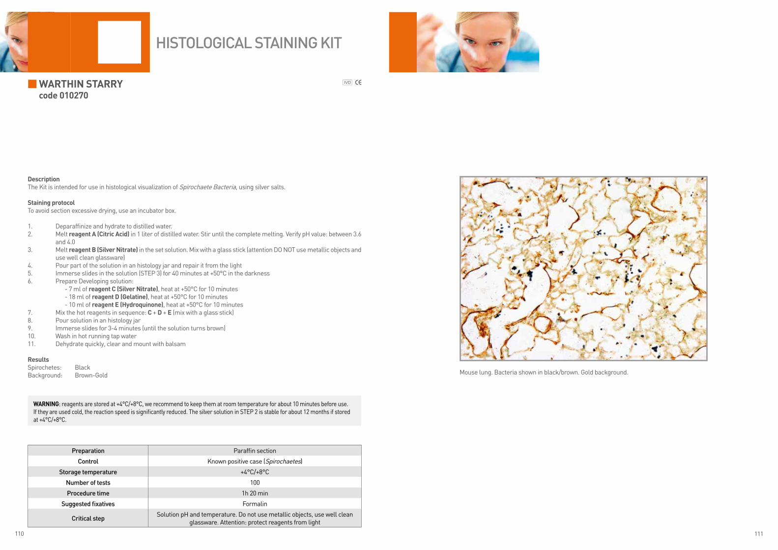
110 111
Warthin starrycode 010270
DescriptionThe Kit is intended for use in histological visualization of Spirochaete Bacteria, using silver salts.
staining protocolTo avoid section excessive drying, use an incubator box.
1. Deparaffinize and hydrate to distilled water.2. Melt reagent a (Citric acid) in 1 liter of distilled water. Stir until the complete melting. Verify pH value: between 3.6
and 4.03. Melt reagent B (silver nitrate) in the set solution. Mix with a glass stick (attention DO NOT use metallic objects and
use well clean glassware)4. Pour part of the solution in an histology jar and repair it from the light 5. Immerse slides in the solution (STEP 3) for 40 minutes at +50°C in the darkness 6. Prepare Developing solution: - 7 ml of reagent C (silver nitrate), heat at +50°C for 10 minutes - 18 ml of reagent D (Gelatine), heat at +50°C for 10 minutes - 10 ml of reagent e (hydroquinone), heat at +50°C for 10 minutes 7. Mix the hot reagents in sequence: C + D + e (mix with a glass stick)8. Pour solution in an histology jar 9. Immerse slides for 3-4 minutes (until the solution turns brown)10. Wash in hot running tap water11. Dehydrate quickly, clear and mount with balsam
resultsSpirochetes: BlackBackground: Brown-Gold
Preparation Paraffin section
Control Known positive case (Spirochaetes)
Storage temperature +4°C/+8°C
Number of tests 100
Procedure time 1h 20 min
Suggested fixatives Formalin
Critical stepSolution pH and temperature. Do not use metallic objects, use well clean
glassware. Attention: protect reagents from light
HiStologiCal StaiNiNg Kit
Mouse lung. Bacteria shown in black/brown. Gold background.
WarninG: reagents are stored at +4°C/+8°C, we recommend to keep them at room temperature for about 10 minutes before use. If they are used cold, the reaction speed is significantly reduced. The silver solution in STEP 2 is stable for about 12 months if stored at +4°C/+8°C.

112 113
WeiGert-Van Gieson, lonG MethoDcode010218
DescriptionThe Kit supplies reagents to highlight elastic fibers in paraffin sections according to Weigert’s stain using Van Gieson as coun-terstaining to highlight connective tissue.Staining with resorcin-fuchsin differentiates elastic fibers. Nuclei are shown with Weigert hematoxylin while the Picrofuchsin highlight the connective tissue.High selectivity due to long time staining protocol.
staining protocolDrain reagents directly on section in a way to cover it completely.To avoid section excessive drying, use an incubator box.
1. Deparaffinize and hydrate to distilled water2. reagent a (Periodic acid) for 5 minutes3. Pour reagent B (resorcine fuchsin) in a vertical jar for histology and immerse the section (cover the jar or prevent
ethyl alcohol evaporation). Incubate overnight in immersion at room temperature (alternatively cover the section and incubate 1 hour at +45°C in incubator box)
4. Wash in distilled water5. reagent C (Jenkins reagent) for 10 minutes6. Wash in distilled water7. Prepare Weigert hematoxylin: mix 10 drops of reagent D (Weigert hematoxylin sol. a) and 10 drops of reagent e
(Weigert hematoxylin sol. B)8. Cover the section with Weigert hematoxylin for 8-10 minutes9. Running tap water for at least 10 minutes10. reagent f (Picrofuchsin) for 5-7 minutes11. Wash quickly in distilled water12. Dehydrate quickly, clear and mount with balsam
resultsElastic fibers: from dark Blue to BlackNuclei: BlackCollagen: different shades of RedConnective tissue and erythrocytes: Yellow
Preparation Paraffin section
Control Skin
Storage temperature +15°C/+25°C
Number of tests 100
Procedure time24h + 40 min with overnight incubation1h e 40 min with incubation at +45°C
Suggested fixatives Formalin
Critical step Avoid section drying
HiStologiCal StaiNiNg Kit
Stomach. Brown-black elastic fibers in arterial vessel. Counterstaining with Van Gieson.
WarninG: reagent B can be used again after filtration.
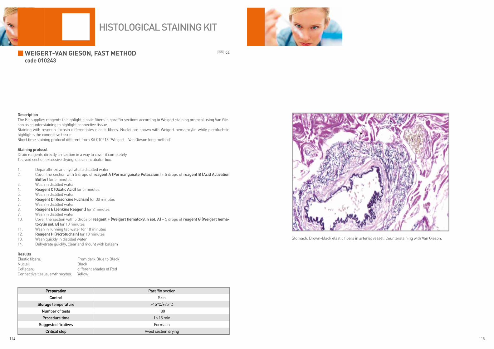
114 115
WeiGert-Van Gieson, fast MethoDcode 010243
DescriptionThe Kit supplies reagents to highlight elastic fibers in paraffin sections according to Weigert staining protocol using Van Gie-son as counterstaining to highlight connective tissue.Staining with resorcin-fuchsin differentiates elastic fibers. Nuclei are shown with Weigert hematoxylin while picrofuchsin highlights the connective tissue.Short time staining protocol different from Kit 010218 “Weigert – Van Gieson long method”.
staining protocolDrain reagents directly on section in a way to cover it completely.To avoid section excessive drying, use an incubator box.
1. Deparaffinize and hydrate to distilled water2. Cover the section with 5 drops of reagent a (Permanganate Potassium) + 5 drops of reagent B (acid activation
Buffer) for 5 minutes3. Wash in distilled water4. reagent C (oxalic acid) for 5 minutes5. Wash in distilled water6. reagent D (resorcine fuchsin) for 30 minutes7. Wash in distilled water8. reagent e (Jenkins reagent) for 2 minutes9. Wash in distilled water10. Cover the section with 5 drops of reagent f (Weigert hematoxylin sol. a) + 5 drops of reagent G (Weigert hema-
toxylin sol. B) for 10 minutes11. Wash in running tap water for 10 minutes12. reagent h (Picrofuchsin) for 10 minutes13. Wash quickly in distilled water14. Dehydrate quickly, clear and mount with balsam
resultsElastic fibers: From dark Blue to Black Nuclei: BlackCollagen: different shades of RedConnective tissue, erythrocytes: Yellow
Preparation Paraffin section
Control Skin
Storage temperature +15°C/+25°C
Number of tests 100
Procedure time 1h 15 min
Suggested fixatives Formalin
Critical step Avoid section drying
HiStologiCal StaiNiNg Kit
Stomach. Brown-black elastic fibers in arterial vessel. Counterstaining with Van Gieson.
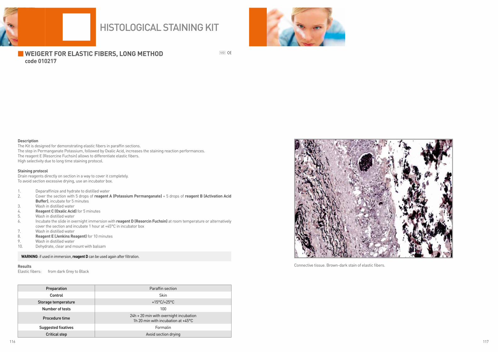
116 117
WeiGert for elastiC fiBers, lonG MethoDcode 010217
DescriptionThe Kit is designed for demonstrating elastic fibers in paraffin sections.The step in Permanganate Potassium, followed by Oxalic Acid, increases the staining reaction performances. The reagent E (Resorcine Fuchsin) allows to differentiate elastic fibers.High selectivity due to long time staining protocol.
staining protocolDrain reagents directly on section in a way to cover it completely.To avoid section excessive drying, use an incubator box.
1. Deparaffinize and hydrate to distilled water2. Cover the section with 5 drops of reagent a (Potassium Permanganate) + 5 drops of reagent B (activation acid
Buffer), incubate for 5 minutes3. Wash in distilled water4. reagent C (oxalic acid) for 5 minutes5. Wash in distilled water6. Incubate the slide in overnight immersion with reagent D (resorcin fuchsin) at room temperature or alternatively
cover the section and incubate 1 hour at +45°C in incubator box7. Wash in distilled water8. reagent e (Jenkins reagent) for 10 minutes9. Wash in distilled water10. Dehydrate, clear and mount with balsam
resultsElastic fibers: from dark Grey to Black
Preparation Paraffin section
Control Skin
Storage temperature +15°C/+25°C
Number of tests 100
Procedure time24h + 20 min with overnight incubation
1h 20 min with incubation at +45°C
Suggested fixatives Formalin
Critical step Avoid section drying
HiStologiCal StaiNiNg Kit
Connective tissue. Brown-dark stain of elastic fibers.
WarninG: if used in immersion, reagent D can be used again after filtration.

118 119
WeiGert for elastiC fiBers, fast MethoDcode 010242
DescriptionThe Kit supplies reagents for Weigert staining protocol with resorcin-fuchsin solution to highlight elastic fibers in tissue section and nuclei counterstaining with Mayer hematoxylin. The step in Potassium Permanganate, followed by Oxalic Acid, increases reaction performances.Short time staining protocol different from Kit 010217 “Weigert for elastic fibers, long method”.
staining protocolDrain reagents directly on section in a way to cover it completely.To avoid section excessive drying, use an incubator box.
1. Deparaffinize and hydrate to distilled water2. Cover the section with 5 drops of reagent a (Potassium Permangate) + 5 drops of reagent B (activation acid Buff-
er) for 5 minutes3. Wash in distilled water4. reagent C (oxalic acid) for 5 minutes5. Wash in distilled water6. reagent D (resorcin fuchsin) for 30 minutes7. Wash in distilled water8. reagent e (Jenkins reagent) for 2 minutes9. Wash in distilled water10. reagent f (Mayer hematoxylin) for 5 minutes11. Wash in running tap water for 5 minutes12. Dehydrate quickly, clear and mount with balsam
resultsElastic fibers: Purple-BrownCell nuclei: Blue-Violet
Preparation Paraffin section
Control Skin
Storage temperature +15°C/+25°C
Number of tests 100
Procedure time 55 min
Suggested fixatives Formalin
Critical step Avoid section drying
HiStologiCal StaiNiNg Kit
Connective tissue. Brown-black stain of elastic fibers. No counterstaining.

120 121
heMatoXylin
hematoxylin/Mayer hematoxylin – histoloGy
reagent features: It is a long lasting hematoxylin, red/violet colour, to be used in progressive way. Oxidizing agent: Sodium Iodate
Code Packaging
C0302 500 ml
C0303 1 ltC0305 2.5 lt
C0306 5 lt
Hematoxylin-eosin staining is the most widely used staining protocol for the study of tissue morphology.
Hematoxylin is a vegetable origin molecule and represents the basis of stains used to show cellular nuclei in histology and cytology. The staining action actually is not due to hematoxylin but to its oxidation product, the hematein. The oxidation process occurs naturally with exposure to light and air. Since it takes a long time, this process is accelerated by adding chemical oxidant agents.
The hematein has a poor affinity with tissues. For this reason, to the solution is added a chemical mordant able to increase the selectivity of the nuclear staining. Depending on the kind of mordant, there are different kind of hematoxylins: aluminum (or hemallum), ferric and phosphotungstic.These formulations allow to obtain different cellular nucleus staining shades.
The most commonly used formulations, in histology, are aluminum (or hemallume) ones with mordant based on aluminum (aluminum potassium sulfate or aluminum ammonium sulfate). They have a reddish stain that, after toning in the running water, has a typical blue stain. This kind of hematoxylin, often, requires a bath in acid alcohol (99 ml of ethyl alcohol 70° + 1 ml of hydrochloric acid) to remove non-specific cytoplasm stain.Due they are very sensitive to acid stains, these hematoxylins are not widely used in special stain, but are widely used in rou-tine stain associated to eosin.
The stain with hematoxylin is very sensitive to pH conditions: for the toning is required running water with a neutral pH be-cause excessively alkaline or chlorinated water may interfere with the staining results.
harris hematoxylin – histoloGy
reagent features: It is the common hematoxylin characterized by a stain between purple and blue. Oxidizing agent: Sodium Iodate (firstly mercuric oxide was used, then replaced due to its dangerousness).
results: Nuclei turn blue/light blue and the stain results very clear.
Code Packaging
C0282 500 ml
C0283 1 ltC0285 2.5 lt
C0286 5 lt
Carazzi hematoxylin - histoloGy
reagent features: The stain is violet/dark blue and is characterized by glycerol presence that makes this hematoxylin long lasting and increases its selective nucleus stain. Oxidizing agent: Potassium Iodate
results: nuclei turn Blue/Violet
Code Packaging
C0202 500 ml
C0203 1 ltC0205 2.5 lt
C0206 5 lt

122 123
hhs hematoxylin – high specificity hematoxylin
reagent feature: This formula has been designed by Diapath. Its slightly acid pH increases nuclear stain selectivity preventing non-specific cytoplasm stain. Recommended for gastric biopsies.Oxidizing agent: Potassium Iodate
results: Nuclei turn blue
Code Packaging
C0292 500 ml
C0293 1 ltC0295 2.5 lt
C0296 5 lt
Gill hematoxylin
reagent features: This hematoxylin is characterized by ethylene glycerol presence. Oxidizing agent: Sodium IodateThere are 3 formula containing different hematoxylin concentration (normal, double or triple concentration):
• Gill i - normal concentration, recommended for paraffin sections • Gill ii - double concentration, recommended for paraffin sections and cytology • Gill iii - triple concentration, recommended for paraffin and cryostat sections
results: The nuclei stain changes from light blue to blue/very dark violet.Gill i hematoxylin is comparable to Mayer hematoxylin.
Code Packaging
C0252 500 ml
C0253 1 ltC0255 2.5 lt
C0256 5 lt
Gill ii hematoxylin is comparable to Harris hematoxylin for histology and requires differentiation with acid alcohol.
Code Packaging
C0262 500 ml
C0263 1 ltC0265 2.5 lt
C0266 5 lt
Gill iii is not widely used in histology in paraffin sections as the nuclear staining is very strong. It is recommended for cryostat due to its quick action.
Code Packaging
C0272 500 ml
C0273 1 ltC0275 2.5 lt
C0276 5 lt
Weigert hematoxylin
reagent features: Weigert hematoxylin is a ferric hematoxylin composed by two reagents that are mixed before their use with relationship 1:1. The ready-to-use mixed solution is stable for some weeks, while origin solutions are stable longer. It is recommended to mix origin solutions at least 20 minutes before the use. It is mainly used in special stains (especially trichrome ones). Not used in routine.Mordant and oxidizing agent: Iron Chloride
results: nuclei turn black
• Weigert hematoxylin (reagent a) (CODE C022X) • Weigert hematoxylin (reagent B) (CODE C023X)
Code Packaging
C0221 125 ml
C0222 500 mlC0223 1 lt
Code Packaging
C0231 125 ml
C0232 500 mlC0233 1 lt
heMatoXylin

124 125
eosin
Polychromatic aqueous eosin 1%
reagent features: watery stain, dark red. The addition of acetic acid, before the use (0.5 ml of acetic acid in 1 liter of solution) makes the stain more intense and bright. Its polychromatic stain allows to show tissue structures with a good nuclei/cytoplasm differentiation. Differentiation is important for the good stain performance. It occurs both in water step and in ethyl alcohol step. Usable with all hematoxylin.
results: cytoplasm turns between Pink and Red
Code Packaging
C0362 500 ml
C0363 1 ltC0365 2.5 lt
C0366 5 lt
The hematoxylin-eosin stain is the most used protocol to study tissue morphology.The eosin is an artificial reagent used as cytoplasmatic stain that, according to cell structure, gives a stain between pink and red. The eosin powder can be dissolved both in water and in ethyl alcohol.
alcoholic eosin 0.5%
reagent features: Alcohol-based stain, red/bright orange. The alcoholic eosin use allows a staining protocol faster and easier than the aqueous eosin. It doesn’t need differentiation in running water. Compatible with all hematoxylins.The staining with alcoholic eosin is more intense and bright than aqueous eosin. The differentiation must be performed quickly. If too long, the eosin is removed by the tissue. Compatible with hematoxylins.
results: the cytoplasm turns from Pink to very bright Red-Orange
Code Packaging
C0352 500 ml
C0353 1 ltC0355 2.5 lt
C0356 5 lt
Phloxin B aqueous solution 3%
reagent features: Dark red, watery stain. The stain is more intense and violaceous than using eosin and has a wide chromatic range. It is often associated to Harris or Gill hematoxylin.Do not use in association with Carazzi hematoxylin because of nuclei turn purplish and they are less visible if associated with Phloxin B.
results: tissue turns from Pink to dark-Cyclamen Pink
Code Packaging
C0401 125 ml
C0402 500 ml
eosin-Phloxin
reagent features: Watery stain composed by a mix of alcoholic eosin and phloxin. This stain has the features both of eosin and phloxin giving to tissue a more intense stain than eosin but less using only phloxin. It differentiates excellently tissue components. It is recommended the use in association with Harris or Gill II hematoxylin.
results: from Pink to bright Red. The muscle tissue and collagen are well differentiated. Erythrocytes are stained in bright red
Code Packaging
C0342 125 ml
C0343 500 ml
erythrosine
reagent features: Watery stain composed by erythrosine powder dissolved in water. It is characterized by a particular bright orange erythrocytes stain. The result is similar to the alcoholic eosin with particularly intense and bright stain. Compatible with all hematoxylin.
results: The tissue turns from Red to bright Pink
Code Packaging
C0371 125 ml
C0372 500 ml

126 127
Special Staining index
AAcid Fast Bacteria acc. Ziehl – Neelsen 8
Acid Fast Bacteria acc. Ziehl – Neelsen modified acc. Fite 10
Acid Orcein 12
AFOG (Acid Fuchsin Orange G) stain 14
AgNOR 16
Alcian Blue pH 0.2 acc. Dorling 18
Alcian Blue pH 0.5 acc. Dorling 18
Alcian Blue pH 1.0 acc. Dorling 18
Alcian Blue pH 1.5 acc. Dorling 18
Alcian Blue pH 2.5 acc. Dorling 20
Alcian Blue pH 3.1 acc. Dorling 22
Alcian Blue pH 2.5-P.A.S. acc. Mowry 24
Alcian Yellow– Toluidine Blue 26
Azan Trichrome 28
CCongo Red 30
DDane Trichrome 32
Diastase Buffer 34
FFast Quick – M.G.G. Rapid 36
Fast Quick hematoxylin – eosin 38
Feulgen and Rossenbeck 40
Fouchet – Van Gieson acc. Kutlick 42
G
Goldner Trichrome (Masson’s trichrome with light green) 44
Gomori’s Trichrome 46
Gram for histological sections 48
Grimelius 50
Grocott acc. Callard 52
HHale Reaction 54
LLong Giemsa acc. Lennert 56
Luxol Fast Blue acc. Kluwer – Barrera 58
MMallory Trichrome acc. McFarlane 60
Masson Fontana 62
Masson’s trichrome acc. Capelli (with aniline blue) 64
May Grunwald- Giemsa acc. Romanowsky for tissue sections 66
May Grunwald Giemsa for smears 68
Movat pentachrome stain 70
Mucicarmine acc. Mayer 72
O
Oil Red O acc. Johnson 74
PParaldeyde Fuchsin acc. Gomori 76
P.A.S. – Periodic Acid Schiff acc. Hotchkiss – McManus 78
P.A.S. – Periodic Acid Schiff acc. Morel – Maronger 80
P.A.S. – Periodic Acid Schiff acc. Pearse 82
P.A.S.M. – Silver Methenamine acc. Callard 84
P.T.A.H. Phosphotungstic acid hematoxylin acc. Mallory 86
Perls 88
Perls – Van Gieson 90
Picro Mallory Trichrome acc. Lendrum 92
R
Rhodamine 94
SSilver impregnation 96
Sirius Red for amyloid 98
Sirius Red for collagen 100
TTwort’s stain 102
VVan Gieson Trichrome acc. Weigert 104
Verhoeff’s stain 106
Von Kossa acc. McGee – Russel 108
WWarthin Starry 110
Weigert – Van Gieson, long method 112
Weigert – Van Gieson, fast method 114
Wiegert for elastic fibers, long method 116
Wiegert for elastic fibers, fast method 118
Revision 1°_March 2013
DIAPATH S.p.A.Via Savoldini, 7124057 Martinengo (BG) ItalyPhone (+39) 0363.986411Fax (+39) [email protected]
www.diapath.com