Embed Size (px)
DESCRIPTION
english
Citation preview
ENGLISH FOR NURSEHELICAL CT SCAN & MRI
Team 1 :AGUS ARIEF SANTOSO
ANNISSA AMELIA IRAWANAYU AUDINA
AYU FITRIANISSADIAN HARDIANTIFERDIYAN SATRIAMIRASASMITHA
MUHAMMAD PANJI SAPUTRA
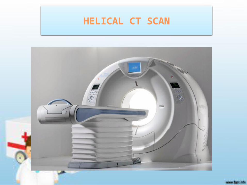
HELICAT CT SCAN
Helical computed axial tomography scan (CAT scan or CT scan) is another name for a CT scan, and is also called a spiral CT scan. Until approximately the mid to late 1990's, CT images were obtained one slice at a time, with the patient table moving step by step through the gantry. Scans obtained using this older technique often required 30 or 45 minutes, and the patient held his or her breath for every slice. Helical or spiral CT scans are obtained usually with one breath hold and obtain a volume of X-rayed tissue while the table moves rapidly through the gantry. Complete exams can be performed in less than five minutes. With the advent of helical or spiral CT scanning and rapid image acquisition, CT scanning has become the main diagnostic imaging tool in the radiology department.•
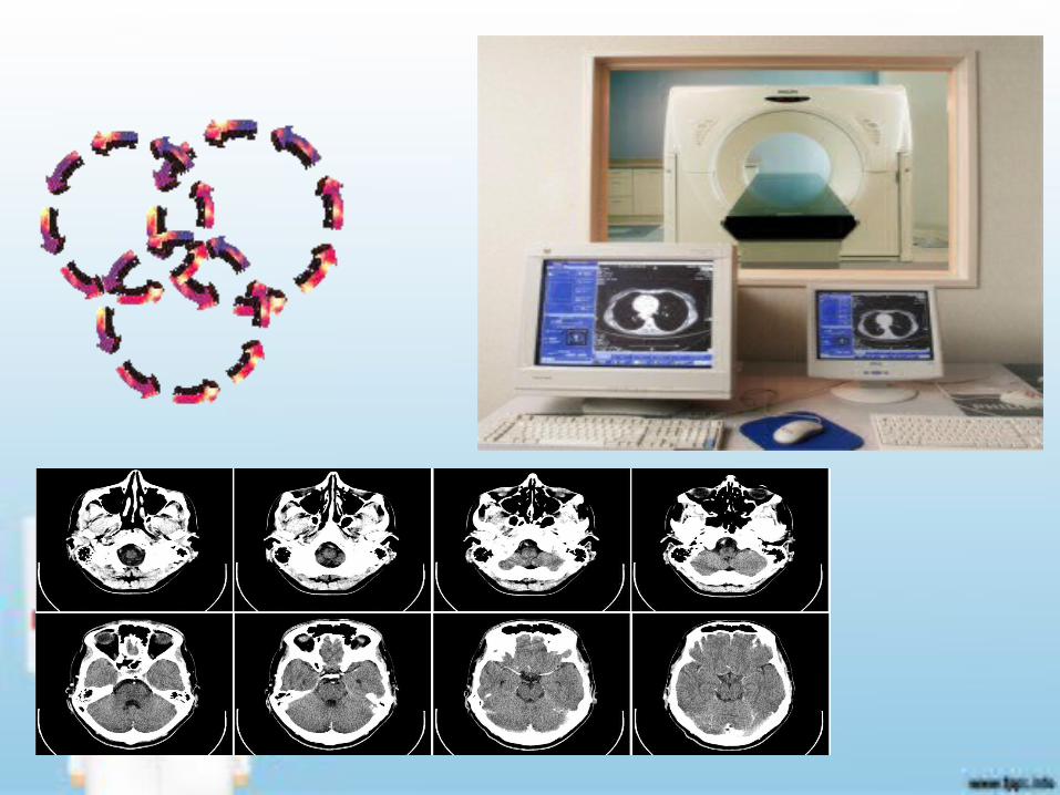
• the function helical CT scan is a radiodiagnostic imaging can produce images and slices or specific areas of the patient's body and provide information processed computer synthesis of x-rays with the data displayed on the video display
FUNCTION HELICAL CT SCAN
• The surplus depiction of organs will be processed more quickly and an advanced three-dimensional images through computer processing. This generation uses technology that has a fixed-ring detector 4800. When the examination of x-ray tube rotates 360 ° around the stationary detector with a scanning time equal to the third generation CT Scan.
ADVANTAGES HELICAL CT SCAN THAN CT SCAN
HELICAL CT SCAN
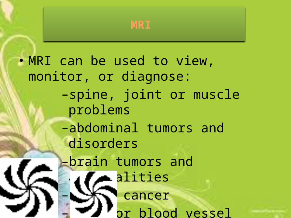
• MRI can be used to view, monitor, or diagnose:
–spine, joint or muscle problems–abdominal tumors and disorders–brain tumors and abnormalities–breast cancer–heart or blood vessel problems
MRI
• An MRI machine or scanner uses a powerful magnet and radio waves linked to a computer to create remarkably clear and detailed cross sectional images of the body. To visualize an MRI, think of your body as a loaf of bread with its many slices. The MRI allows the physician to see many different “slices” of a body part by taking pictures from outside the body. The “slices” can be displayed on a video monitor and saved on film or disk for analysis.
• For some MRI studies, a contrast agent, usually gadolinium may be used to enhance the visibility of certain tissues. The contrast agent is given via a small intravenous (IV) line placed in a vein in your arm. See ACRIN’s “About Imaging Agents or Tracers” Information page for more information.
MRI
• Benefits:• MRI can help evaluate the function as well as
the structure of many organs.• Soft tissue structures (heart, lungs, liver, and
other organs) are clearer and more detailed with MRI than other imaging methods.
• MRI provides a fast, non-invasive alternative to x-ray angiography.
• MRI uses no ionizing radiation.• •
THE BENEFITS OF MRI

MRI

This a video MRI and Helical CT Scan Differences lets see guys :D