Embed Size (px)
Citation preview
lable at ScienceDirect
Polymer 51 (2010) 3926e3933
Contents lists avai
Polymer
journal homepage: www.elsevier .com/locate/polymer
Engineering oligo(ethylene glycol)-based thermosensitive microgels for drugdelivery applications
Ting Zhou, Weitai Wu, Shuiqin Zhou*
Department of Chemistry of College of Staten Island, and The Graduate Center, The City University of New York, 2800 Victory Boulevard, Staten Island, New York 10314, USA
a r t i c l e i n f o
Article history:Received 3 May 2010Received in revised form12 June 2010Accepted 15 June 2010Available online 22 June 2010
Keywords:PEGMicrogelDrug delivery
* Corresponding author. Tel.: þ1 718 982 3897; faxE-mail address: [email protected] (S. Zho
0032-3861/$ e see front matter � 2010 Elsevier Ltd.doi:10.1016/j.polymer.2010.06.030
a b s t r a c t
Novel oligo(ethylene glycol)-based thermosensitive microgels with well engineered core-shell structureswere developed for storage and delivery of chemotherapeutic agents. The core is consisted of hydro-phobic poly[2-(2-methoxyethoxy)ethyl methacrylate], while the shell is consisted of hydrophiliccopolymer of 2-(2-methoxyethoxy)ethyl methacrylate with oligo(ethylene glycol) methyl ether meth-acrylates. These core-shell microgels exhibit tunable volume phase transition temperature and excellentcolloidal stability across the physiologically important temperature range. The thickness of the hydro-philic shell can control the collapsing degree (or mesh size) of the hydrophobic core network, which canbe utilized to significantly increase the loading capacity of the model hydrophobic drugs dipyridamole bytailoring the shell thickness of microgels. While the microgels are nontoxic, the drug molecules releasedfrom the microgels remain active to kill the cancer cells. The presented results provide importantguidelines for the rational design of core-shell structured polymeric microgels for drug uptake andrelease applications.
� 2010 Elsevier Ltd. All rights reserved.
1. Introduction
Microgel particles as drug delivery carriers for biological andbiomedical applications have received increasing attention duringthe last decade due to their unique chemical and physical versatility[1e6]. Microgels offer several advantages over other polymer baseddrug delivery systems: simple synthesis, tunable size from nano-meters to micrometers, large surface area for effective bio-conjugation to add target ligands for site-specific delivery, aninterior network structure for the incorporation of drug moleculesto protect the drug from hydrolysis and other type of chemicaldegradation, and potential biocompatibility. Among the variousapproaches used to enhance the efficacy of chemotherapy is the useof smart carrier systems that can release a drug in response tostimuli, such as changes in pH, temperature, light, or the presenceof specific enzymes that are selectively encountered in relevant cellorganelles [7e10].
So far, the thermo-responsive poly(N-isopropylacrylamide)(PNIPAM)-based microgels have been most extensively studied forapplications in drug delivery [8e21], including the introduction ofother environmental sensitivities such as pH, glucose, and lightsensitive components into the PNIPAM microgels to control the
: þ1 718 982 3910.u).
All rights reserved.
drug release. However, the well studied PNIPAM-based microgelshave not been translated into a biomedical breakthrough althougha recent study on the microgels containing NIPAM and acrylic acid(AA) indicated no adverse effects [16]. To meet the generalrequirement in biocompatibility of the materials used for drugdelivery systems, an important challenge is to develop biocom-patible polymers which have similar properties to PNIPAM [4].Thermo-responsive polymers containing short oligo(ethyleneglycol) side chains were recently proposed as an attractive alter-native to PNIPAM [22e28]. The lower critical solution temperature(LCST) of these graft polymers are tunable through the control inthe compositions of the oligo(ethylene glycol) side chains. Lutzet al. have reported that the LCST of the copolymers composed of2-(2-methoxyethoxy)ethyl methacrylate (MEO2MA) with oligo(ethylene glycol) methyl ether methacrylate (Mn ¼ 475 g/mol,MEO9MA) can be adjusted by varying the comonomer ratios[22e24]. Ishizone et al. found that the LCST of the poly[oligo(ethylene glycol) alkyl ether methacrylates] is tunable by varyingthe oligo(ethylene glycol) side chain length [26,27]. The longer theoligo(ethylene glycol) side chain, the higher the LCST of the graftpolymer. In addition, the star-block copolymers of these oligo(ethylene glycol) methyl ether methacrylate with star poly(ethylene glycol) (PEG) exhibit thermoreversible sol-gel transitionsin physiological media [28]. Although the graft structure composedof a carbonecarbon backbone and multiple oligo(ethylene glycol)side chains is different from the standard linear PEG, these
T. Zhou et al. / Polymer 51 (2010) 3926e3933 3927
nonlinear PEG analogues are mainly composed of oligo(ethyleneglycol) segments and are, in most cases, water-soluble andbiocompatible [25]. Hence, these thermosensitive nonlinear PEGanalogue polymers are very promising for biomedical applications.
Based on the free radical precipitation polymerization techniqueused to synthesize PNIPAM microgels, Hu’s group has successfullysynthesized monodisperse copolymer microgels from the como-nomers of MEO2MA and MEO9MA at different ratios [29]. With thesame two-step polymerization method used for the preparation ofPNIPAM-based core-shell microgels, they also successfullysynthesized core-shell microgels with the poly[oligo(ethyleneglycol) ethyl ethermethacrylate (Mn¼ 246 g/mol, EEO3MA)] as coreand the copolymer of EEO3MA, MEO5MA (Mn ¼ 300 g/mol) andacrylic acid as shell. Similar to PNIPAM microgels, these nonlinearPEG analogue polymer microgels can self-assemble into interestingcolloidal crystalline phases, depending on the concentration andtemperature [30].
The aim of this work is to develop a series of monodisperse oligo(ethylene glycol)-based temperature sensitive core-shell microgelswith different shell thickness, and study their potential applicationas delivery vehicles of hydrophobic drugs. The structures andvolume phase transitions of PNIPAM-based core-shell microgelshave been well studied [31e34]. The core-shell microgels canprovide unique internal structures andmulti-responsive propertiesdue to the mutual influence of core and shell swelling, whichenables us to explore the possibility to control the uptake andrelease of drug molecules in the microgels. In our design, the poly[2-(2-methoxyethoxy)ethyl methacrylate] [P(MEO2MA)] microgelswith a VPTT w22 �C is used as a hydrophobic core and the cross-linked copolymer of P(MEO2MA-co-MEO5MA) with the como-nomer ratio of MEO2MA:MEO5MA ¼ 1:2 and a VPTT of w55 �C isused as a hydrophilic shell. In addition to the good biocompatibility,the hydrophilic P(MEO2MA-co-MEO5MA) shell can influence thecore swelling. Although the free P(MEO2MA) core chain network ishydrophobic and collapsed at temperature above its VPTT, theaddition of hydrophilic shell can mechanically expand thecollapsed core. As the shell thickness increases to certain extent,the hydrophobic core network can be almost fully expanded evenat temperature above the VPTTof core microgels [33,34]. We expectthat the hydrophobic core with an open network structure shouldload hydrophobic drug molecules much more effectively comparedto the collapsed structure. To demonstrate this conception,a hydrophobic drug dipyridamole (DIP) (see Scheme 1) was studiedas model drug. The effects of shell thickness and temperature on
N
N
N
N
N
N
N
N
OH
OH
OH
HO
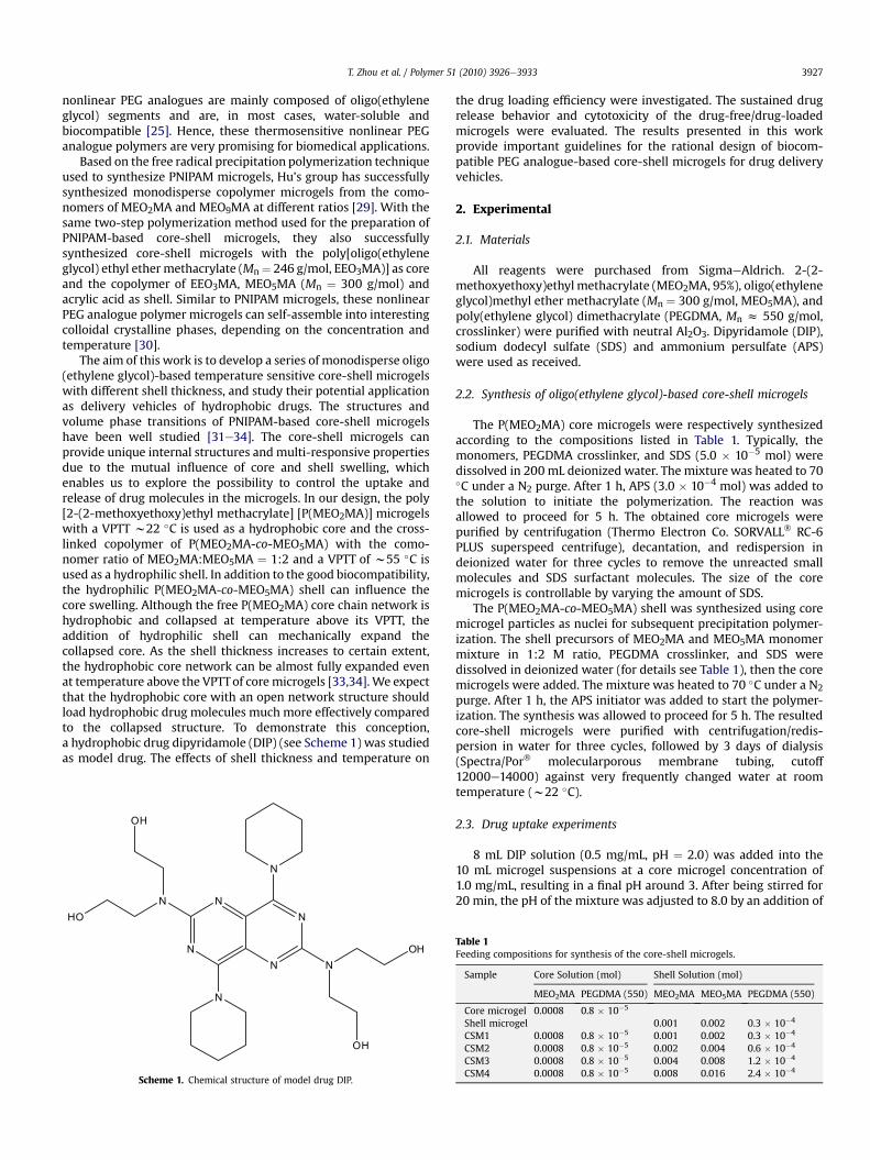
Scheme 1. Chemical structure of model drug DIP.
the drug loading efficiency were investigated. The sustained drugrelease behavior and cytotoxicity of the drug-free/drug-loadedmicrogels were evaluated. The results presented in this workprovide important guidelines for the rational design of biocom-patible PEG analogue-based core-shell microgels for drug deliveryvehicles.
2. Experimental
2.1. Materials
All reagents were purchased from SigmaeAldrich. 2-(2-methoxyethoxy)ethyl methacrylate (MEO2MA, 95%), oligo(ethyleneglycol)methyl ether methacrylate (Mn ¼ 300 g/mol, MEO5MA), andpoly(ethylene glycol) dimethacrylate (PEGDMA, Mn z 550 g/mol,crosslinker) were purified with neutral Al2O3. Dipyridamole (DIP),sodium dodecyl sulfate (SDS) and ammonium persulfate (APS)were used as received.
2.2. Synthesis of oligo(ethylene glycol)-based core-shell microgels
The P(MEO2MA) core microgels were respectively synthesizedaccording to the compositions listed in Table 1. Typically, themonomers, PEGDMA crosslinker, and SDS (5.0 � 10�5 mol) weredissolved in 200 mL deionized water. The mixture was heated to 70�C under a N2 purge. After 1 h, APS (3.0 � 10�4 mol) was added tothe solution to initiate the polymerization. The reaction wasallowed to proceed for 5 h. The obtained core microgels werepurified by centrifugation (Thermo Electron Co. SORVALL� RC-6PLUS superspeed centrifuge), decantation, and redispersion indeionized water for three cycles to remove the unreacted smallmolecules and SDS surfactant molecules. The size of the coremicrogels is controllable by varying the amount of SDS.
The P(MEO2MA-co-MEO5MA) shell was synthesized using coremicrogel particles as nuclei for subsequent precipitation polymer-ization. The shell precursors of MEO2MA and MEO5MA monomermixture in 1:2 M ratio, PEGDMA crosslinker, and SDS weredissolved in deionized water (for details see Table 1), then the coremicrogels were added. The mixture was heated to 70 �C under a N2
purge. After 1 h, the APS initiator was added to start the polymer-ization. The synthesis was allowed to proceed for 5 h. The resultedcore-shell microgels were purified with centrifugation/redis-persion in water for three cycles, followed by 3 days of dialysis(Spectra/Por� molecularporous membrane tubing, cutoff12000e14000) against very frequently changed water at roomtemperature (w22 �C).
2.3. Drug uptake experiments
8 mL DIP solution (0.5 mg/mL, pH ¼ 2.0) was added into the10 mL microgel suspensions at a core microgel concentration of1.0 mg/mL, resulting in a final pH around 3. After being stirred for20 min, the pH of the mixture was adjusted to 8.0 by an addition of
Table 1Feeding compositions for synthesis of the core-shell microgels.
Sample Core Solution (mol) Shell Solution (mol)
MEO2MA PEGDMA (550) MEO2MA MEO5MA PEGDMA (550)
Core microgel 0.0008 0.8 � 10�5
Shell microgel 0.001 0.002 0.3 � 10�4
CSM1 0.0008 0.8 � 10�5 0.001 0.002 0.3 � 10�4
CSM2 0.0008 0.8 � 10�5 0.002 0.004 0.6 � 10�4
CSM3 0.0008 0.8 � 10�5 0.004 0.008 1.2 � 10�4
CSM4 0.0008 0.8 � 10�5 0.008 0.016 2.4 � 10�4
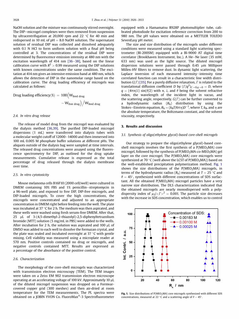
Fig. 1. Size distributions of P(MEO2MA) core microgels synthesized with different SDSconcentrations, measured at 22 �C and a scattering angle of q ¼ 45� .
T. Zhou et al. / Polymer 51 (2010) 3926e39333928
NaOH solution and the mixture was continuously stirred overnight.The DIPemicrogel complexes were then removed from suspensionby ultracentrifugation at 20,000 rpm and 22 �C for 40 min andredispersed in 10 mL of pH ¼ 8.0 NaOH solution. The supernatantsolution of residual DIP was collected and dissolved adequatelywith 0.1 N HCl to form uniform solution with a final pH beingcontrolled at 3. The concentrations of the residual DIP weredetermined by fluorescence emission intensity at 480 nmwith theexcitation wavelength of 414 nm [36e38], based on the linearcalibration curve with R2 > 0.99 measured using the DIP solutionswith known concentrations under the same condition. The exci-tation at 414 nm gives an intensive emission band at 480 nm, whichallows the detection of DIP in the nanomolar range based on thecalibration curve. The drug loading efficiency of microgels wascalculated as follows:
Drug loading efficiencyð%Þ ¼ 100�Wfeed drug
�Wfree drug
�.Wfeed drug
2.4. In vitro drug release
The release of model drug from the microgel was evaluated bythe dialysis method [36,39]. The purified DIP-loaded microgeldispersions (1 mL) were transferred into dialysis tubes withamolecular weight cutoff of 12000e14000 and then immersed into100 mL 0.005 M phosphate buffer solutions at different pHs. Thealiquots outside of the dialysis bag were sampled at time intervals.The released drug concentrations were assayed using the fluores-cence spectrometry for DIP, as outlined for the drug uptakemeasurements. Cumulative release is expressed as the totalpercentage of drug released through the dialysis membraneover time.
2.5. In vitro cytotoxicity
Mouse melanoma cells B16F10 (2000 cell/well) were cultured inDMEM containing 10% FBS and 1% penicillinestreptomycin ina 96-well plate, and exposed to free DIP, DIP-free microgels, andDIP-loaded microgels. To cover the high concentrations, themicrogels were concentrated and adjusted to an appropriateconcentration in DMEM right before feeding into thewell. The platewas incubated at 37 �C for 2 h. Themediumwas then aspirated, andthese wells were washed using fresh serum-free DMEM. After that,25 mL of 3-(4,5-dimethyl-2-thiazolyl)-2,5-diphenyltetrazoliumbromide (MTT) solution (5 mg/mL in PBS) were added to the wells.After incubation for 2 h, the solution was aspirated and 100 mL ofDMSOwas added to each well to dissolve the formazan crystal, andthe plate was sealed and incubated overnight at 37 �C with gentlemixing. Cell viability was measured using a microplate reader at570 nm. Positive controls contained no drug or microgels, andnegative controls contained MTT. Results are expressed asa percentage of the absorbance of the positive control.
2.6. Characterization
The morphology of the core-shell microgels was characterizedwith transmission electron microscopy (TEM). The TEM imageswere taken on a Zeiss EM 902 transmission electron microscopeoperating at an accelerating voltage of 100 kV. Approximately 10 mLof the diluted microgel suspension was dropped on a Formvar-covered copper grid (300 meshes) and then air-dried at roomtemperature for the TEM measurements. The PL spectra wereobtained on a JOBIN YVON Co. FluoroMax�-3 Spectrofluorometer
equipped with a Hamamatsu R928P photomultiplier tube, cali-brated photodiode for excitation reference correction from 200 to980 nm. The pH values were obtained on a METTLER TOLEDOSevenEasy pH meter.
The size and size distribution of the microgels under differentconditions were measured using a standard light scattering spec-trometer (BI-200SM) equipped with a BI-9000 AT digital timecorrelator (Brookhaven Instrument, Inc.). A HeeNe laser (35 mW,633 nm) was used as the light source. The diluted microgeldispersion solutions were passed through 0.45 mm MilliporeMillex-HV filters to remove dust. In dynamic light scattering, theLaplace inversion of each measured intensityeintensity timecorrelated function can result in a characteristic line width distri-bution G(G) [35]. For a purely diffusive relaxation, G is related to thetranslational diffusion coefficient D by (G/q2)C/0,q/0 ¼ D, whereq ¼ (4pn/l) sin(q/2) with n, l, and q being the solvent refractiveindex, the wavelength of the incident light in vacuo, andthe scattering angle, respectively. G(G) can be further converted toa hydrodynamic radius (Rh) distribution by using theStokeseEinstein equation, Rh ¼ (kBT/6ph)D�1, where T, kB, and h arethe absolute temperature, the Boltzmann constant, and the solventviscosity, respectively.
3. Results and discussion
3.1. Synthesis of oligo(ethylene glycol)-based core-shell microgels
Our strategy to prepare the oligo(ethylene glycol)-based core-shell microgels involves the first synthesis of a P(MEO2MA) coremicrogel, followed by the synthesis of P(MEO2MA-co-MEO5MA) gellayer on the core microgel. The P(MEO2MA) core microgels weresynthesized at 70 �C (well above the LCST of P(MEO2MA)) based onthe well-established precipitation polymerization method. Fig. 1shows the size distributions of the P(MEO2MA) microgels, interms of the hydrodynamic radius (Rh) measured at T ¼ 25 �C andq ¼ 45�, synthesized with different concentrations of SDS surfac-tant. All the obtained P(MEO2MA) microgel particles have a verynarrow size distribution. The DLS characterization indicated thatthe obtained microgels are nearly monodispersed with a poly-dispersity index of m2/<G>2 ¼ 0.001. The particle size decreaseswith the increase in SDS concentration, which enables us to control
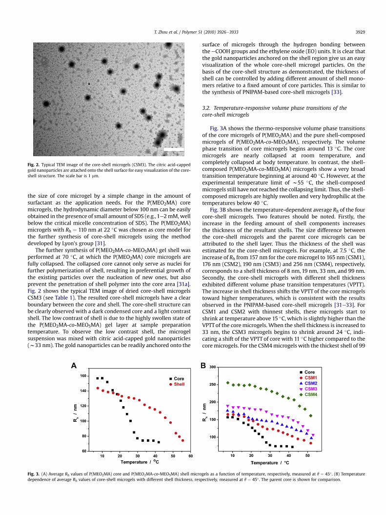
Fig. 2. Typical TEM image of the core-shell microgels (CSM3). The citric acid-cappedgold nanoparticles are attached onto the shell surface for easy visualization of the core-shell structure. The scale bar is 1 mm.
T. Zhou et al. / Polymer 51 (2010) 3926e3933 3929
the size of core microgel by a simple change in the amount ofsurfactant as the application needs. For the P(MEO2MA) coremicrogels, the hydrodynamic diameter below 100 nm can be easilyobtained in the presence of small amount of SDS (e.g.,1e2mM,wellbelow the critical micelle concentration of SDS). The P(MEO2MA)microgels with Rh ¼ 110 nm at 22 �C was chosen as core model forthe further synthesis of core-shell microgels using the methoddeveloped by Lyon’s group [31].
The further synthesis of P(MEO2MA-co-MEO5MA) gel shell wasperformed at 70 �C, at which the P(MEO2MA) core microgels arefully collapsed. The collapsed core cannot only serve as nuclei forfurther polymerization of shell, resulting in preferential growth ofthe existing particles over the nucleation of new ones, but alsoprevent the penetration of shell polymer into the core area [31a].Fig. 2 shows the typical TEM image of dried core-shell microgelsCSM3 (see Table 1). The resulted core-shell microgels have a clearboundary between the core and shell. The core-shell structure canbe clearly observed with a dark condensed core and a light contrastshell. The low contrast of shell is due to the highly swollen state ofthe P(MEO2MA-co-MEO5MA) gel layer at sample preparationtemperature. To observe the low contrast shell, the microgelsuspension was mixed with citric acid-capped gold nanoparticles(w33 nm). The gold nanoparticles can be readily anchored onto the
Fig. 3. (A) Average Rh values of P(MEO2MA) core and P(MEO2MA-co-MEO5MA) shell microdependence of average Rh values of core-shell microgels with different shell thickness, resp
surface of microgels through the hydrogen bonding betweentheeCOOH groups and the ethylene oxide (EO) units. It is clear thatthe gold nanoparticles anchored on the shell region give us an easyvisualization of the whole core-shell microgel particles. On thebasis of the core-shell structure as demonstrated, the thickness ofshell can be controlled by adding different amount of shell mono-mers relative to a fixed amount of core particles. This is similar tothe synthesis of PNIPAM-based core-shell microgels [33].
3.2. Temperature-responsive volume phase transitions of thecore-shell microgels
Fig. 3A shows the thermo-responsive volume phase transitionsof the core microgels of P(MEO2MA) and the pure shell-composedmicrogels of P(MEO2MA-co-MEO5MA), respectively. The volumephase transition of core microgels begins around 13 �C. The coremicrogels are nearly collapsed at room temperature, andcompletely collapsed at body temperature. In contrast, the shell-composed P(MEO2MA-co-MEO5MA) microgels show a very broadtransition temperature beginning at around 40 �C. However, at theexperimental temperature limit of w55 �C, the shell-composedmicrogels still have not reached the collapsing limit. Thus, the shell-composed microgels are highly swollen and very hydrophilic at thetemperatures below 40 �C.
Fig. 3B shows the temperature-dependent average Rh of the fourcore-shell microgels. Two features should be noted. Firstly, theincrease in the feeding amount of shell components increasesthe thickness of the resultant shells. The size difference betweenthe core-shell microgels and the parent core microgels can beattributed to the shell layer. Thus the thickness of the shell wasestimated for the core-shell microgels. For example, at 7.5 �C, theincrease of Rh from 157 nm for the core microgel to 165 nm (CSM1),176 nm (CSM2), 190 nm (CSM3) and 256 nm (CSM4), respectively,corresponds to a shell thickness of 8 nm, 19 nm, 33 nm, and 99 nm.Secondly, the core-shell microgels with different shell thicknessexhibited different volume phase transition temperatures (VPTT).The increase in shell thickness shifts the VPTT of the core microgelstoward higher temperatures, which is consistent with the resultsobserved in the PNIPAM-based core-shell microgels [31e33]. ForCSM1 and CSM2 with thinnest shells, these microgels start toshrink at temperature above 15 �C, which is slightly higher than theVPTTof the core microgels. When the shell thickness is increased to33 nm, the CSM3 microgels begins to shrink around 24 �C, indi-cating a shift of the VPTT of core with 11 �C higher compared to thecore microgels. For the CSM4microgels with the thickest shell of 99
gels as a function of temperature, respectively, measured at q ¼ 45� . (B) Temperatureectively, measured at q ¼ 45� . The parent core is shown for comparison.
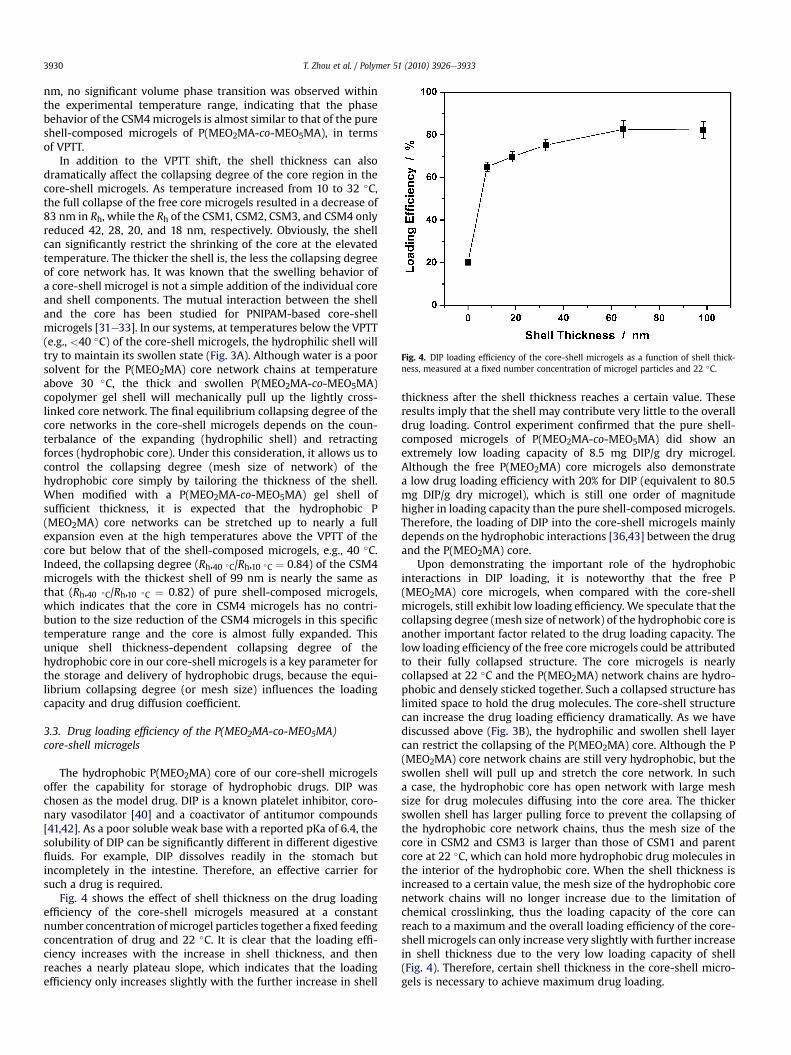
Fig. 4. DIP loading efficiency of the core-shell microgels as a function of shell thick-ness, measured at a fixed number concentration of microgel particles and 22 �C.
T. Zhou et al. / Polymer 51 (2010) 3926e39333930
nm, no significant volume phase transition was observed withinthe experimental temperature range, indicating that the phasebehavior of the CSM4microgels is almost similar to that of the pureshell-composed microgels of P(MEO2MA-co-MEO5MA), in termsof VPTT.
In addition to the VPTT shift, the shell thickness can alsodramatically affect the collapsing degree of the core region in thecore-shell microgels. As temperature increased from 10 to 32 �C,the full collapse of the free core microgels resulted in a decrease of83 nm in Rh, while the Rh of the CSM1, CSM2, CSM3, and CSM4 onlyreduced 42, 28, 20, and 18 nm, respectively. Obviously, the shellcan significantly restrict the shrinking of the core at the elevatedtemperature. The thicker the shell is, the less the collapsing degreeof core network has. It was known that the swelling behavior ofa core-shell microgel is not a simple addition of the individual coreand shell components. The mutual interaction between the shelland the core has been studied for PNIPAM-based core-shellmicrogels [31e33]. In our systems, at temperatures below the VPTT(e.g., <40 �C) of the core-shell microgels, the hydrophilic shell willtry to maintain its swollen state (Fig. 3A). Although water is a poorsolvent for the P(MEO2MA) core network chains at temperatureabove 30 �C, the thick and swollen P(MEO2MA-co-MEO5MA)copolymer gel shell will mechanically pull up the lightly cross-linked core network. The final equilibrium collapsing degree of thecore networks in the core-shell microgels depends on the coun-terbalance of the expanding (hydrophilic shell) and retractingforces (hydrophobic core). Under this consideration, it allows us tocontrol the collapsing degree (mesh size of network) of thehydrophobic core simply by tailoring the thickness of the shell.When modified with a P(MEO2MA-co-MEO5MA) gel shell ofsufficient thickness, it is expected that the hydrophobic P(MEO2MA) core networks can be stretched up to nearly a fullexpansion even at the high temperatures above the VPTT of thecore but below that of the shell-composed microgels, e.g., 40 �C.Indeed, the collapsing degree (Rh,40 �C/Rh,10 �C ¼ 0.84) of the CSM4microgels with the thickest shell of 99 nm is nearly the same asthat (Rh,40 �C/Rh,10 �C ¼ 0.82) of pure shell-composed microgels,which indicates that the core in CSM4 microgels has no contri-bution to the size reduction of the CSM4 microgels in this specifictemperature range and the core is almost fully expanded. Thisunique shell thickness-dependent collapsing degree of thehydrophobic core in our core-shell microgels is a key parameter forthe storage and delivery of hydrophobic drugs, because the equi-librium collapsing degree (or mesh size) influences the loadingcapacity and drug diffusion coefficient.
3.3. Drug loading efficiency of the P(MEO2MA-co-MEO5MA)core-shell microgels
The hydrophobic P(MEO2MA) core of our core-shell microgelsoffer the capability for storage of hydrophobic drugs. DIP waschosen as the model drug. DIP is a known platelet inhibitor, coro-nary vasodilator [40] and a coactivator of antitumor compounds[41,42]. As a poor soluble weak base with a reported pKa of 6.4, thesolubility of DIP can be significantly different in different digestivefluids. For example, DIP dissolves readily in the stomach butincompletely in the intestine. Therefore, an effective carrier forsuch a drug is required.
Fig. 4 shows the effect of shell thickness on the drug loadingefficiency of the core-shell microgels measured at a constantnumber concentration of microgel particles together a fixed feedingconcentration of drug and 22 �C. It is clear that the loading effi-ciency increases with the increase in shell thickness, and thenreaches a nearly plateau slope, which indicates that the loadingefficiency only increases slightly with the further increase in shell
thickness after the shell thickness reaches a certain value. Theseresults imply that the shell may contribute very little to the overalldrug loading. Control experiment confirmed that the pure shell-composed microgels of P(MEO2MA-co-MEO5MA) did show anextremely low loading capacity of 8.5 mg DIP/g dry microgel.Although the free P(MEO2MA) core microgels also demonstratea low drug loading efficiency with 20% for DIP (equivalent to 80.5mg DIP/g dry microgel), which is still one order of magnitudehigher in loading capacity than the pure shell-composed microgels.Therefore, the loading of DIP into the core-shell microgels mainlydepends on the hydrophobic interactions [36,43] between the drugand the P(MEO2MA) core.
Upon demonstrating the important role of the hydrophobicinteractions in DIP loading, it is noteworthy that the free P(MEO2MA) core microgels, when compared with the core-shellmicrogels, still exhibit low loading efficiency. We speculate that thecollapsing degree (mesh size of network) of the hydrophobic core isanother important factor related to the drug loading capacity. Thelow loading efficiency of the free core microgels could be attributedto their fully collapsed structure. The core microgels is nearlycollapsed at 22 �C and the P(MEO2MA) network chains are hydro-phobic and densely sticked together. Such a collapsed structure haslimited space to hold the drug molecules. The core-shell structurecan increase the drug loading efficiency dramatically. As we havediscussed above (Fig. 3B), the hydrophilic and swollen shell layercan restrict the collapsing of the P(MEO2MA) core. Although the P(MEO2MA) core network chains are still very hydrophobic, but theswollen shell will pull up and stretch the core network. In sucha case, the hydrophobic core has open network with large meshsize for drug molecules diffusing into the core area. The thickerswollen shell has larger pulling force to prevent the collapsing ofthe hydrophobic core network chains, thus the mesh size of thecore in CSM2 and CSM3 is larger than those of CSM1 and parentcore at 22 �C, which can hold more hydrophobic drug molecules inthe interior of the hydrophobic core. When the shell thickness isincreased to a certain value, the mesh size of the hydrophobic corenetwork chains will no longer increase due to the limitation ofchemical crosslinking, thus the loading capacity of the core canreach to a maximum and the overall loading efficiency of the core-shell microgels can only increase very slightly with further increasein shell thickness due to the very low loading capacity of shell(Fig. 4). Therefore, certain shell thickness in the core-shell micro-gels is necessary to achieve maximum drug loading.
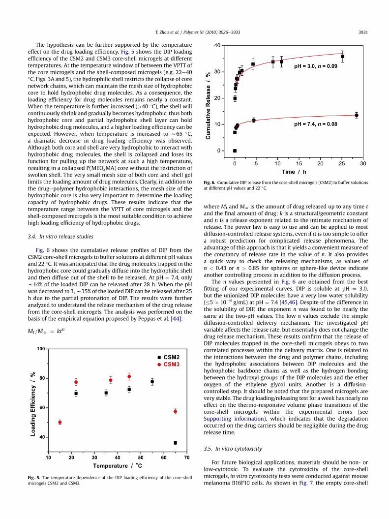
Fig. 6. Cumulative DIP release from the core-shell microgels (CSM2) to buffer solutionsat different pH values and 22 �C.
T. Zhou et al. / Polymer 51 (2010) 3926e3933 3931
The hypothesis can be further supported by the temperatureeffect on the drug loading efficiency. Fig. 5 shows the DIP loadingefficiency of the CSM2 and CSM3 core-shell microgels at differenttemperatures. At the temperature window of between the VPTT ofthe core microgels and the shell-composed microgels (e.g. 22e40�C, Figs. 3A and 5), the hydrophilic shell restricts the collapse of corenetwork chains, which can maintain the mesh size of hydrophobiccore to hold hydrophobic drug molecules. As a consequence, theloading efficiency for drug molecules remains nearly a constant.When the temperature is further increased (>40 �C), the shell willcontinuously shrink and gradually becomes hydrophobic, thus bothhydrophobic core and partial hydrophobic shell layer can holdhydrophobic drug molecules, and a higher loading efficiency can beexpected. However, when temperature is increased to w65 �C,a dramatic decrease in drug loading efficiency was observed.Although both core and shell are very hydrophobic to interact withhydrophobic drug molecules, the shell is collapsed and loses itsfunction for pulling up the network at such a high temperature,resulting in a collapsed P(MEO2MA) core without the restriction ofswollen shell. The very small mesh size of both core and shell gellimits the loading amount of drug molecules. Clearly, in addition tothe drugepolymer hydrophobic interactions, the mesh size of thehydrophobic core is also very important to determine the loadingcapacity of hydrophobic drugs. These results indicate that thetemperature range between the VPTT of core microgels and theshell-composed microgels is the most suitable condition to achievehigh loading efficiency of hydrophobic drugs.
3.4. In vitro release studies
Fig. 6 shows the cumulative release profiles of DIP from theCSM2 core-shell microgels to buffer solutions at different pH valuesand 22 �C. It was anticipated that the drug molecules trapped in thehydrophobic core could gradually diffuse into the hydrophilic shelland then diffuse out of the shell to be released. At pH ¼ 7.4, onlyw14% of the loaded DIP can be released after 28 h. When the pHwas decreased to 3,w35% of the loaded DIP can be released after 25h due to the partial protonation of DIP. The results were furtheranalyzed to understand the release mechanism of the drug releasefrom the core-shell microgels. The analysis was performed on thebasis of the empirical equation proposed by Peppas et al. [44]:
Mt=MN ¼ ktn
Fig. 5. The temperature dependence of the DIP loading efficiency of the core-shellmicrogels CSM2 and CSM3.
where Mt and MN is the amount of drug released up to any time tand the final amount of drug; k is a structural/geometric constantand n is a release exponent related to the intimate mechanism ofrelease. The power law is easy to use and can be applied to mostdiffusion-controlled release systems, even if it is too simple to offera robust prediction for complicated release phenomena. Theadvantage of this approach is that it yields a convenient measure ofthe constancy of release rate in the value of n. It also providesa quick way to check the releasing mechanisms, as values ofn < 0.43 or n > 0.85 for spheres or sphere-like device indicateanother controlling process in addition to the diffusion process.
The n values presented in Fig. 6 are obtained from the bestfitting of our experimental curves. DIP is soluble at pH ¼ 3.0,but the unionized DIP molecules have a very low water solubility(�5 � 10�6 g/mL) at pH ¼ 7.4 [45,46]. Despite of the difference inthe solubility of DIP, the exponent n was found to be nearly thesame at the two-pH values. The low n values exclude the simplediffusion-controlled delivery mechanism. The investigated pHvariable affects the release rate, but essentially does not change thedrug release mechanism. These results confirm that the release ofDIP molecules trapped in the core-shell microgels obeys to twocorrelated processes within the delivery matrix. One is related tothe interactions between the drug and polymer chains, includingthe hydrophobic associations between DIP molecules and thehydrophobic backbone chains as well as the hydrogen bondingbetween the hydroxyl groups of the DIP molecules and the etheroxygen of the ethylene glycol units. Another is a diffusion-controlled step. It should be noted that the prepared microgels arevery stable. The drug loading/releasing test for aweek has nearly noeffect on the thermo-responsive volume phase transitions of thecore-shell microgels within the experimental errors (seeSupporting information), which indicates that the degradationoccurred on the drug carriers should be negligible during the drugrelease time.
3.5. In vitro cytotoxicity
For future biological applications, materials should be non- orlow-cytotoxic. To evaluate the cytotoxicity of the core-shellmicrogels, in vitro cytotoxicity tests were conducted against mousemelanoma B16F10 cells. As shown in Fig. 7, the empty core-shell
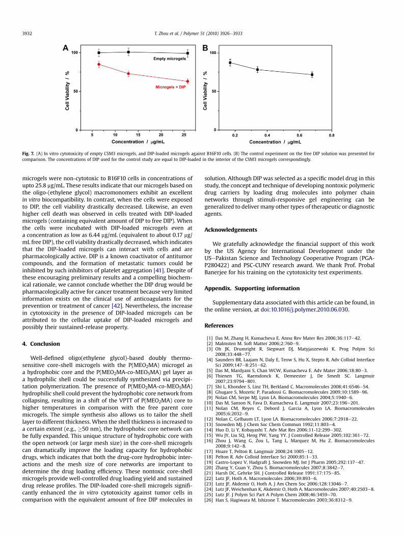
Fig. 7. (A) In vitro cytotoxicity of empty CSM3 microgels, and DIP-loaded microgels against B16F10 cells. (B) The control experiment on the free DIP solution was presented forcomparison. The concentrations of DIP used for the control study are equal to DIP-loaded in the interior of the CSM3 microgels correspondingly.
T. Zhou et al. / Polymer 51 (2010) 3926e39333932
microgels were non-cytotoxic to B16F10 cells in concentrations ofupto 25.8 mg/mL. These results indicate that our microgels based onthe oligo-(ethylene glycol) macromonomers exhibit an excellentin vitro biocompatibility. In contrast, when the cells were exposedto DIP, the cell viability drastically decreased. Likewise, an evenhigher cell death was observed in cells treated with DIP-loadedmicrogels (containing equivalent amount of DIP to free DIP). Whenthe cells were incubated with DIP-loaded microgels even ata concentration as low as 6.44 mg/mL (equivalent to about 0.17 mg/mL free DIP), the cell viability drastically decreased, which indicatesthat the DIP-loaded microgels can interact with cells and arepharmacologically active. DIP is a known coactivator of antitumorcompounds, and the formation of metastatic tumors could beinhibited by such inhibitors of platelet aggregation [41]. Despite ofthese encouraging preliminary results and a compelling biochem-ical rationale, we cannot conclude whether the DIP drug would bepharmacologically active for cancer treatment because very limitedinformation exists on the clinical use of anticoagulants for theprevention or treatment of cancer [42]. Nevertheless, the increasein cytotoxicity in the presence of DIP-loaded microgels can beattributed to the cellular uptake of DIP-loaded microgels andpossibly their sustained-release property.
4. Conclusion
Well-defined oligo(ethylene glycol)-based doubly thermo-sensitive core-shell microgels with the P(MEO2MA) microgel asa hydrophobic core and the P(MEO2MA-co-MEO5MA) gel layer asa hydrophilic shell could be successfully synthesized via precipi-tation polymerization. The presence of P(MEO2MA-co-MEO5MA)hydrophilic shell could prevent the hydrophobic core network fromcollapsing, resulting in a shift of the VPTT of P(MEO2MA) core tohigher temperatures in comparison with the free parent coremicrogels. The simple synthesis also allows us to tailor the shelllayer to different thickness. When the shell thickness is increased toa certain extent (e.g., �50 nm), the hydrophobic core network canbe fully expanded. This unique structure of hydrophobic core withthe open network (or large mesh size) in the core-shell microgelscan dramatically improve the loading capacity for hydrophobicdrugs, which indicates that both the drug-core hydrophobic inter-actions and the mesh size of core networks are important todetermine the drug loading efficiency. These nontoxic core-shellmicrogels provide well-controlled drug loading yield and sustaineddrug release profiles. The DIP-loaded core-shell microgels signifi-cantly enhanced the in vitro cytotoxicity against tumor cells incomparison with the equivalent amount of free DIP molecules in
solution. Although DIP was selected as a specific model drug in thisstudy, the concept and technique of developing nontoxic polymericdrug carriers by loading drug molecules into polymer chainnetworks through stimuli-responsive gel engineering can begeneralized to delivermany other types of therapeutic or diagnosticagents.
Acknowledgements
We gratefully acknowledge the financial support of this workby the US Agency for International Development under theUSePakistan Science and Technology Cooperative Program (PGA-P280422) and PSC-CUNY research award. We thank Prof. ProbalBanerjee for his training on the cytotoxicity test experiments.
Appendix. Supporting information
Supplementary data associated with this article can be found, inthe online version, at doi:10.1016/j.polymer.2010.06.030.
References
[1] Das M, Zhang H, Kumacheva E. Annu Rev Mater Res 2006;36:117e42.[2] Malmsten M. Soft Matter 2006;2:760e9.[3] Oh JK, Drumright R, Siegwart DJ, Matyjaszewski K. Prog Polym Sci
2008;33:448e77.[4] Saunders BR, Laajam N, Daly E, Teow S, Hu X, Stepto R. Adv Colloid Interface
Sci 2009;147e8:251e62.[5] Das M, Mardyani S, Chan WCW, Kumacheva E. Adv Mater 2006;18:80e3.[6] Thienen TG, Raemdonck K, Demeester J, De Smedt SC. Langmuir
2007;23:9794e801.[7] Shi L, Khondee S, Linz TH, Berkland C. Macromolecules 2008;41:6546e54.[8] Ghugare S, Mozetic P, Paradossi G. Biomacromolecules 2009;10:1589e96.[9] Nolan CM, Serpe MJ, Lyon LA. Biomacromolecules 2004;5:1940e6.
[10] Das M, Sanson N, Fava D, Kumacheva E. Langmuir 2007;23:196e201.[11] Nolan CM, Reyes C, Debord J, Garcia A, Lyon LA. Biomacromolecules
2005;6:2032e9.[12] Nolan C, Gelbaum LT, Lyon LA. Biomacromolecules 2006;7:2918e22.[13] Snowden MJ. J Chem Soc Chem Commun 1992;11:803e4.[14] Huo D, Li Y, Kobayashi T. Adv Mat Res 2006;11-12:299e302.[15] Wu JY, Liu SQ, Heng PW, Yang YY. J Controlled Release 2005;102:361e72.[16] Zhou J, Wang G, Zou L, Tang L, Marquez M, Hu Z. Biomacromolecules
2008;9:142e8.[17] Hoare T, Pelton R. Langmuir 2008;24:1005e12.[18] Pelton R. Adv Colloid Interface Sci 2000;85:1e33.[19] Castro-Lopez V, Hadgraft J, Snowden MJ. Int J Pharm 2005;292:137e47.[20] Zhang Y, Guan Y, Zhou S. Biomacromolecules 2007;8:3842e7.[21] Harsh DC, Gehrke SH. J Controlled Release 1991;17:175e85.[22] Lutz JF, Hoth A. Macromolecules 2006;39:893e6.[23] Lutz JF, Akdemir O, Hoth A. J Am Chem Soc 2006;128:13046e7.[24] Lutz JF, Weichenhan K, Akdemir O, Hoth A. Macromolecules 2007;40:2503e8.[25] Lutz JF. J Polym Sci Part A Polym Chem 2008;46:3459e70.[26] Han S, Hagiwara M, Ishizone T. Macromolecules 2003;36:8312e9.

T. Zhou et al. / Polymer 51 (2010) 3926e3933 3933
[27] Ishiznoe T, Seki A, Hagiwara M, Han S, Yokoyama H, Oyane A, et al. Macro-molecules 2008;41:2963e7.
[28] Badi N, Lutz JF. J Controlled Release 2009;140:224e9.[29] Cai T, Marquez M, Hu Z. Langmuir 2007;23:8663e6.[30] Chi C, Cai T, Hu Z. Langmuir 2009;25:3814e9.[31] (a) Jones CD, Lyon A. Macromolecules 2000;33:8301e6;
(b) Jones CD, Lyon A. Macromolecules 2003;36:1988e93;(c) Jones CD, Lyon A. Langmuir 2003;19:4544e7.
[32] (a) Gan D, Lyon A. J Am Chem Soc 2001;123:8203e9;(b) Jones CD, McGrath JG, Lyon A. J Phys Chem B 2004;108:12652e7.
[33] Berndt I, Richtering W. Macromolecules 2003;36:8780e5.[34] Berndt I, Pedersen JS, Lindner P, Richtering W. Langmuir 2006;22:459e68.[35] Chu B. Laser light scattering. 2nd ed. New York: Academic Press; 1991.[36] Tang Y, Liu SY, Armes SP, Billinghan NC. Biomacromolecules 2003;4:
1636e45.
[37] Giacomelli C, Le ML, Borsali R, Lai-Kee-Him J, Brisson A, Armes SP, et al.Biomacromolecules 2006;7:817e28.
[38] Vertzoni M, Pastelli E, Psachoulias D, Kalantzi L, Reppas C. Pharm Res2007;24:909e17.
[39] Jiang X, Ge Z, Xu J, Liu H, Liu S. Biomacromolecules 2007;8:3184e92.[40] Marchandt E, Prichard AD, Casanegra P, Lindsay L. Am J Cardiol 1984;
53:718.[41] Shalinsky DR, Jekunen AP, Alcaraz JE, Christen RD, Kim S, Khatibi S, et al. Br J
Cancer 1993;67(1):30.[42] Hejna M, Raderer M, Zielinski CC. J Natl Cancer Inst 1999;91:22.[43] KozlovYu M, Melik-Nubarov NS, Batrakova EV, Kabanov AV. Macromolecules
2000;33:3305.[44] Siepmann J, Peppas NA. Adv Drug Deliv Rev 2001;48:139.[45] Giacomelli C, Schmidt V, Borsali R. Langmuir 2007;23:6947e55.[46] Avdeef A, Berger CM, Brownell C. Pharm Res 2000;17:85e9.