Embed Size (px)
Citation preview
Ep
AHa
b
c
a
ARRAA
TKPo
KTCMNM
1
Tsihta(ea
c
DB
0d
Virus Research 157 (2011) 35–46
Contents lists available at ScienceDirect
Virus Research
journa l homepage: www.e lsev ier .com/ locate /v i rusres
ngineered Tobacco mosaic virus mutants with distinct physical characteristics inlanta and enhanced metallization properties
nan Kadria,∗, Edgar Maißb, Nadja Amsharovc, Alexander M. Bittnerc,1, Sinan Balci c, Klaus Kernc,olger Jeskea, Christina Wegea,∗∗
Universität Stuttgart, Institute of Biology, Department of Plant Molecular Biology and Plant Virology, Pfaffenwaldring 57, D-70550 Stuttgart, GermanyLeibniz Universität Hannover, Institut für Pflanzenkrankheiten und Pflanzenschutz, Herrenhäuser Straße 2, D-30419 Hannover, GermanyMax Planck Institute for Solid State Research, Heisenbergstraße 1, D-70569 Stuttgart, Germany
r t i c l e i n f o
rticle history:eceived 22 July 2010eceived in revised form 25 January 2011ccepted 29 January 2011vailable online 15 February 2011
he publication is dedicated toarl-Wolfgang Mundry, founder of thelant Virology Department at the University
a b s t r a c t
Tobacco mosaic virus mutants were engineered to alter either the stability or surface chemistry of thevirion: within the coat protein, glutamic acid was exchanged for glutamine in a buried portion to enhancethe inter-subunit binding stability (E50Q), or a hexahistidine tract was fused to the surface-exposedcarboxy terminus of the coat protein (6xHis). Both mutant viruses were expected to possess specificmetal ion affinities. They accumulated to high titers in plants, induced distinct phenotypes, and theirphysical properties during purification differed from each other and from wild type (wt) virus. Whereas6xHis and wt virions contained RNA, the majority of E50Q protein assembled essentially without RNAinto rods which frequently exceeded 2 �m in length. Electroless deposition of nickel metallized the outer
f Stuttgart, who deceased in 2009.
eywords:obacco mosaic virus (TMV)oat proteinutationanotechnologyetallization
surface of 6xHis virions, but the central channel of E50Q rods, with significantly more nanowires ofincreased length in comparison to those formed in wtTMV.
© 2011 Elsevier B.V. All rights reserved.
. Introduction
Tobacco mosaic virus (TMV) is the type member of the genusobamovirus. Its virion encapsidates a helical plus-sense single-tranded RNA (+ssRNA) genome of 6395 nucleotides in 2130dentical coat protein subunits of 17.5 kDa each, and forms a rigidelical tube with a length of 300 nm, a diameter of 18 nm, and a cen-ral channel 4 nm in width. As this structure exposes several distinct
nd repeated functional groups on the inner and outer surfacesClare and Orlova, 2010; Klug, 1999; Namba et al., 1989; Sachset al., 2007) it is suitable for differential modifications such as met-llization and has therefore been the subject of intensive studies∗ Corresponding author. Tel.: +49 711 685 65072; fax: +49 711 685 65096.∗∗ Corresponding author. Tel.: +49 711 685 65073; fax: +49 711 685 65096.
E-mail addresses: [email protected] (A. Kadri),[email protected] (C. Wege).
1 Present address: CIC nanoGUNE Consolider, Tolosa Hiribidea 76, E-20018onostia - San Sebastian, and IKERBASQUE, Basque Foundation for Science, E-48011ilbao, Spain.
168-1702/$ – see front matter © 2011 Elsevier B.V. All rights reserved.oi:10.1016/j.virusres.2011.01.014
in the field of nanotechnology as a versatile biotemplate, yieldingan ever increasing number of close-to-application materials anddevices (e.g. Balci et al., 2007, 2008, 2009; Dujardin et al., 2003;Gerasopoulos et al., 2010; Górzny et al., 2010; Knez et al., 2002,2003, 2004a,b, 2006; Kobayashi et al., 2010; Lee et al., 2006; Limet al., 2010; Miller et al., 2007; Rong et al., 2009; Royston et al.,2008, 2009; Schlick et al., 2005; Shenton et al., 1999; Smith et al.,2006; Tseng et al., 2006; Wu et al., 2010a; Yang et al., 2010; Yi et al.,2005).
TMV exists in a great number of different strains and mutantsand accumulates to high levels in many host plants with variablesymptom responses (reviewed in Culver, 2002; Zaitlin and Israel,1975). Dependent on both the virus strain and the host genome, itmay either spread systemically, or may be restricted to a local infec-tion site, and induce yellowing, mosaic, severe systemic necrosis, orlocal necrotic lesions (Mundry et al., 1990; Zaitlin and Israel, 1975).
Wild type (wt) virions are exceptionally stable under a broad rangeof chemical and physical conditions (Mutombo et al., 1992; Perhamand Wilson, 1978; Zaitlin, 2000) and easily isolated from plants, butchanges in the coat protein sequence may disrupt particle stabilityand impede purification.
36 A. Kadri et al. / Virus Research 157 (2011) 35–46
2x 35S
BA
p843pe35TMVr.1
10104 bp
Amp
pAK133841 bp
AmpT7
Bsi WIXho I
OASRibozymeBsi WI T3
P lac
Nco IXho I
Nco I
wtTMV (1) SYSITTPSQFVFLSSAWADPIELINLCTNALGNQFQTQQARTVVQRQFSEVWKPSPQVTV6xHis (1) ------------------------------------------------------------E50Q (1) -------------------------------------------------Q----------
wtTMV (61) RFPDSDFKVYRYNAVLDPLVTALLGAFDTRNRIIEVENQANPTTAETLDATRRVDDATVA
C
wtTMV (61) RFPDSDFKVYRYNAVLDPLVTALLGAFDTRNRIIEVENQANPTTAETLDATRRVDDATVA6xHis (61) ------------------------------------------------------------E50Q (61) ------------------------------------------------------------
wtTMV (121) IRSAINNLIVELIRGTGSYNRSSFESSSGLVWTSSPAT6xHis (121) –(N)----------------------------------HHHHHHE50Q (121) --------------------------------------
Fig. 1. Constructs and sequences used. Genetic maps at different scales for the infectious wtTMV plasmid construct p843pe35TMVr.1 (A) or the mutagenesis sub-clone pAK13(B), and alignment of the TMV CP amino acid sequences (C). Genetic elements and restriction sites used in cloning strategies, mutagenesis and to generate TMV CP-specificp n; CP,T blacks
SCsRipwaa(bs5l(ip
Cf2eoooc
robes are indicated: RP, RNA-dependent RNA polymerase; MP, movement proteiMV genome and the TMV fragment (nts 5433-6306; NC 001367) are indicated byequences of the indicated constructs.
The virion structure (Clare and Orlova, 2010; Namba et al., 1989;achse et al., 2007) provides some explanation for its stability.arboxyl–carboxylate interactions tighten adjacent coat proteinubunits, and carboxylate–phosphate pairs bind protein subunits toNA (reviewed in Culver, 2002). Upon infection, virions face phys-
ological conditions with lower calcium concentrations and higherH values than in the extracellular environment, and protons asell as calcium ions are removed from the carboxyl–carboxylate
nd carboxylate–phosphate pairs. The resulting repulsive neg-tive electrostatic charges destabilize the subunits’ interactionsCaspar, 1963; Caspar and Namba, 1990), and permit disassem-ly necessary for translation and replication. Hence, one of theo-called “Caspar-carboxylate” groups (glutamic acid at position0) was changed to glutamine in this study, in order to stabi-
ize the rod structure and prevent its disassembly as describedBendahmane et al., 2007; Culver et al., 1995; Lu et al., 1996), tomprove the robustness of the biotemplate for metallization pur-oses.
In addition, amino acids may be replaced or inserted at the-termini, the N-termini, or within the exposed loop in order to
unctionalize the outer surface of the virion specifically (Lee et al.,005, 2006; Smith et al., 2006; Yi et al., 2005). Thus, in order to
xploit the well-known binding affinity of histidine to nickel, cobaltr zinc, a hexahistidine oligopeptide was fused to the C-terminusf the coat protein. The stabilities and metal-binding propertiesf both mutants compared to those of wtTMV virions are dis-ussed.coat protein; OAS, origin of assembly. The resulting transcripts for the full-lengtharcs. Alignment of the CP amino acid sequences (C) deduced from the nucleotide
2. Materials and methods
2.1. Construction of infectious clones for TMV coat proteinmutants
cDNA was generated from the RNA of wt full-length TMV vul-gare strain (PV0107; DSMZ, Germany), inserted into the bacterialplasmid pT7T3 19U (Pharmacia) between a duplicated Cauliflowermosaic virus 35S promoter (Restrepo et al., 1990) and a ham-merhead ribozyme (Shintaku et al., 1996) using standard cloningprocedures (Sambrook and Russell, 2001), resulting in the plant-infectious plasmid p843pe35TMVr.1 (Fig. 1A).
To generate mutants, a sub-clone (pAK13, Fig. 1B) compris-ing nucleotides 5433 to 6306 of TMV cDNA was raised fromp843pe35TMVr.1 using pBluescript-SKII + (Stratagene, Heidelberg,Germany) as vector: the corresponding 874 bp cDNA fragment wasamplified by PCR [95 ◦C, 5 min; 25 cycles 95 ◦C, 30 s, 72 ◦C, 30 s,72 ◦C, 1 min; 72 ◦C, 10 min] using Taq DNA polymerase (Qiagen,Hilden, Germany), forward primer #1 and reverse primer #2, eachadding an Xho I restriction site (Table 1), and inserted into thesame restriction site of the vector following standard protocols(Sambrook and Russell, 2001). Mutants were raised by PCR ampli-
fication of the entire sub-clone pAK13 in the presence of mutantprimers and blunt-end re-ligation, for pAK-6xHis using Taq DNApolymerase (Qiagen) with primers #3 and #4 [95 ◦C, 5 min; 25cycles 95 ◦C, 30 s, 72 ◦C, 30 s, 72 ◦C, 3 min; 72 ◦C, 7 min], or for pAK-E50Q using proofreading DNA polymerase (Qiagen) and primers #5
A. Kadri et al. / Virus Research 157 (2011) 35–46 37
Table 1PCR primers used for TMV CP cloning, mutagenesis, sequencing, or RT-PCR.
# Name Sequencea (5′ to 3′)
1 5′AKp843TMVXhoI5433a CCAACCTCGAGGATTACAAACGTGAGAGACGGAGG2 3′AKp843TMVXhoI6306b CCAACCTCGAGCGCGATCCAAGACACAACCCTTCG3 5′AKpBlCPTMV6206Ma ATAATAAATAACGGATTGTGTCCGTAATCACACGTGGTGCGTACGATAACGC4 3′AKpBlCPTMV6205Mb GCATCTTGACTAGCTCA(ATGGTG)3AGTTGCAGGACTAGAGG5 5′AKpBlCPTMV5851Ma GACAATTCAGTCAGGTGTGGAAACC6 3′AKpBlCPTMV5850Mb TTTGAACGACAGTTCGAGCTTGTTG7 AKcpTMVseq5680.MF GCTACTTGTCGCCGAATCGGATTCG8 AKcpTMVseq5971.MR GCACCTAACAGTGCTGTGACTAGC
CCCG
a7bG#93tomw(6
2
upant(cppanpNrIg
2
vscvwt1wgfiwcs
9 AKcpTMVseq5362.F10 AKcpTMVseq6346.R
a Added restriction sites are underlined. Inserted or altered sequences are bold.
nd #6 [95 ◦C, 5 min; 25 cycles 94 ◦C, 45 s, 68 ◦C, 30 s, 72 ◦C, 4 min;2 ◦C, 10 min]. Correctness of the resultant clones was confirmedy sequencing using a semi-automatic system (LI-COR BiosciencesmbH, Bad Homburg, Germany) and IRD-labeled primers (Table 1)7 or #8 after cycle-sequencing reactions [92 ◦C, 2 min; 30 cycles2 ◦C, 15 s, 64 ◦C for primer #7 or 54 ◦C for primer #8, 15 s, 70 ◦C,0 s]. Accurate sub-clones were digested with Nco I and Bsi WI andhe smaller fragment was inserted into the Nco I/Bsi WI fragmentf the infectious plasmid p843pe35TMVr.1 yielding the infectiousutant clones pTMVcp6xHis and pTMVcpE50Q. Their sequencesere verified using primers #7 and #8 (as above), and #9 or #10
annealing temperature of 61 ◦C). The resultant final TMV mutantsxHis and E50Q were used in all subsequent analyses.
.2. Virus propagation and infectivity assays
Seeds of test plants were germinated in soil and maintainednder greenhouse conditions with auxiliary lighting (24 ◦C/16 h dayeriod and 18 ◦C/8 h night period). TMV CP mutants (6xHis or E50Q)nd wtTMV were propagated in Nicotiana tabacum L. cv. ‘Samsun’n or NN, ‘Xanthi’ nc, Nicotiana benthamiana Domin, Nicotiana rus-ica L. and N. rustica. L. cv. ‘Pavonii’ and ‘Cordata’. Two upper leavesNo. 4 and 5) of plants in the five to six leaf-stage were mechani-ally inoculated using Carborundum® as an abrasive with infectiouslasmid DNA (1 �g per leaf diluted in 100 mM sodium potassiumhosphate buffer [SPP], pH 7.4, prepared according to Sambrooknd Russell, 2001). For lesion assays on ‘Samsun’ NN and ‘Xanthi’c, 1 or 0.5 �g of the corresponding infectious cDNAs was applieder half of same leaves; additionally, plant sap from symptomatic. benthamiana upper leaves (prepared from 10 mg infected mate-
ial in 100 �l SPP per inoculated leaf) was used on separate plants.noculated or buffer-treated control plants were maintained in areenhouse under 20 ◦C/16 h light and 15 ◦C/8 h dark periods.
.3. Virus enrichment and purification
Two methods for virus isolation were compared.Method A. Slightly modifying the protocol of Devash et al. (1981),
irus was purified from symptomatic plants (N. tabacum cv. ‘Sam-un’ nn, 14, 21 and 28 dpi) by differential and density-gradiententrifugation. Young systemically infected leaves (20 g) were har-ested and homogenized (blender) with three volumes per g fresheight in 0.1 M EDTA pH 8.0 at 4 ◦C. The homogenate was filtered
hrough two layers of gauze, and the filtrate centrifuged (5300 × g,5 min, 4 ◦C, Sorvall, SLA-1500 rotor). The resultant supernatantas filtered through a 1 cm thick layer of Celite® (545, 0.02-0.1 mm
rain size, Merck Biosciences, Bad Soden, Germany). The resultingltrate was incubated for 90 min at 50 ◦C. Denatured plant proteinsere removed by sedimentation (as above). The supernatant was
entrifuged (117,000 × g, 90 min, 4 ◦C, Beckman, Ti 60 rotor) and theediment resuspended in 10 mM SPP (pH 7.0) using 0.5 ml per g ini-
GCTTTCTCTGGAGTTTGTGTCGTGCCTGCGGATGTATATGAACC′
tial plant tissue. Virus particles were separated from aggregates bylow-speed centrifugation (4800 × g, 10 min, 4 ◦C, Sorvall, SLA-1500rotor). The supernatant was either loaded onto sucrose gradientsfor rate-zonal centrifugation or for isopycnic centrifugation.
Method B. Virus particles were isolated from N. benthamianafollowing the procedure of Chapman (1998). Frozen (−70 ◦C) sys-temically infected leaves (5 g) from symptomatic plants (21 and28 dpi) were pulverized with mortar and pestle in 50 ml 100 mMSPP and filtered through three layers of gauze. Aliquots of eachpurification step were kept for further analysis and the remain-der processed as follows: filtrates (F; 12.5 ml) were centrifuged(5000 × g, 15 min, 18 ◦C, Sorvall, SS34 rotor), from which pellets(P1) and supernatants (S1) were separated, n-butanol (final concen-tration [f.c.] of 8%) was added dropwise to the supernatants whilestirring the mixture at room temperature (RT) for 45 min prior toits clarification by centrifugation (10,000 × g, 30 min, 4 ◦C, Sorvall,SS34 rotor). The resulting pellets were resuspended (P2), the upperbutanol phases discarded, and the supernatants filtered throughMiracloth® (S2).
2.3.1. Rate-zonal centrifugation in sucrose density gradientsSamples (0.5–1 ml) were layered onto 10–30% sucrose gradi-
ents, centrifuged (32,000 rpm, 2.5 h, 4 ◦C, Beckman, SW40 rotor)and fractionated into 33 aliquots into microtiter plates. Absorbanceprofiles (A260, A280 and A320) were recorded for the fractions and theresuspended pellets. Selected fractions were pooled (50 �l each),dialyzed against 10 mM SPP buffer at 4 ◦C, and analyzed by Westernblotting and electron microscopy.
2.3.2. Isopycnic density-gradient centrifugation in the presence ofCs2SO4
Samples were diluted to a final volume of 13.5 ml 100 mM SPPand adjusted to 35% Cs2SO4 (w/v) and centrifuged (16,000 × g,10 min, 20 ◦C, Sorvall, HB6 rotor) to remove aggregated mate-rial. The resulting supernatants were centrifuged to equilibriumdensity (50,000 rpm, 17 h, 20 ◦C, Beckman, VTi65.1 rotor) and 36fractions of 400 �l were collected from the bottom of the tubes.Their absorbance profiles (A260, A280 and A320) were monitoredusing a SPECTRAFluor plus spectrophotometer (TECAN, Crailsheim,Germany) and their virus content by enzyme-linked immunosor-bent assay (ELISA).
2.4. Nucleic acid isolation, RT-PCR and sequencing
Total nucleic acids were purified from young, systemically
infected N. tabacum cv. ‘Samsun’ nn leaves, or inoculated tobacco‘Samsun’ NN leaves exhibiting local lesions (100 mg each), at 14, 21and 28 dpi according to Boom et al. (1990). TMV RNA from puri-fied TMV particles was obtained by means of phenol/chloroformextraction and alcohol precipitation.
3 esear
sTOw7PT
2
d(G
2
(nawwh
emG2E2Sd
2a
iS(ggHbB1voGaaNiS
2
ahha1b
8 A. Kadri et al. / Virus R
RNA samples were reverse-transcribed using an AMV first-trand cDNA synthesis kit (Invitrogen, Karlsruhe, Germany) andMV primer #2 (Table 1) following the manufacturer’s instructions.ne tenth of the volume of cDNA (2 �l) and the primers #1 and #2ere used for PCR [95 ◦C, 5 min; 35 cycles of 95 ◦C, 45 s, 56 ◦C, 45 s,
2 ◦C, 1 min; 72◦ C, 10 min]. Fragments from 15 �l samples of theCR reaction were separated in 0.8% agarose gels in TBE (89 mMris, 89 mM boric acid, 2 mM EDTA), eluted and sequenced.
.5. TMV-specific probe
To obtain a TMV-plus-strand-specific probe, pAK13 wasigested with Hind III and transcribed in vitro using T7 polymeraseDIG-RNA labeling kit (SP6/T7), Roche Applied Science, Penzberg,ermany).
.6. RNA extraction and Northern blot analysis
For total nucleic acids isolated from plant tissues, samples15 �l) were separated on 1% agarose/MOPS gels and transferred toylon membranes (Hybond-NX, Amersham Biosciences, NJ, USA)ccording to Sambrook and Russell (2001). RNA was hybridizedith a DIG-labeled probe specific for TMV CP RNA, and signalsere developed according to the DIG system user’s guide for filterybridization (Roche Applied Science).
For RNA extraction from TMV particles, samples (33 �l) fromach step of the virus isolation (method B) were treated with aixture of 1 �l proteinase K (10 mg/ml, Sigma–Aldrich, Munich,ermany) and 32 �l solution of 2% SDS, 0.3 M NaCl, 0.1 M EDTA,% bentonite for 20 h at45 ◦C and centrifuged (13,000 rpm, 5 min,ppendorf, 5417C rotor). Supernatants (2 �l) were treated with.5% SDS (f.c.) for 10 min at 65 ◦C, separated on 1% agarose/0.03%DS gels, transferred to nylon membranes, and the RNA wasetected as above.
.7. Polyacrylamide gel electrophoresis (PAGE) and Western blotnalysis
Electrophoresis and Western blotting were performed accord-ng to standard methods (Laemmli, 1970; Towbin et al., 1979).amples were denatured by boiling in Laemmli sample buffer100 mM Tris–HCl pH 6.8, 4% SDS, 0.2% bromophenol blue, 20%lycerin, 200 mM DTT), electrophoresed using 15% polyacrylamideels, stained with Serva-Violet 17 (SERVA Electrophoresis GmbH,eidelberg, Germany) or electro-blotted to nitrocellulose mem-ranes (Protran BA85, Schleicher & Schuell, Dassel, Germany).lots were blocked with 5% (w/v) nonfat milk powder for at leasth at RT, incubated with TMV-specific rabbit IgG (kindly pro-ided by Prof. K.W. Mundry, Stuttgart, Germany, diluted 1/1000)r anti-pentaHis mouse antibodies (Biotrend, Cologne, Germany).oat anti-rabbit IgG or rabbit anti-mouse IgG conjugated withlkaline phosphatase (Biotrend, Cologne, Germany) were useds secondary antibodies. Enzyme activity was detected usingBT/BCIP (nitro blue tetrazolium chloride/5-bromo-4-chloro-3-
ndolyl phosphate, toluidine salt) or CDP-star® (Roche Appliedcience).
.8. Enzyme-linked immunosorbent assay (ELISA)
Aliquots (50 �l) of each fraction from the density gradients werenalyzed by ELISA according to standard procedures using Greiner
igh-binding plates (Greiner Bio-One International AG, Fricken-ausen, Germany) and rabbit anti-TMV IgG (1/1000 dilution) andlkaline phosphatase-conjugate (goat anti-rabbit IgG, Biotrend;/5000). Between treatments, plates were washed with phosphate-uffered saline supplemented with 0.05% Tween-20. After addingch 157 (2011) 35–46
substrate (100 �l p-nitrophenyl phosphate, 1 mg/ml) to each well,enzyme activity was measured at A405 using a SPECTRAFluor Plusspectrophotometer (TECAN).
2.9. Electron microscopy
Samples were adsorbed on Formvar®/carbon-coated 400-meshgrids, rinsed with 40 drops of ddH2O and negatively stainedwith 1% aqueous uranyl acetate containing 250 �g/ml bacitracin.Immunosorbent electron microscopy (ISEM) (Milne and Lesemann,1984) was performed by coating grids with antibodies (rabbitanti-TMV IgG or mouse anti-pentaHis monoclonal IgG (Qiagen,Hilden)) diluted 1/1000 in 10 mM phosphate buffer, pH 7 for15 min. The coated grids were incubated with samples for 15 minat RT, rinsed with water and negatively stained as describedabove.
2.10. Metallization of TMV particles with nickel
Electroless metal deposition was applied to the different virusvariants following a slightly modified method on the basis ofBalci et al. (2006). For activation with Pd(II), samples (50 �l) fromfiltrates (F, see above) of wt or mutant TMV-infected plant mate-rial, respectively, were diluted with H2O (250 �l), dialyzed twiceagainst H2O (10 min, Slide-A-Lyzer 10,000 MWCO), incubated(10 min, RT) with an equal volume of freshly prepared activa-tion bath (1.36 mM Na2PdCl4, 1 M NaCl) at pH 7.0 and centrifuged(13,000 rpm, 10 min, Eppendorf, 5417C rotor). The resulting brown-ish pellets were washed with H2O, sedimented (13,000 rpm, 5 min,Eppendorf, 5417C rotor) and resuspended in 300 �l H2O. Fornickel electroless deposition, the Pd-activated suspensions weremixed with an equal volume of metallization solution (223 mMNi(CH3COO)2·4H2O, 228 mM lactic acid, 42.4 mM (CH3)2NH·BH3,adjusted to pH 7.5 with NaOH). Aliquots were taken as soon as metalwas observed, immediately adsorbed on Formvar®/carbon-coated400-mesh grids, incubated for 2 min and analyzed by electronmicroscopy.
2.11. MALDI-MS analysis
Systemically infected leaves (200 mg) from symptomatic plants(21 dpi) were pulverized with mortar and pestle in 2 ml 100 mMSPP and filtered through three layers of gauze; samples from eachfiltrate (F), pellet (P1 and P2) and second supernatant (S2) were pre-pared for MALDI-MS analysis. Viruses were denatured by adding6 �l guanidine hydrochloride (6 M) to the sample (24 �l) and mix-ing for 5 min at RT. Denatured proteins were spotted on MTP384 massive target plate using Millipore ZipTipsC18
® tips (Milli-pore Corporation Billerica, USA) to remove excess salts and assistthe binding of protein to the sinapic acid matrix (Sigma–Aldrich,Germany). MALDI-TOF-MS analysis was performed using a Brukerreflex IV mass spectrometer (Bruker Daltonics, Germany) and linearpositive mode.
3. Results
3.1. Infectious constructs
The TMV CP gene was amplified from the infectious full-lengthconstruct p843pe35TMVr.1 (Fig. 1A), and TMV CP mutants 6xHis(hexahistidine at the C-terminus) and E50Q (amino acid substi-
tution at position 50) were generated in pAK13 (Fig. 1B), givingpAK-6xHis and pAK-E50Q. Their inserts were transferred backin place of the wt CP gene of the infectious construct, givingpTMVcp6xHis and pTMVcpE50Q. The first resulting clones carriedan unintended second mutation (S123N) in pAKcp6xHis-1, which
A. Kadri et al. / Virus Research 157 (2011) 35–46 39
Table 2Numbers and diameters of local lesions on N gene-carrying tobacco induced by infectious plasmid DNA, or by plant sap from systemically infected N. benthamiana leaves fordifferent TMV variants, analyzed at 7 or 2 days post inoculation, respectively.
Inoculum Infectious cDNA Plant sap
Host Samsun NN Xanthi nc Samsun NN Xanthi nc
TMV variant LL D LL D LL D LL D
wtTMV 2.7 0.5–2.0 4.4 1.0 850 1.0–1.5 800 1.0–1.5TMV-E50Q 2.9 0.5–2.0 4.5 3.5 1.0 0.5 1.0 1.0TMV-His6 1.2 0.2–1.0 5.0 1.5–2.0 180 0.2–1.0 200 0.5–1.0
A leavesD
wudiiemt
I‘FtsHwiii6stchaNfitPg
aalttaahs
bagoracIwep
TMV-His6 (+S123N) 0 – 0
verage numbers of local lesions, and ranges of their diameters on locally infected: diameter in mm.
as absent from a second construct (pAKcp6xHis-2) generated byse of a proofreading DNA-polymerase (Fig. 1C, and sequencingata not shown). Both variants of the 6xHis mutant behaved sim-
larly in the subsequent analysis, except for a distinct responsenduced in local lesion hosts (see below), indicating that the S123Nxchange did not have any detectable influence on properties of theutants’ systemic infection and physicochemical characteristics of
he virions.All constructs were inoculated onto plants for virus propagation.
nitial infectivity assays were carried out with Nicotiana tabacumSamsun’ nn or NN (referred to as nn, NN tobacco in the following).ollowing mechanical inoculation of plasmid DNA, E50Q inducedypical systemic mosaic symptoms on nn tobacco like wtTMV (nothown), but delayed (at 21 dpi compared to 14 dpi for wtTMV).owever, mosaic development stopped later on, and no symptomsere observed on newly developed leaves beyond 40 dpi. On the
noculated leaves of NN and nc tobacco, E50Q-infectious plasmidnduced local lesions, the number and size of which did not signif-cantly differ from those elicited by wtTMV plasmid (see Table 2).xHis revealed inconspicuous up to mild symptoms at 21 dpi on theystemic host. A lower number of lesions of slightly reduced diame-er on plants with the N gene were generated by 6xHis (Table 2). Thelone pAKcp6xHis-1 harboring the second-site CP mutation S123N,owever, did not yield any lesions. A second set of local lesionssays was carried out using plant sap from systemically invaded. benthamiana leaves as inoculum. These results (Table 2) con-rmed that the accurate 6xHis mutant provoked less local lesionshan wtTMV (in contrast to the 6xHis + S132N clone lacking lesions).lant sap from E50Q-infected N. benthamiana produced only negli-ible numbers of local lesions in the same assay.
Total RNAs were isolated from leaves with local lesions from NN,nd from systemic leaves from nn tobacco and analyzed by RT-PCRnd sequencing. For E50Q, viral RNA was successfully obtained fromesions on NN plants, but was below the detection limit in systemicissues of nn plants, which, in contrast, provided RNA readily whenhe TMVcp6xHis-2 plasmid was used as inoculum. Direct sequencenalysis of the RT-PCR products confirmed that the 6xHis as wells the E50Q progeny had retained the engineered mutations andad not undergone revertant or second site CP mutations (data nothown).
wtTMV particles were successfully isolated from N. tabacum nny use of method A, whereas 6xHis purification repeatedly failed,nd E50Q revealed two bands after CsCl density-gradient centrifu-ation. The lower and fainter band complied with a buoyant densityf 1.32, as for wtTMV (data not shown). The upper major band cor-esponded to a reduced density expected for RNA-free TMV proteinssemblies. Electron microscopic analysis verified that both bands
ontained TMV-like particles of variable length (data not shown).n agreement with its lower buoyant density, no genomic TMV RNAas extractable from the predominant E50Q band, yielding furthervidence that E50Q protein had assembled mainly into RNA-freearticles.
– 0 – 0 –
of the indicated hosts. LL: local lesions per leaf (decimals omitted for values >10);
3.2. Virus propagation and symptoms in different host plants
Since the initial purification of 6xHis from N. tabacum nn wasinsufficient, additional hosts were tested. Following mechanicalinoculation with plasmid DNA, both mutants induced systemicsymptoms (vein clearing and leaf curling) in N. benthamiana from7 dpi on, which frequently developed to severe symptoms (14 dpi)and systemic necrosis at 28 dpi (Fig. 2). Symptoms varied and wereeven more enhanced in several combinations of the virus variantswith different N. rustica types (Fig. 2). In the wild N. rustica variant,6xHis or wtTMV provoked stunting and leaf necrosis, whereas E50Qprimarily affected stems, resulting in death of adjoining leaves(Fig. 2). In the cultivar ‘Cordata’, pronounced stunting was observedwith wtTMV and 6xHis, the latter of which also caused severenecrosis. By contrast, E50Q induced only few yellowing spots in sys-temically invaded leaves (Fig. 2). Finally, N. rustica ‘Pavonii’ showedsimilar symptoms for all virus variants, including stunting andexpanded necrotic areas of systemic leaves (Fig. 2).
3.3. Virus isolation
N. benthamiana was used as an alternative host for isola-tion of the 6xHis mutant. Method A was applied for differentialcentrifugation, and combined with rate-zonal sucrose-gradientcentrifugation. wtTMV was treated in parallel. As analyzed by West-ern blotting, part of the viral 6xHis particles accumulated in thesame gradient fractions as wtTMV, but different from it, as themajority pelleted through the gradient, indicating a high tendencyto aggregate for the mutant virus (data not shown). Upon SDSpolyacrylamide gel electrophoresis, the 6xHis mutant exhibitedconsiderable amounts of CP dimers and trimers retained in the gels,with apparent molecular weights of 36 and 54 kDa, respectively(data not shown).
Due to the differences in the behavior of the 6xHis particles com-pared to wtTMV during purification, method B was chosen as analternative enrichment technique for all TMV variants. All interme-diate fractions were scrutinized for the presence of viral particlesby TEM, and the yields of the coat proteins from various plantsand purification steps were quantified in two independent experi-ments using comparative and standardized Serva-violet 17 stainedgels. Filtrates (F), pellets (P1), pellets (P2) and supernatants (S2)for wtTMV-, 6xHis- or E50Q-infected N. benthamiana plants werecollected. In the filtrates, abundant CP was detected on Westernblots for all TMV types (Fig. 3). wtTMV particles accumulated in thesupernatant fractions S2 after butanol clarification (Fig. 3A), whichwas in contrast to both mutants that were mainly present in pel-lets (Fig. 3A; P1, P2) The majority of 6xHis protein (67/71% in two
independent experiments, Table 3) was found in P1, whereas themajority of E50Q protein (54/63%, Table 3) was present in P2. Detec-tion of the 6xHis CP extension with anti-His tag antisera confirmedthe predominant presence of this protein in pellets P1 (Fig. 3B), andthe maintenance of the CP modification.
40 A. Kadri et al. / Virus Research 157 (2011) 35–46
F ctiond er to t
tcefswilcato
3
trEt(ow
TR
wp
ig. 2. Symptoms in different Nicotiana species or cultivars as indicated, after infeifferent hosts, which may interfere with efficient virus purification. For details, ref
Mass spectrometry (MALDI-MS) was used to analyze whetherhe His-tagged protein population was homogeneous. The analysisonfirmed the unique presence of only one type of coat protein inach virion preparation, as obvious from single distinct mass peaksor every preparation (Fig. 3C). In order to further scrutinize theequence identity of the viral progenies, plants were inoculatedith the different variants (27 plants in total per virus in three
ndependent experiments), total RNA was extracted from systemiceaves at 21 dpi, RT-PCR was applied to bacterially clone the CPDNA, and 20–25 colonies were randomly selected for each variantnd their insert DNA sequenced. The sequence analysis confirmedhe sole presence of the intended mutants and ruled out any kindf reversion (data not shown).
.4. Fate of RNA in the variants
In order to examine whether the mutant proteins were ableo package the respective genomes, Northern blots were preparedepresenting all steps of the purification method B (Fig. 3D and
). wtTMV and the 6xHis viral RNAs co-purified with their pro-eins in all fractions, with most intense signals for wtTMV in S2Fig. 3D) and for 6xHis in P1 (Fig. 3D). In contrast, initial detectionf RNA for E50Q failed for all fractions (Fig. 3D), although their CPsere abundantly present (Fig. 3A). Only after prolonged exposureable 3elative accumulation (%) of TMV CP in fractions obtained by differential centrifugation.
Purification steps according to method Ba
F P1
Experiment 1 2 1 2
wtTMV 100 100 3.0 6TMV-His6 100 100 71.0 67TMV-E50Q 100 100 2.0 0
a Values denote percentage amounts of TMV CP in systemically infected N. benthamianaas quantified by means of dilution series analyzed via comparative SDS–PAGE after Serer g plant fresh weight with no significant differences between TMV variants.
with the construct variants at 28 dpi. Note the high necrotic potential of 6xHis inext.
(Fig. 3E), E50Q-RNA was detectable as faint bands in filtrate (F) andS2.
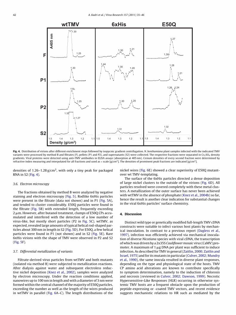
3.5. Variant particles after isopycnic density gradientcentrifugation
Filtrate, pellet and supernatant fractions obtained from methodB were processed in parallel for isopycnic Cs2SO4 centrifugationand the resulting fractions were analyzed by ELISA using anti-TMVantibodies (Fig. 4). The corresponding densities of the fractionswere determined by refractometry, and ELISA values were drawnto the density scale. wtTMV particles with 5% RNA accumulated atdensities of 1.29–1.33 g/cm3 (Fig. 4; wtTMV; major peak), whereasRNA-free TMV protein was found in the upper fractions with den-sities of 1.25–1.27 g/cm3 (Fig. 4; wtTMV; minor peaks). Most ofwtTMV was retrieved from S2 as expected. Of the 6xHis particlesfrom the first pellet fraction (P1), a small portion migrating at adensity of 1.3 g/cm3 indicated that at least some particles wereformed with the canonical concentration of 5% RNA. However, the
majority of their proteins were found in a broad distribution overthe whole of the gradients, indicating that these particles are het-erogeneous and have a tendency to decompose and/or aggregatewith host components. E50Q protein, as expected from the pre-vious experiments, was mainly found in RNA-free fractions withP2 S2
1 2 1 2
.0 8.5 3.0 66.5 59.0
.0 <0.3 <0.3 <0.3 <0.3
.5 62.5 53.5 <0.3 <0.3
leaves at 21 dpi in two independent experiments. TMV CP in the indicated fractionsva-Violet staining. Absolute yields varied between experiments from 1.2 to 18 mg

A. Kadri et al. / Virus Research 157 (2011) 35–46 41
Fig. 3. Detection of CP and RNA for the four TMV variants during enrichment procedure (method B). Western blot (A, B), mass spectrometry analysis (C) and Northern blots(D and E) are shown for two independent N. benthamiana plant samples (lanes #1 and #2). Plants were inoculated with 6xHis (H), E50Q (Q) or wtTMV (T). Mock-inoculatedN. benthamiana plants (C) served as negative control. Filtrates (F), supernatants (S2) and pellet fractions (P1) and (P2) are compared. Proteins were detected either withanti-TMV antibodies (A and B) or anti-pentaHis antibodies (B and D). M: prestained molecular weight marker (NEB). The calculated sizes of TMV CP (A) or His-tagged TMVCP (B) are indicated. MALDI-TOF spectra (C) of the respective proteins are indicated by the abbreviation of the variant and purification step from which they stem. Molecularmasses are indicated above the single peak signals. RNA samples (D and E) were separated on SDS-agarose gels, blotted onto nylon membranes, and viral RNA was detectedwith a plus-strand-specific TMV CP RNA probe. (D) and (E) display the same blots with different exposure. The length of the TMV genome (6.4 kb) is indicated.
42 A. Kadri et al. / Virus Research 157 (2011) 35–46
Q05EVMTtw 6xHis1.30
1.25F
1.211.30
1.27
1,30A
40
5 n
m
1.30
130
P1
1.301.44
1.20
1.21
1.28
1.26 1.241.30
1.25
1.32
1.27
P2
1.201.30
Density (g/cm3)
1.27
1.11.21.31.41.5
S21.31
1.11.21.31.41.5
1.32
1.11.21.31.41.5
F c gradv ernatag (absor cm3].
dR
3
swat2mvetp6(
3
(Atbnfei
ig. 4. Distribution of virions after different enrichment steps followed by isopycniariants were processed by method B and filtrates (F), pellets (P1 and P2), and supradients. Viral proteins were detected using anti-TMV antibodies in ELISA assaysefractive index measuring and interpolated for all fractions and used as × scale [g/
ensities of 1.26–1.28 g/cm3, with only a tiny peak for packagedNA in S2 (Fig. 4).
.6. Electron microscopy
The fractions obtained by method B were analyzed by negativetaining and electron microscopy (Fig. 5). Rodlike 6xHis particlesere present in the filtrate (data not shown) and in P1 (Fig. 5A),
nd tended to cluster considerably. E50Q particles were found inhe filtrate (Fig. 5B) with extended length, frequently exceeding�m. However, after butanol treatment, clumps of E50Q CPs accu-ulated and interfered with the detection of a low number of
irus-like, but mostly short particles (P2 in Fig. 5C). wtTMV, asxpected, revealed large amounts of typical helical rod-shaped par-icles about 300 nm in length in S2 (Fig. 5D). For E50Q, a few helicalarticles were found in P1 (not shown) and in S2 (Fig. 5E). RarexHis virions with the shape of TMV were observed in P2 and S2Fig. 5F).
.7. Differential metallization of variants
Filtrate-derived virus particles from wtTMV and both mutantsisolated via method B) were subjected to metallization reactions.fter dialysis against water and subsequent electroless reduc-
ive nickel deposition (Knez et al., 2002), samples were analyzed
y electron microscopy. Under the reaction conditions applied,anowires up to 100 nm in length and with a diameter of 3 nm wereormed within the central channel of the majority of E50Q particles,xceeding the number as well as the length of the wires producedn wtTMV in parallel (Fig. 6A–C). The length distributions of the
ient centrifugation. N. benthamiana plant samples infected with the indicated TMVnts (S2) were collected. The respective fractions were separated in Cs2SO4 densityrption at 405 nm). Cesium densities of every second fraction were determined byThe densities of prominent peak fractions are indicated [g/cm3].
nickel wires (Fig. 6E) showed a clear superiority of E50Q mutant-over wt TMV-templating.
The surface of the 6xHis particles directed a dense depositionof large nickel clusters to the outside of the virions (Fig. 6D). Allparticles resolved were covered completely with these metal clus-ters. A metallization of the outer surface has never been achievedwith wtTMV in the absence of phosphate (Knez et al., 2004b) so far,hence the result is another clear indication for substantial changesin the viral 6xHis particles’ surface chemistry.
4. Discussion
Distinct wild type or genetically modified full-length TMV cDNAconstructs were suitable to infect various host plants by mechan-ical inoculation. In contrast to a previous report (Dagless et al.,1997), infection was efficiently achieved via mechanical inocula-tion of diverse Nicotiana species with viral cDNA, the transcriptionof which was driven by a 2x35S Cauliflower mosaic virus (CaMV) pro-moter. A maximum of 1 �g DNA per plant was sufficient to induceinfection. As described for TMV in general (Zaitlin, 2000; Zaitlin andIsrael, 1975) and for its mutants in particular (Culver, 2002; Mundryet al., 1990), the same inocula resulted in diverse plant responses,depending on the type and physiological state of the hosts. TMVCP amino acid alterations are known to contribute specificallyto symptom determination, namely to the induction of chlorosis
and necrosis (reviewed in Culver, 2002; Dawson, 1999). NecroticHypersensitive-Like Responses (HLR) occurring in otherwise sys-temic TMV hosts are a frequent obstacle upon the production ofpeptide-expressing or -coated TMV vectors, and recent evidencesuggests mechanistic relations to HR such as mediated by the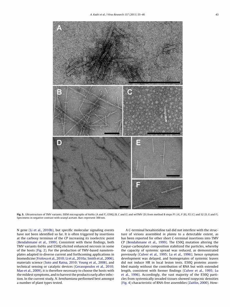
A. Kadri et al. / Virus Research 157 (2011) 35–46 43
F (B, C aS
Nha(TopbmtMtta
ig. 5. Ultrastructure of TMV variants. ISEM micrographs of 6xHis (A and F), E50Qpecimens in negative contrast with uranyl acetate. Bars represent 300 nm.
gene (Li et al., 2010b), but specific molecular signaling eventsave not been identified so far. It is often triggered by insertionst the carboxy terminus of the CP increasing its isoelectric pointBendahmane et al., 1999). Consistent with these findings, bothMV variants 6xHis and E50Q elicited enhanced necrosis in somef the hosts (Fig. 2). For the production of TMV-based nanotem-lates adapted to diverse current and forthcoming applications iniomedicine (Frolova et al., 2010; Li et al., 2010a; Smith et al., 2006),aterials science (Soto and Ratna, 2010; Young et al., 2008), and
echnical sensing or catalytic devices (Gerasopoulos et al., 2010;
ao et al., 2009), it is therefore necessary to choose the hosts withhe mildest symptoms, and to harvest the products early after infec-ion. In the current study, N. benthamiana performed best amongstnumber of plant types tested.
nd E) and wtTMV (D) from method B steps P1 (A), F (B), P2 (C) and S2 (D, E and F).
A C-terminal hexahistidine tail did not interfere with the struc-ture of virions assembled in planta to a detectable extent, ashas been reported for other short C-terminal insertions into TMVCP (Bendahmane et al., 1999). The E50Q mutation altering theCaspar-carboxylate composition stabilized the particles, wherebythe capacity of systemic spread was reduced, as demonstratedpreviously (Culver et al., 1995; Lu et al., 1996); hence symptomdevelopment was delayed, and homogenates of systemic leavesdid not induce HR in local lesion tests. E50Q proteins assem-bled mainly without the contribution of RNA but with extended
length, consistent with former findings (Culver et al., 1995; Luet al., 1996). Accordingly, the vast majority of the E50Q parti-cles from systemically invaded tissues showed isopycnic densities(Fig. 4) characteristic of RNA-free assemblies (Zaitlin, 2000). How-
44 A. Kadri et al. / Virus Resear
Fig. 6. TMV-based biotemplates after nickel metallization. Transmission electronmicroscopy in positive contrast for E50Q (A and B), wtTMV (C), and 6xHis (D).Method B filtrate fractions were adsorbed to grids after electroless deposition ofncaf
ewn3lErooDpe1t(
Clspaddt
ickel. Bars represent 100 nm. The length-distributions of nickel wires in the centralhannel for E50Q and wtTMV are compared (E). Histograms represent the percent-ge frequency distribution of nickel wires in the indicated length classes measuredor wtTMV (n = 114) and E50Q (n = 109) wires.
ver, a small amount of E50Q particles that escaped degradationhen treated with n-butanol, and thus was retained in the super-atant S2, was found to encapsidate RNA and to be approximately00 nm in length. These particles presumably contained viral full-
ength RNA after systemic spread of the construct. The majority of50Q particles had an increased length, which reflects the reducedepulsion between otherwise more negative electrostatic patchesf adjacent CP subunits in this mutant lacking the carboxyl groupf E50, which in wild type TMV is in close proximity to that of the77 residue and contributes to the destabilization of the nucleo-rotein helix in a suitable physicochemical environment (Culvert al., 1995; Kegel and van der Schoot, 2006; Namba and Stubbs,986). Nevertheless, butanol treatment was sufficient to disrupthe elongated protein assemblies lacking continuous RNA scaffoldsFig. 5C).
Oligopeptides can be introduced on the outer face of TMVPs without altering protein folding (Negrouk et al., 2004). Simi-
arly, our hexahistidine fusion to the C-terminus did not preventelf-assembly and revealed particles which resembled wtTMV and
ackaged RNA. In isopycnic density gradient centrifugation (Fig. 4),subfraction of the 6xHis particles exhibited the same buoyantensity as wtTMV, but our data suggest that a significant portionisassembled under the high salt conditions. This may indicate thathe hexahistidine tag reduced the virion stability. Moreover, the
ch 157 (2011) 35–46
particles showed a high tendency to aggregate (Fig. 5A), whichmight be explained by an interaction of the His-tag with hostnucleic acids and has been observed for further His-tagged proteinspecies (Renzi et al., 2006). In addition, the 6xHis mutant inducednecrotic reactions (Fig. 2) with the consequence of polyphenolaccumulation and crosslinking of proteins. In this view, it is con-ceivable that large portions of dimers and multimers of 6xHis CPremained stable during SDS–PAGE (Fig. 3A). Interestingly, the TMV6xHis CP, if expressed in yeast, did not assemble with RNA in vitro(Mueller et al., 2010).
In summary, despite of limitations upon the formation of homo-geneous nucleoprotein nanotubes, both TMV CP mutants did notonly allow accumulation of virion titers sufficient for subsequentapplications in the inoculated leaves, they also were able to mediatelong-distance (LD) transport of infectious nucleic acid via the vascu-lar tissues – another essential function of the viral CP (Dawson et al.,1988). The CP’s molecular roles upon systemic spread have not beenunraveled, but several lines of evidence indicate that TMV virionformation is not required at this stage (Wisniewski et al., 1988,and references therein): tobamoviral CP mutations abolishing itsassembly into helical particles did not prevent LD movement of theviral agent (e.g. Berzal-Herranz et al., 1995; Dawson et al., 1988);and, furthermore, LD trafficking of TMV lacking CP was comple-mented by a nonstructural protein of an umbravirus (Ryabov et al.,1999). Putative roles of TMV CP upon systemic spread may there-fore encompass gating the transported complex into companioncell/sieve element domains inside the phloem, maybe as acces-sory function supporting the viral movement protein (Heinlein,2002; Ryabov et al., 1999, and references therein), and/or interac-tions with host factors directly or indirectly enabling long-distancetransport in vascular tissues (Li et al., 2005).
As with our previous studies (Balci et al., 2006; Knez et al.,2002, 2003, 2004b), the new TMV variants were successfully met-allized with nickel. The electroless deposition process requiresto firstly bind Pd(II) in an “activation” step. In the absence ofphosphate, a reaction with carboxylate groups is possible; theydominate the surface chemistry especially in the channel of wtTMVand E50Q particles. However, nitrogen ligands and namely Hisare favored, so a faster reaction and dense exterior coating of the6xHis nanotubes was expected. Catalytically active Pd nanoparti-cles (d < 4 nm) are then formed by reduction of Pd(II) to Pd(0), andby coalescence. Since the metallization by nickel is autocatalytic, Niinitially deposited on Pd as well as subsequently on Pd/Ni nuclei.catalyzes its own deposition, with the required electrons producedby catalytic oxidation of BH3 (from (CH3)2NH·BH3).
Therefore, the 6xHis mutant was metallized specifically on itsouter coat without any further treatment, where Pd(II) was boundto the His-tags. On the outer surface of wtTMV, nickel accumula-tion can be induced by phosphate conditioning (Knez et al., 2004b).In contrast, for wtTMV and the E50Q virions, Pd is predominantlydirected into the channel promoting Ni nanowire growth (Knezet al., 2002). Interestingly, the wires inside the E50Q mutant parti-cles were significantly longer and formed with increased efficiencyin comparison to those inside wtTMV virions under the conditionsapplied (Fig. 6E). The channel chemistry should be identical for bothvirus variants, as the E50Q mutation affects inaccessible parts of thevirions. However, probably due to their enhanced stability againstchemical and physical changes, the in many cases very long (upto some micrometers) virus-like tubes seem to allow for a largelyunhindered growth of metal wires of improved quality. Assumedlyalso the absence of the RNA scaffold renders beneficial conditions
for reductive nickel deposition.In conclusion, this study demonstrates that genetic engineeringof the tubular plant virus has resulted in new types of biotemplatesthat governed the final nanostructure of inorganic reaction prod-ucts in a predictable manner. Inner-surface metallization of the

esear
scpcmncron
A
StftN
R
B
B
B
B
B
B
B
B
C
C
C
C
C
C
D
D
D
D
D
F
A. Kadri et al. / Virus R
tiff helical capsids may offer a unique access to ultrathin inter-onnects for nanoscale circuits (Kwok and Ellenbogen, 2002), or torintable nanoelectronic arrays (Fan et al., 2009). Magnetic metaloatings of TMV-derived particles with pre-determined aspect ratioight contribute to a novel production route for high-performance
anotube ferrofluids applied for damping purposes in microme-hanical devices: support for this concept comes from our recentesults demonstrating the stable and tunable beneficial effectsf tobamoviral scaffolding additives on commercially availableanosphere ferrofluids (Wu et al., 2010a,b).
cknowledgements
The authors thank the gardeners for taking care of the plants,igrid Kober and Sylvia Pfeiffer for excellent technical assis-ance, and Katharina Kittelmann, Robert G. Milne and Carl Krillor critically reading the manuscript. This work was funded byhe Baden-Wuerttemberg-Stiftung, “Kompetenznetz Funktionelleanostrukturen”, and subsidiarily supported by the DFG-SPP1165.
eferences
alci, S., Bittner, A., Schirra, M., Thonke, K., Sauer, R., Hahn, K., Kadri, A., Wege, C.,Jeske, H., Kern, K., 2009. Catalytic coating of virus particles with zinc oxide.Electrochim. Acta 54, 5149–5154.
alci, S., Bittner, A.M., Hahn, K., Scheu, C., Knez, M., Kadri, A., Wege, C., Jeske, H., Kern,K., 2006. Copper nanowires within the central channel of tobacco mosaic virusparticles. Electrochim. Acta 51, 6251–6257.
alci, S., Leinberger, D.M., Knez, M., Bittner, A.M., Boes, F., Kadri, A., Wege, C., Jeske,H., Kern, K., 2008. Printing and aligning mesoscale patterns of tobacco mosaicviruses on surfaces. Adv. Mater. 20, 2195–2200.
alci, S., Noda, K., Bittner, A.M., Kadri, A., Wege, C., Jeske, H., Kern, K., 2007. Self-assembly of metal-virus nanodumbbells. Angew. Chem. Int. Ed. 46, 3149–3151.
endahmane, M., Chen, I., Asurmendi, S., Bazzini, A.A., Szecsi, J., Beachy, R.N.,2007. Coat protein-mediated resistance to TMV infection of Nicotiana tabacuminvolves multiple modes of interference by coat protein. Virology 366, 107–116.
endahmane, M., Koo, M., Karrer, E., Beachy, R.N., 1999. Display of epitopes on thesurface of tobacco mosaic virus: impact of charge and isoelectric point of theepitope on virus–host interactions. J. Mol. Biol. 290, 9–20.
erzal-Herranz, A., de la Cruz, A., Tenllado, F., Diaz-Ruiz, J.R., Lopez, L., Sanz, A.I.,Vaquero, C., Serra, M.T., Garcia-Luque, I., 1995. The Capsicum L3 gene-mediatedresistance against the tobamoviruses is elicited by the coat protein. Virology209, 498–505.
oom, R., Sol, C.J.A., Salimans, M.M.M., Jansen, C.L., Wertheim-van Dillen, P.M.E., vander Noordaa, J., 1990. Rapid and simple method for purification of nucleic acids.J. Clin. Microbiol. 28, 495–503.
aspar, D.L.D., 1963. Assembly and stability of the tobacco mosaic virus particle. Adv.Protein Chem. 18, 37–121.
aspar, D.L.D., Namba, K., 1990. Switching in the self-assembly of tobacco mosaicvirus. Adv. Biophys. 26, 157–185.
hapman, S.N., 1998. Tobamovirus isolation and RNA extraction. In: Foster, G.D.,Taylor, S.C. (Eds.), Methods in Molecular Biology: Plant Virology Protocols: FromVirus Isolation to Transgenic Resistance, vol. 81. Humana Press Inc., Totowa, NJ,pp. 123–129.
lare, D.K., Orlova, E.V., 2010. 4.6 A Cryo-EM reconstruction of tobacco mosaic virusfrom images recorded at 300 keV on a 4k × 4k CCD camera. J. Struct. Biol.,303–308.
ulver, J.N., 2002. Tobacco mosaic virus assembly and disassembly: determinants inpathogenicity and resistance. Annu. Rev. Phytopathol. 40, 287–308.
ulver, J.N., Dawson, W.O., Plonk, K., Stubbs, G., 1995. Site-directed mutagenesisconfirms the involvement of carboxylate groups in the disassembly of tobaccomosaic virus. Virology 206, 724–730.
agless, E.M., Shintaku, M.H., Nelson, R.S., Foster, G.D., 1997. A CaMV 35S pro-moter driven cDNA clone of tobacco mosaic virus can infect host plant tissuedespite being uninfectious when manually inoculated onto leaves. Arch. Virol.142, 183–191.
awson, W.O., 1999. Tobacco mosaic virus virulence and avirulence. Philos. Trans.R. Soc. Lond. B: Biol. Sci. 354, 645–651.
awson, W.O., Bubrick, P., Grantham, G.L., 1988. Modifications of the tobacco mosaicvirus coat protein gene affecting replication, movement, and symptomatology.Phytopathology 78, 783–789.
evash, Y., Hauschner, A., Sela, I., Chakraburtty, K., 1981. The Anti-Viral Factor (Avf)from virus-infected plants induces discharge of Histidinyl-Tmv-Rna. Virology
111, 103–112.ujardin, E., Peet, C., Stubbs, G., Culver, J.N., Mann, S., 2003. Organization of metallicnanoparticles using tobacco mosaic virus templates. Nano Lett. 3, 413–417.
an, Z., Ho, J.C., Takahashi, T., Yerushalmi, R., Takei, K., Ford, A.C., Chueh, Y.-L., Javey,A., 2009. Toward the development of printable nanowire electronics and sensors.Adv. Mater. 21, 3730–3743.
ch 157 (2011) 35–46 45
Frolova, O.Y., Petrunia, I.V., Komarova, T.V., Kosorukov, V.S., Sheval, E.V., Gleba, Y.Y.,Dorokhov, Y.L., 2010. Trastuzumab-binding peptide display by tobacco mosaicvirus. Virology 407, 7–13.
Gerasopoulos, K., McCarthy, M., Banerjee, P., Fan, X., Culver, J.N., Ghodssi, R., 2010.Biofabrication methods for the patterned assembly and synthesis of viral nan-otemplates. Nanotechnology 21, 11, 055304.
Górzny, M., Walton, A.S., Evans, S.D., 2010. Synthesis of high-surface-area platinumnanotubes using a viral template. Adv. Funct. Mater. 20, 1295–1300.
Heinlein, M., 2002. The spread of tobacco mosaic virus infection: insights into thecellular mechanism of RNA transport. Cell. Mol. Life Sci. 59, 58–82.
Kegel, W.K., van der Schoot, P., 2006. Physical regulation of the self-assembly oftobacco mosaic virus coat protein. Biophys. J. 91, 1501–1512.
Klug, A., 1999. The tobacco mosaic virus particle: structure and assembly. Philos.Trans. R. Soc. Lond. B 354, 531–535.
Knez, M., Bittner, A.M., Boes, F., Wege, C., Jeske, H., Maiß, E., Kern, K., 2003. Biotem-plate synthesis of 3-nm nickel and cobalt nanowires. Nano Lett. 3, 1079–1082.
Knez, M., Kadri, A., Wege, C., Gosele, U., Jeske, H., Nielsch, K., 2006. Atomic layerdeposition on biological macromolecules: metal oxide coating of tobacco mosaicvirus and ferritin. Nano Lett. 6, 1172–1177.
Knez, M., Sumser, M., Bittner, A., Wege, C., Jeske, H., Hoffmann, D.M.P., Kuhnke, K.,Kern, K., 2004a. Binding the tobacco mosaic virus to inorganic surfaces. Langmuir20, 411–447.
Knez, M., Sumser, M., Bittner, A.M., Wege, C., Jeske, H., Kooi, S., Burghard, M., Kern,K., 2002. Electrochemical modification of individual nano-objects. J. Electroanal.Chem. 522, 70–74.
Knez, M., Sumser, M., Bittner, A.M., Wege, C., Jeske, H., Martin, T.P., Kern, K., 2004b.Spatially selective nucleation of metal clusters on the tobacco mosaic virus. Adv.Funct. Mater. 14, 116–124.
Kobayashi, M., Seki, M., Tabata, H., Watanabe, Y., Yamashita, I., 2010. Fabrication ofaligned magnetic nanoparticles using tobamoviruses. Nano Lett. 10, 773–776.
Kwok, K.S., Ellenbogen, J.C., 2002. Moletronics: future electronics. Mater. Today 5,28–37.
Laemmli, U.K., 1970. Cleavage of structural proteins during the assembly of the headof bacteriophage T4. Nature 227, 680–685.
Lee, S.Y., Choi, J., Royston, E., Janes, D.B., Culver, J.N., Harris, M.T., 2006. Deposi-tion of platinum clusters on surface-modified tobacco mosaic virus. J. Nanosci.Nanotechnol. 6, 974–981.
Lee, S.Y., Royston, E., Culver, J.N., Harris, M.T., 2005. Improved metal cluster deposi-tion on a genetically engineered tobacco mosaic virus template. Nanotechnology16, S435–S441.
Li, K., Nguyen, H.G., Lu, X., Wang, Q., 2010a. Viruses and their potential in bioimagingand biosensing applications. Analyst 135, 21–27.
Li, M., Li, P., Song, R., Xu, Z., 2010b. An induced hypersensitive-like response lim-its expression of foreign peptides via a recombinant TMV-based vector in asusceptible tobacco. PLoS ONE 5, e15087.
Li, Y., Wu, M.Y., Song, H.H., Hu, X., Qiu, B.S., 2005. Identification of a tobacco proteininteracting with tomato mosaic virus coat protein and facilitating long-distancemovement of virus. Arch. Virol. 150, 1993–2008.
Lim, J.S., Kim, S.M., Lee, S.Y., Stach, E.A., Culver, J.N., Harris, M.T., 2010. Formation ofAu/Pd alloy nanoparticles on TMV. J. Nanomater. 2010, 6, 620505.
Lu, B., Stubbs, G., Culver, J.N., 1996. Carboxylate interactions involved in the disas-sembly of tobacco mosaic tobamovirus. Virology 225, 11–20.
Mao, C., Liu, A., Cao, B., 2009. Virus-based chemical and biological sensing. Angew.Chem. Int. Ed. 48, 6790–6810.
Miller, R.A., Presley, A.D., Francis, M.B., 2007. Self-assembling light-harvesting sys-tems from synthetically modified tobacco mosaic virus coat proteins. J. Am.Chem. Soc. 129, 3104–3109.
Milne, R.G., Lesemann, D.E., 1984. Immunosorbent electron microscopy in plantvirus studies. In: Maramorosch, K., Koprowski, H. (Eds.), Methods in Virology,vol. VIII. Academic Press, San Diego, pp. 85–101.
Mueller, A., Kadri, A., Jeske, H., Wege, C., 2010. In vitro assembly of tobacco mosaicvirus coat protein variants derived from fission yeast expression clones or plants.J. Virol. Methods 166, 77–85.
Mundry, K.W., Schaible, W., Ellwart-Tschürtz, M., Nitschko, H., Hapke, C., 1990.Hypersensitivity to tobacco mosaic virus in N′-gene hosts: which viral genesare involved? In: Fraser, R.S.S. (Ed.), Recognition and Response in Plant–VirusInteractions, vol. H41. Springer-Verlag, Berlin/Heidelberg, pp. 345–359.
Mutombo, K., Michels, B., Ott, H., Cerf, R., Witz, J., 1992. Scanning calorimetric studiesof the stability of tobacco mosaic virus and aggregates of its coat protein. Eur.Biophys. J. 21, 77–83.
Namba, K., Pattanayek, R., Stubbs, G., 1989. Visualization of protein-nucleic acidinteractions in a virus. Refined structure of intact tobacco mosaic virus at 2.9 Aresolution by X-ray fiber diffraction. J. Mol. Biol. 208, 307–325.
Namba, K., Stubbs, G., 1986. Structure of tobacco mosaic virus at 3.6 A resolution:implications in assembly. Science 231, 1401–1406.
Negrouk, V., Eisner, G., Midha, S., Lee, H., Bascomb, N., Gleba, Y., 2004. Affinity purifi-cation of streptavidin using tobacco mosaic virus particles as purification tags.Anal. Biochem. 333, 230–235.
Perham, R.N., Wilson, T.M.A., 1978. The characterization of intermediates formedduring disassembly of tobacco mosaic virus at alkaline pH. Virology 84, 293–302.
Renzi, F., Panetta, G., Vallone, B., Brunori, M., Arceci, M., Bozzoni, I., Laneve, P.,Caffarelli, E., 2006. Large-scale purification and crystallization of the endori-bonuclease XendoU: troubleshooting with His-tagged proteins. Acta Crystall. F:Struct. Biol. Cryst. Commun. 62, 298–301.
Restrepo, M.A., Freed, D.D., Carrington, J.C., 1990. Nuclear transport of plant potyviralproteins. Plant Cell 2, 987–998.

4 esear
R
R
R
R
S
S
S
S
S
S
Young, M., Willits, D., Uchida, M., Douglas, T., 2008. Plant viruses as biotemplates
6 A. Kadri et al. / Virus R
ong, J.H., Oberbeck, F., Wang, X.N., Li, X.D., Oxsher, J., Niu, Z.W., Wang, Q.,2009. Tobacco mosaic virus templated synthesis of one dimensional inorganic-polymer hybrid fibres. J. Mater. Chem. 19, 2841–2845.
oyston, E., Ghosh, A., Kofinas, P., Harris, M.T., Culver, J.N., 2008. Self-assembly ofvirus-structured high surface area nanomaterials and their application as bat-tery electrodes. Langmuir 24, 906–912.
oyston, E.S., Brown, A.D., Harris, M.T., Culver, J.N., 2009. Preparation of silica sta-bilized tobacco mosaic virus templates for the production of metal and layerednanoparticles. J. Colloid Interface Sci. 332, 402–407.
yabov, E.V., Robinson, D.J., Taliansky, M.E., 1999. A plant virus-encoded proteinfacilitates long-distance movement of heterologous viral RNA. Proc. Natl. Acad.Sci. U.S.A. 96, 1212–1217.
achse, C., Chen, J.Z., Coureux, P.D., Stroupe, M.E., Fandrich, M., Grigorieff, N.,2007. High-resolution electron microscopy of helical specimens: a fresh lookat tobacco mosaic virus. J. Mol. Biol. 371, 812–835.
ambrook, J., Russell, D.W., 2001. Molecular Cloning: A Laboratory Manual, 3rd ed.Cold Spring Harbor Laboratory Press, Cold Spring Harbor, NY.
chlick, T.L., Ding, Z., Kovacs, E.W., Francis, M.B., 2005. Dual-surface modification ofthe tobacco mosaic virus. J. Am. Chem. Soc. 127, 3718–3723.
henton, W., Douglas, T., Young, M., Stubbs, G., Mann, S., 1999. Inorganic–organicnanotube composites from template mineralization of tobacco mosaic virus.Adv. Mater. 11, 253–256.
hintaku, M.H., Carter, S.A., Bao, Y., Nelson, R.S., 1996. Mapping nucleotides in the
126-kDa protein gene that control the differential symptoms induced by twostrains of tobacco mosaic virus. Virology 221, 218–225.mith, M.L., Lindbo, J.A., Dillard-Telm, S., Brosio, P.M., Lasnik, A.B., McCormick, A.A.,Nguyen, L.V., Palmer, K.E., 2006. Modified tobacco mosaic virus particles asscaffolds for display of protein antigens for vaccine applications. Virology 348,475–488.
ch 157 (2011) 35–46
Soto, C.M., Ratna, B.R., 2010. Virus hybrids as nanomaterials for biotechnology. Curr.Opin. Biotechnol. 21, 426–438.
Towbin, H., Staehelin, T., Gordon, J., 1979. Electrophoretic transfer of proteins frompolyacrylamide gels to nitrocellulose sheets: procedure and some applications.Proc. Natl. Acad. Sci. U.S.A. 76, 4350–4354.
Tseng, R.J., Tsai, C., Ma, L.P., Ouyang, J., Ozkan, C.S., Yang, Y., 2006. Digital mem-ory device based on tobacco mosaic virus conjugated with nanoparticles. Nat.Nanotechnol. 1, 72–77.
Wisniewski, L.A., Powell, P.A., Nelson, R.S., Beachy, R.N., 1988. Local and systemicspread of tobacco mosaic virus in transgenic tobacco. Plant Cell 2, 559–567.
Wu, Z., Mueller, A., Degenhard, S., Ruff, S.E., Geiger, F., Bittner, A., Wege, C., KrillIII, C., 2010a. Enhancing the magnetoviscosity of ferrofluids by the addition ofbiological nanotubes. ACS Nano 4, 4531–4538.
Wu, Z., Zierold, R., Mueller, A., Ruff, S.E., Ma, C., Khan, A.A., Geiger, F., Sommer, B.A.,Knez, M., Nielsch, K., Bittner, A.M., Wege, C., Krill III, C., 2010b. Preparation andmagnetoviscosity of nanotube ferrofluids by viral scaffolding and ALD on poroustemplates. Phys. Stat. Sol. B, 2412–2423.
Yang, C., Manocchi, A.K., Lee, B., Yi, H., 2010. Viral templated palladium nanocatalystsfor dichromate reduction. Appl. Catal. B: Environ. 93, 282–291.
Yi, H., Nisar, S., Lee, S.Y., Powers, M.A., Bentley, W.E., Payne, G.F., Ghodssi, R., Rubloff,G.W., Harris, M.T., Culver, J.N., 2005. Patterned assembly of genetically modifiedviral nanotemplates via nucleic acid hybridization. Nano Lett. 5, 1931–1936.
for materials and their use in nanotechnology. Annu. Rev. Phytopathol. 46,361–384.
Zaitlin, M., 2000. Tobacco mosaic virus. AAB Descr. Plant Viruses 370, 1–13.Zaitlin, M., Israel, H.W., 1975. Tobacco mosaic virus (type strain). C.M.I./A.A.B. Descr.
of Plant Viruses, No. 151.


![ScienceDirect cienceirect ScienceDirect · and. {[,], , , : . , /](https://img.pdfslide.us/doc/110x75/608077a6d3af4a2358487f59/-sciencedirect-cienceirect-sciencedirect-and-.jpg)