Embed Size (px)
Citation preview
REVIEW
Engineered cells as biosensing systems in biomedical analysis
Nilesh Raut & Gregory O’Connor & Patrizia Pasini &Sylvia Daunert
Received: 18 November 2011 /Revised: 7 January 2012 /Accepted: 16 January 2012 /Published online: 5 February 2012# Springer-Verlag 2012
Abstract Over the past two decades there have been greatadvances in biotechnology, including use of nucleic acids,proteins, and whole cells to develop a variety of molecularanalytical tools for diagnostic, screening, and pharmaceuti-cal applications. Through manipulation of bacterial plas-mids and genomes, bacterial whole-cell sensing systemshave been engineered that can serve as novel methods foranalyte detection and characterization, and as more efficientand cost-effective alternatives to traditional analytical tech-niques. Bacterial cell-based sensing systems are typicallysensitive, specific and selective, rapid, easy to use, low-cost,and amenable to multiplexing, high-throughput, and minia-turization for incorporation into portable devices. This crit-ical review is intended to provide an overview of availablebacterial whole-cell sensing systems for assessment of avariety of clinically relevant analytes. Specifically, we ex-amine whole-cell sensing systems for detection of bacterialquorum sensing molecules, organic and inorganic toxiccompounds, and drugs, and for screening of antibacterialcompounds for identification of their mechanisms of action.Methods used in the design and development of whole-cellsensing systems are also reviewed.
Keywords Whole-cell sensing system . Biomedicalanalysis . Quorum sensing molecules .Mercury .
Hydroxylated polychlorinated biphenyls . Cytarabine .
Antibiotics
Introduction
The need of living organisms to recognize and respond tochanges in their environment exemplifies biosensing in na-ture. Biosensing involves selective and sensitive molecularrecognition between proteins and a target ligand analyte, evenwhen present at very low concentrations. Researchers havetried to mimic the exquisite properties found in nature byutilizing naturally occurring recognition elements to developbiosensing systems for analytical applications. To that end, abiological recognition element capable of reversible bindingto a target ligand analyte is coupled to a transducer elementthat converts the recognition event into a readable/measurableoutput. Several biological recognition elements, includingproteins, nucleic acids, and intact cells, have been used assensing elements in biosensors for applications in environ-mental, biological, pharmaceutical, and clinical analysis [1, 2].
Proteins with high specificity for analytes of interest havebeen extensively used in the development of biosensors.These proteins include enzymes, antibodies, and bindingproteins, among others. For instance, glucose oxidase cata-lyzes oxidation of β-D-glucose to D-glucono-1,5-lactone,which is further hydrolyzed to gluconic acid [3]. A rangeof commercially available biosensors for blood glucosemonitoring and diabetes management use glucose oxidaseas the sensing element coupled to appropriate mediators andtransducers [4]. Antibodies have exquisite specificity fortheir cognate antigens. In an immunosensor [5], antibodiesare used as recognition elements coupled with a variety of
Published in the topical collection Biomimetic Recognition Elementsfor Sensing Applications with guest editor María Cruz Moreno-Bondi.
N. RautDepartment of Chemistry,University of Kentucky,Lexington, KY 40506, USA
G. O’Connor : P. Pasini : S. Daunert (*)Department of Biochemistry and Molecular Biology,Miller School of Medicine,University of Miami,Miami, FL 33136, USAe-mail: [email protected]
Anal Bioanal Chem (2012) 402:3147–3159DOI 10.1007/s00216-012-5756-6
detection methods, including, electrochemical, piezoelectric,fluorescence, bioluminescence, absorbance, and surface plas-mon resonance; labeled secondary antibodies can also be usedin a different format for detection of target analytes. A varietyof sensing systems have also been developed by using hinge-motion binding proteins (HMBPs), for example periplasmicbinding proteins and the messenger protein calmodulin, withan incorporated signal-generating reporter molecule [6]. Ingeneral, these proteins have extraordinary selectivity to theircorresponding ligand and/or analyte, with affinities, KD, typi-cally in the sub-micromolar range—in some cases as low as inthe nanomolar range—and undergo conformational changesupon binding to their ligands. As representative examples ofperiplasmic binding proteins [7], the KD of the sulfate-bindingprotein is 10 nmol L−1 whereas that of the glucose-bindingprotein is 20 nmol L−1. Specifically, upon ligand binding, twoprotein domains bend around a “hinge” region of the protein.Such conformational change can be used to quantify a targetanalyte and/or ligand by measuring the change in signal inten-sity of the transduction molecule, which can either be anenvironmentally sensitive fluorescent probe strategically con-jugated to the protein or a reporter protein genetically fused tothe HMBP. The advantages of this type of protein-based bio-sensor are their high specificity toward their ligand analyte,thus resulting in high selectivity, low limits of detection, rapidresponse times, and amenability to incorporation into variousanalytical devices. Potential disadvantages of protein-basedbiosensors can be their storage conditions, transport, and shelflife. In most cases, protein biosensors must be stored andtransported refrigerated, which limits their utility and compro-mises their shelf life when used at room temperature.
Intact cells, including bacterial, yeast, and mammaliancells, are used as sensing elements in biosensing systems.Bacterial cell-based biosensing systems use genetically engi-neered bacteria capable of generating a signal on selectiverecognition of the analyte or class of analytes of interest. Theability to produce dose-dependent detectable signals in re-sponse to the analyte, as described in the next section, enablesselective determination of the bioavailable analyte or class ofanalytes present in a given sample. Cell-based sensing sys-tems are relatively easy and inexpensive to prepare and store,and are robust: they tend to be stable to environmentalchanges, for example variations in temperature or pH. Inaddition, these sensing systems can provide physiologicallyrelevant data and evaluate the bioavailability of the analyte ofinterest, because the target chemical must be ingested by thecells to trigger a response. Moreover, by using different rec-ognition element–reporter protein pairs, multiple analytes canbe detected simultaneously in a sample. Cell-based biosensingsystems have high-throughput features because they are ame-nable to miniaturization and incorporation into high-densityanalytical devices, thus enabling assay of large numbers ofsamples in a single analytical run. This is a distinct advantage
over conventional physicochemical analytical methods. How-ever, a whole-cell biosensing system is not without limita-tions. In whole-cell biosensing systems, analytes must enterthe bacteria by diffusion, which may, depending on the rate ofdiffusion, result in a slow sensor response. Additionally, be-cause the cell biochemical machinery must be activated toproduce the reporter protein, the response of a whole-cellsensing system is slow compared with that of protein-basedbiosensors, which is of the order of seconds or minutes.Additional drawbacks include potential interference with thesensor’s response by components of the bacterial cell and highbackground signal when fluorescent proteins are used asreporters, because of the presence of fluorescent moleculesin the cell. Cell batch-to-batch variability, which is intrinsic toliving organisms, is a further aspect that may have to be takeninto consideration [8].
In this critical review, we discuss the design and devel-opment of genetically engineered bacterial cell-based bio-sensing systems and focus on their applications in clinicalanalysis. Specifically, we describe whole-cell sensing sys-tems for detection of clinically relevant analytes, including,bacterial quorum sensing molecules, inorganic ions, toxicorganic compounds, for example hydroxylated polychlori-nated biphenyls, mercury ions, methylmercury, and drugs,for example antibiotics and the chemotherapeutic agent,cytarabine. We also describe the use of whole-cell sensingsystems for screening of antibacterial compounds.
Bacterial whole-cell-based biosensing systems
Bacterial biosensing systems can be categorized into twodifferent types, depending on the mode of expression of thereporter protein [9]. Expression of the reporter can either beconstitutive or inducible. In constitutive expression systems,the reporter is expressed at high basal levels. An increase inthe amount of compounds that are toxic to the cell causes itsdeath thus reducing the reporter protein expressed and itsgeneration of signal. Whole-cell biosensing systems basedon constitutive expression have been used to measure thegeneral toxicity of a sample or test compound. A well-known example is Microtox toxicity testing [10], a stan-dardized, commercially available toxicity testing system thatuses the spontaneously bioluminescent marine bacteria Vib-rio fischeri as bacterial sensor for detection of toxic com-pounds in water samples. When V. fischeri in the test kit isexposed to a sample containing toxic compounds, a dose-dependent reduction in bioluminescence is observed, indi-cating the toxicity level of the sample.
The second class of whole-cell bacterial biosensing sys-tems comprises inducible expression systems in which thecells are genetically engineered to contain a plasmid in whichan inducible promoter fused to a reporter gene controls its
3148 N. Raut et al.
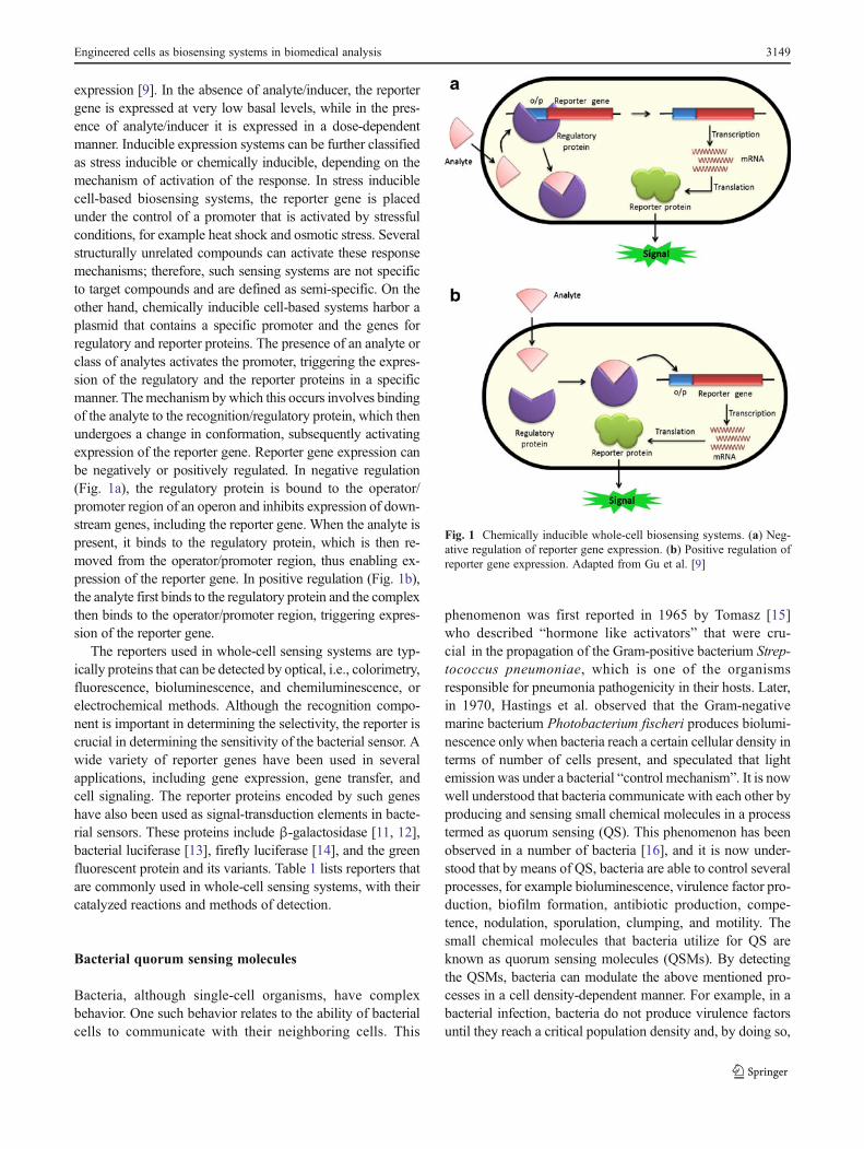
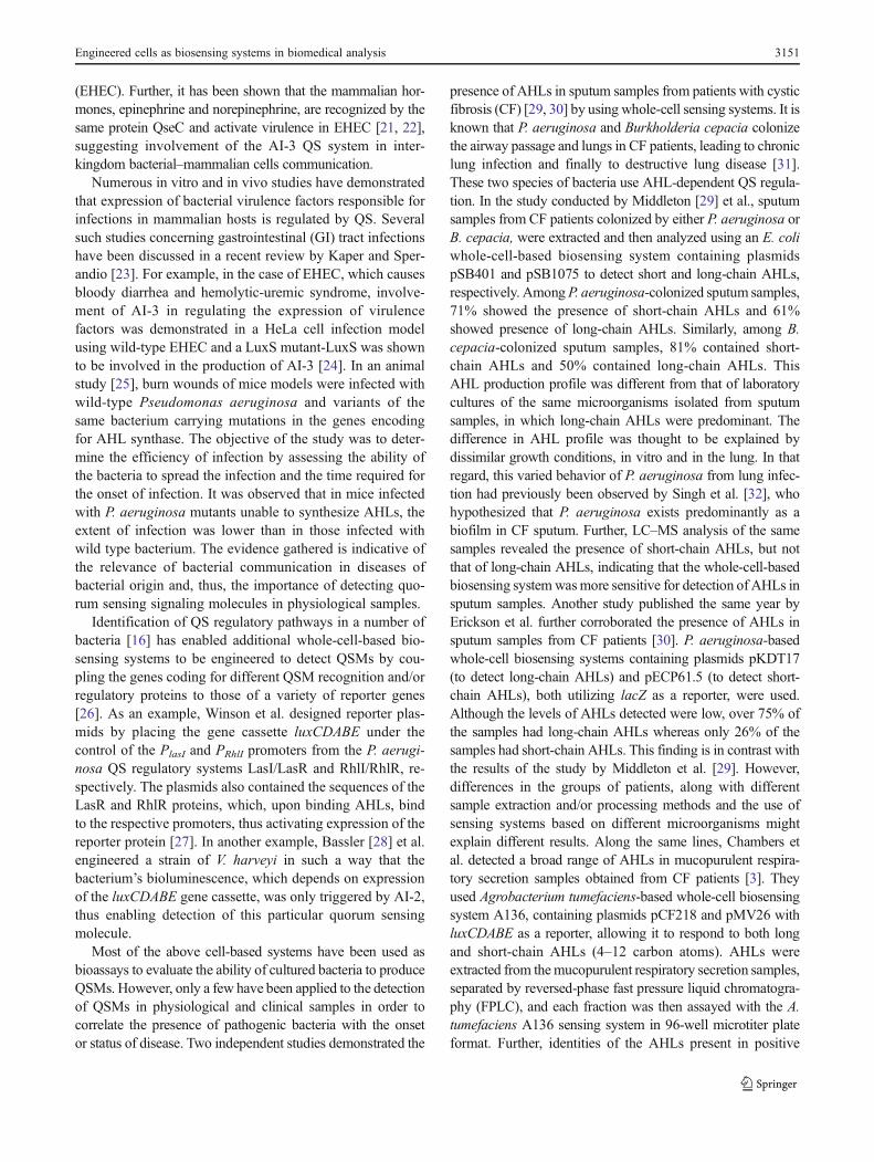
expression [9]. In the absence of analyte/inducer, the reportergene is expressed at very low basal levels, while in the pres-ence of analyte/inducer it is expressed in a dose-dependentmanner. Inducible expression systems can be further classifiedas stress inducible or chemically inducible, depending on themechanism of activation of the response. In stress induciblecell-based biosensing systems, the reporter gene is placedunder the control of a promoter that is activated by stressfulconditions, for example heat shock and osmotic stress. Severalstructurally unrelated compounds can activate these responsemechanisms; therefore, such sensing systems are not specificto target compounds and are defined as semi-specific. On theother hand, chemically inducible cell-based systems harbor aplasmid that contains a specific promoter and the genes forregulatory and reporter proteins. The presence of an analyte orclass of analytes activates the promoter, triggering the expres-sion of the regulatory and the reporter proteins in a specificmanner. Themechanism bywhich this occurs involves bindingof the analyte to the recognition/regulatory protein, which thenundergoes a change in conformation, subsequently activatingexpression of the reporter gene. Reporter gene expression canbe negatively or positively regulated. In negative regulation(Fig. 1a), the regulatory protein is bound to the operator/promoter region of an operon and inhibits expression of down-stream genes, including the reporter gene. When the analyte ispresent, it binds to the regulatory protein, which is then re-moved from the operator/promoter region, thus enabling ex-pression of the reporter gene. In positive regulation (Fig. 1b),the analyte first binds to the regulatory protein and the complexthen binds to the operator/promoter region, triggering expres-sion of the reporter gene.
The reporters used in whole-cell sensing systems are typ-ically proteins that can be detected by optical, i.e., colorimetry,fluorescence, bioluminescence, and chemiluminescence, orelectrochemical methods. Although the recognition compo-nent is important in determining the selectivity, the reporter iscrucial in determining the sensitivity of the bacterial sensor. Awide variety of reporter genes have been used in severalapplications, including gene expression, gene transfer, andcell signaling. The reporter proteins encoded by such geneshave also been used as signal-transduction elements in bacte-rial sensors. These proteins include β-galactosidase [11, 12],bacterial luciferase [13], firefly luciferase [14], and the greenfluorescent protein and its variants. Table 1 lists reporters thatare commonly used in whole-cell sensing systems, with theircatalyzed reactions and methods of detection.
Bacterial quorum sensing molecules
Bacteria, although single-cell organisms, have complexbehavior. One such behavior relates to the ability of bacterialcells to communicate with their neighboring cells. This
phenomenon was first reported in 1965 by Tomasz [15]who described “hormone like activators” that were cru-cial in the propagation of the Gram-positive bacterium Strep-tococcus pneumoniae, which is one of the organismsresponsible for pneumonia pathogenicity in their hosts. Later,in 1970, Hastings et al. observed that the Gram-negativemarine bacterium Photobacterium fischeri produces biolumi-nescence only when bacteria reach a certain cellular density interms of number of cells present, and speculated that lightemission was under a bacterial “control mechanism”. It is nowwell understood that bacteria communicate with each other byproducing and sensing small chemical molecules in a processtermed as quorum sensing (QS). This phenomenon has beenobserved in a number of bacteria [16], and it is now under-stood that by means of QS, bacteria are able to control severalprocesses, for example bioluminescence, virulence factor pro-duction, biofilm formation, antibiotic production, compe-tence, nodulation, sporulation, clumping, and motility. Thesmall chemical molecules that bacteria utilize for QS areknown as quorum sensing molecules (QSMs). By detectingthe QSMs, bacteria can modulate the above mentioned pro-cesses in a cell density-dependent manner. For example, in abacterial infection, bacteria do not produce virulence factorsuntil they reach a critical population density and, by doing so,
Fig. 1 Chemically inducible whole-cell biosensing systems. (a) Neg-ative regulation of reporter gene expression. (b) Positive regulation ofreporter gene expression. Adapted from Gu et al. [9]
Engineered cells as biosensing systems in biomedical analysis 3149
they ensure that they can overwhelm the host’s immuneresponse [17].
Several groups of QSMs have been identified, which in-clude N-acyl homoserine lactones (AHLs) in Gram-negativebacteria and a class of autoinducing peptides (AIPs) in Gram-positive bacteria. Whereas AHLs and AIPs are species-specificand, therefore, used for intra-species communication, a thirdcategory of molecules, autoinducer-2 (AI-2), has been found inboth Gram-positive and Gram-negative bacteria, suggesting apotential role in inter-species communication within bacteria
[18]. Table 2 lists some of these signaling molecules and thebacteria that use them in cell-to-cell communication. Further-more, some bacteria have multiple QS circuits and use morethan one kind of QSM; for example, the marine bacteriumVibrio harveyi uses N-(3-hydroxybutanoyl)-L-homoserine lac-tone, and a furanosyl borate diester form of AI-2 to controlbioluminescence [19]. Recently, Sperandio et al. [20] havediscovered an additional QSM, autoinducer-3, AI-3 (unknownstructure), which binds to the membrane protein QseC andactivates virulence in enterohemorrhagic Escherichia coli
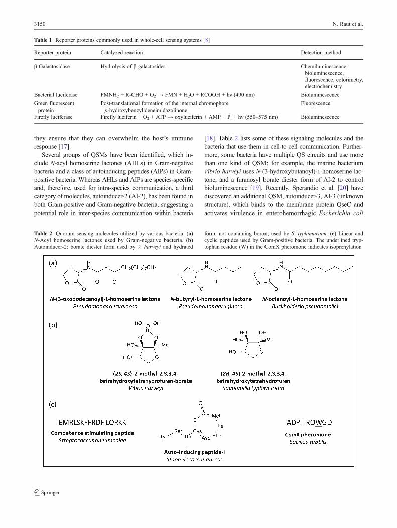
Table 1 Reporter proteins commonly used in whole-cell sensing systems [8]
Reporter protein Catalyzed reaction Detection method
β-Galactosidase Hydrolysis of β-galactosides Chemiluminescence,bioluminescence,fluorescence, colorimetry,electrochemistry
Bacterial luciferase FMNH2 + R-CHO + O2 → FMN + H2O + RCOOH + hν (490 nm) Bioluminescence
Green fluorescentprotein
Post-translational formation of the internal chromophorep-hydroxybenzylideneimidazolinone
Fluorescence
Firefly luciferase Firefly luciferin + O2 + ATP → oxyluciferin + AMP + Pi + hν (550–575 nm) Bioluminescence
Table 2 Quorum sensing molecules utilized by various bacteria. (a)N-Acyl homoserine lactones used by Gram-negative bacteria. (b)Autoinducer-2: borate diester form used by V. harveyi and hydrated
form, not containing boron, used by S. typhimurium. (c) Linear andcyclic peptides used by Gram-positive bacteria. The underlined tryp-tophan residue (W) in the ComX pheromone indicates isoprenylation
3150 N. Raut et al.
(EHEC). Further, it has been shown that the mammalian hor-mones, epinephrine and norepinephrine, are recognized by thesame protein QseC and activate virulence in EHEC [21, 22],suggesting involvement of the AI-3 QS system in inter-kingdom bacterial–mammalian cells communication.
Numerous in vitro and in vivo studies have demonstratedthat expression of bacterial virulence factors responsible forinfections in mammalian hosts is regulated by QS. Severalsuch studies concerning gastrointestinal (GI) tract infectionshave been discussed in a recent review by Kaper and Sper-andio [23]. For example, in the case of EHEC, which causesbloody diarrhea and hemolytic-uremic syndrome, involve-ment of AI-3 in regulating the expression of virulencefactors was demonstrated in a HeLa cell infection modelusing wild-type EHEC and a LuxS mutant-LuxS was shownto be involved in the production of AI-3 [24]. In an animalstudy [25], burn wounds of mice models were infected withwild-type Pseudomonas aeruginosa and variants of thesame bacterium carrying mutations in the genes encodingfor AHL synthase. The objective of the study was to deter-mine the efficiency of infection by assessing the ability ofthe bacteria to spread the infection and the time required forthe onset of infection. It was observed that in mice infectedwith P. aeruginosa mutants unable to synthesize AHLs, theextent of infection was lower than in those infected withwild type bacterium. The evidence gathered is indicative ofthe relevance of bacterial communication in diseases ofbacterial origin and, thus, the importance of detecting quo-rum sensing signaling molecules in physiological samples.
Identification of QS regulatory pathways in a number ofbacteria [16] has enabled additional whole-cell-based bio-sensing systems to be engineered to detect QSMs by cou-pling the genes coding for different QSM recognition and/orregulatory proteins to those of a variety of reporter genes[26]. As an example, Winson et al. designed reporter plas-mids by placing the gene cassette luxCDABE under thecontrol of the PlasI and PRhlI promoters from the P. aerugi-nosa QS regulatory systems LasI/LasR and RhlI/RhlR, re-spectively. The plasmids also contained the sequences of theLasR and RhlR proteins, which, upon binding AHLs, bindto the respective promoters, thus activating expression of thereporter protein [27]. In another example, Bassler [28] et al.engineered a strain of V. harveyi in such a way that thebacterium’s bioluminescence, which depends on expressionof the luxCDABE gene cassette, was only triggered by AI-2,thus enabling detection of this particular quorum sensingmolecule.
Most of the above cell-based systems have been used asbioassays to evaluate the ability of cultured bacteria to produceQSMs. However, only a few have been applied to the detectionof QSMs in physiological and clinical samples in order tocorrelate the presence of pathogenic bacteria with the onsetor status of disease. Two independent studies demonstrated the
presence of AHLs in sputum samples from patients with cysticfibrosis (CF) [29, 30] by using whole-cell sensing systems. It isknown that P. aeruginosa and Burkholderia cepacia colonizethe airway passage and lungs in CF patients, leading to chroniclung infection and finally to destructive lung disease [31].These two species of bacteria use AHL-dependent QS regula-tion. In the study conducted by Middleton [29] et al., sputumsamples from CF patients colonized by either P. aeruginosa orB. cepacia, were extracted and then analyzed using an E. coliwhole-cell-based biosensing system containing plasmidspSB401 and pSB1075 to detect short and long-chain AHLs,respectively. AmongP. aeruginosa-colonized sputum samples,71% showed the presence of short-chain AHLs and 61%showed presence of long-chain AHLs. Similarly, among B.cepacia-colonized sputum samples, 81% contained short-chain AHLs and 50% contained long-chain AHLs. ThisAHL production profile was different from that of laboratorycultures of the same microorganisms isolated from sputumsamples, in which long-chain AHLs were predominant. Thedifference in AHL profile was thought to be explained bydissimilar growth conditions, in vitro and in the lung. In thatregard, this varied behavior of P. aeruginosa from lung infec-tion had previously been observed by Singh et al. [32], whohypothesized that P. aeruginosa exists predominantly as abiofilm in CF sputum. Further, LC–MS analysis of the samesamples revealed the presence of short-chain AHLs, but notthat of long-chain AHLs, indicating that the whole-cell-basedbiosensing systemwasmore sensitive for detection of AHLs insputum samples. Another study published the same year byErickson et al. further corroborated the presence of AHLs insputum samples from CF patients [30]. P. aeruginosa-basedwhole-cell biosensing systems containing plasmids pKDT17(to detect long-chain AHLs) and pECP61.5 (to detect short-chain AHLs), both utilizing lacZ as a reporter, were used.Although the levels of AHLs detected were low, over 75% ofthe samples had long-chain AHLs whereas only 26% of thesamples had short-chain AHLs. This finding is in contrast withthe results of the study by Middleton et al. [29]. However,differences in the groups of patients, along with differentsample extraction and/or processing methods and the use ofsensing systems based on different microorganisms mightexplain different results. Along the same lines, Chambers etal. detected a broad range of AHLs in mucopurulent respira-tory secretion samples obtained from CF patients [3]. Theyused Agrobacterium tumefaciens-based whole-cell biosensingsystem A136, containing plasmids pCF218 and pMV26 withluxCDABE as a reporter, allowing it to respond to both longand short-chain AHLs (4–12 carbon atoms). AHLs wereextracted from themucopurulent respiratory secretion samples,separated by reversed-phase fast pressure liquid chromatogra-phy (FPLC), and each fraction was then assayed with the A.tumefaciens A136 sensing system in 96-well microtiter plateformat. Further, identities of the AHLs present in positive
Engineered cells as biosensing systems in biomedical analysis 3151
fractions were confirmed by comparing their retention timeswith those of standard AHLs. Using the whole-cell sensingsystem combined with FPLC, the authors were able to detectlow concentrations of AHLs in small volumes of samples fromnine (out of thirteen) CF patients and to identify seven differentAHLs.
Our research group used whole-cell biosensing systemsto evaluate QSMs in physiological samples from individualsaffected by bacterial gastrointestinal (GI) disorders, includ-ing inflammatory bowel disease (IBD). Two of the majorconditions of IBD, ulcerative colitis (UC) and Crohn’s dis-ease (CD), involve chronic and relapsing acute inflamma-tion in the large and small intestine, respectively, with CDbeing able to affect any portion of the GI tract. Currentmethods of diagnosis and monitoring rely on endoscopictechniques and analysis of mucosal tissue biopsies taken
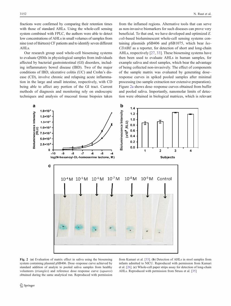
from the inflamed regions. Alternative tools that can serveas non-invasive biomarkers for such diseases can prove verybeneficial. To that end, we have developed and optimized E.coli-based bioluminescent whole-cell sensing systems con-taining plasmids pSB406 and pSB1075, which bear lux-CDABE as a reporter, for detection of short and long-chainAHLs, respectively [27, 33]. These biosensing systems havethen been used to evaluate AHLs in human samples, forexample saliva and stool samples, which bear the advantageof being collected non-invasively. The effect of componentsof the sample matrix was evaluated by generating dose–response curves in spiked pooled samples after minimalprocessing (no sample extraction nor extensive preparation).Figure 2a shows dose–response curves obtained from bufferand pooled saliva. Importantly, nanomolar limits of detec-tion were obtained in biological matrices, which is relevant
Fig. 2 (a) Evaluation of matrix effect in saliva using the biosensingsystem containing plasmid pSB406. Dose–response curve achieved bystandard addition of analyte to pooled saliva samples from healthyvolunteers (triangles) and reference dose–response curve (squares)obtained during the same analytical run. Reproduced with permission
from Kumari et al. [33]. (b) Detection of AHLs in stool samples frominfants admitted to NICU. Reproduced with permission from Kumariet al. [26]. (c) Whole-cell paper strips assay for detection of long-chainAHLs. Reproduced with permission from Struss et al. [35]
3152 N. Raut et al.
in that nanomolar concentrations of QSMs are necessary toinitiate cell-to-cell communication [34]. Saliva samples fromIBD and healthy individuals, and stool samples from newborns(Fig. 2b) admitted to a neonatal intensive care unit (NICU)were then assayed [26]. AHLs were detected at different levelsin the specimens tested, showing for the first time the presenceof these QSMs in such physiological samples. If a correlationis established between the QSM levels in samples and thehealth status of a patient, it may be possible to use QSMs asbiomarkers of bacteria-related disorders, which should aid inthe management of the disease. Studies are currently in prog-ress in our group in which physiological samples from selectedsets of GI patients and matched controls are analyzed for theirQSM content to evaluate relationships between QSM levelsand disease status. One such clinical study involved stoolsamples that were obtained from infants admitted to the NICUfor a variety of illnesses, including bowel inflammation andbacterial sepsis (submitted manuscript). Similarly, we havealso characterized and optimized a V. harveyi-based whole-cell biosensing system [19] to quantitatively detect AI-2 insaliva, stool, and intestinal fluid samples (manuscript inpreparation).
Whole-cell sensing systems using bioluminescent reporters,for example those described above, are advantageous for use inclinical applications because of high sensitivity, high through-put, and multiplexing capabilities. However, they need instru-mentation to measure bioluminescent signals, and thus are notideal for on-site applications. Although portable photodiode-based luminometers exist, they may not be readily available,especially in remote locations and developing countries. Tothat end, we have developed a whole-cell-based paper-stripassay for AHLs using β-galactosidase as reporter, which canbe visualized by the naked eye using a chromogenic substrate[35]. The whole-cell-based sensor consists of E. coli cellsDH5α-T1 transformed with plasmid pSB908, which includesthe genes lasR and lacZ, encoding LasR and β-galactosidase,respectively, under the control of the promoter PlasI. In brief,the sensing cells were vacuum-dried on precut paper strips. Onincubation with a set of standards containing differentAHL concentrations and addition of the β-galactosidasechromogenic substrate 5-bromo-4-chloro-3-indolyl-β-D-galac-topyranoside (X-gal), a blue precipitate (5,5′-dibromo-4,4′-dichloroindigo) formed in the areas where the cells had beendried. The color intensity was visually relatable to the AHLconcentration, as shown in Fig. 2c. The limit of detection wasonly one order of magnitude higher than that obtained with thebioluminescent luxCDABE-based sensing system describedabove (1 × 10⁻⁸ mol L−1 vs. 1 × 10⁻⁹ mol L−1). Furthermore,the paper strip assay was successfully used to detect AHLs insaliva samples. The paper-strip-based analytical test is portable,easy to use, and inexpensive, and can thus be used in thephysician’s office and at the patient’s bedside, even by un-trained personnel. Although simple image processing software
can assist in obtaining quantitative data from analysis of ac-quired digital images of the paper strips, visual evaluation ofthe color intensity provides a semi-quantitative estimate of theamount of AHLs present in a sample.
Hydroxylated polychlorinated biphenyls
In the early 20th century, polychlorinated biphenyls (PCBs)were widely used in several industrial applications, includingelectrical insulating fluid, caulks, adhesives, and printing paper[6]. Their use was banned in the 1970s because of their proventoxicity to humans and animals. Because of resistance to phys-ical, chemical, and biological degradation, PCBs still persist inthe environment, contaminating water and soil. Additionally,their lipophilic nature can cause bioaccumulation in livingorganisms and biomagnification through the food chain. Whenentering the human body, PCBs are metabolized tohydroxylated-PCBs (OH-PCBs), which have been found inserum and other biological fluids [36]. Therefore, detection ofOH-PCBs in physiological samples may serve as a means ofevaluating exposure to PCBs. Furthermore, OH-PCBs, al-though less toxic than PCBs, have significant adverse healtheffects, given that they can act as endocrine disruptors [37] andincrease the formation of reactive oxygen species [1]. Thisemphasizes the importance of OH-PCB detection in physiolog-ical samples. To that end, our group has developed a whole-cellbiosensing system for the detection of OH-PCBs based on E.coli cells harboring plasmid pHYBP109 in which the luxABgenes encoding for bacterial luciferase are under control of thePhbpC promoter. The construct also contains the gene coding forthe protein HbpR, which negatively regulates expression of thereporter gene and is activated by 2-hydroxybyphenyl [38].Given the structural similarities between 2-hydroxybyphenyland OH-PCBs, we postulated that the system could also beactivated by the latter [39]. Indeed, a dose-dependent responseto a variety of OH-PCBs was observed, with limits of detectiondown to 1.0 × 10⁻⁹ mol L−1. The sensing systemwas optimizedand standardized in terms of analytical conditions and furtherused to detect OH-PCBs in spiked human serum samples. Theresults demonstrated that the optimized whole-cell biosensingsystem can be used for sensitive, rapid, direct detection of OH-PCBs in human blood serum, thus confirming its potential foruse as a tool for screening large numbers of samples.
Mercury(II) ions
Mercury is a heavy metal known to be neurotoxic, embryo-toxic, and able to damage various organs. In addition to occu-pational exposure, sources of exposure to mercury include useof man-made products, for example disinfectants, batteries,thermometers, and fluorescent bulbs, dental amalgams, and
Engineered cells as biosensing systems in biomedical analysis 3153
consumption of mercury-contaminated foods. Mercury canexist in three major forms, elemental, inorganic, and organic.Approximately 80% of mercury released from human activi-ties is in the elemental form [37]. Dental amalgams contain50% mercury, with silver, tin, and copper [40]. Althoughmercury is bound within the amalgam, it leaches out in theelemental form Hg(0) as vapor, accounting for 75% of dailymercury exposure. It is then absorbed by the lungs, andoxidized to Hg(II) [41]. Roda et al. have optimized a whole-cell biosensing system based on E. coli cells, in which the lucreporter gene encoding for firefly luciferase is under thecontrol of a mercury-inducible promoter, for detection ofmercury in urine. The whole-cell sensor proved to be selectiveand sensitive for Hg2+ and methylmercury, with a detectionlimit of 1.67 × 10⁻¹³ mol L−1 for Hg2+. The analysis of urinesamples obtained from individuals with amalgam fillings andcontrol individuals showed slightly significantly higher levelsof Hg2+ in the former group. Assays were performed in 384-well microtiter plates and required low volumes of samplesand reagents, thus proving suitable for high-throughputscreening. Therefore, the method could be used for analysisof large numbers of urine samples from individuals with andwithout amalgam fillings to investigate the relationship be-tween urinary mercury levels and dental amalgam.
Methylmercury
Methylmercury (MeHg) is the most toxic of the organomer-cury compounds and among the most toxic environmentalpollutants worldwide [42]. Clinical investigations have shownthat MeHg is the primary form of mercury that accumulates infish and their consumers. This accumulation leads to mercurypoisoning and its associated symptoms, because most speciesare unable to removeMeHg from the body [43, 44]. Diagnosisof mercury poisoning is performed by correlating the clinicalsymptoms with the level of MeHg in the patient’s blood.Normal levels are typically less than 30 nmol L−1 in wholeblood, but can vary, depending on dietary and environmentalfactors [45]. Traditional analytical methods for detection oforganomercury compounds focus on chromatographic techni-ques (gas and liquid chromatography) combined with spectro-scopic detection methods (atomic fluorescence spectrometry,atomic absorption spectrometry) [46] or on capillary zoneelectrophoresis [47]. These methods are impractical for assay-ing clinical and environmental samples because they requiremuch pretreatment of samples, are highly expensive, and arevery time-consuming: they are, furthermore, not amenable tominiaturization and not applicable to on-site analysis. More-over, these methods only determine the total amount of mer-cury present in the sample, which makes it hard, if notimpossible, to distinguish the amount of bioavailable mercury.To develop more efficient and cost effective methods for
detection of organomercury compounds, many groups haveengineered whole-cell biosensing systems (Table 3). Belowwehighlight two approaches for specific detection of MeHg.
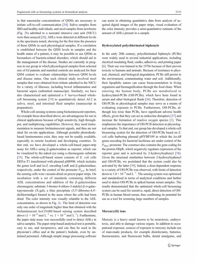
Nagata et al. [48] reported a whole-cell sensing systemspecific for MeHg capable of rapid detection down to 0.01nmol L−1 levels. The whole-cell sensing system was con-structed using two separate plasmids expressed in E. coliDH5α; pHY-mer-lux and pJM-B (Fig. 3a). The plasmidpHY-mer-lux was constructed by fusing the mercury-inducible promoter and its regulatory gene, merR, from themer-operon of Pseudomonas K-62 with the luxAB reportergene from V. harveyi [49]. The second plasmid, pJM-B,was prepared by cloning the merB gene from PsuedomonasK-62 into the pGEM-T Easy cloning vector. The systemfunctions by constitutively producing the organomercurylyase, MerB, which cleaves the C-Hg bond of MeHg, pro-ducing free Hg2+. This free Hg2+ can then bind to the Hg2+-selective regulatory protein, MerR, and activate the mer-operon, thus producing luciferase in an Hg2+-dose-depen-dent fashion (Fig. 3b). This system is selective for free Hg2+
cleaved from MeHg over free Hg2+ that may be present in asample, although the specific mechanism has not been in-vestigated (Fig. 3c). The selectivity is most likely explainedby the high lipid solubility of organic mercury comparedwith inorganic mercury, which may favor passive diffusionof MeHg into the sensing cells [42]. Nagata et al. postulatedthat the constructed pHY-mer-lux may cause selective up-take of MeHg into the cell because of lack of a native activetransport system for inorganic mercury. This type of phe-nomenon has been reported before for phenylmercury,which is specifically transported into cells via the mercurytransporter protein, MerT [50, 51]. As a result, Nagata et al.produced a whole-cell biosensing system that can rapidlyand selectively detect bioavailable MeHg with a limit ofdetection low enough to assess both environmental andphysiological samples.
Rantala et al. [13] took a different approach to the detec-tion of MeHg, one that focused on using an already engi-neered sensor [52], but creating a highly selective, morerobust, reproducible, portable assay with detection limitsdown to 0.3 nmol L−1 levels. The whole-cell sensing systemwas constructed by transforming, in E. coli MC1061, a plas-mid that fused the mercury inducible promoter and its regula-tory gene, merR, from the pDU1358mer-operon with reportergene luxCDABE. In addition, the plasmid contains the merBgene under the control of the lac promoter. The mechanism ofthe system is similar to that of the Nagata et al. system, exceptthat, because the entire luxCDABE cassette is present, there isno need for addition of external substrate to generate theemission of bioluminescence from the luciferase reporter. Asconstructed, the whole-cell sensing system detects total mer-cury in the sample. Selectivity for MeHg was achieved byadding ethylenediaminetetraacetic acid (EDTA), which acts as
3154 N. Raut et al.

a chelating agent, blocking inorganic mercury from enteringthe cells. Multiple concentrations of EDTA were tested fortoxicity and luminescence inhibition, and an optimum
concentration was selected (10 mmol L−1). Use of EDTA asa chelating agent also helps to make the sensor more robust,because the cells are protected from the toxic inorganic mer-cury species that may exist in samples.
Antibiotics
Since the 1940s, tetracycline has been one of the antibioticsmost widely used to treat bacterial infections in humans andanimals. With increasing animal farming, animals are rou-tinely administered tetracycline to control infections; a sideeffect of such practice is the increase in tetracycline-resistantbacterial populations. For example, it has been shown thatsubinhibitory concentrations of tetracycline promote accu-mulation of tetracycline-resistance genes in bacteria colo-nizing the GI tract of mammals [53]. Bahl et al. haveinvestigated the above mentioned effect by determining, invivo, bioavailable concentrations of the antibiotic in theintestines of tetracycline-treated rats, which continuouslyreceived drinking water containing the antibiotic [54]. Thiswas achieved by using a whole-cell biosensing system con-sisting of E. coli MC4100 cells harboring the plasmidpTGFP2, which contains a gene fusion of the tetracyclineinducible promoter Ptet and gfp. Uptake of tetracycline bythe sensing cells activates the tetracycline-inducible promot-er, subsequently inducing dose-dependent expression ofGFP. Fluorescence measurements by flow cytometry dem-onstrated low bioavailability and proved the potential utilityof whole-cell sensing systems as tools to determine antibi-otic intestinal bioavailability and therapy optimization. In asimilar study, Hansen et al. demonstrated the utility of awhole-cell biosensing system using β-galactosidase as a
Fig. 3 (a) pHY-mer-lux and pJM-B plasmids in E. coli DH5α. (b)Simplified reaction mechanism for reporter system. (c) Response ofwhole-cell sensing system to CH3Hg
+ (triangles) and Hg2+ (circles).Adapted from Nagata et al. [48]
Table 3 Comparison of whole-cell sensing systems for detection of organomercury. Adapted from Rantala et al. [13]
Host strain Analyte Reporter plasmid Substrate Limit of detection(nmol L−1)
Dynamic range(nmol L−1)
Assaytime
Ref.
E. coli MC106 Organomercurycompounds
pmerBRBSluc D-Luciferin 0.2 0.2–10 2.5 h [52]
E. coli DH5α Organomercurycompounds(phenylmercuryacetate)
pHYB3LuxAB+pGR1A 10% 1-decanal 50 50–5000 Severalhours
[49]
E. coli MC1061 Total mercury pmerRBPmerluxCDABE No substratec 0.008 8 × 10⁻¹²
–5 × 10−8120 minb [52]
E. coli DH5α Methylmercury pHY-mer-luxAB+pJM-B 10% 1-decanal 0.01 0.01–1.0 40 minb [48]
E. coli MC1061a Total mercuryd pmerRBluxCDABE No substratec 0.3 0.3–100 60–180 min [13]
a Freeze-dried sensor cellsb Growing step of the cells not included, only the measurement with the analytec Produced by the reporter gene operond Chelation of ionic Hg2+ makes the sensor specific for organic mercury
Engineered cells as biosensing systems in biomedical analysis 3155
reporter for quantitative measurement of chlortetracycline inpig feces. These studies showed the application of whole-cell sensing systems in veterinary clinical medicine, withpotential implications in human medicine. Such cell-basedassays can further be developed in high-density formats forhigh-throughput screening of a multitude of samples and forscreening natural and synthetic molecules for antibioticactivity [55].
Cytarabine efficacy
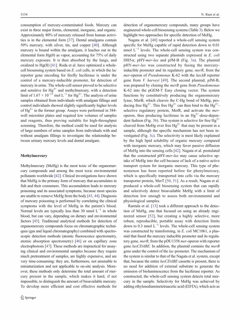
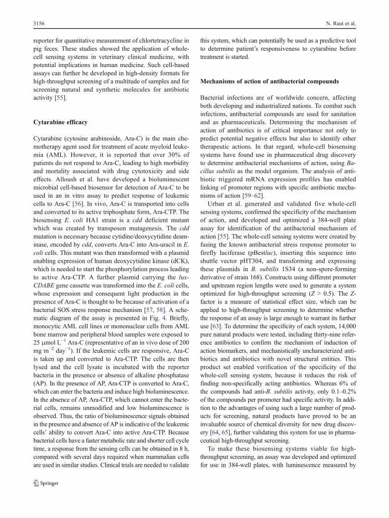
Cytarabine (cytosine arabinoside, Ara-C) is the main che-motherapy agent used for treatment of acute myeloid leuke-mia (AML). However, it is reported that over 30% ofpatients do not respond to Ara-C, leading to high morbidityand mortality associated with drug cytotoxicity and sideeffects. Alloush et al. have developed a bioluminescentmicrobial cell-based biosensor for detection of Ara-C to beused in an in vitro assay to predict response of leukemiccells to Ara-C [56]. In vivo, Ara-C is transported into cellsand converted to its active triphosphate form, Ara-CTP. Thebiosensing E. coli HA1 strain is a cdd deficient mutantwhich was created by transposon mutagenesis. The cddmutation is necessary because cytidine/deoxycytidine deam-inase, encoded by cdd, converts Ara-C into Ara-uracil in E.coli cells. This mutant was then transformed with a plasmidenabling expression of human deoxycytidine kinase (dCK),which is needed to start the phosphorylation process leadingto active Ara-CTP. A further plasmid carrying the lux-CDABE gene cassette was transformed into the E. coli cells,whose expression and consequent light production in thepresence of Ara-C is thought to be because of activation of abacterial SOS stress response mechanism [57, 58]. A sche-matic diagram of the assay is presented in Fig. 4. Briefly,monocytic AML cell lines or mononuclear cells from AMLbone marrow and peripheral blood samples were exposed to25 μmol L−1 Ara-C (representative of an in vivo dose of 200mg m−2 day−1). If the leukemic cells are responsive, Ara-Cis taken up and converted to Ara-CTP. The cells are thenlysed and the cell lysate is incubated with the reporterbacteria in the presence or absence of alkaline phosphatase(AP). In the presence of AP, Ara-CTP is converted to Ara-C,which can enter the bacteria and induce high bioluminescence.In the absence of AP, Ara-CTP, which cannot enter the bacte-rial cells, remains unmodified and low bioluminescence isobserved. Thus, the ratio of bioluminescence signals obtainedin the presence and absence of AP is indicative of the leukemiccells’ ability to convert Ara-C into active Ara-CTP. Becausebacterial cells have a faster metabolic rate and shorter cell cycletime, a response from the sensing cells can be obtained in 8 h,compared with several days required when mammalian cellsare used in similar studies. Clinical trials are needed to validate
this system, which can potentially be used as a predictive toolto determine patient’s responsiveness to cytarabine beforetreatment is started.
Mechanisms of action of antibacterial compounds
Bacterial infections are of worldwide concern, affectingboth developing and industrialized nations. To combat suchinfections, antibacterial compounds are used for sanitationand as pharmaceuticals. Determining the mechanism ofaction of antibiotics is of critical importance not only topredict potential negative effects but also to identify othertherapeutic actions. In that regard, whole-cell biosensingsystems have found use in pharmaceutical drug discoveryto determine antibacterial mechanisms of action, using Ba-cillus subtilis as the model organism. The analysis of anti-biotic triggered mRNA expression profiles has enabledlinking of promoter regions with specific antibiotic mecha-nisms of action [59–62].
Urban et al. generated and validated five whole-cellsensing systems, confirmed the specificity of the mechanismof action, and developed and optimized a 384-well plateassay for identification of the antibacterial mechanism ofaction [55]. The whole-cell sensing systems were created byfusing the known antibacterial stress response promoter tofirefly luciferase (pBestluc), inserting this sequence intoshuttle vector pHT304, and transforming and expressingthese plasmids in B. subtilis 1S34 (a non-spore-formingderivative of strain 168). Constructs using different promoterand upstream region lengths were used to generate a systemoptimized for high-throughput screening (Z > 0.5). The Z-factor is a measure of statistical effect size, which can beapplied to high-throughput screening to determine whetherthe response of an assay is large enough to warrant its furtheruse [63]. To determine the specificity of each system, 14,000pure natural products were tested, including thirty-nine refer-ence antibiotics to confirm the mechanism of induction ofaction biomarkers, and mechanistically uncharacterized anti-biotics and antibiotics with novel structural entities. Thisproduct set enabled verification of the specificity of thewhole-cell sensing system, because it reduces the risk offinding non-specifically acting antibiotics. Whereas 6% ofthe compounds had anti-B. subtilis activity, only 0.1–0.2%of the compounds per promoter had specific activity. In addi-tion to the advantages of using such a large number of prod-ucts for screening, natural products have proved to be aninvaluable source of chemical diversity for new drug discov-ery [64, 65], further validating this system for use in pharma-ceutical high-throughput screening.
To make these biosensing systems viable for high-throughput screening, an assay was developed and optimizedfor use in 384-well plates, with luminescence measured by
3156 N. Raut et al.
using a CCD camera-based luminescence detector. To validatethe developed promoter induction assay, second-line mecha-nism of action studies, which involved incorporation of radio-labeled metabolic precursors for DNA, RNA, protein, and cellwall biosynthesis in Staphylococcus aureus [66], were per-formed for a representative compound, ferrimycin A1, anantibiotic with a poorly studied mechanism of action. Boththe whole-cell sensing systems and the metabolite incorpora-tion studies strongly supported selective protein biosynthesisinhibition as the mechanism of action of this compound. Thus,Urban et al. produced a high-throughput method of screeningcapable of determining themechanism of action of antibacterialcompounds that is fast, inexpensive, reproducible, and highlyspecific. However, this system is not without its flaws. Thesystem is unable to assess antibacterial compounds that func-tion at concentrations less than 1.56 μg mL−1, such as fusidicacid and siomycin A. Also, although 14,000 compounds weretested, only thirty-nine were reference antibiotics—testingmore reference antibiotics would result in a stronger, morerobust comparison matrix.
It is envisaged that continued use of these whole-cellbiosensing systems for high-throughput screening may aidin the discovery of novel pharmaceutical compounds, andadvance our knowledge of the mechanisms of action ofmany different types of compound.
Conclusions and future perspectives
Identification of novel biomarkers of disease created a needfor analytical tools that enable their detection in physiolog-ical fluids for diagnosis and management of disease, and for
assessment of therapeutic efficacy. Whole-cell biosensingsystems have properties that make them ideal for thesepurposes. Specifically, they afford very low limits of detec-tion, thus reporting the early presence of a target biomarker,and have sufficient analytical sensitivity to enable discrim-ination between close biomarker concentrations, whichcould be crucial in some clinical settings. Their high selec-tivity also enables direct detection of target analytes inclinical samples with no or minimal sample pre-treatment,a significant advantage over traditional instrumental analyt-ical methods. This feature, with fast response and amena-bility to incorporation into high-density analytical devices,enable the development of rapid, high-throughput screeningassays requiring small sample volumes. Importantly, minia-turization and integration of whole-cell sensing systems intochip, paper, and microfluidics-based devices should enablethe development of portable equipment suitable for use on-site, in the physician’s office, or at the patient’s bedside.Furthermore, paper strips with immobilized whole-cell sens-ing systems and color visualization of the signal have po-tential for implementation of easy-to-use dip-stick typedevices which could be used by patients for disease man-agement as home-based self-monitoring tests. Such devicescould prove especially useful in remote areas and develop-ing countries. Continued work on cell-based biosensorsshould contribute to further exploiting and enhancing theirunique advantages. The identification of additional regula-tory proteins and receptors that recognize analytes of bio-medical interest should continue to expand the usefulness ofwhole-cell biosensing systems in all branches of clinicalanalysis, including, diagnostics, disease monitoring, drugscreening, and efficacy testing.
Fig. 4 Cell lysis assay using E.coli HA1 biosensing system fordetection of cytarabine.Adapted from Alloush et al.[56]
Engineered cells as biosensing systems in biomedical analysis 3157
Acknowledgments This work was supported in part by the NationalScience Foundation (grant CHE-0416553); the National Institute ofEnvironmental Health Sciences, Superfund Research Program (grantP42ES07380); the Broad Foundation, Broad Medical Research Pro-gram (grant IBD-0198R); the National Institute of Hometown Security;and the Children’s Miracle Network. S.D. is grateful for support fromthe Lucille P. Markey Chair in Biochemistry and Molecular Biology ofthe Miller School of Medicine of the University of Miami, as well asfrom a Gill Eminent Professorship from the University of Kentucky. N.R. acknowledges support from a Research Challenge Trust FundFellowship from the University of Kentucky.
References
1. Rodriguez-Mozaz S, de Alda MJL, Barceló D (2006) Biosensors asuseful tools for environmental analysis and monitoring. Anal BioanalChem 386(4):1025–1041
2. Sanvicens N, Mannelli I, Salvador JP, Valera E, Marco MP (2011)Biosensors for pharmaceuticals based on novel technology. TrendsAnal Chem 30(3):541–553
3. Chambers CE, Visser MB, Schwab U, Sokol PA (2005) Identificationof N-acyl homoserine lactones in mucopurulent respiratory secretionsfrom cystic fibrosis patients. FEMS Microbiol Lett 244(2):297–304
4. Yoo E-H, Lee S-Y (2010) Glucose biosensors: an overview of usein clinical practice. Sens 10(5):4558–4576
5. Conroy PJ, Hearty S, Leonard P, O'Kennedy RJ (2009) Antibodyproduction, design and use for biosensor-based applications.Semin Cell Dev Biol 20(1):10–26
6. Erickson M, Kaley R (2011) Applications of polychlorinatedbiphenyls. Environ Sci Pollut Res 18(2):135–151
7. Moschou EA, Bachas LG, Daunert S, Deo SK (2006) Hinge-motion binding proteins: unraveling their analytical potential. AnalChem 78(19):6692–6700
8. Struss AK, Pasini P, Daunert S (2010). In: Zourob M (ed). SpringerNew York
9. Gu M, Mitchell R, Kim B (2004) Whole-cell-based biosensors forenvironmental biomonitoring and application. Adv Biochem Engin/Biotechnol 87:269–305
10. Microtox® toxicity testing. http://www.leederconsulting.com/toxicology_services_microtox.html
11. Fantino J-R, Barras F, Denizot F (2009) Sposensor: Awhole-bacterialbiosensor that uses immobilized Bacillus subtilis spores and a one-step incubation/detection process. J Mol Microbiol Biotechnol17:90–95
12. Buchinger S, Grill P, Morosow V, Ben-Yoav H, Shacham-Diamand Y, Biran A, Pedahzur R, Belkin S, Reifferscheid G(2010) Evaluation of chrono-amperometric signal detection forthe analysis of genotoxicity by a whole cell biosensor. AnalChim Acta 659(1–2):122–128
13. Rantala A, Utriainen M, Kaushik N, Virta M, Välimaa A-L, KarpM (2011) Luminescent bacteria-based sensing method for methyl-mercury specific determination. Anal Bioanal Chem 400(4):1041–1049
14. Roda A, Cevenini L, Michelini E, Branchini BR (2011) Aportable bioluminescence engineered cell-based biosensor foron-site applications. Biosens Bioelectron 26(8):3647–3653
15. Tomasz A (1965) Control of the competent state in Pneumococcusby a hormone-like cell product: an example for a new type ofregulatory mechanism in bacteria. Nature 208(5006):155–159
16. Miller MB, Bassler BL (2001) Quorum sensing in bacteria. AnnuRev Microbiol 55(1):165–199
17. HentzerM, GivskovM (2003) Pharmacological inhibition of quorumsensing for the treatment of chronic bacterial infections. J Clin Invest112(9):1300–1307
18. Antunes LCM, Ferreira RBR (2009) Intercellular communicationin bacteria. Crit Rev Microbiol 35(2):69–80
19. Bassler BL, Wright M, Showalter RE, Silverman MR (1993)Intercellular signalling in Vibrio harveyi: sequence and functionof genes regulating expression of luminescence. Mol Microbiol 9(4):773–786
20. Sperandio V, Torres AG, Jarvis B, Nataro JP, Kaper JB (2003)Bacteria–host communication: The language of hormones. ProcNatl Acad Sci 100(15):8951–8956
21. Kendall MM, Rasko DA, Sperandio V (2007) Global effects of thecell-to-cell signaling molecules autoinducer-2, autoinducer-3, andepinephrine in a luxs mutant of enterohemorrhagic Escherichiacoli. Infect Immun 75(10):4875–4884
22. Moreira CG, Weinshenker D, Sperandio V (2009) QseC mediatesSalmonella enterica serovar Typhimurium virulence in vitro and invivo. Infect Immun: 914–926
23. Kaper JB, Sperandio V (2005) Bacterial cell-to-cell signaling inthe gastrointestinal tract. Infect Immun 73(6):3197–3209
24. Walters M, Sperandio V (2006) Autoinducer 3 and epinephrinesignaling in the kinetics of locus of enterocyte effacement geneexpression in enterohemorrhagic Escherichia coli. Infect Immun74(10):5445–5455
25. Rumbaugh KP, Griswold JA, Iglewski BH, Hamood AN (1999)Contribution of quorum sensing to the virulence of Pseudomonasaeruginosa in burn wound infections. Infect Immun 67(11):5854–5862
26. Toxicological profile for mercury. http://www.atsdr.cdc.gov/toxprofiles/tp46.pdf
27. Winson MK, Swift S, Fish L, Throup JP, Jørgensen F, Chhabra SR,Bycroft BW, Williams P, Stewart GSAB (1998) Construction andanalysis of luxCDABE-based plasmid sensors for investigating N-acyl homoserine lactone-mediated quorum sensing. FEMSMicrobiolLett 163(2):185–192
28. Berglund A (1993) An in vitro and in vivo study of the release ofmercury vapor from different types of amalgam alloys. J Dent Res72(5):939–946
29. Middleton B, Rodgers HC, Cámara M, Knox AJ, Williams P,Hardman A (2002) Direct detection of N-acyl homoserine lactonesin cystic fibrosis sputum. FEMS Microbiol Lett 207(1):1–7
30. Erickson DL, Endersby R, Kirkham A, Stuber K, Vollman DD,Rabin HR, Mitchell I, Storey DG (2002) Pseudomonas aeruginosaquorum-sensing systems may control virulence factor expressionin the lungs of patients with cystic fibrosis. Infect Immun 70(4):1783–1790
31. Govan J, Deretic V (1996) Microbial pathogenesis in cystic fibrosis:mucoid Pseudomonas aeruginosa and Burkholderia cepacia.MicrobiolRev 60(3):539–574
32. Singh PK, Schaefer AL, Parsek MR, Moninger TO, Welsh MJ,Greenberg EP (2000) Quorum-sensing signals indicate that cysticfibrosis lungs are infected with bacterial biofilms. Nature 407(6805):762–764
33. Kumari A, Pasini P, Deo SK, Flomenhoft D, Shashidhar H, Daunert S(2006) Biosensing systems for the detection of bacterial quorumsignaling molecules. Anal Chem 78(22):7603–7609
34. Pearson JP, Passador L, Iglewski BH, Greenberg EP (1995) Asecond N-acyl homoserine lactone signal produced by Pseudomonasaeruginosa. Proc Natl Acad Sci 92(5):1490–1494
35. Struss A, Pasini P, Ensor CM, Raut N, Daunert S (2010) Paperstrip whole cell biosensors: A portable test for the semiquantitativedetection of bacterial quorum signaling molecules. Anal Chem 82(11):4457–4463
36. JamesMO (2001) PCBs, recent advances in environmental toxicologyand health effects., pp 35–46
37. Bakayan A, Vaquero CF, Picazo F, Llopis J (2011) Red fluorescentprotein-aequorin fusions as improved bioluminescent Ca2+ reportersin single cells and mice. PLoS One 6(5):e19520
3158 N. Raut et al.
38. Jaspers MCM, Schmid A, Sturme MHJ, Goslings DAM, Kohler H-PE, Roelof van der Meer J (2001) Transcriptional organization anddynamic expression of the hbpCAD genes, which encode the firstthree enzymes for 2-hydroxybiphenyl degradation in Pseudomonasazelaica HBP1. J Bacteriol 183(1):270–279
39. Turner K, Xu S, Pasini P, Deo S, Bachas L, Daunert S (2007)Hydroxylated polychlorinated biphenyl detection based on agenetically engineered bioluminescent whole-cell sensing system.Anal Chem 79(15):5740–5745
40. Lorscheider F, Vimy M, Summers A (1995) Mercury exposure from"silver" tooth fillings: emerging evidence questions a traditionaldental paradigm. FASEB J 9(7):504–508
41. Roda A, Pasini P, Mirasoli M, Guardigli M, Russo C, Musiani M,Baraldini M (2001) Sensitive determination of urinary Mercury(II)by a bioluminescent transgenic bacteria-based biosensor. Anal Lett34(1):29
42. Jan A, Murtaza I, Ali A, Rizwanul Haq Q (2009) Mercury pollution:an emerging problem and potential bacterial remediation strategies.World J Microbiol Biotechnol 25(9):1529–1537
43. Halbach S (1985) The octanol/water distribution of mercury com-pounds. Arch Toxicol 57(2):139–141
44. Zahir F, Rizvi S, Haq S, Khan R (2006) Effect of methyl mercuryinduced free radical stress on nucleic acids and protein: Implicationson cognitive and motor functions. Indian J Clin Biochem 21(2):149–152
45. Ibrahim D, Froberg B, Wolf A, Rusyniak DE (2006) Heavy metalpoisoning: clinical presentations and pathophysiology. Clin LabMed 26(1):67–97
46. Stoichev T, Amouroux D, Martin-Doimeadios RCR, Monperrus M,Donard OFX, Tsalev DL (2006) Speciation analysis of mercury inaquatic environment. Appl Spectrosc Rev 41(6):591–619
47. Kubánˇ P, Pelcová P, Margetínová J, Kubánˇ V (2009) Mercuryspeciation by CE: An update. Electrophoresis 30(1):92–99
48. Nagata T, Muraoka T, Kiyonoa M, Pan-Hou H (2010) Developmentof a luminescence-based biosensor for detection of methylmercury. JToxicol Sci 35(2):231–234
49. Matsui K, Yusa K, Sugawara H, Narita M, Endo G (2007)Development of bacterial biosensor for detecting organomercurialsusing organomercurial lyase gene and bioluminescence reporter sys-tem. J Japan Soc Water Environ 30(2):77–81
50. Masako K, Yoshio U, Tomoko O, Hidemitsu P-H (2000) Involvementof merB in the expression of the pMR26 mer operon induced byorganomercurials. J Heal Sci 46(2):142–145
51. Yoshio U, Masako K, Toshiyuki T, Hidemitsu P-H (1997)Phenylmercury transport mediated by merT-merP genes ofPseudomonas K-62 plasmid pMR26. Biol Pharm Bull 20(1):107–109
52. Ivask A, Hakkila K, Virta M (2001) Detection of organomercurialswith sensor bacteria. Anal Chem 73(21):5168–5171
53. Blake DP, Humphry RW, Scott KP, Hillman K, Fenlon DR, Low JC(2003) Influence of tetracycline exposure on tetracycline resistance
and the carriage of tetracycline resistance genes within commensalEscherichia coli populations. J Appl Microbiol 94(6):1087–1097
54. Bahl MI, Hansen LH, Licht TR, Sorensen SJ (2004) In vivodetection and quantification of Tetracycline by use of a whole-cell biosensor in the rat intestine. Antimicrob Agents Chemother48(4):1112–1117
55. Urban A, Eckermann S, Fast B, Metzger S, Gehling M, ZiegelbauerK, Rubsamen-Waigmann H, Freiberg C (2007) Novel whole-cell antibiotic biosensors for compound discovery. Appl EnvironMicrobiol 73(20):6436–6443
56. Alloush HM, Anderson E, Martin AD, Ruddock MW, AngellJE, Hill PJ, Mehta P, Smith MA, Smith JG, Salisbury VC(2010) A bioluminescent microbial biosensor for in vitro pretreatmentassessment of Cytarabine efficacy in leukemia. Clin Chem 56(12):1862–1870
57. Kozakiewicz J, Gajewska M, Łyźeń R, Czyź A, W grzyn G (2005)Bioluminescence-mediated stimulation of photoreactivation inbacteria. FEMS Microbiol Lett 250(1):105–110
58. Czyz A, Plata K, Wegrzyn G (2002) Induction of light emission byluminescent bacteria treated with UV light and chemical mutagens.J Appl Genet 43(3):377–389
59. Fischer HP, Brunner NA, Wieland B, Paquette J, Macko L,Ziegelbauer K, Freiberg C (2004) Identification of antibioticstress-inducible promoters: a systematic approach to novel pathway-specific reporter assays for antibacterial drug discovery. Genome Res14(1):90–98
60. Freiberg C, Fischer HP, Brunner NA (2005) Discovering themechanism of action of novel antibacterial agents throughtranscriptional profiling of conditional mutants. AntimicrobAgents Chemother 49(2):749–759
61. Hutter B, Fischer C, Jacobi A, Schaab C, Loferer H (2004) Panelof Bacillus subtilis reporter strains indicative of various modes ofaction. Antimicrob Agents Chemother 48(7):2588–2594
62. Hutter B, Schaab C, Albrecht S, Borgmann M, Brunner NA,Freiberg C, Ziegelbauer K, Rock CO, Ivanov I, Loferer H (2004)Prediction of mechanisms of action of antibacterial compounds bygene expression profiling. Antimicrob Agents Chemother 48(8):2838–2844
63. Zhang J-H, Chung TDY, Oldenburg KR (1999) A simple statisticalparameter for use in evaluation and validation of high throughputscreening assays. J Biomol Screen 4(2):67–73
64. Chapman T (2004) Drug discovery: The leading edge. Nature 430(6995):109–115
65. Newman DJ, Cragg GM, Snader KM (2003) Natural products assources of new drugs over the period 1981−2002. J Nat Prod 66(7):1022–1037
66. Freiberg C, Brunner NA, Schiffer G, Lampe T, Pohlmann J, BrandsM, Raabe M, Häbich D, Ziegelbauer K (2004) Identification andcharacterization of the first class of potent bacterial Acetyl-CoAcarboxylase inhibitors with antibacterial activity. J Biol Chem 279(25):26066–26073
Engineered cells as biosensing systems in biomedical analysis 3159