Embed Size (px)
Citation preview
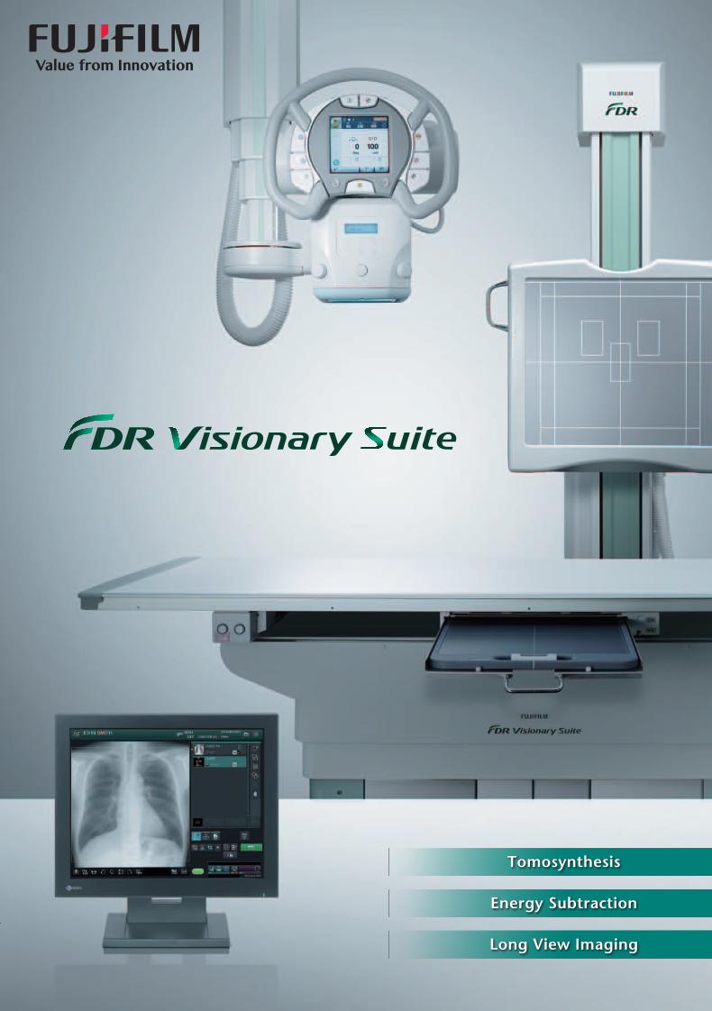
Tomosynthesis
Energy Subtraction
Long View Imaging
Specifications are subject to change without notice.All brand names or trademarks are the property of their respective owners.All products require the regulatory approval of the importing country.For details on their availability, contact our local representative.Actual X-ray images are varied by conditions of X-ray system or subjects or other factors.
Ref. No. XB-1029ER (SK·16·05·F1079·F9711) Printed in Japan ©2015 FUJIFILM Corporationhttp://www.fujifilm.com/products/medical/ 26-30, NISHIAZABU 2-CHOME, MINATO-KU, TOKYO 106-8620, JAPAN
FDR Visionary Suite Specifications
� X-ray Generator• Rated output : 50 kW / 65 kW / 80 kW• Tube voltage : 40 to 150 kV• Tube current : 10 to 630 mA (50 kW model) 10 to 800 mA (65 kW model) 10 to 1000 mA (80 kW model)• AEC : Xe detector-type phototimer receiver combination up to three receivers
� X-ray Tube Support• Ceiling fixture : Fixed rail of 4 / 5.5 m Moving rail of 2 / 2.6 /3.3 m• Movement range : Longitudinal 2.95 m (4 m fixed rail) Longitudinal 4.45 m (5.5 m fixed rail) Transversal 1.4 m (2 m moving rail) Transversal 2.0 m (2.6 m moving rail) Transversal 2.7 m (3.3 m moving rail) Vertical 1.6 m• Rotation : Vertical axis ±180° Horizontal axis -180° to +120°
� X-ray Tube Unit• Maximum anode heat content : 400 kHU• Maximum anode heat dissipation rate : 2200 HU/s • Focal spot : 0.6 / 1.2 mm
� Collimator• Filtration : Inherent filtration 1.1 mmAl eq. Added filter of Cu 0.1 / 0.2 / 0.3 mm • Standard accessories : Auto-filter Line marker Detent (fitted at the home position) • Area dosimeter adapter (Option) : An adapter for dosimeter manufactured by VACUTEC/PTW
� Table• Tabletop size : 810 × 2350 mm • Table height : 535 to 930 mm• Longitudinal range : ±375 mm• Transversal range : ±125 mm• Bucky moving range : 800 mm • Max. load : 295 kg• Standard accessories : Tracking device Bucky tracking driver• Options : Compression belt Side cassette holder Grip switch CFRP tabletop Hand grip Drip hanger Rear foot switch
� Stand• Distance between Bucky top edge and floor surface : Manual : 643 to 2143 mm Motorized : 671 to 2113 mm• Tilting angle (Function for BR-120T) : -20° to 90°• Standard accessories : Tracking device Stop switch Foot switch• Options : Hand grip (mounted on top edge of the Bucky) Hand grip (mounted on back side of the Bucky) Cassette holder Front handle Both side operation Compression belt Patient stand (for long view radiography) Wall mounting option (for BR-120)
FDR D-EVO Advanced C43A Specifications
• Scintillator : CsI • Detector external size : 464.5±1(W) × 516.7±1(D) × 18±1(H) mm *excluding convex part of the cable• Weight : Approx. 4.5 kg (including battery)• Pixel size : 150 µm• Maximum detecting area : 2816 × 2816 pixels • Image preview : less than 2 sec• Cycle time : less than 8 sec
FDR D-EVO Advanced C43A

FNC processing
In contrast to conventional FPD offerings this system features an indirectconversion FPD using the “ISS method,” where the TFT sensor is placedin front of the scintillation layer instead of behind it. With this proprietarymethod the scattering/dissipation of X-ray signals is significantly reduced,achieving sharper images with a lower X-ray dose.
X-ray X-ray
Conventional method ISS method
2. Improved sensitivity in low-density areas using noise reduction circuitry*
1. Using Fujifilm’s “ISS method” reading technology to achieve sharper images
Image noise is reduced by using a proprietary noise reduction circuitry. Granularity in low-density areas is improved, helping to boost image quality.
The system features imaging processing technologies that enable desirable image display. These technologies include “Dynamic Visualization,” which optimizes images for diagnosis on-screen, and “Flexible Noise Control (FNC) Processing,” which reduces granularity by automatically extracting and excluding image noise components.
3. Image processing technologies producing optimized images
Freedom and Flexibility in Imaging
02 03
Experience a wide range of applications
targeted to improve diagnostic capabilities combined
with a precise design that facilitates imaging.
With unique image processing functions that
enhance quality while reducing the operational dose rate,
the FDR Visionary Suite is the next generation
in functional X-ray systems — offering ease of operation
with minimal impact for patients.
Using the latest technological developments to allow further quality improvementswith a low operational dose rate.
TFT panel
TFT panel
Optical signalimage reaching
TFT panel
Optical signalimage reaching
TFT panel
Scintillationlayer
Scintillationlayer
* FDR D-EVO Advanced C43A
Wide Array of Applications Support Diagnosis
Stress-free, Optimized Imaging Workflow
Compatibility with a Broad Range of CassetteDR Panels Ensures Maximum Flexibility
Tomosynthesis Energy Subtraction Long View Imaging
04 05
Freedom to reconstruct and display image slicesWith this technology the X-ray tube moves through an arc, acquiring a series of images in a single sweep, which can then be reconstructed to create cross-sectional image slices.
Separates images of soft tissue and bone for improved viewing
Display full-length images of spine or lower limb
High-precision, high-quality imaging to 150µmBy controlling such items as metal artifacts, high-precision imaging down to 150µm is possible.
Automatic X-ray dose control and background reconstructionUsing the imaging conditions for a single preliminary image as reference, the conditions for Tomosynthesis imaging are set automatically.
Tomosynthesis*
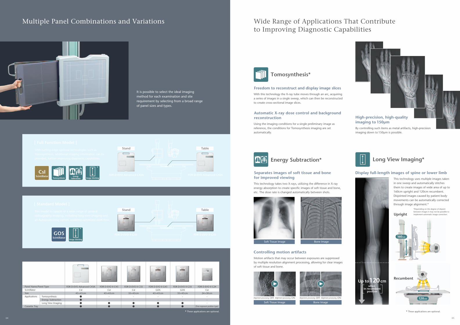
With cutting-edge optional technologies such as Tomosynthesis, additional imaging information can be provided, further improving diagnostic capabilities.
[ Full Function Model ]
This model is capable of a wide-range of general radioagraphy imaging, including long view imaging and an Autopositioning option, enabling optimized work flow.
[ Standard Model ]
This technology uses multiple images taken in one sweep and automatically stitches them to create images of wide area of up to 160cm upright and 120cm recumbent. Disjointed images caused by patient body movements can be automatically corrected through image alignment.*
Recumbent
Upright
160 cm
120 cm
* These applications are optional.* These applications are optional.
It is possible to select the ideal imaging method for each examination and site requirement by selecting from a broad range of panel sizes and types.
Alignment processing: OFF Alighment processing: ON Alignment processing: OFF Alighment processing: ON
This technology takes two X-rays, utilizing the difference in X-ray energy absorption to create specific images of soft tissue and bone, etc. The dose rate is changed automatically between shots.
Controlling motion artifactsMotion artifacts that may occur between exposures are suppressed by multiple resolution alignment processing, allowing for clear images of soft tissue and bone.
Soft Tissue Image Bone Image
Soft Tissue Image Bone Image
Multiple Panel Combinations and Variations Wide Range of Applications That Contribute to Improving Diagnostic Capabilities
Up to120cm
*Depending on the degree of disjoint between images it may not be possible to implement automatic image correction.
when in recumbent
position
FDR D-EVO Advanced C43A FDR D-EVO Advanced C43A
Stand Table
FDR D-EVO II G43 FDR D-EVO II G43
Stand Table
Panel Name/Panel TypeScintillatorSizeApplications*
Cassette Tray
TomosynthesisEnergy SubtractionLong View Imaging
FDR D-EVO Advanced C43ACsI
43×43cm
FDR D-EVO II C35CsI
35×43cm--
FDR D-EVO II G43GOS
43×43cm--
FDR D-EVO II G35GOS
35×43cm--
FDR D-EVO II C24CsI
24×30cm---
(Free exposure position type)
FDR D-EVO II C43CsI
43×43cm--
Long View Imaging*
CsIScintillator
Tomosynthesis Image StitchingEnergy
Subtraction
GOSScintillator
Image Stitching
Energy Subtraction** * *
*
06 07
Move to desired position using remote control
• “Sound and light” notifies those away from the machine when an X-ray is being taken“Ready up” and “X-ray in progress” notifications can be clearly understood by sounds and lights on the frame and hand switch. There is a choice of seven colors for the notification lights.
• Setting made easy with an LCD touch panelThe touch panel presents image-related information clearly and also enables settings to be changed easily. It is also possible to change the angle of the square LCD panel by 90 degrees to match the direction of the X-ray tube, making it easy to see at all times.
A movable scope of 40 to 190cm from the center of the exposure makes it possible to take images of the entire lower limb from the cervical vertebrae down. The exposure platform can be adjusted from -20 to 90 degrees*, making it possible to take images of the head and upper limbs.
• X-ray stand
Using the foot switch and grip switch*it is possible to adjust the height quickly and easily between 53 and 85cm.
• X-ray table
Manual Operation Automatic Tracking
• Area to be imaged
• X-ray image conditions
• Procedure
• Panel type
Provision of Easy-to-useAdvanced Applications
Determine the image position and take an X-ray.
Imaging1
Imaging conditions are automatically calculated from the pre-shot and up to 60 images are collected.
Collection of images2
Metal artifacts are suppressed to create a high-precision image.
Images are reconstructedinto slices
3
Image Stitching
TomosynthesisWork flow
The parameters to obtain an image of wide area are set and an exposure is performed.
Imaging1
Multiple images are taken automatically within the pre-set parameters.
Collection of images2
Multiple images are stitched automatically. Disjoints in stitched images caused by patient body movements are also automatically corrected.
Automatic stitching3
In the X-ray room Outside the X-ray room
• Completion of room preparation without touching the systemThe system features an auto-positioning function that moves the X-ray tube into position automatically. It is possible to pre-set and restore positions from the image guidance menu.
• Easily define the imaging position for each individual patientWith the auto-tracking function the panel and X-ray tube are automatically kept in alignment, making it possible to focus on patient positioning and care. By switching between automatic and manual functions positioning can be simplified, allowing the operator to maintain full control.
• Change conditions in the X-ray room using the touch panelAll conditions can be changed using the LCD touch panel on the X-ray tube supporting arm, making it possible to set conditions in the X-ray room alone. The changed conditions are relayed in real time to the controller outside the X-ray room.
• Radiation field linking functionThe pre-selected radiation field size for the area to be imaged is automatically set and alignment of the field to the upper or lower potion of the detector is also automatically performed.
Smoothly and Surely Ensuring a Stress-free Imaging Environment
*Option
*Option
Preparation 1 2 Patient Guidance and Positioning 3 Taking Images
1
Work flow 2

Tomosynthesis
Energy Subtraction
Long View Imaging
Specifications are subject to change without notice.All brand names or trademarks are the property of their respective owners.All products require the regulatory approval of the importing country.For details on their availability, contact our local representative.Actual X-ray images are varied by conditions of X-ray system or subjects or other factors.
Ref. No. XB-1029ER (SK·16·07·F1079·F9711) Printed in Japan ©2015 FUJIFILM Corporationhttp://www.fujifilm.com/products/medical/ 26-30, NISHIAZABU 2-CHOME, MINATO-KU, TOKYO 106-8620, JAPAN
FDR Visionary Suite Specifications
� X-ray Generator• Rated output : 50 kW / 65 kW / 80 kW• Tube voltage : 40 to 150 kV• Tube current : 10 to 630 mA (50 kW model) 10 to 800 mA (65 kW model) 10 to 1000 mA (80 kW model)• AEC : Xe detector-type phototimer receiver combination up to three receivers
� X-ray Tube Support• Ceiling fixture : Fixed rail of 4 / 5.5 m Moving rail of 2 / 2.6 /3.3 m• Movement range : Longitudinal 2.95 m (4 m fixed rail) Longitudinal 4.45 m (5.5 m fixed rail) Transversal 1.4 m (2 m moving rail) Transversal 2.0 m (2.6 m moving rail) Transversal 2.7 m (3.3 m moving rail) Vertical 1.6 m• Rotation : Vertical axis ±180° Horizontal axis -180° to +120°
� X-ray Tube Unit• Maximum anode heat content : 400 kHU• Maximum anode heat dissipation rate : 2200 HU/s • Focal spot : 0.6 / 1.2 mm
� Collimator• Filtration : Inherent filtration 1.1 mmAl eq. Added filter of Cu 0.1 / 0.2 / 0.3 mm • Standard accessories : Auto-filter Line marker Detent (fitted at the home position) • Area dosimeter adapter (Option) : An adapter for dosimeter manufactured by VACUTEC/PTW
� Table• Tabletop size : 810 × 2350 mm • Table height : 535 to 930 mm• Longitudinal range : ±375 mm• Transversal range : ±125 mm• Bucky moving range : 800 mm • Max. load : 295 kg• Standard accessories : Tracking device Bucky tracking driver• Options : Compression belt Side cassette holder Grip switch CFRP tabletop Hand grip Drip hanger Rear foot switch
� Stand• Distance between Bucky top edge and floor surface : Manual : 643 to 2143 mm Motorized : 671 to 2113 mm• Tilting angle (Function for BR-120T) : -20° to 90°• Standard accessories : Tracking device Stop switch Foot switch• Options : Hand grip (mounted on top edge of the Bucky) Hand grip (mounted on back side of the Bucky) Cassette holder Front handle Both side operation Compression belt Patient stand (for long view radiography) Wall mounting option (for BR-120)
FDR D-EVO Advanced C43A Specifications
• Scintillator : CsI • Detector external size : 464.5±1(W) × 516.7±1(D) × 18±1(H) mm *excluding convex part of the cable• Weight : Approx. 4.5 kg (including battery)• Pixel size : 150 µm• Maximum detecting area : 2816 × 2816 pixels • Image preview : less than 2 sec• Cycle time : less than 8 sec
FDR D-EVO Advanced C43A


![Medical Imaging Methods, in Brief - Universitetet i oslo€¦ · ter responsible for central vision] Slide 26: Digital subtraction angiography • Angiographyimages: made while injecting](https://img.pdfslide.us/doc/110x75/5f1bffa392cc18723b221b9d/medical-imaging-methods-in-brief-universitetet-i-oslo-ter-responsible-for-central.jpg)