Embed Size (px)
Citation preview
EndoVenousEndoVenousLaser AblationLaser Ablation
DR WALID ASAADDR WALID ASAADMBBS,MD,DABRMBBS,MD,DABR
ASS. PROF. DIAGNOSTIC &INTERVENTIONAL RADIOLOGYASS. PROF. DIAGNOSTIC &INTERVENTIONAL RADIOLOGYKING ABDULAZIZ UNIVERSITY HOSPITALKING ABDULAZIZ UNIVERSITY HOSPITAL
DefinitionDefinition TelangiectasiasTelangiectasias - are a confluence of dilated - are a confluence of dilated
intradermal venules less than one millimeter in diameter. intradermal venules less than one millimeter in diameter.
Reticular veinsReticular veins - are dilated bluish subdermal veins, - are dilated bluish subdermal veins, one to three millimeters in diameter. Usually tortuous.one to three millimeters in diameter. Usually tortuous.
Varicose veinsVaricose veins - are subcutaneous dilated veins three - are subcutaneous dilated veins three millimeters or greater in size. They may involve the millimeters or greater in size. They may involve the saphenous veins, saphenous tributaries, or saphenous veins, saphenous tributaries, or nonsaphenous superficial leg veins. nonsaphenous superficial leg veins.
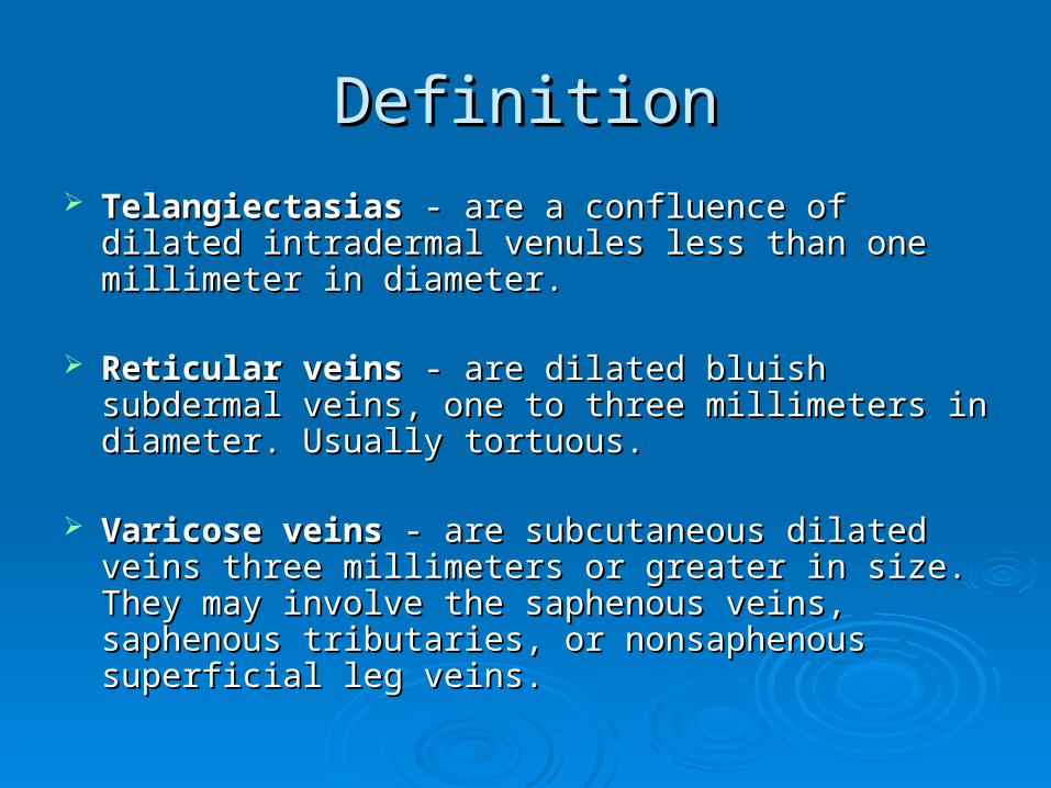
Abnormal VeinsAbnormal Veins
Telangiectasias
Reticular veinsVaricose vein
Common QuestionsCommon Questions
Are they dangerous?Are they dangerous? How do they form?How do they form? Why does it happen? Why does it happen? Did I inherit it?Did I inherit it? What tests can we use?What tests can we use? What treatments are available?What treatments are available?
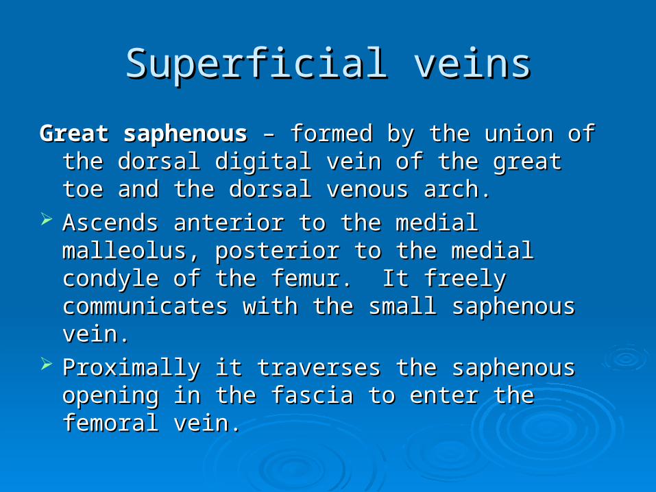
Superficial veinsSuperficial veins
Great saphenousGreat saphenous – formed by the union of the – formed by the union of the dorsal digital vein of the great toe and the dorsal dorsal digital vein of the great toe and the dorsal venous arch.venous arch.
Ascends anterior to the medial malleolus, Ascends anterior to the medial malleolus, posterior to the medial condyle of the femur. It posterior to the medial condyle of the femur. It freely communicates with the small saphenous freely communicates with the small saphenous vein. vein.
Proximally it traverses the saphenous opening in Proximally it traverses the saphenous opening in the fascia to enter the femoral vein.the fascia to enter the femoral vein.
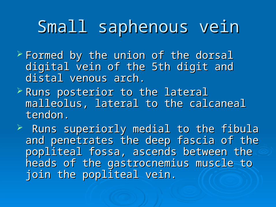
Small saphenous veinSmall saphenous vein
Formed by the union of the dorsal digital Formed by the union of the dorsal digital vein of the 5th digit and distal venous arch. vein of the 5th digit and distal venous arch.
Runs posterior to the lateral malleolus, Runs posterior to the lateral malleolus, lateral to the calcaneal tendon. lateral to the calcaneal tendon.
Runs superiorly medial to the fibula and Runs superiorly medial to the fibula and penetrates the deep fascia of the popliteal penetrates the deep fascia of the popliteal fossa, ascends between the heads of the fossa, ascends between the heads of the gastrocnemius muscle to join the popliteal gastrocnemius muscle to join the popliteal vein.vein.

Perforating veins Perforating veins Penetrate the deep Penetrate the deep
fascia, tributaries of the fascia, tributaries of the saphenous veins, valves saphenous veins, valves are located just distal to are located just distal to penetration of the deep penetration of the deep fascia. fascia.
Veins cross the deep Veins cross the deep fascia obliquely fascia obliquely
Muscle contraction Muscle contraction causes the valves to causes the valves to close prior to venous close prior to venous compression so blood is compression so blood is forced proximally forced proximally (musculo-venous pump). (musculo-venous pump).
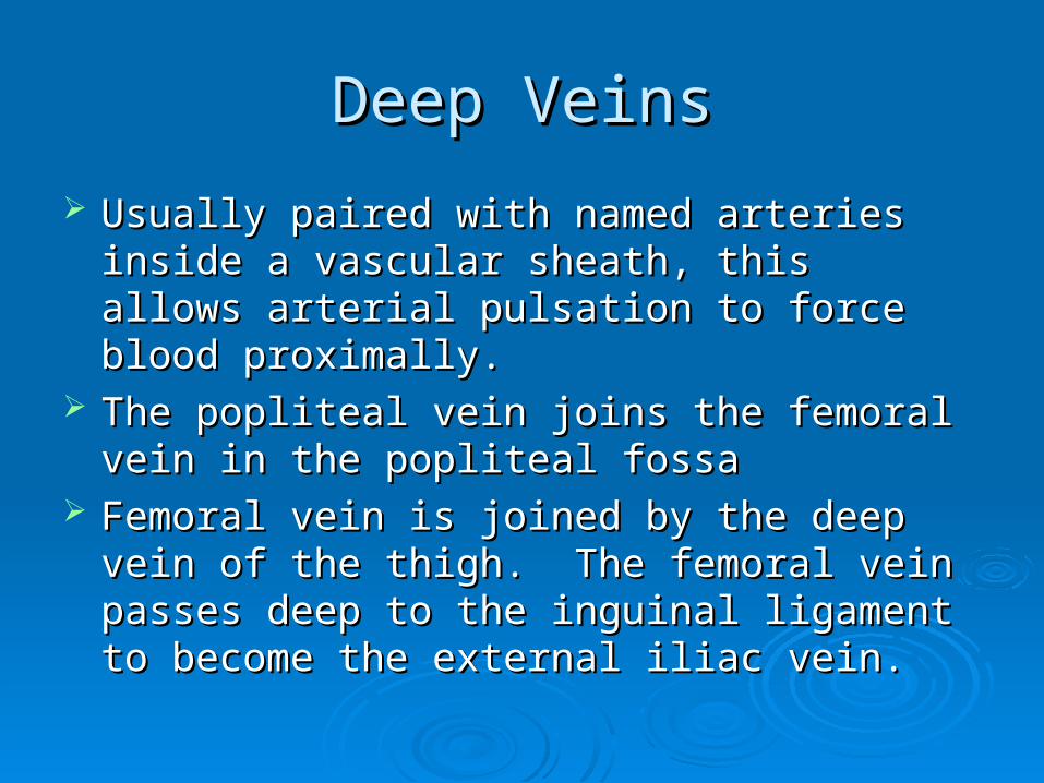
Deep VeinsDeep Veins
Usually paired with named arteries inside a Usually paired with named arteries inside a vascular sheath, this allows arterial pulsation to vascular sheath, this allows arterial pulsation to force blood proximally. force blood proximally.
The popliteal vein joins the femoral vein in the The popliteal vein joins the femoral vein in the popliteal fossapopliteal fossa
Femoral vein is joined by the deep vein of the Femoral vein is joined by the deep vein of the thigh. The femoral vein passes deep to the thigh. The femoral vein passes deep to the inguinal ligament to become the external iliac inguinal ligament to become the external iliac vein.vein.
EtiologyEtiology
Reflux 80%Reflux 80% Venous obstruction 18-28%Venous obstruction 18-28%
Resultant edema and skin changes = Resultant edema and skin changes = Postthrombotic syndromePostthrombotic syndrome
Muscle Pump Dysfunction Muscle Pump Dysfunction
Stasis PathophysiologyStasis Pathophysiology
Usually associated with venous Usually associated with venous incompetenceincompetence
Primary and secondary refluxPrimary and secondary reflux Edema Edema Vein wall dilatation Vein wall dilatation Inflammation/Pigmentation (Hemosiderin Inflammation/Pigmentation (Hemosiderin
deposits)deposits) ““Fibrin cuffing”Fibrin cuffing” UlcerationUlceration
Risk factorsRisk factors
Age: Age: Aging causes wear and tear. Eventually, Aging causes wear and tear. Eventually, that wear causes the valves to malfunction. that wear causes the valves to malfunction.
Sex: Sex: Women > Men. Hormonal changes during Women > Men. Hormonal changes during pregnancy or menopause. Progesterone pregnancy or menopause. Progesterone relaxes venous walls. OCP may increase the relaxes venous walls. OCP may increase the risk of varicose veins. risk of varicose veins.
GeneticsGenetics Obesity: Obesity: Increases venous HTN. Increases venous HTN. Standing for long periods of time.Standing for long periods of time. Prolonged Prolonged
immobile standing impairs venous return.immobile standing impairs venous return.
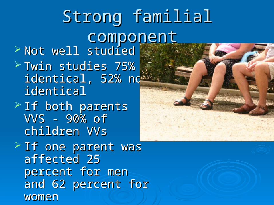
Strong familial component Strong familial component Not well studiedNot well studied Twin studies 75% Twin studies 75%
identical, 52% non identical, 52% non identicalidentical
If both parents VVS - If both parents VVS - 90% of children VVs90% of children VVs
If one parent was If one parent was affected 25 percent for affected 25 percent for men and 62 percent for men and 62 percent for womenwomen
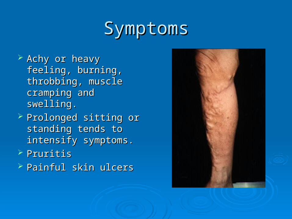
SymptomsSymptoms
Achy or heavy feeling, Achy or heavy feeling, burning, throbbing, burning, throbbing, muscle cramping and muscle cramping and swelling. swelling.
Prolonged sitting or Prolonged sitting or standing tends to standing tends to intensify symptoms. intensify symptoms.
Pruritis Pruritis Painful skin ulcers Painful skin ulcers
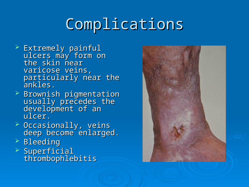
ComplicationsComplications Extremely painful ulcers Extremely painful ulcers
may form on the skin may form on the skin near varicose veins, near varicose veins, particularly near the particularly near the ankles.ankles.
Brownish pigmentation Brownish pigmentation usually precedes the usually precedes the development of an ulcer. development of an ulcer.
Occasionally, veins deep Occasionally, veins deep become enlarged.become enlarged.
Bleeding Bleeding Superficial Superficial
thrombophlebitis thrombophlebitis
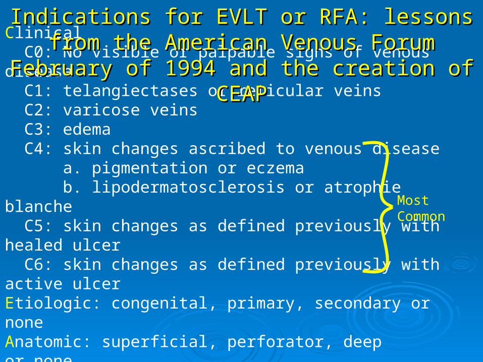
Clinical C0: No visible or palpable signs of venous disease C1: telangiectases or reticular veins C2: varicose veins C3: edema C4: skin changes ascribed to venous disease a. pigmentation or eczema b. lipodermatosclerosis or atrophie blanche C5: skin changes as defined previously with healed ulcer C6: skin changes as defined previously with active ulcer Etiologic: congenital, primary, secondary or noneAnatomic: superficial, perforator, deepor nonePathophysiologic: reflux, obstruction, both or none
Most Common
Indications for EVLT or RFA: lessons from the Indications for EVLT or RFA: lessons from the American Venous ForumAmerican Venous Forum
February of 1994 and the creation of CEAPFebruary of 1994 and the creation of CEAP
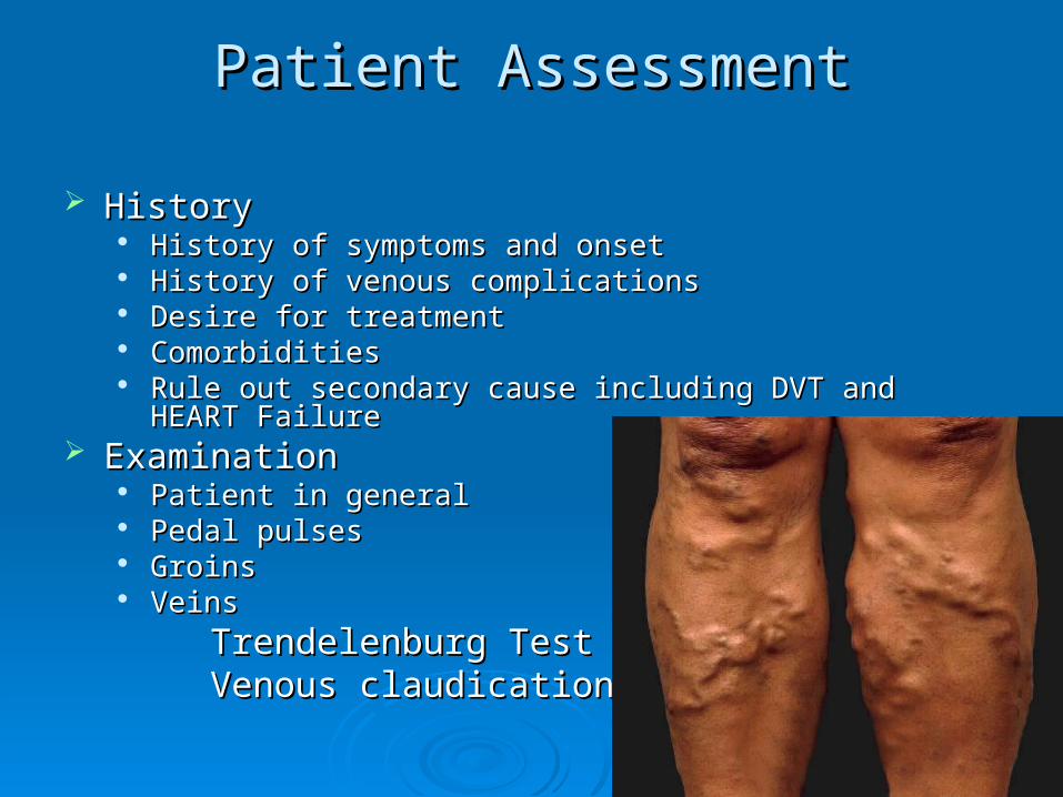
Patient AssessmentPatient Assessment
HistoryHistory History of symptoms and onsetHistory of symptoms and onset History of venous complicationsHistory of venous complications Desire for treatmentDesire for treatment ComorbiditiesComorbidities Rule out secondary cause including DVT and HEART FailureRule out secondary cause including DVT and HEART Failure
ExaminationExamination Patient in generalPatient in general Pedal pulsesPedal pulses GroinsGroins VeinsVeins
Trendelenburg TestTrendelenburg TestVenous claudicationVenous claudication
InvestigationInvestigation
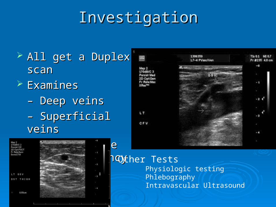
All get a Duplex scan All get a Duplex scan ExaminesExamines
– – Deep veinsDeep veins
– – Superficial veinsSuperficial veins
– – Incompetence and Incompetence and patencypatency
Other TestsPhysiologic testingPhlebographyIntravascular Ultrasound
Duplex scanDuplex scan
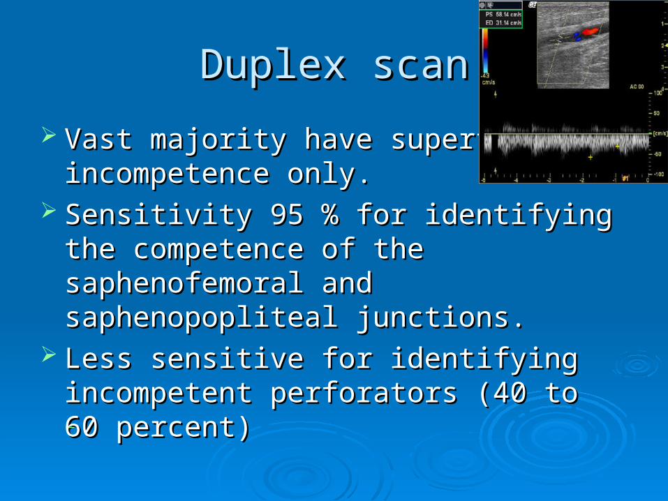
Vast majority have superficial Vast majority have superficial incompetence only.incompetence only.
Sensitivity 95 % for identifying the Sensitivity 95 % for identifying the competence of the saphenofemoral and competence of the saphenofemoral and saphenopopliteal junctions. saphenopopliteal junctions.
Less sensitive for identifying incompetent Less sensitive for identifying incompetent perforators (40 to 60 percent) perforators (40 to 60 percent)
.
TreatmentTreatment
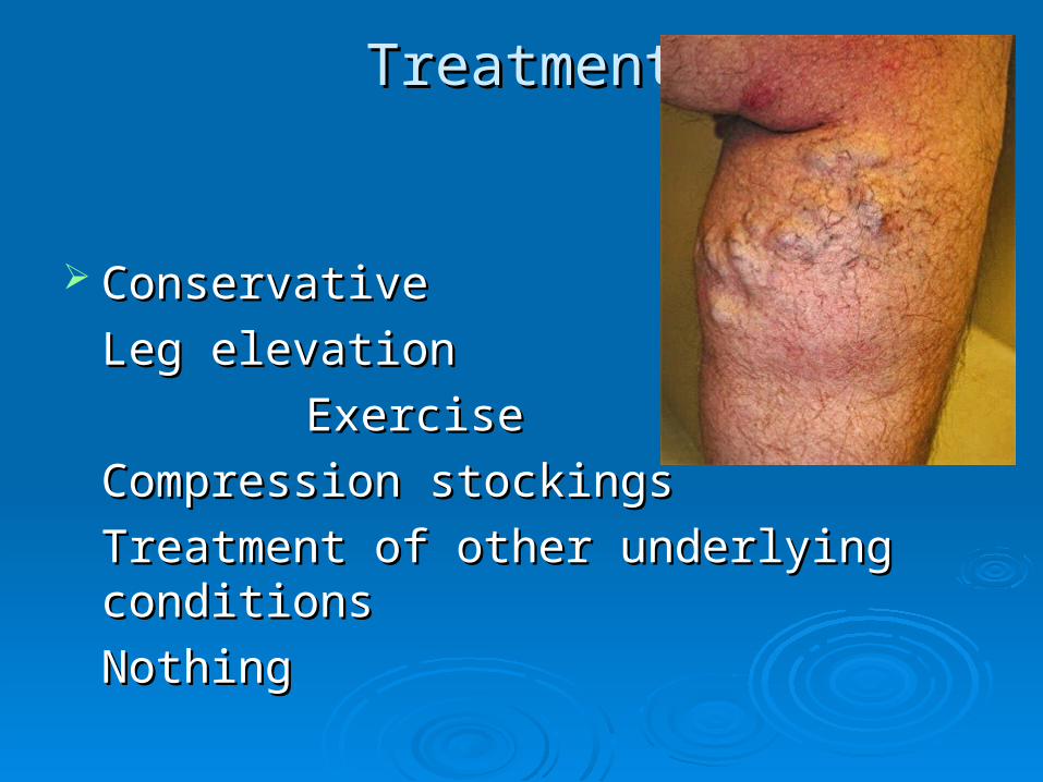
ConservativeConservative
Leg elevationLeg elevation
ExerciseExercise
Compression stockings Compression stockings
Treatment of other underlying Treatment of other underlying conditionsconditions
Nothing Nothing
Vein ablation therapies Vein ablation therapies
Classified by method of vein destruction:Classified by method of vein destruction:
1. Chemical (sclerotherapy)1. Chemical (sclerotherapy)
2. Thermal (laser or endovenous ablation)2. Thermal (laser or endovenous ablation)
3. Mechanical (surgical excision or 3. Mechanical (surgical excision or stripping) stripping)
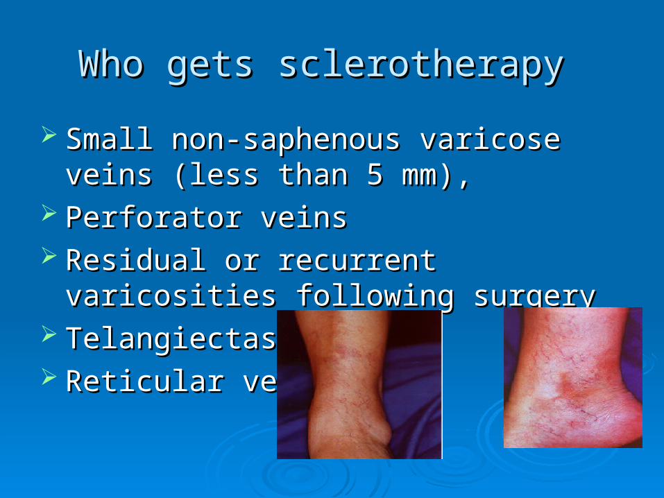
Who gets sclerotherapy Who gets sclerotherapy
Small non-saphenous varicose veins (less Small non-saphenous varicose veins (less than 5 mm), than 5 mm),
Perforator veinsPerforator veins Residual or recurrent varicosities following Residual or recurrent varicosities following
surgery surgery TelangiectasiaTelangiectasia Reticular veinsReticular veins

Who gets SclerotherapyWho gets Sclerotherapy
Who elseWho else
– – Good control with TrendelenburgGood control with Trendelenburg
– – Recurrent veinsRecurrent veins
– – Frail with resistant/healed ulcersFrail with resistant/healed ulcers
Sclerosing AgentsSclerosing Agents
Sodium tetradecyl sulfateSodium tetradecyl sulfate Hypertonic SalineHypertonic Saline PolidocanolPolidocanol Monoethanolamine oleateMonoethanolamine oleate Glucose combinationsGlucose combinations
Damage endothelium leading to thrombosis of Damage endothelium leading to thrombosis of the vein.the vein.
Pressure to try and reduce the amount of Pressure to try and reduce the amount of thrombus.thrombus.
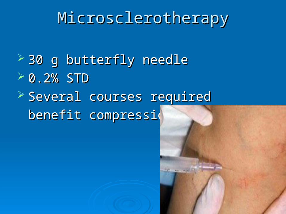
MicrosclerotherapyMicrosclerotherapy
30 g butterfly needle30 g butterfly needle 0.2% STD0.2% STD Several courses requiredSeveral courses required
benefit compressionbenefit compression
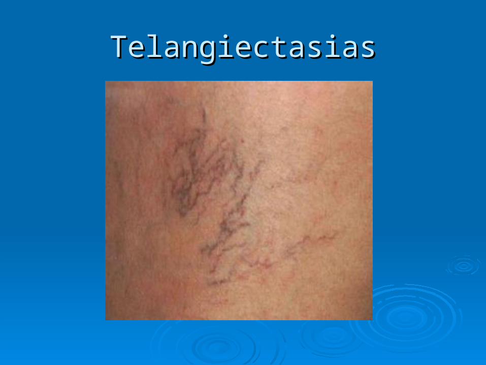
TelangiectasiasTelangiectasias
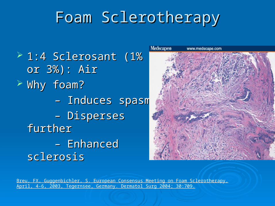
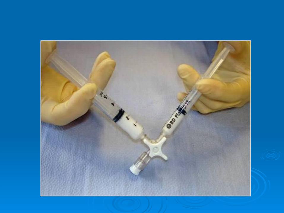
Foam SclerotherapyFoam Sclerotherapy
1:4 Sclerosant (1% or 1:4 Sclerosant (1% or 3%): Air3%): Air
Why foam?Why foam?
– – Induces spasmInduces spasm
– – Disperses furtherDisperses further
– – Enhanced Enhanced sclerosissclerosis
Breu, FX, Guggenbichler, S. European Consensus Meeting on Foam Sclerotherapy, April, 4-6, 2003, Tegernsee, Germany. Dermatol Surg 2004; 30:709.
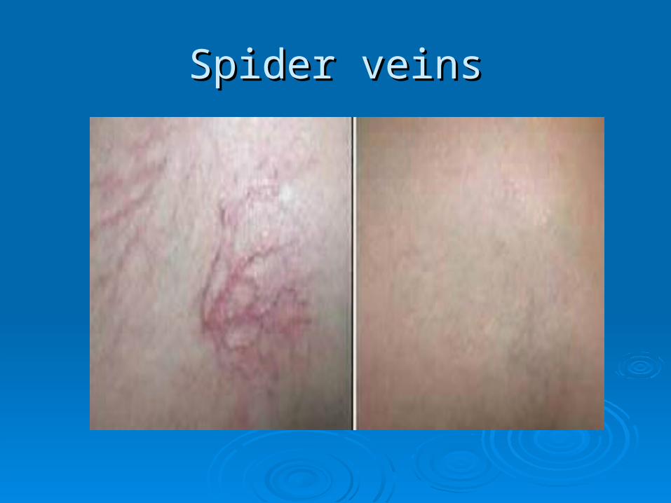
Spider veinsSpider veins
Foam Sclerotherapy:Foam Sclerotherapy:ComplicationsComplications
PhlebitisPhlebitis Skin stainingSkin staining FailureFailure Residual lumpsResidual lumps MattingMatting Embolus (CVA)Embolus (CVA) DVTDVT Ulceration (rare)Ulceration (rare) Anaphylaxis (very rare)Anaphylaxis (very rare)
Foam Sclerotherapy ResultsFoam Sclerotherapy Results
Variable depending on seriesVariable depending on series Long-term recurrence rates are as high as Long-term recurrence rates are as high as
65 percent in five years, however, patients 65 percent in five years, however, patients can also be retreated when veins recurcan also be retreated when veins recur
Large veins can be a problemLarge veins can be a problem Currently randomized trialCurrently randomized trial
Catheter-based TreatmentsCatheter-based Treatments
Endovenous laser EVLAEndovenous laser EVLA Radiofrequency ablation RFARadiofrequency ablation RFA Primarily to treat saphenous insufficiency Primarily to treat saphenous insufficiency
(great or small)(great or small) EVLA and RFA, are equally efficacious & EVLA and RFA, are equally efficacious &
have similar recanalization rates. have similar recanalization rates.
Boros, MJ, O'Brien, SP, McLaren, JT, Collins, JT. High ligation of the saphenofemoral junction in endovenous obliteration of varicose veins. Vasc Endovascular Surg 2008; 42:235.
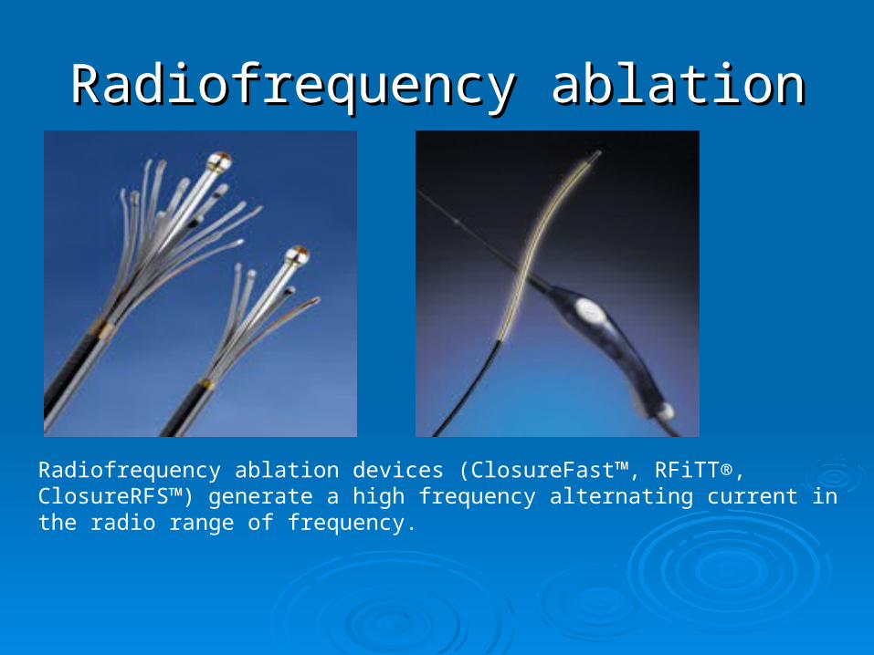
Radiofrequency ablationRadiofrequency ablation
Radiofrequency ablation devices (ClosureFast™, RFiTT®, ClosureRFS™) generate a high frequency alternating current in the radio range of frequency.
- By directing resistive radiofrequency energy through a vein, a narrow rim of tissue less than 1mm is heated by an electrode. - The amount of heating is modulated using both a microprocessor and manual movement, resulting in controlled collagen contraction, thermocoagulation and absorption of the vein.
Mechanism RFA
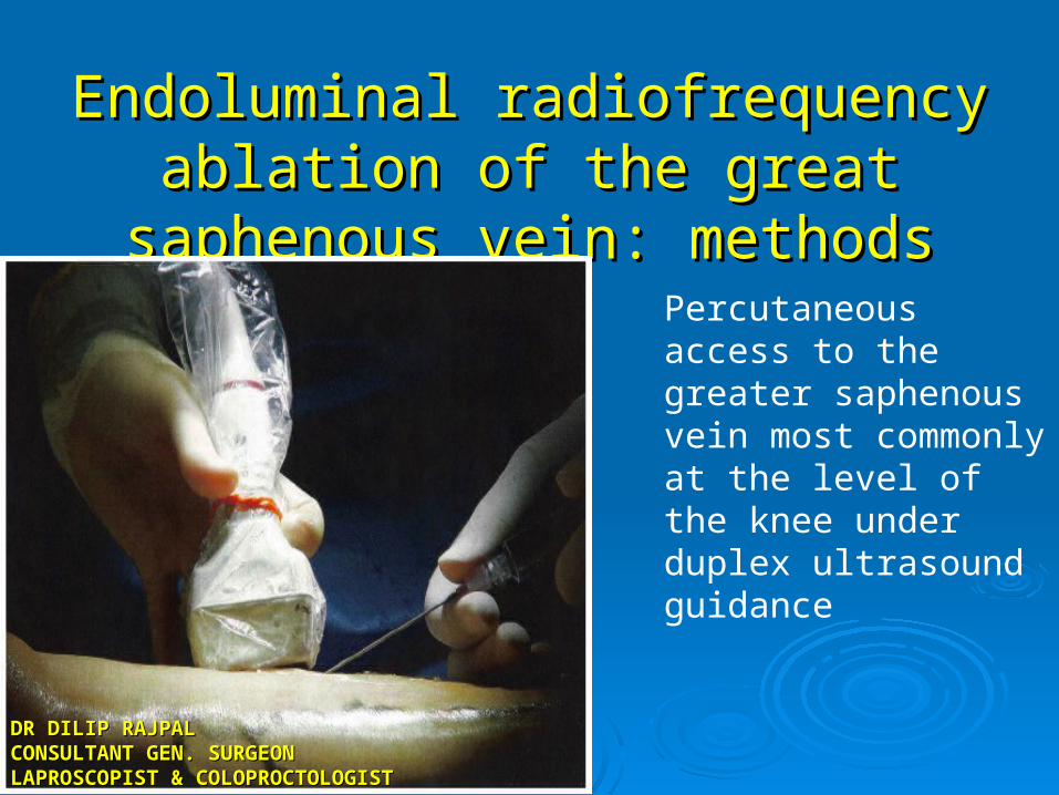
Endoluminal radiofrequency ablation of Endoluminal radiofrequency ablation of the great saphenous vein: methodsthe great saphenous vein: methods
Percutaneous access to the greater saphenous vein most commonly at the level of the knee under duplex ultrasound guidance
DR DILIP RAJPALDR DILIP RAJPALCONSULTANT GEN. SURGEONCONSULTANT GEN. SURGEONLAPROSCOPIST & LAPROSCOPIST & COLOPROCTOLOGISTCOLOPROCTOLOGIST
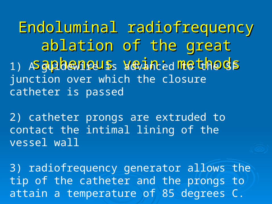
Endoluminal radiofrequency ablation of Endoluminal radiofrequency ablation of the great saphenous vein: methodsthe great saphenous vein: methods
1) A guidewire is advanced to the SF junction over which the closure catheter is passed
2) catheter prongs are extruded to contact the intimal lining of the vessel wall
3) radiofrequency generator allows the tip of the catheter and the prongs to attain a temperature of 85 degrees C.
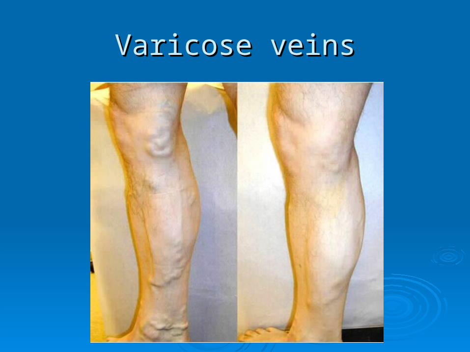
Varicose veinsVaricose veins
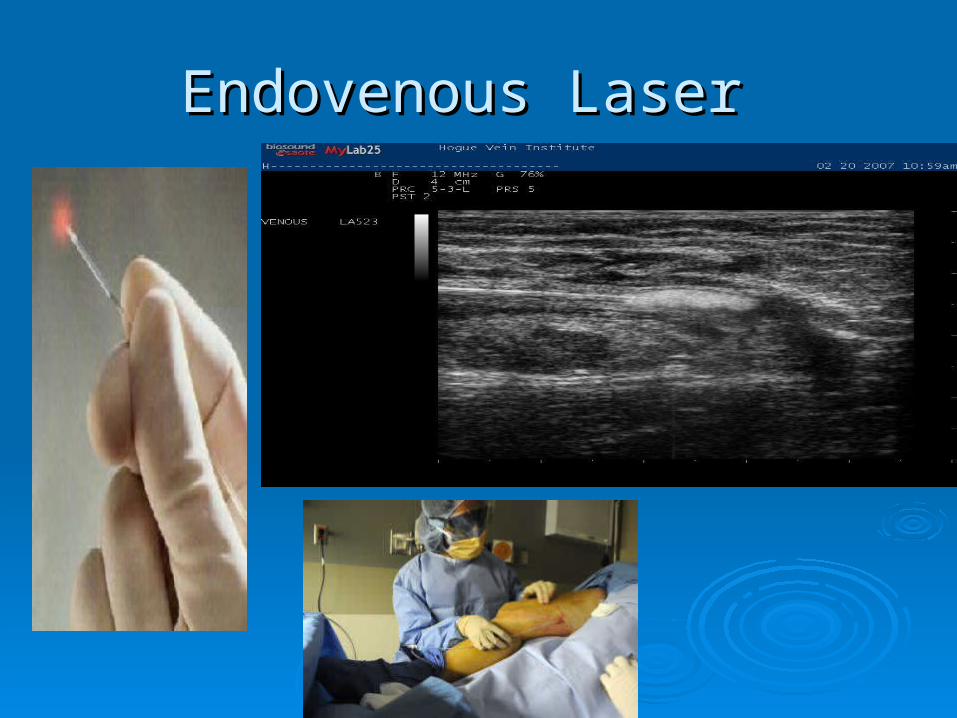
Endovenous Laser Endovenous Laser
Endovenous LaserEndovenous Laser
Devices (EVLT®, ClosurePlus™) Devices (EVLT®, ClosurePlus™) Use a bare tipped optical fiber which Use a bare tipped optical fiber which
applies laser light energy to the vein.applies laser light energy to the vein. Therapy based on photothermolysis (light Therapy based on photothermolysis (light
induced thermal damage). induced thermal damage). Laser light heats the target tissue inducing Laser light heats the target tissue inducing
thermal injurythermal injury Wavelength of light is chosen based on Wavelength of light is chosen based on
the target structure's chromophore.the target structure's chromophore.Bush, RG, Shamma, HN, Hammond, K. Histological changes occurring after endoluminal ablation with two diode
lasers (940 and 1319 nm) from acute changes to 4 months. Lasers Surg Med 2008; 40:676.
Endovenous laser therapy (EVLT): mechanism
- Thermal reaction after laser exposure is essential.
- Damages endothelial, intimal internal elastic lamina, and to some degree the media. Adventitia is rarely affected. - In vitro studies suggest that energy results in ‘boiling of blood’ and generation of ‘steam bubbles’ that indirectly, homogenously affect the varicose vein.
Endovenous laser therapyEndovenous laser therapy: : methodsmethods
1) GSV entered at the knee2) Guidewire passed through hollow needle into the vein can be difficult if:
a. tortuosities b. local venous spasmc. sclerotic fragments
3) Needle removed4) 3mm cutaneous incision made5) Introducer sheath placed over guide wire6) Guidewire removed when at the SFJ7) Longitudinal US visualization of sheath 1-2 cm distally to the SFJ
Endovenous laser therapy and Endovenous laser therapy and radiofrequencyradiofrequency: methods: methods
Tumescent anesthesia (5 ml epi, 5 ml bicarb, 35ml 1% lidocaine in 500ml saline) is administered to the perivenous space resulting in
a) reduction in pain
b) protection of perivenous tissue through cooling
c) increase in surface area of laser tip and vein wall
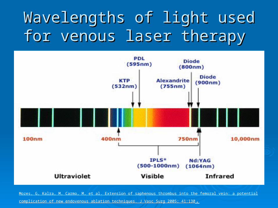
Wavelengths of light used for Wavelengths of light used for venous laser therapy venous laser therapy
Mozes, G, Kalra, M, Carmo, M, et al. Extension of saphenous thrombus into the femoral vein: a potential complication of new endovenous
ablation techniques. J Vasc Surg 2005; 41:130.
Endovenous laser therapy and Endovenous laser therapy and radiofrequencyradiofrequency: specifics: specifics
Pulsed vs. continuous:pulsed mode is associated with higher adverse events
Wavelengths:Higher wavelengths (1320nm) reported less
postoperative pain, and less likely to have ecchymoses
Fluence (J/ cm2):Single most important parameter to quantifyabove 60-100 J/ cm2 for durable GSV occlusion
Wattage:high, short duration wattage vaporizing effectlow prolonged wattage coagulating effect
Pullback Speed:if performed at fixed wattage then energy is
solely dependent on pullback speed
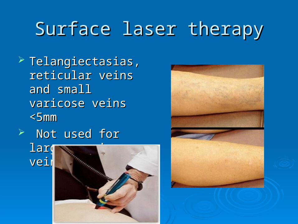
Surface laser therapySurface laser therapy
Telangiectasias, Telangiectasias, reticular veins and reticular veins and small varicose veins small varicose veins <5mm<5mm
Not used for larger Not used for larger varicose veins varicose veins
Post op carePost op care
Graduated compression stockings are Graduated compression stockings are worn following the procedure.worn following the procedure.
F/U duplex ultrasound is performed within F/U duplex ultrasound is performed within one week to evaluate for thrombus in the one week to evaluate for thrombus in the common femoral vein.common femoral vein.
Pt recovery averages two and four daysPt recovery averages two and four days Significantly shorter interval than is seen Significantly shorter interval than is seen
with surgical ligation and stripping with surgical ligation and stripping
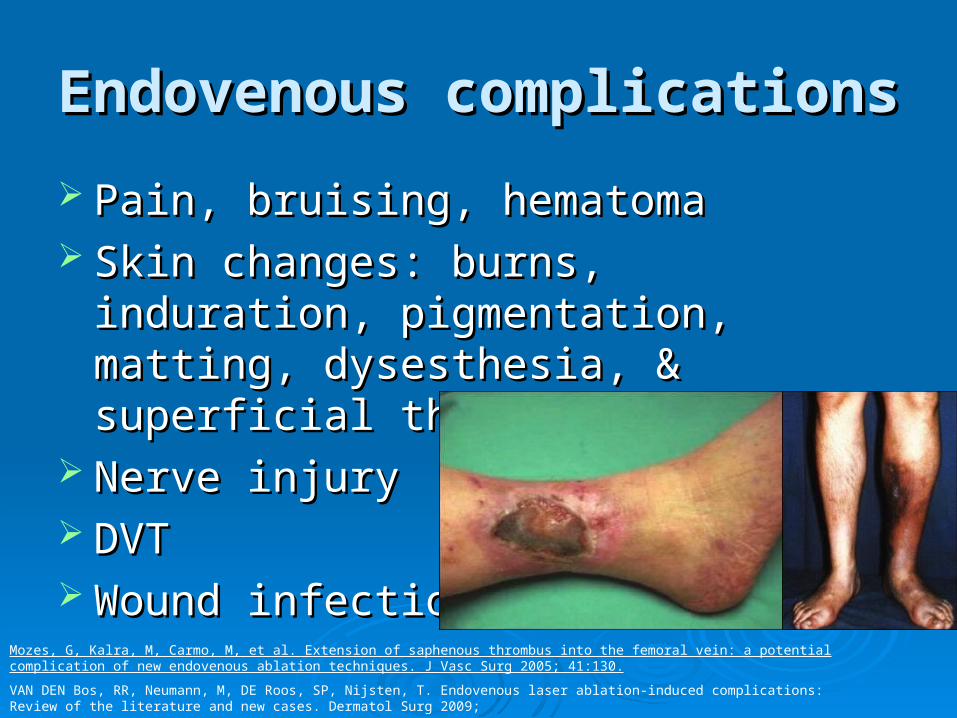
Endovenous complicationsEndovenous complications
Pain, bruising, hematoma Pain, bruising, hematoma Skin changes: burns, induration, Skin changes: burns, induration,
pigmentation, matting, dysesthesia, & pigmentation, matting, dysesthesia, & superficial thrombophlebitis.superficial thrombophlebitis.
Nerve injury Nerve injury DVT DVT Wound infection Wound infection
Mozes, G, Kalra, M, Carmo, M, et al. Extension of saphenous thrombus into the femoral vein: a potential complication of new endovenous ablation techniques. J Vasc Surg 2005; 41:130.
VAN DEN Bos, RR, Neumann, M, DE Roos, SP, Nijsten, T. Endovenous laser ablation-induced complications: Review of the literature and new cases. Dermatol Surg 2009;
Which is Better ???Which is Better ???
Endoluminal thermal ablation versus Endoluminal thermal ablation versus stripping of the saphenous vein: Meta-stripping of the saphenous vein: Meta-analysis of recurrence of reflux.analysis of recurrence of reflux.
ES Xenos, G Bietz, DJ Minion, et alES Xenos, G Bietz, DJ Minion, et al
Endoluminal thermal ablation versus stripping of Endoluminal thermal ablation versus stripping of the saphenous vein: Meta-analysis of recurrence the saphenous vein: Meta-analysis of recurrence
of refluxof reflux..
Method: Systematic search of Method: Systematic search of Medline/Pubmed, OVID, EMBASE, Medline/Pubmed, OVID, EMBASE, CINAHL, Clinicaltrials.gov and Cochrane CINAHL, Clinicaltrials.gov and Cochrane central registercentral register 1966-2009 in all lanuages1966-2009 in all lanuages
MethodMethod
Randomized prospective clinical trials with Randomized prospective clinical trials with > 365 days f/u.> 365 days f/u.
Analyzed outcomes included recurrence of Analyzed outcomes included recurrence of varicosities and reflux, as documented by varicosities and reflux, as documented by duplex ultrasound, and recurrence of signs duplex ultrasound, and recurrence of signs and symptomsand symptoms
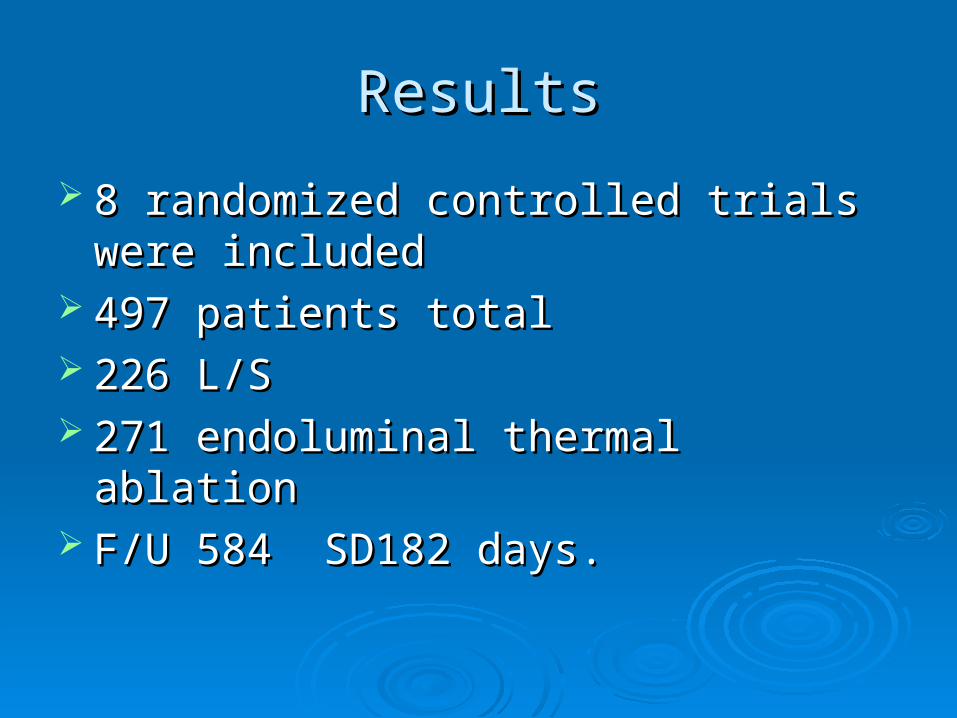
ResultsResults
8 randomized controlled trials were 8 randomized controlled trials were includedincluded
497 patients total497 patients total 226 L/S226 L/S 271 endoluminal thermal ablation271 endoluminal thermal ablation F/U 584 SD182 days.F/U 584 SD182 days.
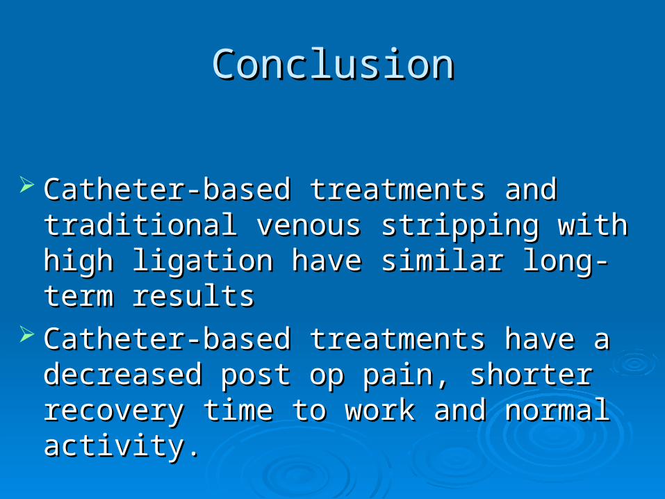
ConclusionConclusion
Catheter-based treatments and traditional Catheter-based treatments and traditional venous stripping with high ligation have similar venous stripping with high ligation have similar long-term resultslong-term results
Catheter-based treatments have a decreased Catheter-based treatments have a decreased post op pain, shorter recovery time to work post op pain, shorter recovery time to work and normal activity.and normal activity.