Embed Size (px)
Citation preview
459Copyrights © 2013 The Korean Society of Radiology
INTRODUCTION
Chronic mesenteric ischemia (CMI) is an uncommon disor-der caused by atherosclerotic occlusion or stenosis of the mes-enteric arteries (1). CMI symptoms classically develop when there is significant stenosis or occlusion of at least two of the three mesenteric arteries (2). Endovascular therapy is increas-ingly accepted as the first line of therapy for CMI due to its low-er morbidity and mortality rate and similar outcome compared with open surgery (1-5).
In celiacomesenteric trunk (CMT), the celiac artery (CA) and superior mesenteric artery (SMA) arise from a common origin (6). Patients with CMT variation develop CMI when atheroma-tous disease causes occlusion of both the proximal CA and SMA at their common trunk. We report a case of CMT variant with severe stenosis of the trunk ostium and a totally occluded SMA that was successfully treated with overlapping CA and SMA
stents in the trunk portion.
CASE REPORT
A 73-year-old woman with a 3-month history of postprandial abdominal pain and weight loss was referred to our department for evaluation. Contrast-enhanced computed tomography (CT) scan revealed a common trunk of the CA and SMA, with total SMA occlusion. There were no signs of bowel ischemia, such as bowel wall thickening or abnormal contrast enhancement.
A 5-Fr RH catheter (Cook, Bloomington, IN, USA) was in-serted in the right common femoral artery. Angiography con-firmed CMT with significant stenosis of the trunk and complete occlusion of the proximal portion of the SMA (Fig. 1A). The distal portion of the SMA showed faint retrograde filling via the pancreaticoduodenal arcade. We first attempted to cross the ob-structed SMA through the stenotic trunk, using a 5-Fr Cobra
Case ReportpISSN 1738-2637J Korean Soc Radiol 2013;68(6):459-462http://dx.doi.org/10.3348/jksr.2013.68.6.459
Received February 26, 2013; Accepted March 1, 2013Corresponding author: Jae Myeong Lee, MDDepartment of Radiology, Soonchunhyang University College of Medicine, Bucheon Hospital, 170 Jomaru-ro, Wonmi-gu, Bucheon 420-767, Korea.Tel. 82-32-621-6409 Fax. 82-32-621-5016E-mail: [email protected]
This is an Open Access article distributed under the terms of the Creative Commons Attribution Non-Commercial License (http://creativecommons.org/licenses/by-nc/3.0) which permits unrestricted non-commercial use, distri-bution, and reproduction in any medium, provided the original work is properly cited.
A 73-year-old woman was presented with a 3-month history of postprandial ab-dominal pain and weight loss. The patient had an anatomic variant of celiacomes-enteric trunk (CMT), with complete occlusion of the superior mesenteric artery (SMA) and significant stenosis of the CMT ostium, resulting in chronic mesenteric ischemia. After several unsuccessful attempts at revascularization of the SMA, celi-ac artery stenting was performed, followed by SMA stent placement through the previously placed stent mesh into the occlusion site. The patient’s symptoms com-pletely resolved after treatment.
Index termsChronic Mesenteric IschemiaEndovascular TreatmentStentCeliac ArterySuperior Mesenteric Artery
Endovascular Treatment of Chronic Mesenteric Ischemia by Crossing of Two Stents in a Patient with Celiacomesenteric Trunk 복강위창자간막동맥간을 보인 환자에서 두 개의 스텐트의 교차에 의한 만성 장간막 허혈의 혈관내 치료 Eun Kyung Khil, MD, Jae Myeong Lee, MDDepartment of Radiology, Soonchunhyang University College of Medicine, Bucheon Hospital, Bucheon, Korea
Endovascular Treatment of Chronic Mesenteric Ischemia by Crossing of Two Stents in a Patient with Celiacomesenteric Trunk
submit.radiology.or.krJ Korean Soc Radiol 2013;68(6):459-462460
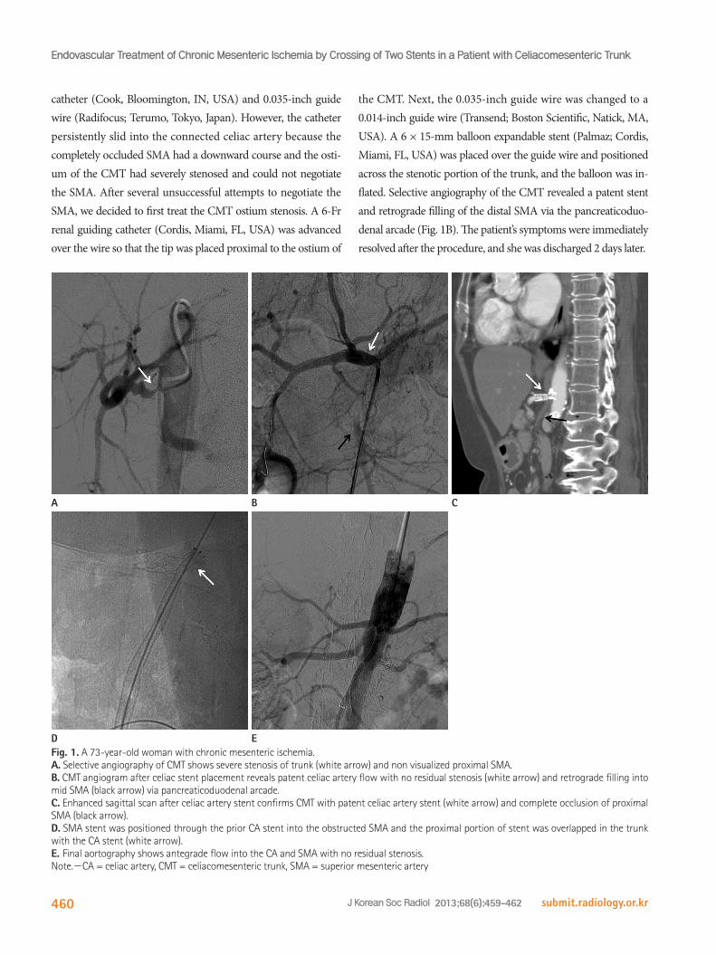
the CMT. Next, the 0.035-inch guide wire was changed to a 0.014-inch guide wire (Transend; Boston Scientific, Natick, MA, USA). A 6 × 15-mm balloon expandable stent (Palmaz; Cordis, Miami, FL, USA) was placed over the guide wire and positioned across the stenotic portion of the trunk, and the balloon was in-flated. Selective angiography of the CMT revealed a patent stent and retrograde filling of the distal SMA via the pancreaticoduo-denal arcade (Fig. 1B). The patient’s symptoms were immediately resolved after the procedure, and she was discharged 2 days later.
catheter (Cook, Bloomington, IN, USA) and 0.035-inch guide wire (Radifocus; Terumo, Tokyo, Japan). However, the catheter persistently slid into the connected celiac artery because the completely occluded SMA had a downward course and the osti-um of the CMT had severely stenosed and could not negotiate the SMA. After several unsuccessful attempts to negotiate the SMA, we decided to first treat the CMT ostium stenosis. A 6-Fr renal guiding catheter (Cordis, Miami, FL, USA) was advanced over the wire so that the tip was placed proximal to the ostium of
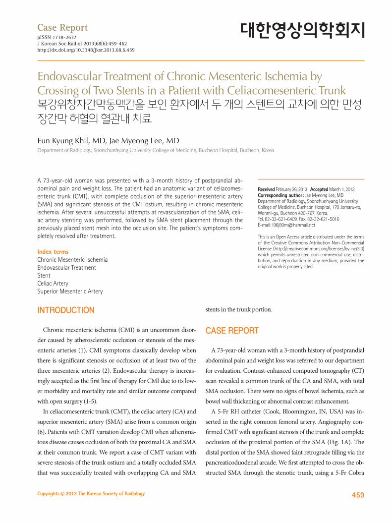
Fig. 1. A 73-year-old woman with chronic mesenteric ischemia.A. Selective angiography of CMT shows severe stenosis of trunk (white arrow) and non visualized proximal SMA. B. CMT angiogram after celiac stent placement reveals patent celiac artery flow with no residual stenosis (white arrow) and retrograde filling into mid SMA (black arrow) via pancreaticoduodenal arcade. C. Enhanced sagittal scan after celiac artery stent confirms CMT with patent celiac artery stent (white arrow) and complete occlusion of proximal SMA (black arrow). D. SMA stent was positioned through the prior CA stent into the obstructed SMA and the proximal portion of stent was overlapped in the trunk with the CA stent (white arrow). E. Final aortography shows antegrade flow into the CA and SMA with no residual stenosis.Note.-CA = celiac artery, CMT = celiacomesenteric trunk, SMA = superior mesenteric artery
D
A
E
B C

Eun Kyung Khil, et al
submit.radiology.or.kr J Korean Soc Radiol 2013;68(6):459-462 461
a higher incidence of recurrent symptoms compared with open surgery, repeat therapy is possible. Therefore, the endovascular treatment of CMI has become the first-line therapy (2, 4).
CMT, an anatomic variant where the CA and SMA have a common origin from the aorta, accounts for only 1.5% of all splanchnic artery anomalies (6). When more than one mesenteric artery is occluded or stenotic, endovascular treatment should be-gin with the vessel that is technically easier to access (1). Although ischemic symptoms improve when only one of the two affected arteries is successfully revascularized, treatment of the other in-creases bowel perfusion and prevents recurrent symptoms in the case of restenosis (3). In our case, the ostium of the CMT showed severe stenosis, and there was complete occlusion of the proximal SMA. The wire required catheter or sheath support to pass through the SMA occlusion site, but because the origin of the CMT was too stenotic, our initial attempt to access the SMA was unsuccessful. Therefore, we decided to first stent the significant stenosis of the CMT ostium, and treat the SMA occlusion later.
Another treatment option for this patient was percutaneous transluminal angioplasty (PTA) of ostial stenosis of the trunk, followed by stent placement through the occluded SMA. How-ever, we did not choose this method, because of the risk of distal embolization of the hepatic artery and significant recoil after PTA (5). Retrograde recanalization of SMA via collaterals from the celiac artery was another treatment option. In our patient, however, SMA recanalization in retrograde fashion via the pan-creaticoduodenal arcade was technically impossible because of tight stenosis at the CMT origin (8).
To our knowledge, this is the first report of endovascular treat-ment of CMT occlusion by crossing the two stents. Ailawadi et al. (9) reported a case of CMT occlusion treated surgically using a bypass graft. Ayers et al. (10) treated a patient with CMT oc-clusion using blunt microdissection catheter (Frontrunner X39 CTO Catheter; LuMend, Redwood City, CA, USA) and stent placement. However, in this case, unlike ours, the occluded CMT showed a residual normal stump that could support a catheter or sheath and enable SMA stenting.
In conclusion, in cases where SMA negotiation is technically difficult or impossible, endovascular recanalization of a stenotic CMT can be achieved by crossing the two stents. This treatment option may be used to treat CMI associated with other anatomic variations.
One month later, she complained of recurrent abdominal pain and weight loss. Abdominal CT revealed a patent CA stent and complete occlusion of a 3.0 cm length of the proximal SMA. Retrograde flow into the distal SMA occurred via the pancreati-coduodenal arcade. No signs of ischemia, such as bowel wall thickening or abnormal contrast enhancement, were observed (Fig. 1C). Endovascular revascularization for SMA recanaliza-tion via the left brachial artery route was decided because of the downward course of the SMA and because of the difficulty in wire selection and stent placement due to prior insertion of the CA stent making the femoral approach difficult. After inserting a 6-Fr sheath, a 5-Fr Cobra catheter was inserted along the 0.035-inch guide wire. The wire was successfully negotiated through the previously inserted stent mesh into the SMA occlusion site. A 6 × 60-mm stent (S.M.A.R.T.; Cordis, Miami, FL, USA) was inserted along the wire and was carefully positioned across the obstructed SMA site. The proximal portion of the stent was over-lapped in the trunk with the CA stent, and the 6-mm balloon (Synergy; Boston Scientific, Natick, MA, USA) was inflated (Fig. 1D). Final angiogram showed normal position of the overlapped CA and SMA stents and antegrade flow into the SMA and CA without residual stenosis (Fig. 1E). There were no complica-tions, and the patient’s symptoms were immediately resolved. Six months later, the patient had no clinical symptoms, and she regained her weight.
DISCUSSION
The cause of CMI is progressive atherosclerotic stenosis or oc-clusion of one or more mesenteric arteries (1). However, clinical manifestations of CMI are rare because atheromatous disease usually involves the proximal portion of the mesenteric arteries, allowing collateral blood flow to the intestine (2). Treatment of stenosis or occlusion of mesenteric vessels is indicated for intes-tinal ischemia-related symptoms, such as postprandial pain, weight loss, nausea, and diarrhea (3). The aims of treatment are to improve symptoms and prevent ischemic injury to the bowel.
Historically, CMI has been treated by surgical revascularization. However, surgery has a 15-47% morbidity rate and a 0-17% mor-tality rate, higher than that of endovascular treatment (1). The overall outcome of the endovascular treatment of CMI compares favorably with surgery (7). Although endovascular treatment has
Endovascular Treatment of Chronic Mesenteric Ischemia by Crossing of Two Stents in a Patient with Celiacomesenteric Trunk
submit.radiology.or.krJ Korean Soc Radiol 2013;68(6):459-462462
6.YiSQ,TerayamaH,NaitoM,HayashiS,MoriyamaH,Tsu-
chidaA,etal.Acommonceliacomesenterictrunk,anda
briefreviewoftheliterature.AnnAnat2007;189:482-488
7.KasirajanK,O’HaraPJ,GrayBH,HertzerNR,ClairDG,
GreenbergRK,etal.Chronicmesenteric ischemia:open
surgeryversuspercutaneousangioplastyandstenting.J
VascSurg2001;33:63-71
8.RobkenJ,ShammasNW.Treatmentofatotallyoccluded
superiormesentericarteryfacilitatedbyretrogradecross-
ingviacollateralsfromtheceliacartery.JEndovascTher
2007;14:745-747
9.AilawadiG,CowlesRA,StanleyJC,EliasonJL,Williams
DM,CollettiLM,etal.Commonceliacomesenterictrunk:
aneurysmalandocclusivedisease.JVascSurg 2004;40:
1040-1043
10.AyersNP,ZachariasSJ,Abu-FadelMS,HennebryTA.Suc-
cessfuluseofbluntmicrodissectioncatheterinachronic
totalocclusionofaceliomesentericartery.CatheterCar-
diovascInterv2007;69:546-549
REFERENCES
1.LoffroyR,GuiuB,CercueilJP,KrauséD.Chronicmesen-
teric ischemia:efficacyandoutcomeofendovascular
therapy.AbdomImaging2010;35:306-314
2.AllenRC,MartinGH,ReesCR,RiveraFJ,TalkingtonCM,
GarrettWV,etal.Mesentericangioplastyinthetreatment
ofchronicintestinal ischemia.JVascSurg1996;24:415-
421;discussion421-423
3.RazaviM,ChungHH.Endovascularmanagementofchronic
mesentericischemia.TechVascIntervRadiol2004;7:155-
159
4.FiooleB,vandeRestHJ,MeijerJR,vanLeersumM,van
KoeverdenS,MollFL,etal.Percutaneoustransluminalan-
gioplastyandstentingasfirst-choicetreatment inpa-
tientswithchronicmesentericischemia.JVascSurg2010;
51:386-391
5.SilvaJA,WhiteCJ,CollinsTJ,JenkinsJS,AndryME,Reilly
JP,etal.Endovascular therapyforchronicmesenteric
ischemia.JAmCollCardiol2006;47:944-950
복강위창자간막동맥간을 보인 환자에서 두 개의 스텐트의 교차에 의한 만성 장간막 허혈의 혈관내 치료
길은경 · 이재명
73세된 여자 환자가 3달간의 식후통증과 체중감소를 주소로 내원했다. 이 환자는 해부학적 변이인 복강위창자간막동맥
간을 가지고 있었는데 그 중에서 위창자간막동맥은 완전히 폐색되어 있었고 복강위창자간막동맥간의 입구는 심한 협착을
보였다. 위창자간막동맥을 개통 시키기 위한 몇 차례의 시도는 실패했으며 그래서 복강동맥의 스텐트 시술 후 스텐트 메
시를 통해서 폐색된 위창자간막동맥내로 두 번째 스텐트가 삽입되었으며 시술 후 환자의 증상은 즉시 사라졌다.
순천향대학교 의과대학 부천병원 영상의학과


![Challenges Encountered during the Treatment of Acute ...acute mesenteric ischemia is a great clinical challenge [1–6]. ... of acute mesenteric ischemia, occurring in 38 (92.68%)](https://img.pdfslide.us/doc/110x75/60f89ecd2be9754e8c1fff31/challenges-encountered-during-the-treatment-of-acute-acute-mesenteric-ischemia.jpg)