Embed Size (px)
Citation preview
ENDOVASCULAR ACUTE STROKE THERAPY:INTEGRATING IMAGING INTO TREATMENT DECISIONS
T U D OR G. J OV I N , M .D .
AS S OCI AT E PROFE S S OR OF NE UROL OGY AND NE UROS URGE RY
D I R E C TOR , U PM C S T R OKE I N S T I T U TE
D I R E C TOR U PM C C E N T E R FOR N E U R OE N DOVA S CU LAR T HE R A PY
U N I V E R S IT Y OF PI T T S B UR GH M E D I C A L C E N T E R
DISCLOSURES
Consultant/Advisory Board: Ownership Interest: Silk Road Medical – modest
Consultant/Advisory Board: Covidien/Medtronic: unpaid
Consultant: Stryker Neurovascular unpaid
PI: REVASCAT (Fundacio Ictus Malaltia Vascular), unpaid
PI: DAWN (Stryker Neurovascular), unpaid
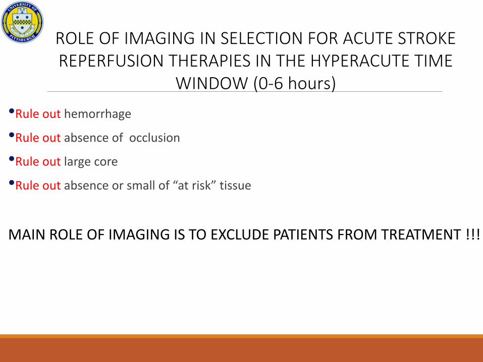
ROLE OF IMAGING IN SELECTION FOR ACUTE STROKEREPERFUSION THERAPIES IN THE HYPERACUTE TIME
WINDOW (0-6 hours)
•Rule out hemorrhage
•Rule out absence of occlusion
•Rule out large core
•Rule out absence or small of “at risk” tissue
MAIN ROLE OF IMAGING IS TO EXCLUDE PATIENTS FROM TREATMENT !!!
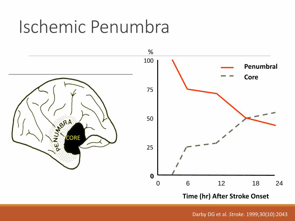
Darby DG et al. Stroke. 1999;30(10):2043
0 6 12 18 24
%
Time (hr) After Stroke Onset
Penumbral
Core
00
25
50
75
100
Ischemic Penumbra
CORE
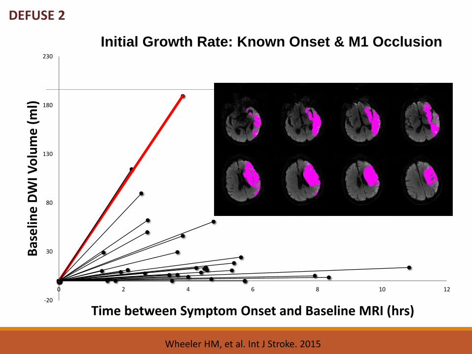
DEFUSE 2
-20
30
80
130
180
230
0 2 4 6 8 10 12
Bas
elin
e D
WI V
olu
me
(m
l)
Time between Symptom Onset and Baseline MRI (hrs)
Initial Growth Rate: Known Onset & M1 Occlusion
Wheeler HM, et al. Int J Stroke. 2015
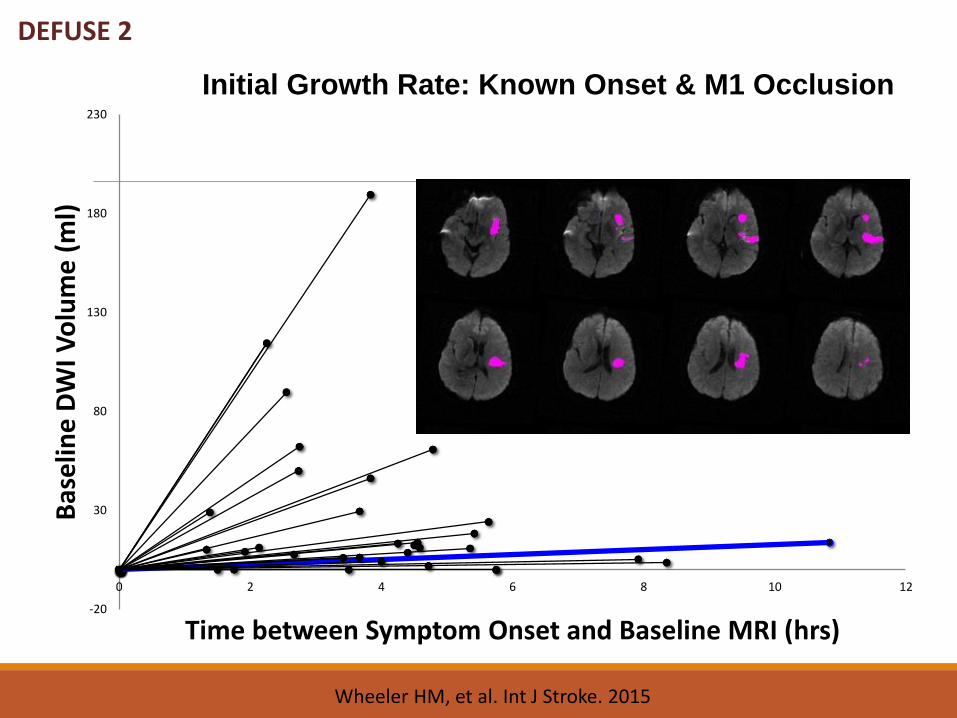
DEFUSE 2
-20
30
80
130
180
230
0 2 4 6 8 10 12
Bas
elin
e D
WI V
olu
me
(m
l)
Time between Symptom Onset and Baseline MRI (hrs)
Initial Growth Rate: Known Onset & M1 Occlusion
Wheeler HM, et al. Int J Stroke. 2015
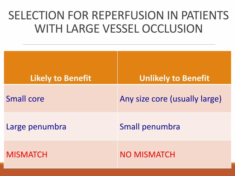
SELECTION FOR REPERFUSION IN PATIENTS WITH LARGE VESSEL OCCLUSION
Likely to Benefit Unlikely to Benefit
Small core Any size core (usually large)
Large penumbra Small penumbra
MISMATCH NO MISMATCH
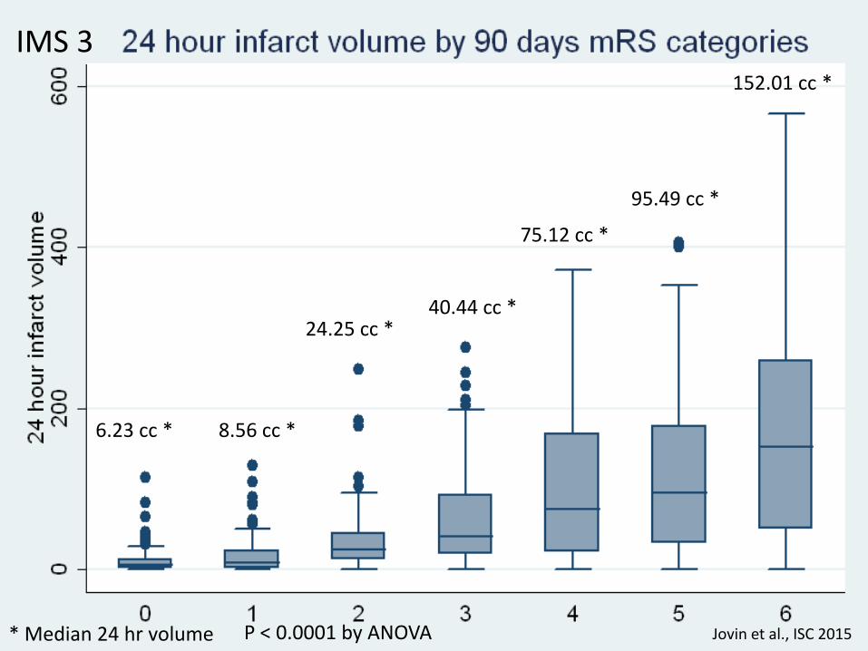
6.23 cc * 8.56 cc *
24.25 cc *40.44 cc *
75.12 cc *
95.49 cc *
152.01 cc *
* Median 24 hr volume P < 0.0001 by ANOVA
IMS 3
Jovin et al., ISC 2015
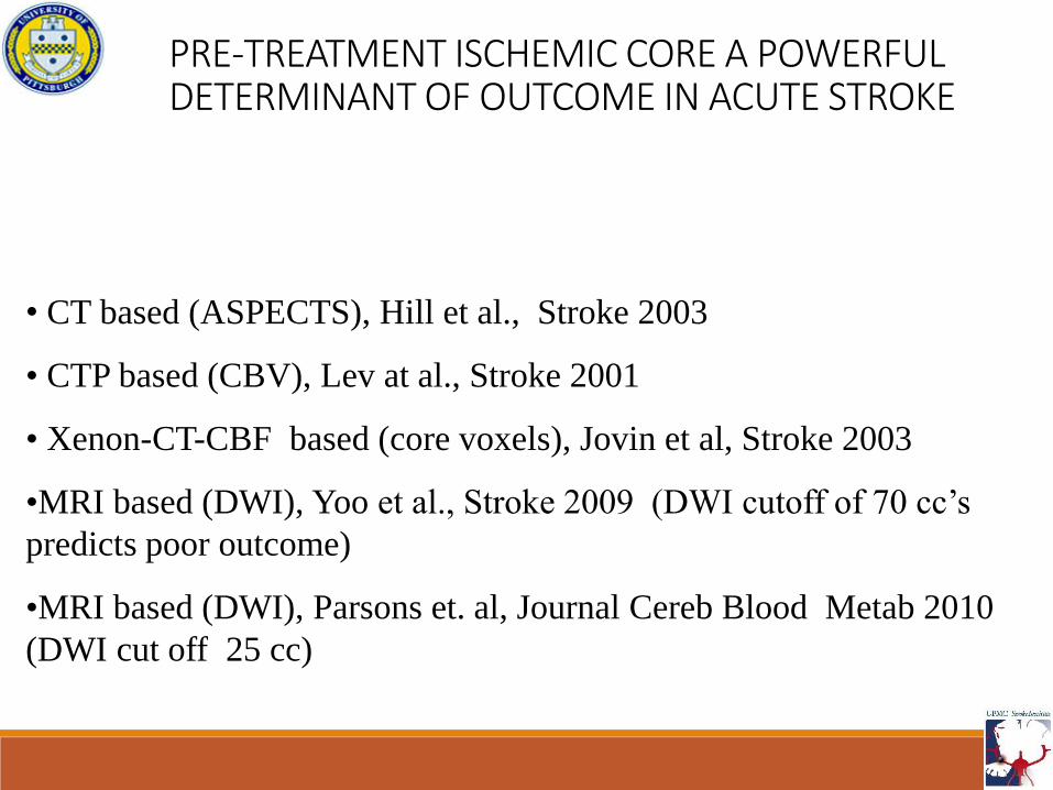
PRE-TREATMENT ISCHEMIC CORE A POWERFUL DETERMINANT OF OUTCOME IN ACUTE STROKE
• CT based (ASPECTS), Hill et al., Stroke 2003
• CTP based (CBV), Lev at al., Stroke 2001
• Xenon-CT-CBF based (core voxels), Jovin et al, Stroke 2003
•MRI based (DWI), Yoo et al., Stroke 2009 (DWI cutoff of 70 cc’s
predicts poor outcome)
•MRI based (DWI), Parsons et. al, Journal Cereb Blood Metab 2010
(DWI cut off 25 cc)
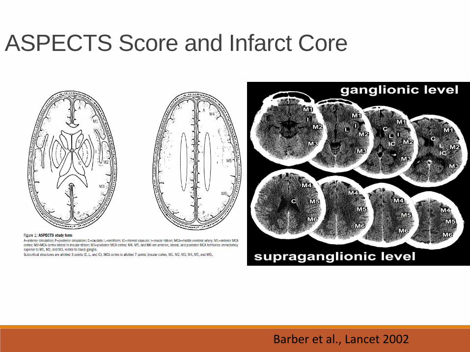
ASPECTS Score and Infarct Core
Barber et al., Lancet 2002
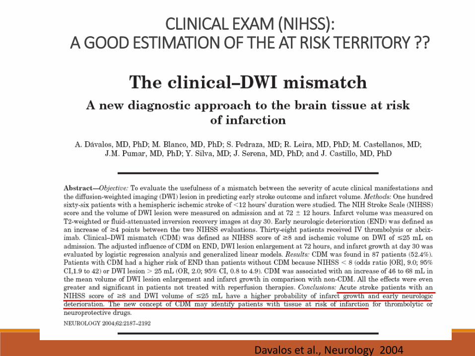
CLINICAL EXAM (NIHSS): A GOOD ESTIMATION OF THE AT RISK TERRITORY ??
Davalos et al., Neurology 2004
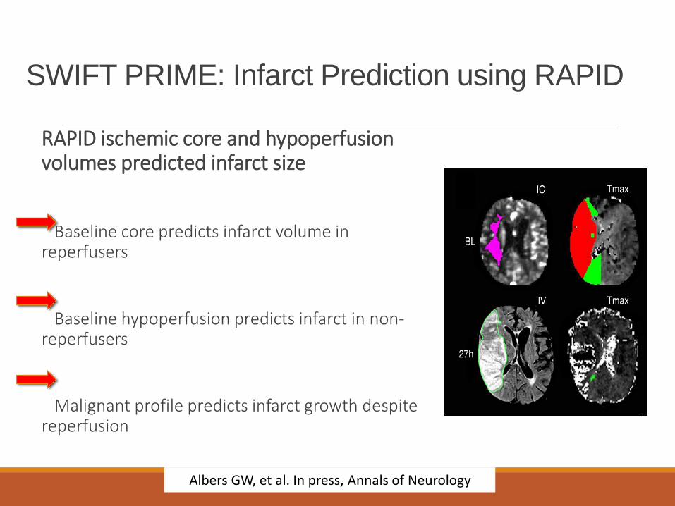
SWIFT PRIME: Infarct Prediction using RAPID
RAPID ischemic core and hypoperfusionvolumes predicted infarct size
Baseline core predicts infarct volume in reperfusers
Baseline hypoperfusion predicts infarct in non-reperfusers
Malignant profile predicts infarct growth despite reperfusion
Albers GW, et al. In press, Annals of Neurology
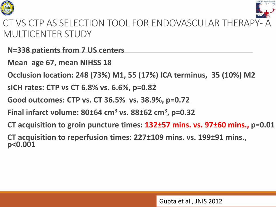
CT VS CTP AS SELECTION TOOL FOR ENDOVASCULAR THERAPY- A MULTICENTER STUDY
N=338 patients from 7 US centers
Mean age 67, mean NIHSS 18
Occlusion location: 248 (73%) M1, 55 (17%) ICA terminus, 35 (10%) M2
sICH rates: CTP vs CT 6.8% vs. 6.6%, p=0.82
Good outcomes: CTP vs. CT 36.5% vs. 38.9%, p=0.72
Final infarct volume: 80±64 cm3 vs. 88±62 cm3, p=0.32
CT acquisition to groin puncture times: 132±57 mins. vs. 97±60 mins., p=0.01
CT acquisition to reperfusion times: 227±109 mins. vs. 199±91 mins., p<0.001
Gupta et al., JNIS 2012
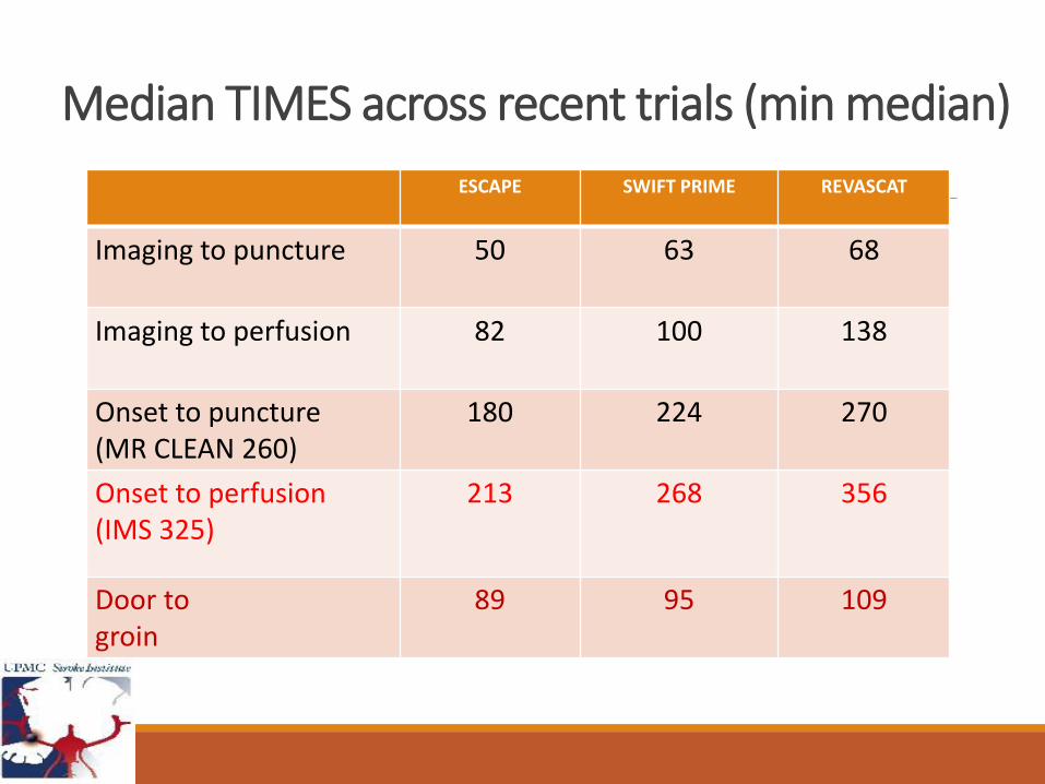
Median TIMES across recent trials (min median)
ESCAPE SWIFT PRIME REVASCAT
Imaging to puncture 50 63 68
Imaging to perfusion 82 100 138
Onset to puncture(MR CLEAN 260)
180 224 270
Onset to perfusion(IMS 325)
213 268 356
Door to groin
89 95 109
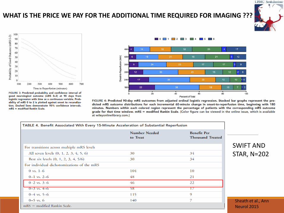
WHAT IS THE PRICE WE PAY FOR THE ADDITIONAL TIME REQUIRED FOR IMAGING ???
SWIFT AND STAR, N=202
Sheath et al., Ann Neurol 2015
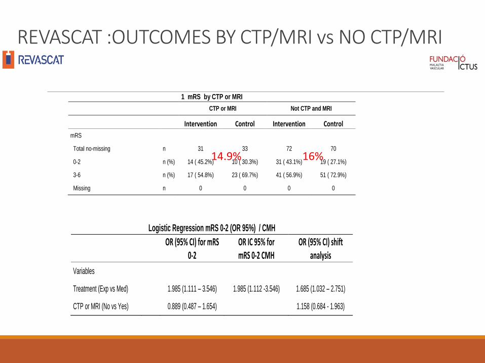
REVASCAT :OUTCOMES BY CTP/MRI vs NO CTP/MRI
Logistic Regression mRS 0-2 (OR 95%) / CMH
OR (95% CI) for mRS 0-2
OR IC 95% for mRS 0-2 CMH
OR (95% CI) shift analysis
Variables
Treatment (Exp vs Med) 1.985 (1.111 – 3.546) 1.985 (1.112 -3.546) 1.685 (1.032 – 2.751)
CTP or MRI (No vs Yes) 0.889 (0.487 – 1.654) 1.158 (0.684 - 1.963)
1 mRS by CTP or MRI
CTP or MRI Not CTP and MRI
Intervention Control Intervention Control
mRS
Total no-missing n 31 33 72 70
0-2 n (%) 14 ( 45.2%) 10 ( 30.3%) 31 ( 43.1%) 19 ( 27.1%)
3-6 n (%) 17 ( 54.8%) 23 ( 69.7%) 41 ( 56.9%) 51 ( 72.9%)
Missing n 0 0 0 0
14.9% 16%
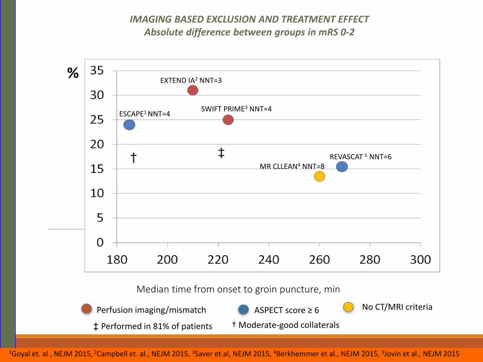
IMAGING BASED EXCLUSION AND TREATMENT EFFECTAbsolute difference between groups in mRS 0-2
%
Median time from onset to groin puncture, min
Perfusion imaging/mismatch ASPECT score ≥ 6 No CT/MRI criteria
†
† Moderate-good collaterals‡ Performed in 81% of patients
‡
ESCAPE1 NNT=4
EXTEND IA2 NNT=3
SWIFT PRIME3 NNT=4
REVASCAT 5 NNT=6MR CLLEAN4 NNT=8
1Goyal et. al , NEJM 2015, 2Campbell et. al., NEJM 2015, 3Saver et.al, NEJM 2015, 4Berkhemmer et al., NEJM 2015, 5Jovin et al., NEJM 2015
0
5
10
15
20
Death Death (shock
excl.)
Non-Fatal MI CVA Death CVA/MI
PCI Lytic
23 Randomized Trials of PCI vs Lytics:
30 day Events (n=7739)
P=0.0002P=0.0002
P=0.0003P=0.0003 P<0.0001P<0.0001
P=0.0004P=0.0004
P<0.0001P<0.0001
7
9
57
2.5
6.8
1 2
8
14
Fre
qu
en
cy (
%)
Keeley Keeley & Grines,& Grines, Lancet Lancet 2003;361:132003;361:13--2020
ARR 6%, NNT= 29
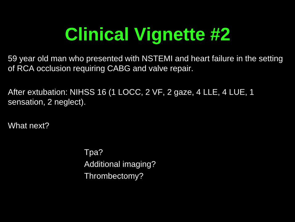
Clinical Vignette #259 year old man who presented with NSTEMI and heart failure in the setting
of RCA occlusion requiring CABG and valve repair.
After extubation: NIHSS 16 (1 LOCC, 2 VF, 2 gaze, 4 LLE, 4 LUE, 1
sensation, 2 neglect).
What next?
Tpa?
Additional imaging?
Thrombectomy?
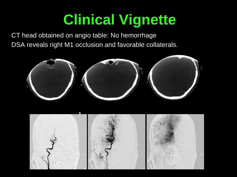
Clinical Vignette CT head obtained on angio table: No hemorrhage
DSA reveals right M1 occlusion and favorable collaterals.
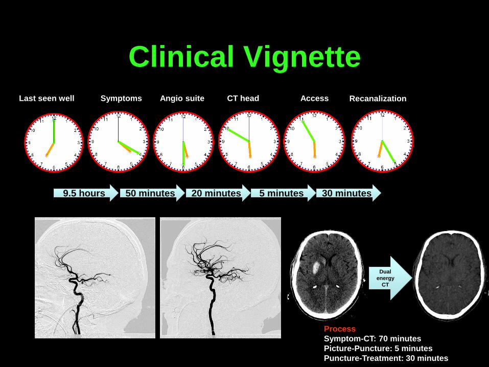
Clinical Vignette Last seen well Symptoms Angio suite CT head Access
9.5 hours 50 minutes 20 minutes 5 minutes 30 minutes
Recanalization
Dual
energy
CT
Process
Symptom-CT: 70 minutes
Picture-Puncture: 5 minutes
Puncture-Treatment: 30 minutes
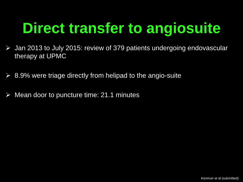
Direct transfer to angiosuite Jan 2013 to July 2015: review of 379 patients undergoing endovascular
therapy at UPMC
8.9% were triage directly from helipad to the angio-suite
Mean door to puncture time: 21.1 minutes
Kenmuir et al (submitted)
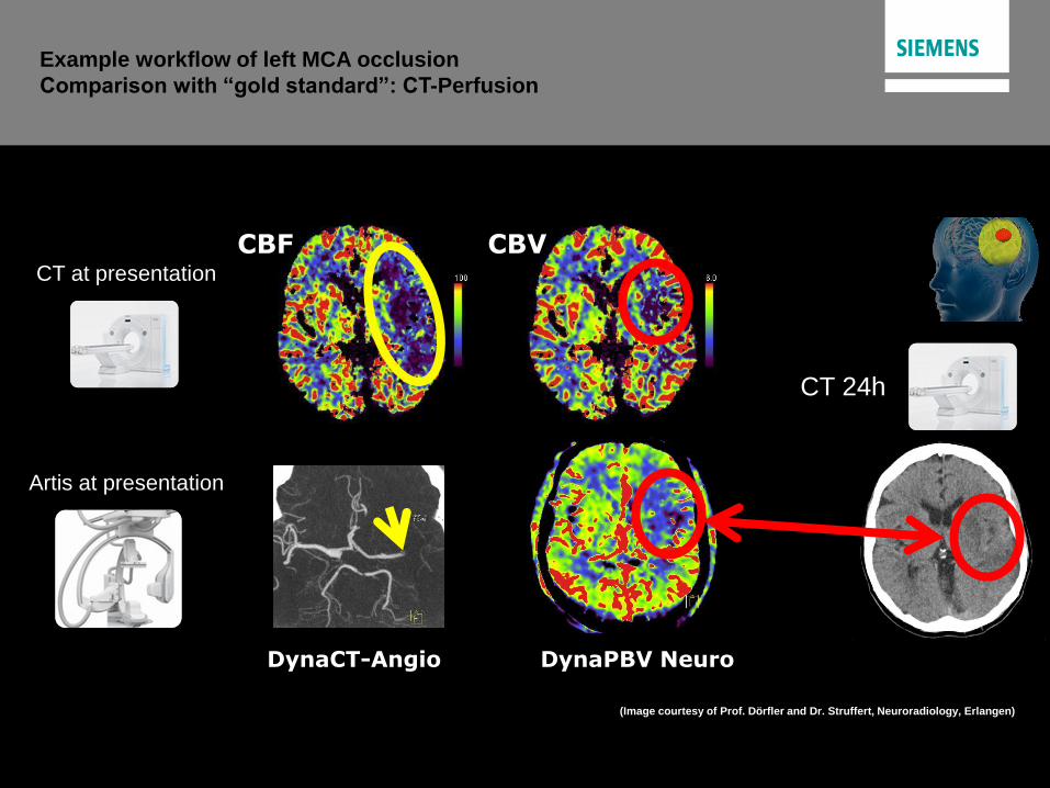
Page 23 Siemens Healthcare, Angiography & Interventional X-Ray Systems
DynaPBV Neuro
Example workflow of left MCA occlusion
Comparison with “gold standard”: CT-Perfusion
DynaCT-Angio
(Image courtesy of Prof. Dörfler and Dr. Struffert, Neuroradiology, Erlangen)
Artis at presentation
Size and location of
“tissue at risk” is known!
Size and location of
“infarct core” is known!
CT at presentation
CBF CBVCBV
CT 24h
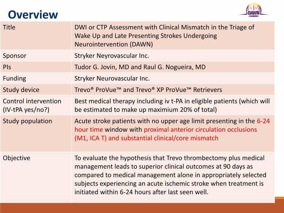
OverviewTitle DWI or CTP Assessment with Clinical Mismatch in the Triage of
Wake Up and Late Presenting Strokes Undergoing Neurointervention (DAWN)
Sponsor Stryker Neyrovascular Inc.
PIs Tudor G. Jovin, MD and Raul G. Nogueira, MD
Funding Stryker Neurovascular Inc.
Study device Trevo® ProVue™ and Trevo® XP ProVue™ Retrievers
Control intervention(IV-tPA yes/no?)
Best medical therapy including iv t-PA in eligible patients (which will be estimated to make up maximium 20% of total)
Study population Acute stroke patients with no upper age limit presenting in the 6-24 hour time window with proximal anterior circulation occlusions (M1, ICA T) and substantial clinical/core mismatch
Objective To evaluate the hypothesis that Trevo thrombectomy plus medical management leads to superior clinical outcomes at 90 days as compared to medical management alone in appropriately selected subjects experiencing an acute ischemic stroke when treatment is initiated within 6-24 hours after last seen well.
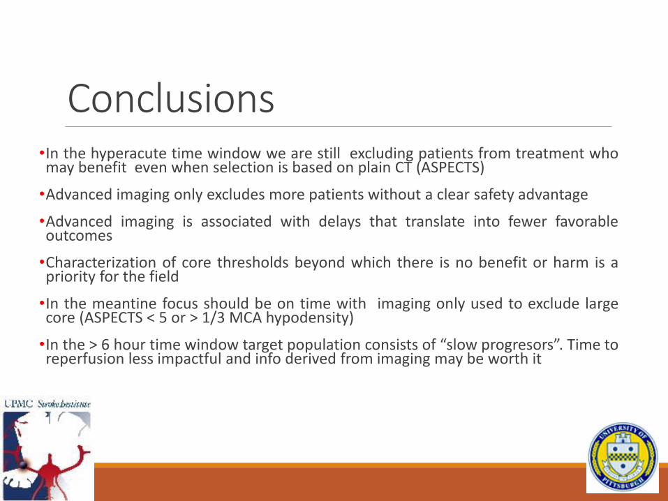
Conclusions•In the hyperacute time window we are still excluding patients from treatment whomay benefit even when selection is based on plain CT (ASPECTS)
•Advanced imaging only excludes more patients without a clear safety advantage
•Advanced imaging is associated with delays that translate into fewer favorableoutcomes
•Characterization of core thresholds beyond which there is no benefit or harm is apriority for the field
•In the meantine focus should be on time with imaging only used to exclude largecore (ASPECTS < 5 or > 1/3 MCA hypodensity)
•In the > 6 hour time window target population consists of “slow progresors”. Time toreperfusion less impactful and info derived from imaging may be worth it
Stroke/Interventional Neurology
◦ Tudor Jovin, MD
◦ Ashu Jadhav, MD PhD
◦ Lawrence Wechsler, MD
◦ Maxim Hammer, MD
◦ Vivek Reddy, MD
◦ Matt Starr, MD
◦ Viktoria Totoraitis, MD
◦ Nima Aghaebrahim, mD
◦ Dan Victor Giurgiutiu, MD
◦ Vascular Endovascular Neurosurgery
◦ Brian Jankowitz, MD
◦ Andrew Ducruet, MD
◦ Robert Friedlander, MD
◦ Paul Gardner, MD
◦ Dan Wecht, MD
◦ Stroke Institute Nursing Staff
◦ Lori Massaro – CRNP
◦ Susan Kim - CRNP
◦ Maria Abraham – PA
◦ Christina Bonaccorsi – PA
◦ Kathy Seiler, RN
◦ Cherie Adams RN
◦ Jonya Brooks, RN
◦ Ken Coval ,RN
◦ Patti Williams, RN
◦ Neurocritical Care
◦ Brad Molyneaux, MD
◦ Lori Shutter, MD
◦ Sherry Chou, MD
◦ Ruchi Jha, MD
◦ Kees Polderman, MD
◦ Neuroanesthesia
◦ Coleen Moran, MD
◦ Frank Gyulai, MD
◦ Theresa Gelzinis , MD
Emergency Medicine
◦ Charissa Pacella, MD
◦ Frank Guyette, MD
◦ Chris Gill-Martin, MD
◦ Cliff Callaway, MD
◦ Don Yealy, MD
◦ Mike Turturro, MD
◦ Maria Guyette, MD
◦ Neuroradiology
◦ Bill Delfyett, MD
◦ Char Branstetter, MD
◦ Emanuel Kanal, MD
◦ Stroke Institute Research Staff
◦ Lisa Baxendell – Research coord
◦ Carlynn Graves – Research coord
◦ Patricia Porter – Research coord
◦ Kara Armbruster – Research coord
◦ Yvonne Cannon – Research coord
◦ Holly Kromer- Data Management
◦ NeuroIR Radiology Techs & Nursing Staff
◦ Kitty O’Toole, RTVI
◦ Casey Foster, RTVI
◦ Jennifer Hamil, RTVI
◦ Jim Bozak,RTVI
◦ Jason Paul,RTVI
◦ Candace Acklin, RN
◦ Josie Stashko, RN
◦ Neurorahabiltiation
◦ Jennifer Shen
◦ Cara Camiolo
◦ Mike Bonimnger
◦ Neuroscience Nursing
◦ Melanie Smith, RN
◦ Ben Morrow, RN
◦ Kate Spiering, RN
◦ Therese Dawson, RN
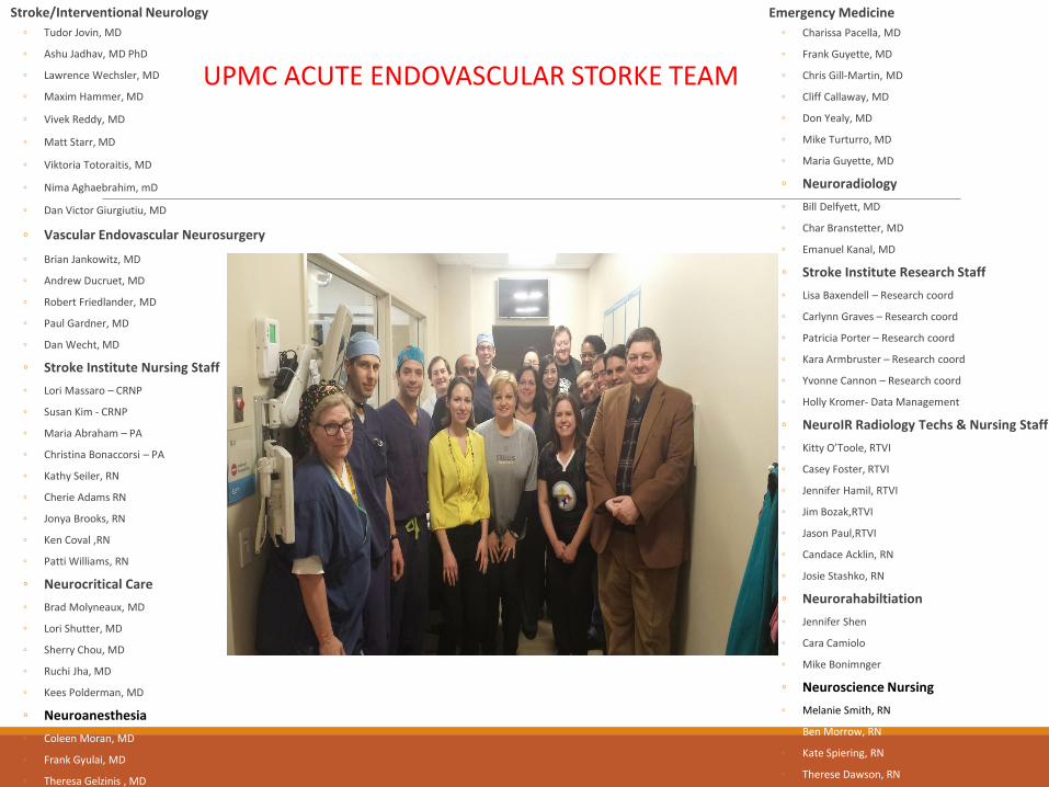
UPMC ACUTE ENDOVASCULAR STORKE TEAM


![Endovascular therapy in acute ischemic stroke: where we ...neurology.mcgill.ca/neurodocs/AHD 2010-2011... · standard of care in many stroke centers across the globe [18–20]. Endovascular](https://img.pdfslide.us/doc/110x75/5ea01a4853f7473169025d9d/endovascular-therapy-in-acute-ischemic-stroke-where-we-2010-2011-standard.jpg)