Embed Size (px)
Citation preview
Neuron
Article
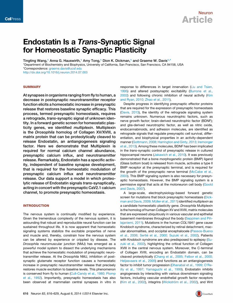
Endostatin Is a Trans-Synaptic Signalfor Homeostatic Synaptic PlasticityTingting Wang,1 Anna G. Hauswirth,1 Amy Tong,1 Dion K. Dickman,1 and Graeme W. Davis1,*1Department of Biochemistry and Biophysics, University of California, San Francisco, San Francisco, CA 94158, USA
*Correspondence: [email protected]
http://dx.doi.org/10.1016/j.neuron.2014.07.003
SUMMARY
At synapses inorganisms ranging fromfly tohuman, adecrease in postsynaptic neurotransmitter receptorfunction elicits a homeostatic increase in presynapticrelease that restores baseline synaptic efficacy. Thisprocess, termed presynaptic homeostasis, requiresa retrograde, trans-synaptic signal of unknown iden-tity. In a forward genetic screen for homeostatic plas-ticity genes, we identified multiplexin. Multiplexinis the Drosophila homolog of Collagen XV/XVIII, amatrix protein that can be proteolytically cleaved torelease Endostatin, an antiangiogenesis signalingfactor. Here we demonstrate that Multiplexin isrequired for normal calcium channel abundance,presynaptic calcium influx, and neurotransmitterrelease. Remarkably, Endostatin has a specific activ-ity, independent of baseline synapse development,that is required for the homeostatic modulation ofpresynaptic calcium influx and neurotransmitterrelease. Our data support a model in which proteo-lytic release of Endostatin signals trans-synaptically,acting in concertwith thepresynapticCaV2.1 calciumchannel, to promote presynaptic homeostasis.
INTRODUCTION
The nervous system is continually modified by experience.
Given the tremendous complexity of the nervous system, it is
astounding that robust and reproducible neural function can be
sustained throughout life. It is now apparent that homeostatic
signaling systems stabilize the excitable properties of nerve
and muscle and, thereby, constrain how the nervous system
can be altered by experience or crippled by disease. The
Drosophila neuromuscular junction (NMJ) has emerged as a
powerful model system to dissect the underlying mechanisms
that achieve the homeostatic modulation of presynaptic neuro-
transmitter release. At the Drosophila NMJ, inhibition of post-
synaptic glutamate receptor function causes a homeostatic
increase in presynaptic neurotransmitter release that precisely
restores muscle excitation to baseline levels. This phenomenon
is conserved from fly to human (Cull-Candy et al., 1980; Plomp
et al., 1992). Importantly, presynaptic homeostasis has also
been observed at mammalian central synapses in vitro in
616 Neuron 83, 616–629, August 6, 2014 ª2014 Elsevier Inc.
response to differences in target innervation (Liu and Tsien,
1995) and altered postsynaptic excitability (Burrone et al.,
2002) and following chronic inhibition of neural activity (Kim
and Ryan, 2010; Zhao et al., 2011).
Despite progress in identifying presynaptic effector proteins
that are required for the expression of presynaptic homeostasis
(Davis, 2013), the identity of the retrograde signaling system
remains unknown. Numerous neurotrophic factors, such as
nerve growth factor; brain-derived neurotrophic factor (BDNF);
and glia-derived neurotrophic factor, as well as nitric oxide,
endocannabinoids, and adhesion molecules, are identified as
retrograde signals that regulate presynaptic cell survival, differ-
entiation, and biophysical properties in an activity-dependent
manner (Gottmann, 2008; Harrington andGinty, 2013; Iremonger
et al., 2013). Among thesemolecules, BDNF has been implicated
in the trans-synaptic control of presynaptic release in cultured
hippocampal neurons (Jakawich et al., 2010). It was previously
demonstrated that a bone morphogenetic protein (BMP) ligand
(Glass bottom boat) is released from muscle, activates a type II
BMP receptor at the presynaptic terminal, and is required for
the growth of the presynaptic nerve terminal (McCabe et al.,
2003). This BMP signaling system is also necessary for presyn-
aptic homeostasis. However, the BMP signaling system is a
permissive signal that acts at the motoneuron cell body (Goold
and Davis, 2007).
A large-scale, electrophysiology-based forward genetic
screen for mutations that block presynaptic homeostasis (Dick-
man and Davis, 2009; Muller et al., 2011) identifiedmultiplexin as
a candidate homeostatic plasticity gene. Drosophila Multiplexin
is the homolog of humanCollagen XV and XVIII, matrixmolecules
that are expressed ubiquitously in various vascular and epithelial
basement membranes throughout the body (Seppinen and Pih-
lajaniemi, 2011). Mutations in the human COL18A1 gene cause
Knobloch syndrome, characterized by retinal detachment, mac-
ular abnormalities, and occipital encephalocele (Passos-Bueno
et al., 2006; Sertie et al., 2000; Suzuki et al., 2002). Patients
with Knobloch syndrome are also predisposed to epilepsy (Su-
zuki et al., 2002), highlighting the critical function of Collagen
XVIII in the central nervous system. Moreover, the C-terminal
of Collagen XVIII, encoding an Endostatin domain, can be
cleaved proteolytically (Chang et al., 2005; Felbor et al., 2000;
Heljasvaara et al., 2005) and functions as an antiangiogenesis
factor to inhibit tumor progression (Dhanabal et al., 1999; O’Re-
illy et al., 1997; Yamaguchi et al., 1999). Endostatin inhibits
angiogenesis by interacting with various downstream signaling
factors, including vascular endothelial growth factor receptors
(Kim et al., 2002), integrins (Wickstrom et al., 2002), and Wnt
A
B
C
D E
F G H
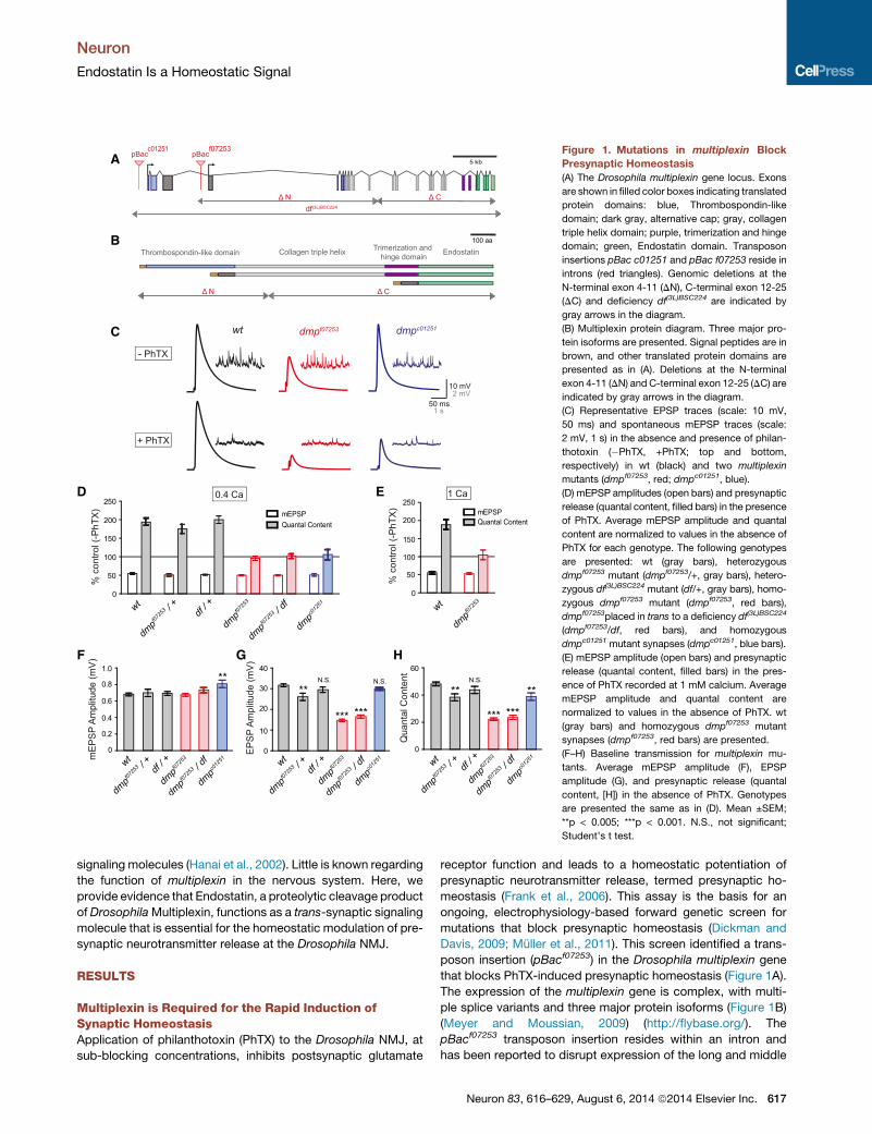
Figure 1. Mutations in multiplexin Block
Presynaptic Homeostasis
(A) The Drosophila multiplexin gene locus. Exons
are shown in filled color boxes indicating translated
protein domains: blue, Thrombospondin-like
domain; dark gray, alternative cap; gray, collagen
triple helix domain; purple, trimerization and hinge
domain; green, Endostatin domain. Transposon
insertions pBac c01251 and pBac f07253 reside in
introns (red triangles). Genomic deletions at the
N-terminal exon 4-11 (DN), C-terminal exon 12-25
(DC) and deficiency df(3L)BSC224 are indicated by
gray arrows in the diagram.
(B) Multiplexin protein diagram. Three major pro-
tein isoforms are presented. Signal peptides are in
brown, and other translated protein domains are
presented as in (A). Deletions at the N-terminal
exon 4-11 (DN) and C-terminal exon 12-25 (DC) are
indicated by gray arrows in the diagram.
(C) Representative EPSP traces (scale: 10 mV,
50 ms) and spontaneous mEPSP traces (scale:
2 mV, 1 s) in the absence and presence of philan-
thotoxin (�PhTX, +PhTX; top and bottom,
respectively) in wt (black) and two multiplexin
mutants (dmpf07253, red; dmpc01251, blue).
(D) mEPSP amplitudes (open bars) and presynaptic
release (quantal content, filled bars) in the presence
of PhTX. Average mEPSP amplitude and quantal
content are normalized to values in the absence of
PhTX for each genotype. The following genotypes
are presented: wt (gray bars), heterozygous
dmpf07253 mutant (dmpf07253/+, gray bars), hetero-
zygous df(3L)BSC224 mutant (df/+, gray bars), homo-
zygous dmpf07253 mutant (dmpf07253, red bars),
dmpf07253placed in trans to a deficiency df(3L)BSC224
(dmpf07253/df, red bars), and homozygous
dmpc01251 mutant synapses (dmpc01251, blue bars).
(E) mEPSP amplitude (open bars) and presynaptic
release (quantal content, filled bars) in the pres-
ence of PhTX recorded at 1 mM calcium. Average
mEPSP amplitude and quantal content are
normalized to values in the absence of PhTX. wt
(gray bars) and homozygous dmpf07253 mutant
synapses (dmpf07253, red bars) are presented.
(F–H) Baseline transmission for multiplexin mu-
tants. Average mEPSP amplitude (F), EPSP
amplitude (G), and presynaptic release (quantal
content, [H]) in the absence of PhTX. Genotypes
are presented the same as in (D). Mean ±SEM;
**p < 0.005; ***p < 0.001. N.S., not significant;
Student’s t test.
Neuron
Endostatin Is a Homeostatic Signal
signaling molecules (Hanai et al., 2002). Little is known regarding
the function of multiplexin in the nervous system. Here, we
provide evidence that Endostatin, a proteolytic cleavage product
of DrosophilaMultiplexin, functions as a trans-synaptic signaling
molecule that is essential for the homeostatic modulation of pre-
synaptic neurotransmitter release at the Drosophila NMJ.
RESULTS
Multiplexin is Required for the Rapid Induction ofSynaptic HomeostasisApplication of philanthotoxin (PhTX) to the Drosophila NMJ, at
sub-blocking concentrations, inhibits postsynaptic glutamate
receptor function and leads to a homeostatic potentiation of
presynaptic neurotransmitter release, termed presynaptic ho-
meostasis (Frank et al., 2006). This assay is the basis for an
ongoing, electrophysiology-based forward genetic screen for
mutations that block presynaptic homeostasis (Dickman and
Davis, 2009; Muller et al., 2011). This screen identified a trans-
poson insertion (pBacf07253) in the Drosophila multiplexin gene
that blocks PhTX-induced presynaptic homeostasis (Figure 1A).
The expression of the multiplexin gene is complex, with multi-
ple splice variants and three major protein isoforms (Figure 1B)
(Meyer and Moussian, 2009) (http://flybase.org/). The
pBacf07253 transposon insertion resides within an intron and
has been reported to disrupt expression of the long and middle
Neuron 83, 616–629, August 6, 2014 ª2014 Elsevier Inc. 617
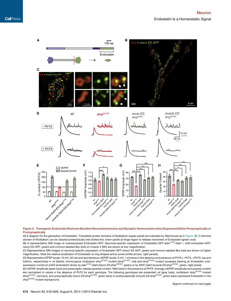
Figure 2. Transgenic Endostatin Restores BaselineNeurotransmission and Synaptic Homeostasis when Expressed Either Presynaptically or
Postsynaptically
(A) A diagram for the generation of Endostatin. Translated protein domains of Multiplexin (upper panel) are indicated by filled boxes as in Figure 1B. C-terminal
domain of Multiplexin can be cleaved proteolytically (red dotted line, lower panel) at hinge region to release monomers of Endostatin (green oval).
(B) A representative SIM image of overexpressed Endostatin-GFP. Neuronal-specific expression of Endostatin-GFP (elavC155-Gal4 > UAS-endostatin-GFP,
neuro ES-GFP, green) and immuno-labeled Brp (red) on muscle 4 (M4) are shown at low magnification.
(C) Representative SIM images of neuronal specific expression of Endostatin-GFP (neuro ES-GFP, green) and immuno-labeled Brp (red) are shown at higher
magnification. Note the relative localization of Endostatin to ring-shaped active zones (white arrows, right panels).
(D) Representative EPSP (scale: 10 mV, 50 ms) and spontaneous mEPSP (scale: 2 mV, 1 s) traces in the absence and presence of PhTX (�PhTX, +PhTX; top and
bottom, respectively) in wt (black), homozygous multiplexin dmpf07253 mutant (dmpf07253, red) and dmpf07253 mutant synapses bearing an Endostatin over-
expression construct (UAS-endostatin) driven by elavC155-Gal4 (neuro ES;dmpf07253, green) or by MHC-Gal4 (muscle ES;dmpf07253, green, right panel).
(E) mEPSP amplitude (open bars) and presynaptic release (quantal content, filled bars) in the presence of PhTX. Average mEPSP amplitude and quantal content
are normalized to values in the absence of PhTX for each genotype. The following genotypes are presented: wt (gray bars), multiplexin dmpf07253 mutant
(dmpf07253, red bars), and presynaptically (neuro ES;dmpf07253, green bars) or postsynaptically (muscle ES;dmpf07253, green bars) expressed Endostatin in the
dmpf07253 mutant background.
(legend continued on next page)
Neuron
Endostatin Is a Homeostatic Signal
618 Neuron 83, 616–629, August 6, 2014 ª2014 Elsevier Inc.
Neuron
Endostatin Is a Homeostatic Signal
isoforms of Multiplexin, indicating it is a loss-of-function allele
(Momota et al., 2011).
First, we confirmed that loss of multiplexin disrupts the rapid
induction of synaptic homeostasis. Bath application of 20 mM
PhTX induced a significant decrease in mEPSP amplitude at
both wild-type (wt) and dmpf07253 mutant synapses (Figures 1C
and 1D). At wt synapses, an increase in presynaptic release (Fig-
ures 1C and 1D; quantal content) was observed to offset the
change in mEPSP amplitude and restore postsynaptic excitation
to baseline values (Figure 1C). By contrast, following application
of PhTX to the dmpf07253mutant, there was no change in quantal
content, and EPSPs were significantly smaller than that
observed in the dmpf07253 mutant in the absence of PhTX (Fig-
ures 1C and 1D). Presynaptic homeostasis was also blocked
when dmpf07253was placed in trans to a deficiency [Df(3L)
BSC224] that uncovers the entire multiplexin locus (Figure 1D).
Thus, loss-of-functionmutations inmultiplexin block presynaptic
homeostasis. Throughout this paper, average values are pre-
sented in figures with sample sizes reported in figure legends.
Absolute values of mEPSP amplitudes, EPSP amplitudes, and
quantal contents for each data set are included in Table S1
(available online).
In these initial experiments, we discovered that baseline
EPSP amplitudes and quantal contents recorded in
dmpf07253and dmpf07253/df mutant synapses were significantly
decreased compared to wt and heterozygous controls (Fig-
ures 1G and 1H; p < 0.001). Since there was no change in
average mEPSP amplitude, the data imply a defect in baseline
presynaptic release (Figure 1F). However, analysis of a sec-
ond transposon insertion mutation, dmpc01251, demonstrates
that synaptic homeostasis is blocked without a concomitant
change in baseline release (Figure 1A; http://flybase.org/). At
dmpc01251 mutant synapses, there was a minor deficit in
baseline spontaneous mEPSP amplitude (Figure 1F; p <
0.005) and quantal content (Figure 1H; p < 0.005), but the
average EPSP amplitude is similar to wt (Figure 1G; N.S.,
p = 0.24). When PhTX was applied to the dmpc01251 synap-
ses, homeostasis was blocked. Specifically, the average
mEPSP amplitude at dmpc01251 synapses was decreased by
�50%, but quantal content was unaltered (Figures 1C and
1D). Finally, to address whether the efficient induction of pre-
synaptic homeostasis is sensitive to the extracellular concen-
tration of calcium, we assayed presynaptic homeostasis at
dmpf07253 mutant synapses at an elevated extracellular cal-
cium concentration (1 mM). Raising extracellular calcium in-
creases the average EPSP amplitude from �15 mV to
�30 mV at dmpf07253 synapses, a value similar to wt at
0.4 mM extracellular calcium, but presynaptic homeostasis re-
mained blocked (Figure 1E). Taken together, these data
demonstrate that Multiplexin is necessary for presynaptic ho-
meostasis and has a separable, required function to promote
baseline presynaptic neurotransmitter release (see below for
additional evidence).
(F–H) Average mEPSP amplitude (F), EPSP amplitude (G), and presynaptic relea
presented: wt (gray bars), multiplexin dmpf07253 mutant (dmpf07253, red bars), n
Endostatin in dmpf07253 mutant background (neuro ES;dmpf07253, green bars),
expressed Endostatin in the dmpf07253 mutant background (muscle ES;dmpf0725
Endostatin Is Sufficient to Rescue SynapticHomeostasis in multiplexin MutantsMultiplexin is the Drosophila homolog of human Collagen
XV/XVIII. In mammals, Collagen XVIII is proteolytically cleaved
to release a C-terminal domain termed ‘‘Endostatin,’’ which
has been shown to have antiangiogenic activity (Figure 2A) (Myl-
lyharju and Kivirikko, 2004; O’Reilly et al., 1997). DrosophilaMul-
tiplexin also contains a C-terminal Endostatin domain and is
expressed in both neurons and muscles (Momota et al., 2011).
In order to test whether Drosophila Endostatin might participate
in presynaptic homeostasis, we attempted to rescuemultiplexin
mutants by overexpressing a UAS-endostatin transgene either
presynaptically or postsynaptically.
First, we demonstrate that Endostatin is trafficked to the NMJ
(Figures 2B and 2C). We generated and expressed a UAS-endo-
statin-GFP transgene in wt animals using pan-neuronal
(elavC155-Gal4) or muscle-specific drivers (MHC-Gal4). Struc-
tured Illumination Microscopy (SIM) images were acquired
from synapses colabeled for the active zone marker Bruchpilot
(Brp) and Endostatin-GFP (ES-GFP) (Figures 2B, 2C, and S1A).
We find that presynaptically expressed Endostatin-GFP forms
punctate structures that are distributed throughout the presyn-
aptic nerve terminal (Figure 2B). In some cases, Endostatin-
GFP is localized in the center of ring-shaped active zones
(Figure 2C, right upper panels) facing the extracellular space
(Figure 2C, right lower panels), indicating that Endostatin-GFP
is trafficked to synapses. When expressed by a muscle-specific
driver (MHC-Gal4), Endostatin-GFP is found across the entire
muscle, without enrichment at the postsynaptic NMJ (Fig-
ure S1A). In order to determine whether Endostatin-GFP is ex-
pressed and secreted to the extracellular matrix, we performed
immunolabeling to examine the surface expression of Endosta-
tin-GFP achieved by staining without permeabilizing the cell
(Figures S1B–S1D). We find that Endostatin-GFP is expressed
and forms punctuate structures on the cell surface when
expressed presynaptically (Figure S1B; elavC155-Gal4 > UAS-
endostatin-GFP) or postsynaptically (Figure S1C; BG57-Ga-
l4>UAS-endostatin-GFP). As a control, we showed that a
GFP-tagged cytoplasmic protein is not detectable in the
absence of cell permeabilization (Figure S1D; elavC155-Gal4 >
UAS-s6k-GFP).
Next, we examined whether overexpression of Endostatin
can functionally restore baseline transmission and synaptic
homeostasis in multiplexin mutants. We expressed Endostatin
(UAS-endostatin; Meyer and Moussian, 2009) in neurons and
muscles of dmpf07253 mutant animals, using either pan-
neuronal (elavC155-Gal4) or muscle-specific (MHC-Gal4) drivers
(Figures 2D–2H). First, we find that both neuron-specific and
muscle-specific expression of Endostatin rescues baseline
neurotransmission at the dmpf07253 mutant NMJ (Figures 2D
and 2F–2H). Second, we find that both neuron-specific and
muscle-specific expression of Endostatin rescues PhTX-
induced synaptic homeostasis at the dmpf07253 mutant NMJ
se (quantal content, [H]) in the absence of PhTX. The following genotypes are
eural-specific Gal4 driver (neuro-Gal4, gray bars), presynaptically expressed
muscle-specific Gal4 driver (muscle-Gal4, gray bars), and postsynaptically3, green bars). Mean ±SEM; *p < 0.05; ***p < 0.001; Student’s t test.
Neuron 83, 616–629, August 6, 2014 ª2014 Elsevier Inc. 619
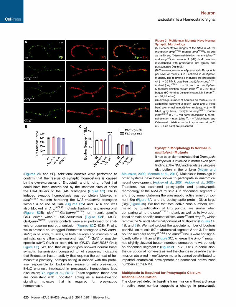
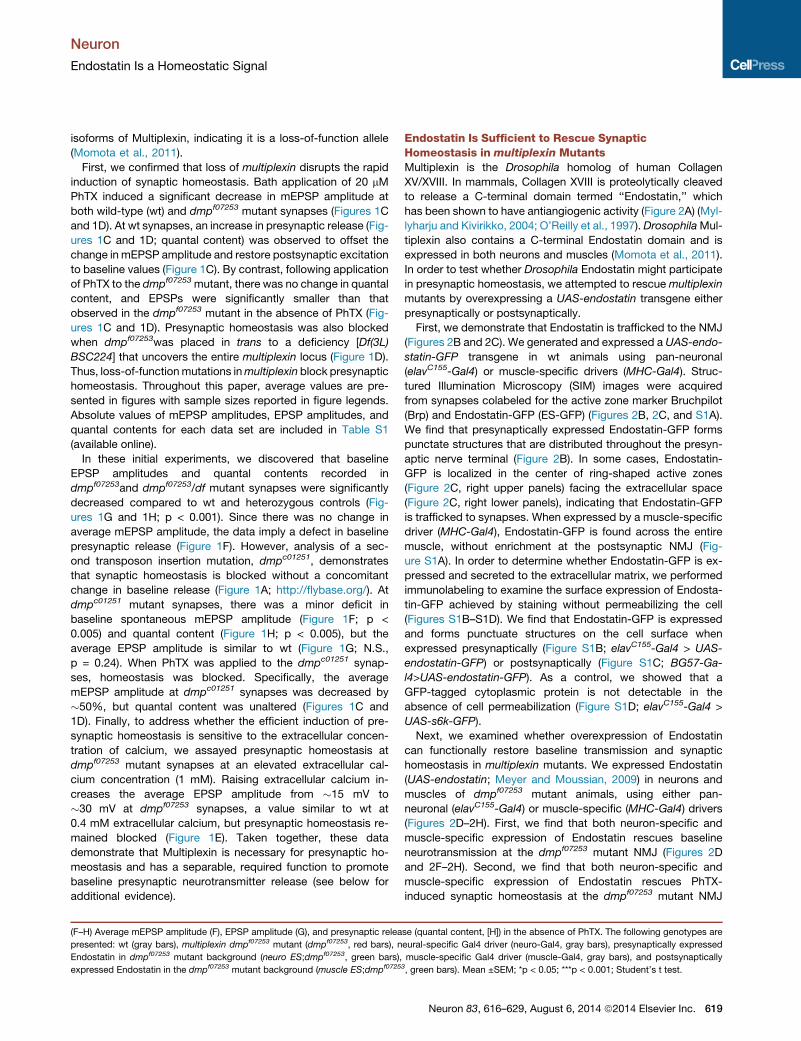
Figure 3. Multiplexin Mutants Have Normal
Synaptic Morphology
(A) Representative images of the NMJ in wt, the
multiplexin dmpf07253 mutant (dmpf07253), as well
as the N- and C-terminal deletion mutants (dmpDN
and dmpDC) on muscle 4 (M4). NMJ are im-
munolabeled with presynaptic Brp (green) and
postsynaptic Dlg (red).
(B) The average number of presynaptic Brp puncta
per NMJ at muscle 4 is unaltered in multiplexin
mutants. The following genotypes are presented:
wt (n = 26 NMJ, gray bar), multiplexin dmpf07253
mutant (dmpf07253, n = 19, red bar), multiplexin
N-terminal deletion mutant (dmpDN, n = 20, blue
bar), and C-terminal deletion mutant NMJ (dmpDC,
n = 18, blue bar).
(C) Average number of boutons on muscle 6/7 in
abdominal segment 2 (open bars) and 3 (filled
bars) are normal in multiplexin mutants. wt (n = 19
NMJ, gray bars), multiplexin dmpf07253 mutant
(dmpf07253, n = 19, red bars), multiplexin N-termi-
nal deletion mutant (dmpDN, n = 7, blue bars), and
C-terminal deletion mutant synapses (dmpDC,
n = 8, blue bars) are presented.
Neuron
Endostatin Is a Homeostatic Signal
(Figures 2D and 2E). Additional controls were performed to
confirm that the rescue of synaptic homeostasis is caused
by the overexpression of Endostatin and is not an effect that
could have been contributed by the insertion sites of either
the Gal4 drivers or the UAS transgene (Figure S2). PhTX-
induced synaptic homeostasis was completely blocked in
dmpf07253 mutants harboring the UAS-endostatin transgene
without a source of Gal4 (Figures S2A and S2B) and was
also blocked in dmpf07253 mutants harboring a pan-neuronal
(Figure S2B; elavC155-Gal4;;dmpf07253) or muscle-specific
Gal4 driver without UAS-endostatin (Figure S2B, MHC-
Gal4,dmpf07253). Similar controls were also performed for anal-
ysis of baseline neurotransmission (Figures S2C–S2E). Finally,
we expressed an untagged Endostatin transgene (UAS-endo-
statin) in neurons, muscles, or both neurons and muscles of wt
animals, using either pan-neuronal (elavC155-Gal4) or muscle-
specific (MHC-Gal4) or both drivers (OK371-Gal4;BG57-Gal4;
Figure S3). We find that all genotypes showed normal basal
synaptic transmission compared to wt synapses, indicating
that Endostatin has an activity that requires the context of ho-
meostatic plasticity, perhaps acting in concert with the prote-
ase responsible for Endostatin release or with presynaptic
ENaC channels implicated in presynaptic homeostasis (see
discussion; Younger et al., 2013). Taken together, these data
are consistent with Endostatin acting as a trans-synaptic
signaling molecule that is required for presynaptic
homeostasis.
620 Neuron 83, 616–629, August 6, 2014 ª2014 Elsevier Inc.
Synaptic Morphology Is Normal inmultiplexin MutantsIt has been demonstrated thatDrosophila
multiplexin is involved in motor axon path
finding at the NMJ and regulates wingless
distribution in the embryo (Meyer and
Moussian, 2009; Momota et al., 2011). Multiplexin homologs in
other systems have been shown to participate in anatomical
neural development (Ackley et al., 2001; Ackley et al., 2003).
Therefore, we examined presynaptic and postsynaptic
morphology at the NMJ of muscle 4 in abdominal segment 2
and 3 by immunolabeling the presynaptic active zone compo-
nent Brp (Figure 3A) and the postsynaptic protein Discs-large
(Dlg) (Figure 3A). We find that total active zone numbers, esti-
mated by quantification of Brp puncta, are similar when
comparing wt to the dmpf07253 mutant, as well as to two addi-
tional domain-specific mutant alleles, dmpDN and dmpDC, which
remove the N- andC-terminal portions ofMultiplexin (Figures 1A,
1B, and 3B). We next probed the absolute number of boutons
per NMJ on muscle 6/7 at abdominal segment 2 and 3. The total
bouton numbers at dmpf07253 and dmpDN NMJs were not signif-
icantly different than wt (Figure 3C), whereas the dmpDC mutant
had slightly elevated bouton numbers compared to wt, but only
on abdominal segment 2 (Figure 3C; p < 0.001). In conclusion,
the disruption of homeostasis and the change in baseline trans-
mission observed in multiplexin mutants cannot be attributed to
impaired anatomical development or decreased active zone
numbers at the NMJ.
Multiplexin Is Required for Presynaptic CalciumChannel LocalizationThe observed defect in baseline transmission without a change
in active zone number suggests a change in presynaptic
E F G H
wtdmpf07253
Cum
ulat
ive
Fre
quen
cy
Average Fluorescence Intensity (a.u.)
0
0.5
1.0
0 10 20 30 40 50 60 70 80
Aver
age
Volu
me
(μm
3 )
wt
dmpf07253
***2.0
1.5
1.0
0.5
0
x10-2
dmpf07253
1 μm 0.4 μm0.4 μm
wt
1 μm
Cac-GFP + Brp
C D
dmpf07253
2.5 μm
wt Cac-GFP + Brp
10 μmA B 2.5 μm10 μm
Aver
age
Fluo
resc
ence
In
tens
ity (a
.u.)
0
30
20
wt
dmpf07253
***
10 wtdmpf07253
Average Volume (μm3)
Cum
ulat
ive
Freq
uenc
y
1.0
0.5
0 x10-2
00.0
10.0
20.0
30.0
40.0
50.0
6
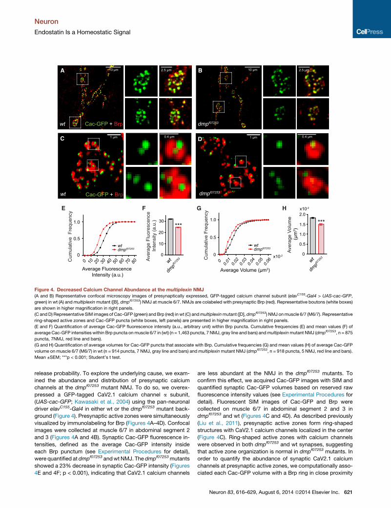
Figure 4. Decreased Calcium Channel Abundance at the multiplexin NMJ
(A and B) Representative confocal microscopy images of presynaptically expressed, GFP-tagged calcium channel subunit (elavC155-Gal4 > UAS-cac-GFP,
green) in wt (A) andmultiplexinmutant ([B], dmpf07253) NMJ at muscle 6/7. NMJs are colabeled with presynaptic Brp (red). Representative boutons (white boxes)
are shown in higher magnification in right panels.
(C and D) Representative SIM images of Cac-GFP (green) and Brp (red) in wt (C) andmultiplexinmutant ([D], dmpf07253) NMJ onmuscle 6/7 (M6/7). Representative
ring-shaped active zones and Cac-GFP puncta (white boxes, left panels) are presented in higher magnification in right panels.
(E and F) Quantification of average Cac-GFP fluorescence intensity (a.u., arbitrary unit) within Brp puncta. Cumulative frequencies (E) and mean values (F) of
average Cac-GFP intensities within Brp puncta onmuscle 6/7 in (wt) (n = 1,463 puncta, 7 NMJ, gray line and bars) andmultiplexinmutant NMJ (dmpf07253, n = 875
puncta, 7NMJ, red line and bars).
(G and H) Quantification of average volumes for Cac-GFP puncta that associate with Brp. Cumulative frequencies (G) and mean values (H) of average Cac-GFP
volume on muscle 6/7 (M6/7) in wt (n = 914 puncta, 7 NMJ, gray line and bars) andmultiplexinmutant NMJ (dmpf07253, n = 918 puncta, 5 NMJ, red line and bars).
Mean ±SEM; ***p < 0.001; Student’s t test.
Neuron
Endostatin Is a Homeostatic Signal
release probability. To explore the underlying cause, we exam-
ined the abundance and distribution of presynaptic calcium
channels at the dmpf07253 mutant NMJ. To do so, we overex-
pressed a GFP-tagged CaV2.1 calcium channel a subunit,
(UAS-cac-GFP; Kawasaki et al., 2004) using the pan-neuronal
driver elavC155-Gal4 in either wt or the dmpf07253 mutant back-
ground (Figure 4). Presynaptic active zones were simultaneously
visualized by immunolabeling for Brp (Figures 4A–4D). Confocal
images were collected at muscle 6/7 in abdominal segment 2
and 3 (Figures 4A and 4B). Synaptic Cac-GFP fluorescence in-
tensities, defined as the average Cac-GFP intensity inside
each Brp punctum (see Experimental Procedures for detail),
were quantified at dmpf07253 andwt NMJ. The dmpf07253mutants
showed a 23% decrease in synaptic Cac-GFP intensity (Figures
4E and 4F; p < 0.001), indicating that CaV2.1 calcium channels
are less abundant at the NMJ in the dmpf07253 mutants. To
confirm this effect, we acquired Cac-GFP images with SIM and
quantified synaptic Cac-GFP volumes based on reserved raw
fluorescence intensity values (see Experimental Procedures for
detail). Fluorescent SIM images of Cac-GFP and Brp were
collected on muscle 6/7 in abdominal segment 2 and 3 in
dmpf07253 and wt (Figures 4C and 4D). As described previously
(Liu et al., 2011), presynaptic active zones form ring-shaped
structures with CaV2.1 calcium channels localized in the center
(Figure 4C). Ring-shaped active zones with calcium channels
were observed in both dmpf07253 and wt synapses, suggesting
that active zone organization is normal in dmpf07253 mutants. In
order to quantify the abundance of synaptic CaV2.1 calcium
channels at presynaptic active zones, we computationally asso-
ciated each Cac-GFP volume with a Brp ring in close proximity
Neuron 83, 616–629, August 6, 2014 ª2014 Elsevier Inc. 621
Neuron
Endostatin Is a Homeostatic Signal
(see Experimental Procedures for detail). Only Cac-GFP puncta
paired with nearby Brp-positive active zones were defined as
synaptic calcium channels and were used for quantification.
Fluorescence intensity values, non-normalized during image
processing, were used to estimate Cac-GFP volume (see
Experimental Procedures). The average Cac-GFP volume at
dmpf07253 synapses decreased 19% compared to wt (Figures
4G and 4H; p < 0.001), supporting the conclusion that
presynaptic CaV2.1 channels are less abundant in dmpf07253
mutants. From these data, we conclude Multiplexin is necessary
for the normal abundance of presynaptic, active-zone-localized,
CaV2.1 calcium channels.
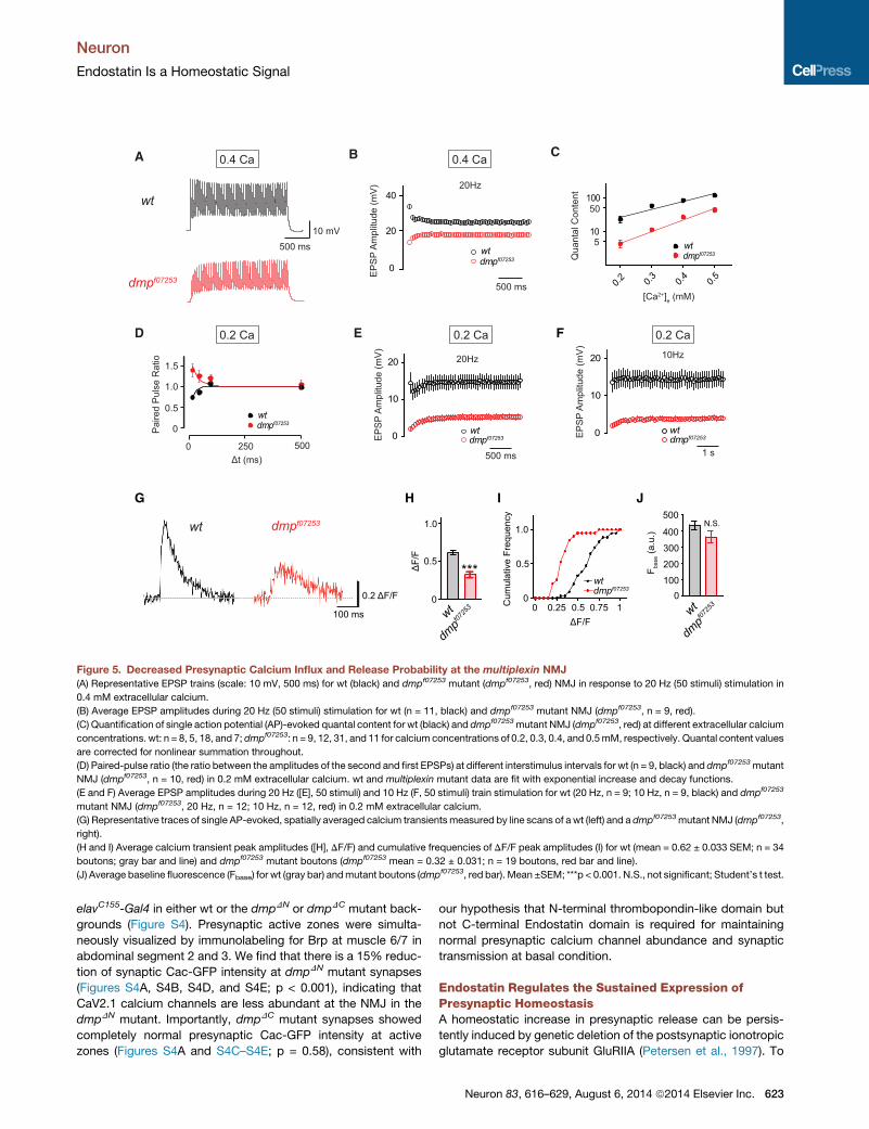
Multiplexin Mutants Exhibit Defects in Short-TermPlasticity and Presynaptic Calcium InfluxWe next sought to determine whether fewer synaptic calcium
channels at dmpf07253 mutant synapses alter synaptic function.
If there are fewer calcium channels per active zone, then
we expect to observe a decrease in presynaptic calcium influx
and a decrease in presynaptic release probability. We con-
firmed both predictions. To assess a change in presynaptic
release probability, we examined short-term facilitation and
depression during stimulus trains. At dmpf07253 mutant synap-
ses, we observed significant facilitation during a 20 Hz stimulus
train, whereas there was pronounced synaptic depression at wt
synapses (Figures 5A and 5B). It should be noted that the
steady-state EPSP amplitude in dmpf07253 mutants during a
20 Hz train plateaus and never reaches wt levels (Figure 5B).
This indicates that the defect in presynaptic release probability
cannot be restored during a stimulus train by an increase of
intracellular calcium that occurs during a train. Consistent
with this finding, the dmpf07253 mutant synapses also show a
decrease in presynaptic release over a range of extracellular
calcium concentrations (0.2–0.5 mM), while the apparent
cooperativity of release remains similar to wt (Figure 5C).
When we examined synaptic facilitation at low extracellular cal-
cium (0.2 mM), we again observed that dmpf07253 mutant syn-
apses showed facilitation compared to wt synapses that
depressed over a range of interstimulus intervals (Figure 5D).
The steady-state EPSP amplitude in dmpf07253 mutants was
also significantly smaller than that in wt animals during 20 Hz
and 10 Hz trains at low extracellular calcium (Figures 5E and
5F). Taken together, these data support the conclusion that
there is a drop in presynaptic release probability at dmpf07253
mutant synapses compared to wt.
Next, we examined presynaptic calcium influx by imaging
the spatially averaged presynaptic calcium signal at wt and
dmpf07253 mutant boutons following single action potential stim-
ulation. The spatially averaged calcium transients were attained
by loading presynaptic terminals with the calcium indicator Ore-
gon green 488 BAPTA-1 (OGB-1) and a reference dye, Alexa
568. Single AP-evoked calcium transients were quantified using
line scans across type 1b boutons on muscle 6/7 in abdominal
segments 2 and 3 (see Experimental Procedures for detail). As
exemplified in Figure 5G, we found that the average peak ampli-
tude of single AP-evoked calcium transients in the dmpf07253
mutant was significantly smaller than observed at wt synapses
(Figures 5H and 5I; p < 0.001). As a control, baseline OGB-1 fluo-
622 Neuron 83, 616–629, August 6, 2014 ª2014 Elsevier Inc.
rescence before stimulus onset was not different between geno-
types (Figure 5J; p = 0.11), indicating similar OGB-1 loading in
dmpf07253 mutant and wt synapses. Taken together, our data
provide evidence that loss of the multiplexin gene causes a
decrease in presynaptic calcium channel abundance with a
corresponding drop in both presynaptic calcium influx and pre-
synaptic release probability. We conclude that Multiplexin is
necessary for establishing or maintaining wt calcium channel
abundance at the presynaptic active zone.
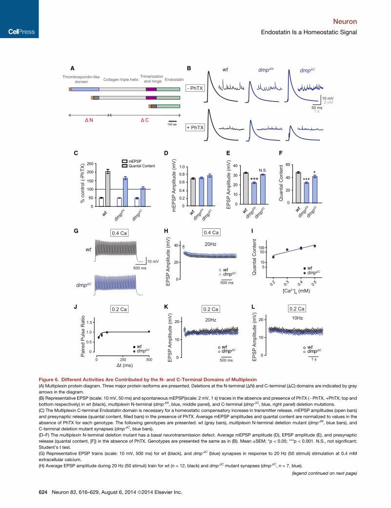
The N- and C-Terminal Domains of Multiplexin HaveDiscrete Functions Controlling Baseline Transmissionand Presynaptic HomeostasisDifferent structural domains have been identified in Multiplexin,
including a Thrombospondin-like domain, collagen triple helix,
trimerization region, hinge region, and C-terminal Endostatin
domain (Meyer and Moussian, 2009). In order to dissect the
contribution of these domains to baseline release and presynap-
tic homeostasis, we examined the induction of synaptic homeo-
stasis in two deletion mutants, one that removes the N-terminal
Thrombospondin-like domain and a second that removes
the C-terminal Endostatin domain (Figure 6A). Intriguingly, the
N- and C-terminal domains confer different activities (Figure 6).
The N-terminal deletion mutant (dmpDN) exhibits a significant
decrease in average baseline EPSP amplitude and quantal con-
tent (Figures 6B, 6E, and 6F; p < 0.001) but showed only minor
suppression of presynaptic homeostasis (Figures 6B and 6C).
By contrast, the C-terminal deletion mutant (dmpDC) has little
or no effect on baseline EPSP and quantal content (Figure 6E,
p = 0.08; Figure 6F, p < 0.05) but completely blocks presynaptic
homeostasis (Figures 6B and 6C). These data suggest that the
N-terminal Thrombospondin-like domain is required for basal
transmission, while the C-terminal Endostatin domain is required
for presynaptic homeostasis. By extension, the phenotype
observed in the dmpDC mutant clearly dissociates the function
of Endostatin during presynaptic homeostasis and baseline
transmission.
As a control, we examined short-term plasticity in the dmpDC
mutant. If the dmpDC mutant NMJ has wt baseline transmission,
then short-term synaptic plasticity should also be similar to wt.
We examined baseline synaptic transmission during a 20 Hz
stimulus train and at different extracellular calcium concentra-
tions. Similar to wt, dmpDC mutant synapses showed synaptic
depression and wt steady-state EPSP amplitudes during a
20 Hz train (Figures 6G and 6H). Furthermore, the apparent cal-
cium cooperativity of neurotransmitter release in dmpDC mutant
was not different compared to wt synapses (Figure 6I). Finally,
dmpDC mutant synapses had normal paired-pulse ratios and
short-term synaptic plasticity at low extracellular calcium con-
centration compared to wt synapses (Figures 6J–6L). Thus, the
Endostatin domain has a highly specific, required function for
the homeostatic modulation of neurotransmitter release.
Finally, we explored the underlying mechanism that may
cause the discrete effect on basal synaptic transmission in the
dmpDN and dmpDC mutants. We examined the abundance of
presynaptic calcium channels at the dmpDN and dmpDC mutant
NMJ. We overexpressed a GFP-tagged CaV2.1 calcium channel
a subunit, (UAS-cac-GFP) using the pan-neuronal driver
A
10 mV500 ms
wt
dmpf07253
Qua
ntal
Con
tent
[Ca2+]e (mM)
wtdmpf07253
0.2 0.3 0.4 0.5
10
100
5
50
B
D
0
20
40
wtdmpf07253
EP
SP
Am
plitu
de (m
V)
500 ms
20Hz
C
E
0
10
20
wtdmpf07253
20Hz
EP
SP
Am
plitu
de (m
V)
500 ms
0.4 Ca
0.2 Ca
Pai
red
Pul
se R
atio
Δt (ms)250 500
0
0.5
1.0
1.5
wtdmpf07253
0
wt
dmpf07253
ΔF/
F
0
1.0
0.5***
0 0.25 0.5 0.75 1
1.0
0.5
0
wtdmpf07253
ΔF/F
Cum
ulat
ive
Freq
uenc
yH I J
0
10
20
wtdmpf07253E
PS
P A
mpl
itude
(mV
)
1 s
10Hz
F
0.4 Ca
0.2 Ca 0.2 Ca
500
400
300
200
1000
wt
dmpf07253
F base
(a.u
.)
N.S.
0.2 ΔF/F
100 ms
wt dmpf07253
G
Figure 5. Decreased Presynaptic Calcium Influx and Release Probability at the multiplexin NMJ
(A) Representative EPSP trains (scale: 10 mV, 500 ms) for wt (black) and dmpf07253 mutant (dmpf07253, red) NMJ in response to 20 Hz (50 stimuli) stimulation in
0.4 mM extracellular calcium.
(B) Average EPSP amplitudes during 20 Hz (50 stimuli) stimulation for wt (n = 11, black) and dmpf07253 mutant NMJ (dmpf07253, n = 9, red).
(C) Quantification of single action potential (AP)-evoked quantal content for wt (black) and dmpf07253mutant NMJ (dmpf07253, red) at different extracellular calcium
concentrations. wt: n = 8, 5, 18, and 7; dmpf07253: n = 9, 12, 31, and 11 for calcium concentrations of 0.2, 0.3, 0.4, and 0.5mM, respectively. Quantal content values
are corrected for nonlinear summation throughout.
(D) Paired-pulse ratio (the ratio between the amplitudes of the second and first EPSPs) at different interstimulus intervals for wt (n = 9, black) and dmpf07253mutant
NMJ (dmpf07253, n = 10, red) in 0.2 mM extracellular calcium. wt and multiplexin mutant data are fit with exponential increase and decay functions.
(E and F) Average EPSP amplitudes during 20 Hz ([E], 50 stimuli) and 10 Hz (F, 50 stimuli) train stimulation for wt (20 Hz, n = 9; 10 Hz, n = 9, black) and dmpf07253
mutant NMJ (dmpf07253, 20 Hz, n = 12; 10 Hz, n = 12, red) in 0.2 mM extracellular calcium.
(G) Representative traces of single AP-evoked, spatially averaged calcium transientsmeasured by line scans of a wt (left) and a dmpf07253mutant NMJ (dmpf07253,
right).
(H and I) Average calcium transient peak amplitudes ([H], DF/F) and cumulative frequencies of DF/F peak amplitudes (I) for wt (mean = 0.62 ± 0.033 SEM; n = 34
boutons; gray bar and line) and dmpf07253 mutant boutons (dmpf07253 mean = 0.32 ± 0.031; n = 19 boutons, red bar and line).
(J) Average baseline fluorescence (Fbase) for wt (gray bar) andmutant boutons (dmpf07253, red bar). Mean ±SEM; ***p < 0.001. N.S., not significant; Student’s t test.
Neuron
Endostatin Is a Homeostatic Signal
elavC155-Gal4 in either wt or the dmpDN or dmpDC mutant back-
grounds (Figure S4). Presynaptic active zones were simulta-
neously visualized by immunolabeling for Brp at muscle 6/7 in
abdominal segment 2 and 3. We find that there is a 15% reduc-
tion of synaptic Cac-GFP intensity at dmpDN mutant synapses
(Figures S4A, S4B, S4D, and S4E; p < 0.001), indicating that
CaV2.1 calcium channels are less abundant at the NMJ in the
dmpDN mutant. Importantly, dmpDC mutant synapses showed
completely normal presynaptic Cac-GFP intensity at active
zones (Figures S4A and S4C–S4E; p = 0.58), consistent with
our hypothesis that N-terminal thrombopondin-like domain but
not C-terminal Endostatin domain is required for maintaining
normal presynaptic calcium channel abundance and synaptic
transmission at basal condition.
Endostatin Regulates the Sustained Expression ofPresynaptic HomeostasisA homeostatic increase in presynaptic release can be persis-
tently induced by genetic deletion of the postsynaptic ionotropic
glutamate receptor subunit GluRIIA (Petersen et al., 1997). To
Neuron 83, 616–629, August 6, 2014 ª2014 Elsevier Inc. 623
A B
C D E
G H I
J K L
F
Figure 6. Different Activities Are Contributed by the N- and C-Terminal Domains of Multiplexin
(A) Multiplexin protein diagram. Three major protein isoforms are presented. Deletions at the N-terminal (DN) and C-terminal (DC) domains are indicated by gray
arrows in the diagram.
(B) Representative EPSP (scale: 10 mV, 50 ms) and spontaneous mEPSP(scale: 2 mV, 1 s) traces in the absence and presence of PhTX (�PhTX, +PhTX; top and
bottom respectively) in wt (black), multiplexin N-terminal (dmpDN, blue, middle panel), and C-terminal (dmpDC, blue, right panel) deletion mutations.
(C) The Multiplexin C-terminal Endostatin domain is necessary for a homeostatic compensatory increase in transmitter release. mEPSP amplitudes (open bars)
and presynaptic release (quantal content, filled bars) in the presence of PhTX. Average mEPSP amplitudes and quantal content are normalized to values in the
absence of PhTX for each genotype. The following genotypes are presented: wt (gray bars), multiplexin N-terminal deletion mutant (dmpDN, blue bars), and
C-terminal deletion mutant synapses (dmpDC, blue bars).
(D–F) The multiplexin N-terminal deletion mutant has a basal neurotransmission defect. Average mEPSP amplitude (D), EPSP amplitude (E), and presynaptic
release (quantal content, [F]) in the absence of PhTX. Genotypes are presented the same as in (B). Mean ±SEM; *p < 0.05; ***p < 0.001. N.S., not significant;
Student’s t test.
(G) Representative EPSP trains (scale: 10 mV, 500 ms) for wt (black), and dmpDC (blue) synapses in response to 20 Hz (50 stimuli) stimulation at 0.4 mM
extracellular calcium.
(H) Average EPSP amplitude during 20 Hz (50 stimuli) train for wt (n = 12, black) and dmpDC mutant synapses (dmpDC, n = 7, blue).
(legend continued on next page)
Neuron
Endostatin Is a Homeostatic Signal
624 Neuron 83, 616–629, August 6, 2014 ª2014 Elsevier Inc.
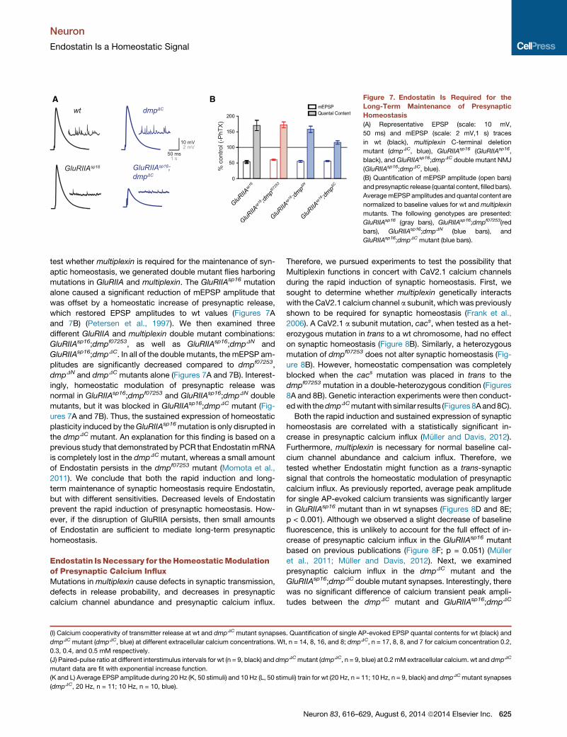
A B Figure 7. Endostatin Is Required for the
Long-Term Maintenance of Presynaptic
Homeostasis
(A) Representative EPSP (scale: 10 mV,
50 ms) and mEPSP (scale: 2 mV,1 s) traces
in wt (black), multiplexin C-terminal deletion
mutant (dmpDC, blue), GluRIIAsp16 (GluRIIAsp16,
black), and GluRIIAsp16;dmpDC double mutant NMJ
(GluRIIAsp16;dmpDC, blue).
(B) Quantification of mEPSP amplitude (open bars)
andpresynaptic release (quantal content, filledbars).
AveragemEPSPamplitudes and quantal content are
normalized to baseline values for wt andmultiplexin
mutants. The following genotypes are presented:
GluRIIAsp16 (gray bars), GluRIIAsp16;dmpf07253(red
bars), GluRIIAsp16;dmpDN (blue bars), and
GluRIIAsp16;dmpDC mutant (blue bars).
Neuron
Endostatin Is a Homeostatic Signal
test whether multiplexin is required for the maintenance of syn-
aptic homeostasis, we generated double mutant flies harboring
mutations in GluRIIA and multiplexin. The GluRIIAsp16 mutation
alone caused a significant reduction of mEPSP amplitude that
was offset by a homeostatic increase of presynaptic release,
which restored EPSP amplitudes to wt values (Figures 7A
and 7B) (Petersen et al., 1997). We then examined three
different GluRIIA and multiplexin double mutant combinations:
GluRIIAsp16;dmpf07253, as well as GluRIIAsp16;dmpDN and
GluRIIAsp16;dmpDC. In all of the double mutants, the mEPSP am-
plitudes are significantly decreased compared to dmpf07253,
dmpDN and dmpDC mutants alone (Figures 7A and 7B). Interest-
ingly, homeostatic modulation of presynaptic release was
normal in GluRIIAsp16;dmpf07253 and GluRIIAsp16;dmpDN double
mutants, but it was blocked in GluRIIAsp16;dmpDC mutant (Fig-
ures 7A and 7B). Thus, the sustained expression of homeostatic
plasticity induced by theGluRIIAsp16mutation is only disrupted in
the dmpDC mutant. An explanation for this finding is based on a
previous study that demonstrated by PCR that EndostatinmRNA
is completely lost in the dmpDC mutant, whereas a small amount
of Endostatin persists in the dmpf07253 mutant (Momota et al.,
2011). We conclude that both the rapid induction and long-
term maintenance of synaptic homeostasis require Endostatin,
but with different sensitivities. Decreased levels of Endostatin
prevent the rapid induction of presynaptic homeostasis. How-
ever, if the disruption of GluRIIA persists, then small amounts
of Endostatin are sufficient to mediate long-term presynaptic
homeostasis.
Endostatin IsNecessary for theHomeostaticModulationof Presynaptic Calcium InfluxMutations in multiplexin cause defects in synaptic transmission,
defects in release probability, and decreases in presynaptic
calcium channel abundance and presynaptic calcium influx.
(I) Calcium cooperativity of transmitter release at wt and dmpDC mutant synapses
dmpDC mutant (dmpDC, blue) at different extracellular calcium concentrations. Wt
0.3, 0.4, and 0.5 mM respectively.
(J) Paired-pulse ratio at different interstimulus intervals for wt (n = 9, black) and dm
mutant data are fit with exponential increase function.
(K and L) Average EPSP amplitude during 20 Hz (K, 50 stimuli) and 10 Hz (L, 50 stim
(dmpDC, 20 Hz, n = 11; 10 Hz, n = 10, blue).
Therefore, we pursued experiments to test the possibility that
Multiplexin functions in concert with CaV2.1 calcium channels
during the rapid induction of synaptic homeostasis. First, we
sought to determine whether multiplexin genetically interacts
with the CaV2.1 calcium channel a subunit, whichwas previously
shown to be required for synaptic homeostasis (Frank et al.,
2006). A CaV2.1 a subunit mutation, cacs, when tested as a het-
erozygous mutation in trans to a wt chromosome, had no effect
on synaptic homeostasis (Figure 8B). Similarly, a heterozygous
mutation of dmpf07253 does not alter synaptic homeostasis (Fig-
ure 8B). However, homeostatic compensation was completely
blocked when the cacs mutation was placed in trans to the
dmpf07253 mutation in a double-heterozygous condition (Figures
8A and 8B). Genetic interaction experiments were then conduct-
edwith thedmpDCmutantwith similar results (Figures 8Aand8C).
Both the rapid induction and sustained expression of synaptic
homeostasis are correlated with a statistically significant in-
crease in presynaptic calcium influx (Muller and Davis, 2012).
Furthermore, multiplexin is necessary for normal baseline cal-
cium channel abundance and calcium influx. Therefore, we
tested whether Endostatin might function as a trans-synaptic
signal that controls the homeostatic modulation of presynaptic
calcium influx. As previously reported, average peak amplitude
for single AP-evoked calcium transients was significantly larger
in GluRIIAsp16 mutant than in wt synapses (Figures 8D and 8E;
p < 0.001). Although we observed a slight decrease of baseline
fluorescence, this is unlikely to account for the full effect of in-
crease of presynaptic calcium influx in the GluRIIAsp16 mutant
based on previous publications (Figure 8F; p = 0.051) (Muller
et al., 2011; Muller and Davis, 2012). Next, we examined
presynaptic calcium influx in the dmpDC mutant and the
GluRIIAsp16;dmpDC double mutant synapses. Interestingly, there
was no significant difference of calcium transient peak ampli-
tudes between the dmpDC mutant and GluRIIAsp16;dmpDC
. Quantification of single AP-evoked EPSP quantal contents for wt (black) and
, n = 14, 8, 16, and 8; dmpDC, n = 17, 8, 8, and 7 for calcium concentration 0.2,
pDCmutant (dmpDC, n = 9, blue) at 0.2 mM extracellular calcium. wt and dmpDC
uli) train for wt (20 Hz, n = 11; 10 Hz, n = 9, black) and dmpDCmutant synapses
Neuron 83, 616–629, August 6, 2014 ª2014 Elsevier Inc. 625
A
B C
D E F
G
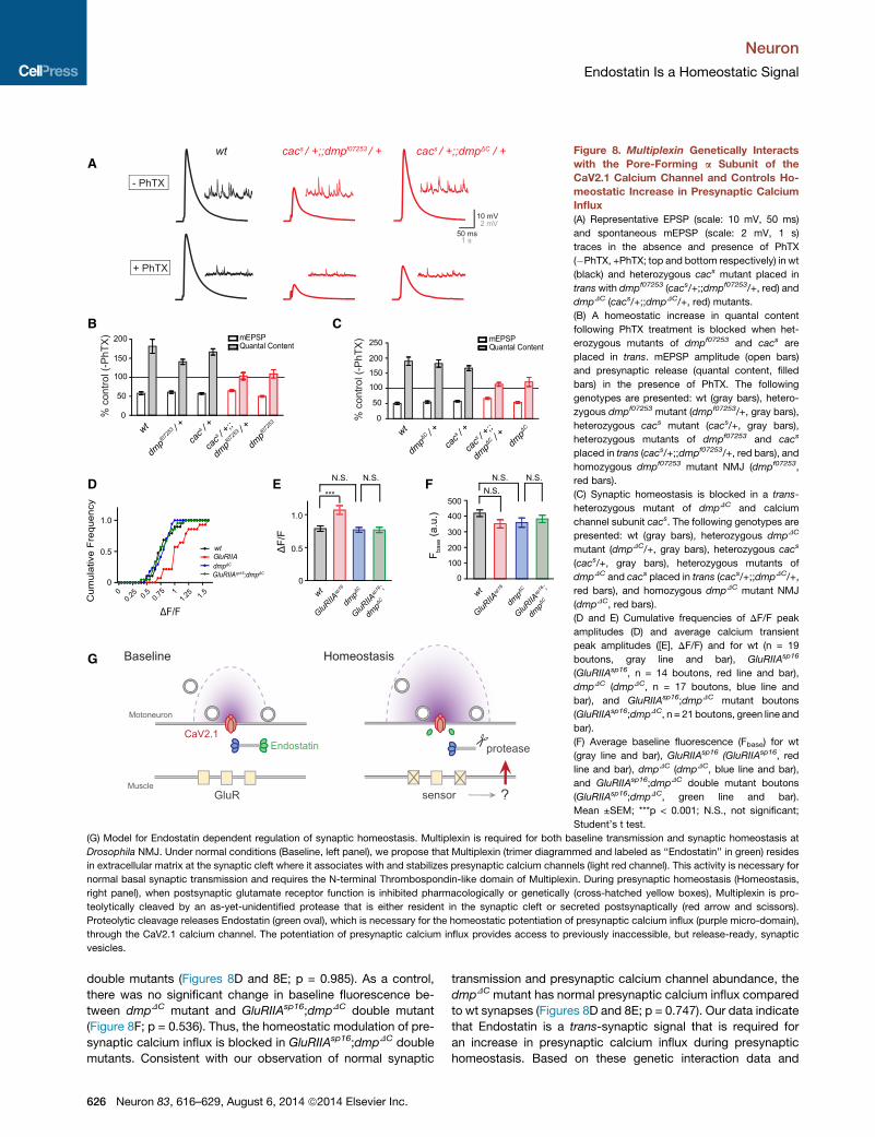
Figure 8. Multiplexin Genetically Interacts
with the Pore-Forming a Subunit of the
CaV2.1 Calcium Channel and Controls Ho-
meostatic Increase in Presynaptic Calcium
Influx
(A) Representative EPSP (scale: 10 mV, 50 ms)
and spontaneous mEPSP (scale: 2 mV, 1 s)
traces in the absence and presence of PhTX
(�PhTX, +PhTX; top and bottom respectively) in wt
(black) and heterozygous cacs mutant placed in
trans with dmpf07253 (cacs/+;;dmpf07253/+, red) and
dmpDC (cacs/+;;dmpDC/+, red) mutants.
(B) A homeostatic increase in quantal content
following PhTX treatment is blocked when het-
erozygous mutants of dmpf07253 and cacs are
placed in trans. mEPSP amplitude (open bars)
and presynaptic release (quantal content, filled
bars) in the presence of PhTX. The following
genotypes are presented: wt (gray bars), hetero-
zygous dmpf07253 mutant (dmpf07253/+, gray bars),
heterozygous cacs mutant (cacs/+, gray bars),
heterozygous mutants of dmpf07253 and cacs
placed in trans (cacs/+;;dmpf07253/+, red bars), and
homozygous dmpf07253 mutant NMJ (dmpf07253,
red bars).
(C) Synaptic homeostasis is blocked in a trans-
heterozygous mutant of dmpDC and calcium
channel subunit cacs. The following genotypes are
presented: wt (gray bars), heterozygous dmpDC
mutant (dmpDC/+, gray bars), heterozygous cacs
(cacs/+, gray bars), heterozygous mutants of
dmpDC and cacs placed in trans (cacs/+;;dmpDC/+,
red bars), and homozygous dmpDC mutant NMJ
(dmpDC, red bars).
(D and E) Cumulative frequencies of DF/F peak
amplitudes (D) and average calcium transient
peak amplitudes ([E], DF/F) and for wt (n = 19
boutons, gray line and bar), GluRIIAsp16
(GluRIIAsp16, n = 14 boutons, red line and bar),
dmpDC (dmpDC, n = 17 boutons, blue line and
bar), and GluRIIAsp16;dmpDC mutant boutons
(GluRIIAsp16;dmpDC, n = 21 boutons, green line and
bar).
(F) Average baseline fluorescence (Fbase) for wt
(gray line and bar), GluRIIAsp16 (GluRIIAsp16, red
line and bar), dmpDC (dmpDC, blue line and bar),
and GluRIIAsp16;dmpDC double mutant boutons
(GluRIIAsp16;dmpDC, green line and bar).
Mean ±SEM; ***p < 0.001; N.S., not significant;
Student’s t test.
(G) Model for Endostatin dependent regulation of synaptic homeostasis. Multiplexin is required for both baseline transmission and synaptic homeostasis at
Drosophila NMJ. Under normal conditions (Baseline, left panel), we propose that Multiplexin (trimer diagrammed and labeled as ‘‘Endostatin’’ in green) resides
in extracellular matrix at the synaptic cleft where it associates with and stabilizes presynaptic calcium channels (light red channel). This activity is necessary for
normal basal synaptic transmission and requires the N-terminal Thrombospondin-like domain of Multiplexin. During presynaptic homeostasis (Homeostasis,
right panel), when postsynaptic glutamate receptor function is inhibited pharmacologically or genetically (cross-hatched yellow boxes), Multiplexin is pro-
teolytically cleaved by an as-yet-unidentified protease that is either resident in the synaptic cleft or secreted postsynaptically (red arrow and scissors).
Proteolytic cleavage releases Endostatin (green oval), which is necessary for the homeostatic potentiation of presynaptic calcium influx (purple micro-domain),
through the CaV2.1 calcium channel. The potentiation of presynaptic calcium influx provides access to previously inaccessible, but release-ready, synaptic
vesicles.
Neuron
Endostatin Is a Homeostatic Signal
double mutants (Figures 8D and 8E; p = 0.985). As a control,
there was no significant change in baseline fluorescence be-
tween dmpDC mutant and GluRIIAsp16;dmpDC double mutant
(Figure 8F; p = 0.536). Thus, the homeostatic modulation of pre-
synaptic calcium influx is blocked in GluRIIAsp16;dmpDC double
mutants. Consistent with our observation of normal synaptic
626 Neuron 83, 616–629, August 6, 2014 ª2014 Elsevier Inc.
transmission and presynaptic calcium channel abundance, the
dmpDC mutant has normal presynaptic calcium influx compared
to wt synapses (Figures 8D and 8E; p = 0.747). Our data indicate
that Endostatin is a trans-synaptic signal that is required for
an increase in presynaptic calcium influx during presynaptic
homeostasis. Based on these genetic interaction data and
Neuron
Endostatin Is a Homeostatic Signal
the finding that Endostatin is necessary for the modulation of
presynaptic calcium influx during homeostatic plasticity, we
conclude that Multiplexin functions in concert with the CaV2.1
calcium channels to control the homeostatic modulation of
neurotransmitter release.
DISCUSSION
Here we provide evidence that Endostatin is a trans-synaptic
signal for the homeostatic modulation of presynaptic neuro-
transmitter release. Specifically, we show that loss of Endostatin
blocks the homeostatic modulation of presynaptic calcium influx
and presynaptic neurotransmitter release. This activity is
remarkably specific to presynaptic homeostasis, since loss of
Endostatin has no effect on baseline neurotransmission or syn-
apse morphology. Endostatin also interacts genetically with the
pore-forming subunit of the CaV2.1 calcium channel and is
required for the homeostatic increase of presynaptic calcium
influx during synaptic homeostatic plasticity. Finally, transgenic
overexpression of Endostatin is sufficient to rescue synaptic
homeostasis and baseline neurotransmitter release when it is
supplied to either the presynaptic or postsynaptic side of the
synapse. Although deletion of Endostatin does not impair base-
line transmission, overexpression of Endostatin in the dmpf07253
mutant is sufficient to restore baseline transmission release even
in the absence of the Thrombospondin-like domain. As a work-
ing model, we propose that inhibition of postsynaptic glutamate
receptors initiates the proteolytic cleavage of Multiplexin, which
resides in the synaptic cleft. We further propose that release of
Endostatin acts upon presynaptic calcium channels, directly or
indirectly, to potentiate calcium influx and presynaptic neuro-
transmitter release (Figure 8G). This model is consistent with
data from other systems demonstrating that activation of Endo-
statin requires proteolytic cleavage of Collagen XVIII by matrix
metalloproteases (MMPs) and cysteine cathepsins. Moreover,
only free Endostatin released by cleavage functions as an antian-
giogenesis factor (Heljasvaara et al., 2005).
Endostatin Regulates Homeostatic PlasticityThe means by which presynaptic calcium channel function
is modulated by Endostatin (Figures 4 and 5) remains to be eluci-
dated. Recently, it has been shown that a presynaptic Deg/ENaC
channel is also necessary for the homeostatic modulation of pre-
synaptic release (Younger et al., 2013). In this previous study, a
model is presented in which ENaC channel insertion causes a
sodium leak and modest depolarization of the presynaptic
resting membrane potential that, in turn, potentiates presynaptic
calcium influx. One possibility is that the interaction of Endostatin
with the presynaptic CaV2.1 channels enables the channels to
respond to low-voltage modulation. This would be consistent
with both Endostatin and the ENaC channel being strictly neces-
sary for presynaptic homeostasis. It remains formally possible
that Endostatin stabilizes presynaptic ENaC channels and,
thereby, influences presynaptic calcium influx. For example, it
was demonstrated that the interaction between ENaC channels
and extracellular collagens mediates the mechanosensory
transduction in the touch reception systems (Chalfie, 2009; Liu
et al., 1996).
Proteolytic Cleavage of Synaptic Proteins as a Triggerfor Retrograde Trans-Synaptic SignalingActivation of Endostatinin in other systems requires proteolyti-
cal cleavage of Collagen XVIII by MMPs and cysteine cathep-
sins. This raises an intriguing possibility that, during synaptic
homeostasis, Multiplexin could be cleaved by synaptic MMPs,
releasing Endostatin to trigger a homeostatic change in presyn-
aptic release. In this model, inhibition of postsynaptic glutamate
receptors would lead to the activation of MMPswithin the synap-
tic cleft (Figure 8G). Thus, the retrograde signal would be a
multistage system, providing opportunity for both amplification
and multilevel control of the signaling event. At glutamatergic
synapses in hippocampal neurons, proteolytic cleavage of
neuroligin-1, a synaptic adhesion molecule residing in postsyn-
aptic terminals, is triggered by postsynaptic NMDA receptor
activation. Cleavage of neuroligin-1 depresses presynaptic
transmission by reducing presynaptic release probability in a
trans-synaptic manner (Peixoto et al., 2012). Thus, the activity-
dependent cleavage of cell adhesion and extracellular matrix
proteins could provide a robust and evolutionarily conserved
feedback paradigm for trans-synaptic signaling to regulate syn-
aptic efficacy in diverse neuronal circuits.
EXPERIMENTAL PROCEDURES
Fly Stocks and Genetics
All fly stocks were maintained at 22�C–25�C on normal food. All flies were
obtained from Bloomington Drosophila Stock Center or the Exelixis Collec-
tion (Harvard Medical School), unless otherwise noted. The multiplexin N-
(dmpDN) and C-terminal (dmpDC) deletion mutations and Endostatin rescue
allele (UAS-endostatin) were generous gifts from Bernard Moussian (Max-
Planck-Institute for Developmental Biology, Tubingen). For pan-neuronal
expression, we used driver elavC155-Gal4 on the X chromosome (male
larvae); for muscle expression, we used MHC-Gal4 or BG57-Gal4 (as spec-
ified in figure legends or Results); for neuronal and muscle expression, we
used OK371-Gal4;BG57-Gal4. Unless otherwise noted, the w1118 strain
was used as a wt control.
Electrophysiology
Electrophysiology was performed as described previously (Muller et al., 2011)
using modified HL3 saline at the specified calcium concentrations containing
the following components: NaCl (70 mM), KCl (5 mM), MgCl2 (10 mM),
NaHCO3 (10 mM), sucrose (115 mM), trehalose (5 mM), HEPES (5 mM), and
CaCl2 (0.4 mM, unless specified otherwise). Mean EPSP, mEPSP amplitude,
and quantal content were obtained by averaging values across all NMJs for
a given genotype. EPSP andmEPSP traces were analyzed in IGOR Pro (Wave-
Metrics) and MiniAnalysis (Synaptosoft).
Immunocytochemistry
Standard immunocytochemistry was performed as previously described. For
surface GFP immuno-labeling, dissected third instar larvae were incubated
with primary antibody for 20 min at room temperature and fixed with ice-
cold ethanol. The following primary antibodies were used: mouse anti-Brp
(1:100) (Kittel et al., 2006), rabbit anti-Discs large (Dlg, 1:1,000) (Pielage
et al., 2008), rabbit anti-GFP (1:1,000, Invitrogen G10362), and mouse anti-
GFP (1:1,000, Invitrogen clone 3E6). Rabbit and mouse anti-GFP antibodies
were used to detect surface and total GFP expression respectively. Alexa-con-
jugated secondary antibodies were used at 1:300 (Jackson Immuno-research
Laboratories).
Image Acquisition and Analysis
Deconvolution imaging for synapse morphology was performed using a 1003
(1.4 NA) plan Apochromat objective (Carl Zeiss) on an Axiovert 200 inverted
Neuron 83, 616–629, August 6, 2014 ª2014 Elsevier Inc. 627
Neuron
Endostatin Is a Homeostatic Signal
microscope (Carl Zeiss) equipped with a cooled CCD camera (CoolSNAP HQ;
Roper Scientific). Image acquisition and analysis were performed in SlideBook
software (Intelligent Imaging Innovation). Maximum projections of decon-
volved images were used for analyses. Quantification of Brp and bouton num-
ber was performed as previously described (Pielage et al., 2008). Confocal im-
aging was performed on a Yokagawa CSU22 spinning disk confocal with a
603/1.4 plan Apochromat objective. Structured illumination fluorescence
imaging for Endostatin-GFP and calcium channels was performed using an
ELYRA PS.1 system (Carl Zeiss) with an inverted LSM 710 microscope equip-
ped with a 633 (1.4 NA) plan Apochromat objective (Carl Zeiss) and an Andor
iXon 885 EMCCD camera. For quantitative calcium channel abundance anal-
ysis, raw fluorescence intensities were preserved with position information
using a customized algorithm (Carl Zeiss). See Supplemental Experimental
Proceduresfor details.
Calcium Imaging
Calcium imaging experiments at the third instar NMJ were performed as
previously described (Muller and Davis, 2012) using 5 mM OBG-1 (Invitrogen)
and 1 mM Alexa 568 (Invitrogen). Image acquisition was performed at
room temperature using a confocal laser-scanning system (Ultima, Prairie
Technologies) equipped with excitation light (488 nm) from an air-cooled
krypton-argon laser, 603 objective (1.0 NA, Olympus), and a gallium arsenide
phosphide-based photocathode photomultiplier tube (Hamamatsu). Line
scans across single boutons were made at a frequency of 568 Hz. Data of
experimental and control groups were collected side by side. Calcium imaging
data were acquired using Prairie View (Prairie Technologies) and analyzed in
Igor Pro (Wavemetrics).
SUPPLEMENTAL INFORMATION
Supplemental Information includes four figures, one table, and Supplemental
Experimental Procedures and can be found with this article online at http://dx.
doi.org/10.1016/j.neuron.2014.07.003.
ACKNOWLEDGMENTS
We thank Meg Younger, Mike Gavino, Kevin Ford, and Cody Locke for critical
reading of the manuscript. We thank Martin Muller for help with train stimula-
tion data analysis in IGOR. We thank Bryant Chhun (Carl Zeiss) for help with
custom algorithm to preserve raw fluorescence intensity in SIM images. We
thank Tao Cui (California Institute of Technology) for help with SIM data pro-
cessing by generating custom analysis program in Matlab. We thank Gama
Ruiz for technical assistance. We thank Zeiss USA for use of their structured
illumination microcopy system. Work in the laboratory of G.W.D. was sup-
ported by NIH grants NS059867 and NS039313. T.W. was supported by NIH
NRSA postdoc fellowship NIH NINDS 1F32NS081884-01.
Accepted: June 16, 2014
Published: July 24, 2014
REFERENCES
Ackley, B.D., Crew, J.R., Elamaa, H., Pihlajaniemi, T., Kuo, C.J., and Kramer,
J.M. (2001). The NC1/endostatin domain of Caenorhabditis elegans type XVIII
collagen affects cell migration and axon guidance. J. Cell Biol. 152,
1219–1232.
Ackley, B.D., Kang, S.H., Crew, J.R., Suh, C., Jin, Y., and Kramer, J.M. (2003).
The basement membrane components nidogen and type XVIII collagen regu-
late organization of neuromuscular junctions in Caenorhabditis elegans.
J. Neurosci. 23, 3577–3587.
Burrone, J., O’Byrne, M., and Murthy, V.N. (2002). Multiple forms of synaptic
plasticity triggered by selective suppression of activity in individual neurons.
Nature 420, 414–418.
Chalfie, M. (2009). Neurosensory mechanotransduction. Nat. Rev. Mol. Cell
Biol. 10, 44–52.
628 Neuron 83, 616–629, August 6, 2014 ª2014 Elsevier Inc.
Chang, J.H., Javier, J.A., Chang, G.Y., Oliveira, H.B., and Azar, D.T. (2005).
Functional characterization of neostatins, the MMP-derived, enzymatic cleav-
age products of type XVIII collagen. FEBS Lett. 579, 3601–3606.
Cull-Candy, S.G., Miledi, R., Trautmann, A., and Uchitel, O.D. (1980). On the
release of transmitter at normal, myasthenia gravis and myasthenic syndrome
affected human end-plates. J. Physiol. 299, 621–638.
Davis, G.W. (2013). Homeostatic signaling and the stabilization of neural func-
tion. Neuron 80, 718–728.
Dhanabal, M., Ramchandran, R., Waterman, M.J., Lu, H., Knebelmann, B.,
Segal, M., and Sukhatme, V.P. (1999). Endostatin induces endothelial cell
apoptosis. J. Biol. Chem. 274, 11721–11726.
Dickman, D.K., and Davis, G.W. (2009). The schizophrenia susceptibility gene
dysbindin controls synaptic homeostasis. Science 326, 1127–1130.
Felbor, U., Dreier, L., Bryant, R.A., Ploegh, H.L., Olsen, B.R., and Mothes, W.
(2000). Secreted cathepsin L generates endostatin from collagen XVIII. EMBO
J. 19, 1187–1194.
Frank, C.A., Kennedy, M.J., Goold, C.P., Marek, K.W., and Davis, G.W. (2006).
Mechanisms underlying the rapid induction and sustained expression of
synaptic homeostasis. Neuron 52, 663–677.
Goold, C.P., and Davis, G.W. (2007). The BMP ligand Gbb gates the expres-
sion of synaptic homeostasis independent of synaptic growth control.
Neuron 56, 109–123.
Gottmann, K. (2008). Transsynaptic modulation of the synaptic vesicle cycle
by cell-adhesion molecules. J. Neurosci. Res. 86, 223–232.
Hanai, J., Gloy, J., Karumanchi, S.A., Kale, S., Tang, J., Hu, G., Chan, B.,
Ramchandran, R., Jha, V., Sukhatme, V.P., and Sokol, S. (2002). Endostatin
is a potential inhibitor of Wnt signaling. J. Cell Biol. 158, 529–539.
Harrington, A.W., and Ginty, D.D. (2013). Long-distance retrograde neurotro-
phic factor signalling in neurons. Nat. Rev. Neurosci. 14, 177–187.
Heljasvaara, R., Nyberg, P., Luostarinen, J., Parikka, M., Heikkila, P., Rehn,M.,
Sorsa, T., Salo, T., and Pihlajaniemi, T. (2005). Generation of biologically active
endostatin fragments from human collagen XVIII by distinct matrix metallopro-
teases. Exp. Cell Res. 307, 292–304.
Iremonger, K.J., Wamsteeker Cusulin, J.I., and Bains, J.S. (2013). Changing
the tune: plasticity and adaptation of retrograde signals. Trends Neurosci.
36, 471–479.
Jakawich, S.K., Nasser, H.B., Strong, M.J., McCartney, A.J., Perez, A.S.,
Rakesh, N., Carruthers, C.J., and Sutton, M.A. (2010). Local presynaptic activ-
ity gates homeostatic changes in presynaptic function driven by dendritic
BDNF synthesis. Neuron 68, 1143–1158.
Kawasaki, F., Zou, B., Xu, X., and Ordway, R.W. (2004). Active zone localiza-
tion of presynaptic calcium channels encoded by the cacophony locus of
Drosophila. J. Neurosci. 24, 282–285.
Kim, S.H., and Ryan, T.A. (2010). CDK5 serves as a major control point in
neurotransmitter release. Neuron 67, 797–809.
Kim, Y.M., Hwang, S., Kim, Y.M., Pyun, B.J., Kim, T.Y., Lee, S.T., Gho, Y.S.,
and Kwon, Y.G. (2002). Endostatin blocks vascular endothelial growth fac-
tor-mediated signaling via direct interaction with KDR/Flk-1. J. Biol. Chem.
277, 27872–27879.
Kittel, R.J., Wichmann, C., Rasse, T.M., Fouquet, W., Schmidt, M., Schmid, A.,
Wagh, D.A., Pawlu, C., Kellner, R.R., Willig, K.I., et al. (2006). Bruchpilot pro-
motes active zone assembly, Ca2+ channel clustering, and vesicle release.
Science 312, 1051–1054.
Liu, G., and Tsien, R.W. (1995). Properties of synaptic transmission at single
hippocampal synaptic boutons. Nature 375, 404–408.
Liu, J., Schrank, B., andWaterston, R.H. (1996). Interaction between a putative
mechanosensory membrane channel and a collagen. Science 273, 361–364.
Liu, K.S., Siebert, M., Mertel, S., Knoche, E., Wegener, S., Wichmann, C.,
Matkovic, T., Muhammad, K., Depner, H., Mettke, C., et al. (2011). RIM-bind-
ing protein, a central part of the active zone, is essential for neurotransmitter
release. Science 334, 1565–1569.
Neuron
Endostatin Is a Homeostatic Signal
McCabe, B.D., Marques, G., Haghighi, A.P., Fetter, R.D., Crotty, M.L., Haerry,
T.E., Goodman, C.S., and O’Connor, M.B. (2003). The BMP homolog Gbb pro-
vides a retrograde signal that regulates synaptic growth at the Drosophila
neuromuscular junction. Neuron 39, 241–254.
Meyer, F., and Moussian, B. (2009). Drosophila multiplexin (Dmp) modulates
motor axon pathfinding accuracy. Dev. Growth Differ. 51, 483–498.
Momota, R., Naito, I., Ninomiya, Y., and Ohtsuka, A. (2011). Drosophila type
XV/XVIII collagen, Mp, is involved in Wingless distribution. Matrix Biol. 30,
258–266.
Muller, M., and Davis, G.W. (2012). Transsynaptic control of presynaptic Ca2+
influx achieves homeostatic potentiation of neurotransmitter release. Curr.
Biol. 22, 1102–1108.
Muller, M., Pym, E.C., Tong, A., and Davis, G.W. (2011). Rab3-GAP controls
the progression of synaptic homeostasis at a late stage of vesicle release.
Neuron 69, 749–762.
Myllyharju, J., and Kivirikko, K.I. (2004). Collagens, modifying enzymes and
their mutations in humans, flies and worms. Trends Genet. 20, 33–43.
O’Reilly, M.S., Boehm, T., Shing, Y., Fukai, N., Vasios, G., Lane, W.S., Flynn,
E., Birkhead, J.R., Olsen, B.R., and Folkman, J. (1997). Endostatin: an endog-
enous inhibitor of angiogenesis and tumor growth. Cell 88, 277–285.
Passos-Bueno, M.R., Suzuki, O.T., Armelin-Correa, L.M., Sertie, A.L., Errera,
F.I., Bagatini, K., Kok, F., and Leite, K.R. (2006). Mutations in collagen 18A1
and their relevance to the human phenotype. An. Acad. Bras. Cienc. 78,
123–131.
Peixoto, R.T., Kunz, P.A., Kwon, H., Mabb, A.M., Sabatini, B.L., Philpot, B.D.,
and Ehlers, M.D. (2012). Transsynaptic signaling by activity-dependent cleav-
age of neuroligin-1. Neuron 76, 396–409.
Petersen, S.A., Fetter, R.D., Noordermeer, J.N., Goodman, C.S., and
DiAntonio, A. (1997). Genetic analysis of glutamate receptors in Drosophila
reveals a retrograde signal regulating presynaptic transmitter release.
Neuron 19, 1237–1248.
Pielage, J., Cheng, L., Fetter, R.D., Carlton, P.M., Sedat, J.W., and Davis, G.W.
(2008). A presynaptic giant ankyrin stabilizes the NMJ through regulation of
presynaptic microtubules and transsynaptic cell adhesion. Neuron 58,
195–209.
Plomp, J.J., van Kempen, G.T., and Molenaar, P.C. (1992). Adaptation of
quantal content to decreased postsynaptic sensitivity at single endplates in
alpha-bungarotoxin-treated rats. J. Physiol. 458, 487–499.
Seppinen, L., and Pihlajaniemi, T. (2011). The multiple functions of collagen
XVIII in development and disease. Matrix Biol. 30, 83–92.
Sertie, A.L., Sossi, V., Camargo, A.A., Zatz, M., Brahe, C., and Passos-Bueno,
M.R. (2000). Collagen XVIII, containing an endogenous inhibitor of angiogen-
esis and tumor growth, plays a critical role in the maintenance of retinal struc-
ture and in neural tube closure (Knobloch syndrome). Hum. Mol. Genet. 9,
2051–2058.
Suzuki, O.T., Sertie, A.L., Der Kaloustian, V.M., Kok, F., Carpenter, M., Murray,
J., Czeizel, A.E., Kliemann, S.E., Rosemberg, S., Monteiro, M., et al. (2002).
Molecular analysis of collagen XVIII reveals novel mutations, presence of a
third isoform, and possible genetic heterogeneity in Knobloch syndrome.
Am. J. Hum. Genet. 71, 1320–1329.
Wickstrom, S.A., Alitalo, K., and Keski-Oja, J. (2002). Endostatin associates
with integrin alpha5beta1 and caveolin-1, and activates Src via a tyrosyl phos-
phatase-dependent pathway in human endothelial cells. Cancer Res. 62,
5580–5589.
Yamaguchi, N., Anand-Apte, B., Lee, M., Sasaki, T., Fukai, N., Shapiro, R.,
Que, I., Lowik, C., Timpl, R., and Olsen, B.R. (1999). Endostatin inhibits
VEGF-induced endothelial cell migration and tumor growth independently of
zinc binding. EMBO J. 18, 4414–4423.
Younger, M.A., Muller, M., Tong, A., Pym, E.C., and Davis, G.W. (2013). A pre-
synaptic ENaC channel drives homeostatic plasticity. Neuron 79, 1183–1196.
Zhao, C., Dreosti, E., and Lagnado, L. (2011). Homeostatic synaptic plasticity
through changes in presynaptic calcium influx. J. Neurosci. 31, 7492–7496.
Neuron 83, 616–629, August 6, 2014 ª2014 Elsevier Inc. 629