Embed Size (px)
Citation preview
ENTUK Student Undergraduate Essay Prize 2013
i | P a g e
Endoscopic nasal surgery- a surgical fashion or
genuine progress?
Author: Alexander Yao Final Year Medical Student (Direct Entry Programme) Imperial College London Word count: 2870
ENTUK Student Undergraduate Essay Prize 2013
ii | P a g e
Table of Contents
1. Introduction ............................................................................................................................................................................. 1
2. Historical progress towards the endoscopic ideal .............................................................................................. 2
3. Advancement in endoscope components ................................................................................................................. 3
3.1 The xenon light source ............................................................................................................................................... 3
3.2 Angled telescopes ......................................................................................................................................................... 3
3.3 High definition (HD) monitors ............................................................................................................................... 3
4. Instruments in endoscopic nasal surgery ................................................................................................................ 4
4.1 Microdebriders .............................................................................................................................................................. 4
4.2 The suction irrigation drill ....................................................................................................................................... 4
4.3 Balloon dilatation technology, or balloon sinuplasty................................................................................. 4
5. Radiology in endoscopic nasal surgery ..................................................................................................................... 5
6. Progress in the management of nasal and paranasal pathology .................................................................. 5
6.1 Function Endoscopic Sinus Surgery (FESS) .................................................................................................... 5
6.2 Septoplasty ....................................................................................................................................................................... 6
6.3 Rhinoplasty ...................................................................................................................................................................... 6
6.4 Skull base surgery......................................................................................................................................................... 6
6.5 Orbital surgery ............................................................................................................................................................... 7
7. The future of endoscopic nasal surgery .................................................................................................................... 7
7.1 3D endoscopes and monitors ................................................................................................................................. 7
7.2 Robotic systems ............................................................................................................................................................. 7
7.3 Intra-operative imaging ............................................................................................................................................ 8
8. Conclusion ................................................................................................................................................................................ 8
9. References ................................................................................................................................................................................ 9
10. Appendix- References for Figure 1 ........................................................................................................................ 13
ENTUK Student Undergraduate Essay Prize 2013
1 | P a g e
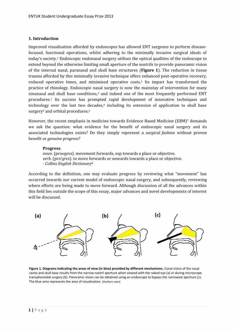
1. Introduction
Improved visualisation afforded by endoscopes has allowed ENT surgeons to perform disease-
focused, functional operations, whilst adhering to the minimally invasive surgical ideals of
today’s society.1 Endoscopic endonasal surgery utilises the optical qualities of the endoscope to
extend beyond the otherwise limiting small aperture of the nostrils to provide panoramic vision
of the internal nasal, paranasal and skull base structures (Figure 1). The reduction in tissue
trauma afforded by this minimally invasive technique offers enhanced post-operative recovery,
reduced operative times, and minimised operative costs.2 Its impact has transformed the
practice of rhinology. Endoscopic nasal surgery is now the mainstay of intervention for many
sinunasal and skull base conditions,3 and indeed one of the most frequently performed ENT
procedures.1 Its success has prompted rapid development of innovative techniques and
technology over the last two decades,4 including its extension of application to skull base
surgery5 and orbital procedures.6
However, the recent emphasis in medicine towards Evidence Based Medicine (EBM)7 demands
we ask the question: what evidence for the benefit of endoscopic nasal surgery and its
associated technologies exists? Do they simply represent a surgical fashion without proven
benefit or genuine progress?
Progress. noun. (prəʊɡrɛs). movement forwards, esp towards a place or objective. verb. (prə’ɡrɛs). to move forwards or onwards towards a place or objective. - Collins English Dictionary8
According to the definition, one may evaluate progress by reviewing what “movement” has
occurred towards our current model of endoscopic nasal surgery, and subsequently, reviewing
where efforts are being made to move forward. Although discussion of all the advances within
this field lies outside the scope of this essay, major advances and novel developments of interest
will be discussed.
Figure 1. Diagrams indicating the areas of view (in blue) provided by different mechanisms. Conal vision of the nasal cavity and skull base results from the narrow nostril aperture when viewed with the naked eye (a) or during microscope transphenoidal surgery (b). Panoramic vision can be obtained using an endoscope to bypass the narrowest aperture (c). The blue area represents the area of visualisation. (Authors own)
ENTUK Student Undergraduate Essay Prize 2013
2 | P a g e
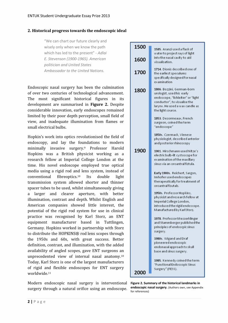
2. Historical progress towards the endoscopic ideal
Endoscopic nasal surgery has been the culmination
of over two centuries of technological advancement.
The most significant historical figures in its
development are summarised in Figure 2. Despite
considerable innovation, early endoscopes remained
limited by their poor depth perception, small field of
view, and inadequate illumination from flames or
small electrical bulbs.
Hopkin’s work into optics revolutionised the field of
endoscopy, and lay the foundations to modern
minimally invasive surgery.9 Professor Harold
Hopkins was a British physicist working as a
research fellow at Imperial College London at the
time. His novel endoscope employed true optical
media using a rigid rod and lens system, instead of
conventional fibreoptics.10 Its double light
transmission system allowed shorter and thinner
spacer tubes to be used, whilst simultaneously giving
a larger and clearer aperture, with better
illumination, contrast and depth. Whilst English and
American companies showed little interest, the
potential of the rigid rod system for use in clinical
practice was recognised by Karl Storz, an ENT
equipment manufacturer based in Tuttlingen,
Germany. Hopkins worked in partnership with Storz
to distribute the HOPKINS® rod lens scopes through
the 1950s and 60s, with great success. Better
definition, contrast, and illumination, with the added
availability of angled scopes, gave ENT surgeons an
unprecedented view of internal nasal anatomy.10
Today, Karl Storz is one of the largest manufacturers
of rigid and flexible endoscopes for ENT surgery
worldwide.11
Modern endoscopic nasal surgery is interventional
surgery through a natural orifice using an endoscope.
“We can chart our future clearly and
wisely only when we know the path
which has led to the present” - Adlai
E. Stevenson (1900-1965). American
politician and United States
Ambassador to the United Nations.
Figure 2. Summary of the historical landmarks in endoscopic nasal surgery. (Authors own, see Appendix
for references)
ENTUK Student Undergraduate Essay Prize 2013
3 | P a g e
It is defined by parameters consisting of the endoscope, the instruments, adjunctive radiology,
perioperative care and the surgeon. Certainly the former three have seen significant advances
and their contribution to progress will be discussed subsequently.
3. Advancement in endoscope components
The endoscope defines the value
of endoscopic nasal surgery. It
consists of relatively few
principle components: a light
source, a telescopic component, a
camera head, a processor and a
monitor.
3.1 The xenon light source
It wasn’t until 1876 that Nitze
used Edison’s invention of the
light bulb to introduce electricity
as a source of illumination for
endoscopy.12 Modern light
sources have progressed from
using halogen elements to using
more durable xenon. Xenon
offers numerous advantages
including triple the light output
compared to standard halogen,
and a true white image rather
than a yellow hue.10
Furthermore, xenon has been
shown to be more efficient, by
consuming less energy, and
generating less heat.
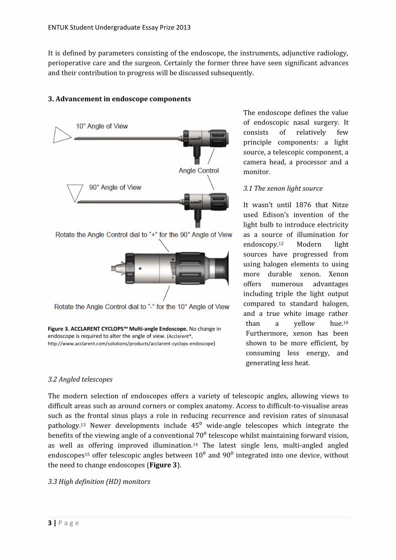
3.2 Angled telescopes
The modern selection of endoscopes offers a variety of telescopic angles, allowing views to
difficult areas such as around corners or complex anatomy. Access to difficult-to-visualise areas
such as the frontal sinus plays a role in reducing recurrence and revision rates of sinunasal
pathology.13 Newer developments include 45⁰ wide-angle telescopes which integrate the
benefits of the viewing angle of a conventional 70⁰ telescope whilst maintaining forward vision,
as well as offering improved illumination.14 The latest single lens, multi-angled angled
endoscopes15 offer telescopic angles between 10⁰ and 90⁰ integrated into one device, without
the need to change endoscopes (Figure 3).
3.3 High definition (HD) monitors
Figure 3. ACCLARENT CYCLOPS™ Multi-angle Endoscope. No change in endoscope is required to alter the angle of view. (Acclarent®,
http://www.acclarent.com/solutions/products/acclarent-cyclops-endoscope)
ENTUK Student Undergraduate Essay Prize 2013
4 | P a g e
Prior to the use of monitors surgeons would perform endoscopic nasal surgery through the
direct vision of the endoscope. The introduction of monitors improved surgery ergonomics,16
increased theatre staff awareness of the procedure, facilitated image/video capture, and played
a role in education. The replacement of one-chip camera processors with three-chip processors
(one for each primary colour) significantly improved monitors further by increasing the levels
of contrast and balance that could be achieved on the monitor display.14 Modern monitors
potentially offer HD definition (from 640x480 to 1980x1080), wider aspect ratios (from 4:3 to
16:9), and improved refresh rates (from 50Hz to 60Hz). The result of these changes have been
improvements in visual acuity, colour, contrast, visualisation of surgical field, less flicker and
less eye fatigue.14
Overall, few studies have evaluated the effects of these advances on surgical performance or
operative outcomes. A systematic review by Ayad et al. into ergonomics of endoscopic
endonasal surgery including the use of monitors vs direct visualisation concluded there were no
advantages in terms of surgical performance.16 A limited number of studies indicate that high
definition monitors may be of benefit over standard definition monitors in procedures requiring
high levels of detail such as diagnostic laryngoscopy17 and laparoscopic surgical skills.18 Both
studies acknowledge a strong subjective preference for high definition monitor use.
4. Instruments in endoscopic nasal surgery
Early instruments, consisting mainly of simple grasping forceps, used to remove mucosa
resulted in excess exposed bone, scarring, chronic inflammation and mucocele formation.19 To
overcome this problem, fine, through-cutting instruments, were adopted to reduce mucosal
trauma.
4.1 Microdebriders
Powered, replaceable cutting blades, and concurrent suction capabilities meant microdebriders
were able to offer faster, more efficient methods of tissue removal whilst maintaining excellent
visualisation, even in the presence of bleeding.20,21 The impact of the powered microdebrider on
rhinology has led some to hail it as one of the four most significant technological advances in the
specialty.22
4.2 The suction irrigation drill
This device quickly became adopted as an important tool in the removal of thickened osteotic
bone found in osseous and fibro-osseous neoplasms, which were otherwise difficult to remove
with forceps or the microdebrider.14
4.3 Balloon dilatation technology, or balloon sinuplasty
Introduced in 2005, balloon sinuplasty represents an important progression in minimally
invasive CRS treatment.22 The catheter-based system for dilatation of the paranasal sinuses has
subsequently been shown to be safe and effective.23 The CLEAR study by Bolger et al.
demonstrated a post-procedure patency rate of 80.5% at 24 weeks follow-up.24 The indications
and applications of balloon sinuplasty as a stand-alone vs adjunctive procedure to FESS have
been controversial.

ENTUK Student Undergraduate Essay Prize 2013
5 | P a g e
5. Radiology in endoscopic nasal surgery
Operating in close proximity to the paranasal sinuses, orbit and skull base makes endoscopic
nasal surgery potentially hazardous.25 The use of radiological imaging has been a crucial adjunct
in its success and safety. Computerised tomography (CT) offers detailed evaluation regarding
the anatomy and extent of sinunasal disease, making it the current gold standard.26 Magnetic
resonance imaging (MRI) has a role in soft tissue and skull base pathology including gliomas,
meningoceles, meningoencephaloceles, and benign tumours.14
Image-guided, or computer-aided, endoscopic nasal surgery uses CT or MRI for anatomical
navigation on triplanar radiologic images to create a pre-operative road map. High accuracy
within 2mm affords surgeons safe navigation in complex cases such as where anatomical
distortion is present secondary to alteration from disease or previous surgery.1 Fried at al.27
noted a significant reduction in major complications (from 11% to 1%) in the 97 patients who
underwent image-guided surgery compared to those who underwent non-image-guided
surgery. Image-guided endoscopic nasal surgery has a foreseeable role in teaching trainees as
well as high compatibility with robotic systems.
6. Progress in the management of nasal and paranasal pathology
6.1 Function Endoscopic Sinus Surgery (FESS)
Functional endoscopic sinus surgery (FESS), the archetypal endoscopic nasal procedure, is one
of the most performed ENT procedures, and the mainstay of treatment for Chronic
rhinosinusitis (CRS) refractory to medical therapy. FESS is an intranasal procedure involving
the endoscope to improve ventilation and drainage in addition to polyp removal.28
FESS has been shown to be both safe and effective in surgical management of both CRS with
nasal polyps (CRSwNP) and without nasal polyps (CRSsNP). A systematic review of 33 articles
by Dalziel et al.28 included three studies comparing FESS with Caldwell Luc or another
endonasal procedure (n=240), three non-randomised trials comparing different surgical
approaches (n=2699), and 27 case series. Symptom improvement in FESS was “greatly
improved” in 75-95%, comparable to traditional procedures. Overall complication rate was low
(1.4% for FESS and 0.8% for traditional procedures).
Most patients with CRS seek treatment when the burden of symptoms negatively impacts on
their quality of life. Therefore the degree to which the quality of life improves after sinus
surgery is a critical indicator of surgical success. A systemic review of 289 studies by Chester et
al. found Endoscopic sinus surgery was particularly effective in CRSsNP in relieving subjective
symptoms of nasal obstruction, facial pain, and post-nasal discharge.29 Similarly in CRSwNP, the
National Comparative Audit of Surgery for Nasal Polyposis and Chronic Rhinosinusitis reported
a high level of patient satisfaction with the surgery, and clinically significant improvement in
SNOT-22 scores at 3, 12, 36, 60 months post-operatively.30
Studies have shown endoscopic sinus surgery to be at least as effect as medical therapy in
treating CRS. A randomised controlled trial by Raghab et al. comparing long term antibiotics
with endoscopic sinus surgery in CRSwNP management found both to be equally effective in
significantly improving objective and subjective measures of CRS (p<0.01).31 No significant
ENTUK Student Undergraduate Essay Prize 2013
6 | P a g e
difference was found between quality of life measures when CRSwNP was treated surgical vs
medically.32
There is also evidence to suggest FESS is superior to conventional non-endoscopic or open
surgical approaches in the treatment of CRS, except for sphenoethmoidectomy where no studies
have yet been conducted.3,33–35
6.2 Septoplasty
Septoplasty is a common procedure used to treat nasal obstruction secondary to nasal septal
deviation.36 Good intra-operative visualisation is crucial to minimising complications and
achieving functional airway. A study of 2,730 patients undergoing power-assisted endoscopic
septoplasties demonstrated a role in minimise flap tears, and addressing septal spurs.37
Endoscopic septoplasty may better address discrete, localised lesions such isolated deflection,
spurs, perforations, contact points compared to traditional headlight septoplasty.38
Symptomatic outcomes also compare favourably to traditional techniques. A retrospective
review of 160 patients undergoing endoscopic septoplasty for nasal airway obstruction
demonstrated 70% resolution, and 20% improvement on symptoms after a 13 month mean
follow-up.39
6.3 Rhinoplasty
Rhinoplasty is an increasingly popular procedure, restoring form and function in nasal
deformity. Rhinoplasty can be extremely challenging,40 and parts of the procedure which are not
in direct vision of the surgeon, or poorly visualised, may benefit from endoscopic assistance.41
However, the evidence for its use has largely been limited to case series, usually with small
numbers and short follow-up. Data have suggested that the endoscope may reduce secondary
revisions and precision contouring of the bony nasal dorsum.42,43
6.4 Skull base surgery
The whole ventral skull base can be approached endonasally, via open craniotomy or via a
transphenoidal microscopic approach.5 In addition to benefitting from a greatly improved view
(Figure 1), endoscopic approaches avoid the need for extensive bone drilling, brain retraction,
and nerve manipulation that is sometimes required in transcranial approaches.44
Endoscopic repair is now generally regarded as the procedure of choice for cerebral spinal fluid
(CSF) leak repair.45 By approaching the anterior skull base endonasally, one can minimise
complications such as anosmia, intracranial haemorrhage or oedema, seizures, and changes in
memory and behaviour.46 A systematic review including 55 studies by Psaltis et al.47
demonstrated endoscope repair was effective, with a 90% overall success rate for primary
repairs, and 97% for secondary repairs. It is also regarded as safe with a low complication rate
of <0.03%. No studies have compared open invasive intracranial approaches with the endonasal
approach.
Tumours such as craniopharyngiomas, clivalchordomas, and meningiomas may also be resected
endoscopically with equivalent or greater rates of gross total resection compared to traditional
open approaches.44 Similarly, rates of post-operative complications are at least comparable with
traditional approaches. However, CSF leak still remains a problem.
ENTUK Student Undergraduate Essay Prize 2013
7 | P a g e
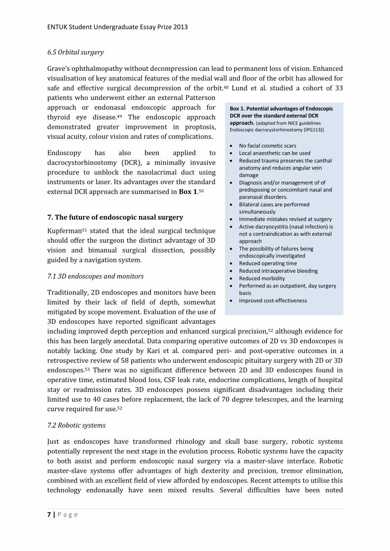
6.5 Orbital surgery
Grave’s ophthalmopathy without decompression can lead to permanent loss of vision. Enhanced
visualisation of key anatomical features of the medial wall and floor of the orbit has allowed for
safe and effective surgical decompression of the orbit.48 Lund et al. studied a cohort of 33
patients who underwent either an external Patterson
approach or endonasal endoscopic approach for
thyroid eye disease.49 The endoscopic approach
demonstrated greater improvement in proptosis,
visual acuity, colour vision and rates of complications.
Endoscopy has also been applied to
dacrocystorhinostomy (DCR), a minimally invasive
procedure to unblock the nasolacrimal duct using
instruments or laser. Its advantages over the standard
external DCR approach are summarised in Box 1.50
7. The future of endoscopic nasal surgery
Kupferman51 stated that the ideal surgical technique
should offer the surgeon the distinct advantage of 3D
vision and bimanual surgical dissection, possibly
guided by a navigation system.
7.1 3D endoscopes and monitors
Traditionally, 2D endoscopes and monitors have been
limited by their lack of field of depth, somewhat
mitigated by scope movement. Evaluation of the use of
3D endoscopes have reported significant advantages
including improved depth perception and enhanced surgical precision,52 although evidence for
this has been largely anecdotal. Data comparing operative outcomes of 2D vs 3D endoscopes is
notably lacking. One study by Kari et al. compared peri- and post-operative outcomes in a
retrospective review of 58 patients who underwent endoscopic pituitary surgery with 2D or 3D
endoscopes.53 There was no significant difference between 2D and 3D endoscopes found in
operative time, estimated blood loss, CSF leak rate, endocrine complications, length of hospital
stay or readmission rates. 3D endoscopes possess significant disadvantages including their
limited use to 40 cases before replacement, the lack of 70 degree telescopes, and the learning
curve required for use.52
7.2 Robotic systems
Just as endoscopes have transformed rhinology and skull base surgery, robotic systems
potentially represent the next stage in the evolution process. Robotic systems have the capacity
to both assist and perform endoscopic nasal surgery via a master-slave interface. Robotic
master-slave systems offer advantages of high dexterity and precision, tremor elimination,
combined with an excellent field of view afforded by endoscopes. Recent attempts to utilise this
technology endonasally have seen mixed results. Several difficulties have been noted
Box 1. Potential advantages of Endoscopic DCR over the standard external DCR approach. (adapted from NICE guidelines
Endoscopic dacrocystorhinostomy (IPG113)) No facial cosmetic scars Local anaesthetic can be used
Reduced trauma preserves the canthal anatomy and reduces angular vein damage
Diagnosis and/or management of of predisposing or concomitant nasal and paranasal disorders.
Bilateral cases are performed simultaneously
Immediate mistakes revised at surgery
Active dacryocystitis (nasal infection) is not a contraindication as with external approach
The possibility of failures being endoscopically investigated
Reduced operating time
Reduced intraoperative bleeding
Reduced morbidity Performed as an outpatient, day surgery
basis
Improved cost-effectiveness
ENTUK Student Undergraduate Essay Prize 2013
8 | P a g e
concerning the most popular model (da Vinci Surgical System by Intuitive Surgical Inc.)
including; size and number of instruments fitting through the nose, difficulty in instrument
exchange, and paucity of haptic feedback.54
Prototype robots have been developed to overcome these issues. Some models have the
capacity to handle the endoscope autonomously via speech commands from the surgeon,
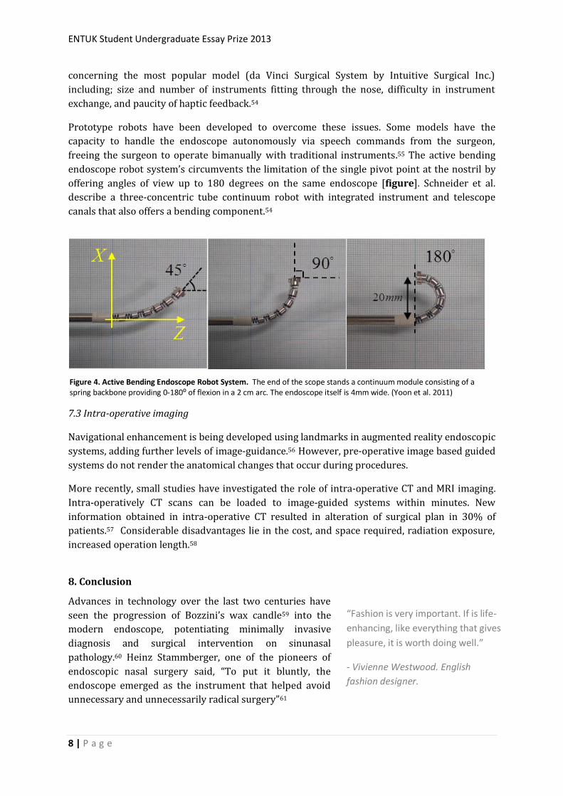
freeing the surgeon to operate bimanually with traditional instruments.55 The active bending
endoscope robot system’s circumvents the limitation of the single pivot point at the nostril by
offering angles of view up to 180 degrees on the same endoscope [figure]. Schneider et al.
describe a three-concentric tube continuum robot with integrated instrument and telescope
canals that also offers a bending component.54
7.3 Intra-operative imaging
Navigational enhancement is being developed using landmarks in augmented reality endoscopic
systems, adding further levels of image-guidance.56 However, pre-operative image based guided
systems do not render the anatomical changes that occur during procedures.
More recently, small studies have investigated the role of intra-operative CT and MRI imaging.
Intra-operatively CT scans can be loaded to image-guided systems within minutes. New
information obtained in intra-operative CT resulted in alteration of surgical plan in 30% of
patients.57 Considerable disadvantages lie in the cost, and space required, radiation exposure,
increased operation length.58
8. Conclusion
Advances in technology over the last two centuries have
seen the progression of Bozzini’s wax candle59 into the
modern endoscope, potentiating minimally invasive
diagnosis and surgical intervention on sinunasal
pathology.60 Heinz Stammberger, one of the pioneers of
endoscopic nasal surgery said, “To put it bluntly, the
endoscope emerged as the instrument that helped avoid
unnecessary and unnecessarily radical surgery”61
“Fashion is very important. If is life-
enhancing, like everything that gives
pleasure, it is worth doing well.”
- Vivienne Westwood. English
fashion designer.
Figure 4. Active Bending Endoscope Robot System. The end of the scope stands a continuum module consisting of a spring backbone providing 0-180⁰ of flexion in a 2 cm arc. The endoscope itself is 4mm wide. (Yoon et al. 2011)
ENTUK Student Undergraduate Essay Prize 2013
9 | P a g e
Unfortunately, rigorous objective evaluation of the progress in endoscopic nasal surgery
remains limited by the lack of high quality comparative studies and objective outcome measures
in many areas. Insufficient robust evidence fails to provide a clear guide to policies on behalf of
the NHS28 and makes informed decisions concerning commissioning particularly difficult.
However, it is important to recognise a lack of level 1a evidence for effectiveness does not
automatically warrant a label of ineffectiveness or dismissal as surgical fashion. Indeed, the
parachute has never been subjected to such rigorous evaluation.62 Evidently, robust
observational data can be extremely valuable.
Further research is required to evaluate merits, limitations and costs of expensive new
technologies such as 3D endoscopes, intra-operative imaging, and robotic systems. Many
current procedures require further clarification in regards to indications, and optimising patient
selection.28
Overall, it remains undeniable that endoscopic nasal surgery represents genuine progress. The
field of endoscopic surgery is constantly evolving, and the future seems extremely promising to
patient and surgeon alike.
9. References
1. Wise SK, DelGaudio JM. Computer-aided surgery of the paranasal sinuses and skull base. Expert review of medical devices. 2005. 2(4):395–408.
2. Welch KC, Stankiewicz JA. Application of minimally invasive endoscopic sinus surgery techniques. Otolaryngologic clinics of North America. 2010. 43(3):565–78, ix.
3. Fokkens WJ, Lund VJ, Mullol J, Bachert C, Alobid I, Baroody F, et al. European Position Paper on Rhinosinusitis and Nasal Polyps 2012. Rhinology. Supplement. 2012.(23):3, 1–298.
4. Kennedy DW. Technical innovations and the evolution of endoscopic sinus surgery. The Annals of otology, rhinology & laryngology. Supplement. 2006. 196:3–12.
5. Paluzzi A, Gardner P, Fernandez-Miranda JC, Snyderman C. The expanding role of endoscopic skull base surgery. British journal of neurosurgery. 2012. 26(5):649–61.
6. Metson R, Pletcher SD. Endoscopic Orbital and Optic Nerve Decompression. Otolaryngologic Clinics of North America. 2006 Jun;39(3):551–61.
7. Sackett DL, Rosenberg WM, Gray JA, Haynes RB, Richardson WS. Evidence based medicine: what it is and what it isn’t. BMJ. 1996.312(7023):71–2.
8. Collins Dictionary. Definition of “Progress”. 2013. Available at http://www.collinsdictionary.com/dictionary/english/progress?showCookiePolicy=trueLast Accessed 2013 Aug 4.
9. Jennings CR. Harold Hopkins. Archives of Otolaryngology–Head & Neck Surgery. 1998 Sep 1;124(9):1042.
ENTUK Student Undergraduate Essay Prize 2013
10 | P a g e
10. Chandra RK, Conley DB, Kern RC. Evolution of the endoscope and endoscopic sinus surgery. Otolaryngologic clinics of North America. 2009.42(5):747–52, vii.
11. Storz: Karl Storz Endoskope. Ear, Nose, Throat. Website. 2013. Website available from: https://www.karlstorz.com/cps/rde/xchg/SID-E1C8421C-D260A1DB/karlstorz-en/hs.xsl/45.htm. Last Accessed: 2013 Aug 31
12. Mouton WG, Bessell JR, Maddern GJ. Looking back to the advent of modern endoscopy: 150th birthday of Maximilian Nitze. World journal of surgery. 1998. 22(12):1256–8.
13. Gore MR, Ebert CS, Zanation AM, Senior BA. Beyond the “central sinus”: radiographic findings in patients undergoing revision functional endoscopic sinus surgery. International forum of allergy & rhinology. 2013. 3(2):139–46.
14. Govindaraj S, Adappa ND, Kennedy DW. Endoscopic sinus surgery: evolution and technical innovations. The Journal of laryngology and otology. 2010. 124(3):242–50.
15. Acclarent. ACCLARENT CYCLOPSTM Multi-angle Endoscope. 2013. Website available from: http://www.acclarent.com/solutions/products/acclarent-cyclops-endoscope. Last Accessed: 2013 Aug 31
16. Ayad T, Péloquin L, Prince F. Ergonomics in endoscopic sinus surgery: systematic review of the literature. The Journal of otolaryngology. 200. 34(5):333–40.
17. Otto KJ, Hapner ER, Baker M, Johns MM. Blinded evaluation of the effects of high definition and magnification on perceived image quality in laryngeal imaging. The Annals of otology, rhinology, and laryngology. 2006. 115(2):110–3.
18. Hagiike M, Phillips EH, Berci G. Performance differences in laparoscopic surgical skills between true high-definition and three-chip CCD video systems. Surgical endoscopy. 2007. 21(10):1849–54.
19. Kennedy DW. Diseases of the Sinuses: Diagnosis and Management. Ontario: BC Decker; 2000.
20. Parsons DS. Rhinologic uses of powered instrumentation in children beyond sinus surgery. Otolaryngologic clinics of North America . 1996. 29(1):105–14.
21. Setliff RC. The hummer: a remedy for apprehension in functional endoscopic sinus surgery. Otolaryngologic clinics of North America. 1996. 29(1):95–104.
22. Catalano PJ. Balloon dilation technology: let the truth be told. Current allergy and asthma reports . 2013. 13(2):250–4.
23. Levine HL, Sertich AP, Hoisington DR, Weiss RL, Pritikin J. Multicenter registry of balloon catheter sinusotomy outcomes for 1,036 patients. The Annals of otology, rhinology, and laryngology. 2008. 117(4):263–70.
24. Bolger WE, Brown CL, Church CA, Goldberg AN, Karanfilov B, Kuhn FA, et al. Safety and outcomes of balloon catheter sinusotomy: a multicenter 24-week analysis in 115 patients. Otolaryngology--head and neck surgery : official journal of American Academy of Otolaryngology-Head and Neck Surgery. 2007. 137(1):10–20.
ENTUK Student Undergraduate Essay Prize 2013
11 | P a g e
25. Lynn-Macrae AG, Lynn-Macrae RA, Emani J, Kern RC, Conley DB. Medicolegal analysis of injury during endoscopic sinus surgery. The Laryngoscope. 2004. 114(8):1492–5.
26. Lund VJ, Savy L, Lloyd G. Imaging for endoscopic sinus surgery in adults. The Journal of laryngology and otology. 2000. 114(5):395–7.
27. Fried MP, Moharir VM, Shin J, Taylor-Becker M, Morrison P. Comparison of endoscopic sinus surgery with and without image guidance. American journal of rhinology. 2002. 16(4):193–7.
28. Dalziel K, Stein K, Round A, Garside R, Royle P. Systematic review of endoscopic sinus surgery for nasal polyps. Health technology assessment (Winchester, England). 2003. 7(17):iii, 1–159.
29. Chester AC, Antisdel JL, Sindwani R. Symptom-specific outcomes of endoscopic sinus surgery: a systematic review. Otolaryngology--head and neck surgery : official journal of American Academy of Otolaryngology-Head and Neck Surgery. 2009. 140(5):633–9.
30. Browne J, Hopkins C, Slack R, Jan van der M, Lund V, Topham J, et al. The National Comparative Audit of Surgery for Nasal Polyposis and Chronic Rhinosinusitis. 2003.
31. Ragab SM, Lund VJ, Scadding G. Evaluation of the medical and surgical treatment of chronic rhinosinusitis: a prospective, randomised, controlled trial. The Laryngoscope. 2004. 114(5):923–30.
32. Alobid I, Benítez P, Bernal-Sprekelsen M, Roca J, Alonso J, Picado C, et al. Nasal polyposis and its impact on quality of life: comparison between the effects of medical and surgical treatments. Allergy. 2005. 60(4):452–8.
33. Lund VJ. The results of inferior and middle meatal antrostomy under endoscopic control. Acta oto-rhino-laryngologica Belgica. 1993.47(1):65–71.
34. Penttilä MA, Rautiainen ME, Pukander JS, Karma PH. Endoscopic versus Caldwell-Luc approach in chronic maxillary sinusitis: comparison of symptoms at one-year follow-up. Rhinology. 1994 Dec. 32(4):161–5.
35. Venkatachalam VP, Jain A. Comparative evaluation of functional endoscopic sinus surgery and conventional surgery in the management of chronic sinusitis. Journal of the Indian Medical Association. 2002. 100(2):78–9, 82–3.
36. Ketcham AS, Han JK. Complications and management of septoplasty. Otolaryngologic clinics of North America. 2010. 43(4):897–904.
37. Sousa A de, Iniciarte L, Levine H. Powered Endoscopic Nasal Septal Surgery. Acta médica portuguesa. 18(4):249–55.
38. Getz AE, Hwang PH. Endoscopic septoplasty. Current opinion in otolaryngology & head and neck surgery. 2008. 16(1):26–31.
39. Chung BJ, Batra PS, Citardi MJ, Lanza DC. Endoscopic septoplasty: revisitation of the technique, indications, and outcomes. American journal of rhinology. 21(3):307–11.
ENTUK Student Undergraduate Essay Prize 2013
12 | P a g e
40. Tasman A-J. Rhinoplasty - indications and techniques. GMS current topics in otorhinolaryngology, head and neck surgery. 2007.
41. Tasca I. Endoscopy-Assisted Rhinoplasty. Archives of Facial Plastic Surgery. 2002. 4(3):190–3.
42. Mitz V. Endoscopic control during rhinoplasty. Aesthetic plastic surgery. 1994. 18(2):153–6.
43. Krouse JH. Endoscopic-powered rhinoplasty. The Journal of otolaryngology . 1999. 28(5):282–4.
44. Raper DMS, Komotar RJ, Starke RM, Anand VK, Schwartz TH. Endoscopic versus open approaches to the skull base: A comprehensive literature review. Operative Techniques in Otolaryngology-Head and Neck Surgery . 2011. 22(4):302–7.
45. Martin TJ, Loehrl TA. Endoscopic CSF leak repair. Current opinion in otolaryngology & head and neck surgery . 2007. 15(1):35–9.
46. Daele JJM, Goffart Y, Machiels S. Traumatic, iatrogenic, and spontaneous cerebrospinal fluid (CSF) leak: endoscopic repair. B-ENT . 2011. 7 Suppl 17:47–60.
47. Psaltis AJ, Schlosser RJ, Banks CA, Yawn J, Soler ZM. A systematic review of the endoscopic repair of cerebrospinal fluid leaks. Otolaryngology--head and neck surgery : official journal of American Academy of Otolaryngology-Head and Neck Surgery . 2012. 147(2):196–203.
48. Pletcher SD, Sindwani R, Metson R. Endoscopic orbital and optic nerve decompression. Otolaryngologic clinics of North America . 2006 Oct. 39(5):943–58, vi.
49. Lund VJ, Larkin G, Fells P, Adams G. Orbital decompression for thyroid eye disease: a comparison of external and endoscopic techniques. The Journal of laryngology and otology . 1997. 111(11):1051–5.
50. National Institute of Clinical Excellence (NICE). Endoscopic dacrocystorhinostomy (IPG113) . 2005;Available from: http://guidance.nice.org.uk/IPG113
51. Kupferman M, Demonte F, Holsinger FC, Hanna E. Transantral robotic access to the pituitary gland. Otolaryngology--head and neck surgery : official journal of American Academy of Otolaryngology-Head and Neck Surgery . 2009. 141(3):413–5.
52. Manes RP, Barnett S, Batra PS. Utility of novel 3-dimensional stereoscopic vision system for endoscopic sinonasal and skull-base surgery. International forum of allergy & rhinology. 2011. 1(3):191–7.
53. Kari E, Oyesiku NM, Dadashev V, Wise SK. Comparison of traditional 2-dimensional endoscopic pituitary surgery with new 3-dimensional endoscopic technology: intraoperative and early postoperative factors. International forum of allergy & rhinology. 2012. 2(1):2–8.
ENTUK Student Undergraduate Essay Prize 2013
13 | P a g e
54. Schneider JS, Burgner J, Webster RJ, Russell PT. Robotic surgery for the sinuses and skull base: what are the possibilities and what are the obstacles? Current opinion in otolaryngology & head and neck surgery . 2013 Mar. 21(1):11–6.
55. Rilk M, Kubus D, Wahl FM, Eichhorn KWG, Wagner I, Bootz F. Demonstration of a prototype for robot assisted Endoscopic Sinus Surgery . In: 2010 IEEE International Conference on Robotics and Automation. IEEE; 2010. p. 1090–1.
56. Thoranaghatte RU, Giraldez JG, Zheng G. Landmark based augmented reality endoscope system for sinus and skull-base surgeries. Conference proceedings : ... Annual International Conference of the IEEE Engineering in Medicine and Biology Society. IEEE Engineering in Medicine and Biology Society. Conference . 2008. 2008:74–7.
57. Jackman AH, Palmer JN, Chiu AG, Kennedy DW. Use of intraoperative CT scanning in endoscopic sinus surgery: a preliminary report. American journal of rhinology. 2008. 22(2):170–4.
58. Cartellieri M, Vorbeck F. Endoscopic sinus surgery using intraoperative computed tomography imaging for updating a three-dimensional navigation system. The Laryngoscope. 2000. 110(2 Pt 1):292–6.
59. Verger-Kuhnke AB, Reuter MA, Beccaria ML. [Biography of Phillip Bozzini (1773-1809) an idealist of the endoscopy]. Actas urologicas españolas . 2007. 31(5):437–44.
60. Linder TE, Simmen D, Stool SE. Revolutionary Inventions in the 20th Century: The History of Endoscopy. Archives of Otolaryngology - Head and Neck Surgery . 1997. 123(11):1161–3.
61. Stammberger H. The evolution of functional endoscopic sinus surgery. Ear, nose, & throat journal . 1994. 73(7):451, 454–5.
62. Smith GCS, Pell JP. Parachute use to prevent death and major trauma related to gravitational challenge: systematic review of randomised controlled trials. BMJ (Clinical research ed.) . 2003. 327(7429):1459–61.
10. Appendix- References for Figure 1
References for Figure 1. Summary of the historical landmarks in endoscopic nasal surgery.
1. Essentials of Pediatric Endoscopic Surgery. Springer; 2008.
2. Verger-Kuhnke AB, Reuter MA, Beccaria ML. [Biography of Phillip Bozzini (1773-1809)
an idealist of the endoscopy]. Actas urologicas españolas. 2007 May 4;31(5):437–44.
3. Garcia M. Observations on the Human Voice. Proceedings of the Royal Society of London
(1854-1905). 1854;7:399–410.
4. Czermak J. Der Kehlkopspiegel und seine Verwerthung fur Physiologie und Medicin. 2nd
ed. Leipzig: Engelmann; 1893.
5. Howard DJ, Lund VJ. Endoscopic surgery in otolaryngology. Br. Med. Bull. 1986 Jan
1;42(3):234–9.
ENTUK Student Undergraduate Essay Prize 2013
14 | P a g e
6. Killian G. Uber Rhinoskopia media. Munchen Med Wochenschr. 1896;33.
7. Hirschmann A. Uber Endoskopie der Nase und deren Nebenhohlen. Arch Laryngol
Rhinol. 1903;14:195–202.
8. Reichert M. Über eine neue Unterscuhungsmethode der Oberkieferhöhle mittels des
Antroskops. Berl klin Wochenschr. 1902;401:478.
9. Mouton WG, Bessell JR, Maddern GJ. Looking back to the advent of modern endoscopy:
150th birthday of Maximilian Nitze. World journal of surgery. 1998 Dec;22(12):1256–8.
10. Chandra RK, Conley DB, Kern RC. Evolution of the endoscope and endoscopic sinus
surgery. Otolaryngologic clinics of North America. 2009 Oct;42(5):747–52, vii.
11. Govindaraj S, Adappa ND, Kennedy DW. Endoscopic sinus surgery: evolution and
technical innovations. The Journal of laryngology and otology. 2010 Mar;124(3):242–50.
12. Messerklinger W. Endoscopy of the Nose. Baltimore, Maryland: Urban & Schwarzenberg;
1978.
13. Kennedy DW. Functional endoscopic sinus surgery: Technique. Archives of
otolaryngology. 1985 Oct.111(10):643–9.