Embed Size (px)
Citation preview
Endoscopic Follow-up of Digestive Anastomosis
123
Giuseppe GalloroEditor
Endoscopic Follow-up of Digestive Anastomosis

Giuseppe Galloro Editor
Endoscopic Follow-up of Digestive Anastomosis
ISBN 978-88-470-5369-4 ISBN 978-88-470-5370-0 (eBook) DOI 10.1007/978-88-470-5370-0 Springer Milan Heidelberg New York Dordrecht London
Library of Congress Control Number: 2014934307
© Springer-Verlag Italia 2014 This work is subject to copyright. All rights are reserved by the Publisher, whether the whole or part of the material is concerned, specifi cally the rights of translation, reprinting, reuse of illustrations, recitation, broadcasting, reproduction on microfi lms or in any other physical way, and transmission or information storage and retrieval, electronic adaptation, computer software, or by similar or dissimilar methodology now known or hereafter developed. Exempted from this legal reservation are brief excerpts in connection with reviews or scholarly analysis or material supplied specifi cally for the purpose of being entered and executed on a computer system, for exclusive use by the purchaser of the work. Duplication of this publication or parts thereof is permitted only under the provisions of the Copyright Law of the Publisher's location, in its current version, and permission for use must always be obtained from Springer. Permissions for use may be obtained through RightsLink at the Copyright Clearance Center. Violations are liable to prosecution under the respective Copyright Law. The use of general descriptive names, registered names, trademarks, service marks, etc. in this publication does not imply, even in the absence of a specifi c statement, that such names are exempt from the relevant protective laws and regulations and therefore free for general use. While the advice and information in this book are believed to be true and accurate at the date of publication, neither the authors nor the editors nor the publisher can accept any legal responsibility for any errors or omissions that may be made. The publisher makes no warranty, express or implied, with respect to the material contained herein.
Printed on acid-free paper
Springer is part of Springer Science+Business Media (www.springer.com)
Editor Giuseppe Galloro University of Naples Federico II Napoli Italy
v
There are undoubtedly several books and atlases, available on the shelves of scientifi c bookstores, regarding digestive endoscopy and investigating both diagnostic and therapeutic techniques, and all those publications are surely very useful from a didactic and technical point of view.
What is, then, the rationale of this new text? My purpose in undertaking the editorship of this volume was to develop a monograph about a topic often treated in a superfi cial or even vague way.
The study of anastomosis is one of the most frequent indications in diag-nostic digestive endoscopy, and the endoscopist is frequently asked to treat some complications of the surgical interventions, such as bleeding, benign strictures, neoplastic recurrences, and dehiscences, by the means of operative procedures. Moreover, the evaluation of a digestive anastomosis can repre-sent a source of worries and anxiety, especially for the junior professionals, because they are confronted with the new anatomy modifi ed by the surgeon.
In spite of this, in most cases, textbooks and atlases available for practitio-ners devote just a few pages or short paragraphs to the endoscopic follow-up of digestive anastomosis and to the endoscopic treatment of their complications.
Finally, beyond the technical aspects of the topic, it appears very important to clarify the logistic points of view of the problem: what is the appropriateness of the endoscopic follow-up, who should be put under surveillance, how and when to perform surveillance, has biopsy been performed, and what about the useful tools of endo-ultrasonography, chromoendoscopy, and magnifi cation?
The main goal of this text is to present the knowledge about endoscopic follow-up of digestive anastomosis as much completely as possible, both illustrating diagnostic protocols and operative techniques, in the global per-spective of a systematic and multidisciplinary monograph.
I would like to seize the opportunity to express my thankfulness to col-laborators and colleagues. In the fi rst place, my sincere thanks go to all the authors and contributors of the book: with their efforts they have been able to share and communicate their scientifi c knowledge and enthusiasm to all those who will read and study this volume. Secondly, my thanks to the Springer editorial team, who believed in this endeavor and followed it with profes-sionalism. Finally, my thoughts go to the readers: we hope this volume will be a contribution to their professional growth and foster a comprehensive vision of digestive endoscopy.
Naples, Italy Giuseppe Galloro
Pref ace

vii
Contents
Part I Diagnostic Procedures and Follow-Up
1 Analysis of Surgical Risk Factors in Tailoring Digestive Anastomosis . . . . . . . . . . . . . . . . . . . . . . . . . . . . . . . . . . . . . . . . . 3Mario Testini, Ilaria Fabiola Franco, Valentina Ferraro, Angela Gurrado, and Germana Lissidini
2 Impact of Flexible Endoscopy in the Evaluation of Digestive Anastomosis . . . . . . . . . . . . . . . . . . . . . . . . . . . . . . . . . . . . . . . . . 11Antonello Trecca, Raffaele Manta, Amitabh Naik, Mario De Bellis, Alberto Arezzo, and Giuseppe Galloro
3 Methodology and Appropriateness of Follow-Up in Digestive Endoscopy . . . . . . . . . . . . . . . . . . . . . 17Gianluca Rotondano, Stefano Sansone, and Claudia Cesaro
4 Timing and Protocols of Endoscopic Follow-Up in Operated Patients After Esophageal Surgery . . . . . . . . . . . . 23Giorgio Battaglia, Matteo Cagol, Stefano Realdon, Carlo Castoro, Giorgio Diamantis, and Alberto Ruol
5 Timing and Protocols of Endoscopic Follow-Up After Gastric Surgery . . . . . . . . . . . . . . . . . . . . . . . . . . . . . . . . . 35Rita Conigliaro, Angelo Caruso, and Marzio Frazzoni
6 Timing and Protocols of Endoscopic Follow-Up in Operated Patients After Small Bowel Surgery . . . . . . . . . . . 41Emanuele Rondonotti and Marco Pennazio
7 Timing and Protocols of Endoscopic Follow-Up in Operated Patients After Colorectal Surgery. . . . . . . . . . . . . 49Mario de Bellis, Elena Di Girolamo, Ugo Pace, Guglielmo Nasti, Maura Claire Tracey, Alberto Arezzo, Raffaele Manta, Antonello Trecca, and Giuseppe Galloro
8 Intraoperative Endoscopy in the Evaluation of Digestive Anastomoses . . . . . . . . . . . . . . . . . . . . . . . . . . . . . . . . . . . . . . . . . 61Raffaele Manta, Amitabh Naik, Marzio Frazzoni, Mauro Manno, Alberto Arezzo, Mario de Bellis, Antonello Trecca, Gabrio Bassotti, Gianluigi Melotti, Rita Conigliaro, and Giuseppe Galloro
viii
9 Contribution of Endo-ultrasonography . . . . . . . . . . . . . . . . . . . 67Vincenzo Napolitano, Maria C. Bondanese, and Manuela Avellino
10 Augmented Endoscopy Imaging in the Study of Digestive Anastomosis: Does It Really Work and How?. . . . . . . . . . . . . . 77Makomo Makazu, Takahisa Matsuda, Taku Sakamoto, Takeshi Nakajima, and Yutaka Saito
Part II Therapeutic Procedures of Anastomotic Complications
11 Physiopathology and Treatment of Anastomotic Ulcer: An Emerging Pathology? . . . . . . . . . . . . . . . . . . . . . . . . . 85Angelo Zullo, Lorenzo Ridola, and Cesare Hassan
12 Endoscopic Treatment of Anastomotic Recurrences in Oncologic Patients . . . . . . . . . . . . . . . . . . . . . . . . . . . . . . . . . . 93Antonella De Ceglie, Andrea Parodi, and Massimo Conio
13 Therapeutic Endoscopy for the Treatment of Benign Anastomotic Strictures . . . . . . . . . . . . . . . . . . . . . . . . . . . . . . . . 105Alessandro Casadei, Angelo De Padova, Ilaria Manzi, and Enrico Ricci
14 Review: Therapeutic Endoscopy for the Treatment of Anastomotic Dehiscences . . . . . . . . . . . . . . . . . . . . . . . . . . . . 119Alberto Arezzo, Mauro Verra, Giuseppe Galloro, Mario de Bellis, Antonello Trecca, Raffaele Manta, and Mario Morino
15 Hemostatic Procedures in the Bleeding Anastomosis . . . . . . . . 131Bjorn Rembacken
16 Endoscopic Treatment of Anastomotic Complications After Bariatric Surgery . . . . . . . . . . . . . . . . . . . . . . . . . . . . . . . . . . . . . 137Alfredo Genco, Roberta Maselli, Massimiliano Cipriano, Giovanni Casella, and Adriano Redler
Index . . . . . . . . . . . . . . . . . . . . . . . . . . . . . . . . . . . . . . . . . . . . . . . . . . . 149
Contents

Part I
Diagnostic Procedures and Follow-Up
3G. Galloro (ed.), Endoscopic Follow-up of Digestive Anastomosis, DOI 10.1007/978-88-470-5370-0_1, © Springer-Verlag Italia 2014
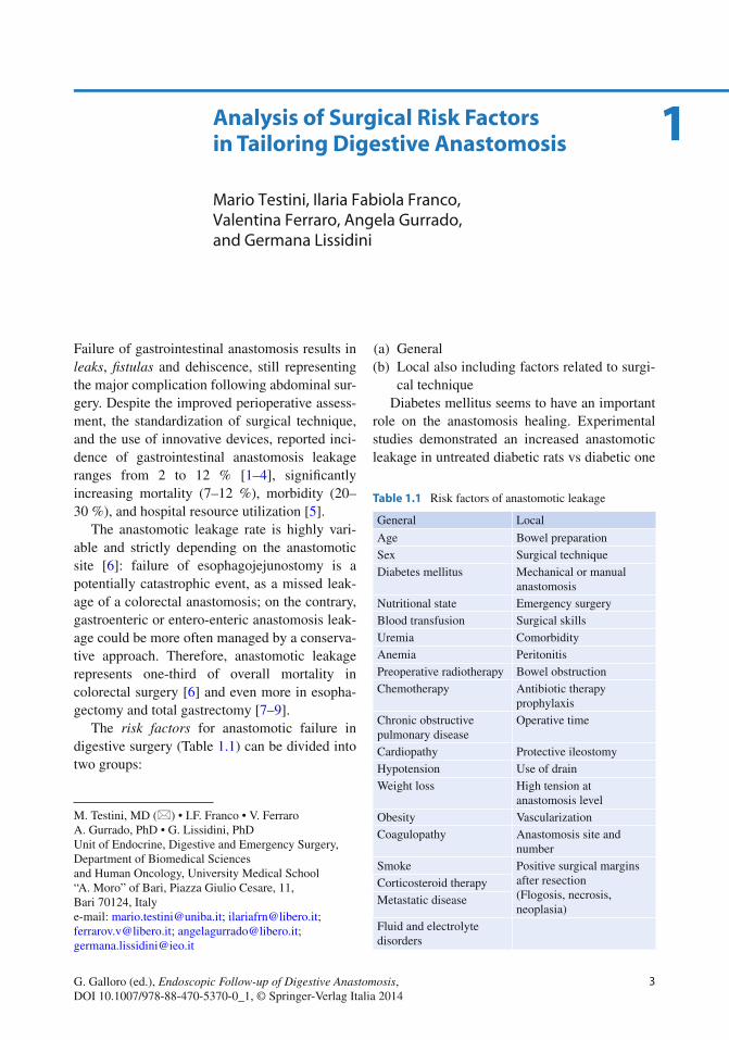
Failure of gastrointestinal anastomosis results in leaks , fi stulas and dehiscence, still representing the major complication following abdominal sur-gery. Despite the improved perioperative assess-ment, the standardization of surgical technique, and the use of innovative devices, reported inci-dence of gastrointestinal anastomosis leakage ranges from 2 to 12 % [ 1 – 4 ], signifi cantly increasing mortality (7–12 %), morbidity (20–30 %), and hospital resource utilization [ 5 ].
The anastomotic leakage rate is highly vari-able and strictly depending on the anastomotic site [ 6 ]: failure of esophagojejunostomy is a potentially catastrophic event, as a missed leak-age of a colorectal anastomosis; on the contrary, gastroenteric or entero-enteric anastomosis leak-age could be more often managed by a conserva-tive approach. Therefore, anastomotic leakage represents one-third of overall mortality in colorectal surgery [ 6 ] and even more in esopha-gectomy and total gastrectomy [ 7 – 9 ].
The risk factors for anastomotic failure in digestive surgery (Table 1.1 ) can be divided into two groups:
(a) General (b) Local also including factors related to surgi-
cal technique Diabetes mellitus seems to have an important
role on the anastomosis healing. Experimental studies demonstrated an increased anastomotic leakage in untreated diabetic rats vs diabetic one
M. Testini , MD (*) • I. F. Franco • V. Ferraro A. Gurrado , PhD • G. Lissidini , PhD Unit of Endocrine, Digestive and Emergency Surgery, Department of Biomedical Sciences and Human Oncology , University Medical School “A. Moro” of Bari , Piazza Giulio Cesare, 11 , Bari 70124 , Italy e-mail: [email protected]; [email protected]; [email protected]; [email protected]; [email protected]
1 Analysis of Surgical Risk Factors in Tailoring Digestive Anastomosis
Mario Testini , Ilaria Fabiola Franco , Valentina Ferraro , Angela Gurrado , and Germana Lissidini
Table 1.1 Risk factors of anastomotic leakage
General Local
Age Bowel preparation Sex Surgical technique Diabetes mellitus Mechanical or manual
anastomosis Nutritional state Emergency surgery Blood transfusion Surgical skills Uremia Comorbidity Anemia Peritonitis Preoperative radiotherapy Bowel obstruction Chemotherapy Antibiotic therapy
prophylaxis Chronic obstructive pulmonary disease
Operative time
Cardiopathy Protective ileostomy Hypotension Use of drain Weight loss High tension at
anastomosis level Obesity Vascularization Coagulopathy Anastomosis site and
number Smoke Positive surgical margins
after resection (Flogosis, necrosis, neoplasia)
Corticosteroid therapy Metastatic disease
Fluid and electrolyte disorders
4
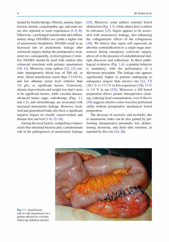
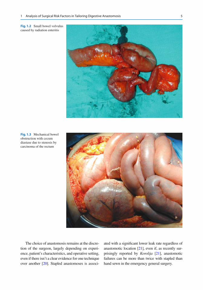
treated by insulin therapy. Obesity, anemia, hypo-tension, uremia, coagulopathy, age, and male sex are also reported in some experiences [ 1 – 5 , 8 ]. Otherwise, a prolonged nonsteroidal anti-infl am-matory drugs (NSAIDs) use yields a higher risk of anastomotic breakdown. NSAIDs result in an increased rate of anastomotic leakage after colorectal surgery during the postoperative treat-ment too; consequently, cyclooxygenase-2 selec-tive NSAIDs should be used with caution after colorectal resections with primary anastomosis [ 10 , 11 ]. Moreover, some authors [ 12 , 13 ] con-sider intraoperative blood loss of 200 mL or more, blood transfusions (more than 2 U/24 h), and low albumin serum level (inferior than 3.0 g/L) as signifi cant factors. Conversely, chronic hypovolemia and weight loss don’t seem to be signifi cant factors, while vascular disease, advanced tumor stage, radiotherapy (Figs. 1.1 and 1.2 ), and chemotherapy are associated with increased anastomotic leakage. However, local-ized and generalized leaks also have a signifi cant negative impact on overall, cancer-related, and disease-free survival [ 1 – 8 , 12 – 14 ].
Among the local factors, compelling evidence exists that intestinal bacteria play a predominant role in the pathogenesis of anastomotic leakage
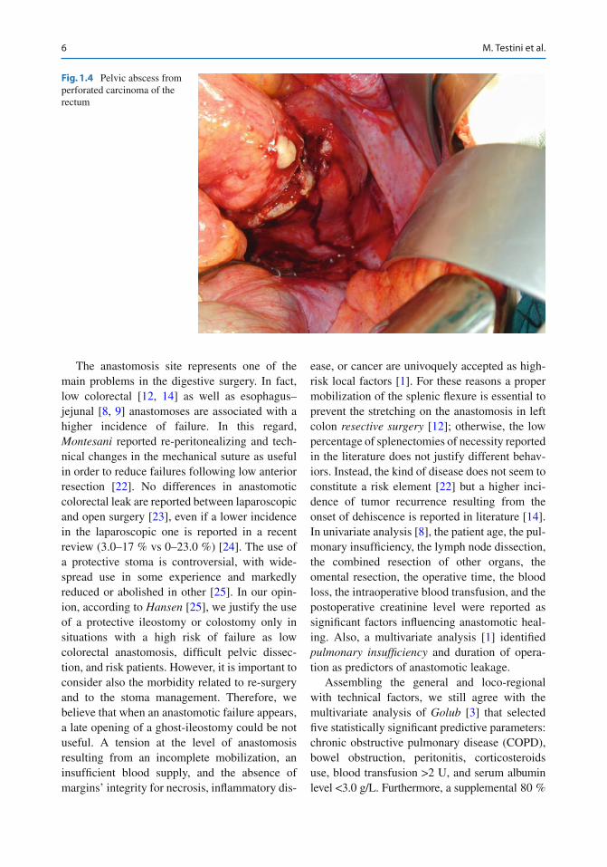
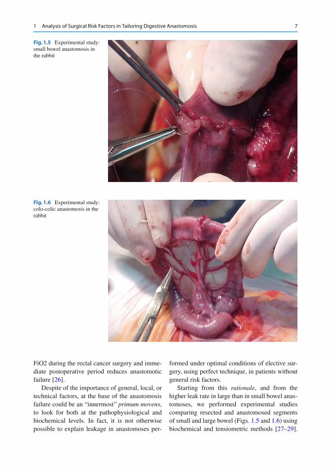
[ 15 ]. Moreover, some authors consider bowel obstruction (Fig. 1.3 ), while others don’t confi rm its relevance [ 13 ]. Sepsis appears to be associ-ated with anastomosis leakage, also enhancing the collagenolytic effects of the collagenosis [ 16 ]. We believe that sepsis still represents an absolute contraindication to a single-stage anas-tomosis during emergency colorectal surgery, above all in the presence of endoabdominal mul-tiple abscesses and collections. In these patho-logical evidences (Fig. 1.4 ), a prudent behavior is mandatory, with the performance of a Hartmann procedure. The leakage rate appears signifi cantly higher in patients undergoing to emergency surgery than elective one [ 12 , 17 ] (38.1 % vs 13.3 % in Kim experience [ 18 ], 13 % vs 3.9 % in our [ 13 ]). Moreover, a full bowel preparation allows greater intraoperative clean-ing, reducing fecal contamination, even if Harris [ 19 ] suggests elective colon resection performed safely without preoperative mechanical bowel preparation.
The decrease of mortality and morbidity due to anastomotic leaks can be also gained by per-forming intraoperative pneumatic test, defunc-tioning ileostomy, and drain tube insertion, as reported by Boccola [ 14 , 20 ].
Fig. 1.1 Small bowel side-to-side anastomosis in a patient affected by volvulus following radiation enteritis
M. Testini et al.
5
The choice of anastomosis remains at the discre-tion of the surgeon, largely depending on experi-ence, patient’s characteristics, and operative setting, even if there isn’t a clear evidence for one technique over another [ 20 ]. Stapled anastomoses is associ-
ated with a signifi cant lower leak rate regardless of anastomotic location [ 21 ], even if, as recently sur-prisingly reported by Korolija [ 21 ], anastomotic failures can be more than twice with stapled than hand sewn in the emergency general surgery.
Fig. 1.2 Small bowel volvulus caused by radiation enteritis
Fig. 1.3 Mechanical bowel obstruction with cecum diastase due to stenosis by carcinoma of the rectum
1 Analysis of Surgical Risk Factors in Tailoring Digestive Anastomosis
6
The anastomosis site represents one of the main problems in the digestive surgery. In fact, low colorectal [ 12 , 14 ] as well as esophagus–jejunal [ 8 , 9 ] anastomoses are associated with a higher incidence of failure. In this regard, Montesani reported re-peritonealizing and tech-nical changes in the mechanical suture as useful in order to reduce failures following low anterior resection [ 22 ]. No differences in anastomotic colorectal leak are reported between laparoscopic and open surgery [ 23 ], even if a lower incidence in the laparoscopic one is reported in a recent review (3.0–17 % vs 0–23.0 %) [ 24 ]. The use of a protective stoma is controversial, with wide-spread use in some experience and markedly reduced or abolished in other [ 25 ]. In our opin-ion, according to Hansen [ 25 ], we justify the use of a protective ileostomy or colostomy only in situations with a high risk of failure as low colorectal anastomosis, diffi cult pelvic dissec-tion, and risk patients. However, it is important to consider also the morbidity related to re-surgery and to the stoma management. Therefore, we believe that when an anastomotic failure appears, a late opening of a ghost-ileostomy could be not useful. A tension at the level of anastomosis resulting from an incomplete mobilization, an insuffi cient blood supply, and the absence of margins’ integrity for necrosis, infl ammatory dis-
ease, or cancer are univoquely accepted as high- risk local factors [ 1 ]. For these reasons a proper mobilization of the splenic fl exure is essential to prevent the stretching on the anastomosis in left colon resective surgery [ 12 ]; otherwise, the low percentage of splenectomies of necessity reported in the literature does not justify different behav-iors. Instead, the kind of disease does not seem to constitute a risk element [ 22 ] but a higher inci-dence of tumor recurrence resulting from the onset of dehiscence is reported in literature [ 14 ]. In univariate analysis [ 8 ], the patient age, the pul-monary insuffi ciency, the lymph node dissection, the combined resection of other organs, the omental resection, the operative time, the blood loss, the intraoperative blood transfusion, and the postoperative creatinine level were reported as signifi cant factors infl uencing anastomotic heal-ing. Also, a multivariate analysis [ 1 ] identifi ed pulmonary insuffi ciency and duration of opera-tion as predictors of anastomotic leakage.
Assembling the general and loco-regional with technical factors, we still agree with the multivariate analysis of Golub [ 3 ] that selected fi ve statistically signifi cant predictive parameters: chronic obstructive pulmonary disease (COPD), bowel obstruction, peritonitis, corticosteroids use, blood transfusion >2 U, and serum albumin level <3.0 g/L. Furthermore, a supplemental 80 %
Fig. 1.4 Pelvic abscess from perforated carcinoma of the rectum
M. Testini et al.
7
FiO2 during the rectal cancer surgery and imme-diate postoperative period reduces anastomotic failure [ 26 ].
Despite of the importance of general, local, or technical factors, at the base of the anastomosis failure could be an “innermost” primum movens , to look for both at the pathophysiological and biochemical levels. In fact, it is not otherwise possible to explain leakage in anastomoses per-
formed under optimal conditions of elective sur-gery, using perfect technique, in patients without general risk factors.
Starting from this rationale , and from the higher leak rate in large than in small bowel anas-tomoses, we performed experimental studies comparing resected and anastomosed segments of small and large bowel (Figs. 1.5 and 1.6 ) using biochemical and tensiometric methods [ 27 – 29 ].
Fig. 1.5 Experimental study: small bowel anastomosis in the rabbit
Fig. 1.6 Experimental study: colo-colic anastomosis in the rabbit
1 Analysis of Surgical Risk Factors in Tailoring Digestive Anastomosis
8
Previous experimental studies showed an early and massive deposition of collagen and a greater distress of the large compared with the small bowel. It is also well known the importance of the maturation of collagen in the anastomosis healing process and that an adequate metabolic energy is needed to realize healing process. Starting from these assumptions, our fi rst study [ 27 ] was to analyze the process of oxidative phosphorylation (mitochondrial func-tion) in colon and small bowel during the anasto-motic process. The results of polarographic, spectrophotometric, and gel-electrophoresis analysis showed a prevalence of oxidative metab-olism in the colic mitochondria compared with the small bowel, demonstrated by an increased activity of oxygen consumption and enzymatic respiratory. On the contrary, the small bowel showed a prevalence of glycolytic metabolism. Summarizing these results, the small bowel burns sugars through anaerobic glycolysis to produce energy for collagen deposition and healing pro-cess of anastomosis, and therefore is less infl u-enced by the decrease of available oxygen occurring in the anastomotic area during surgical stress. By contrast, colon shows a metabolism mainly linked to the oxidative phosphorylation, presents a more diffi cult anastomotic healing process in absence of oxygen, and shows a greater risk of leak. This observation is confi rmed by the decrement of biochemical parameters in colonic cells. In fact, at the end of the study, we observed a small bowel tissue biochemically identical to the preoperative one, while the colon tissue showed marked differences.
In the second phase of our experiments [ 29 ], we investigated if biochemical differences were also associated with motility and peristalsis. In fact, the aim was to verify in vitro how much the surgical stress could affect contractility of the smooth muscle (both spontaneous and agonist induced) of both organs, correlating these results to the biochemical parameters too. The results showed an anarchist contractility and late restart of colic peristalsis compared with an early and regular contractile activity of the small bowel. Such motor abnormalities may be the consequence of abnormal biochemical changes,
because the ATP is necessary in the mainte-nance of membrane potentials, in calcium homeostasis, and in the actin–myosin interac-tions. The study showed that surgical stress determines abnormalities in the mitochondria of the smooth muscle, damaging the contractility. In consequence of a diffi cult process of collagen maturation and deposition, these changes are prevalent in the colon and may explain unex-pected anastomotic leakage in the absence of apparent risk factors.
At confi rm of these experimental results, an other retrospective study [ 30 ] showed a signifi -cant leakage rate (24.1 % vs 2.7 %, P = 0.001) in patients who underwent colic resection, affected by COPD compared with patients not affected by COPD. COPD is characterized by a condition of chronic hypoxemia that determines a reduced peripheral oxygen delivery (DaO2). However, the mechanism of control of blood fl ow and of oxygen extraction at intestinal level let the con-sumption of oxygen (VO2) to be independent from DaO2; thus, the reduced DaO2 does not infl uence the VO2 in patients with COPD. On the contrary, during the healing process of colic anastomosis, the need of oxygen increases, both for higher metabolic request related to the oxida-tive phosphorylation and for the synthesis of col-lagen. In patients with COPD undergoing to resective surgery and colic anastomosis, these pathophysiologic changes inevitably relate the VO2 to the insuffi cient DaO2. Therefore, the cor-rection of impaired oxygen tension could reduce the high incidence of anastomotic leak in patients with COPD. On the basis of these results, a pre-operative evaluation of respiratory tract (chest X-ray, CT, spirometric tests, hemogasanalysis) is essential before colic resective surgery, espe-cially in aged patients affected by COPD. Moreover, a perioperative oxygen therapy also may facilitate anastomotic healing.
In a further experimental study [ 31 ] we investigated in pigs if pericardium bovine patch (Tutomesh®) wrapping ileoileal and colo-colic anastomosis seals the suture line and promotes anastomotic healing. By using integrated and trans-lational methodologies, we described intraopera-tive, histological, biochemical, tensiometric, and
M. Testini et al.
9
electrophysiological evaluations performed on intestinal specimens.
Biologic materials have been introduced in general surgery as reinforcement of abdominal wall hernia in contaminated or potentially con-taminated settings, when the use of alloplastic meshes is contraindicated [ 26 – 31 ]. In this respect, an innovative application of biologic patch could be their use as reinforcement of the gastrointestinal anastomotic suture line [ 7 – 9 ].Therefore, the aim of the study was to verify if bovine pericardium patch improved the healing of anastomosis, when in vivo affi xed on the hand- sewn suture line of large and small bowel anasto-mosis of the pigs.
A further end point was to verify if the patch was able to avoid anastomotic leakage in the presence of a deliberately incomplete left suture.
The results showed that the application of a patch wrapping the colic anastomosis produces a positive effect in the healing compared with untreated samples also showing, during follow- up, an almost full recovery [ 1 – 3 , 26 ]. In the large bowel patch anastomosis group, the delay of oxi-dative stress in the early stage of reparative pro-cesses could prevent the damage of noble cells (like tissue stem cells), allowing a full restoration of tissue functions and also decreasing fi brotic reaction during the next stages of healing pro-cess. Under a condition of cellular oxidative stress, the protective effect of the patch is com-patible with the histological observation of a moderate infl ammatory infi ltrate; moreover, the late increase of reacting oxygen species can be correlated with an appearance of a granulation tissue, without damages during the repairing pro-cess. Therefore, tensiometric evaluations in colic specimens suggested that the use of patch can preserve smooth muscle response to acetylcho-line similar to the response of controls (speci-mens without anastomosis) in the early postoperative time (48 h–14 days), while the colic preparations with traditional anastomosis showed contractility alterations. In the ileum, the presence of pericardium bovine patch clearly pre-vents the alterations following the traumatic effect of surgery. However, pericardium bovine patch appears to modulate and counteract the
traumatic effect of surgery. Overall, our results suggest that the application of the patch also improves the intestinal mucosal function, restor-ing the almost normal transport properties. In conclusion, the use of the pericardium bovine patch as reinforcement of the intestinal anasto-mosis could be safe and effective. Moreover, the leakage prevention in the presence of iatrogenic perforation is also unpublished before and it rep-resents a surprising histopathological data. On the basis of these experimental results, we started a multicenter-controlled clinical trial in humans, comparing the outcomes of intestinal anastomosis performed with and without the bovine pericar-dium patch in risk patients.
In conclusion, despite studies regarding risk factors and prevention, the anastomotic leakage continues to be the most serious complication after gastrointestinal tract surgery . A thorough surgical technique, avoiding hazardous anasto-moses without protective stoma, or without two- stage surgery in patients at risk, could allow a signifi cant reduction of healing process failure. A tailored surgical approach to both patient’s physi-ology and disease is the most important factor that infl uences anastomotic integrity after resec-tive surgery. Further studies regarding innovative devices able to improve the healing process of anastomosis are needed.
References
1. Telem D, Chin E, Nguyen S et al (2010) Risk factors for anastomotic leak following colorectal surgery. A case–control study. Arch Surg 145:371–375
2. Kang CY, Halabi WJ, Chaudhry OO et al (2013) Risk factors for anastomotic leakage after anterior resec-tion for rectal cancer. JAMA Surg 148(1):65–71
3. Golub R, Golub RW, Cantu R Jr et al (1997) A multi-variate analysis of factors contributing to leakage of intestinal anastomoses. J Am Coll Surg 184:364–372
4. Trencheva K, Morrissey KP, Wells M et al (2013) Identifying important predictors for anastomotic leak after colon and rectal resection: prospective study on 616 patients. Ann Surg 257(1):108–113
5. Snijders HS, Wouters MW, van Leersum NJ et al (2012) Meta-analysis of the risk for anastomotic leak-age, the postoperative mortality caused by leakage in relation to the overall postoperative mortality. Eur J Surg Oncol 38(11):1065–1070
1 Analysis of Surgical Risk Factors in Tailoring Digestive Anastomosis
10
6. Branagan G, Finnis D, Colorectal Cancer Audit Working Group et al (2005) Prognosis after anasto-motic leakage in colorectal surgery. Dis Colon Rectum 48:1021–1026
7. Sierzega M, Kolodziejczyk P, Kulig J, Polish Gastric Cancer Study Group et al (2010) Impact of anasto-motic leakage on long-term survival after total gas-trectomy for carcinoma of the stomach. Br J Surg 97(7):1035–1042
8. Deguchi Y, Fukagawa T, Morita S et al (2012) Identifi cation of risk factors for esophagojejunal anas-tomotic leakage after gastric surgery. World J Surg 36(7):1617–1622
9. Markar SR, Arya S, Karthikesalingam A et al (2013) Technical factors that affect anastomotic integrity fol-lowing esophagectomy: systematic review and meta- analysis. Ann Surg Oncol 20(13):4274–81
10. Rutegård J, Rutegård M (2012) Non-steroidal anti- infl ammatory drugs in colorectal surgery: a risk factor for anastomotic complications? World J Gastrointest Surg 4(12):278–280
11. Klein M, Gögenur I, Rosenberg J et al (2012) Postoperative use of non-steroidal anti-infl ammatory drugs in patients with anastomotic leakage requiring reoperation after colorectal resection: cohort study based on prospective data. BMJ 26(9):345, 1–13
12. Warschkow R, Steffen T, Thierbach J et al (2011) Risk factors for anastomotic leakage after rectal can-cer resection and reconstruction with colorectostomy. A retrospective study with bootstrap analysis. Ann Surg Oncol 18(10):2772–2782
13. Testini M, Margari A, Amoruso M et al (2000) The dehiscence of colorectal anastomoses: the risk fac-tors. Ann Ital Chir 71:433–440
14. Boccola MA, Buettner PG, Rozen WM et al (2011) Risk factors and outcomes for anastomotic leakage in colorectal surgery: a single-institution analysis of 1576 patients. World J Surg 35(1):186–195
15. Shogan BD, Carlisle EM, Alverdy JC et al (2013) Do we really know why colorectal anastomoses leak? J Gastrointest Surg 17(9):1698–1707
16. Miccini M, Borghese O, Scarpini M et al (2011) Anastomotic leakage and septic complications: impact on local recurrence in surgery of low rectal cancer. Ann Ital Chir 82(2):117–123
17. Matthiessen P, Hallböök O, Rutegård J et al (2007) Defunctioning stoma reduces symptomatic anasto-motic leakage after low anterior resection of the rec-tum for cancer: a randomized multicenter trial. Ann Surg 246:207–214
18. Kim JJ, Liang MK, Subramanian A et al (2011) Predictors of relaparotomy after nontrauma emer-gency general surgery with initial fascial closure. Am J Surg 202(5):549–552
19. Harris LJ, Moudgill N, Hager E et al (2009) Incidence of anastomotic leak in patients undergoing elective colon resection without mechanical bowel prepara-tion: our updated experience and two-year review. Am Surg 75(9):828–833
20. Boccola MA, Lin J, Rozen WM et al (2010) Reducing anastomotic leakage in oncologic colorectal surgery: an evidence-based review. Anticancer Res 30(2):601–607
21. Korolija D (2008) The current evidence on stapled versus hand-sewn anastomoses in the digestive tract. Minim Invasive Ther Allied Technol 17(3):151–154
22. Montesani C, De Milito R, Chiappalone S et al (1992) Critical evaluation of the anastomoses in large bowel experience in 533 cases. Hepatogastroenterology 39:304–308
23. El-Gazzaz G, Geisler D, Hull T et al (2010) Surgery: risk of clinical leak after laparoscopic versus open bowel anastomosis. Surg Endosc 24(8):1898–1903
24. Hotta T, Yamaue H (2011) Laparoscopic surgery for rectal cancer: review of published literature 2000–2009. Surg Today 41(12):1583–1591
25. Hansen O, Schwenk W, Hucke HP et al (1996) Colorectal stapled anastomoses. Dis Colon Rectum 39:30–36
26. Schietroma M, Carlei F, Cecilia EM et al (2012) Colorectal infraperitoneal anastomosis: the effects of perioperative supplemental oxygen administration on the anastomotic dehiscence. J Gastrointest Surg 16(2):427–434
27. Testini M, Scacco S, Loiotila L et al (1998) Comparison of oxidative phosphorylation in the small vs large bowel anastomosis. Eur Surg Res 30(1):1–7
28. Testini M, Piccinni G et al (1999) Wound healing of intestinal anastomosis after digestive surgery under septic condition. World J Surg 23:1315–1316
29. Testini M, Portincasa P, Scacco S et al (2002) Contractility in vitro and mitochondrial response in small and large anastomized rabbit bowel. World J Surg 26:493–498
30. Testini M, Miniello S, Piccinni G et al (2003) Correlation between chronic obstructive bronchial disease and colonic anastomosis dehiscence in the elderly. Ann Ital Chir 74:247–250
31. Portincasa P, Testini M et al (2011) The apposition of a resorbable pericardial Bovine patch (Tutomesh®) on intestinal anastomoses improves functional mucosal recovery in pig ileum and colon assessed by using chamber electrophysiological studies. Gastroenterology 140(5 suppl 1):S-656
M. Testini et al.
11G. Galloro (ed.), Endoscopic Follow-up of Digestive Anastomosis, DOI 10.1007/978-88-470-5370-0_2, © Springer-Verlag Italia 2014
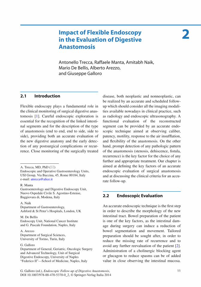
2.1 Introduction
Flexible endoscopy plays a fundamental role in the clinical monitoring of surgical digestive anas-tomosis [ 1 ]. Careful endoscopic exploration is essential for the recognition of the linked intesti-nal segments and for the description of the type of anastomosis (end to end, end to side, side to side), providing both an accurate evaluation of the new digestive anatomy and the early detec-tion of any postsurgical complications or recur-rence. Close monitoring of the surgically treated
disease, both neoplastic and nonneoplastic, can be realized by an accurate and scheduled follow- up which should consider all the imaging modali-ties available nowadays in clinical practice, such as radiology and endoscopic ultrasonography. A functional evaluation of the reconstructed segment can be provided by an accurate endo-scopic technique aimed at observing caliber, patency, motility, response to the air insuffl ation, and fl exibility of the anastomosis. On the other hand, prompt detection of any pathologic pattern of the anastomosis (stenosis, dehiscence, fi stula, recurrence) is the key factor for the choice of any further and appropriate treatment. Our chapter is aimed at defi ning the key factors of an accurate endoscopic evaluation of surgical anastomosis and at discussing the clinical criteria for an accu-rate follow-up.
2.2 Endoscopic Evaluation
An accurate endoscopic technique is the fi rst step in order to describe the morphology of the new intestinal tract. Bowel preparation of the patient is one of the key factors, as the intestinal dam-age during surgery can induce a reduction of bowel segmentation and movement. Tailored preparation should be sought after, in order to reduce the missing rate of recurrence and to avoid any further reevaluation of the patient [ 2 ]. Administration of a cholinergic blocking agent or glucagon to reduce spasms can be of added value in close observing the intestinal mucosa.
A. Trecca , MD, PhD (*) Endoscopic and Operative Gastroenterology Units , USI Group , Via Baccina, 45 , Rome 00184 , Italy e-mail: [email protected]
R. Manta Gastroenterology and Digestive Endoscopy Unit , Nuovo Ospedale Civile S. Agostino-Estense, Baggiovara di , Modena , Italy
A. Naik Department of Gastroenterology , Ashford & St Peter’s Hospitals , London , UK
M. De Bellis Endoscopy Unit , National Cancer Institute and G. Pascale Foundation , Naples , Italy
A. Arezzo Department of Surgical Sciences , University of Torino , Turin , Italy
G. Galloro Department of General, Geriatric, Oncologic Surgery and Advanced Technology, Unit of Surgical Digestive Endoscopy , University of Naples “Federico II”—School of Medicine , Naples , Italy
2 Impact of Flexible Endoscopy in the Evaluation of Digestive Anastomosis
Antonello Trecca , Raffaele Manta , Amitabh Naik , Mario De Bellis , Alberto Arezzo , and Giuseppe Galloro
12
Some authors underline possible side effects and suggest the intracolonic administration of pep-permint oil during colonoscopy for the control of colonic spasms. Asao [ 3 ] refers on a satisfac-tory spasmolytic effect in 88.5 % of the patients treated with a mixed solution of peppermint oil, water, and indigo carmine by using a hand pump attached to the accessory channel of the colono-scope, with a continuing effect of at least 20 min. Endoscopic observation should consider the use-ful role of the air in the evaluation of intestinal lumen with its adequate introduction and aspira-tion during the exploration of the anastomosis. Injection of a saline solution directly or using an irrigation pump through the accessory channel of the endoscope is another tool in the hand of the endoscopist to improve the quality of gastroin-testinal exploration. Flexible endoscopy should always evaluate the caliber of the intestinal lumen which can be measured by using an opened biopsy forcep and the main longitudinal axis of the new reconstructed intestinal tract (Fig. 2.1 ). The description of the type and morphology of the surgical anastomosis should always be pro-vided in the endoscopic report. After a complete evaluation of the functional status of the anas-tomotic site, including its patency and motility, fl exible endoscopy should be prolonged to the evaluation of the proximal and distal parts and to all the reconstructed segments in order not to miss any morphologic change of the intestinal tract. The presence of metallic clips or suture stitches along the border of the anastomosis are often visible during upper and lower endoscopy as far as the presence of connecting venules, which refl ects the healing process of the mucosa and rarely can cause impairment of the anasto-mosis. After an accurate cleaning of the intes-tinal lumen, the surgical anastomosis should be accurately checked for any mucosal defect such as discolorations, atrophic changes, and nodular irregularities which can be the expression of a redundant mucosal response or can mimic the presence of an endoluminal recurrence (Figs. 2.2 and 2.3 ). In this scenario the role of histol-ogy is mandatory to complete the endoscopic
evaluation of the anastomosis and to detect any infl ammatory or neoplastic change. We have to consider that any surgical intervention creates a new and different environment, and it should be taken into account when we study upper or lower gastrointestinal tract. So far the gastric remnant has been considered at higher risk for gastric cancer with an increasing postoperative inter-val, with a well- established clinical entity after remote surgery for peptic ulcer, called gastric stump carcinoma [ 4 ]. Many factors are involved in the pathogenesis such as achlorhydria, hyper-gastrinemia, biliary refl ux, Epstein–Barr virus, atrophic gastritis, and also some polymorphisms in interleukin-1β and maybe cyclooxygenase-2. The microscopy of the anastomosis changes from the chronic active H. pylori gastritis into the typical refl ux gastritis with foveolar hyper-plasia, congestion, paucity of infl ammatory infi ltrate, reactive epithelial change, and smooth muscle fi ber proliferation which slowly evolve to preneoplastic conditions, particularly dys-plasia. Endoscopic surveillance is mandatory particularly in this clinical condition where the detection of premalignant or early neoplastic lesions is more frequent [ 5 ]. Concerning the lower tract, ileal-pouch anastomosis after proc-tocolectomy represents another example of how the modifi ed clinical environment can lead to a new disease condition, named as pouchitis and
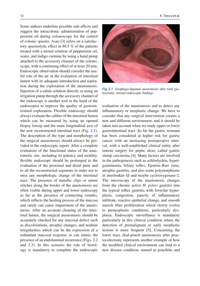
Fig. 2.1 Esophago-digiunal anastomosis after total gas-trectomy: normal endoscopic fi ndings
A. Trecca et al.
13
characterized by a nonspecifi c infl ammation of the ileal reservoir. Bacterial overgrowth, chronic infl ammation, and villous atrophy, even if always present, can evolve in pouchitis in some cases, mainly after surgery for ulcerative colitis, and for this reason pouchitis is considered an infl amma-tory bowel disease. Lower endoscopy, together with an accurate histopathological evaluation, is mandatory for studying and monitoring this condition [ 6 ].
A signifi cant reduction of the intestinal lumen, even if asymptomatic, should be described and monitored, while in case of intestinal stenosis, fi stula, or dehiscence, other imaging modalities together with prompt treatment should be sched-uled and selected among the different options
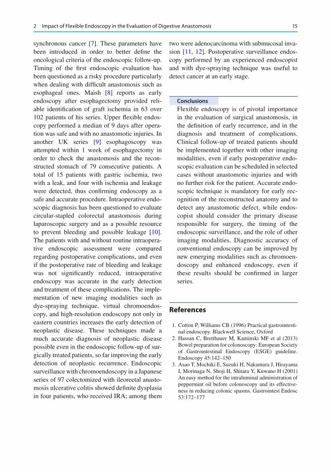
(endoscopic dilation, stent placement, or surgical reconstruction) (Fig. 2.4 ).
2.3 Oncological Criteria of Follow-Up
Endoscopists should keep in mind clinical crite-ria for an accurate follow-up of the patient: syn-chronous cancer is defi ned as a cancer detected within 1 year of follow-up, while metachronous cancer is that one detected after 1 year of follow- up, while concomitant cancers are defi ned as multiple cancers detected before the surgical treatment. In this setting, we defi ne the miss rate as the proportion of missed cancer out of all
a b
c d
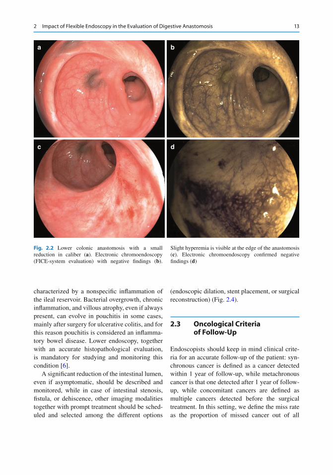
Fig. 2.2 Lower colonic anastomosis with a small reduction in caliber ( a ). Electronic chromoendoscopy (FICE-system evaluation) with negative fi ndings ( b ).
Slight hyperemia is visible at the edge of the anastomosis ( c ). Electronic chromoendoscopy confi rmed negative fi ndings ( d )
2 Impact of Flexible Endoscopy in the Evaluation of Digestive Anastomosis
14
a
c
b
d
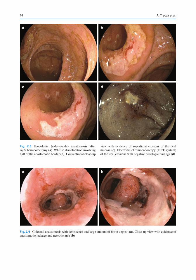
Fig. 2.3 Ileocolonic (side-to-side) anastomosis after right hemicolectomy ( a ). Whitish discoloration involving half of the anastomotic border ( b ). Conventional close-up
view with evidence of superfi cial erosions of the ileal mucosa ( c ). Electronic chromoendoscopy (FICE system) of the ileal erosions with negative histologic fi ndings ( d )
a b
Fig. 2.4 Coloanal anastomosis with dehiscence and large amount of fi brin deposit ( a ). Close-up view with evidence of anastomotic leakage and necrotic area ( b )
A. Trecca et al.
15
synchronous cancer [ 7 ]. These parameters have been introduced in order to better defi ne the oncological criteria of the endoscopic follow-up. Timing of the fi rst endoscopic evaluation has been questioned as a risky procedure particularly when dealing with diffi cult anastomosis such as esophageal ones. Maish [ 8 ] reports as early endoscopy after esophagectomy provided reli-able identifi cation of graft ischemia in 63 over 102 patients of his series. Upper fl exible endos-copy performed a median of 9 days after opera-tion was safe and with no anastomotic injuries. In another UK series [ 9 ] esophagoscopy was attempted within 1 week of esophagectomy in order to check the anastomosis and the recon-structed stomach of 79 consecutive patients. A total of 15 patients with gastric ischemia, two with a leak, and four with ischemia and leakage were detected, thus confi rming endoscopy as a safe and accurate procedure. Intraoperative endo-scopic diagnosis has been questioned to evaluate circular-stapled colorectal anastomosis during laparoscopic surgery and as a possible resource to prevent bleeding and possible leakage [ 10 ]. The patients with and without routine intraopera-tive endoscopic assessment were compared regarding postoperative complications, and even if the postoperative rate of bleeding and leakage was not signifi cantly reduced, intraoperative endoscopy was accurate in the early detection and treatment of these complications. The imple-mentation of new imaging modalities such as dye-spraying technique, virtual chromoendos-copy, and high-resolution endoscopy not only in eastern countries increases the early detection of neoplastic disease. These techniques made a much accurate diagnosis of neoplastic disease possible even in the endoscopic follow-up of sur-gically treated patients, so far improving the early detection of neoplastic recurrence. Endoscopic surveillance with chromoendoscopy in a Japanese series of 97 colectomized with ileorectal anasto-mosis ulcerative colitis showed defi nite dysplasia in four patients, who received IRA; among them
two were adenocarcinoma with submucosal inva-sion [ 11 , 12 ]. Postoperative surveillance endos-copy performed by an experienced endoscopist and with dye- spraying technique was useful to detect cancer at an early stage.
Conclusions
Flexible endoscopy is of pivotal importance in the evaluation of surgical anastomosis, in the defi nition of early recurrence, and in the diagnosis and treatment of complications. Clinical follow- up of treated patients should be implemented together with other imaging modalities, even if early postoperative endo-scopic evaluation can be scheduled in selected cases without anastomotic injuries and with no further risk for the patient. Accurate endo-scopic technique is mandatory for early rec-ognition of the reconstructed anatomy and to detect any anastomotic defect, while endos-copist should consider the primary disease responsible for surgery, the timing of the endoscopic surveillance, and the role of other imaging modalities. Diagnostic accuracy of conventional endoscopy can be improved by new emerging modalities such as chromoen-doscopy and enhanced endoscopy, even if these results should be confi rmed in larger series.
References
1. Cotton P, Williams CB (1996) Practical gastrointesti-nal endoscopy. Blackwell Science, Oxford
2. Hassan C, Bretthauer M, Kaminski MF et al (2013) Bowel preparation for colonoscopy: European Society of Gastrointestinal Endoscopy (ESGE) guideline. Endoscopy 45:142–150
3. Asao T, Mochiki E, Suzuki H, Nakamura J, Hirayama I, Morinaga N, Shoji H, Shitara Y, Kuwano H (2001) An easy method for the intraluminal administration of peppermint oil before colonoscopy and its effective-ness in reducing colonic spasms. Gastrointest Endosc 53:172–177
2 Impact of Flexible Endoscopy in the Evaluation of Digestive Anastomosis
16
4. Sitarz R, Maciejewski R, Polkowski WP, Offerhaus JA (2012) Gastroenterostoma after Billroth antrec-tomy as a premalignant condition. World J Gastroenterol 18:3201–3206
5. Lee Y, Tokunaga A, Tajiri T et al (2004) Infl ammation of the gastric remnant after gastrectomy: mucosal ery-thema is associated with bile refl ux and infl ammatory cellular infi ltration is associated with Helicobacter pylori infection. J Gastroenterol 39:520–526
6. Salemans JM, Nagengast FM (1995) Clinical and physiological aspects of ileal pouch-anal anastomosis. Scand J Gastroenterol Suppl 212:3–12
7. Lee JY, Choi I, Cho S (2012) Routine follow-up biop-sies after complete endoscopic resection for early gas-tric cancer may be unnecessary. J Gastric Cancer 12:88–98
8. Maish MS, DeMeester SR, Choustoulakis E, Briel JW, Hagen JA, Peters JH, Lipham JC, Bremner CG, DeMeester TR (2005) The safety and usefulness of
endoscopy for evaluation of the graft and anastomosis early after esophagectomy and reconstruction. Surg Endosc 19:1093–12
9. Page RD, Asmat A, McShane J, Russell GN, Pennefather SH (2013) Routine endoscopy to detect anastomotic leakage after esophagectomy. Ann Thorac Surg 95:292–298
10. Shamiyeh A, Szabo K, Ulf Wayand W, Zehetner J (2012) Intraoperative endoscopy for the assessment of circular-stapled anastomosis in laparoscopic colon surgery. Surg Laparosc Endosc Percutan Tech 22:65–67
11. Herline AJ, Meisinge LL, Rusin LC et al (2003) Is routine pouch surveillance for dysplasia indicated for ileoanal pouches? Dis Colon Rectum 46:156–159
12. Shuno Y, Hata K, Sunami E et al (2011) Is surveil-lance endoscopy necessary after colectomy in ulcer-ative colitis?. Int Scholarly Res Netw Gastroenterol Article ID 509251. doi: 10.5402/2011/509251
A. Trecca et al.
17G. Galloro (ed.), Endoscopic Follow-up of Digestive Anastomosis, DOI 10.1007/978-88-470-5370-0_3, © Springer-Verlag Italia 2014
3.1 General Criteria
Endoscopic follow-up is defi ned as the perfor-mance of endoscopic examination(s) subsequent to an index endoscopy aimed at both: (a) Monitoring neoplastic or pre-neoplastic con-
ditions or patients at increased risk (more properly termed “surveillance”)
(b) Monitoring the therapeutic response to a determinate treatment (be it pharmacologi-cal, endoscopic, or surgical)
The end point of any endoscopic follow-up is that of reducing morbidity and mortality associ-ated with or deriving from the pathologic condi-tion that is under specifi c surveillance.
Examples of surveillance of pre-neoplastic conditions are Barrett’s metaplasia of esophagus, infl ammatory bowel diseases, colorectal adeno-mas, and polyposis syndromes. Another type of follow-up is the surveillance of patients submit-ted to surgery for a malignant disease, where the patient undergoes scheduled periodical postop-erative examinations aimed at early identifi cation of any recurrence of the primary cancer.
Among the endoscopic controls to assess the outcome of a treatment are the healing of gastro-duodenal ulcers or erosive refl ux esophagitis, the mucosal healing in patients with ulcerative colitis
or Crohn’s disease, a second look after endo-scopic hemostasis of bleeding peptic ulcers, the eradication of esophageal varices, the histological control of gastritis or of the eradication of H. pylori , etc.
As a general rule, the rationale for any follow- up in digestive endoscopy raises from the epide-miologic evidence of its necessity and clinical benefi t: the disease under surveillance should, in fact, be epidemiologically relevant (it would be illogical to monitor patients to identify a rare dis-ease) and its detection and subsequent early treat-ment should entail a prognostic gain or advantage for the patient as compared to the treatment of the same condition identifi ed when symptomatic.
The sticky issue of endoscopic follow-up would inevitably lead us to face up with two awkward truths that are overuse of surveillance and poor quality of surveillance. In fact, not only do we perform too much surveillance, with inevi-table working overloads for our endoscopy ser-vices, but the quality of surveillance is often poor. This last condition can be due to conceptual defi ciency (inconsistent evidence of clinical util-ity of the follow-up or inadequate knowledge of guidelines) or sometimes due to professional and/or supply shortage (technical competency not homogenous between different operators, equipment not always adequate for the specifi c needs of surveillance of specifi c conditions).
In general, the appropriateness is the quality of being just right for the requirements. In clini-cal medicine, it means that a test or an interven-tion is adequate and pertinent, i.e., suitable for a
G. Rotondano (*) • S. Sansone • C. Cesaro Division of Gastroenterology and Endoscopy , “A. Maresca” Hospital , Via Montedoro , Torre del Greco 80059 , Italy e-mail: [email protected]; [email protected]; [email protected]
3 Methodology and Appropriateness of Follow-Up in Digestive Endoscopy
Gianluca Rotondano , Stefano Sansone , and Claudia Cesaro
18
particular subject and performed for a correct indication in a given moment with the aim of pro-ducing health without wasting resources.
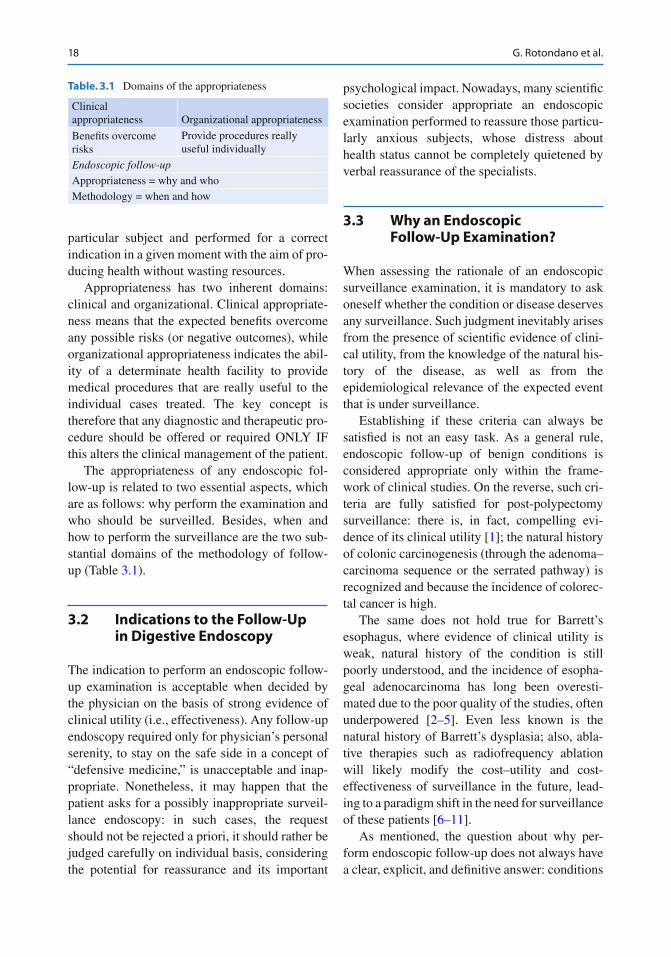
Appropriateness has two inherent domains: clinical and organizational. Clinical appropriate-ness means that the expected benefi ts overcome any possible risks (or negative outcomes), while organizational appropriateness indicates the abil-ity of a determinate health facility to provide medical procedures that are really useful to the individual cases treated. The key concept is therefore that any diagnostic and therapeutic pro-cedure should be offered or required ONLY IF this alters the clinical management of the patient.
The appropriateness of any endoscopic fol-low- up is related to two essential aspects, which are as follows: why perform the examination and who should be surveilled. Besides, when and how to perform the surveillance are the two sub-stantial domains of the methodology of follow- up (Table 3.1 ).
3.2 Indications to the Follow-Up in Digestive Endoscopy
The indication to perform an endoscopic follow- up examination is acceptable when decided by the physician on the basis of strong evidence of clinical utility (i.e., effectiveness). Any follow-up endoscopy required only for physician’s personal serenity, to stay on the safe side in a concept of “defensive medicine,” is unacceptable and inap-propriate. Nonetheless, it may happen that the patient asks for a possibly inappropriate surveil-lance endoscopy: in such cases, the request should not be rejected a priori, it should rather be judged carefully on individual basis, considering the potential for reassurance and its important
psychological impact. Nowadays, many scientifi c societies consider appropriate an endoscopic examination performed to reassure those particu-larly anxious subjects, whose distress about health status cannot be completely quietened by verbal reassurance of the specialists.
3.3 Why an Endoscopic Follow-Up Examination?
When assessing the rationale of an endoscopic surveillance examination, it is mandatory to ask oneself whether the condition or disease deserves any surveillance. Such judgment inevitably arises from the presence of scientifi c evidence of clini-cal utility, from the knowledge of the natural his-tory of the disease, as well as from the epidemiological relevance of the expected event that is under surveillance.
Establishing if these criteria can always be satisfi ed is not an easy task. As a general rule, endoscopic follow-up of benign conditions is considered appropriate only within the frame-work of clinical studies. On the reverse, such cri-teria are fully satisfi ed for post-polypectomy surveillance: there is, in fact, compelling evi-dence of its clinical utility [ 1 ]; the natural history of colonic carcinogenesis (through the adenoma–carcinoma sequence or the serrated pathway) is recognized and because the incidence of colorec-tal cancer is high.
The same does not hold true for Barrett’s esophagus, where evidence of clinical utility is weak, natural history of the condition is still poorly understood, and the incidence of esopha-geal adenocarcinoma has long been overesti-mated due to the poor quality of the studies, often underpowered [ 2 – 5 ]. Even less known is the natural history of Barrett’s dysplasia; also, abla-tive therapies such as radiofrequency ablation will likely modify the cost–utility and cost- effectiveness of surveillance in the future, lead-ing to a paradigm shift in the need for surveillance of these patients [ 6 – 11 ].
As mentioned, the question about why per-form endoscopic follow-up does not always have a clear, explicit, and defi nitive answer: conditions
Table. 3.1 Domains of the appropriateness
Clinical appropriateness Organizational appropriateness
Benefi ts overcome risks
Provide procedures really useful individually
Endoscopic follow-up Appropriateness = why and who Methodology = when and how
G. Rotondano et al.
19
that today are kept under surveillance may in the future be not as the progress in medicine removes the grey shadows from our knowledge.
3.4 Who Should Be Surveilled?
Once established that the condition actually deserves surveillance, the other key question is the selection of candidates , i.e., which patient affected with a given risk condition should be really kept under surveillance?
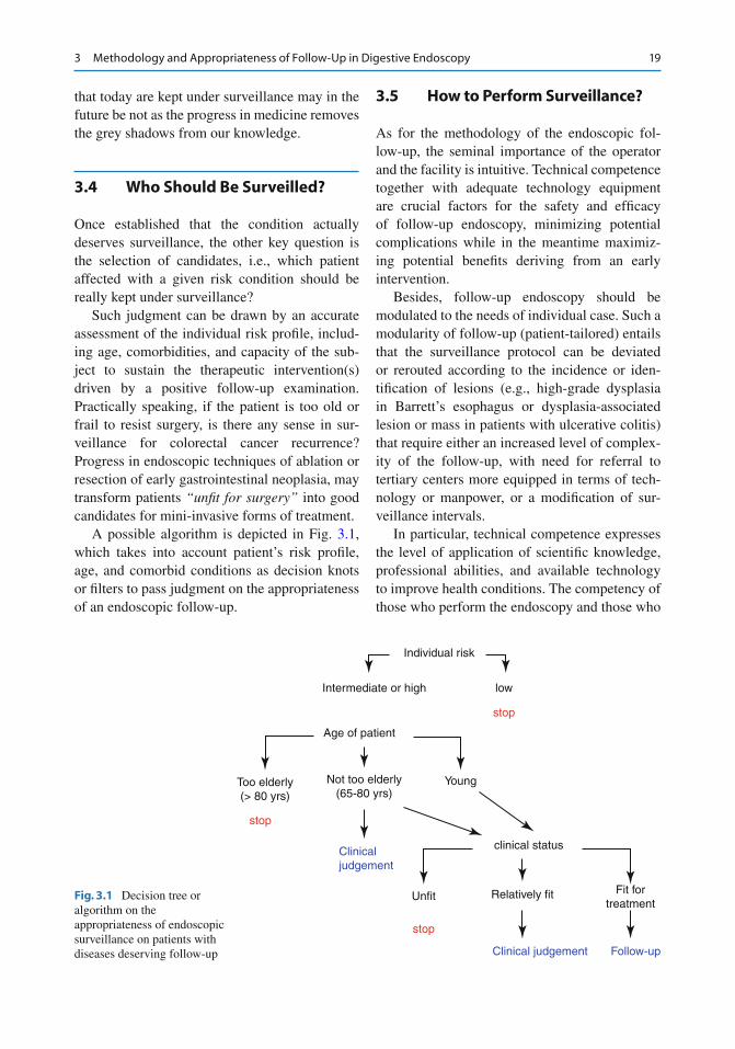
Such judgment can be drawn by an accurate assessment of the individual risk profi le, includ-ing age, comorbidities, and capacity of the sub-ject to sustain the therapeutic intervention(s) driven by a positive follow-up examination. Practically speaking, if the patient is too old or frail to resist surgery, is there any sense in sur-veillance for colorectal cancer recurrence? Progress in endoscopic techniques of ablation or resection of early gastrointestinal neoplasia, may transform patients “unfi t for surgery” into good candidates for mini-invasive forms of treatment.
A possible algorithm is depicted in Fig. 3.1 , which takes into account patient’s risk profi le, age, and comorbid conditions as decision knots or fi lters to pass judgment on the appropriateness of an endoscopic follow-up.
3.5 How to Perform Surveillance?
As for the methodology of the endoscopic fol-low- up, the seminal importance of the operator and the facility is intuitive. Technical competence together with adequate technology equipment are crucial factors for the safety and effi cacy of follow- up endoscopy, minimizing potential complications while in the meantime maximiz-ing potential benefi ts deriving from an early intervention.
Besides, follow-up endoscopy should be modulated to the needs of individual case. Such a modularity of follow-up (patient-tailored) entails that the surveillance protocol can be deviated or rerouted according to the incidence or iden-tifi cation of lesions (e.g., high-grade dysplasia in Barrett’s esophagus or dysplasia-associated lesion or mass in patients with ulcerative colitis) that require either an increased level of complex-ity of the follow-up, with need for referral to tertiary centers more equipped in terms of tech-nology or manpower, or a modifi cation of sur-veillance intervals.
In particular, technical competence expresses the level of application of scientifi c knowledge, professional abilities, and available technology to improve health conditions. The competency of those who perform the endoscopy and those who
Individual risk
Intermediate or high
Age of patient
low
stop
stop
stop
Clinicaljudgement
Clinical judgement Follow-up
Too elderly(> 80 yrs)
Not too elderly(65-80 yrs)
Unfit
Young
clinical status
Relatively fit Fit fortreatment
Fig. 3.1 Decision tree or algorithm on the appropriateness of endoscopic surveillance on patients with diseases deserving follow-up
3 Methodology and Appropriateness of Follow-Up in Digestive Endoscopy
20
draw the histology report that infl uences all the subsequent decisional chain is by no way a sec-ondary aspect. This represents a big problem at all latitudes: ability to match up with the situation should never be taken for granted .
A major aspect in terms of operator’s compe-tence for surveillance endoscopy is the diagnos-tic accuracy and particularly its negative, i.e., the rate of missed diagnosis of pre-neoplastic or neo-plastic lesions (missing rate). Any cancer detected within 3 years of a previous “negative” colonos-copy is termed “interval” cancer and should be considered as a missed lesion [ 12 – 18 ]. The miss-ing rate is not a trivial problem: also, in expert hands, it can be as high as 25 % for small or fl at adenomas [ 19 – 21 ] and can reach up to 7 % for overt cancers [ 15 – 17 ]. While the missing rate may not heavily impact effi cacy of screening endoscopy, where majority of patients are healthy and does not have cancer, it is potentially devas-tating in the fi eld of surveillance, which, in turn, is directed toward subgroups of patients at risk of having or developing cancer.
The critical question: is this exam really nega-tive? It urges that everyone performing a surveil-lance colonoscopy knows and applies those techniques allowing for an accurate evaluation of bowel mucosa during apparently negative exami-nations (chromoendoscopy, magnifi cation, light technology, etc.) [ 22 , 23 ]. Such competency requires formal and adequate training and con-tinuous updating (maintenance curve), possibly periodically audited or assessed by external inde-pendent subjects (credentialing) and that is when things begin to get diffi cult.
Health systems often taken as an example in terms of quality have made tremendous efforts to improve patients’ outcomes through systematic quality improvement programs. In UK, screening colonoscopists have improved their overall cecal intubation rate from less than 60 % in 2006 [ 24 ] to over 90 % in 2011 [ 25 ] thanks to a nationwide training and retraining process of all those involved in CRC screening. Improving colono-scopic skills and bowel preparation may also decrease nonadherence to the recommended post-polypectomy surveillance interval. Inadequate training and absence of retraining inevitably lead
to insuffi cient endoscopic practice not up to the high-quality standards often required by an accu-rate follow-up.
3.6 When to Perform Surveillance?
Last but not least there is the issue of when to perform surveillance, that is, the appropriate tim-ing of endoscopic follow-up start up and the opti-mal interval between examinations. Such information, at best, can be derived by specifi c guidelines on the disease, when available [ 26 , 27 ]. The personal clinical practice should then be tailored accordingly.
As a paradigmatic example, follow-up after curative surgery for colorectal cancer remains controversial, with no consensus on a protocol. Its evolution has largely lacked an evidence base. Current guidelines from the UK, the USA, Europe, and Canada all have differing recommended schedules for clinic visits, carcinoembryonic antigen (CEA) levels, colonoscopy, and abdomi-nal and chest imaging [ 28 ]. However, there is a global lack of consistency. Standardized follow-up regimens need to be developed. Many institutions continue to have their own follow-up regimen. As life expectancy increases, with a reduction in all-cause mortality and spiralling costs of sophis-ticated imaging modalities, intensive follow-up regimens are becoming more expensive. The cost- effectiveness and cost benefi t of such regimens are still unevaluated. The economic burden of this unsystematic follow-up is immense.
A further blow to the faith in follow-up comes from the awareness that cancers can arise between follow-up examinations. This may have an important effect on how patients perceive the benefi ts of follow-up. When faced with a risk, people tend to assign great value to the complete abolition of risk. For example, when asked how much they would pay to reduce the risk of a hypothetical disease, they would be willing to pay signifi cantly more to reduce the risk from 10 to 0 % than they are willing to pay to reduce the risk from 20 to 10 %, a reduction of the same magnitude.
G. Rotondano et al.
21
Although people are willing to pay a higher premium to achieve certainty, such certainty is rarely achievable in real life. Colorectal cancer cannot be completely eliminated as a possibility, even with very intense surveillance. Oncologic surveillance is often perceived by patients as “insurance” against future cancer. I would argue that their enthusiasm for follow-up endoscopy would be greatly mitigated when realistically informed on its true potential benefi ts.
Another important burden is cost evaluation . The potential costs of surveillance are not only direct and indirect health-care costs for the soci-ety or related to the occurrence of complications but also patient’s personal costs that cannot be priced, represented by the burden of anxiety and concern regarding their specifi c pathologic con-dition requiring surveillance. It is therefore self- evident that the more inappropriate the follow-up procedure, the more unacceptable is the induced waste of health resources along with the risk of adverse events to which the patient is exposed.
Overused procedures are those unnecessarily repeated: the excess number of post-polypectomy surveillance colonoscopies is a suitable example [ 29 ] or a colonoscopy repeated 1 year after a neg-ative examination in the absence of a specifi c objective (identifi cation of early missed neoplasia).
3.7 Conclusions and Perspectives
Notwithstanding the absence of a grade A level of evidence (i.e., derived from properly conducted randomized clinical trials or meta-analyses), endoscopic follow-up will likely continue to be a recommended strategy for the surveillance of many conditions.
In this view, every physician involved in the clinical management of cancer patients or patients with precancerous conditions should assure that all potential benefi ciaries of the fol-low- up have access to the procedures, with an inevitable improvement in communication when-ever those who require follow-up endoscopy are not those who perform it.
If the desirable aim is to improve both appro-priateness and effi cacy of follow-up endoscopy, we then need to:• Determine the true incidence and prevalence
of the primary/recurrent cancer and identify predictive factors
• Defi ne the most appropriate modality of follow- up (be it radiologic imaging, endo-scopic, laboratory, or else) and the optimal interval of surveillance examinations
• Increase the adoption of risk stratifi cation sys-tems and then propose a selective follow-up program only to the high-risk subgroup(s)
• Periodically update guidelines and protocols for surveillance on the basis of new clinical proofs of effi cacy
References
1. Zauber AG, Winawer SJ, O'Brien MJ et al (2012) Colonoscopic polypectomy and long-term prevention of colorectal-cancer deaths. N Engl J Med 366:687–696
2. Spechler SJ (2011) Screening and surveillance for complications related to gastroesophageal refl ux dis-ease. Am J Med 111(Suppl 8A):130S–136S
3. Wani S, Puli SR, Shaheen NJ et al (2009) Esophageal adenocarcinoma in Barrett’s esophagus after endo-scopic ablative therapy: a meta-analysis and system-atic review. Am J Gastroenterol 104:502–513
4. Wani S, Falk G, Hall M et al (2011) Patients with non-dysplastic Barrett’s esophagus have low risks for developing dysplasia or esophageal adenocarcinoma. Clin Gastroenterol Hepatol 9:220–227
5. American Gastroenterological Association, Spechler SJ, Sharma P, Souza RF, Inadomi JM, Shaheen NJ (2011) AGA medical position statement on the man-agement of Barrett's esophagus. Gastroenterology 140:1084–1091
6. Pech O, Ell C (2009) Endoscopic therapy of Barrett’s esophagus. Curr Opin Gastroenterol 25:405–411
7. Das A, Wells C, Kim HJ et al (2009) An economic analysis of endoscopic ablative therapy for manage-ment of nondysplastic Barrett’s esophagus. Endoscopy 41:400–408
8. Fleischer DE, Odze R, Overholt BF et al (2010) The case for endoscopic treatment of non-dysplastic and low-grade dysplastic Barrett’s esophagus. Dig Dis Sci 55:1918–1931
9. Inadomi JM, Somsouk M, Madanick RD et al (2009) A cost-utility analysis of ablative therapy for Barrett’s esophagus. Gastroenterology 136:2101–2114
10. Bulsiewicz WJ, Shaheen NJ (2011) The role of radio-frequency ablation in the management of Barrett’s
3 Methodology and Appropriateness of Follow-Up in Digestive Endoscopy
22
esophagus. Gastrointest Endosc Clin N Am 21:95–109
11. Shaheen NJ, Overholt BF, Sampliner RE et al (2011) Durability of radiofrequency ablation in Barrett’s esoph-agus with dysplasia. Gastroenterology 141:460–468
12. Hosokawa O, Shirasaki S, Kaizaki Y et al (2003) Invasive colorectal cancer detected up to 3 years after a colonoscopy negative for cancer. Endoscopy 35:506–510
13. Bressler B, Paszat LF, Vinden C et al (2004) Colonoscopic miss rates for right-sided colon cancer: a population-based analysis. Gastroenterology 127:452–456
14. Pabby A, Schoen RE, Weissfeld JL et al (2005) Analysis of colorectal cancer occurrence during sur-veillance colonoscopy in the dietary polyp prevention trial. Gastrointest Endosc 61:385–391
15. Singh H, Nugent Z, Demers AA et al (2010) Rate and predictors of early/missed colorectal cancers after colonoscopy in Manitoba: a population-based study. Am J Gastroenterol 105:2588–2596
16. Cooper GS, Xu F, Barnholtz Sloan JS et al (2012) Prevalence and predictors of interval colorectal cancers in medicare benefi ciaries. Cancer 118:3044–3052
17. Brenner H, Chang-Claude J, Seiler CM et al (2012) Interval cancers after negative colonoscopy: population- based case–control study. Gut 61:1576–1582
18. Leufkens AM, van Oijen MG, Vleggaar FP et al (2012) Factors infl uencing the miss rate of polyps in a back-to-back colonoscopy study. Endoscopy 44:470–475
19. van Rijn JC, Reitsma JB, Stoker J et al (2006) Polyp miss rate determined by tandem colonoscopy: a sys-tematic review. Am J Gastroenterol 101:343–350
20. Heresbach D, Barrioz T, Lapalus MG et al (2008) Miss rate for colorectal neoplastic polyps: a prospec-tive multicenter study of back-to-back video colonos-copies. Endoscopy 40:284–290
21. Sint Nicolaas J, de Jonge V, Korfage IJ et al (2012) Benchmarking patient experiences in colonoscopy using the global rating scale. Endoscopy 44:462–472
22. Rotondano G, Bianco MA, Sansone S et al (2012) Trimodal endoscopic imaging for the detection and differentiation of colorectal adenomas: a prospective single-centre clinical evaluation. Int J Colorectal Dis 27:331–336
23. Fujiya M, Kohgo Y (2013) Image-enhanced endoscopy for the diagnosis of colon neoplasms. Gastrointest Endosc 77:111–118
24. Bowles CJ, Leicester R, Romaya C et al (2004) A pro-spective study of colonoscopy practice in the UK today: are we adequately prepared for national colorectal cancer screening tomorrow? Gut 53:277–283
25. Lee TJ, Rutter MD, Blanks RG et al (2012) Colonoscopy quality measures: experience from the NHS bowel cancer screening programme. Gut 61:1050–1057
26. Lieberman DA, Rex DK, Winawer SJ, et al, United States Multi-Society Task Force on Colorectal Cancer (2012) Guidelines for colonoscopy surveillance after screening and polypectomy: a consensus update by the US Multi-Society Task Force on Colorectal Cancer. Gastroenterology 143:844–857
27. Mulder SA, Kranse R, Damhuis RA et al (2012) The incidence and risk factors of metachronous colorectal cancer: an indication for follow-up. Dis Colon Rectum 55:522–531
28. Sinclair P, Singh A, Riaz AA et al (2012) An unsolved conundrum: the ideal follow-up strategy after curative surgery for colorectal cancer. Gastrointest Endosc 75:1072–1079
29. Mysliwiec PA, Brown ML, Klabunde CN et al (2004) Are physicians doing too much colonoscopy? A national survey of colorectal surveillance after polyp-ectomy. Ann Intern Med 141:264–271
G. Rotondano et al.
23G. Galloro (ed.), Endoscopic Follow-up of Digestive Anastomosis, DOI 10.1007/978-88-470-5370-0_4, © Springer-Verlag Italia 2014
4.1 Introduction
The reconstructive techniques used to restart feed-ing following a total or partial esophagectomy are as follows: stomach interposition, colon or jejunal interposition, cervical and intestinal loop autograft, and musculocutaneous fl ap. According to the reconstruction technique used, the anastomosis can be at the intrathoracic or cervical level [ 1 , 2 ] and if a colon or jejunal segment is used, a second distal anastomosis is placed under the diaphragm. The indications may be benign diseases like refractory peptic stenosis, decompensated achalasia, caustic ingestion, etc., but, above all, esophageal surgery is linked to a neoplastic disease which is the aspect which mainly concerns us.
Despite the improvement in surgical tech-niques and materials over the last 10 years, com-plications in interposed viscera (necrosis, fi stulation, perforation, stenosis, obstruction, and esophagitis refl ux) still remain a problem even if less common in centers with more experience and treated cases [ 3 , 4 ]. Furthermore, in patients who have undergone an esophagectomy for esophago-cardio malignant cancer, the risk of local neoplastic recurrence and metachronous neoplasia in the residual esophagus has to be considered.
Finally, even if rarely, surgically induced esophageal refl ux could lead to cancer through the well-known chain of esophagitis → intestinal metaplasia → dysplasia → cancer. The failure to diagnose these complications in the early stages after surgery or during follow-up can lead to a serious outcome which could be fatal [ 5 ].
A consensus on a regular postoperative fol-low- up exists, but there is no consensus on the type and the frequency.
Despite endoscopy being undoubtedly the best technique to diagnose and, in many cases, to treat the majority of local early and late compli-cations (both benign and malignant), clear indi-cations for endoscopic controls are lacking. As Park and colleagues underlined in a recent study, in many centers, the majority of complications, including neoplastic recurrence, are only diag-nosed when clinical symptoms appear and at this time the treatment is very diffi cult and in some cases useless [ 6 ].
G. Battaglia (*) • S. Realdon • G. Diamantis High Technology Endoscopy , Veneto Institute of Oncology (IOV-IRCCS) , Via Gattamelata 64 , Padua 35128 , Italy e-mail: [email protected]; [email protected]; [email protected]
M. Cagol • C. Castoro Oncological Surgery Unit , Veneto Institute of Oncology (IOV-IRCCS) , Via Gattamelata 64 , Padua 35128 , Italy e-mail: [email protected]; [email protected]
A. Ruol Department of Surgical and Gastrointestinal Sciences, Clinica Chirurgica III , University of Padua , Via Giustiniani 2 , Padua 35128 , Italy e-mail: [email protected]
4 Timing and Protocols of Endoscopic Follow-Up in Operated Patients After Esophageal Surgery
Giorgio Battaglia , Matteo Cagol , Stefano Realdon , Carlo Castoro , Giorgio Diamantis , and Alberto Ruol
24
4.2 Literature Search
With the aim of fi nding follow-up indications in literature, a research was carried out in the Medline, Embase, and Cochrane databases which include publications in English language since 1980. The research was done by using the terms MESH “esophageal cancer,” “adenocarci-noma of gastroesophageal junction,” “follow-up guidelines,” “disease recurrence,” “clinical prac-tice guidelines,” “prognosis,” and “survival.” All correlated articles on these subjects were ana-lyzed. In this ample literature, four guidelines were chosen, dealing with the follow-up of patients who had undergone curative esophago-gastric resections: the guideline by the British Society of Gastroenterology 2002 [ 5 ], the Scottish Intercollegiate Guidelines Network (SIGN) 2006 [ 7 ], the European Society of Medical Oncology 2009 [ 8 ] and the National Comprehensive Cancer Network (NCCN) release 2–2011 [ 9 ]. Only anecdotal data from individual centers was found, while no random-ized or controlled studies, which specifi cally investigated the duration, the frequency, and the type of follow-up (in particular through endos-copy investigation) after esophageal surgery for cancer, were made available.
Therefore, in the absence of precise indica-tions, it is diffi cult to present sound scientifi c data to back a follow-up program for asymptomatic patients in order to detect complications or dis-ease recurrence. This is also because according to related literature, early diagnosis does not seem to affect survival.
Nevertheless, several studies have shown that cancer patients prefer a regular follow-up for treating benign complications, for feeding management, and as psychological support [ 8 , 10 , 11 ].
The aim of this paper is to verify if and when, in association with other procedures, there is indication for an endoscopic follow-up after esophageal surgery, combining published data with our fi ndings, based on a long and consider-able experience acquired at the Centro Veneto for esophageal diseases, which has treated and
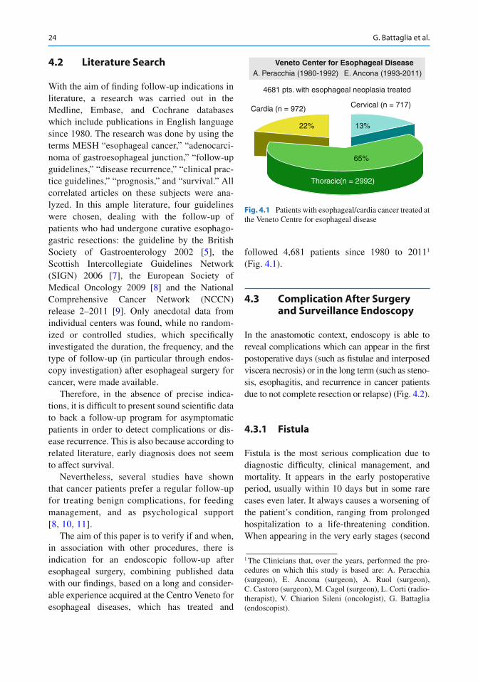
followed 4,681 patients since 1980 to 2011 1 (Fig. 4.1 ).
4.3 Complication After Surgery and Surveillance Endoscopy
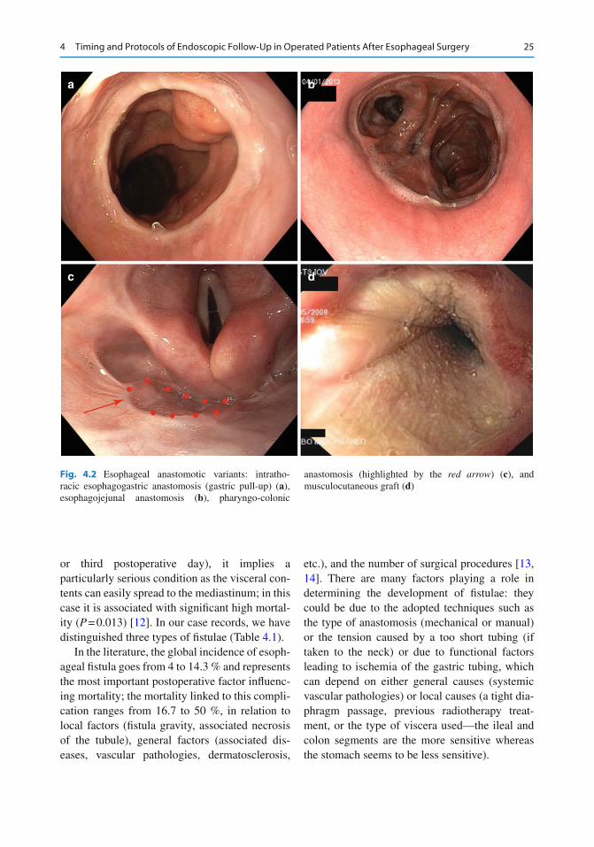
In the anastomotic context, endoscopy is able to reveal complications which can appear in the fi rst postoperative days (such as fi stulae and interposed viscera necrosis) or in the long term (such as steno-sis, esophagitis, and recurrence in cancer patients due to not complete resection or relapse) (Fig. 4.2 ).
4.3.1 Fistula
Fistula is the most serious complication due to diagnostic diffi culty, clinical management, and mortality. It appears in the early postoperative period, usually within 10 days but in some rare cases even later. It always causes a worsening of the patient’s condition, ranging from prolonged hospitalization to a life-threatening condition. When appearing in the very early stages (second
1 The Clinicians that, over the years, performed the pro-cedures on which this study is based are: A. Peracchia (surgeon), E. Ancona (surgeon), A. Ruol (surgeon), C. Castoro (surgeon), M. Cagol (surgeon), L. Corti (radio-therapist), V. Chiarion Sileni (oncologist), G. Battaglia (endoscopist).
Veneto Center for Esophageal DiseaseA. Peracchia (1980-1992) E. Ancona (1993-2011)
4681 pts. with esophageal neoplasia treated
Cardia (n = 972) Cervical (n = 717)
22% 13%
65%
Thoracic(n = 2992)
Fig. 4.1 Patients with esophageal/cardia cancer treated at the Veneto Centre for esophageal disease
G. Battaglia et al.
25
or third postoperative day), it implies a particularly serious condition as the visceral con-tents can easily spread to the mediastinum; in this case it is associated with signifi cant high mortal-ity ( P = 0.013) [ 12 ]. In our case records, we have distinguished three types of fi stulae (Table 4.1 ).
In the literature, the global incidence of esoph-ageal fi stula goes from 4 to 14.3 % and represents the most important postoperative factor infl uenc-ing mortality; the mortality linked to this compli-cation ranges from 16.7 to 50 %, in relation to local factors (fi stula gravity, associated necrosis of the tubule), general factors (associated dis-eases, vascular pathologies, dermatosclerosis,
etc.), and the number of surgical procedures [ 13 , 14 ]. There are many factors playing a role in determining the development of fi stulae: they could be due to the adopted techniques such as the type of anastomosis (mechanical or manual) or the tension caused by a too short tubing (if taken to the neck) or due to functional factors leading to ischemia of the gastric tubing, which can depend on either general causes (systemic vascular pathologies) or local causes (a tight dia-phragm passage, previous radiotherapy treat-ment, or the type of viscera used—the ileal and colon segments are the more sensitive whereas the stomach seems to be less sensitive).
a
c d
b
Fig. 4.2 Esophageal anastomotic variants: intratho-racic esophagogastric anastomosis (gastric pull-up) ( a ), esophagojejunal anastomosis ( b ), pharyngo-colonic
anastomosis (highlighted by the red arrow ) ( c ), and musculocutaneous graft ( d )
4 Timing and Protocols of Endoscopic Follow-Up in Operated Patients After Esophageal Surgery
26
Thanks to new techniques, above all the mechanical suturer, a technical error while making an anastomosis is rare to observe today. A more common problem is the ischemia of the mobilized viscera which, to a certain degree, happens in all patients but rarely leads to the inability of healing. Therefore, when a combination of ischemia and mechanical stress has a negative effect on the anas-tomosis, a fi stula can occur. According to some authors, but without clear scientifi c evidence, radiotherapy may represent another risk factor, especially for an anastomosis at the neck.
A meta-analysis by Biere et al. [ 15 ] on four randomized studies analyzing anastomotic risk factors, showed that fi stula incidence was signifi -cantly higher for cervical anastomoses in respect to intrathoracic ones (2–30 % vs 0–10 % P = 0.03), even if the thoracic ones generally have a more complicated course. The author explains that this difference could be due to the vascular suffering caused by tubing or to the manual technique that is used for performing a cervical anastomosis. On the contrary, in another study by Kayani et al. [ 16 ] examining 47 published articles, no clear evi-dence was found concerning a higher incidence
for cervical anastomoses, but the studies compar-ing the two types of anastomoses are few, with a low number of cases, and they are not standard-ized concerning surgery approaches, anastomosis techniques, and the neo-adjuvant therapy.
Analyzing the gastric tube preparation, Collard showed 1 % fi stulae with the whole stomach vs 7.9 % with tubular stomach while, regarding the intrathoracic interposition, Shu observed less fi stulae with the tubular stomach (5.5 % vs 9.3 % ( P < 0.05)) compared to the whole viscera [ 17 ]. Age does not appear to be a risk factor if vascular diseases are not involved; Tapias observed anastomotic leakage in 4.8 % of patients <70 years 4.8 % in 70–80 age and 0 % < 80 years ( P = 0.685).
Regarding seriousness of fi stulae, Price and colleagues [ 18 ] report a global incidence of 11 % with a superior incidence of neck anastomoses (6/34, 21 %) against intrathoracic (16/268, 5.9 %) but without signifi cant difference regarding the percentage of more serious fi stulae with partial or extended necrosis of the viscera (4.8 % vs 4.8 %). In our global experience, we have had 7.3 % of fi stulae of which only 1.1 % were serious, with a signifi cant reduction to 5.9 % over the last decade. The level of the anastomosis was decisive for the development of a slight fi stula (11.3 % cervical vs 4.3 % thoracic ( P = 0.0001)), as it was also for the type of viscera interposed at the cer-vical level, even if not signifi cant (colon 16.8 % vs stomach 10.7 % ( P = NS)).
Whenever an interpositioning is performed with the colon or the jejunum, the lower anasto-mosis also needs to be monitored as it is the area where, in our experience, there is a fi stula inci-dence of 4.7 % (12/255: 8 slight 4 serious).
Appearance of necrosis has a low rate (1.1 %) and here as well a signifi cant difference between cervical and thoracic levels is to be found (22/774 vs 6/1,468 ( P < 0.001)). Our study did not observe a correlation with comorbidities like cirrhosis, diabetes, obesity, nephropathy, or arteriopathy as shown by the following data:• Cirrhosis (1/48 (2.1 %) in patients with cir-
rhosis vs 27/2,194 (1.7 %); P = 0.46) • Diabetes (3/159 (1.9 %) in patients with
diabetes vs 25/2,083 (1.2 %); P = 0.44)
Table 4.1 Fistulas classifi cation employed at our department
1 Subclinical (radiologic)
2a Minor without borders ischemia
Affects less than ¼ of the esophageal circumference and mainly caused by a technical error; the borders are well vascularized
2b Minor with ischemic borders
Affects less than ¼ of the esophageal circumference; may be related to an insuffi cient vascularization of the graft or a prolonged hypotension during or immediately after surgery
3a Major with necrosis of borders
Affects more than ¼ of the esophageal circumference; related to an important non-transitory vascular insuffi ciency; necrosis of the anastomotic borders
3b Major with extensive necrosis of the graft
Affects more than ¼ of the esophageal circumference with an extensive wall necrosis of the graft
G. Battaglia et al.
27
• Obesity (0/21 in obese patients vs 28/222 (1.3 %); P = 0.99)
• Nephropathy (1/55 (1.8 %) in patient with nephropathy vs 27/2,187 (1.2 %); P = 0.50)
• Arteriopathy (2/93 (2.2 %) in patients with arteriopathy vs 26/2,149 (1.2 %); P = 0.32) Anastomotic fi stula diagnosis may be diffi -
cult [ 19 ]. Normally surgeons control the anasto-mosis with a digestive tube on the seventh day before feeding starts and the control is manda-tory in the presence of risk factors like a diffi -cult anastomosis or a serious blood pressure decrease in the early postoperative period or a suspected septic state. These are the conditions to be aware of as the anastomotic fi stula or tubu-lar necrosis has to be diagnosed as early as pos-sible in order to quickly start the appropriate treatment.
In suspect cases, as the fi rst approach, a diges-tive tube and, if positive, a CT scan should be requested; even if 50 % of fi stulae are not detected with these exams, they are the basis for the next endoscopic exam. Endoscopy is the only exam able to show suffering in the interposed viscera or am anastomotic leakage: it should be performed by an expert endoscopist using a weak insuffl a-tion and, possibly, a hood placed on the tip to enable the detection of the suture line. While per-forming the retroversion (which is a very risky maneuver), the gastric side of the anastomosis and any eventual vascular problem or necrosis should be described. This procedure allows to determine the presence a critical anastomosis before fi stuli-zation in order take the correct measures that
often can be done only to delay the refeeding (Figs. 4.3 and 4.4 ).
Once the diagnosis has been done, the monitor-ing of the clinical process of the viscera can be done by daily endoscopic controls if a conservative treat-ment has been decided. In fact there is not always an indication to surgical treatment even when there is a major fi stula, especially if the patient’s general condition is not serious and if the disassembling of the anastomosis is technically diffi cult.
In our experience, if there is no extended necrosis; we always prefer a conservative treat-ment which includes internal drainage placed endoscopically in the fi stula and a gastric tube. We monitor the clinical state carefully and the viscera with daily endoscopic exams, if neces-sary, and surgery is performed only if the clinical parameters get worse.
a b c
Fig. 4.3 Large anastomotic fi stula without necrosis ( a ), conservative treatment by positioning one tube in the stomach and the other one in the mediastinum ( b ), and X-ray control after 10 days of parenteral nutrition ( c )
Fig. 4.4 Anastomotic leakage with necrotic tissue
4 Timing and Protocols of Endoscopic Follow-Up in Operated Patients After Esophageal Surgery
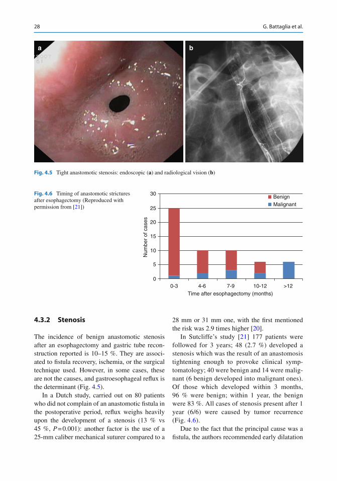
28
4.3.2 Stenosis
The incidence of benign anastomotic stenosis after an esophagectomy and gastric tube recon-struction reported is 10–15 %. They are associ-ated to fi stula recovery, ischemia, or the surgical technique used. However, in some cases, these are not the causes, and gastroesophageal refl ux is the determinant (Fig. 4.5 ).
In a Dutch study, carried out on 80 patients who did not complain of an anastomotic fi stula in the postoperative period, refl ux weighs heavily upon the development of a stenosis (13 % vs 45 %, P = 0.001): another factor is the use of a 25-mm caliber mechanical suturer compared to a
28 mm or 31 mm one, with the fi rst mentioned the risk was 2.9 times higher [ 20 ].
In Sutcliffe’s study [ 21 ] 177 patients were followed for 3 years; 48 (2.7 %) developed a stenosis which was the result of an anastomosis tightening enough to provoke clinical symp-tomatology; 40 were benign and 14 were malig-nant (6 benign developed into malignant ones). Of those which developed within 3 months, 96 % were benign; within 1 year, the benign were 83 %. All cases of stenosis present after 1 year (6/6) were caused by tumor recurrence (Fig. 4.6 ).
Due to the fact that the principal cause was a fi stula, the authors recommended early dilatation
a b
Fig. 4.5 Tight anastomotic stenosis: endoscopic ( a ) and radiological vision ( b )
30
25
20
Num
ber
of c
ases
15
10
5
00-3 4-6 7-9
Time after esophagectomy (months)10-12 >12
BenignMalignant
Fig. 4.6 Timing of anastomotic strictures after esophagectomy (Reproduced with permission from [ 21 ])
G. Battaglia et al.
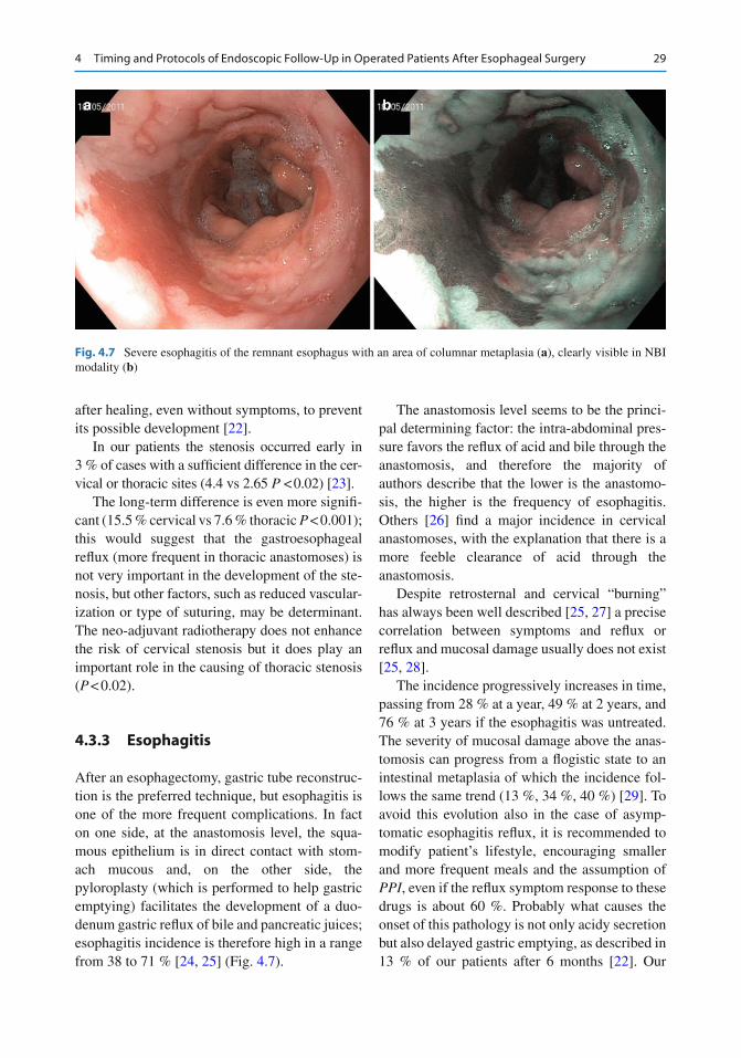
29
after healing, even without symptoms, to prevent its possible development [ 22 ].
In our patients the stenosis occurred early in 3 % of cases with a suffi cient difference in the cer-vical or thoracic sites (4.4 vs 2.65 P < 0.02) [ 23 ].
The long-term difference is even more signifi -cant (15.5 % cervical vs 7.6 % thoracic P < 0.001); this would suggest that the gastroesophageal refl ux (more frequent in thoracic anastomoses) is not very important in the development of the ste-nosis, but other factors, such as reduced vascular-ization or type of suturing, may be determinant. The neo-adjuvant radiotherapy does not enhance the risk of cervical stenosis but it does play an important role in the causing of thoracic stenosis ( P < 0.02).
4.3.3 Esophagitis
After an esophagectomy, gastric tube reconstruc-tion is the preferred technique, but esophagitis is one of the more frequent complications. In fact on one side, at the anastomosis level, the squa-mous epithelium is in direct contact with stom-ach mucous and, on the other side, the pyloroplasty (which is performed to help gastric emptying) facilitates the development of a duo-denum gastric refl ux of bile and pancreatic juices; esophagitis incidence is therefore high in a range from 38 to 71 % [ 24 , 25 ] (Fig. 4.7 ).
The anastomosis level seems to be the princi-pal determining factor: the intra-abdominal pres-sure favors the refl ux of acid and bile through the anastomosis, and therefore the majority of authors describe that the lower is the anastomo-sis, the higher is the frequency of esophagitis. Others [ 26 ] fi nd a major incidence in cervical anastomoses, with the explanation that there is a more feeble clearance of acid through the anastomosis.
Despite retrosternal and cervical “burning” has always been well described [ 25 , 27 ] a precise correlation between symptoms and refl ux or refl ux and mucosal damage usually does not exist [ 25 , 28 ].
The incidence progressively increases in time, passing from 28 % at a year, 49 % at 2 years, and 76 % at 3 years if the esophagitis was untreated. The severity of mucosal damage above the anas-tomosis can progress from a fl ogistic state to an intestinal metaplasia of which the incidence fol-lows the same trend (13 %, 34 %, 40 %) [ 29 ]. To avoid this evolution also in the case of asymp-tomatic esophagitis refl ux, it is recommended to modify patient’s lifestyle, encouraging smaller and more frequent meals and the assumption of PPI , even if the refl ux symptom response to these drugs is about 60 %. Probably what causes the onset of this pathology is not only acidy secretion but also delayed gastric emptying, as described in 13 % of our patients after 6 months [ 22 ]. Our
a b
Fig. 4.7 Severe esophagitis of the remnant esophagus with an area of columnar metaplasia ( a ), clearly visible in NBI modality ( b )
4 Timing and Protocols of Endoscopic Follow-Up in Operated Patients After Esophageal Surgery
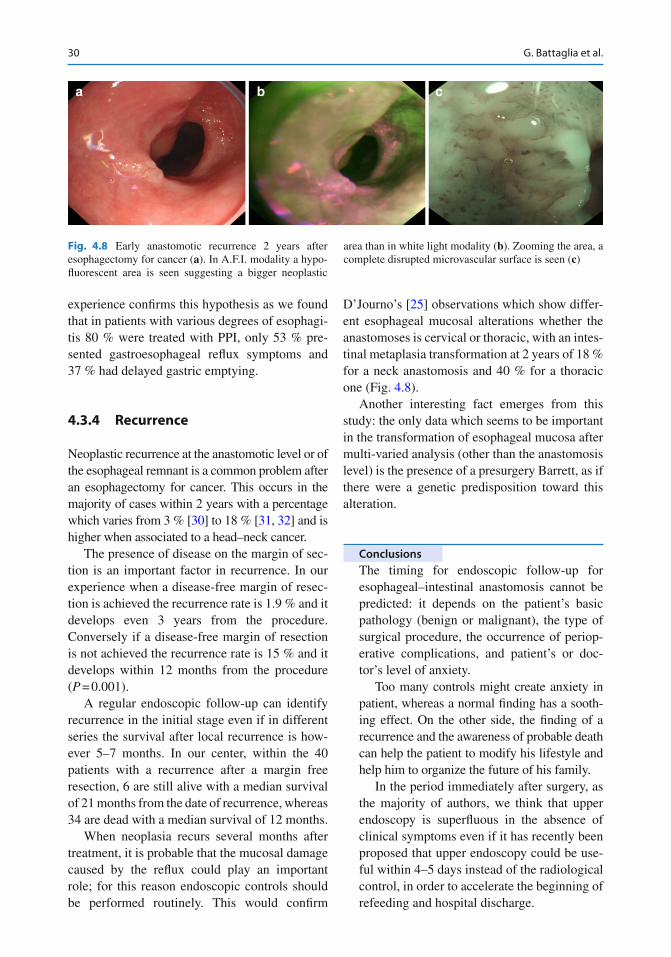
30
experience confi rms this hypothesis as we found that in patients with various degrees of esophagi-tis 80 % were treated with PPI, only 53 % pre-sented gastroesophageal refl ux symptoms and 37 % had delayed gastric emptying.
4.3.4 Recurrence
Neoplastic recurrence at the anastomotic level or of the esophageal remnant is a common problem after an esophagectomy for cancer. This occurs in the majority of cases within 2 years with a percentage which varies from 3 % [ 30 ] to 18 % [ 31 , 32 ] and is higher when associated to a head–neck cancer.
The presence of disease on the margin of sec-tion is an important factor in recurrence. In our experience when a disease-free margin of resec-tion is achieved the recurrence rate is 1.9 % and it develops even 3 years from the procedure. Conversely if a disease-free margin of resection is not achieved the recurrence rate is 15 % and it develops within 12 months from the procedure ( P = 0.001).
A regular endoscopic follow-up can identify recurrence in the initial stage even if in different series the survival after local recurrence is how-ever 5–7 months. In our center, within the 40 patients with a recurrence after a margin free resection, 6 are still alive with a median survival of 21 months from the date of recurrence, whereas 34 are dead with a median survival of 12 months.
When neoplasia recurs several months after treatment, it is probable that the mucosal damage caused by the refl ux could play an important role; for this reason endoscopic controls should be performed routinely. This would confi rm
D’Journo’s [ 25 ] observations which show differ-ent esophageal mucosal alterations whether the anastomoses is cervical or thoracic, with an intes-tinal metaplasia transformation at 2 years of 18 % for a neck anastomosis and 40 % for a thoracic one (Fig. 4.8 ).
Another interesting fact emerges from this study: the only data which seems to be important in the transformation of esophageal mucosa after multi-varied analysis (other than the anastomosis level) is the presence of a presurgery Barrett, as if there were a genetic predisposition toward this alteration.
Conclusions
The timing for endoscopic follow-up for esophageal–intestinal anastomosis cannot be predicted: it depends on the patient’s basic pathology (benign or malignant), the type of surgical procedure, the occurrence of periop-erative complications, and patient’s or doc-tor’s level of anxiety.
Too many controls might create anxiety in patient, whereas a normal fi nding has a sooth-ing effect. On the other side, the fi nding of a recurrence and the awareness of probable death can help the patient to modify his lifestyle and help him to organize the future of his family.
In the period immediately after surgery, as the majority of authors, we think that upper endoscopy is superfl uous in the absence of clinical symptoms even if it has recently been proposed that upper endoscopy could be use-ful within 4–5 days instead of the radiological control, in order to accelerate the beginning of refeeding and hospital discharge.
a b c
Fig. 4.8 Early anastomotic recurrence 2 years after esophagectomy for cancer ( a ). In A.F.I. modality a hypo-fl uorescent area is seen suggesting a bigger neoplastic
area than in white light modality ( b ). Zooming the area, a complete disrupted microvascular surface is seen ( c )
G. Battaglia et al.
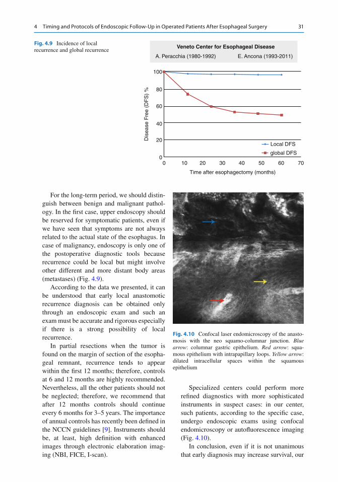
31
For the long-term period, we should distin-guish between benign and malignant pathol-ogy. In the fi rst case, upper endoscopy should be reserved for symptomatic patients, even if we have seen that symptoms are not always related to the actual state of the esophagus. In case of malignancy, endoscopy is only one of the postoperative diagnostic tools because recurrence could be local but might involve other different and more distant body areas (metastases) (Fig. 4.9 ).
According to the data we presented, it can be understood that early local anastomotic recurrence diagnosis can be obtained only through an endoscopic exam and such an exam must be accurate and rigorous especially if there is a strong possibility of local recurrence.
In partial resections when the tumor is found on the margin of section of the esopha-geal remnant, recurrence tends to appear within the fi rst 12 months; therefore, controls at 6 and 12 months are highly recommended. Nevertheless, all the other patients should not be neglected; therefore, we recommend that after 12 months controls should continue every 6 months for 3–5 years. The importance of annual controls has recently been defi ned in the NCCN guidelines [ 9 ]. Instruments should be, at least, high defi nition with enhanced images through electronic elaboration imag-ing (NBI, FICE, I-scan).
Specialized centers could perform more refi ned diagnostics with more sophisticated instruments in suspect cases: in our center, such patients, according to the specifi c case, undergo endoscopic exams using confocal endomicroscopy or autofl uorescence imaging (Fig. 4.10 ).
In conclusion, even if it is not unanimous that early diagnosis may increase survival, our
Veneto Center for Esophageal Disease
A. Peracchia (1980-1992) E. Ancona (1993-2011)
100
80
60
40
Dis
ease
Fre
e (D
FS
) %
20
00 10 20 30 40
Time after esophagectomy (months)
50 60 70
Local DFS
global DFS
Fig. 4.9 Incidence of local recurrence and global recurrence
Fig. 4.10 Confocal laser endomicroscopy of the anasto-mosis with the neo squamo-columnar junction. Blue arrow : columnar gastric epithelium. Red arrow : squa-mous epithelium with intrapapillary loops. Yellow arrow : dilated intracellular spaces within the squamous epithelium
4 Timing and Protocols of Endoscopic Follow-Up in Operated Patients After Esophageal Surgery
32
experience suggests that a standardized follow-up program could help monitoring and possi-bly improving results of esophageal surgery.
References
1. Davis PA, Law S, Wong J (2003) Colonic interposi-tion after esophagectomy for cancer. Arch Surg 138:303–308
2. Coleman JJ 3rd (1995) Reconstruction of the pharynx and cervical esophagus. Semin Surg Oncol 11:208–220
3. Briel JW, Tamhankar AP, Hagen JA et al (2004) Prevalence and risk factors for ischemia, leak, and stricture of esophageal anastomosis: gastric pull-up versus colon interposition. J Am Coll Surg 198:536–541
4. Mansour KA, Bryan FC, Carlson GW (1997) Bowel interposition for esophageal replacement: twentyfi ve- year experience. Ann Thorac Surg 64:752–756
5. Allum WH, Griffi n SM, Watson S et al (2002) Guidelines for the management of oesophageal and gastric cancer. Gut 50:1–23
6. Park SY, Lee HS, Jang HJ et al (2013) The role of one-year endoscopic follow-up for the esophageal remnant and gastric conduit after esophagectomy with gastric reconstruction for esophageal squamous cell carcinoma. Yonsei Med J 54(2):381–388
7. Scottish Intercollegiate Guidelines Network (2006) Management of oesophageal and gastric cancer – a national clinical guideline. http://www.sign.ac.uk
8. Stahl M, Oliveira J (2009) Esophageal cancer: ESMO clinical recommendations for diagnosis, treatment and follow-up. Ann Oncol 20(S4):32–33
9. Ajani JA, Barthel JS, Bentrem, DJ et al (2011) Esophageal and Esophagogastric Junction Cancers Clinical Practice Guidelines in Oncology. J Natl Compr Canc Netw 9:830 –887
10. McCorry NK, Dempster M, Clarke C et al (2009) Adjusting to life after oesophagectomy: the experi-ence of survivors and carers. Qual Health Res 19(10):1485–1494
11. Kiebert GM, Welvaart K, Kievit J (1993) Psychological effects of routine follow-up on cancer patients after surgery. Eur J Surg 159:601–607
12. Patil PK, Patel SG, Mistry RC et al (1992) Cancer of the esophagus: esophagogastric anastomotic leak—a retrospective study of predisposing factors. J Surg Oncol 49(3):163–167
13. Hölscher AH, Vallböhmer D, Brabender J (2006) The prevention and management of perioperative compli-cations. Best Pract Res Clin Gastroenterol 20(5):907–923
14. Rutegård M, Lagergren P, Rouvelas I et al (2012) Intrathoracic anastomotic leakage and mortality after esophageal cancer resection: a population-based study. Ann Surg Oncol 19(1):99–103
15. Biere SS, Maas KW, Cuesta MA et al (2011) Cervical or thoracic anastomosis after esophagectomy for can-cer: a systematic review and meta-analysis. Dig Surg 28(1):29–35
16. Kayani B, Jarral OA, Athanasiou T et al (2012) Should oesophagectomy be performed with cervical or intrathoracic anastomosis? Interact Cardiovasc Thorac Surg 14(6):821–826
17. Shu YS, Sun C, Shi WP (2013) Tubular stomach or whole stomach for esophagectomy through cervico-thoraco- abdominal approach: a comparative clinical study on anastomotic leakage. Ir J Med Sci 182(3):477–480
18. Price TN, Nichols FC, Harmsen WS, Allen MS et al (2013) A comprehensive review of anastomotic tech-nique in 432 esophagectomies. Ann Thorac Surg 95(4):1154–1160
19. Griffi n SM, Lamb PJ, Dresner DL et al (2001) Diagnosis and management of a mediastinal leak following radical oesophagectomy. Br J Surg 88:1346–1351
20. Johansson J, Oberg S, Wenner J et al (2009) Impact of proton pump inhibitors on benign anastomotic stric-ture formations after esophagectomy and gastric tube reconstruction: results from a randomized clinical trial. Ann Surg 250(5):667–673
21. Sutcliffe RP, Forshaw MJ, Tandon R et al (2008) Anastomotic strictures and delayed gastric emptying after esophagectomy: incidence, risk factors and man-agement. Dis Esophagus 21(8):712–717
22. Parekh K, Iannettoni MD (2007) Complications of esophageal resection and reconstruction. Semin Thorac Cardiovasc Surg 19(1):79–88
23. Petrin G, Ruol A, Battaglia G, Buin F, Merigliano S, Constantini M, Pavei P, Cagol M, Scappin S (2000) Ancona anastomotic stenoses occurring after circular stapling in esophageal cancer surgery. Surg Endosc 14:670–674
24. Gutschow C, Collard JM, Romagnoli R et al (2001) Denervated stomach as an esophageal substitute recovers intraluminal acidity with time. Ann Surg 233:509–514
25. Shibuya S, Fukudo S, Shineha R et al (2003) High incidence of refl ux esophagitis observed by routine endoscopic examination after gastric pull-up esopha-gectomy. World J Surg 27:580–583
26. Johansson J, Johnsson F, Groshen S et al (1999) Pharyngeal refl ux after gastric pull-up esophagec-tomy with neck and chest anastomoses. J Thorac Cardiovasc Surg 118(6):1078–1083
27. Wang Q, Liu J, Zhao X et al (1999) Can esophagogas-tric anastomosis prevent gastroesophageal refl ux. Zhonghua Wai Ke Za Zhi 37:71–73
G. Battaglia et al.
33
28. Yamamoto S, Makuuchi H, Shimada H et al (2007) Clinical analysis of refl ux esophagitis following esophagectomy with gastric tube reconstruction. J Gastroenterol 42:342–345
29. D’Journo XB, Martin J, Rakovich G et al (2009) Mucosal damage in the esophageal remnant after esophagectomy and gastric transposition. Ann Surg 249(2):262–268
30. Kato H, Tachimori Y, Watanabe H et al (1998) Anastomotic recurrence of oesophageal squamous cell carcinoma after transthoracic oesophagectomy. Eur J Surg 164(10):759–764
31. Tu C, Lin J, Lee C et al (2010) Usefulness of endo-scopic evaluation after esophageal reconstruction surgery. Retrieved from http://www.edah.org.tw/edh/EDJM/allissues/vol.1/UsefulnessofEndoscopic-Evaluationafter-EsophagealReconstructionSurgery
32. Li CL, Zhang FL, Wang YD et al (2012) Characteristics of recurrence after radical esophagectomy with two- fi eld lymph node dissection for thoracic esophageal cancer. Oncol Lett 5(1):355–359
4 Timing and Protocols of Endoscopic Follow-Up in Operated Patients After Esophageal Surgery
35G. Galloro (ed.), Endoscopic Follow-up of Digestive Anastomosis, DOI 10.1007/978-88-470-5370-0_5, © Springer-Verlag Italia 2014
5.1 Introduction
After gastric surgery, indications to endoscopic follow-up have not yet been defi nitely estab-lished. We will consider the most common gas-tric surgical interventions and indications of endoscopic follow-up will be based on available literature.
5.1.1 Partial Gastrectomy
The question of whether partial gastrectomy car-ries an increased risk of subsequent development of stomach cancer is debated.
A number of studies have reported an increased risk after Billroth-II gastrectomy com-pared with Billroth-I gastrectomy [ 1 ]. The exact mechanism for the development of gastric stump cancer (GSC) remains unclear, although many
causative factors have been reported. It is widely accepted that the predominant factor underlying the development of GSC is duodenogastric refl ux including bile and pancreatic juice, reportedly carcinogenic and related with GSC [ 2 ]. GSC has been used to defi ne all cancers occurring in the remnant stomach after gastrectomy, regardless of whether the primary disease was benign or malig-nant [ 3 ].
The incidence of GSC following distal gas-trectomy has been reported to account for 1–2 % of all gastric cancer in Japan [ 4 ]. Specifi cally, GSC is commonly found at an advanced stage, resulting in low rates of curative resections (38–40 %) and a consequently poor prognosis [ 5 ]. However, the incidence and etiology of GSC have changed in recent years because of the long latency periods, the decreasing prevalence of gastrectomy for benign disease, and the early detection and improved outcomes in patients with gastric cancers [ 6 ]. Despite the previous reports where GSC was commonly found at an advanced stage, resulting in poor prognosis, recent advances in diagnostic methods and less- invasive treatment techniques have led to a higher detection rate of early GSC following dis-tal gastrectomy and decreased mortality and mor-bidity rates. Consequently, endoscopic therapy such as endoscopic mucosal resection or submu-cosal dissection can be adopted for treatment of early-stage GSC [ 7 ].
Thus, oncologic and mainly endoscopic follow- ups are important in all patients after
R. Conigliaro (*) • A. Caruso (*) Gastroenterology and Digestive Endoscopy Unit , New S. Agostino-Estense Hospital , Viale Giardini 1355 , Baggiovara (MO) 41100 , Italy e-mail: [email protected]; [email protected]
M. Frazzoni Digestive Pathophysiology Unit , New S. Agostino-Estense Hospital , Via Giardini 1355 , Baggiovara–Modena (MO) 41100 , Italy e-mail: [email protected]
5 Timing and Protocols of Endoscopic Follow-Up After Gastric Surgery
Rita Conigliaro , Angelo Caruso , and Marzio Frazzoni
36
gastric surgery. Data from literature should be subdivided as follows:• Risk of GSC after gastric surgery for benign
peptic ulcer (partial gastrectomy was the treat-ment of choice for ulcer disease until the development of antisecretory drugs)
• Risk of GSC after gastric surgery for gastric cancer Retrospective cohort studies in patients oper-
ated for benign disease found mixed results: increased [ 8 ], unaltered [ 1 ], and decreased [ 9 ] risk. Overall, results from the fi ve largest case control studies indicate that the relative risk of GSC after gastric surgery for benign peptic ulcer is two (RR=2).
Twelve prospective studies on patients oper-ated on for peptic ulcer have been published. These studies are based on the follow-up of a cohort over a certain period of time. Results are expressed and analyzed using the incidence ratio observed in the cohort, comparing observed with expected cases in a similar general population. The results of seven of these studies favor the hypothesis of an increased risk of stomach cancer in patients operated on for peptic ulcer disease, while those of fi ve do not support this hypothesis. The results of two of these studies were obtained by multivariate analyses, which represent the best statistical methodology known for assessing the respective role of confounding variables. Authors believe that the evidence is good enough to identify patients who underwent partial gas-trectomy more than 20 years previously as a high-risk group for the development of carci-noma. Therefore, it is recommended that these patients should be offered regular endoscopy, especially if they underwent a Billroth-II surgical procedure [ 10 ].
Green et al. [ 11 ] suggested endoscopic and histological surveillance to enable early diagno-sis. Seven early gastric adenocarcinomas were detected in 163 patients followed up for a mean duration of 8 years with yearly upper GI endos-copy gastroscopies. Screening was commenced at least 10 years after surgery.
More recent studies, mostly from Japan, have been published on patients operated on for gastric cancer. Thanks to recent advances in diagnosis, an
increased early detection of gastric cancer has been found. Consequently, the number of cured patients has increased and some of these patients are at risk of acquiring a second primary cancer in the remnant stomach. This implies that more cases of GSC will be encountered in the future [ 12 ].
Surveillance systems for early detection and curative treatment of GSC with periodic endo-scopic examinations of the gastric remnant are mandatory. The time interval to the occurrence of GSC in patients with previous benign and malig-nant disease was 23.8–32.4 and 6.8–18.8 years, respectively [ 13 ]. In the study of Komatsu et al. [ 14 ], the follow-up interval was signifi cantly associated with the stage of progression in rem-nant gastric cancer: an early detection of GSC required less-invasive curative treatment (endo-scopic submucosal dissection) and a better prog-nosis (Fig. 5.1 ). More than half of the GSC cases were T1 or T2 undifferentiated, node-negative, and early-stage cancers. Accordingly, annual surveillance endoscopy is recommended for at least 12 years following distal gastrectomy to obtain an early diagnosis of GSC Thereafter, sur-veillance endoscopy could be performed every second year (Table 5.1 ). According to authorita-tive suggestions, careful endoscopic examination should be performed near the suture line and the remnant gastric wall after Billroth-I reconstruc-tion and near the anastomosis after Billroth-II reconstruction [ 15 ]. However, more cohort stud-ies are warranted to defi ne optimal surveillance and treatment strategies.
5.1.2 Total Gastrectomy
After total gastrectomy , tumor recurrence can be subdivided as follows:• Distant tumor recurrence • Peritoneal tumor recurrence • Loco-regional tumor recurrence
Loco-regional tumor recurrence includes both endoscopically accessible tumor recur-rence (at the anastomosis site or within loop) and endoscopically inaccessible tumor recur-rence (lymph nodes or mass near the resected site) [ 16 ].
R. Conigliaro et al.
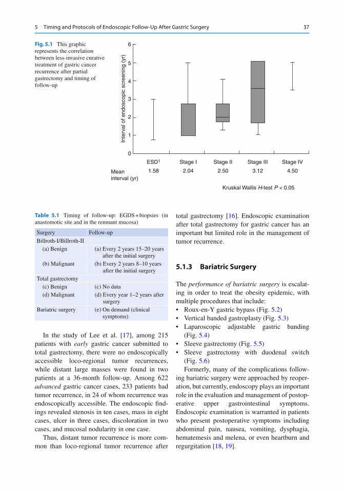
37
In the study of Lee et al. [ 17 ], among 215 patients with early gastric cancer submitted to total gastrectomy, there were no endoscopically accessible loco-regional tumor recurrences, while distant large masses were found in two patients at a 36-month follow-up. Among 622 advanced gastric cancer cases, 233 patients had tumor recurrence, in 24 of whom recurrence was endoscopically accessible. The endoscopic fi nd-ings revealed stenosis in ten cases, mass in eight cases, ulcer in three cases, discoloration in two cases, and mucosal nodularity in one case.
Thus, distant tumor recurrence is more com-mon than loco-regional tumor recurrence after
total gastrectomy [ 16 ]. Endoscopic examination after total gastrectomy for gastric cancer has an important but limited role in the management of tumor recurrence.
5.1.3 Bariatric Surgery
The performance of bariatric surgery is escalat-ing in order to treat the obesity epidemic, with multiple procedures that include:• Roux-en-Y gastric bypass (Fig. 5.2 )• Vertical banded gastroplasty (Fig. 5.3 )• Laparoscopic adjustable gastric banding
(Fig. 5.4 )• Sleeve gastrectomy (Fig. 5.5 )• Sleeve gastrectomy with duodenal switch
(Fig. 5.6 ) Formerly, many of the complications follow-
ing bariatric surgery were approached by reoper-ation, but currently, endoscopy plays an important role in the evaluation and management of postop-erative upper gastrointestinal symptoms. Endoscopic examination is warranted in patients who present postoperative symptoms including abdominal pain, nausea, vomiting, dysphagia, hematemesis and melena, or even heartburn and regurgitation [ 18 , 19 ].
6
5
4
3
2
1
0
Inte
rval
of e
ndos
copi
c sc
reen
ing
(yr)
Meaninterval (yr)
ESD1 Stage I Stage II Stage III Stage IV
1.58 2.04 2.50 3.12 4.50
Kruskal Wallis H-test P < 0.05
Fig. 5.1 This graphic represents the correlation between less-invasive curative treatment of gastric cancer recurrence after partial gastrectomy and timing of follow-up
Table 5.1 Timing of follow-up: EGDS + biopsies (in anastomotic site and in the remnant mucosa)
Surgery Follow-up
Billroth-I/Billroth-II (a) Benign (a) Every 2 years 15–20 years
after the initial surgery (b) Malignant (b) Every 2 years 8–10 years
after the initial surgery Total gastrectomy (c) Benign (c) No data (d) Malignant (d) Every year 1–2 years after
surgery Bariatric surgery (e) On demand (clinical
symptoms)
5 Timing and Protocols of Endoscopic Follow-Up After Gastric Surgery
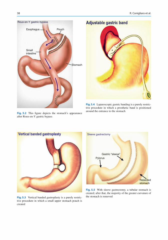
38
Roux-en-Y gastric bypass
Esophagus Pouch
Smallintestine
Stomach
Fig. 5.2 This fi gure depicts the stomach’s appearance after Roux-en-Y gastric bypass
Fig. 5.3 Vertical banded gastroplasty is a purely restric-tive procedure in which a small upper stomach pouch is created
Fig. 5.4 Laparoscopic gastric banding is a purely restric-tive procedure in which a prosthetic band is positioned around the entrance to the stomach
Gastric “sleeve”
Pylorus
Resectedstomach
Sleeve gastrectomy
Fig. 5.5 With sleeve gastrectomy, a tubular stomach is created; after that, the majority of the greater curvature of the stomach is removed
R. Conigliaro et al.
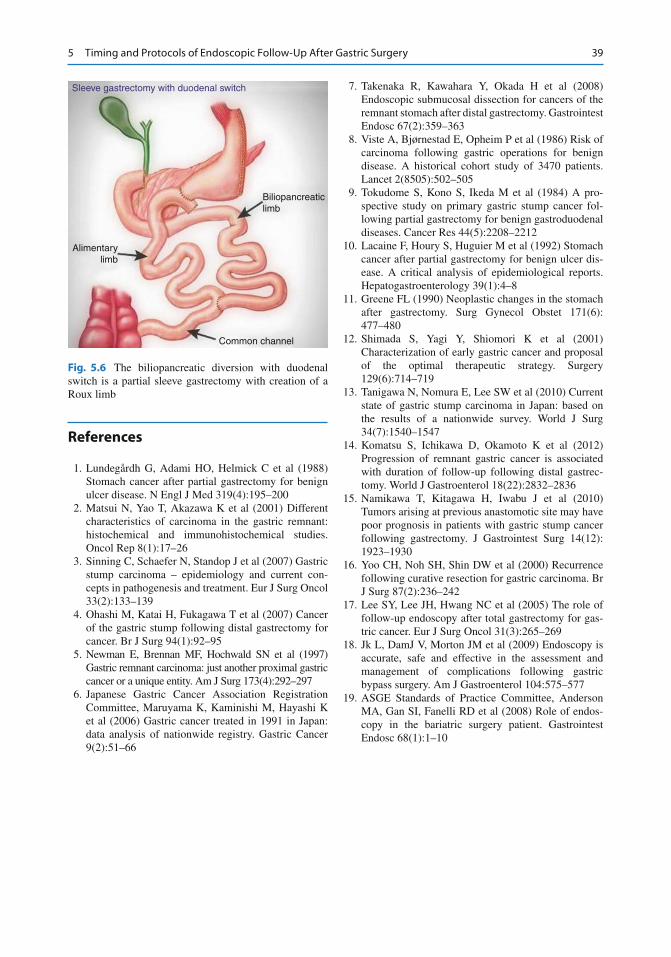
39
References
1. Lundegårdh G, Adami HO, Helmick C et al (1988) Stomach cancer after partial gastrectomy for benign ulcer disease. N Engl J Med 319(4):195–200
2. Matsui N, Yao T, Akazawa K et al (2001) Different characteristics of carcinoma in the gastric remnant: histochemical and immunohistochemical studies. Oncol Rep 8(1):17–26
3. Sinning C, Schaefer N, Standop J et al (2007) Gastric stump carcinoma – epidemiology and current con-cepts in pathogenesis and treatment. Eur J Surg Oncol 33(2):133–139
4. Ohashi M, Katai H, Fukagawa T et al (2007) Cancer of the gastric stump following distal gastrectomy for cancer. Br J Surg 94(1):92–95
5. Newman E, Brennan MF, Hochwald SN et al (1997) Gastric remnant carcinoma: just another proximal gastric cancer or a unique entity. Am J Surg 173(4):292–297
6. Japanese Gastric Cancer Association Registration Committee, Maruyama K, Kaminishi M, Hayashi K et al (2006) Gastric cancer treated in 1991 in Japan: data analysis of nationwide registry. Gastric Cancer 9(2):51–66
7. Takenaka R, Kawahara Y, Okada H et al (2008) Endoscopic submucosal dissection for cancers of the remnant stomach after distal gastrectomy. Gastrointest Endosc 67(2):359–363
8. Viste A, Bjørnestad E, Opheim P et al (1986) Risk of carcinoma following gastric operations for benign disease. A historical cohort study of 3470 patients. Lancet 2(8505):502–505
9. Tokudome S, Kono S, Ikeda M et al (1984) A pro-spective study on primary gastric stump cancer fol-lowing partial gastrectomy for benign gastroduodenal diseases. Cancer Res 44(5):2208–2212
10. Lacaine F, Houry S, Huguier M et al (1992) Stomach cancer after partial gastrectomy for benign ulcer dis-ease. A critical analysis of epidemiological reports. Hepatogastroenterology 39(1):4–8
11. Greene FL (1990) Neoplastic changes in the stomach after gastrectomy. Surg Gynecol Obstet 171(6):477–480
12. Shimada S, Yagi Y, Shiomori K et al (2001) Characterization of early gastric cancer and proposal of the optimal therapeutic strategy. Surgery 129(6):714–719
13. Tanigawa N, Nomura E, Lee SW et al (2010) Current state of gastric stump carcinoma in Japan: based on the results of a nationwide survey. World J Surg 34(7):1540–1547
14. Komatsu S, Ichikawa D, Okamoto K et al (2012) Progression of remnant gastric cancer is associated with duration of follow-up following distal gastrec-tomy. World J Gastroenterol 18(22):2832–2836
15. Namikawa T, Kitagawa H, Iwabu J et al (2010) Tumors arising at previous anastomotic site may have poor prognosis in patients with gastric stump cancer following gastrectomy. J Gastrointest Surg 14(12):1923–1930
16. Yoo CH, Noh SH, Shin DW et al (2000) Recurrence following curative resection for gastric carcinoma. Br J Surg 87(2):236–242
17. Lee SY, Lee JH, Hwang NC et al (2005) The role of follow-up endoscopy after total gastrectomy for gas-tric cancer. Eur J Surg Oncol 31(3):265–269
18. Jk L, DamJ V, Morton JM et al (2009) Endoscopy is accurate, safe and effective in the assessment and management of complications following gastric bypass surgery. Am J Gastroenterol 104:575–577
19. ASGE Standards of Practice Committee, Anderson MA, Gan SI, Fanelli RD et al (2008) Role of endos-copy in the bariatric surgery patient. Gastrointest Endosc 68(1):1–10
Sleeve gastrectomy with duodenal switch
Biliopancreaticlimb
Common channel
Alimentarylimb
Fig. 5.6 The biliopancreatic diversion with duodenal switch is a partial sleeve gastrectomy with creation of a Roux limb
5 Timing and Protocols of Endoscopic Follow-Up After Gastric Surgery
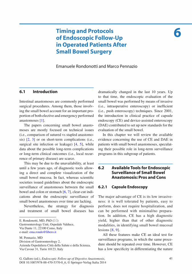
41G. Galloro (ed.), Endoscopic Follow-up of Digestive Anastomosis, DOI 10.1007/978-88-470-5370-0_6, © Springer-Verlag Italia 2014
6.1 Introduction
Intestinal anastomoses are commonly performed surgical procedures. Among them, those involv-ing the small bowel account for an important pro-portion of both elective and emergency performed anastomoses [ 1 ].
The papers concerning small bowel anasto-moses are mostly focused on technical issues (i.e., comparison of sutured vs stapled anastomo-sis) [ 2 , 3 ] or on short-term complications (i.e., surgical site infection or leakage) [ 4 , 5 ], while data about the possible long-term complications or long-term clinical outcomes (i.e., local recur-rence of primary disease) are scarce.
This may be due to the unavailability, at least until a few years ago, of diagnostic tools allow-ing a direct and complete visualization of the small bowel mucosa. In fact, whereas scientifi c societies issued guidelines about the endoscopic surveillance of anastomoses between the small bowel and colon or stomach [ 6 , 7 ], clear-cut indi-cations about the endoscopic surveillance of small bowel anastomoses over time are lacking.
Nevertheless, the strategy for diagnosis and treatment of small bowel diseases has
dramatically changed in the last 10 years. Up to that time, the endoscopic evaluation of the small bowel was performed by means of invasive (i.e., intraoperative enteroscopy) or ineffi cient (i.e., push enteroscopy) techniques. Since 2001, the introduction in clinical practice of capsule endoscopy (CE) and device-assisted enteroscopy (DAE) contributed to set up new standards for the evaluation of the small bowel.
In this chapter we will review the available evidence concerning the use of CE and DAE in patients with small bowel anastomoses, speculat-ing their possible role in long-term surveillance programs in this subgroup of patients.
6.2 Available Tools for Endoscopic Surveillance of Small Bowel Anastomosis: Pros and Cons
6.2.1 Capsule Endoscopy
The major advantage of CE is its low invasive-ness: it is well tolerated by patients, easy to perform, does not require hospitalization, and can be performed with minimal/no prepara-tion. In addition, CE has a high diagnostic yield, higher than that of other diagnostic modalities, in identifying small bowel mucosal lesions [ 8 , 9 ].
All these features make CE an ideal test for surveillance programs, in which the same proce-dure should be repeated over time. However, CE has a low specifi city in differentiating the nature
E. Rondonotti , MD, PhD (*) Gastroenterology Unit , Ospedale Valduce , Via Dante 11 , 22100 Como , Italy e-mail: [email protected]
M. Pennazio , MD Division of Gastroenterology 2 , Azienda Ospedaliera Città della Salute e della Scienza , Via Cavour 31 , Turin 10123 , Italy
6 Timing and Protocols of Endoscopic Follow-Up in Operated Patients After Small Bowel Surgery
Emanuele Rondonotti and Marco Pennazio
42
of small bowel lesions. This limitation may have a particular relevance in case of small bowel anastomosis, where, without taking biopsies, it may be diffi cult to distinguish between postsurgi-cal complications and recurrence of the primary disease (Fig. 6.1 ).
Another possible limitation of CE in this set-ting is related to the delayed capsule transit, which prevents, in operated patients, the com-plete evaluation of the small bowel. In the study by De Palma [ 10 ], where CE was performed in previously operated patients, the rate of complete evaluation of the small intestine was lower (about 70 %) than usually expected. The authors advo-cated the altered motility, resulting from the small bowel resection, as possible cause of the slow capsule transit.
Interestingly, in this study, all the ten enrolled patients excreted the capsule naturally; neverthe-less, the surgically altered anatomy is a well- known risk factor for capsule retention, particularly in case of side-to-side anastomoses [ 11 ] in which the capsule can enter blind loops. In the study of De Palma [ 10 ], all patients were therefore screened, before undergoing CE by means of the small bowel follow-through to exclude critical stenosis. Subsequent studies
have proposed to use, in patients with surgical anastomoses at risk for capsule retention, the patency capsule as “screening test” [ 12 ]. Although some studies [ 13 ] reported that the patency capsule can cause acute obstruction, the majority of available data [ 12 , 14 – 16 ] especially in cases in which the Agile® patency capsule [ 17 ] was ingested, seem to suggest that this test is both safe, even in case of tight stenosis, and effective in selecting patients in whom CE could be performed safely.
6.2.2 Device-Assisted Enteroscopy
With the increased detection rate of small bowel lesions, by means of purely diagnostic proce-dures such as CE or radiological examinations, innovations in overtube-assisted deep enteros-copy have been crucial. Although some studies report that DAE is helpful in the diagnostic pro-cess (sometimes it is able to identify neoplastic lesions missed by other techniques) [ 18 , 19 ], the main advantage of this procedure over CE is rep-resented by its therapeutic and operative capa-bilities. DAE allows delivering therapies (i.e., hemostasis), but more importantly, in patients with small bowel anastomoses, to take biopsies, to place tattoos, and to perform balloon dilations.
In a recently published paper [ 20 ], the authors reported that all cases of small bowel neoplasm were histologically diagnosed on the of ground biopsies obtained during DAE and, interestingly, the tattoo placed during the procedure made the laparoscopic approach feasible in about 80 % of them.
Recent studies have also suggested a possible role for DAE in the endoscopic balloon dilation of small bowel strictures mostly in patients with Crohn’s disease or ischemic enteritis [ 21 , 22 ]. In these patients, where the dilation was usually performed for disease-related stenosis, the tech-nical success rate and the complication rate range between 80–100 % and 0–6 %, respectively [ 23 ]. Nevertheless, data about long-term outcome are lacking as well as those concerning results of postsurgical strictures dilation.
Fig. 6.1 Small bowel anastomosis at CE: arrows indicate the presence of a double lumen, a fi nding consistent with a small bowel anastomosis provide that a diverticulum has been excluded
E. Rondonotti and M. Pennazio
43
As far as limitations of DAE are concerned, this procedure is invasive, challenging, and time- consuming and requires trained endoscopists and, often, deep sedation or general anesthesia with intubation. Moreover, even when guided by other diagnostic procedures (i.e., CE or radio-logic techniques), DAE can fail in reaching the small bowel fi nding. This often requires a new examination performed through the opposite route to access the small bowel [ 24 ]. The rate of entire small bowel examination, even when per-formed by DBE, which, although controversial results exist, seems to be the DAE with the high-est small bowel completion rate [ 25 , 26 ] is on average 40–50 % [ 23 ], with a wide range between the various published studies. Last but not least, although the complication rate of DAE appears to be low; severe complications (such as pancre-atitis and bowel perforation) occur in about 1 % of all diagnostic procedures, whereas the compli-cation rate of therapeutic procedures is reported to be higher, up to 4–5 % [ 27 , 28 ]. Focusing the attention on operated patients, some authors reported an increased risk of perforation in patients with recently performed anastomosis [ 29 ]. On the other hand, the abdominal adhe-sions, which can arise after small bowel resec-tion, can make DAE diffi cult to perform, less successful in exploring the small bowel, and more risky for the patients [ 27 ].
6.3 Endoscopic Surveillance of Small Bowel Anastomoses: Timing and Protocols
6.3.1 Preventing Postsurgical Complications
The most common complication of intestinal anastomosis is the development of ulcers [ 30 ]. Anastomotic ulcers may occur a few months to many years after surgery; in the study by Weinstock and Shatz [ 31 ], focused on ileocolonic anastomoses, the mean time frame between sur-gery and detection of ulcer was 5.1 years. Reaction to foreign body (Fig. 6.2 ) has been pos-tulated as the main cause for the ulceration [ 32 ];
however, the majority of patients who have their anastomosis either with hand-sewn sutures or stapled do not develop ulcers. The local isch-emia, secondary to scar formation, as well as abnormal motility and local intussusceptions, has also been advocated as mechanisms contributing to ulcer formation [ 30 ]. Although it has been pos-tulated that some conditions (i.e., radiation ther-apy) can facilitate anastomotic ulceration, this complication appears to be unrelated to the clini-cal indication to small bowel resection and some-what unpredictable. Therefore, although both CE (Fig. 6.3 ) and DAE are able to recognize the pres-ence of small bowel anastomotic ulcers, the rou-tine endoscopic surveillance of small bowel anastomoses, aimed at preventing this complica-tion, is not recommended.
Conversely, at least from a theoretical point of view, there may be an indication to the endo-scopic surveillance of small bowel anastomosis for those diseases with the potential for local recurrence, at the site of the anastomosis, such as Crohn’s disease or small bowel tumors.
6.3.2 Crohn’s Disease
Crohn’s disease (CD) most commonly affects the ileocolonic region involving the small bowel up
Fig. 6.2 Retained postsurgical suture at CE; arrows indi-cate the suture stitch
6 Timing and Protocols of Endoscopic Follow-Up in Operated Patients After Small Bowel Surgery
44
to 80 % of cases, while in about 30 % of patients, the disease is limited to the SB alone [ 33 – 35 ]. Therefore, in case of complication and/or failure of medical therapy, a signifi cant proportion of patients with CD receive surgical interventions, and particularly ileocolonic resections, during their lifetime.
After ileal or ileocolonic resection, most patients have a postsurgical CD recurrence in the neoileum (endoscopic recurrence is indeed observed in almost 73 % of CD patients at 1 year and in 90 % at 3 years after curative resection) [ 36 – 38 ]. This recurrence follows a sequence of endoscopic lesions in the anastomotic and pre-anastomotic regions, followed by the develop-ment of clinical symptoms. The presence of extensive lesions in the neoileum area, identifi ed through ileocolonoscopy in the months following surgery, predicts a rapid evolution to recurrent symptoms and eventual complications [ 39 ]. There are patient- and disease-related risk factors for postoperative recurrence: fi stulizing disease, ileocolonic location, and a smoking habit increase the risk of recurrence [ 40 – 42 ]. Thus, these patients usually receive immunosuppressive therapy immediately after surgery. However, patients with a low risk of recurrence, such as nonsmokers and those with fi brostenotic disease,
do not usually receive prophylactic treatment to prevent the development of new lesions. In such patients, clinical practice guidelines recommend that they undergo an ileocolonoscopy, grading the severity of lesions according to the Rutgeerts’ score, 6–12 months after resection [ 43 , 44 ]. In this setting, the feasibility, the diagnostic perfor-mances, and the safety of CE have been explored.
Some studies reported CE diagnostic perfor-mances similar to that of ileocolonoscopy in rec-ognizing lesions located at the site of anastomosis (sensitivity and specifi city of CE 50–80 % and 94–100 %, respectively) [ 43 , 45 – 47 ]. In addition, in these studies, CE was able to depict in a rel-evant proportion of patients, about 60 % [ 45 , 47 ], infl ammatory changes in the small bowel proxi-mal to the anastomosis, although the clinical rel-evance of such lesions remains to be determined. Nevertheless, in these studies, about 10 % of patients developed, over time, anastomotic stric-tures and they could not undergo CE because of being tested positive to a patency capsule test [ 46 ].
In patients with CD, as far as the small intes-tine resections are concerned, strictureplasty is often performed. This way to restore the intesti-nal continuity allows avoiding extensive resec-tions and consequently the risk of short bowel, but on the other hand, it creates large dilated loops with altered motility, potentially causing capsule retention [ 48 ].
Therefore, trying to translate the data collected with CE on the evaluation of ileocolonic anasto-moses to small bowel anastomoses, and taking into account possible risks (capsule retention) and the low recurrence rate (lower than that observed in ileocolonic anastomoses) [ 49 ], it seems that a surveillance program, with CE, for evaluating the small bowel anastomoses in patients with CD cannot be proposed at the present time.
In this setting one should ask whether it is worthwhile to perform DAE for evaluating the small bowel. Theoretically, in these patients, the DAE could provide the same information of CE without the risk of capsule retention (Fig. 6.4 ). On the other hand, we have to take into account that DAE may be diffi cult to per-form in patients with previously abdominal operations and an increased risk of perforation
Fig. 6.3 Small bowel anastomotic ulcer at CE (inside the blue circle the ulcer covered by fi brin)
E. Rondonotti and M. Pennazio
45
has been reported in case of severe infl amma-tion of the small bowel wall and recently per-formed anastomoses [ 27 ].
Unfortunately, to the best of our knowledge, there are no studies that have specifi cally addressed this type of assessment and this may represent an area for future research [ 50 ].
Endoscopic balloon dilation of small bowel strictures by DAE has the potential to obviate surgery in carefully selected patients [ 35 , 51 ]. One study [ 52 ] showed that, an anastomotic stric-ture is an independent marker of the symptom- free outcome after enteroscopic balloon dilations. This emphasizes that, even with the limitations above mentioned, DAE has the potential to improve outcome of patients who previously underwent small bowel surgery.
6.3.3 Small Bowel Tumors
Small bowel tumors are a small proportion of gastrointestinal neoplasms; accounting for 1–3 % of all primary gastrointestinal tumors [ 53 ]. Nevertheless, recent studies suggest that the inci-dence of these diseases is increasing [ 53 , 54 ]. Among malignant tumors, about 30–50 % are adenocarcinomas, 25–30 % are carcinoids, and 15–20 % are lymphomas [ 54 ]. As long as for some subtypes of small bowel lymphomas (i.e., follicular lymphoma) at the early stage, chemo-therapy has been proposed as the primary cura-
tive therapy [ 55 , 56 ], in the majority of cases, the surgical intervention, with en bloc resection, remains the cornerstone for the treatment of small bowel neoplams. Bilimoria et al. [ 54 ], col-lecting over the last 20 years more than 67,800 patients diagnosed with small bowel neoplasm, reported that about 80 % of them received surgi-cal intervention. These data have also been con-fi rmed by a recently published study [ 57 ] in which, in 141 patients with small bowel neo-plasms, a segmental bowel resection was the most commonly used surgical procedure (about 70 % of cases).
After surgical resection, despite the differ-ences between the various neoplasms, the restag-ing is usually carried out with imaging modalities (e.g., CT scan and/or FDG PET for lymphoma). There are no data, however, at present about the systematic use of CE or DAE in the follow-up of these patients.
The main limitation of CE in this specifi c set-ting, as reported above, is its low specifi city (dif-fi culty in distinguishing between surgical outcomes, postsurgical complications, or possi-ble local recurrence of primary disease). This limitation could be easily overcome by DAE, which has also shown, at the time of the diagno-sis, a diagnostic yield that, in some cases (i.e., lymphomas) (Figs. 6.5 and 6.6 ), was higher than that of other diagnostic methods (i.e., radiologi-cal) commonly used in the process of staging and restaging [ 58 , 59 ].
Fig. 6.4 Small bowel anastomotic stricture at DAE in a patient with Crohn’s disease
Fig. 6.5 Anastomotic recurrence of non-Hodgkin lym-phoma identifi ed at DAE
6 Timing and Protocols of Endoscopic Follow-Up in Operated Patients After Small Bowel Surgery
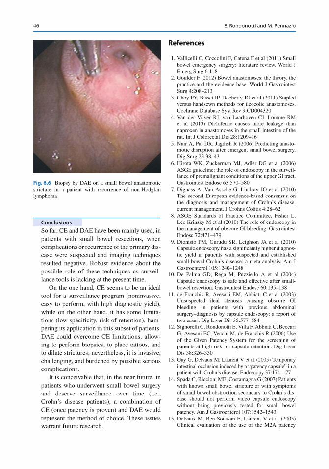
46
Conclusions
So far, CE and DAE have been mainly used, in patients with small bowel resections, when complications or recurrence of the primary dis-ease were suspected and imaging techniques resulted negative. Robust evidence about the possible role of these techniques as surveil-lance tools is lacking at the present time.
On the one hand, CE seems to be an ideal tool for a surveillance program (noninvasive, easy to perform, with high diagnostic yield), while on the other hand, it has some limita-tions (low specifi city, risk of retention), ham-pering its application in this subset of patients. DAE could overcome CE limitations, allow-ing to perform biopsies, to place tattoos, and to dilate strictures; nevertheless, it is invasive, challenging, and burdened by possible serious complications.
It is conceivable that, in the near future, in patients who underwent small bowel surgery and deserve surveillance over time (i.e., Crohn’s disease patients), a combination of CE (once patency is proven) and DAE would represent the method of choice. These issues warrant future research.
References
1. Vallicelli C, Coccolini F, Catena F et al (2011) Small bowel emergency surgery: literature review. World J Emerg Surg 6:1–8
2. Goulder F (2012) Bowel anastomoses: the theory, the practice and the evidence base. World J Gastrointest Surg 4:208–213
3. Choy PY, Bisset IP, Docherty JG et al (2011) Stapled versus handsewn methods for ileocolic anastomoses. Cochrane Database Syst Rev 9:CD004320
4. Van der Vijver RJ, van Laarhoven CJ, Lomme RM et al (2013) Diclofenac causes more leakage than naproxen in anastomoses in the small intestine of the rat. Int J Colorectal Dis 28:1209–16
5. Nair A, Pai DR, Jagdish R (2006) Predicting anasto-motic disruption after emergent small bowel surgery. Dig Surg 23:38–43
6. Hirota WK, Zuckerman MJ, Adler DG et al (2006) ASGE guideline: the role of endoscopy in the surveil-lance of premalignant conditions of the upper GI tract. Gastrointest Endosc 63:570–580
7. Dignass A, Van Assche G, Lindsay JO et al (2010) The second European evidence-based consensus on the diagnosis and management of Crohn’s disease: current management. J Crohns Colitis 4:28–62
8. ASGE Standards of Practice Committee, Fisher L, Lee Krinsky M et al (2010) The role of endoscopy in the management of obscure GI bleeding. Gastrointest Endosc 72:471–479
9. Dionisio PM, Gurudu SR, Leighton JA et al (2010) Capsule endoscopy has a signifi cantly higher diagnos-tic yield in patients with suspected and established small-bowel Crohn’s disease: a meta-analysis. Am J Gastroenterol 105:1240–1248
10. De Palma GD, Rega M, Puzziello A et al (2004) Capsule endoscopy is safe and effective after small- bowel resection. Gastrointest Endosc 60:135–138
11. de Franchis R, Avesani EM, Abbiati C et al (2003) Unsuspected ileal stenosis causing obscure GI bleeding in patients with previous abdominal surgery–diagnosis by capsule endoscopy: a report of two cases. Dig Liver Dis 35:577–584
12. Signorelli C, Rondonotti E, Villa F, Abbiati C, Beccari G, Avesani EC, Vecchi M, de Franchis R (2006) Use of the Given Patency System for the screening of patients at high risk for capsule retention. Dig Liver Dis 38:326–330
13. Gay G, Delvaux M, Laurent V et al (2005) Temporary intestinal occlusion induced by a “patency capsule” in a patient with Crohn’s disease. Endoscopy 37:174–177
14. Spada C, Riccioni ME, Costamagna G (2007) Patients with known small bowel stricture or with symptoms of small bowel obstruction secondary to Crohn’s dis-ease should not perform video capsule endoscopy without being previously tested for small bowel patency. Am J Gastroenterol 107:1542–1543
15. Delvaux M, Ben Soussan E, Laurent V et al (2005) Clinical evaluation of the use of the M2A patency
Fig. 6.6 Biopsy by DAE on a small bowel anastomotic stricture in a patient with recurrence of non-Hodgkin lymphoma
E. Rondonotti and M. Pennazio
47
capsule system before a capsule endoscopy proce-dure, in patients with known or suspected intestinal stenosis. Endoscopy 37:801–807
16. Caunedo-Alvarez A, Romero-Vazquez J, Herrerias- Gutierrez JM (2008) Patency and agile capsules. World J Gastroenterol 14:5269–5273
17. Herrerias JM, Leighton JA, Costamagna G et al (2008) Agile patency system eliminates risk of cap-sule retention in patients with known intestinal stric-tures who undergo capsule endoscopy. Gastrointest Endosc 67:902–909
18. Postgate A, Despott E, Burling D et al (2008) Signifi cant small-bowel lesions detected by alterna-tive diagnostic modalities after negative capsule endoscopy. Gastrointest Endosc 68:1209–1214
19. Ross A, Mehdizadeh S, Tokar J et al (2008) Double balloon enteroscopy detects small bowel mass lesions missed by capsule endoscopy. Dig Dis Sci 53:2140–2143
20. Riccioni ME, Cianci R, Urgesi R et al (2012) Advance in diagnosis and treatment of small bowel tumors: a single-center report. Surg Endosc 26:438–441
21. Nishimura N, Yamamoto H, Yano T et al (2011) Balloon dilation when using double-balloon enteros-copy for small-bowel strictures associated with isch-emic enteritis. Gastrointest Endosc 74:1157–1161
22. Sunada K, Yamamoto H, Kita H et al (2005) Clinical outcomes of enteroscopy using the double-balloon method for strictures of the small intestine. World J Gastroenterol 11:1087–1089
23. Rondonotti E, Sunada K, Yano T et al (2012) Double- balloon endoscopy in clinical practice: where are we now? Dig Endosc 24:209–219
24. Gay G, Delvaux M, Fassler I (2006) Outcome of capsule endoscopy in determining indication and route for push-and-pull enteroscopy. Endoscopy 38:49–58
25. Messer I, May A, Manner H et al (2013) Prospective, randomized, single-center trial comparing double- balloon enteroscopy and spiral enteroscopy in patients with suspected small-bowel disorders. Gastrointest Endosc 77:241–249
26. Lenz P, Domagk D (2012) Double- vs. single-balloon vs. spiral enteroscopy. Best Pract Res Clin Gastroenterol 26:303–313
27. Gerson LB, Tokar J, Chiorean M et al (2009) Complications associated with double balloon enter-oscopy at nine US centers. Clin Gastroenterol Hepatol 7:1177–1182
28. Moschler O, May A, Muller MK et al (2011) Complications in and performance of double-balloon enteroscopy (DBE): results from a large prospective DBE database in Germany. Endoscopy 43:484–489
29. Mehdizadeh S, Ross A, Gerson L et al (2006) What is the learning curve associated with double-balloon enter-oscopy? Technical details and early experience in 6 U.S. tertiary care centers. Gastrointest Endosc 64:740–750
30. Suaresh T, Chari MD, Keate RF (2000) Ileocolonic anastomotic ulcers: a case series and review of the lit-erature. Am J Gastroenterol 95:1239–1243
31. Weinstock LB, Shatz BA (1994) Endoscopic abnor-malities of the anastomosis following resection of colonic neoplasm. Gastrointest Endosc 40:558–561
32. Walker RS, Vanajunas AD (1995) Colon anastomotic ulcers. Gastrointest Endosc 42:597–598
33. Cosnes J, Gower-Rousseau C, Seksik P et al (2011) Epidemiology and natural history of infl ammatory bowel diseases. Gastroenterology 140:1785–1794
34. Mensink PB, Groenen MJ, van Buuren HR et al (2009) Double-balloon enteroscopy in Crohn’s disease patients suspected of small bowel activity: fi ndings and clinical impact. J Gastroenterol 44:271–276
35. Despott EJ, Fraser C (2012) Small bowel endoscopy in infl ammatory bowel disease. Best Pract Res Clin Gastroenterol 26:279–291
36. Rutgeerts P, Geboes K, Vantrappen G et al (1990) Predictability of the postoperative course of Crohn’s disease. Gastroenterology 99:956–963
37. Rutgeerts P (2003) Strategies in the prevention of post-operative recurrence in Crohn’s disease. Best Pract Res Clin Gastroenterol 17(1):63–73
38. Biancone L, Onali S, Calabrese E et al (2008) Non- invasive techniques for assessing postoperative recurrence in Crohn’s disease. Dig Liver Dis 40:S265–S270
39. Olaison G, Smedh K, Sjordaahl R (1992) Natural course of Crohn’s disease after ileocolonic resection: endoscopically visualised ileal ulcers preceding symptoms. Gut 33:331–335
40. Cottone M, Rosselli M, Orlando A et al (1994) Smoking habits and recurrence in Crohn’s disease. Gastroenterology 106:643–648
41. Sachar DB, Wolfson DM, Greenstein AJ et al (1983) Risk factors for postoperative recurrence of Crohn’s disease. Gastroenterology 85:917–921
42. Tibble JA, Sigthorsson G, Bridger S et al (2000) Surrogate markers of intestinal infl ammation are pre-dictive of relapse in patients with infl ammatory bowel disease. Gastroenterology 119:15–22
43. Buisson A, Chevaux JB, Bommelaer G et al (2012) Diagnosis, prevention and treatment of postoperative Crohn’s disease recurrence. Dig Liver Dis 4:453–460
44. Van Assche G, Dignass A, Reinisch W et al (2010) The second European evidence-based Consensus on the diagnosis and management of Crohn’s disease: special situations. J Crohns Colitis 4:63–101
45. Bourreille A, Jarry M, D'Halluin PN et al (2006) Wireless capsule endoscopy versus ileocolonoscopy for the diagnosis of postoperative recurrence of Crohn’s disease: a prospective study. Gut 55:978–983
46. Biancone L, Calabrese E, Petruzziello C et al (2007) Wireless capsule endoscopy and small intestine con-trast ultrasonography in recurrence of Crohn’s dis-ease. Infl amm Bowel Dis 13:1256–1265
47. Pons Beltran V, Nos P, Bastida G et al (2007) Evaluation of postsurgical recurrence in Crohn’s dis-ease: a new indication for capsule endoscopy? Gastrointest Endosc 66:533–540
6 Timing and Protocols of Endoscopic Follow-Up in Operated Patients After Small Bowel Surgery
48
48. Sciaudone G, Pelino G, Gudagni I et al (2010) Wireless capsule endoscopy years after Michelassi Stricturoplasty for Crohn’s disease. Acta Chir Belg 110:213–215
49. Onali S, Petruzziello C, Calabrese E et al (2009) Frequency, pattern, and risk factors of postoperative recurrence of Crohn’s disease after resection different from ileo-colonic. J Gastrointest Surg 13:246–252
50. Bourreille A, Ignjatovic A, Aabakken L et al (2009) Role of small-bowel endoscopy in the management of patients with infl ammatory bowel disease: an international OMED-ECCO consensus. Endoscopy 41:618–637
51. Pennazio M (2007) Crohn’s disease: diagnostic and therapeutic potential of modern small bowel endos-copy. Gastrointest Endosc 66(3 Suppl):S91–S93
52. Ohmiya N, Arakawa D, Nakamura M et al (2009) Small-bowel obstruction: diagnostic comparison between double-balloon endoscopy and fl uoroscopic enteroclysis, and the outcome of enteroscopic treat-ment. Gastrointest Endosc 69:84–93
53. Jemal A, Thomas A, Murray T et al (2002) Cancer statistics 2001. CA Cancer J Clin 52:23–47
54. Bilimoria KY, Bentrem DJ, Wayne JD et al (2009) Small bowel cancer in the United States: changes in epidemiology, treatment, and survival over the last 20 years. Ann Surg 249:63–71
55. Beaton C, Davies M, Beynon J (2012) The manage-ment of primary small bowel and colon lymphoma-a review. Int J Colorectal Dis 27:555–563
56. Ruskonè-Fourmestraux A, Audouin J (2010) Primary gastrointestinal tract mantle cell lymphoma as multi-ple lymphomatous polyposis. Best Pract Res Clin Gastroenterol 24:35–42
57. Han SL, Cheng J, Zhou HZ et al (2010) Surgically treated primary malignant tumor of small bowel: a clinical analysis. World J Gastroenterol 16:1527–1532
58. Akamatsu T, Kaneko Y, Ota H et al (2010) Usefulness of double balloon enteroscopy and video capsule endoscopy for the diagnosis and management of pri-mary follicular lymphoma of the gastrointestinal tract in its early stages. Dig Endosc 22:33–38
59. Higuchi N, Sumida Y, Nakamura K et al (2009) Impact of double ballon on endoscopy on the diagnosis of jejuno-ileal involvement in primary intestinal follicu-lar lymphoma: a case series. Endoscopy 41:175–178
E. Rondonotti and M. Pennazio
49G. Galloro (ed.), Endoscopic Follow-up of Digestive Anastomosis, DOI 10.1007/978-88-470-5370-0_7, © Springer-Verlag Italia 2014
7.1 Introduction
Endoscopic follow-up (FU) after colorectal surgery remains controversial, and it is addressed mainly to patients operated on for colorectal can-cer (CRC). CRC is the third most common can-cer in the US, where this type of cancer was diagnosed in 140,000 individuals during 2012 [ 1 ]. Similarly, in Europe, CRC is the second most frequent cancer, with 376,400 incident cases diagnosed in 2004 [ 2 ]. Approximately two-thirds of patients with CRC undergo curative surgery,
and every year 230,000 Western patients are entered into follow-up [ 3 ].
Endoscopic FU has the objective to detect both early recurrences and metachronous colorectal adenomas and/or cancer in order to increase sur-vival among CRC survivors [ 4 ]. In fact, endoscopic FU does not improve survival from recurrent colon cancer, while it is useful in the detection of luminal recurrences in patients with rectal cancer who underwent either endoscopic or surgical resection, because of high recurrence rates [ 4 – 8 ]. The most signifi cant potential benefi t of endoscopic FU is the prevention of metachronous cancer, by means of
M. de Bellis , MD (*) • E. Di Girolamo , MD Endoscopy Unit , National Cancer Institute and G Pascale Foundation , Via Mariano Semmola , Naples 80131 , Italy e-mail: [email protected]; [email protected]
U. Pace , MD Division of Colorectal Surgery , National Cancer Institute and G Pascale Foundation , Via Mariano Semmola , Naples 80131 , Italy e-mail: [email protected]
G. Nasti , MD Division of Abdominal Medical Oncology , National Cancer Institute and G Pascale Foundation , Via Mariano Semmola , Naples 80131 , Italy e-mail: [email protected]
M. C. Tracey , BSN, RN Division of Thoracic Surgery , National Cancer Institute and G Pascale Foundation , Via Mariano Semmola , Naples 80131 , Italy e-mail: [email protected]
7 Timing and Protocols of Endoscopic Follow-Up in Operated Patients After Colorectal Surgery
Mario de Bellis , Elena Di Girolamo , Ugo Pace , Guglielmo Nasti , Maura Claire Tracey , Alberto Arezzo , Raffaele Manta , Antonello Trecca , and Giuseppe Galloro
A. Arezzo , MD Department of Surgical Sciences , School of Medicine, University of Torino , Torino , Italy e-mail: [email protected]
R. Manta , MD Gastroenterology and Digestive Endoscopy Unit , New S. Agostino-Estense Hospital , Baggiovara (MO) , Italy e-mail: [email protected]
A. Trecca , MD Operative Endoscopy Unit , USI Group , Rome , Italy e-mail: [email protected]
G. Galloro , MD Digestive Surgical Endoscopy Unit, Department of Clinical Medicine and Surgery , School of Medicine, University of Naples Federico II , Via Pansini , Naples 80131 , Italy e-mail: [email protected]
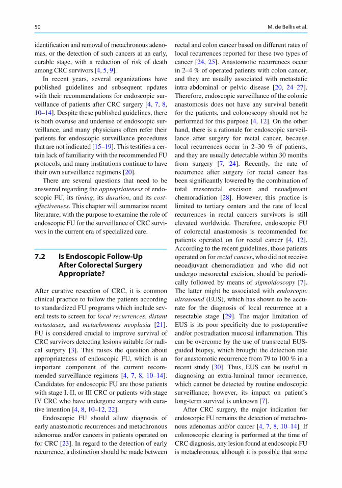
50
identifi cation and removal of metachronous adeno-mas, or the detection of such cancers at an early, curable stage, with a reduction of risk of death among CRC survivors [ 4 , 5 , 9 ].
In recent years, several organizations have published guidelines and subsequent updates with their recommendations for endoscopic sur-veillance of patients after CRC surgery [ 4 , 7 , 8 , 10 – 14 ]. Despite these published guidelines, there is both overuse and underuse of endoscopic sur-veillance, and many physicians often refer their patients for endoscopic surveillance procedures that are not indicated [ 15 – 19 ]. This testifi es a cer-tain lack of familiarity with the recommended FU protocols, and many institutions continue to have their own surveillance regimens [ 20 ].
There are several questions that need to be answered regarding the appropriateness of endo-scopic FU , its timing , its duration , and its cost- effectiveness . This chapter will summarize recent literature, with the purpose to examine the role of endoscopic FU for the surveillance of CRC survi-vors in the current era of specialized care.
7.2 Is Endoscopic Follow-Up After Colorectal Surgery Appropriate?
After curative resection of CRC, it is common clinical practice to follow the patients according to standardized FU programs which include sev-eral tests to screen for local recurrences , distant metastases , and metachronous neoplasia [ 21 ]. FU is considered crucial to improve survival of CRC survivors detecting lesions suitable for radi-cal surgery [ 3 ]. This raises the question about appropriateness of endoscopic FU, which is an important component of the current recom-mended surveillance regimens [ 4 , 7 , 8 , 10 – 14 ]. Candidates for endoscopic FU are those patients with stage I, II, or III CRC or patients with stage IV CRC who have undergone surgery with cura-tive intention [ 4 , 8 , 10 – 12 , 22 ].
Endoscopic FU should allow diagnosis of early anastomotic recurrences and metachronous adenomas and/or cancers in patients operated on for CRC [ 23 ]. In regard to the detection of early recurrence, a distinction should be made between
rectal and colon cancer based on different rates of local recurrences reported for these two types of cancer [ 24 , 25 ]. Anastomotic recurrences occur in 2–4 % of operated patients with colon cancer, and they are usually associated with metastatic intra-abdominal or pelvic disease [ 20 , 24 – 27 ]. Therefore, endoscopic surveillance of the colonic anastomosis does not have any survival benefi t for the patients, and colonoscopy should not be performed for this purpose [ 4 , 12 ]. On the other hand, there is a rationale for endoscopic surveil-lance after surgery for rectal cancer, because local recurrences occur in 2–30 % of patients, and they are usually detectable within 30 months from surgery [ 7 , 24 ]. Recently, the rate of recurrence after surgery for rectal cancer has been signifi cantly lowered by the combination of total mesorectal excision and neoadjuvant chemoradiation [ 28 ]. However, this practice is limited to tertiary centers and the rate of local recurrences in rectal cancers survivors is still elevated worldwide. Therefore, endoscopic FU of colorectal anastomosis is recommended for patients operated on for rectal cancer [ 4 , 12 ]. According to the recent guidelines, those patients operated on for rectal cancer , who did not receive neoadjuvant chemoradiation and who did not undergo mesorectal excision, should be periodi-cally followed by means of sigmoidoscopy [ 7 ]. The latter might be associated with endoscopic ultrasound (EUS), which has shown to be accu-rate for the diagnosis of local recurrence at a resectable stage [ 29 ]. The major limitation of EUS is its poor specifi city due to postoperative and/or postradiation mucosal infl ammation. This can be overcome by the use of transrectal EUS-guided biopsy, which brought the detection rate for anastomotic recurrence from 79 to 100 % in a recent study [ 30 ]. Thus, EUS can be useful in diagnosing an extra- luminal tumor recurrence, which cannot be detected by routine endoscopic surveillance; however, its impact on patient’s long-term survival is unknown [ 7 ].
After CRC surgery, the major indication for endoscopic FU remains the detection of metachro-nous adenomas and/or cancer [ 4 , 7 , 8 , 10 – 14 ]. If colonoscopic clearing is performed at the time of CRC diagnosis, any lesion found at endoscopic FU is metachronous, although it is possible that some
M. de Bellis et al.
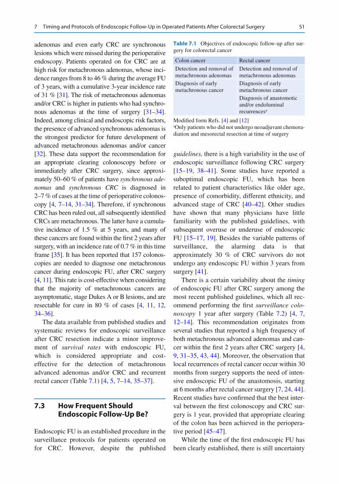
51
adenomas and even early CRC are synchronous lesions which were missed during the perioperative endoscopy. Patients operated on for CRC are at high risk for metachronous adenomas, whose inci-dence ranges from 8 to 46 % during the average FU of 3 years, with a cumulative 3-year incidence rate of 31 % [ 31 ]. The risk of metachronous adenomas and/or CRC is higher in patients who had synchro-nous adenomas at the time of surgery [ 31 – 34 ]. Indeed, among clinical and endoscopic risk factors, the presence of advanced synchronous adenomas is the strongest predictor for future development of advanced metachronous adenomas and/or cancer [ 32 ]. These data support the recommendation for an appropriate clearing colonoscopy before or immediately after CRC surgery, since approxi-mately 50–60 % of patients have synchronous ade-nomas and synchronous CRC is diagnosed in 2–7 % of cases at the time of perioperative colonos-copy [ 4 , 7 – 14 , 31 – 34 ]. Therefore, if synchronous CRC has been ruled out, all subsequently identifi ed CRCs are metachronous. The latter have a cumula-tive incidence of 1.5 % at 5 years, and many of these cancers are found within the fi rst 2 years after surgery, with an incidence rate of 0.7 % in this time frame [ 35 ]. It has been reported that 157 colonos-copies are needed to diagnose one metachronous cancer during endoscopic FU, after CRC surgery [ 4 , 11 ]. This rate is cost-effective when considering that the majority of metachronous cancers are asymptomatic, stage Dukes A or B lesions, and are resectable for cure in 80 % of cases [ 4 , 11 , 12 , 34 – 36 ].
The data available from published studies and systematic reviews for endoscopic surveillance after CRC resection indicate a minor improve-ment of survival rates with endoscopic FU, which is considered appropriate and cost- effective for the detection of metachronous advanced adenomas and/or CRC and recurrent rectal cancer (Table 7.1 ) [ 4 , 5 , 7 – 14 , 35 – 37 ].
7.3 How Frequent Should Endoscopic Follow-Up Be?
Endoscopic FU is an established procedure in the surveillance protocols for patients operated on for CRC. However, despite the published
guidelines , there is a high variability in the use of endoscopic surveillance following CRC surgery [ 15 – 19 , 38 – 41 ]. Some studies have reported a suboptimal endoscopic FU, which has been related to patient characteristics like older age, presence of comorbidity, different ethnicity, and advanced stage of CRC [ 40 – 42 ]. Other studies have shown that many physicians have little familiarity with the published guidelines, with subsequent overuse or underuse of endoscopic FU [ 15 – 17 , 19 ]. Besides the variable patterns of surveillance, the alarming data is that approximately 30 % of CRC survivors do not undergo any endoscopic FU within 3 years from surgery [ 41 ].
There is a certain variability about the timing of endoscopic FU after CRC surgery among the most recent published guidelines, which all rec-ommend performing the fi rst surveillance colo-noscopy 1 year after surgery (Table 7.2 ) [ 4 , 7 , 12 – 14 ]. This recommendation originates from several studies that reported a high frequency of both metachronous advanced adenomas and can-cer within the fi rst 2 years after CRC surgery [ 4 , 9 , 31 – 35 , 43 , 44 ]. Moreover, the observation that local recurrences of rectal cancer occur within 30 months from surgery supports the need of inten-sive endoscopic FU of the anastomosis, starting at 6 months after rectal cancer surgery [ 7 , 24 , 44 ]. Recent studies have confi rmed that the best inter-val between the fi rst colonoscopy and CRC sur-gery is 1 year, provided that appropriate clearing of the colon has been achieved in the periopera-tive period [ 45 – 47 ].
While the time of the fi rst endoscopic FU has been clearly established, there is still uncertainty
Table 7.1 Objectives of endoscopic follow-up after sur-gery for colorectal cancer
Colon cancer Rectal cancer
Detection and removal of metachronous adenomas
Detection and removal of metachronous adenomas
Diagnosis of early metachronous cancer
Diagnosis of early metachronous cancer Diagnosis of anastomotic and/or endoluminal recurrences a
Modifi ed form Refs. [ 4 ] and [ 12 ] a Only patients who did not undergo neoadjuvant chemora-diation and mesorectal resection at time of surgery
7 Timing and Protocols of Endoscopic Follow-Up in Operated Patients After Colorectal Surgery
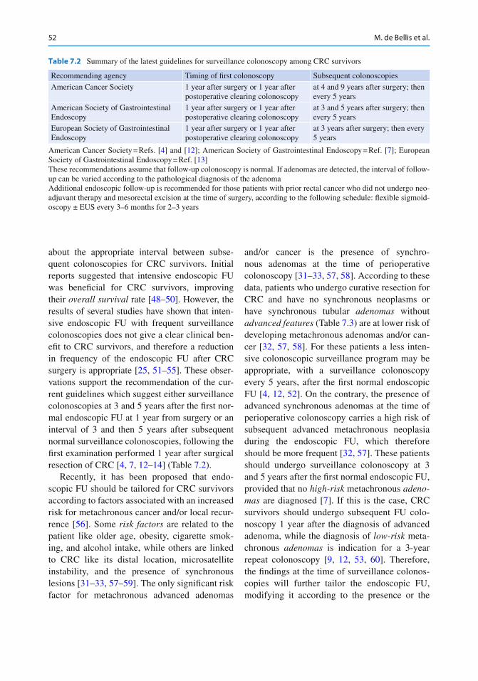
52
about the appropriate interval between subse-quent colonoscopies for CRC survivors. Initial reports suggested that intensive endoscopic FU was benefi cial for CRC survivors, improving their overall survival rate [ 48 – 50 ]. However, the results of several studies have shown that inten-sive endoscopic FU with frequent surveillance colonoscopies does not give a clear clinical ben-efi t to CRC survivors, and therefore a reduction in frequency of the endoscopic FU after CRC surgery is appropriate [ 25 , 51 – 55 ]. These obser-vations support the recommendation of the cur-rent guidelines which suggest either surveillance colonoscopies at 3 and 5 years after the fi rst nor-mal endoscopic FU at 1 year from surgery or an interval of 3 and then 5 years after subsequent normal surveillance colonoscopies, following the fi rst examination performed 1 year after surgical resection of CRC [ 4 , 7 , 12 – 14 ] (Table 7.2 ).
Recently, it has been proposed that endo-scopic FU should be tailored for CRC survivors according to factors associated with an increased risk for metachronous cancer and/or local recur-rence [ 56 ]. Some risk factors are related to the patient like older age, obesity, cigarette smok-ing, and alcohol intake, while others are linked to CRC like its distal location, microsatellite instability, and the presence of synchronous lesions [ 31 – 33 , 57 – 59 ]. The only signifi cant risk factor for metachronous advanced adenomas
and/or cancer is the presence of synchro-nous adenomas at the time of perioperative colonoscopy [ 31 – 33 , 57 , 58 ]. According to these data, patients who undergo curative resection for CRC and have no synchronous neoplasms or have synchronous tubular adenomas without advanced features (Table 7.3 ) are at lower risk of developing metachronous adenomas and/or can-cer [ 32 , 57 , 58 ]. For these patients a less inten-sive colonoscopic surveillance program may be appropriate, with a surveillance colonoscopy every 5 years, after the fi rst normal endoscopic FU [ 4 , 12 , 52 ]. On the contrary, the presence of advanced synchronous adenomas at the time of perioperative colonoscopy carries a high risk of subsequent advanced metachronous neoplasia during the endoscopic FU, which therefore should be more frequent [ 32 , 57 ]. These patients should undergo surveillance colonoscopy at 3 and 5 years after the fi rst normal endoscopic FU, provided that no high- risk metachronous adeno-mas are diagnosed [ 7 ]. If this is the case, CRC survivors should undergo subsequent FU colo-noscopy 1 year after the diagnosis of advanced adenoma, while the diagnosis of low-risk meta-chronous adenomas is indication for a 3-year repeat colonoscopy [ 9 , 12 , 53 , 60 ]. Therefore, the fi ndings at the time of surveillance colonos-copies will further tailor the endoscopic FU, modifying it according to the presence or the
Table 7.2 Summary of the latest guidelines for surveillance colonoscopy among CRC survivors
Recommending agency Timing of fi rst colonoscopy Subsequent colonoscopies
American Cancer Society 1 year after surgery or 1 year after postoperative clearing colonoscopy
at 4 and 9 years after surgery; then every 5 years
American Society of Gastrointestinal Endoscopy
1 year after surgery or 1 year after postoperative clearing colonoscopy
at 3 and 5 years after surgery; then every 5 years
European Society of Gastrointestinal Endoscopy
1 year after surgery or 1 year after postoperative clearing colonoscopy
at 3 years after surgery; then every 5 years
American Cancer Society = Refs. [ 4 ] and [ 12 ]; American Society of Gastrointestinal Endoscopy = Ref. [ 7 ]; European Society of Gastrointestinal Endoscopy = Ref. [ 13 ] These recommendations assume that follow-up colonoscopy is normal. If adenomas are detected, the interval of follow- up can be varied according to the pathological diagnosis of the adenoma Additional endoscopic follow-up is recommended for those patients with prior rectal cancer who did not undergo neo-adjuvant therapy and mesorectal excision at the time of surgery, according to the following schedule: fl exible sigmoid-oscopy ± EUS every 3–6 months for 2–3 years
M. de Bellis et al.
53
absence of metachronous adenomas and to their features [ 9 , 12 , 14 , 32 , 55 ].
Patients with prior rectal cancer should undergo a tailored endoscopic FU, which is based on the type of surgery and the administration of neoadjuvant chemoradiation. Postoperative sur-veillance sigmoidoscopy is not recommended in patients treated with neoadjuvant chemoradiation and mesorectal excision, given the decreased likelihood of local cancer recurrence [ 7 ]. These patients should undergo standard endoscopic FU. On the contrary, an intensive endoscopic FU with sigmoidoscopy ± EUS every 3–6 months, for the fi rst 3 years after surgery, is highly recommended for patients with rectal cancer who have under-gone surgery without mesorectal excision and neoadjuvant chemoradiation [ 7 , 60 ]. This inten-sive endoscopic FU helps to identify resectable local recurrences; however, it has not been shown to improve patient’s survival, and its only ratio-nale is the high rate of local recurrences within the fi rst 2–3 years from rectal surgery [ 4 , 7 , 24 , 61 ]. Finally, a tailored approach is mandatory for patients with inherited syndromes of CRC. Hereditary non Polyposis Colorectal Cancer (HNPCC) patients diagnosed with CRC should undergo proctocolectomy, because of the high risk of metachronous cancer, which is 25 % after segmental colectomy and 8% after total colec-tomy with ileorectal anastomosis [ 62 – 64 ]. However, many HNPCC patients still undergo either segmental colectomy or total colectomy with ileorectal anastomosis, and therefore they require yearly surveillance colonoscopy or rec-toscopy to remove premalignant adenomas [ 62 – 64 ]. Similarly, patients with Familial
Adenomatous Polyposis (FAP) require strict endoscopic FU of the rectal stump or the ileal pouch [ 62 , 65 , 66 ]. Rectal cancer has been reported in 17 % of FAP patients with ileorectal anastomosis and in 8 % of cases with an ileal pouch [ 65 ]. These data confi rm the recommenda-tion that FAP patients should undergo intensive endoscopic FU, which forecast a proctosigmoid-oscopy every 6–12 months, according to the number of adenomas detected [ 62 , 65 , 66 ].
7.4 How Long Should Endoscopic Follow-Up Last?
According to the natural history of CRC, FU should be discontinued 5 years after surgery [ 67 ]. However, the published guidelines recommend long-term endoscopic surveillance of CRC survi-vors because they have a lifelong risk of develop-ing metachronous CRC [ 4 , 7 , 8 , 10 – 14 , 34 , 68 – 73 ]. A retrospective study reported that meta-chronous CRCs occur in approximately 30 % of patients 5 years after the resection of the primary CRC [ 34 ]. The authors of another retrospective study calculated that the cumulative risk of meta-chronous CRCs ranges from 2 %, for 5 years CRC survivors, to 7 % for those patients who are alive 20 years after colorectal surgery [ 73 ]. Therefore, the risk of developing metachronous CRCs seems to increase continuously over time, after surgical resection of the primary CRC. Metachronous CRCs occur from 1 to 25 years after surgery among various studies, and this data justifi es a lifelong endoscopic FU [ 34 , 68 – 73 ]. The latter may explain the high rate of resectable metachronous CRCs which are diagnosed at an early stage, as compared to the general popula-tion [ 72 , 73 ]. However, endoscopic FU does not prevent the occurrence of metachronous cancers in CRC survivors, probably because patients with a history of CRC cancer may differ biologically from patients with adenomatous polyps and are at higher risk for metachronous cancer [ 69 ]. This hypothesis is supported by the observation that the incidence rate of metachronous cancer in
Table 7.3 Features of colorectal adenomas
Low-risk adenomas High-risk adenomas
No more than three adenomas
Multiple adenomas (≥3)
Size < 1 cm Size > 1 cm Tubular histology Villous or tubulovillous
histology Low-grade dysplasia High-grade dysplasia
Modifi ed form Ref. [ 13 ]
7 Timing and Protocols of Endoscopic Follow-Up in Operated Patients After Colorectal Surgery
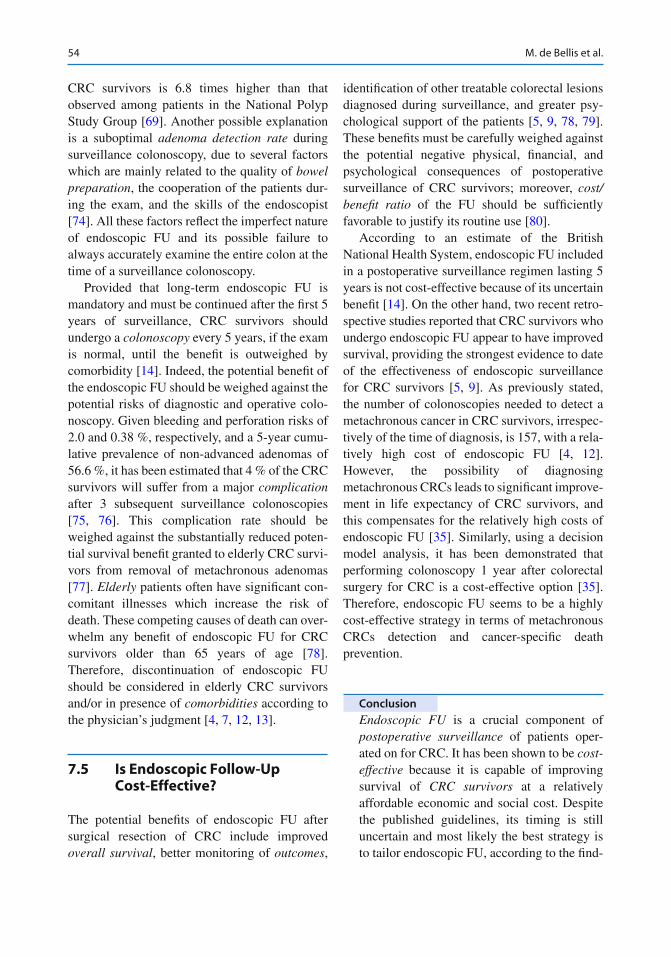
54
CRC survivors is 6.8 times higher than that observed among patients in the National Polyp Study Group [ 69 ]. Another possible explanation is a suboptimal adenoma detection rate during surveillance colonoscopy, due to several factors which are mainly related to the quality of bowel preparation , the cooperation of the patients dur-ing the exam, and the skills of the endoscopist [ 74 ]. All these factors refl ect the imperfect nature of endoscopic FU and its possible failure to always accurately examine the entire colon at the time of a surveillance colonoscopy.
Provided that long-term endoscopic FU is mandatory and must be continued after the fi rst 5 years of surveillance, CRC survivors should undergo a colonoscopy every 5 years, if the exam is normal, until the benefi t is outweighed by comorbidity [ 14 ]. Indeed, the potential benefi t of the endoscopic FU should be weighed against the potential risks of diagnostic and operative colo-noscopy. Given bleeding and perforation risks of 2.0 and 0.38 %, respectively, and a 5-year cumu-lative prevalence of non-advanced adenomas of 56.6 %, it has been estimated that 4 % of the CRC survivors will suffer from a major complication after 3 subsequent surveillance colonoscopies [ 75 , 76 ]. This complication rate should be weighed against the substantially reduced poten-tial survival benefi t granted to elderly CRC survi-vors from removal of metachronous adenomas [ 77 ]. Elderly patients often have signifi cant con-comitant illnesses which increase the risk of death. These competing causes of death can over-whelm any benefi t of endoscopic FU for CRC survivors older than 65 years of age [ 78 ]. Therefore, discontinuation of endoscopic FU should be considered in elderly CRC survivors and/or in presence of comorbidities according to the physician’s judgment [ 4 , 7 , 12 , 13 ].
7.5 Is Endoscopic Follow-Up Cost-Effective?
The potential benefi ts of endoscopic FU after surgical resection of CRC include improved overall survival , better monitoring of outcomes ,
identifi cation of other treatable colorectal lesions diagnosed during surveillance, and greater psy-chological support of the patients [ 5 , 9 , 78 , 79 ]. These benefi ts must be carefully weighed against the potential negative physical, fi nancial, and psychological consequences of postoperative surveillance of CRC survivors; moreover, cost/benefi t ratio of the FU should be suffi ciently favorable to justify its routine use [ 80 ].
According to an estimate of the British National Health System, endoscopic FU included in a postoperative surveillance regimen lasting 5 years is not cost-effective because of its uncertain benefi t [ 14 ]. On the other hand, two recent retro-spective studies reported that CRC survivors who undergo endoscopic FU appear to have improved survival, providing the strongest evidence to date of the effectiveness of endoscopic surveillance for CRC survivors [ 5 , 9 ]. As previously stated, the number of colonoscopies needed to detect a metachronous cancer in CRC survivors, irrespec-tively of the time of diagnosis, is 157, with a rela-tively high cost of endoscopic FU [ 4 , 12 ]. However, the possibility of diagnosing metachronous CRCs leads to signifi cant improve-ment in life expectancy of CRC survivors, and this compensates for the relatively high costs of endoscopic FU [ 35 ]. Similarly, using a decision model analysis, it has been demonstrated that performing colonoscopy 1 year after colorectal surgery for CRC is a cost-effective option [ 35 ]. Therefore, endoscopic FU seems to be a highly cost- effective strategy in terms of metachronous CRCs detection and cancer-specifi c death prevention.
Conclusion
Endoscopic FU is a crucial component of postoperative surveillance of patients oper-ated on for CRC. It has been shown to be cost-effective because it is capable of improving survival of CRC survivors at a relatively affordable economic and social cost. Despite the published guidelines, its timing is still uncertain and most likely the best strategy is to tailor endoscopic FU, according to the fi nd-
M. de Bellis et al.
55
Perioperative colonoscopy
high-risk “advanced”adenomas
Normal findings orlow-risk adenomas
Colonoscopy at 1 year Colonoscopyat 1 year*Early
metachronouscolorectal cancer
Normal findings orlow-risk adenomas
Colonoscopy at 3 years
Normal findings or low-risk adenomas
Colonoscopy every 5 years
Normal findings orlow-risk adenomas
Colonoscopy at 5 years
Normal findings orlow-risk adenomas
Colonoscopy at 5 years
Normal findings orlow-risk adenomas
Colonoscopy every 5 years
high-risk adenomashigh-risk adenomas
Repeat colonoscopy after 1 year
3-year repeat colonoscopy
Normal findings or low-risk adenomas
Normal findings orlow-risk adenomas
Colonoscopy every 5 years
surgery
Diagnosis of colonic or rectal cancer
Fig. 7.1 Proposed algorithm for a tailored endoscopic follow-up of CRC survivors. *The diagnosis of early metachronous cancer is indication for surgery at any time during endoscopic follow-up. HNPCC patients who
undergo segmental colectomy require yearly surveillance colonoscopy. Endoscopic FU will be continued until the benefi t is outweighed by comorbidity
7 Timing and Protocols of Endoscopic Follow-Up in Operated Patients After Colorectal Surgery
56
ings of both perioperative colonoscopy and surveillance endoscopies (Figs. 7.1 and 7.2 ).
Acknowledgements The authors are grateful to Dr. Alessandra Trocino, Librarian at the National Cancer Institute and G Pascale Foundation of Naples, Italy, for her bibliographic assistance.
References
1. Brawarsky P, Neville BA, Fitzmaurice GM (2013) Surveillance after resection for colorectal cancer. Cancer 119:1235–1242
2. Boyle P, Ferlay J (2005) Cancer incidence and mortal-ity in Europe, 2004. Ann Oncol 16:481–488
3. Scheer A, Auer RAC (2009) Surveillance after cura-tive resection of colorectal cancer. Clin Colon Rectal Surg 22:242–250
4. Rex DK, Kahi CJ, Levin B et al (2006) Guidelines for colonoscopy surveillance after cancer resection: a consensus update by the American Cancer Society and the US Multi-Society Task Force on Colorectal Cancer. Gastroenterology 130:1865–1871
5. Fisher DA, Jeffreys A, Grambow SC, Provenzale D (2003) Mortality and follow-up colonoscopy after colorectal cancer. Am J Gastroenterol 98:901–906
6. Speake D, Lees N, McMahon RFT, Hill J (2008) Who should be followed up after transanal endoscopic resec-tion of rectal tumours? Colorectal Dis 10:330–335
7. Davila RE, Rajan E, Baron TH, ASGE Standard of Practice Committee (2006) ASGE guideline: colorectal cancer screening and surveillance. Gastrointest Endosc 63(4):546–557
8. Glimelius B, Påhlman L, Cervantes A, ESMO Guidelines Working Group (2010) Rectal cancer: ESMO clinical practice guidelines for diagnosis, treatment and follow-up. Ann Oncol 21(Suppl 5):v82–v86
9. Rulyak SJ, Lieberman DA, Wagner EK et al (2007) Outcome of follow-up colon examination among a population-based cohort of colorectal cancer patients. Clin Gastroenterol Hepatol 5:470–476
10. Labianca R, Nordlinger B, Beretta GD, ESMO Guidelines Working Group et al (2010) Primary colon cancer ESMO clinical practice guidelines for diagno-sis, treatment and follow-up. Ann Oncol 21(Suppl 5):v70–v77
11. Brooks DD, Winawer SJ, Rex DK et al (2008) Colonoscopy surveillance after polypectomy and colorectal cancer resection. Am Fam Physician 77(7):995–1002
12. Rex DK, Kahi CJ, Levin B et al (2006) Guidelines for colonoscopy surveillance after cancer resection: a consensus update by the American Cancer Society and US Multi-Society Task Force on Colorectal Cancer. CA Cancer J Clin 56:160–167
Diagnosis of Rectal Cancer
Standard surgerywithout neoadjuvant chemoradiation
Neoadjuvant chemoradiationMesorectal excision
Standard endoscopicfollow-up
Sigmoidoscopy ± EUSat 3-6 months
Normal findings at colorectal anastomosis
Sigmoidoscopy ± EUSevery 3-6 months
up to 3 years from surgery
*Earlyanastomoticrecurrence
surgery
Fig. 7.2 Proposed algorithm for additional tailored endoscopic follow-up of CRC survivors after rectal surgery. *The diagnosis of early anastomotic recurrence is indication for surgery at any time during endoscopic follow-up. HNPCC patients who have undergone total colectomy with ileorectal anastomosis require yearly surveillance proctoscopy. Operated FAP patients require strict endoscopic FU of the rectal stump or the ileal pouch. FAP patients should undergo proctosigmoidoscopy every 6–12 months, based on the number of adenomas detected during the exam
M. de Bellis et al.
57
13. Arditi C, Gonvers JJ, Burnand B, EPAGE II Study Group et al (2009) Appropriateness of colonoscopy in Europe (EPAGE II). Surveillance after polypectomy and after resection of colorectal cancer. Endoscopy 41:209–217
14. Cairns SR, Scholefi eld JH, Steele RJ et al on behalf of The British Society of Gastroenterology, and the Association of Coloproctology for Great Britain and Ireland (2010) Guidelines for colorectal cancer screening and surveillance in moderate and high risk groups (update from 2002). Gut 59:666–690
15. Zbidi I, Hazazi R, Niv Y (2007) Colonoscopy screen-ing and surveillance of colorectal cancer and polyps: physicians’ knowledge. IMAJ 9:862–865
16. Singh A, Kuo YF, Goodwin JS (2013) Many patients who undergo surgery for colorectal cancer receive surveillance colonoscopies earlier than recommended by guidelines. Clin Gastroenterol Hepatol 11:65–72
17. Cooper GS, Kou TD, Reynolds HL Jr (2008) Receipt of guideline-recommended follow-up in older colorectal cancer survivors. Cancer 113:2029–2037
18. John BJ, Irukulla S, Mendall MA, Abulafi AM (2010) Do guidelines improve clinical practice? A national survey on surveillance colonoscopies. Colorectal Dis 12:642–645
19. Viehl CT, Ochsner A, von Holzen U et al (2010) Inadequate quality of surveillance after curative surgery for colon cancer. Ann Surg Oncol 17:2663–2669
20. Salz T, Weinberger M, Ayanian JZ et al (2010) Variation in use of surveillance colonoscopy among colorectal cancer survivors in the United States. BMC Health Services Research 10. http://www.biomedcen-tral.com/1472-6963/10/256 . Accessed 1 Mar 2013
21. Jeffery M, Hickey BE, Hider PN (2008) Follow-up strat-egies for patients treated for non-metastatic colorectal cancer (Review). The Cochrane Library, Issue 4. http://www.thecochranelibrary.com . Accessed 25 Feb 2013
22. Rodríguez-Moranta F, Saló J, Arcusa A et al (2006) Postoperative surveillance in patients with colorectal cancer who have undergone curative resection: a pro-spective, multicenter, randomized, controlled trial. J Clin Oncol 24:386–393
23. Lautenbach E, Forde KA, Neugut A (1994) Benefi ts of colonoscopy surveillance after curative resection of colorectal cancer. Ann Surg 220(2):206–211
24. Barillari P, Ramacciato G, Manetti G et al (1996) Surveillance of colorectal cancer: effectiveness of early detection of intraluminal recurrences on progno-sis and survival of patients treated for cure. Dis Colon Rectum 39:388–393
25. Schoemaker D, Black R, Giles L, Toouli J (1998) Yearly colonoscopy, liver CT, and chest radiography do not infl uence 5-year survival of colorectal cancer patients. Gastroenterology 114:7–14
26. Porter GA, Soskolne CL, Yakimets WW, Newman SC (1998) Surgeon-related factors and outcome in rectal cancer. Ann Surg 227:157–167
27. Panageas KS, Schrag D, Riedel E et al (2003) The effect of clustering of outcomes on the association of
procedure volume and surgical outcomes. Ann Intern Med 139:658–665
28. Kapiteijn E, Marijnen CA, Nagtegaal ID, Dutch Colorectal Cancer Group et al (2001) Preoperative radio-therapy combined with total mesorectal excision for resectable rectal cancer. N Engl J Med 345:638–646
29. Wiersema MJ, Harewood GC (2002) Endoscopic ultrasound for rectal cancer. Gastroenterol Clin N Am 31:1093–1105
30. Lohnert M, Doniec JM, Henne-Bruns D (2000) Effectiveness of endoluminal sonography in the iden-tifi cation of occult local rectal cancer. Dis Colon Rectum 43:483–491
31. Chu DZJ, Chansky K, Alberts DS (2003) Adenoma recurrences after resection of colorectal carcinoma: results from the Southwest Oncology Group 9041 Calcium Chemoprevention Pilot Study. Ann Surg Oncol 10(8):870–875
32. Moon CM, Cheon JH, Choi EH (2010) Advanced synchronous adenoma but not simple adenoma pre-dicts the future development of metachronous neopla-sia in patients with resected colorectal cancer. J Clin Gastroenterol 44:495–501
33. Ballesté B, Bessa X, Piñol V (2007) Detection of meta-chronous neoplasms in colorectal cancer patients: iden-tifi cation of risk factors. Dis Colon Rectum 50:1–10
34. Park IJ, Yu CS, Kim HC et al (2006) Metachronous colorectal cancer. Colorectal Dis 8:323–327
35. Hassan C, Pickhardt PJ, Zullo A (2009) Cost- effectiveness of early colonoscopy surveillance after cancer resection. Dig Liver Dis 41:881–885
36. Bochud M, Burnand B, Froehlich F (1999) Appropriateness of colonoscopy: surveillance after curative resection of colorectal cancer. Endoscopy 31(8):664–672
37. Eckardt VF, Stamm H, Kanzler G, Bernhard G (1994) Improved survival after colorectal cancer in patients complying with a postoperative endoscopic surveil-lance program. Endoscopy 26(8):523–527
38. Cooper GS, Yuan Z, Chak A, Rimm AA (2000) Patterns of endoscopic follow-up after surgery for nonmetastatic colorectal cancer. Gastrointest Endosc 52:33–38
39. Hilsden RJ, Bryant HE, Sutherland LR et al (2004) A retrospective study on the use of post-operative colo-noscopy following potentially curative surgery for colorectal cancer in a Canadian province. BMC Cancer 4:14. http://www.biomedcentral.com/1471- 2407/4/14 . Accessed 11 Feb 2013
40. Foley KL, Song EY, Klepin H et al (2012) Screening colonoscopy among colorectal cancer survivors insured by Medicaid. Am J Clin Oncol 35:205–211
41. Cooper GS, Payes JD (2006) Temporal trends in colorectal procedure use after colorectal cancer resec-tion. Gastrointest Endosc 64:933–940
42. Cooper GS, Yuan Z, Chak A, Rimm AA (1999) Geographic and patient variation among Medicare benefi ciaries in the use of follow-up testing after sur-gery for nonmetastatic colorectal carcinoma. Cancer 85:2124–2131
7 Timing and Protocols of Endoscopic Follow-Up in Operated Patients After Colorectal Surgery
58
43. Platell C, Salama P, Barwood N, Makin G (2005) Performing a colonoscopy 12 months after surgery for colorectal neoplasia. ANZ J Surg 75:282–285
44. Yun HR, Lee LJ, Park JH (2008) Local recurrence after curative resection in patients with colon and rec-tal cancers. Int J Colorectal Dis 23:1081–1087
45. Hyman N, Moore J, Cataldo P, Osler T (2010) The high yield of 1-year colonoscopy after resection: is it the handoff? Surg Endosc 24:648–652
46. Hollington P, Tiong L, Young G (2011) Timing and detection of metachronous colorectal cancer. ANZ J Surg 81:272–274
47. Couch DG, Bullen N, Ward-Booth SE, Adams C (2012) What interval between colorectal cancer resec-tion and fi rst surveillance colonoscopy? An audit of practice and yield. Colorectal Dis 15:317–322
48. Larson GM, Bond SJ, Shallcross C et al (1986) Colonoscopy after curative resection of colorectal cancer. Arch Surg 121:535–540
49. Juhl G, Larson GM, Mullins R (1990) Six year results of annual colonoscopy after resection of colorectal cancer. World J Surg 14:255–261, 19
50. Khoury DA, Opelka FG, Beck DE et al (1996) Colon surveillance after colorectal cancer surgery. Dis Colon Rectum 39:252–256
51. Yusoff IF, Hoffman EN, Ee HC (2003) Colonoscopic surveillance after surgery for colorectal cancer. ANZ J Surg 73:3–7
52. McFall MR, Woods WGA, Miles WFA (2003) Colonoscopic surveillance after curative colorectal resection: results of an empirical surveillance pro-gramme. Colorectal Dis 5:233–240
53. Sakamoto T, Matsuda T, Nakajima T, Saito Y (2012) How often should we perform surveillance colonos-copy after surgery for colorectal cancer? Int J Colorectal Dis. doi: 10.1007/s00384-012-1613-5
54. Wang T, Cui Y, Huang WS et al (2009) The role of postoperative colonoscopic surveillance after radical surgery for colorectal cancer: a prospective, random-ized clinical study. Gastrointest Endosc 69:609–615
55. Mathew J, Saklani AK, Borgho M (2006) Surveillance colonoscopy in patients with colorectal cancer: how often should we be doing it? Surgeon 4(1):3–5
56. Søreide K (2010) Endoscopic surveillance after cura-tive surgery for sporadic colorectal cancer: patient- tailored, tumor-targeted or biology-driven? Scand J Gastroenterol 45:1255–1261
57. Rajaratnam SG, Dennett ER (2009) Development of metachronous neoplasms after colorectal cancer resection: absence of synchronous neoplasms predicts a lower risk. N Z Med J 122:61–66
58. Borda A, Martínez-Peñuela MJ, Borda F et al (2012) Drawing up an individual risk index for development of metachronous neoplastic lesions in resected colorectal cancer. Rev Esp Enferm Dig 104(6):291–297
59. Kang KI, Sinn DH, Park SH (2010) Adenoma incidence after resection of sporadic colorectal
cancer with microsatellite instability. J Surg Oncol 101:577–581
60. Figueredo A, Bryan Rumble R, Maroun J et al (2003) Follow-up of patients with curatively resected colorec-tal cancer: a practice guideline. BMC Cancer 3:26. http://www.biomedcentral.com/1471- 2407/3/26 . Accessed 1 Feb 2013
61. Davila RE, Rajan E, Adler D et al (2005) ASGE guideline: the role of endoscopy in the diagnosis, staging, and management of colorectal cancer. Gastrointest Endosc 61:1–7
62. Balmaña J, Castells A, Cervantes A, ESMO Guidelines Working Group (2010) Familial colorectal cancer risk: ESMO clinical practice guidelines. Ann Oncol 21(Suppl 5):v78–v81
63. Kalady MF, McGannon E, Vogel JD et al (2010) Risk of colorectal adenoma and carcinoma after colectomy for colorectal cancer in patients meeting Amsterdam criteria. Ann Surg 252:507–513
64. Win AK, Parry S, Parry B (2013) Risk of metachro-nous colon cancer following surgery for rectal cancer in mismatch repair gene mutation carriers. Ann Surg Oncol. doi: 10.1245/s10434-012-2858-5
65. Guilherme Campos F, Imperiale AR, Seid VE (2009) Rectal and pouch recurrences after surgical treatment for familial adenomatous polyposis. J Gastrointest Surg 13:129–136
66. Yamaguchi T, Yamamoto S, Fujita S et al (2010) Long-term outcome of metachronous rectal cancer following ileorectal anastomosis for familial adeno-matous polyposis. J Gastrointest Surg 14:500–505
67. Sinclair P, Singh A, Riaz AA et al (2012) An unsolved conundrum: the ideal follow-up strategy after curative surgery for colorectal cancer. Gastrointest Endosc 75(5):1072–1079
68. Enblad P, Adami H-O, Glimelius B et al (1990) The risk of subsequent primary malignant diseases after cancers of the colon and rectum. A nationwide cohort study. Cancer 65:2091–2100
69. Green RJ, Metlay JP, Propert K et al (2002) Surveillance for second primary colorectal cancer after adjuvant chemotherapy: an analysis of inter-group 0089. Ann Intern Med 136:261–269
70. Evans HS, Møller H, Robinson D et al (2002) The risk of subsequent primary cancers after colorectal cancer in southeast England. Gut 50:647–652
71. Papadopoulos V, Michalopoulos A, Basdanis G (2004) Synchronous and metachronous colorectal carcinoma. Tech Coloproctol 8:S97–S100. doi: 10.1007/s10151-004-0124-y
72. LanY-T LJK, Li AF-Y et al (2005) Metachronous colorectal cancer: necessity of post-operative colono-scopic surveillance. Int J Colorectal Dis 20:121–125. doi: 10.1007/s00384-004-0635-z
73. Bouvier A-M, Latournerie M, Jooste V et al (2008) The lifelong risk of metachronous colorectal cancer justifi es long-term colonoscopic follow-up. Eur J Cancer 44:522–527
M. de Bellis et al.

59
74. de Groen PC (2010) Advanced systems to assess colo-noscopy. Gastrointest Endosc Clin N Am 20:699–716
75. Sonnenberg A, Delco F, Inadomi JM (2000) Cost- effectiveness of colonoscopy in screening for colorec-tal cancer. Ann Intern Med 133:573–584
76. Hassan C, Gaglia P, Zullo A et al (2006) Endoscopic follow-up after colorectal cancer resection: an Italian multicentre study. Dig Liver Dis 38:45–50
77. Ramsey DS, Howlader N, Etzioni R (2007) Surveillance endoscopy does not improve survival for patients with local and regional stage colorectal can-cer. Cancer 109:2222–2228
78. Augestad KM, Vonen B, Ranveig A (2008) Should the surgeon or the general practitioner (GP) follow up patients after surgery for colon cancer? A randomized controlled trial protocol focusing on quality of life, cost-effectiveness and serious clinical events. BMC Health Serv Res 8:137. doi: 10.1186/1472-6963-8-137
79. Salz T, Brewer NT, Sandler RS (2009) Association of health beliefs and colonoscopy use among survivors of colorectal cancer. J Cancer Surviv 3(4):193–201. doi: 10.1007/s11764-009-0095-0
80. Northover J (2003) Follow-up after curative surgery for colorectal cancer. Scand J Surg 92:84–89
7 Timing and Protocols of Endoscopic Follow-Up in Operated Patients After Colorectal Surgery
61G. Galloro (ed.), Endoscopic Follow-up of Digestive Anastomosis,DOI 10.1007/978-88-470-5370-0_8, © Springer-Verlag Italia 2014
8.1 Introduction
Most GI tract surgical interventions require anastomotic procedures which are associated with complications such as leakage, bleeding, impaired transit, or strictures. Since the 1970s, intraoperative endoscopy was applied as a diag-nostic tool during colorectal surgery to help the
surgeon locating the exact area of interest [ 1 – 3 ]. Afterwards, intraoperative endoscopy has been utilized for evaluation of patency and integrity of GI tract anastomoses during the surgical proce-dure [ 4 , 5 ]. Since endoscopic intraoperative anas-tomotic testing allows direct visualization of anastomosis, complication rates can be reduced by the use of this technique.
R. Manta , MD (*) • M. Manno • R. Conigliaro Gastroenterology and Digestive Endoscopy Unit , New S.Agostino-Estense Hospital , Viale Giardini 1355 , Baggiovara (MO) 41100 , Italy e-mail: [email protected]; [email protected]; [email protected]
A. Naik Department of Gastroenterology , Ashford and St Peter’s Hospitals , Guildford Road , London , UK e-mail: [email protected]
M. Frazzoni Digestive Pathophysiology Unit , New S. Agostino-Estense Hospital , Via Giardini 1355 , Baggiovara–Modena (MO) 41100 , Italy e-mail: [email protected]
A. Arezzo Department of Surgical Sciences , School of Medicine, University of Turin , corso Achille Mario Dogliotti 14 , Turin 10126 , Italy e-mail: [email protected]
M. de Bellis Endoscopy Unit , National Cancer Institute and G. Pascale Foundation , Via Mariano Semmola , Naples 80131 , Italy e-mail: [email protected]
8 Intraoperative Endoscopy in the Evaluation of Digestive Anastomoses
Raffaele Manta , Amitabh Naik , Marzio Frazzoni , Mauro Manno , Alberto Arezzo , Mario de Bellis , Antonello Trecca , Gabrio Bassotti , Gianluigi Melotti , Rita Conigliaro , and Giuseppe Galloro
A. Trecca Operative Endoscopy Unit , USI Group , Rome , Italy e-mail: [email protected]
G. Bassotti Gastroenterology and Hepatology Section, Department of Clinical and Experimental Medicine , University of Perugia , Piazzale Menghini, 1 , Perugia 06156 , Italy e-mail: [email protected]
G. Melotti General Surgery , New S. Agostino-Estense Hospital, Baggiovara – Modena , Viale Giardini 1355 , Baggiovara–Modena (MO) 41100 , Italy e-mail: [email protected]
G. Galloro Digestive Surgical Endoscopy Unit, Department of Clinical Medicine and Surgery , School of Medicine, University of Naples Federico II , Via Pansini , Naples 80131 , Italy e-mail: [email protected]
62
8.2 Intraoperative Endoscopy in Upper GI Tract Surgery
8.2.1 Gastric Bypass
Morbid obesity is a global epidemic. As the demand for bariatric surgery increases, endos-copy is being increasingly used both intra- and postoperatively.
In 1994, Wittgrove et al. [ 6 ] introduced lapa-roscopic Roux-en-Y gastric bypass (LRYGBP). Anastomotic leakage is the most serious early postoperative complication following LRYGBP. Leakage can occur at the gastrojejunal anastomo-sis, at the gastric pouch, or at the jejunojejunal anastomosis. The commonest site for a leak after a LRYGBP is the gastrojejunal anastomosis (GJA) site with reported incidences from 0 to 5.2 % [ 7 – 10 ].
The detection of early postoperative leaks tra-ditionally was based on upper GI series. Intraoperative assessment of the GJA, however, in a study performed in 245 patients (Madan et al.) showed that an upper GI series has a sensi-tivity of 75 % and a positive predictive value of 67 % in the detection of anastomotic leaks [ 11 ].
The main advantage of intraoperative endos-copy is that it can be used not only to detect leaks but also to treat them in the operating room. Moreover, intraoperative endoscopy can also ensure that the anastomosis isn’t too tight. To treat leaks from the GJA and from the gastric pouch, application of fi brin sealant, bovine peri-cardial strips, and omental patches has been used with variable success [ 12 – 14 ].
Fernandez et al. [ 15 ] analyzed more than 3.000 patients undergoing gastric bypass sur-gery and identifi ed GJA leakage as an indepen-dent risk factor associated with postoperative mortality besides weight, hypertension and the type of bypass operation.
A number of techniques have emerged over the years to eliminate anastomotic or staple-line disruption. In the past, postoperative leaks were detected intraoperatively for GJA by simple visu-alization of the anastomosis by the surgeon. Later on, air insuffl ation under saline or methylene blue instillation was used, but more recently various groups have used intraoperative endoscopic examination of the anastomosis.
Methylene blue can be instilled through a nasogastric tube or even through an upper GI endoscope, with the patient in the anti- Trendelenburg position [ 16 ]. The nasogastric tube is positioned in the gastric pouch and the jejunum is occluded. However, the technique is demanding and retesting is problematic as the tis-sue remains stained after the fi rst evaluation.
Ramanathan et al. [ 17 ] compared endoscopy to the methylene blue test and found the low pressure infusion of methylene blue to be less sensitive to leak detection than air insuffl ation by endoscopy. This study showed that a 10 % inci-dence of leaks was reduced to 3.8 % owing to suture strengthening after intraoperative gastros-copy (IOG).
Champion et al. [ 10 ] demonstrated that post-operative leakage can be reduced from 3.8 to 0.36 % with the aid of IOG.
With the advent of intraoperative endoscopy, many surgeons raised a concern that the insuffl a-tion caused by the endoscope may damage the anastomosis. There is also a concern raised that insuffl ation of air during IOG may produce more false-positive air leak test. Mohos et al. [ 18 ] showed that the pressure leading to damage of the staple line in the animal model is more than three times higher than the maximal pressure measured in humans during IOG. However, the recent use of CO 2 insuffl ation instead of air will probably solve these problems.
Intraoperative endoscopy has been shown to reduce postoperative morbidity. One study has reported a 0 % postoperative anastomotic leak-age rate in more than 250 patients using IOG [ 8 ]. In 340 patients who underwent LRYGB, Sekhar et al. [ 12 ] found no postoperative leaks because 56 intraoperative leaks were detected and corrected by IOG. In a study assessing 400 patients undergoing LRGYB, Alaedeen et al. demonstrated that IOG reduced postoperative leak rates from 4 to 0.5 % and mortality from 1 to 0 % [ 19 ].
GI bleeding is another important complication after gastric bypass surgery. The incidence can vary from 0.5 % for open procedures to 4.4 % for the laparoscopic approach. Intraluminal bleeding can occur during intervention from any of the staple lines and can be missed unless an IOG is carried out [ 6 , 10 ].
R. Manta et al.
63
– Technique for IOG During Laparoscopic Gastric Bypass The upper esophageal sphincter is intubated under vision. Occasionally a jaw thrust may be of help during the introduction of the endoscope. The proximal pouch is initially inspected, and then the endoscope is gently guided through the anastomosis into the Roux limb. A bowel clamp is placed on the intestinal limb distal to the GJA. The table is leveled and operative fi eld contain-ing the anastomoses is fi lled with sterile normal saline to cover the proximal pouch and anasto-mosis. The area is then irrigated until it is cleared from blood and operative debris. The gastro-scope is then withdrawn into the proximal pouch, and the anastomosis reinspected with continuous insuffl ation. Before withdrawal the air that has been introduced is aspirated completely. In case of persistent air leak, the endoscope is left in situ till the repair of gastrojejunostomy suture line. The procedure is repeated.
What can go wrong during an intra–operative endoscopy is shown below : 1. An intraoperative gastroscopy has to be carried
out with due care especially during intubation of the esophagus. Intubation must be carried out under vision to avoid pharyngeal tears.
2. In gastric bypass surgery, the proximal gastric pouch should be examined carefully to prevent pouch tear, and this can be done by ensuring that the gastroscope controls are in the unlocked position.
3. It is mandatory to carefully perform endo-scopic maneuvers and air insuffl ation in order to avoid an important complication that can occur during IOG well described in some cases report: air embolism can occurred in patients of all ages in association with an interruption in the mucosal barrier [ 20 , 21 ].
8.2.2 Other Gastric Interventions
Besides LRYGB, vertical banded gastroplasty , sleeve gastrectomy , laparoscopic adjustable gas-tric bands , and placement of gastric pacemakers can also be assisted with intraoperative endos-copy to minimize early complications [ 10 ].
Usefulness of IOG during sleeve gastrectomy for obesity was demonstrated by Frezza et al. in 2008.
They used a 29 F endoscope instead of bougies in order to caliber the anastomosis width and to help in the sleeve gastrectomy procedure [ 22 ].
No report has been published about the use of intraoperative endoscopy during video- laparoscopic GIST removal. At the New S. Agostino-Estense Hospital in Baggiovara-Modena (Italy), we elected to routinely perform IOG during laparoscopic removal of gastric GIST located in the gastric body or fundus wall (Fig. 8.1 ). In our experience (10 cases), after adoption of IOG leakages and early hemorrhages were not registered. The only adverse event that we registered was entrapment of the endoscope in the stapler valves that was immediately resolved without further complication.
Finally, IOG has also been proved useful in patients undergoing total gastrectomy for gastric cancer [ 23 , 24 ].
8.3 Intraoperative Endoscopy in Colorectal Surgery
Anastomotic complications such as leakage and bleeding still represent the most serious compli-cations of laparoscopic colorectal surgery. Intraoperative colonoscopy allows detection of the bleeding source and of leaks. Surgeons have tried testing the anastomosis, especially in the distal colon and rectum by using rectal probes fi lled with air [ 25 – 27 ] or saline [ 28 – 30 ] or by using methylene blue enema [ 31 ]. Adopting intraoperative endoscopy, the pelvis is fi lled with saline and air is insuffl ated in the rectum using a sigmoidoscope. The presence of air bub-bles indicates anastomotic leaks which can be repaired without delay. In a recent study carried out in 60 patients, intraoperative air testing of colorectal anastomosis proved to be an effective method for prevention of anastomotic dehis-cence , incidence being 50 % lower in compari-son with the control group [ 32 ]. A more novel and recent application of intraoperative endos-copy to evaluate the adequacy of GI anastomo-ses is based on narrow band imaging (NBI). As vascularization is the primary factor determining anastomotic viability, NBI could prove useful to ascertain tissue vascularity after the anastomotic procedure [ 33 ].
8 Intraoperative Endoscopy in the Evaluation of Digestive Anastomoses
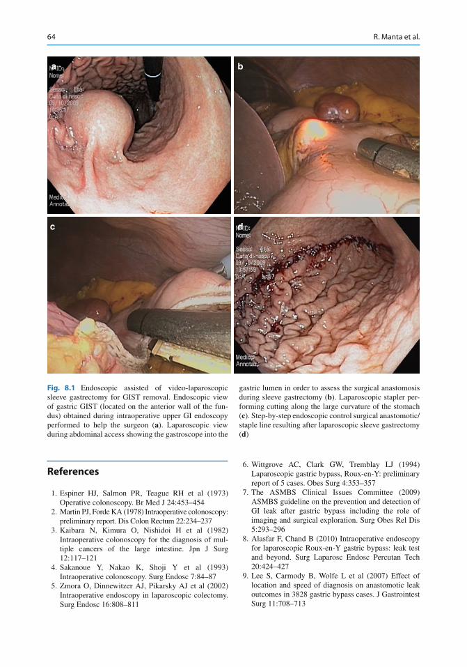
64
References
1. Espiner HJ, Salmon PR, Teague RH et al (1973) Operative colonoscopy. Br Med J 24:453–454
2. Martin PJ, Forde KA (1978) Intraoperative colonoscopy: preliminary report. Dis Colon Rectum 22:234–237
3. Kaibara N, Kimura O, Nishidoi H et al (1982) Intraoperative colonoscopy for the diagnosis of mul-tiple cancers of the large intestine. Jpn J Surg 12:117–121
4. Sakanoue Y, Nakao K, Shoji Y et al (1993) Intraoperative colonoscopy. Surg Endosc 7:84–87
5. Zmora O, Dinnewitzer AJ, Pikarsky AJ et al (2002) Intraoperative endoscopy in laparoscopic colectomy. Surg Endosc 16:808–811
6. Wittgrove AC, Clark GW, Tremblay LJ (1994) Laparoscopic gastric bypass, Roux-en-Y: preliminary report of 5 cases. Obes Surg 4:353–357
7. The ASMBS Clinical Issues Committee (2009) ASMBS guideline on the prevention and detection of GI leak after gastric bypass including the role of imaging and surgical exploration. Surg Obes Rel Dis 5:293–296
8. Alasfar F, Chand B (2010) Intraoperative endoscopy for laparoscopic Roux-en-Y gastric bypass: leak test and beyond. Surg Laparosc Endosc Percutan Tech 20:424–427
9. Lee S, Carmody B, Wolfe L et al (2007) Effect of location and speed of diagnosis on anastomotic leak outcomes in 3828 gastric bypass cases. J Gastrointest Surg 11:708–713
a
c d
b
Fig. 8.1 Endoscopic assisted of video-laparoscopic sleeve gastrectomy for GIST removal. Endoscopic view of gastric GIST (located on the anterior wall of the fun-dus) obtained during intraoperative upper GI endoscopy performed to help the surgeon ( a ). Laparoscopic view during abdominal access showing the gastroscope into the
gastric lumen in order to assess the surgical anastomosis during sleeve gastrectomy ( b ). Laparoscopic stapler per-forming cutting along the large curvature of the stomach ( c ). Step-by-step endoscopic control surgical anastomotic/staple line resulting after laparoscopic sleeve gastrectomy ( d )
R. Manta et al.
65
10. Champion JK, Hunt T, Delisle N (2002) Role of routine intraoperative endoscopy in laparoscopic bar-iatric surgery. Surg Endosc 16:1663–1665
11. Madan AK, Stocklein HH, Ternovits CA et al (2007) Predictive value of upper gastrointestinal studies ver-sus clinical signs for gastrointestinal leaks after lapa-roscopic gastric bypass. Surg Endosc 21:194–196
12. Sekhar N, Tourquati A, Lutfi R et al (2006) Endoscopic evaluation of the gastrojejunostomy in laparoscopic gastric bypass. Surg Endosc 20:199–201
13. Panieri E, Dent DM (2003) Implications of anasto-motic leakage after total gastrectomy for gastric carci-noma. S Afr J Surg 41:66–69
14. Lamb PJ, Griffi n SM, Chandrashekar MV et al (2004) Prospective study of routine contrast radiology after total gastrectomy. Br J Surg 91:1015–1019
15. Fernandez AZ, DeMaria EJ, Tichansky DS et al (2004) Experience with over 3,000 open and laparo-scopic bariatric procedures. Surg Endosc 18:193–197
16. Sensenig DM, Jurgelbit HC (1980) The use of methy-lene blue solution to test for leaks in gastric bypass operations for morbid obesity. J Maine Med Assoc 71:234
17. Ramanathan R, Ikramuddin S, Gourash W et al (2000) The value of intraoperative endoscopy during laparo-scopic roux-en-Y gastric bypass. Surg Endosc 14:212
18. Mohos E, Schmaldienst E, Richter D et al (2011) Examination of the effi cacy and safety of intraopera-tive gastroscopic testing of the gastrojejunal anasto-mosis in laparoscopic Roux Y gastric bypass surgery. Obes Surg 21(10):1592–1596
19. Alaedeen D, Madan AK, Ro CY et al (2009) Intraoperative endoscopy and leaks after laparoscopic Roux-en-Y gastric bypass. Am Surg 75(6):485–488
20. Green BT, Tendler DA (2005) Cerebral air embolism during upper endoscopy: case report and review. Gastrointest Endosc 61:620–623
21. Akhtar N, Jafri W, Mozaffar T (2001) Cerebral artery air embolism following an esophagogastroscopy: a case report. Neurology 56:136–137
22. Frezza EE, Barton A, Herbert H et al (2008) Laparoscopic sleeve gastrectomy with endoscopic
guidance in morbid obesity. Surg Obes Relat Dis 4(5):575–579
23. Nishikawa K, Yanaga K, Kashiwagi H et al (2010) Signifi cance of intraoperative endoscopy in total gastrec-tomy for gastric cancer. Surg Endosc 24(10):2633–2636
24. Hyodo M, Hosoya Y, Hirashima Y et al (2007) Minimum leakage rate (0.5 %) of stapled esophagoje-junostomy with sacrifi ce of a small part of the jeju-num after total gastrectomy in 390 consecutive patients. Dig Surg 24:169–172
25. Lazorthes F, Chiotassol P (1986) Stapled colorectal anastomoses: preoperative integrity of the anastomo-sis and risk of postoperative leakage. Int J Colorectal Dis 1:96–98
26. Davies AH, Bartolo DC, Richards AE et al (1988) Intraoperative air testing: an audit on rectal anastomo-sis. Ann R Coll Surg Engl 70:345–347
27. Griffi th CD, Hardcastle JD (1990) Intraoperative test-ing of anastomotic integrity after stapled anterior resection for cancer. J R Coll Surg Edinb 35:106–108
28. Gilbert JM, Trapnell JE (1988) Intraoperative testing of the integrity of left-sided colorectal anastomoses: a technique of value to the surgeon in training. Ann R Coll Surg Engl 70:158–160
29. Dixon AR, Holmes JT (1991) Colorectal anastomotic integrity after anterior resection: is there a role for intraoperative testing? J R Coll Surg Edinb 36:35–36
30. Wheeler JM, Gilbert JM (1999) Controlled intraop-erative water testing of left-sided colorectal anasto-moses: are ileostomies avoidable? Ann R Coll Surg Engl 81:105–108
31. Smith S, McGeehin W, Kozol RA et al (2007) The effi cacy of intraoperative methylene blue enemas to assess the integrity of a colonic anastomosis. BMC Surg 1:15–20
32. Ivanov D, Cvijanović R, Gvozdenović L (2011) Intraoperative air testing of colorectal anastomoses. Srp Arh Celok Lek 139(5–6):333–338
33. Milsom JW, Pavoor RS, Shukla PJ (2011) Evaluating the vascularity of intestinal anastomoses – can narrow band imaging play a role? Med Hypotheses 77(2):290–293
8 Intraoperative Endoscopy in the Evaluation of Digestive Anastomoses
67G. Galloro (ed.), Endoscopic Follow-up of Digestive Anastomosis,DOI 10.1007/978-88-470-5370-0_9, © Springer-Verlag Italia 2014
9.1 Introduction
The potential role of endo-ultrasonography in the follow-up of gastrointestinal anastomoses mainly concerns the diagnosis of local recurrences after surgical resection of esophageal, gastric, or rectal cancer.
Despite the introduction of new modalities of treatment (multimodal therapy) and the improve-ment of surgical techniques, local recurrences still represent a signifi cant problem, though their incidence ranges widely depending on many fac-tors such as the initial stage of the disease, hospi-tal case load, and surgeons’ procedure volume.
At least for rectal cancer, some patients with local recurrence can undergo a reoperation with a curative intent but only on the condition of
making a diagnosis as early as possible. Most of the local recurrences are perianastomotic with no or very late mucosal involvement, so the lesion is not accessible for an endoscopic biopsy procedure.
Endo-ultrasonography can overcome the diag-nostic limits of endoscopy permitting the evalua-tion of both the gastrointestinal wall and the adjacent organs.
Before discussing the diagnostic yield of EUS in the follow-up of gastrointestinal anastomoses, it is useful to briefl y describe the instruments and technical principles of endo-ultrasonography.
9.2 Technical Aspects of Endo-ultrasonography
Different types of instruments are currently avail-able to perform endo-ultrasonography. There are fl exible and rigid instruments. The latter, which are blind ultrasound probes, can only be used for transrectal ultrasonography ( TRUS ).
Flexible instruments (echoendoscopes) have been conceived for endoscopic ultrasound ( EUS ), an imaging technique that combines both endoscopy and ultrasound in one. There are radial echoendoscopes which provide a 360° image scanning plane perpendicular to the long axis of the instrument and linear echoendoscopes which provide a scanning plane which is parallel to the long axis of the scope. These types of instruments allow real-time visualization of a needle introduced in the operative channel,
V. Napolitano (*) Department of Anaesthetiologic, Surgical and Emergency Sciences , School of Medicine, Second University of Naples , Torquato Tasso no 284 , Naples 80127 , Italy e-mail: [email protected]
M. C. Bondanese Department of Anaesthetiologic, Surgical and Emergency Sciences , School of Medicine, Second University of Naples , Firenze no 72 , Lucera (FG) 71036 , Italy e-mail: [email protected]
M. Avellino Department of Anaesthetiologic, Surgical and Emergency Sciences , School of Medicine, Second University of Naples , S. Abbondio 53 , Pompei (NA) 80045 , Italy e-mail: [email protected]
9 Contribution of Endo-ultrasonography
Vincenzo Napolitano , Maria C. Bondanese , and Manuela Avellino
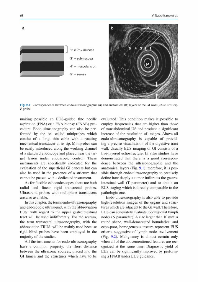
68
making possible an EUS-guided fi ne needle aspiration (FNA) or a FNA biopsy (FNAB) pro-cedure. Endo-ultrasonography can also be per-formed by the so- called miniprobes which consist of a long, thin cable with a rotating mechanical transducer at its tip. Miniprobes can be easily introduced along the working channel of a standard endoscope and placed near the tar-get lesion under endoscopic control. These instruments are specifi cally indicated for the evaluation of the superfi cial GI cancers but can also be used in the presence of a stricture that cannot be passed with a dedicated instrument.
As for fl exible echoendoscopes, there are both radial and linear rigid transrectal probes. Ultrasound probes with multiplane transducers are also available.
In this chapter, the terms endo- ultrasonography and endoscopic ultrasound, with the abbreviation EUS, with regard to the upper gastrointestinal tract will be used indifferently. For the rectum, the term transrectal ultrasonography, with the abbreviation TRUS, will be mainly used because rigid blind probes have been employed in the majority of the studies.
All the instruments for endo-ultrasonography have a common property: the short distance between the ultrasonic sources, placed into the GI lumen and the structures which have to be
evaluated. This condition makes it possible to employ frequencies that are higher than those of transabdominal US and produce a signifi cant increase of the resolution of images. Above all endo-ultrasonography is capable of provid-ing a precise visualization of the digestive tract wall. Usually EUS imaging of GI consists of a fi ve- layered echostructure. In vitro studies have demonstrated that there is a good correspon-dence between the ultrasonographic and the anatomical layers (Fig. 9.1 ); therefore, it is pos-sible through endo-ultrasonography to precisely defi ne how deeply a tumor infi ltrates the gastro-intestinal wall (T parameter) and to obtain an EUS staging which is directly comparable to the pathologic one.
Endo-ultrasonography is also able to provide high-resolution images of the organs and struc-tures which are adjacent to the GI wall. Therefore, EUS can adequately evaluate locoregional lymph nodes (N parameter). A size larger than 10 mm; a round shape, well-demarcated boundaries; and echo-poor, homogeneous texture represent EUS criteria suggestive of lymph node involvement (Fig. 9.2 ). Malignancy is almost certain only when all of the abovementioned features are rec-ognized at the same time. Diagnostic yield of EUS can be signifi cantly improved by perform-ing a FNAB under EUS guidance.
1° e 2° = mucosa
3° = submucosa
4° = muscolaris pr.
5° = serosa
a b
Fig. 9.1 Correspondence between endo-ultrasonographic ( a ) and anatomical ( b ) layers of the GI wall ( white arrows ). P probe
V. Napolitano et al.
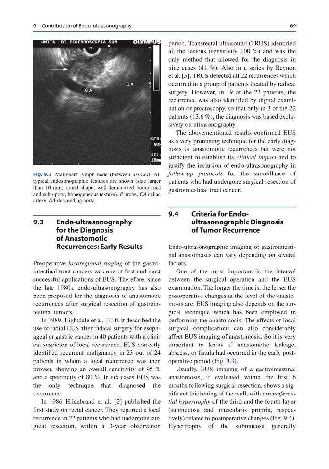
69
9.3 Endo-ultrasonography for the Diagnosis of Anastomotic Recurrences: Early Results
Preoperative locoregional staging of the gastro-intestinal tract cancers was one of fi rst and most successful applications of EUS. Therefore, since the late 1980s, endo-ultrasonography has also been proposed for the diagnosis of anastomotic recurrences after surgical resection of gastroin-testinal tumors.
In 1989, Lightdale et al. [ 1 ] fi rst described the use of radial EUS after radical surgery for esoph-ageal or gastric cancer in 40 patients with a clini-cal suspicion of local recurrence. EUS correctly identifi ed recurrent malignancy in 23 out of 24 patients in whom a local recurrence was then proven, showing an overall sensitivity of 95 % and a specifi city of 80 %. In six cases EUS was the only technique that diagnosed the recurrence.
In 1986 Hildebrand et al. [ 2 ] published the fi rst study on rectal cancer. They reported a local recurrence in 22 patients who had undergone sur-gical resection, within a 3-year observation
period. Transrectal ultrasound (TRUS) identifi ed all the lesions (sensitivity 100 %) and was the only method that allowed for the diagnosis in nine cases (41 %). Also in a series by Beynon et al. [ 3 ], TRUS detected all 22 recurrences which occurred in a group of patients treated by radical surgery. However, in 19 of the 22 patients, the recurrence was also identifi ed by digital exami-nation or proctoscopy, so that only in 3 of the 22 patients (13.6 %), the diagnosis was based exclu-sively on ultrasonography.
The abovementioned results confi rmed EUS as a very promising technique for the early diag-nosis of anastomotic recurrences but were not suffi cient to establish its clinical impact and to justify the inclusion of endo-ultrasonography in follow - up protocols for the surveillance of patients who had undergone surgical resection of gastrointestinal tract cancer.
9.4 Criteria for Endo- ultrasonographic Diagnosis of Tumor Recurrence
Endo-ultrasonographic imaging of gastrointesti-nal anastomoses can vary depending on several factors.
One of the most important is the interval between the surgical operation and the EUS examination. The longer the time is, the lesser the postoperative changes at the level of the anasto-mosis are. EUS imaging also depends on the sur-gical technique which has been employed in performing the anastomosis. The effects of local surgical complications can also considerably affect EUS imaging of anastomosis. So it is very important to know if anastomotic leakage, abscess, or fi stula had occurred in the early post-operative period (Fig. 9.3 ).
Usually, EUS imaging of a gastrointestinal anastomosis, if evaluated within the fi rst 6 months following surgical resection, shows a sig-nifi cant thickening of the wall, with circumferen-tial hypertrophy of the third and the fourth layer (submucosa and muscularis propria, respec-tively) related to postoperative changes (Fig. 9.4 ). Hypertrophy of the submucosa generally
Fig. 9.2 Malignant lymph node (between arrows ). All typical endosonographic features are shown (size larger than 10 mm, round shape, well-demarcated boundaries and echo-poor, homogeneous texture). P probe, CA celiac artery, DA descending aorta
9 Contribution of Endo-ultrasonography
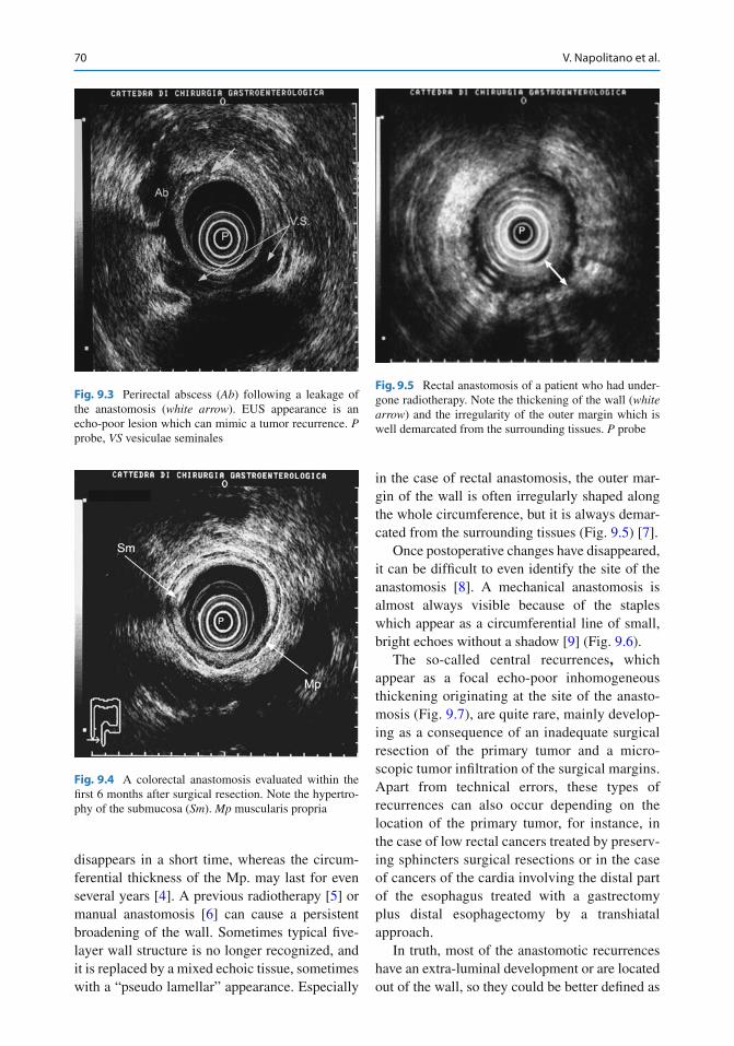
70
disappears in a short time, whereas the circum-ferential thickness of the Mp. may last for even several years [ 4 ]. A previous radiotherapy [ 5 ] or manual anastomosis [ 6 ] can cause a persistent broadening of the wall. Sometimes typical fi ve-layer wall structure is no longer recognized, and it is replaced by a mixed echoic tissue, sometimes with a “pseudo lamellar” appearance. Especially
in the case of rectal anastomosis, the outer mar-gin of the wall is often irregularly shaped along the whole circumference, but it is always demar-cated from the surrounding tissues (Fig. 9.5 ) [ 7 ].
Once postoperative changes have disappeared, it can be diffi cult to even identify the site of the anastomosis [ 8 ]. A mechanical anastomosis is almost always visible because of the staples which appear as a circumferential line of small, bright echoes without a shadow [ 9 ] (Fig. 9.6 ).
The so-called central recurrences , which appear as a focal echo-poor inhomogeneous thickening originating at the site of the anasto-mosis (Fig. 9.7 ), are quite rare, mainly develop-ing as a consequence of an inadequate surgical resection of the primary tumor and a micro-scopic tumor infi ltration of the surgical margins. Apart from technical errors, these types of recurrences can also occur depending on the location of the primary tumor, for instance, in the case of low rectal cancers treated by preserv-ing sphincters surgical resections or in the case of cancers of the cardia involving the distal part of the esophagus treated with a gastrectomy plus distal esophagectomy by a transhiatal approach.
In truth, most of the anastomotic recurrences have an extra-luminal development or are located out of the wall, so they could be better defi ned as
Fig. 9.3 Perirectal abscess ( Ab ) following a leakage of the anastomosis ( white arrow ). EUS appearance is an echo-poor lesion which can mimic a tumor recurrence. P probe, VS vesiculae seminales
Fig. 9.4 A colorectal anastomosis evaluated within the fi rst 6 months after surgical resection. Note the hypertro-phy of the submucosa ( Sm ). Mp muscularis propria
Fig. 9.5 Rectal anastomosis of a patient who had under-gone radiotherapy. Note the thickening of the wall ( white arrow ) and the irregularity of the outer margin which is well demarcated from the surrounding tissues. P probe
V. Napolitano et al.
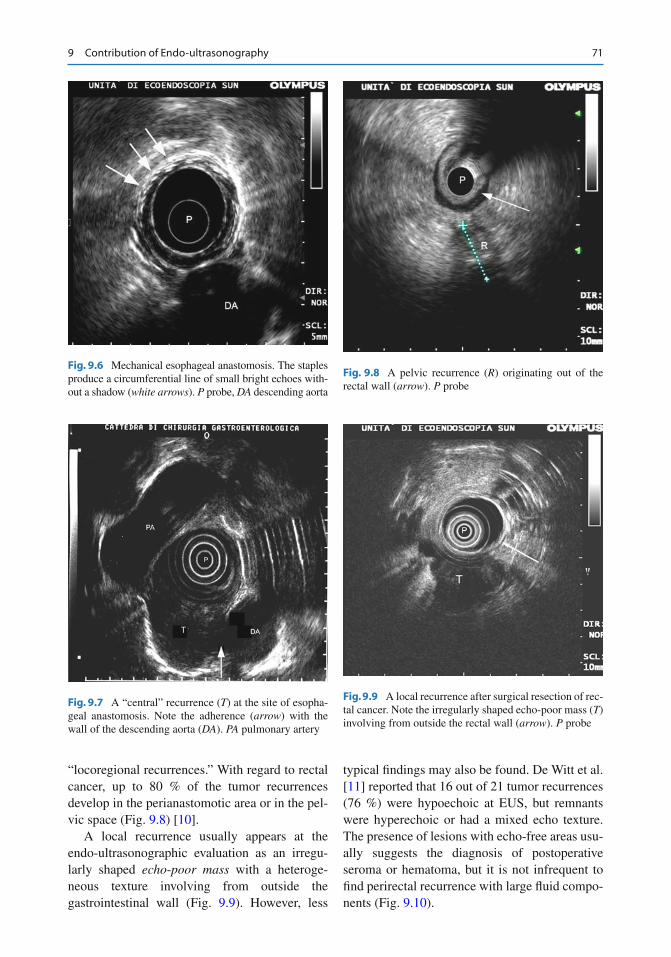
71
“locoregional recurrences.” With regard to rectal cancer, up to 80 % of the tumor recurrences develop in the perianastomotic area or in the pel-vic space (Fig. 9.8 ) [ 10 ].
A local recurrence usually appears at the endo- ultrasonographic evaluation as an irregu-larly shaped echo - poor mass with a heteroge-neous texture involving from outside the gastrointestinal wall (Fig. 9.9 ). However, less
typical fi ndings may also be found. De Witt et al. [ 11 ] reported that 16 out of 21 tumor recurrences (76 %) were hypoechoic at EUS, but remnants were hyperechoic or had a mixed echo texture. The presence of lesions with echo-free areas usu-ally suggests the diagnosis of postoperative seroma or hematoma, but it is not infrequent to fi nd perirectal recurrence with large fl uid compo-nents (Fig. 9.10 ).
Fig. 9.6 Mechanical esophageal anastomosis. The staples produce a circumferential line of small bright echoes with-out a shadow ( white arrows ). P probe, DA descending aorta
Fig. 9.7 A “central” recurrence ( T ) at the site of esopha-geal anastomosis. Note the adherence ( arrow ) with the wall of the descending aorta ( DA ). PA pulmonary artery
Fig. 9.8 A pelvic recurrence ( R ) originating out of the rectal wall ( arrow ). P probe
Fig. 9.9 A local recurrence after surgical resection of rec-tal cancer. Note the irregularly shaped echo-poor mass ( T ) involving from outside the rectal wall ( arrow ). P probe
9 Contribution of Endo-ultrasonography
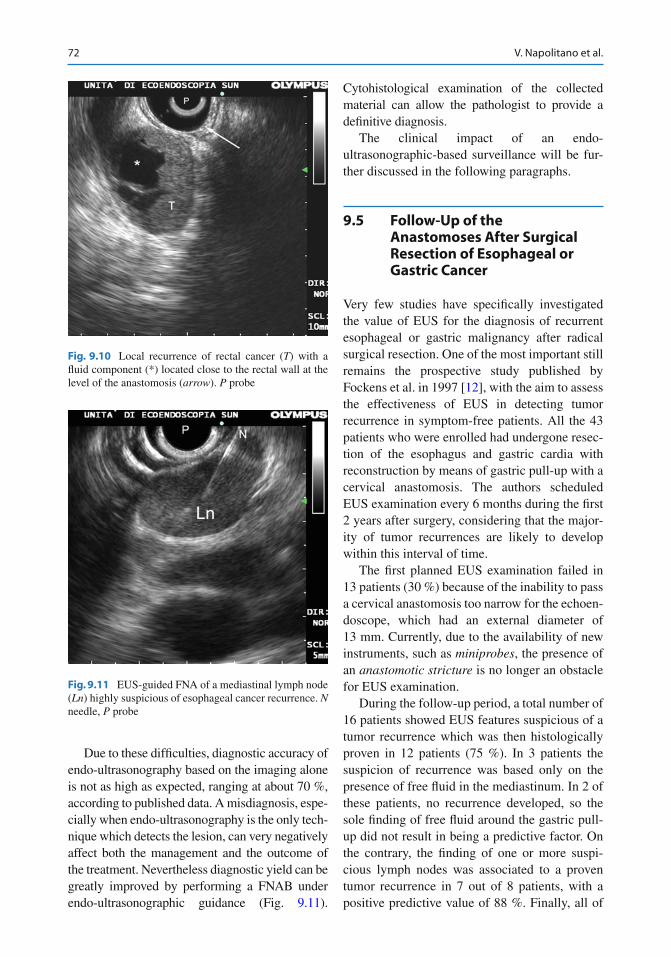
72
Due to these diffi culties, diagnostic accuracy of endo-ultrasonography based on the imaging alone is not as high as expected, ranging at about 70 %, according to published data. A misdiagnosis, espe-cially when endo-ultrasonography is the only tech-nique which detects the lesion, can very negatively affect both the management and the outcome of the treatment. Nevertheless diagnostic yield can be greatly improved by performing a FNAB under endo-ultrasonographic guidance (Fig. 9.11 ).
Cytohistological examination of the collected material can allow the pathologist to provide a defi nitive diagnosis.
The clinical impact of an endo-ultrasonographic- based surveillance will be fur-ther discussed in the following paragraphs.
9.5 Follow-Up of the Anastomoses After Surgical Resection of Esophageal or Gastric Cancer
Very few studies have specifi cally investigated the value of EUS for the diagnosis of recurrent esophageal or gastric malignancy after radical surgical resection. One of the most important still remains the prospective study published by Fockens et al. in 1997 [ 12 ], with the aim to assess the effectiveness of EUS in detecting tumor recurrence in symptom-free patients. All the 43 patients who were enrolled had undergone resec-tion of the esophagus and gastric cardia with reconstruction by means of gastric pull-up with a cervical anastomosis. The authors scheduled EUS examination every 6 months during the fi rst 2 years after surgery, considering that the major-ity of tumor recurrences are likely to develop within this interval of time.
The fi rst planned EUS examination failed in 13 patients (30 %) because of the inability to pass a cervical anastomosis too narrow for the echoen-doscope, which had an external diameter of 13 mm. Currently, due to the availability of new instruments, such as miniprobes , the presence of an anastomotic stricture is no longer an obstacle for EUS examination.
During the follow-up period, a total number of 16 patients showed EUS features suspicious of a tumor recurrence which was then histologically proven in 12 patients (75 %). In 3 patients the suspicion of recurrence was based only on the presence of free fl uid in the mediastinum. In 2 of these patients, no recurrence developed, so the sole fi nding of free fl uid around the gastric pull- up did not result in being a predictive factor. On the contrary, the fi nding of one or more suspi-cious lymph nodes was associated to a proven tumor recurrence in 7 out of 8 patients, with a positive predictive value of 88 %. Finally, all of
Fig. 9.10 Local recurrence of rectal cancer ( T ) with a fl uid component (*) located close to the rectal wall at the level of the anastomosis ( arrow ). P probe
Fig. 9.11 EUS-guided FNA of a mediastinal lymph node ( Ln ) highly suspicious of esophageal cancer recurrence. N needle, P probe
V. Napolitano et al.
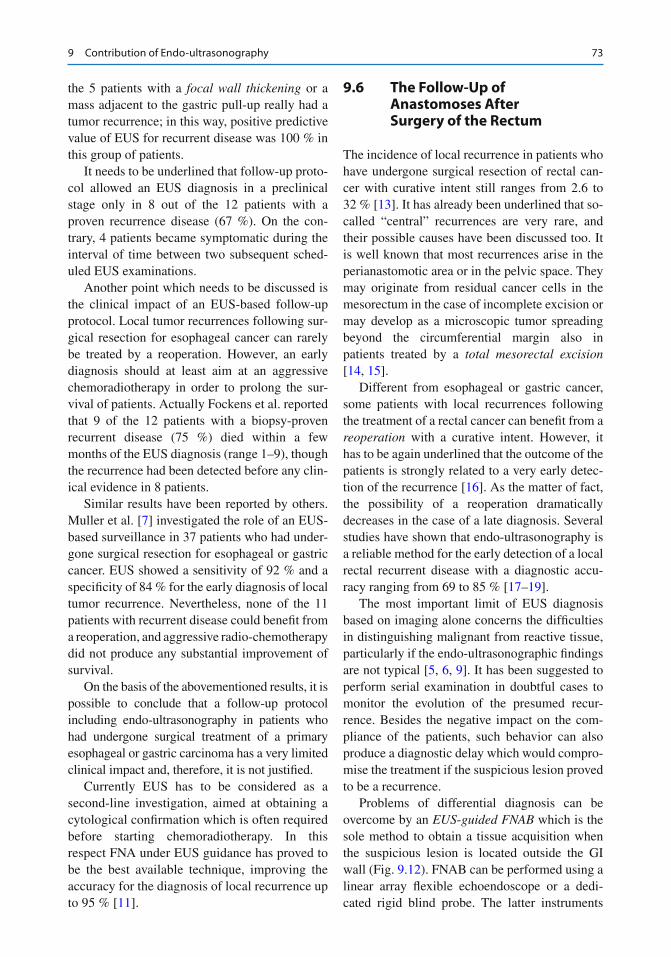
73
the 5 patients with a focal wall thickening or a mass adjacent to the gastric pull-up really had a tumor recurrence; in this way, positive predictive value of EUS for recurrent disease was 100 % in this group of patients.
It needs to be underlined that follow-up proto-col allowed an EUS diagnosis in a preclinical stage only in 8 out of the 12 patients with a proven recurrence disease (67 %). On the con-trary, 4 patients became symptomatic during the interval of time between two subsequent sched-uled EUS examinations.
Another point which needs to be discussed is the clinical impact of an EUS-based follow-up protocol. Local tumor recurrences following sur-gical resection for esophageal cancer can rarely be treated by a reoperation. However, an early diagnosis should at least aim at an aggressive chemoradiotherapy in order to prolong the sur-vival of patients. Actually Fockens et al. reported that 9 of the 12 patients with a biopsy-proven recurrent disease (75 %) died within a few months of the EUS diagnosis (range 1–9), though the recurrence had been detected before any clin-ical evidence in 8 patients.
Similar results have been reported by others. Muller et al. [ 7 ] investigated the role of an EUS- based surveillance in 37 patients who had under-gone surgical resection for esophageal or gastric cancer. EUS showed a sensitivity of 92 % and a specifi city of 84 % for the early diagnosis of local tumor recurrence. Nevertheless, none of the 11 patients with recurrent disease could benefi t from a reoperation, and aggressive radio- chemotherapy did not produce any substantial improvement of survival.
On the basis of the abovementioned results, it is possible to conclude that a follow-up protocol including endo-ultrasonography in patients who had undergone surgical treatment of a primary esophageal or gastric carcinoma has a very limited clinical impact and, therefore, it is not justifi ed.
Currently EUS has to be considered as a second- line investigation, aimed at obtaining a cytological confi rmation which is often required before starting chemoradiotherapy. In this respect FNA under EUS guidance has proved to be the best available technique, improving the accuracy for the diagnosis of local recurrence up to 95 % [ 11 ].
9.6 The Follow-Up of Anastomoses After Surgery of the Rectum
The incidence of local recurrence in patients who have undergone surgical resection of rectal can-cer with curative intent still ranges from 2.6 to 32 % [ 13 ]. It has already been underlined that so- called “central” recurrences are very rare, and their possible causes have been discussed too. It is well known that most recurrences arise in the perianastomotic area or in the pelvic space. They may originate from residual cancer cells in the mesorectum in the case of incomplete excision or may develop as a microscopic tumor spreading beyond the circumferential margin also in patients treated by a total mesorectal excision [ 14 , 15 ].
Different from esophageal or gastric cancer, some patients with local recurrences following the treatment of a rectal cancer can benefi t from a reoperation with a curative intent. However, it has to be again underlined that the outcome of the patients is strongly related to a very early detec-tion of the recurrence [ 16 ]. As the matter of fact, the possibility of a reoperation dramatically decreases in the case of a late diagnosis. Several studies have shown that endo-ultrasonography is a reliable method for the early detection of a local rectal recurrent disease with a diagnostic accu-racy ranging from 69 to 85 % [ 17 – 19 ].
The most important limit of EUS diagnosis based on imaging alone concerns the diffi culties in distinguishing malignant from reactive tissue, particularly if the endo-ultrasonographic fi ndings are not typical [ 5 , 6 , 9 ]. It has been suggested to perform serial examination in doubtful cases to monitor the evolution of the presumed recur-rence. Besides the negative impact on the com-pliance of the patients, such behavior can also produce a diagnostic delay which would compro-mise the treatment if the suspicious lesion proved to be a recurrence.
Problems of differential diagnosis can be overcome by an EUS - guided FNAB which is the sole method to obtain a tissue acquisition when the suspicious lesion is located outside the GI wall (Fig. 9.12 ). FNAB can be performed using a linear array fl exible echoendoscope or a dedi-cated rigid blind probe. The latter instruments
9 Contribution of Endo-ultrasonography
74
have been used in the majority of the published series concerning FNAB for diagnosis of rectal recurrences. Using a rigid ultrasound probe, tis-sue sampling is generally carried out by a spring- loaded core biopsy needle, which allows for the obtainment of histological specimens. On the contrary, standard needles used with fl exible echoendoscopes only allow a fi ne needle aspira-tion procedure, although a new type of needle with a small core trap at its tip is now also available.
Transrectal US-guided FNA is a relatively simple and safe procedure with a rate of compli-cations ranging from 0.5 to 2 % [ 20 ]. Nevertheless perirectal, even severe infections may occur, albeit rarely. Therefore, an antibiotic prophy-laxis should be mandatory before performing the procedure, especially when a cystic component is found. FNAB signifi cantly improves diagnos-tic yield of transrectal US. Its accuracy is esti-mated to range from 87 to 100 % [ 18 , 19 ]. Additional information provided by FNA is able to modify therapeutic strategy in 25–35 of the cases [ 18 , 19 ].
However, once again the question concerns the real usefulness of a follow-up program including both proctoscopy and transrectal US.
In 2004 Hernandez de Anda et al. published a study on transrectal US follow-up in 275 patients with invasive rectal cancer treated by curative- intent local excision or radical surgery [ 9 ]. FU protocol consisted in an anal digital examination, proctoscopy, and transrectal US every 4 months for the fi rst 3 years after surgery and every 6 months for the subsequent 2 years. In the local excision group, most recurrences (81 %) were diagnosed during scheduled examinations in asymptomatic patients. However, only 30 % of recurrences were detected by TRUS alone. The proportion of salvage surgery was similar in patients whose recurrence was diagnosed by transrectal US or by other techniques (respec-tively, 80 % vs 86 %). In the radical surgery group, the proportion of recurrences diagnosed in patients still asymptomatic was 42 %, and the lesion was only detected by transrectal US in 33 % of the cases. As expected, the proportion of salvage surgery in the patients with recurrences enrolled in the follow-up protocol was higher compared to a control group without follow-up, but the difference was not signifi cant. Not even the results of this large study were able to assess the impact of an endo-ultrasonographic follow- up on the survival of the patients with local recur-rences after surgical resection of rectal cancer.
According to the guidelines for posttreatment follow-up of patients with colorectal cancer of the American Society of Colon and Rectal Surgeons published in 2004 [ 21 ], periodic anas-tomotic evaluation is recommended for patients who have undergone resection/anastomosis or local excision of rectal cancer (level of evidence III, grade of evidence B). Authors, however, underlined that the role of endo-ultrasonography in this fi eld would need further confi rmation with larger multi-institutional clinical trials. So far these trials are not yet available.
Maybe, especially in patients with AJCC II or III disease , a follow-up protocol including endo- ultrasonography would be justifi ed, because of the higher risk of local recurrence, but neither the interval nor the total duration of the follow-up has been defi ned yet.
At present, the programs for the postoperative follow-up of patients with GI cancers contemplate
Fig. 9.12 EUS-guided FNA of an extra-luminal tumor recurrence ( T ) after surgical resection of rectal cancer. N needle, P probe
V. Napolitano et al.
75
multi-slice CT as the fi rst-line exam and PET as the second-line tool. PET is the most sensitive method currently available, but its specifi city is not completely satisfactory. Thus, endo-ultraso-nography, associated with FNAB, can play a fun-damental role in obtaining a defi nite diagnosis of malignancy, which is very often required before planning a therapeutic strategy.
References
1. Lightdale CJ, Botet JF, Keksen DP, Turnbull AD, Brennan MF (1989) Diagnosis of recurrent upper gas-trointestinal cancer at the surgical anastomosis by endo-scopic ultrasound. Gastrointest Endosc 35:407–412
2. Hildebrandt U, Feifel G, Schwarz HP, Scherr O (1986) Endorectal ultrasound: instrumentation and clinical aspects. Int Colorect Dis 1:203–207
3. Beynon J, Mortensen NJ, Foy DMA, Channer JL, Rigby H, Virjee J (1989) Preoperative assessment of mesorectal lymph node involvement in rectal cancer. Br J Surg 76:276–279
4. Nakajima S (2001) The effi cacy of the EUS for the detection of recurrent disease in the anastomosis of colon. Diagn Ther Endosc 7:149–158
5. Lindmark G, Elvin A, Pahlman L, Glimelius B (1992) The value of endosonography in preoperative staging of rectal cancer. Int J Colorectal Dis 7:162–166
6. Hizawa A, Aoyagi K, Suekane H, Mibu R, Yao T, Fujishima M (1996) Suture granuloma in rectal anastomosis mistaken for local recurrent cancer. J Clin Gastroenterol 23:78–79
7. Müller C, Kähler G, Scheele J (2000) Endosonographic examination of gastrointestinal anastomoses with sus-pected locoregional tumor recurrence. Surg Endosc 14:45–50
8. Rösch T, Classen M (1992) Colorectal carcinoma. In: Elder D (ed) Gastroenterologic endosonography. Thieme Medical, New York, pp 170–185
9. Hernandez de Anda E, Lee S, Finne CO, Rothenberger DA, Madoff RD, Garcia-Aguilar J (2004) Endorectal ultrasound in the follow-up of rectal cancer patients treated by local excision or radical surgery. Dis Colon Rectum 47:818–824
10. Hoffman JP, Riley L, Carp NZ, Litwin S (1993) Isolated locally recurrent rectal cancer: a review of
incidence, presentation, and management. Semin Oncol 20:506–519
11. DeWitt J, Ghorai S, Kahi C, LeBlanc J, McHenry L, Chappo J, Cramer H, McGreevy K, Chriswell M, Sherman S (2003) EUS-FNA of recurrent postopera-tive extraluminal and metastatic malignancy. Gastrointest Endosc 58:542–548
12. Fockens P, Manshanden CG, van Lanschot JJ, Obertop H, Tytgat GNJ (1997) Prospective study on the value of endosonographic follow-up after surgery for esophageal carcinoma. Gastrointest Endosc 46:487–491
13. Abulafi AM, Williams NS (1994) Local recurrence of colorectal cancer: the problem, mechanisms, manage-ment and adjuvant therapy. Br J Surg 81:7–19
14. Qiurke P, Durdey P, Dixon MF, Williams NS (1986) Local recurrence of rectal adenocarcinoma due to inadequate surgical resection. Histopathological study of lateral tumour spread and surgical excision. Lancet 2:996–999
15. Emslie J, Beart R, Mohiuddin M, Marks G (1998) Use of rectal cancer position as a prognostic indicator. Am Surg 64:958–961
16. Mellgren A, Sirivongs P, Rothenberger DA, Madoff RD, Garcia-Aguilar J (2000) Is local excision ade-quate therapy for early rectal cancer? Dis Colon Rectum 43:1064–1071
17. Romano G, Esercizio L, Santangelo M, Vallone G, Santangelo ML (1993) Impact of computer tomogra-phy vs. intrarectal ultrasound on the diagnosis, resect-ability, and prognosis of locally recurrent rectal cancer. Dis Colon Rectum 36:261–266
18. Hünerbein M, Totkas S, Moesta KT, Ulmer C, Handke T, Schlag PM (2001) The role of transrectal ultrasound- guided biopsy in the postoperative follow- up of patients with rectal cancer. Surgery 129:164–169
19. Löhnert MS, Doniec JM, Henne-Bruns D (2000) Effectiveness of endoluminal sonography in the iden-tifi cation of occult rectal cancer recurrences. Dis Colon Rectum 43:483–491
20. Wiersema MJ, Vilmann P, Giovannini M, Chang KJ, Wiersema IM (1997) Endosonography-guided fi ne- needle aspiration biopsy: diagnostic accuracy and complication assessment. Gastroenterology 112:1087–1095
21. Anthony T, Simmang C, Hyman N et al (2004) Parameters for the surveillance and follow up of patients with colon and rectal cancer. Dis Colon Rectum 47:807–817
9 Contribution of Endo-ultrasonography
77G. Galloro (ed.), Endoscopic Follow-up of Digestive Anastomosis, DOI 10.1007/978-88-470-5370-0_10, © Springer-Verlag Italia 2014
10.1 Augmented Endoscopy Imaging
10.1.1 Magnifying Chromoendoscopy
0.4 % indigo carmine (IC) dye is used to accentu-ate the outline and surface pattern of the lesions. It does not stain the lesion, but remains only in the depressed area and makes a clear contrast between the higher and lower parts of the lesion (contrast method ). The pit pattern proposed by Kudo is evaluated by magnifying view [ 1 ]. The orifi ces of the colonic crypts are described as “pits,” and the specifi c arrangement of the pits is described as “pit pattern.” Pit pattern is useful to predict the histological structure of colorectal lesions. It is divided into fi ve groups: type I, II, III, IV, and V.
Type I pit pattern is seen in normal mucosa. It consists of roundish pits with a regular distribu-tion. Type II pit pattern is found in the majority of the lesions classifi ed as nonneoplastic at his-tology. It is larger than the normal pits and aster-oid or star shaped. The distribution of the pit is
regular. Type III pit pattern is divided into two categories, III S and III L . III S pit pattern is com-posed of tubular or roundish pits smaller than the normal ones (“S” stands for “small” or “short”). It is seen mainly in depressed lesion and tends to be early cancers. III L pit pattern is composed of tubular or roundish pits larger than normal ones (“L” stands for “long” or “large”). It is seen mainly in adenoma. Type IV pit pattern is a branched or gyrus- like pattern and is also seen mainly in adenoma. Type V pit pattern is divided into V I and V N . V I pit pattern (the “I” stands for “irregular”) has pits which are irregular in shape, size, and arrangement. V N pit pattern shows an absence of pit pattern (the “N” stands for “non-structural”). V N pit pattern is seen in deep sub-mucosal invasive cancer and in advanced cancer. V I pit pattern is mainly seen in adenoma with high-grade dysplasia, intramucosal cancer, and superfi cial submucosal invasive cancer. However, sometimes it is seen in deep submucosal invasive cancer.
A clinical classifi cation which modifi ed Kudo’s classifi cation was described by Fujii with the aim to discriminate between m-sm1 (intramucosal cancer and superfi cial submuco-sal invasive cancer) and sm2 (deep submucosal cancer) or beyond [ 2 ]. In this classifi cation, type V I pit pattern is divided into two categories, V I noninvasive pattern and V I invasive pattern. Noninvasive pattern composed of regular crypts with or without demarcated area or irregular pits without a demarcated area. This pattern
M. Makazu , MD (*) • T. Matsuda , MD, PhD (*) T. Sakamoto , MD • T. Nakajima , MD, PhD Y. Saito , MD, PhD Endoscopy Division , National Cancer Center Hospital , Tsukiji 5-1-1, Chuo-ku , Tokyo 104-0045 , Japan e-mail: [email protected]; [email protected]; [email protected]; [email protected]; [email protected]
10 Augmented Endoscopy Imaging in the Study of Digestive Anastomosis: Does It Really Work and How?
Makomo Makazu , Takahisa Matsuda , Taku Sakamoto , Takeshi Nakajima , and Yutaka Saito
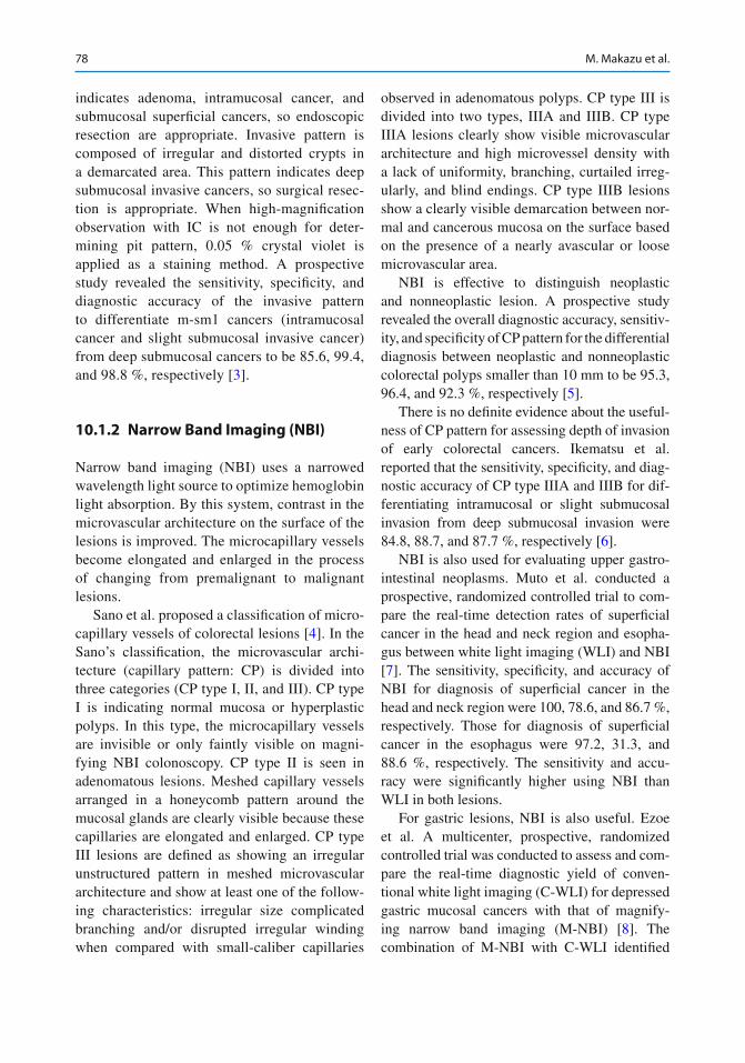
78
indicates adenoma, intramucosal cancer, and submucosal superfi cial cancers, so endoscopic resection are appropriate. Invasive pattern is composed of irregular and distorted crypts in a demarcated area. This pattern indicates deep submucosal invasive cancers, so surgical resec-tion is appropriate. When high- magnifi cation observation with IC is not enough for deter-mining pit pattern, 0.05 % crystal violet is applied as a staining method. A prospective study revealed the sensitivity, specifi city, and diagnostic accuracy of the invasive pattern to differentiate m-sm1 cancers (intramucosal cancer and slight submucosal invasive cancer) from deep submucosal cancers to be 85.6, 99.4, and 98.8 %, respectively [ 3 ].
10.1.2 Narrow Band Imaging (NBI)
Narrow band imaging (NBI) uses a narrowed wavelength light source to optimize hemoglobin light absorption. By this system, contrast in the microvascular architecture on the surface of the lesions is improved. The microcapillary vessels become elongated and enlarged in the process of changing from premalignant to malignant lesions.
Sano et al. proposed a classifi cation of micro-capillary vessels of colorectal lesions [ 4 ]. In the Sano’s classifi cation , the microvascular archi-tecture (capillary pattern: CP) is divided into three categories (CP type I, II, and III). CP type I is indicating normal mucosa or hyperplastic polyps. In this type, the microcapillary vessels are invisible or only faintly visible on magni-fying NBI colonoscopy. CP type II is seen in adenomatous lesions. Meshed capillary vessels arranged in a honeycomb pattern around the mucosal glands are clearly visible because these capillaries are elongated and enlarged. CP type III lesions are defi ned as showing an irregular unstructured pattern in meshed microvascular architecture and show at least one of the follow-ing characteristics: irregular size complicated branching and/or disrupted irregular winding when compared with small-caliber capillaries
observed in adenomatous polyps. CP type III is divided into two types, IIIA and IIIB. CP type IIIA lesions clearly show visible microvascular architecture and high microvessel density with a lack of uniformity, branching, curtailed irreg-ularly, and blind endings. CP type IIIB lesions show a clearly visible demarcation between nor-mal and cancerous mucosa on the surface based on the presence of a nearly avascular or loose microvascular area.
NBI is effective to distinguish neoplastic and nonneoplastic lesion. A prospective study revealed the overall diagnostic accuracy, sensitiv-ity, and specifi city of CP pattern for the differential diagnosis between neoplastic and nonneoplastic colorectal polyps smaller than 10 mm to be 95.3, 96.4, and 92.3 %, respectively [ 5 ].
There is no defi nite evidence about the useful-ness of CP pattern for assessing depth of invasion of early colorectal cancers. Ikematsu et al. reported that the sensitivity, specifi city, and diag-nostic accuracy of CP type IIIA and IIIB for dif-ferentiating intramucosal or slight submucosal invasion from deep submucosal invasion were 84.8, 88.7, and 87.7 %, respectively [ 6 ].
NBI is also used for evaluating upper gastro-intestinal neoplasms . Muto et al. conducted a prospective, randomized controlled trial to com-pare the real-time detection rates of superfi cial cancer in the head and neck region and esopha-gus between white light imaging (WLI) and NBI [ 7 ]. The sensitivity, specifi city, and accuracy of NBI for diagnosis of superfi cial cancer in the head and neck region were 100, 78.6, and 86.7 %, respectively. Those for diagnosis of superfi cial cancer in the esophagus were 97.2, 31.3, and 88.6 %, respectively. The sensitivity and accu-racy were signifi cantly higher using NBI than WLI in both lesions.
For gastric lesions, NBI is also useful. Ezoe et al. A multicenter, prospective, randomized controlled trial was conducted to assess and com-pare the real-time diagnostic yield of conven-tional white light imaging (C-WLI) for depressed gastric mucosal cancers with that of magnify-ing narrow band imaging (M-NBI) [ 8 ]. The combination of M-NBI with C-WLI identifi ed
M. Makazu et al.
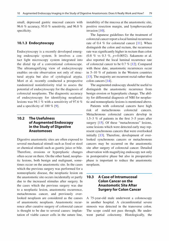
79
small, depressed gastric mucosal cancers with 96.6 % accuracy, 95.0 % sensitivity, and 96.8 % specifi city.
10.1.3 Endocytoscopy
Endocytoscopy is a recently developed emerg-ing endoscopic system. It involves a con-tact light microscopy system integrated into the distal tip of a conventional colonoscope. The ultramagnifying view of endocytoscopy enables on-site observation not only of struc-tural atypia but also of cytological atypia. Mori et al. recently conducted a prospective randomized noninferiority trial to assess the potential of endocytoscopy for the diagnosis of colorectal neoplasms. The diagnostic accuracy of endocytoscopy for identifying neoplastic lesions was 94.1 % with a sensitivity of 97.6 % and a specifi city of 100 % [ 9 ].
10.2 The Usefulness of Augmented Endoscopy in the Study of Digestive Anastomoses
Digestive anastomotic sites are often exposed to several mechanical stimuli such as food or stool or chemical stimuli such as gastric juice or bile. Therefore, erosions or hyperplastic changes often occur on there. On the other hand, neoplas-tic lesions, both benign and malignant, some-times occur on the anastomotic site. In the cases which the previous surgery was performed for a nonneoplastic disease, the neoplastic lesion on the anastomotic site occurs incidentally or partly due to the increased stimulus after surgery. In the cases which the previous surgery was due to a neoplastic lesion, anastomotic recurrence , metachronous cancer, and previously over-looked neoplasm are considered as the causes of anastomotic neoplasm. Anastomotic recur-rence after curative surgery of colorectal cancer is thought to be due to several causes: implan-tation of viable cancer cells in the suture line,
instability of the mucosa at the anastomotic site, positive resection margin, and lymphovascular invasion [ 10 ].
The Japanese guidelines for the treatment of colorectal cancer report a local luminal recurrence rate of 0.4 % for colorectal cancer [ 11 ]. When distinguish the colon and rectum, the recurrence rate was signifi cantly higher in rectum than colon (0.8 % vs 0.3 %, p = 0.0052). Sakamoto et al. also reported the local luminal recurrence rate of colorectal cancer to be 0.7 % [ 12 ]. Compared with these date, anastomotic recurrences occur in 5–10 % of patients in the Western countries [ 13 ]. The majority are recurrent rectal rather than colon cancers [ 14 ].
The augmented endoscopy should be useful to distinguish the anastomotic recurrence from benign erosion or hyperplastic change. The abil-ity for differential diagnosis of NBI for neoplas-tic and nonneoplastic lesions is mentioned above.
Patients with colorectal cancers have high risk of metachronous colorectal cancers. Metachronous colorectal cancers develop in 1.5–3 % of patients in the fi rst 3–5 years after surgery [ 15 ]. Of these “metachronous” lesions, some lesions which were detected early may rep-resent synchronous cancers that were overlooked initially [ 13 ]. Therefore, development of over-looked synchronous cancers or metachronous cancers may be occurred on the anastomotic site after surgery of colorectal cancer. Detailed observation with magnifying endoscopy not only in postoperative phase but also in preoperative phase is important to reduce the anastomotic neoplasm.
10.3 A Case of Intramucosal Colon Cancer on the Anastomotic Site After Surgery for Colon Cancer
A 75-year-old male underwent a colonoscopy in another hospital. A circumferential severe stenosis was detected in the transverse colon. The scope could not pass through. He under-went partial colectomy. Histologically, the
10 Augmented Endoscopy Imaging in the Study of Digestive Anastomosis: Does It Really Work and How?
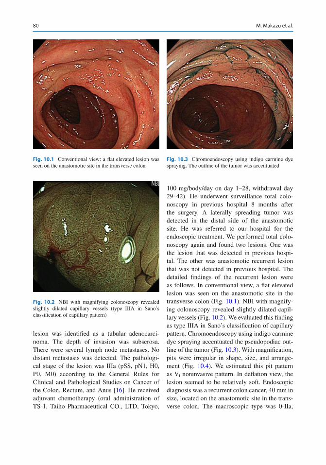
80
lesion was identifi ed as a tubular adenocarci-noma. The depth of invasion was subserosa. There were several lymph node metastases. No distant metastasis was detected. The pathologi-cal stage of the lesion was IIIa (pSS, pN1, H0, P0, M0) according to the General Rules for Clinical and Pathological Studies on Cancer of the Colon, Rectum, and Anus [ 16 ]. He received adjuvant chemotherapy (oral administration of TS-1, Taiho Pharmaceutical CO., LTD, Tokyo,
100 mg/body/day on day 1–28, withdrawal day 29–42). He underwent surveillance total colo-noscopy in previous hospital 8 months after the surgery. A laterally spreading tumor was detected in the distal side of the anastomotic site. He was referred to our hospital for the endoscopic treatment. We performed total colo-noscopy again and found two lesions. One was the lesion that was detected in previous hospi-tal. The other was anastomotic recurrent lesion that was not detected in previous hospital. The detailed fi ndings of the recurrent lesion were as follows. In conventional view, a fl at elevated lesion was seen on the anastomotic site in the transverse colon (Fig. 10.1 ). NBI with magnify-ing colonoscopy revealed slightly dilated capil-lary vessels (Fig. 10.2 ). We evaluated this fi nding as type IIIA in Sano’s classifi cation of capillary pattern. Chromoendoscopy using indigo carmine dye spraying accentuated the pseudopodiac out-line of the tumor (Fig. 10.3 ). With magnifi cation, pits were irregular in shape, size, and arrange-ment (Fig. 10.4 ). We estimated this pit pattern as V I noninvasive pattern. In defl ation view, the lesion seemed to be relatively soft. Endoscopic diagnosis was a recurrent colon cancer, 40 mm in size, located on the anastomotic site in the trans-verse colon. The macroscopic type was 0-IIa,
Fig. 10.1 Conventional view: a fl at elevated lesion was seen on the anastomotic site in the transverse colon
Fig. 10.2 NBI with magnifying colonoscopy revealed slightly dilated capillary vessels (type IIIA in Sano’s classifi cation of capillary pattern)
Fig. 10.3 Chromoendoscopy using indigo carmine dye spraying. The outline of the tumor was accentuated
M. Makazu et al.
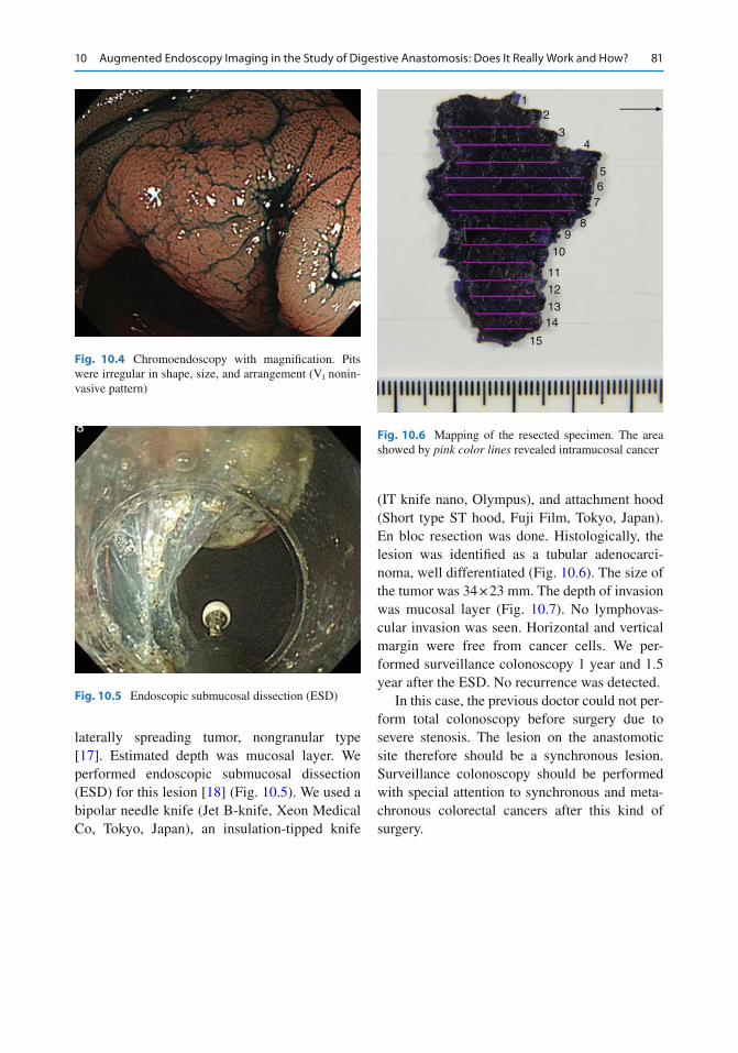
81
laterally spreading tumor , nongranular type [ 17 ]. Estimated depth was mucosal layer. We performed endoscopic submucosal dissection (ESD) for this lesion [ 18 ] (Fig. 10.5 ). We used a bipolar needle knife (Jet B-knife, Xeon Medical Co, Tokyo, Japan), an insulation-tipped knife
(IT knife nano, Olympus), and attachment hood (Short type ST hood, Fuji Film, Tokyo, Japan). En bloc resection was done. Histologically, the lesion was identifi ed as a tubular adenocarci-noma, well differentiated (Fig. 10.6 ). The size of the tumor was 34 × 23 mm. The depth of invasion was mucosal layer (Fig. 10.7 ). No lymphovas-cular invasion was seen. Horizontal and vertical margin were free from cancer cells. We per-formed surveillance colonoscopy 1 year and 1.5 year after the ESD. No recurrence was detected.
In this case, the previous doctor could not per-form total colonoscopy before surgery due to severe stenosis. The lesion on the anastomotic site therefore should be a synchronous lesion. Surveillance colonoscopy should be performed with special attention to synchronous and meta-chronous colorectal cancers after this kind of surgery.
Fig. 10.4 Chromoendoscopy with magnifi cation. Pits were irregular in shape, size, and arrangement (V I nonin-vasive pattern)
Fig. 10.5 Endoscopic submucosal dissection (ESD)
12
34
56
7
89
10
11
12
1314
15
Fig. 10.6 Mapping of the resected specimen. The area showed by pink color lines revealed intramucosal cancer
10 Augmented Endoscopy Imaging in the Study of Digestive Anastomosis: Does It Really Work and How?
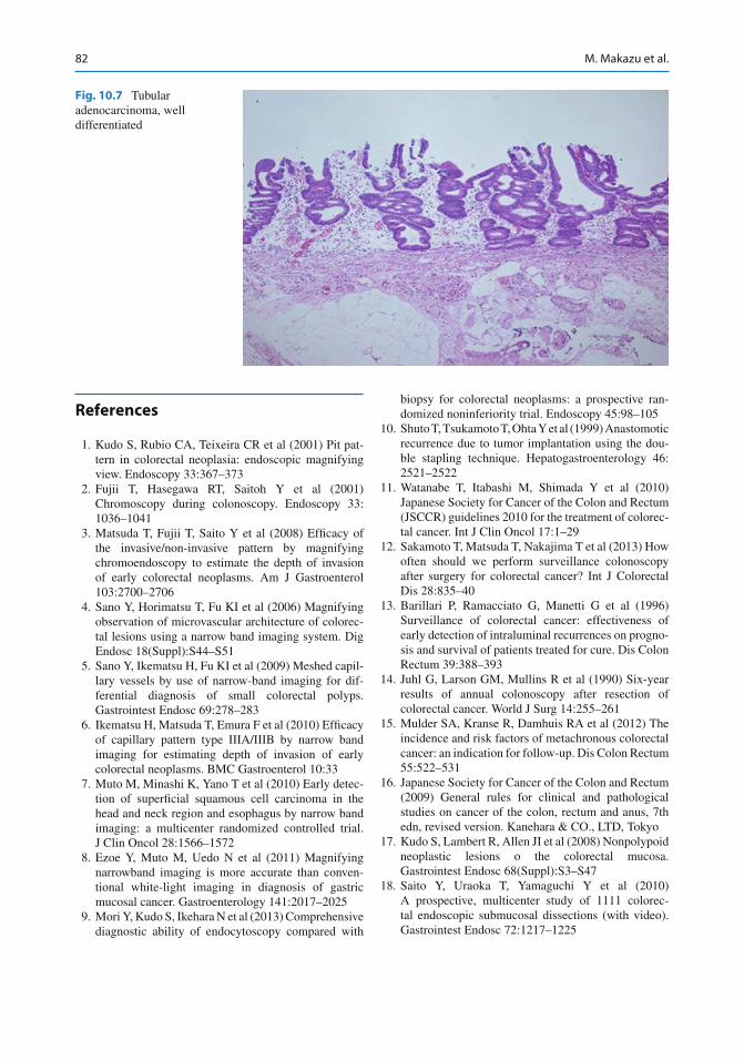
82
References
1. Kudo S, Rubio CA, Teixeira CR et al (2001) Pit pat-tern in colorectal neoplasia: endoscopic magnifying view. Endoscopy 33:367–373
2. Fujii T, Hasegawa RT, Saitoh Y et al (2001) Chromoscopy during colonoscopy. Endoscopy 33:1036–1041
3. Matsuda T, Fujii T, Saito Y et al (2008) Effi cacy of the invasive/non-invasive pattern by magnifying chromoendoscopy to estimate the depth of invasion of early colorectal neoplasms. Am J Gastroenterol 103:2700–2706
4. Sano Y, Horimatsu T, Fu KI et al (2006) Magnifying observation of microvascular architecture of colorec-tal lesions using a narrow band imaging system. Dig Endosc 18(Suppl):S44–S51
5. Sano Y, Ikematsu H, Fu KI et al (2009) Meshed capil-lary vessels by use of narrow-band imaging for dif-ferential diagnosis of small colorectal polyps. Gastrointest Endosc 69:278–283
6. Ikematsu H, Matsuda T, Emura F et al (2010) Effi cacy of capillary pattern type IIIA/IIIB by narrow band imaging for estimating depth of invasion of early colorectal neoplasms. BMC Gastroenterol 10:33
7. Muto M, Minashi K, Yano T et al (2010) Early detec-tion of superfi cial squamous cell carcinoma in the head and neck region and esophagus by narrow band imaging: a multicenter randomized controlled trial. J Clin Oncol 28:1566–1572
8. Ezoe Y, Muto M, Uedo N et al (2011) Magnifying narrowband imaging is more accurate than conven-tional white-light imaging in diagnosis of gastric mucosal cancer. Gastroenterology 141:2017–2025
9. Mori Y, Kudo S, Ikehara N et al (2013) Comprehensive diagnostic ability of endocytoscopy compared with
biopsy for colorectal neoplasms: a prospective ran-domized noninferiority trial. Endoscopy 45:98–105
10. Shuto T, Tsukamoto T, Ohta Y et al (1999) Anastomotic recurrence due to tumor implantation using the dou-ble stapling technique. Hepatogastroenterology 46:2521–2522
11. Watanabe T, Itabashi M, Shimada Y et al (2010) Japanese Society for Cancer of the Colon and Rectum (JSCCR) guidelines 2010 for the treatment of colorec-tal cancer. Int J Clin Oncol 17:1–29
12. Sakamoto T, Matsuda T, Nakajima T et al (2013) How often should we perform surveillance colonoscopy after surgery for colorectal cancer? Int J Colorectal Dis 28:835–40
13. Barillari P, Ramacciato G, Manetti G et al (1996) Surveillance of colorectal cancer: effectiveness of early detection of intraluminal recurrences on progno-sis and survival of patients treated for cure. Dis Colon Rectum 39:388–393
14. Juhl G, Larson GM, Mullins R et al (1990) Six-year results of annual colonoscopy after resection of colorectal cancer. World J Surg 14:255–261
15. Mulder SA, Kranse R, Damhuis RA et al (2012) The incidence and risk factors of metachronous colorectal cancer: an indication for follow-up. Dis Colon Rectum 55:522–531
16. Japanese Society for Cancer of the Colon and Rectum (2009) General rules for clinical and pathological studies on cancer of the colon, rectum and anus, 7th edn, revised version. Kanehara & CO., LTD, Tokyo
17. Kudo S, Lambert R, Allen JI et al (2008) Nonpolypoid neoplastic lesions o the colorectal mucosa. Gastrointest Endosc 68(Suppl):S3–S47
18. Saito Y, Uraoka T, Yamaguchi Y et al (2010) A prospective, multicenter study of 1111 colorec-tal endoscopic submucosal dissections (with video). Gastrointest Endosc 72:1217–1225
Fig. 10.7 Tubular adenocarcinoma, well differentiated
M. Makazu et al.

Part II
Therapeutic Procedures of Anastomotic Complications
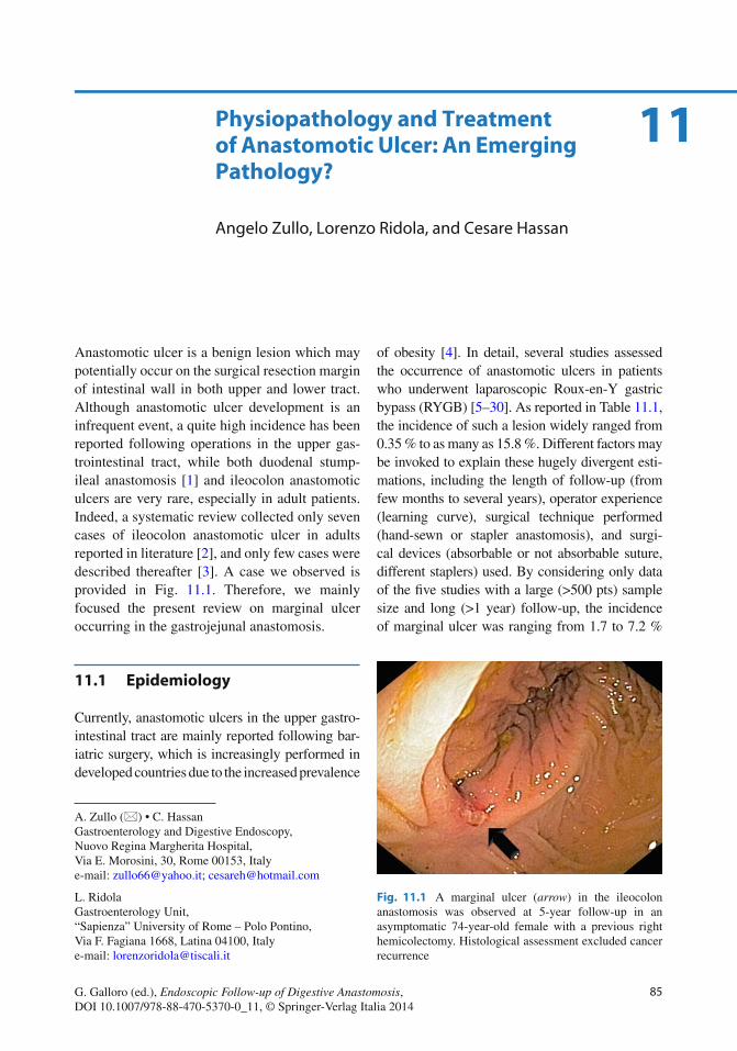
85G. Galloro (ed.), Endoscopic Follow-up of Digestive Anastomosis, DOI 10.1007/978-88-470-5370-0_11, © Springer-Verlag Italia 2014
Anastomotic ulcer is a benign lesion which may potentially occur on the surgical resection margin of intestinal wall in both upper and lower tract. Although anastomotic ulcer development is an infrequent event, a quite high incidence has been reported following operations in the upper gas-trointestinal tract, while both duodenal stump- ileal anastomosis [ 1 ] and ileocolon anastomotic ulcers are very rare, especially in adult patients. Indeed, a systematic review collected only seven cases of ileocolon anastomotic ulcer in adults reported in literature [ 2 ], and only few cases were described thereafter [ 3 ]. A case we observed is provided in Fig. 11.1 . Therefore, we mainly focused the present review on marginal ulcer occurring in the gastrojejunal anastomosis .
11.1 Epidemiology
Currently, anastomotic ulcers in the upper gastro-intestinal tract are mainly reported following bar-iatric surgery, which is increasingly performed in developed countries due to the increased prevalence
of obesity [ 4 ]. In detail, several studies assessed the occurrence of anastomotic ulcers in patients who underwent laparoscopic Roux-en-Y gastric bypass (RYGB) [ 5 – 30 ]. As reported in Table 11.1 , the incidence of such a lesion widely ranged from 0.35 % to as many as 15.8 %. Different factors may be invoked to explain these hugely divergent esti-mations, including the length of follow-up (from few months to several years), operator experience (learning curve), surgical technique performed (hand-sewn or stapler anastomosis), and surgi-cal devices (absorbable or not absorbable suture, different staplers) used. By considering only data of the fi ve studies with a large (>500 pts) sample size and long (>1 year) follow-up, the incidence of marginal ulcer was ranging from 1.7 to 7.2 %
A. Zullo (*) • C. Hassan Gastroenterology and Digestive Endoscopy , Nuovo Regina Margherita Hospital , Via E. Morosini, 30 , Rome 00153 , Italy e-mail: [email protected]; [email protected]
L. Ridola Gastroenterology Unit , “Sapienza” University of Rome – Polo Pontino , Via F. Fagiana 1668 , Latina 04100 , Italy e-mail: [email protected]
11 Physiopathology and Treatment of Anastomotic Ulcer: An Emerging Pathology?
Angelo Zullo , Lorenzo Ridola , and Cesare Hassan
Fig. 11.1 A marginal ulcer ( arrow ) in the ileocolon anastomosis was observed at 5-year follow-up in an asymptomatic 74-year-old female with a previous right hemicolectomy. Histological assessment excluded cancer recurrence
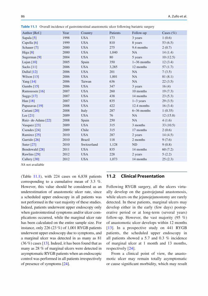
86
(Table 11.1 ), with 224 cases on 6,838 patients corresponding to a cumulative mean of 3.3 %. However, this value should be considered as an underestimation of anastomotic ulcer rate, since a scheduled upper endoscopy in all patients was not performed in the vast majority of these studies. Indeed, patients underwent upper endoscopy only when gastrointestinal symptoms and/or ulcer com-plications occurred, while the marginal ulcer rate has been calculated on the entire sample size. For instance, only 226 (23 %) of 1,001 RYGB patients underwent upper endoscopy due to symptoms, and a marginal ulcer was detected in as many as 81 (36 %) cases [ 13 ]. Indeed, it has been found that as many as 28 % of marginal ulcers were detected in asymptomatic RYGB patients when an endoscopic control was performed in all patients irrespectively of presence of symptoms [ 24 ].
11.2 Clinical Presentation
Following RYGB surgery, all the ulcers virtu-ally develop on the gastrojejunal anastomosis, while ulcers on the jejunojejunostomy are rarely detected. In these patients, marginal ulcers may develop either in the early (few days) postop-erative period or at long-term (several years) follow- up. However, the vast majority (95 %) of anastomotic ulcer develops within 12 months [ 13 ]. In a prospective study on 441 RYGB patients, the scheduled upper endoscopy in all patients showed a 5.7 and 0.3 % incidence of marginal ulcer at 1 month and 13 months, respectively [ 24 ].
From a clinical point of view, the anasto-motic ulcer may remain totally asymptomatic or cause signifi cant morbidity, which may result
Table 11.1 Overall incidence of gastrointestinal anastomotic ulcer following bariatric surgery
Author [Ref.] Year Country Patients Follow-up Cases (%)
Sapala [ 5 ] 1998 USA 173 3 years 1 (0.6) Capella [ 6 ] 1999 USA 810 8 years 53 (6.5) Schauer [ 7 ] 2000 USA 275 9.4 months 2 (0.7) Higa [ 8 ] 2000 USA 1,040 NA 14 (1.4) Sugerman [ 9 ] 2004 USA 80 5 years 10 (12.5) Lujan [ 10 ] 2005 Spain 350 1−36 months 12 (3.4) Sacks [ 11 ] 2006 USA 3,285 12 months 57 (1.7) Dallal [ 12 ] 2006 USA 201 NA 7 (3.5) Wilson [ 13 ] 2006 USA 1,001 NA 81 (8.1) Yang [ 14 ] 2006 Taiwan 636 NA 22 (3.5) Gumbs [ 15 ] 2006 USA 347 3 years 16 (4) Ramsussen [ 16 ] 2007 USA 260 10 months 19 (7.3) Suggs [ 17 ] 2007 USA 438 14 months 23 (5.3) Han [ 18 ] 2007 USA 835 1−3 years 29 (3.5) Papasavas [ 19 ] 2008 USA 422 12.4 months 16 (3.4) Cariani [ 20 ] 2008 Italy 287 6−36 months 1 (0.35) Lee [ 21 ] 2009 USA 76 NA 12 (15.8) Ruiz- de-Adana [ 22 ] 2008 Spain 250 NA 4 (1.6) Vasquez [ 23 ] 2009 USA 315 3 months 33 (10.5) Csendes [ 24 ] 2009 Chile 315 17 months 2 (0.6) Ramirez [ 25 ] 2010 USA 287 2 years 14 (4.5) Garrido [ 26 ] 2010 Brazil 118 2 months 9 (7.6) Suter [ 27 ] 2010 Switzerland 1,128 ND 9 (0.8) Bendewald [ 28 ] 2011 USA 835 14 months 60 (7.2) Rawlins [ 29 ] 2012 USA 228 2 years 5 (2.2) Callery [ 30 ] 2012 USA 1,073 14 months 25 (2.3)
NA not available
A. Zullo et al.
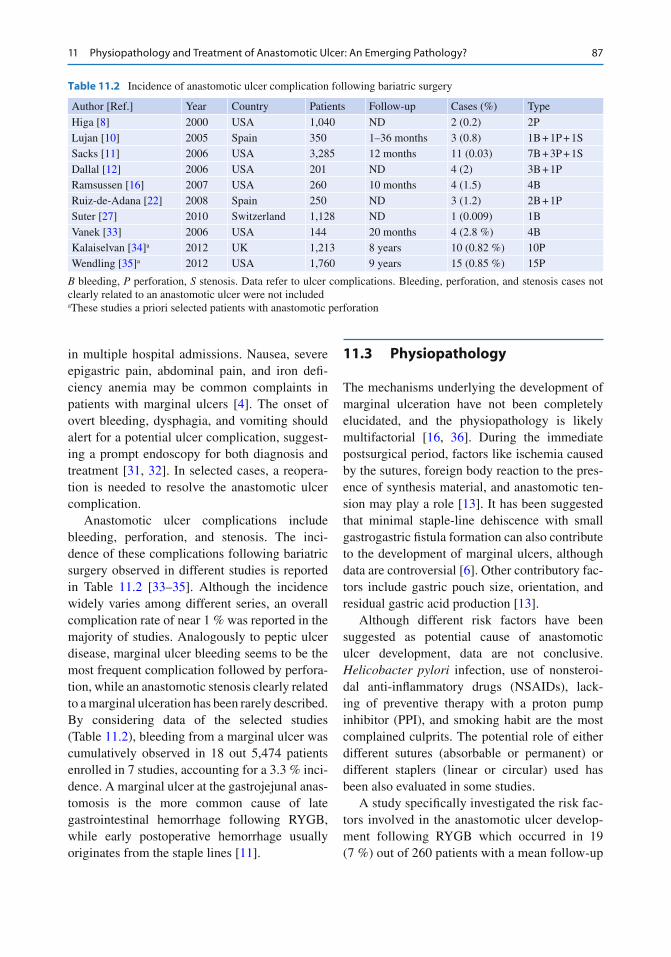
87
in multiple hospital admissions. Nausea, severe epigastric pain, abdominal pain, and iron defi -ciency anemia may be common complaints in patients with marginal ulcers [ 4 ]. The onset of overt bleeding, dysphagia, and vomiting should alert for a potential ulcer complication , suggest-ing a prompt endoscopy for both diagnosis and treatment [ 31 , 32 ]. In selected cases, a reopera-tion is needed to resolve the anastomotic ulcer complication.
Anastomotic ulcer complications include bleeding, perforation, and stenosis. The inci-dence of these complications following bariatric surgery observed in different studies is reported in Table 11.2 [ 33 – 35 ]. Although the incidence widely varies among different series, an overall complication rate of near 1 % was reported in the majority of studies. Analogously to peptic ulcer disease, marginal ulcer bleeding seems to be the most frequent complication followed by perfora-tion , while an anastomotic stenosis clearly related to a marginal ulceration has been rarely described. By considering data of the selected studies (Table 11.2 ), bleeding from a marginal ulcer was cumulatively observed in 18 out 5,474 patients enrolled in 7 studies, accounting for a 3.3 % inci-dence. A marginal ulcer at the gastrojejunal anas-tomosis is the more common cause of late gastrointestinal hemorrhage following RYGB, while early postoperative hemorrhage usually originates from the staple lines [ 11 ].
11.3 Physiopathology
The mechanisms underlying the development of marginal ulceration have not been completely elucidated, and the physiopathology is likely multifactorial [ 16 , 36 ]. During the immediate postsurgical period, factors like ischemia caused by the sutures, foreign body reaction to the pres-ence of synthesis material, and anastomotic ten-sion may play a role [ 13 ]. It has been suggested that minimal staple-line dehiscence with small gastrogastric fi stula formation can also contribute to the development of marginal ulcers, although data are controversial [ 6 ]. Other contributory fac-tors include gastric pouch size, orientation, and residual gastric acid production [ 13 ].
Although different risk factors have been suggested as potential cause of anastomotic ulcer development, data are not conclusive. Helicobacter pylori infection, use of nonsteroi-dal anti-infl ammatory drugs (NSAIDs), lack-ing of preventive therapy with a proton pump inhibitor (PPI), and smoking habit are the most complained culprits. The potential role of either different sutures (absorbable or permanent) or different staplers (linear or circular) used has been also evaluated in some studies.
A study specifi cally investigated the risk fac-tors involved in the anastomotic ulcer develop-ment following RYGB which occurred in 19 (7 %) out of 260 patients with a mean follow-up
Table 11.2 Incidence of anastomotic ulcer complication following bariatric surgery
Author [Ref.] Year Country Patients Follow-up Cases (%) Type
Higa [ 8 ] 2000 USA 1,040 ND 2 (0.2) 2P Lujan [ 10 ] 2005 Spain 350 1–36 months 3 (0.8) 1B + 1P + 1S Sacks [ 11 ] 2006 USA 3,285 12 months 11 (0.03) 7B + 3P + 1S Dallal [ 12 ] 2006 USA 201 ND 4 (2) 3B + 1P Ramsussen [ 16 ] 2007 USA 260 10 months 4 (1.5) 4B Ruiz-de-Adana [ 22 ] 2008 Spain 250 ND 3 (1.2) 2B + 1P Suter [ 27 ] 2010 Switzerland 1,128 ND 1 (0.009) 1B Vanek [ 33 ] 2006 USA 144 20 months 4 (2.8 %) 4B Kalaiselvan [ 34 ] a 2012 UK 1,213 8 years 10 (0.82 %) 10P Wendling [ 35 ] a 2012 USA 1,760 9 years 15 (0.85 %) 15P
B bleeding, P perforation, S stenosis. Data refer to ulcer complications. Bleeding, perforation, and stenosis cases not clearly related to an anastomotic ulcer were not included a These studies a priori selected patients with anastomotic perforation
11 Physiopathology and Treatment of Anastomotic Ulcer: An Emerging Pathology?
88
of 12 months [ 16 ]. The study found that, at univariate analysis, only the seroprevalence for H. pylori infection was signifi cantly higher in patients who developed the marginal ulcer as compared to those who did not (23 % vs 12 %; p = 0.02). A reduced incidence of marginal ulcers was observed in a group of patients who tested positive for H. pylori and were treated before RYGB as compared with a group of patients not tested for the infection (2.4 % vs 6.8 %; P = 0.02) [ 29 ]. Similarly, H. pylori -nega-tive patients had a lower incidence of postopera-tive marginal ulcers compared with a group of untreated H. pylori - positive patients (19 % vs 48 %; P = 0.01) [ 37 ]. In addition, it has been found that patients with preoperatively H. pylori had a threefold higher incidence of postopera-tive GI bleeding or ulcer (18 % vs 6 %), although the difference was not statistically signifi cant due to the low sample size [ 33 ]. On the contrary, several studies failed to demonstrate that the preoperatively test and treat strategy for H. pylori infection would affect marginal ulcer incidence in RYGB patients [ 19 , 29 , 38 – 40 ]. However, some pitfalls may have hampered the accurateness of these studies. For instance, H. pylori infection was assessed only by serology, which is not accurate to diagnose an ongoing infection. In detail, no study evaluated whether H. pylori bacteria were actually present in the small gastric pouch which remain following RYGB. It is counterintuitive that an active infec-tion in this particular niche is prerequisite for a potential role on marginal ulcer development. However, different studies failed to fi nd H. pylori bacteria in gastric pouch mucosa of anas-tomotic ulcer patients previously treated for the infection [ 20 , 26 ]. Therefore, further well- designed studies are needed in such a fi eld.
Although a very small (20−30 ml) gastric pouch remains following RYGB, parietal cells in the remnant gastric mucosa are able to pro-duce gastric acid which may play a role in the anastomotic ulcer onset. Indeed, peptic digestion of the unprotected jejunal mucosa leads to mar-ginal ulceration. Therefore, a preventive therapy reducing gastric acidity is generally performed. However, in some studies a proton pump inhibi-
tor therapy was started the day after surgery and maintained for only 1 month to prevent the development of marginal ulceration [ 27 ]. In other series, a less effective postoperative H2-blocker therapy was administered for a longer period (3−6 months) [ 16 , 19 ]. In a study [ 35 ], as many as 10 (55 %) of 18 patients who developed mar-ginal ulcer were not taking PPI therapy . It has been also found that lacking of PPI therapy was associated with marginal ulcer perforation [ 34 ]. Studies aimed to identify the correct dose and duration of gastric acid inhibition therapy in RYGB patients are needed.
NSAIDs, including low-dose antiplatelets drugs used for cardiovascular prevention, are associated with an increased risk of gastroduode-nal ulcers, erosions, and bleeding [ 41 ]. Therefore, NSAID therapy has been considered as a factor also involved in marginal ulcer development in RYGB patients, but data are confl icting. Indeed, the prevalence of NSAID therapy in the anas-tomotic ulcer patients was widely ranging from 0 % [ 26 ], 10 % [ 25 ], 11 % [ 16 ] 7−16 % [ 19 ], 16 % [ 12 ], and 28 % [ 35 ]. Moreover, in a large series, the majority of patients who developed marginal ulceration were taking NSAIDs despite written and verbal precautions [ 8 ]. In addition, over-the-counter use of NSAIDs in some patients could be not excluded. Of note, NSAIDs use has been also associated with anastomotic ulcer per-foration [ 8 , 34 ].
Tobacco use has been associated with mar-ginal ulcer development. Smoking and nicotine elevate levels of gastric ulcerogens and inhibit nitric oxide synthesis, which leads to a reduction in angiogenesis in the gastric mucosa causing ischemia [ 13 ]. The potential role of smoking on anastomotic ulcer development and ulcer per-foration has been suggested in different studies [ 12 , 20 , 40 ]. A multivariate analysis performed on data of 81 marginal ulcer cases detected on 226 RYGB patients showed that both smok-ing (OR = 30.6, 95 % CI: 6.4–146) and NSAID use (OR = 11.5, 95 % CI: 4.8–28) signifi cantly increase risk of marginal ulcers following surgery, while PPI therapy was protective (OR = 0.33, 95 % CI 0.11–0.97) [ 13 ]. Alcohol consumption, open and laparoscopic surgery, age, gender, and
A. Zullo et al.
89
surgeon experience were not signifi cant predic-tive factors, while H. pylori infection was not taken into account in this study.
Different studies evaluated the role of surgical- related procedures on marginal ulcer onset. The use of absorbable suture was found to signifi -cantly reduce anastomotic ulcer onset as com-pared to permanent suture , the incidence being 1.3 % vs 2.6 % [ 11 ], 2.4 % vs 13.4 % [ 23 ], and 0 % vs 1.6 % [ 6 ]. However, others found no dif-ference between the sutures used (7 % vs 8 %) [ 16 ]. A study found that marginal ulcer incidence tended to be lower by using reinforced stapler with bovine pericardium strips as compared with standard stapler (2.9 % vs 6 %; P = 0.06) [ 25 ], while no improvement emerged with collagen matrix staple-line reinforcement [ 29 ]. Other studies found no difference between the use of 21-mm and 25-mm stapler [ 17 ], as well as among hand-sewn , linear-stapled, and 25-mm circular- stapled anastomosis [ 28 ].
The orientation of the gastric pouch also has been hypothesized to play a role in acid secretion and marginal ulceration development, and the formation of a small gastric pouch with a lower acid production seems to reduce the incidence of marginal ulcer [ 42 ]. Moreover, by excluding parietal cells from the gastric pouch, creating a smaller reservoir, and building the gastrojejunos-tomy using the greater rather than the lesser cur-vature, the anastomotic ulcer incidence was as low as 0.6 % [ 5 ]. Finally, incidence of marginal ulcer was signifi cantly lower following vertical banded gastroplasty as compared to RYGB (12/60 vs 10/22; P = 0.027) [ 14 ].
11.4 Therapy
The fi rst attempt to minimize the marginal ulcer development should be its prevention by improv-ing the surgical techniques. The use of absorbable suture and building a very small gastric pouch through the greater curvature are considered sur-gical adaptations able to reduce anastomotic ulcer development [ 5 , 11 ]. In addition, a therapy aimed to reduce gastric acid secretion is advisable as postoperative prophylaxis. Such a preventive
therapy has been found to be effective [ 16 ], but it was not always performed [ 44 ]. However, the appropriate drug, dosage, and duration of such a preventive therapy have not been established.
When a marginal ulcer develops, a therapy is required to both heal the lesion and to prevent potential ulcer complications. Medical therapy mainly consists of antisecretory therapy with PPIs. Differently from peptic ulcers, these lesions tend to require a prolonged therapy, usually for 3–4 months [ 5 ], and repeat endoscopy is recom-mended to confi rm ulcer resolution. Such a ther-apy is generally effective in healing marginal ulcers [ 16 , 43 , 44 ]. In some studies, sucralfate suspension has been added to PPI therapy, although there are not robust evidence on its effi -cacy. In addition, potential causative factors—such as NSAID use, the presence of a remnant of suture at the ulcer base, and smoking—should be removed [ 11 ]. In detail, long-term cessation of tobacco use could be useful, since some evidences would suggest that resuming smoking is associ-ated with marginal ulcer recurrence and compli-cations [ 12 , 20 ]. Unfortunately, some marginal ulcers may occur despite the absence of H. pylori infection and NSAIDs use, and a preventive PPI therapy was performed [ 26 ]. In selected refrac-tory cases a reoperation is needed [ 45 ].
11.5 Experimental Therapies
Some experimental therapeutic approaches have been tested as a potential therapy for anasto-motic ulcer prevention and healing [ 46 ]. Platelet- derived factors (VEGF, TGF-β factor, platelet factor-4) have been found to be able to promote tissue growth, new angiogenesis, and ulcer heal-ing. In rat model, oral administration of platelet- rich plasma signifi cantly accelerated healing of acetic acid-induced gastric ulcer [ 47 ], suggest-ing a potential role of platelet factors also in anastomotic ulcer healing. The use of becapler-min gel —a recombinant human PDGF-BB—on the anastomotic line just after completion of the colon anastomosis reversed the detrimental effects of ischemia and promoted anastomotic healing in an experimental model [ 48 ]. In a pig
11 Physiopathology and Treatment of Anastomotic Ulcer: An Emerging Pathology?
90
model, a suture-free glued small bowel anas-tomosis using a fi brin-covered collagen fl eece achieved an improved healing without onset of a deep ulcer at the anastomotic line which occurred at conventional sutured anastomosis [ 49 ]. Similarly, the subserosal injection with recom-binant human granulocyte–macrophage colony- stimulating factor in the perianastomotic area improved the healing of ischemic colon anasto-moses in rats [ 50 ]. Leptin potentiates endothelial cell proliferation and increases collagen synthe-sis, and its potential role in improving anastomo-sis healing when administered intraperitoneally in rats has been suggested [ 51 ]. Finally, a recent experimental study in rats showed a halved inci-dence of ischemia-induced colonic anastomotic ulcer by local injection of adipose-tissue-derived stem cells as compared to controls [ 52 ]. Although interesting, data of these studies need to be veri-fi ed in humans. In addition, the effi cacy of such potential therapeutic approaches has been mainly investigated on colonic anastomoses, and conse-quently, these results may not be directly applied to gastrojejunal anastomosis where gastric acid plays an adjunctive detrimental role.
References
1. de Melo SW, Gupta S, Arenas J et al (2008) Single- balloon enteroscopy-guided hemostasis of an anastomotic ulcer in a patient with simultane-ous enteric-drained pancreas-kidney transplant. Endoscopy 40:E164
2. Peter Z, Bodoky G, Szabò Z et al (2004) Ileocolic anastomotic ulcer after surgery in adulthood: case report and review of the literature. Z Gastroenterol 42:605–608
3. Monsanto P, Almeida N, Lérias C et al (2012) Is there still a role for intraoperative enteroscopy in patients with obscure gastrointestinal bleeding? Rev Esp Enferm Dig 104:190–196
4. Karmali S, Stoklossa CJ, Sharma A et al (2010) Bariatric surgery. Can Fam Physician 56:873–879
5. Sapala JA, Wood MH, Sapala MA et al (1998) Marginal ulcer after gastric bypass: a prospective 3-year study of 173 patients. Obes Surg 8:505–516
6. Capella JF, Capella RF (1999) Gastro-gastric fi stulas and marginal ulcers in gastric bypass procedures for weight reduction. Obes Surg 9:22–27
7. Schauer R, Ikramuddin S, Gourash W et al (2000) Outcomes after laparoscopic Roux-en-Y gastric bypass for morbid obesity. Ann Surg 232:515–529
8. Higa KD, Boone KB, Ho T (2000) Complications of the laparoscopic Roux-en-Y gastric bypass: 1,040 patients – what have we learned? Obes Surg 10:509–513
9. Sugerman HJ, DeMaria EJ, Kellum JM et al (2004) Effects of bariatric surgery in older patients. Ann Surg 240:243–247
10. Lujan JA, Frutos MD, Hernández Q et al (2005) Experience with the circular stapler for the gastrojeju-nostomy in laparoscopic gastric bypass (350 cases). Obes Surg 15:1096–1102
11. Sacks BC, Mattar SG, Qureshi FG et al (2006) Incidence of marginal ulcers and the use of absorbable anastomotic sutures in laparoscopic Roux-en-Y gas-tric bypass. Surg Obes Relat Dis 2:11–16
12. Dallal RM, Bailey LA (2006) Ulcer disease after gas-tric bypass surgery. Surg Obes Relat Dis 2:455–459
13. Wilson JA, Romagnuolo J, Byrne K et al (2006) Predictors of endoscopic fi ndings after Roux-en-Y gastric bypass. Am J Gastroenterol 101:2194–2199
14. Yang CS, Lee WJ, Wang HH et al (2006) The infl u-ence of Helicobacter pylori infection on the develop-ment of gastric ulcer in symptomatic patients after bariatric surgery. Obes Surg 16:735–739
15. Gumbs AA, Duffy AJ, Bell RL (2006) Incidence and management of marginal ulceration after laparo-scopic Roux-Y gastric bypass. Surg Obes Relat Dis 4:460–463
16. Ramsussen JJ, Fuller W, Ali MR (2007) Marginal ulceration after laparoscopic gastric bypass: an analy-sis of predisposing factors in 260 patients. Surg Endosc 21:1090–1094
17. Suggs WJ, Kouli W, Lupovic M et al (2007) Complications at gastrojejunostomy after laparo-scopic Roux-en-Y gastric bypass: comparison between 21- and 25-mm circular staplers. Surg Obes Relat Dis 3:508–514
18. Han SH, Gracia C, Mehran A et al (2007) Improved outcomes using a systematic and evidence-based approach to the laparascopic Roux-en-Y gastric bypass in a single academic institution. Am Surg 73:955–958
19. Papasavas P, Gagné DJ, Donnelly PE et al (2008) Prevalence of Helicobacter pylori infection and value of preoperative testing and treatment in patients undergoing laparoscopic Roux-en-Y gastric bypass. Surg Obes Relat Dis 4:383–388
20. Cariani S, Palandri P, Della Valle E et al (2008) Italian multicenter experience of Roux-en-Y gastric bypass on vertical banded gastroplasty: four-year results of effec-tive and safe innovative procedure enabling traditional endoscopic and radiographic study of bypassed stom-ach and biliary tract. Surg Obes Relat Dis 4:16–25
21. Lee JK, Van Dam J, Morton JM et al (2009) Endoscopy is accurate, safe, and effective in the assessment and management of complications following gastric bypass surgery. Am J Gastroenterol 104:575–582
22. Ruiz-de-Adana C, López-Herrero J, Hernández- Matías A et al (2008) Laparoscopic hand-sewn gastro-jejunal anastomosis. Obes Surg 18:1074–1076
A. Zullo et al.
91
23. Vasquez JC, Wayne Overby D, Farrell TM (2009) Fewer gastrojejunostomy strictures and mar-ginal ulcers with absorbable suture. Surg Endosc 23:2011–2015
24. Csendes A, Burgos AM, Altuve J et al (2009) Incidence of marginal ulcer 1 month and 1 to 2 years after gastric bypass: a prospective consecutive endo-scopic evaluation of 442 patients with morbid obesity. Obes Surg 19:135–138
25. Ramirez MC, Rodriguez J, Varghese F et al (2010) Reinforced circular stapler in bariatric surgery. JSLS 14:358–363
26. Garrido AB Jr, Rossi M, Lima SE Jr et al (2010) Early marginal ulcer following Roux-en-Y gastric bypass under proton pump inhibitor treatment – prospective multicentric study. Arq Gastroenterol 47:130–134
27. Suter M, Donadini A, Calmes JM et al (2010) Improved surgical technique for laparoscopic Roux-en- Y gastric bypass reduces complications at the gas-trojejunostomy. Obes Surg 20:841–845
28. Bendewald FP, Choi JN, Blythe LS et al (2011) Comparison of hand-sewn, linear-stapled, and circular- stapled gastrojejunostomy in laparoscopic Roux-en-Y gastric bypass. Obes Surg 21:1671–1675
29. Rawlins L, Rawlins MP, Brown CC et al (2013) Effect of Helicobacter pylori on marginal ulcer and stomal stenosis after Roux-en-Y gastric bypass. Surg Obes Relat Dis 9:760–764
30. Callery CD, Filiciotto S, Neil KL (2012) Collagen matrix staple line reinforcement in gastric bypass. Surg Obes Relat Dis 8:185–189
31. Levitzky BE, Wassef WY (2010) Endoscopic man-agement in the bariatric surgical patient. Curr Opin Gastroenterol 26:632–639
32. Mayer G, Lingenfelser T, Ell TC (2004) The role of endoscopy in early postoperative haemorrhage. Best Pract Res Clin Gastroenterol 18:799–807
33. Vanek VW, Catania M, Triveri K et al (2006) Retrospective review of the preoperative biliary and gastrointestinal evaluation for gastric bypass surgery. Surg Obes Relat Dis 2:17–23
34. Kalaiselvan R, Exarchos G, Hamza N et al (2012) Incidence of perforated gastrojejunal anastomotic ulcers after laparoscopic gastric bypass for morbid obesity and role of laparoscopy in their management. Surg Obes Relat Dis 8:423–428
35. Wendling MR, Linn JG, Keplinger KM et al (2013) Omental patch repair effectively treats perforated marginal ulcer following Roux-en-Y gastric bypass. Surg Endosc 27:384–389
36. Mason EE (1996) Ulcerogenesis in surgery for obe-sity. Obes Surg 6:180–181
37. Ramaswamy A, Lin E, Ramshaw BJ, Smith CD (2004) Early effects of Helicobacter pylori infection in patients undergoing bariatric surgery. Arch Surg 139:1094–1096
38. Hartin CW, ReMine DS, Lucktong TA (2009) Preoperative bariatric screening and treatment of Helicobacter pylori. Surg Endosc 23:2531–2534
39. Marano BJ (2005) Endoscopy after Roux-en-Y gastric bypass: a community hospital experience. Obes Surg 15:342–345
40. Schreiber H, Ben-Meir A, Sonpal I et al (2005) Cigarette smoking, but not the presence of H. pylori , is associated with anastomotic ulcers in Roux-en-Y gastric bypass patients. Surg Obes Relat Dis 1:257 (abstract)
41. Zullo A, Hassan C, Campo SMA et al (2007) Bleeding peptic ulcer in the elderly risk factors and prevention strategies. Drugs Aging 24:815–828
42. MacLean LD, Rhode BM, MacLean AP et al (1997) Stomal ulcer after gastric bypass. J Am Coll Surg 185:1–7
43. Hedberg J, Hedenstrom H, Nilsson S et al (2005) Role of gastric acid in stomal ulcer after gastric bypass. Obes Surg 15:1375–1378
44. Pope GD, Goodney PP, Burchard KW (2002) Peptic ulcer/stricture after gastric bypass: a comparison of technique and acid suppression variables. Obes Surg 12:30–33
45. Steinemann DC, Schiesser M, Clavien PA et al (2011) Laparoscopic gastric pouch and remnant resection: a novel approach to refractory anastomotic ulcers after Roux-en-Y gastric bypass: case report. BMC Surg 11:33
46. Zimmermann R, Jakubietz R, Jakubietz M et al (2001) Different preparation methods to obtain platelet com-ponents as a source of growth factors for local appli-cation. Transfusion 41:1217–1224
47. Wallace JL, Dicay M, McKnight W et al (2006) Platelets accelerate gastric ulcer healing through pre-sentation of vascular endothelial growth factor. Br J Pharmacol 148:274–278
48. Sarıbeyoglu G, Baca B, Hamzaoglu I et al (2003) Does becaplermin (Platelet-Derived Growth Factor-BB) reverse detrimental effects of ischemia on colonic anastomosis? Dis Colon Rectum 46:516–520
49. Stumpf M, Junge K, Rosch R et al (2009) Suture-free small bowel anastomoses using collagen fl eece covered with fi brin glue in pigs. J Invest Surg 22:138–147
50. Dinca S, Gulcelika MA, Kurua B et al (2004) Effects of locally applied recombinant Human Granulocyte- Macrophage Colony-Stimulating factor on ischemic bowel anastomoses in rat. Eur Surg Res 36:59–63
51. Tasdlen A, Algin C, Ates E et al (2004) Effect of leptin on healing of colonic anastomoses in rats. Hepatogastroenterology 51:994–997
52. Yoo HJ, Shin JH, An MS et al (2012) Adipose-tissue- derived stem cells enhance the healing of ischemic colonic anastomoses: an experimental study in rats. J Korean Soc Coloproctol 28:132–139
11 Physiopathology and Treatment of Anastomotic Ulcer: An Emerging Pathology?
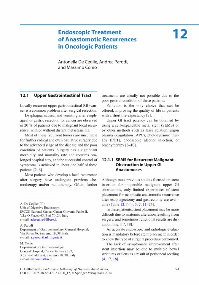
93G. Galloro (ed.), Endoscopic Follow-up of Digestive Anastomosis, DOI 10.1007/978-88-470-5370-0_12, © Springer-Verlag Italia 2014
12.1 Upper Gastrointestinal Tract
Locally recurrent upper gastrointestinal (GI) can-cer is a common problem after surgical resection.
Dysphagia , nausea, and vomiting after esoph-ageal or gastric resection for cancer are observed in 20 % of patients due to malignant local recur-rence , with or without distant metastasis [ 1 ].
Most of these recurrent tumors are unsuitable for further radical and even palliative surgery due to the advanced stage of the disease and the poor condition of patients. Surgery has a signifi cant morbidity and mortality rate and requires pro-longed hospital stay, and the successful control of symptoms is achieved in about one half of these patients [ 2 – 6 ].
Most patients who develop a local recurrence after surgery have undergone previous che-motherapy and/or radiotherapy. Often, further
treatments are usually not possible due to the poor general condition of these patients.
Palliation is the only choice that can be offered, improving the quality of life in patients with a short life expectancy [ 7 ].
Upper GI tract patency can be obtained by using a self-expandable metal stent (SEMS ) or by other methods such as laser ablation, argon plasma coagulation (APC), photodynamic ther-apy (PDT), endoscopic alcohol injection, or brachytherapy [ 8 – 10 ].
12.1.1 SEMS for Recurrent Malignant Obstruction in Upper GI Anastomoses
Although most previous studies focused on stent insertion for inoperable malignant upper GI obstructions, only limited experiences of stent placement for neoplastic anastomotic recurrence after esophagectomy and gastrectomy are avail-able (Table 12.1 ) [ 4 , 5 , 7 , 11 – 24 ].
In these patients, stent placement may be more diffi cult due to anatomic alteration resulting from surgery, and sometimes functional results are dis-appointing [ 17 , 18 ].
An accurate endoscopic and radiologic evalua-tion is mandatory before stent placement in order to know the type of surgical procedure performed.
The lack of symptomatic improvement after stent insertion may be due to multiple bowel strictures or ileus as a result of peritoneal seeding [ 4 , 17 , 18 ].
A. De Ceglie (*) Unit of Digestive Endoscopy , IRCCS National Cancer Center Giovanni Paolo II , V.Le O Flacco 65 , Bari 70124 , Italy e-mail: [email protected]
A. Parodi Department of Gastroenterology , General Hospital , Via Borea 56 , Sanremo 18038 , Italy e-mail: [email protected]
M. Conio Department of Gastroenterology , General Hospital , Corso Garibaldi 187, 3 (private address) , Sanremo 18038 , Italy e-mail: [email protected]
12 Endoscopic Treatment of Anastomotic Recurrences in Oncologic Patients
Antonella De Ceglie , Andrea Parodi , and Massimo Conio
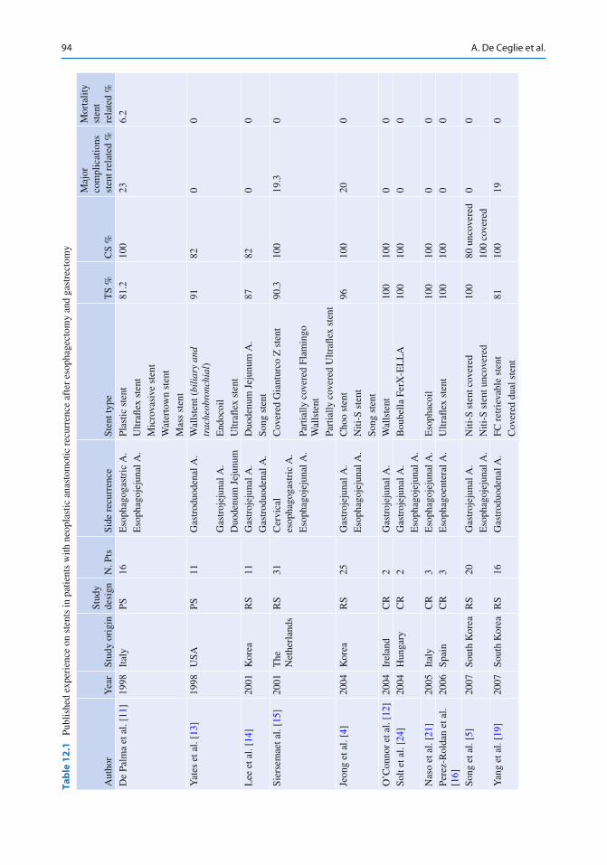
94
Tab
le 1
2.1
Pu
blis
hed
expe
rien
ce o
n st
ents
in p
atie
nts
with
neo
plas
tic a
nast
omot
ic r
ecur
renc
e af
ter
esop
hage
ctom
y an
d ga
stre
ctom
y
Aut
hor
Yea
r St
udy
orig
in
Stud
y de
sign
N
. Pts
Si
de r
ecur
renc
e St
ent t
ype
TS
%
CS
%
Maj
or
com
plic
atio
ns
sten
t rel
ated
%
Mor
talit
y st
ent
rela
ted
%
De
Palm
a et
al.
[ 11 ]
19
98
Ital
y PS
16
E
soph
agog
astr
ic A
. Pl
astic
ste
nt
81.2
10
0 23
6.
2 E
soph
agoj
ejun
al A
. U
ltrafl
ex
sten
t M
icro
vasi
ve s
tent
W
ater
tow
n st
ent
Mas
s st
ent
Yat
es e
t al.
[ 13 ]
19
98
USA
PS
11
G
astr
oduo
dena
l A.
Wal
lste
nt (
bili
ary
and
trac
heob
ronc
hial
) 91
82
0
0
Gas
troj
ejun
al A
. E
ndoc
oil
Duo
denu
m J
ejun
um
Ultr
afl e
x st
ent
Lee
et a
l. [ 1
4 ]
2001
K
orea
R
S 11
G
astr
ojej
unal
A.
Duo
denu
m J
ejun
um A
. 87
82
0
0 G
astr
oduo
dena
l A.
Song
ste
nt
Sier
sem
aet a
l. [ 1
5 ]
2001
T
he
Net
herl
ands
R
S 31
C
ervi
cal
esop
hago
gast
ric
A.
Cov
ered
Gia
ntur
co Z
ste
nt
90.3
10
0 19
.3
0
Eso
phag
ojej
unal
A.
Part
ially
cov
ered
Fla
min
go
Wal
lste
nt
Part
ially
cov
ered
Ultr
afl e
x st
ent
Jeon
g et
al.
[ 4 ]
2004
K
orea
R
S 25
G
astr
ojej
unal
A.
Cho
o st
ent
96
100
20
0 E
soph
agoj
ejun
al A
. N
iti-S
ste
nt
Song
ste
nt
O’C
onno
r et
al.
[ 12 ]
20
04
Irel
and
CR
2
Gas
troj
ejun
al A
. W
alls
tent
10
0 10
0 0
0 So
lt et
al.
[ 24 ]
20
04
Hun
gary
C
R
2 G
astr
ojej
unal
A.
Bou
bella
Fer
X-E
LL
A
100
100
0 0
Eso
phag
ojej
unal
A.
Nas
o et
al.
[ 21 ]
20
05
Ital
y C
R
3 E
soph
agoj
ejun
al A
. E
soph
acoi
l 10
0 10
0 0
0 Pe
rez-
Rol
dan
et a
l. [ 1
6 ]
2006
Sp
ain
CR
3
Eso
phag
oent
eral
A.
Ultr
afl e
x st
ent
100
100
0 0
Song
et a
l. [ 5
] 20
07
Sout
h K
orea
R
S 20
G
astr
ojej
unal
A.
Niti
-S s
tent
cov
ered
10
0 80
unc
over
ed
0 0
Eso
phag
ojej
unal
A.
Niti
-S s
tent
unc
over
ed
100
cove
red
Yan
g et
al.
[ 19 ]
20
07
Sout
h K
orea
R
S 16
G
astr
oduo
dena
l A.
FC r
etri
evab
le s
tent
81
10
0 19
0
Cov
ered
dua
l ste
nt
A. De Ceglie et al.
95
Kim
et a
l. [ 2
3 ]
2007
So
uth
Kor
ea
RS
32
Eso
phag
ojej
unal
A.
FC r
etri
evab
le s
tent
94
91
31
.2
0 C
over
ed d
ual s
tent
So
ng e
t al.
[ 18 ]
20
07
Sout
h K
orea
R
S 39
G
astr
ojej
unal
A.
Song
FC
ste
nt
100
90
25.6
5.
1 FC
ret
riev
able
ste
nt
Bar
e ni
tinol
ste
nt
Dua
l ste
nt
Cho
et a
l. [ 2
0 ]
2009
So
uth
Kor
ea
RS
20
Eso
phag
ojej
unal
A
Niti
-S s
tent
10
0 70
30
0
Gas
trod
uode
nal A
. C
hoo
sten
t G
astr
ojej
unal
A.
Kim
et a
l. [ 1
7 ]
2009
So
uth
Kor
ea
RS
47
Eso
phag
ojej
unal
A.
Niti
-S s
tent
( c
over
ed / u
ncov
ered
) 95
.7
87.2
27
.6
0
Gas
troj
ejun
al A
. C
hoo
sten
t ( co
vere
d / un
cove
red )
To
ng e
t al.
[ 7 ]
2010
C
hina
PR
35
E
soph
agoj
ejun
al A
. U
ltrafl
ex
sten
t 97
.6
100
14
0 E
soph
agea
l rem
nant
C
hoo
sten
t G
OO
N
iti-S
ste
nt
Ext
rins
ic
com
pres
sion
ER
F K
im e
t al.
[ 22 ]
20
11
Sout
h K
orea
R
S 35
E
soph
agoj
ejun
al A
. C
hoo
esop
hage
al s
tent
92
of
39
90 o
f 39
44
of
39
0 39
ste
nt
Gas
trod
uode
nal A
. H
anar
o es
opha
geal
ste
nt
Gas
troj
ejun
al A
. N
iti-S
eso
phag
eal,
pylo
ric/
duod
enal
ste
nt
Wal
lste
nt c
olon
ic s
tent
W
allF
lex
duod
enal
ste
nt
N n
umbe
r, P
ts p
atie
nts,
TS
tech
nica
l suc
cess
, CS
clin
ical
suc
cess
, PS
pros
pect
ive
stud
y, A
ana
stom
osis
, RS
retr
ospe
ctiv
e st
udy,
CR
cas
e re
port
, FC
ful
ly c
over
ed, G
OO
gas
tric
ou
tlet o
bstr
uctio
n, E
RF
eso
phag
ores
pira
tory
fi st
ula
12 Endoscopic Treatment of Anastomotic Recurrences in Oncologic Patients
96
Multiple strictures occur in 11 % of cases, and stent placement for two successive synchronous strictures is exceptional [ 13 ].
SEMS insertion may be performed both by endoscopically guided and by fl uoroscopically guided methods. Similar high technical success (TS) and clinical success (CS) rates have been reported (92 and 90 % TS and CS, respectively, in endoscopic approach vs TS at 94–100 % and CS at 90–96 % in fl uoroscopic approach) [ 4 , 5 , 22 , 23 ].
However, the advantages of the endoscopic method in patients with distal gastrectomy are the ease of accessing the stricture site and the avoidance of looping the delivery system through the dilated gastric lumen due to the stiffness of the endoscope [ 20 ].
The friction between the working channel of the endoscope and the long delivery system may result in a diffi cult stent insertion when the endoscope is in an angulated position [ 22 , 25 ]. Balloon dilatation can be exceptionally per-formed when the tightness of strictures does not allow the introduction of the delivery system; excessive dilatation should be avoided to prevent stent migration [ 24 ].
A stent at least 2–4 cm longer than the stric-ture should be chosen to allow for a 1–2 cm extension beyond the proximal as well as distal tumor margins [ 15 ].
Generally, in patients treated with SEMS, dys-phagia is relieved in approximately 90 % of cases, and these patients undergo signifi cantly fewer procedures, thus spending fewer days in the hospital [ 26 , 27 ].
Stent placement has several advantages over surgery: it is a less-invasive procedure is pre-ferred both by the patient and surgeon because of its lower morbidity, shorter procedural time and hospital stay, as well as faster recovery of gut function [ 17 , 18 , 28 ].
This is important for patients whose life expectancy is of 4–6 months, or less.
The overall survival rate for stent-treated patients has been reported to be similar to that for patients undergoing surgery, whose hospitaliza-tion is three times longer and costs are three times greater [ 29 ].
The recurrent rate of stenosis in patients with SEMS placed for anastomotic malignant obstruc-tion ranges from 8 to 46 % after an interval of 2–21 weeks. It is generally due to tumor in- overgrowth [ 5 , 30 ].
Some studies report that early occlusion of SEMS (within 4 weeks after their implantation) occurs more frequently in patients with the stent at the anastomotic level, than in those treated for unresectable cancer, but the causes are unknown [ 5 , 6 , 31 ].
Chemotherapy could decrease the tumor ingrowth and overgrowth; however, chemother-apy after stent placement is likely to shrink the tumor, increasing stent migration, if a covered SEMS has been used [ 5 , 19 , 32 ].
Stent migration rates range from 4.2 to 27.8 %; a pooled analysis of 21 studies reports that migra-tion rate was 2.7 and 16 % for uncovered and covered stents, respectively [ 33 ].
An appropriate choice among the marketed SEMS according to the type of surgery performed may reduce the migration rate of stents placed in malignant recurrences [ 17 ].
A retrospective study reported that a double coaxial stent had a longer patency and lower migration rate than an uncovered stent in anasto-motic recurrence [ 5 ].
At present, several types of SEMS are avail-able, varying by type of alloy, confi guration, degree of shortening after their release, lengths and diameters, presence, type, and extent of covering, delivery system, expandable force, presence or absence of anti-refl ux valve, and removability.
Most SEMS are made of nitinol, an alloy of nickel and titanium, whose peculiarity is super- elasticity and shape memory.
The covering is either polyurethane, silicone, or polytetrafl uoroethylene (PTFE). Their fl exibil-ity, the small diameter of the delivery system, and the large availability of models allow the treat-ment of any type of malignant obstruction [ 26 , 34 ].
There are no data to date demonstrating sig-nifi cant differences in outcomes or complications among SEMS types. Therefore, the choice of spe-cifi c SEMS is often based on the endoscopist’s
A. De Ceglie et al.
97
experience, although the initial stent selection has a signifi cant impact on the clinical outcome in patients with inoperable malignancy [ 35 ].
Use of a self-expandable plastic stent (Polyfl ex® stent, Boston Scientifi c , Natick , MA , USA ) is not recommended in GI anastomotic malignant recurrence. The major disadvantage of the current version of plastic stents is the large diameter and stiffness of the stent delivery sys-tem when compared with metal stents. Therefore, plastic stents cannot be released in patients with angulated strictures as in anastomotic ones.
SEMS may be placed hardly in some cases, especially in the presence of narrow bowel loops. In such situations, SEMS cannot fully expand, and they may migrate or cause perforation due to excessive stretching of gut wall, determined by the device. New D-Weave Niti-S colonic stent ( TaeWoong Medical Co ., Seoul , South Korea ) has characteristics that may reduce the risk of such complications. It is a self-expandable nitinol stent, whose particular confi guration confers some inter-esting properties to the device, such as compliant fl exibility, high expansible force, and negligible foreshortening, in order to reduce the risk of migra-tion, perforation, and inadequate expansion [ 36 ].
SEMS for malignant anastomotic obstruction after :• Esophagectomy
For patients with a gastric tube interposition after esophagectomy, recurrent tumor growth occurs at the level of cervical anastomosis [ 37 ]. Endoscopic placement of a stent for lesions within a few centimeters of the upper esopha-geal sphincter (UES ) is challenging. Complications such as migration, perforation, and tracheal compression can occur. The opening of the upper fl ared end of a regular esophageal SEMS is often incomplete and causes an intolerable foreign body sensation, leading to an inadequate swallowing capacity [ 15 , 38 , 39 ]. The use of SEMS with a diameter <18 mm could minimize patient intolerance and stent- related complications [ 15 , 39 ]. The risk of distal migration of the stent is high as the length of the anastomotic stricture is
short, preventing an adequate adhesion. In fact, even placing a 6-cm stent, most of it is free into the stomach. Siersema et al. inserted 4 covered Gianturco Z stents, (body diameter/fl ared ends: 18–25 mm) and 6 partially covered Ultrafl ex stents (body/fl ared ends diameter: 18–23 mm) in 10 patients with a recurrent tumor located in the proximal part of the gastric tube interposition. They obtained technical success rate of 90 %, improvement of dysphagia of at least 1 grade, and complications and recurrent dysphagia due to tumor overgrowth in 30 and 20 % of patients, respectively [ 15 ]. In selected cases, as in the one mentioned above, anatomic morphology may prevent the use of SEMS. The only palliative option we use is thermal ablation with APC or neodym-ium: yttrium–aluminum–garnet (Nd:YAG) laser. Small diameters SEMS (12, 14, 16 mm the body; 14, 16, 18 mm the upper fl ared end) may be used in patients with neoplastic recur-rence occurring after chemo/radiation treat-ment and surgery, for laryngeal cancer. Montgomery salivary bypass tube (MSBT) ( Boston Medical Products , Westborough , USA ) may also be used as a palliative measure in this subgroup of patients. MSBT is a silicone prosthesis with a fl ared upper end. It is available in 7 sizes, ranging 8–20 mm. The length of the stent is 191 mm. It can be placed trough the stricture using a Savary-Gilliard dilator. The stent is released with a fi nger, inserted deep in the throat, when the dilator is partially retracted. This maneu-ver allows the mobilization of the MSBT. The fi nal adjustment is made pushing gently the stent with the Savary and, in the end, with the tip of the scope. However, its upper fl ared end has a large diameter, and it should be adapted according to the modifi ed anatomic morphology, by cut-ting it with scissors before its introduction [ 40 – 42 ]. The onset of an esophagorespiratory fi stula (ERF) is an ominous complication that can occur both in patients with esophageal cancer
12 Endoscopic Treatment of Anastomotic Recurrences in Oncologic Patients
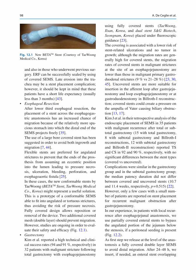
98
and also in those who underwent previous sur-gery. ERF can be successfully sealed by using of covered SEMS. Late erosion into the tra-chea may be a stent placement complication; however, it should be kept in mind that these patients have a short life expectancy (usually less than 3 months) [ 43 ].
• Esophageal Resection After lower third esophageal resection, the placement of a stent across the esophagogas-tric anastomosis has an increased chance of migration because of the relatively more spa-cious stomach into which the distal end of the SEMS projects freely [ 35 ]. The use of a large bore covered stent has been suggested in order to avoid both ingrowth and migration [ 7 , 44 ]. Flexible stents are preferred for angulated strictures to prevent that the ends of the pros-thesis from assuming an eccentric position into the lumen leading to pressure necro-sis, ulceration, bleeding, perforation, and esophagoaortic fi stula [ 25 ]. In these cases, the new conformable stents by TaeWoong ( BETA TM Stent , TaeWoong Medical Co ., Korea ) might represent a useful solution. This is a prototype of a conformable SEMS able to fi t into angulated or tortuous strictures, thus avoiding the risk of pressure necrosis. Fully covered design allows reposition or removal of the device. Two additional covered mesh (double layer) should prevent migration. However, studies are ongoing in order to eval-uate their safety and effi cacy (Fig. 12.1 ).
• Gastrectomy Kim et al. reported a high technical and clini-cal success rates (94 and 91 %, respectively) in 32 patients with malignant stricture following total gastrectomy with esophagojejunostomy
using fully covered stents ( TaeWoong , Ilsan , Korea , and dual stent S&G Biotech , Seongnam , Korea ) placed under fl uoroscopic guidance [ 23 ]. The covering is associated with a lower risk of stent-related ulcerations and no tumor in growth; although the migration rates are gen-erally high for covered stents, the migration rates of covered stents in malignant strictures at the site of an esophagojejunostomy are lower than those in malignant primary gastro-duodenal strictures (9 % vs 21–28 %) [ 23 , 30 , 45 ]. Uncovered stents are more suitable for insertion in the afferent loop after gastrojeju-nostomy and loop esophagojejunostomy or at gastroduodenostomy in Billroth-I reconstruc-tion; covered stents could create a pressure on the ampulla of Vater causing biliary obstruc-tion [ 13 , 17 ]. Kim J et al. in their retrospective analysis of the endoscopic placement of SEMS in 35 patients with malignant recurrence after total or sub-total gastrectomy (15 with total gastrectomy, 8 with subtotal gastrectomy and Billroth-I reconstructions, 12 with subtotal gastrectomy and Billroth-II reconstruction) reported TS and CS in 92 and 90 %, respectively, with no signifi cant differences between the stent types (covered vs uncovered). Complications were similar in the gastrectomy group and in the subtotal gastrectomy group; the median patency duration did not differ between covered and uncovered stents (10.7 and 11.4 weeks, respectively, p = 0.515) [ 22 ]. However, only a few cases with a small num-ber of patients are reported on stent placement for recurrent malignant obstruction after gastrojejunostomy. In our experience, in patients with local recur-rence after esophagojejunal anastomosis, we use partially covered enteral stents to bypass the angulated portion of the jejunum below the stenosis, if a peritoneal seeding is present (Fig. 12.2 ). As fi rst step we release at the level of the anas-tomosis a fully covered double layer SEMS to avoid distal migration. After 24–48 h, we insert, if needed, an enteral stent overlapping
Fig. 12.1 New BETA TM Stent (Courtesy of TaeWoong Medical Co., Korea)
A. De Ceglie et al.
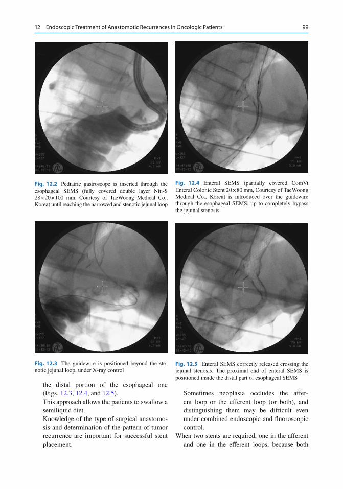
99
the distal portion of the esophageal one (Figs. 12.3 , 12.4 , and 12.5 ). This approach allows the patients to swallow a semiliquid diet. Knowledge of the type of surgical anastomo-sis and determination of the pattern of tumor recurrence are important for successful stent placement.
Sometimes neoplasia occludes the affer-ent loop or the efferent loop (or both), and distinguishing them may be diffi cult even under combined endoscopic and fl uoroscopic control.
When two stents are required, one in the afferent and one in the efferent loops, because both
Fig. 12.2 Pediatric gastroscope is inserted through the esophageal SEMS (fully covered double layer Niti-S 28 × 20 × 100 mm, Courtesy of TaeWoong Medical Co., Korea) until reaching the narrowed and stenotic jejunal loop
Fig. 12.3 The guidewire is positioned beyond the ste-notic jejunal loop, under X-ray control
Fig. 12.4 Enteral SEMS (partially covered ComVi Enteral Colonic Stent 20 × 80 mm, Courtesy of TaeWoong Medical Co., Korea) is introduced over the guidewire through the esophageal SEMS, up to completely bypass the jejunal stenosis
Fig. 12.5 Enteral SEMS correctly released crossing the jejunal stenosis. The proximal end of enteral SEMS is positioned inside the distal part of esophageal SEMS
12 Endoscopic Treatment of Anastomotic Recurrences in Oncologic Patients
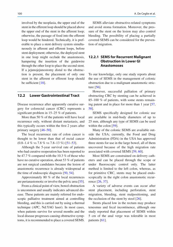
100
involved by the neoplasia, the upper end of the stent in the efferent loop should be placed above the upper end of the stent in the afferent loop; otherwise, the passage of food into the efferent loop would be hindered. Technically, it is pref-erable to place a stent delivery system simulta-neously in afferent and efferent loops, before stent deployment; otherwise, the deployed stent in one loop might occlude the anastomosis, hampering the insertion of the guidewire through the other loop to place the second stent. If a jejunojejunostomy distal to the obstruc-tion is present, the placement of only one stent in the afferent or efferent loop should be suffi cient [ 18 ].
12.2 Lower Gastrointestinal Tract
Disease recurrence after apparently curative sur-gery for colorectal cancer (CRC) represents a signifi cant problem in 15–25 % of patients.
More than 50 % of the patients will have local recurrence only, without distant metastasis, and this typically occurs within the fi rst 2 years after primary surgery [ 46 – 50 ].
The local recurrence rate of colon cancer is thought to be lower than that of rectal cancer (0.8–1.4 % vs 7.8 % vs 7.8–13 %) [ 51 – 53 ].
Although the 5-year survival rate of patients who had curative reoperation has been reported to be 47.7 % compared with the 10.3 % of those who have no curative operation, about 53 % of patients are not surgical candidates because the lesion of anastomotic recurrence is already widespread at the time of endoscopic diagnosis [ 50 , 54 ].
Approximately 80 % of the local recurrences are perianastomotic or involve the pelvic area [ 55 ].
From a clinical point of view, bowel obstruction is uncommon and usually indicates advanced dis-ease. These patients are mainly referred for endo-scopic palliative treatment aimed at controlling bleeding, and this is carried out by using a thermal technique (APC, Nd:YAG laser). In most cases, when patients survive for several months and the local disease progresses causing obstructive symp-toms, it is recommended to place a covered SEMS.
SEMS alleviate obstructive-related symptoms and avoid stoma formation. Moreover, the pres-sure of the stent on the lesion may also control bleeding. The possibility of placing a partially covered SEMS can be considered for the preven-tion of migration.
12.2.1 SEMS for Recurrent Malignant Obstruction in Lower GI Anastomoses
To our knowledge, only one study reports about the use of SEMS in the management of colonic obstruction due to a malignant anastomotic stric-ture [ 56 ].
However, successful palliation of primary obstructing CRC by stenting can be achieved in 85–100 % of patients, with some stents remain-ing patent and in place for more than 1 year [ 57 , 58 ].
SEMS specifi cally designed for colonic use are available in mid-body diameters of up to 25 mm, although any type of SEMS can be used within the colon [ 59 ].
Many of the colonic SEMS are available out-side the USA; currently, the Food and Drug Administration (FDA) in the USA has approved three stents for use in the large bowel, all of them uncovered because of the high migration rate associated with covered SEMS [ 59 , 60 ].
Most SEMS are constrained on delivery cath-eters and can be placed through the scope or under fl uoroscopic control only. The latter method is limited to the left colon, whereas, as for primitive CRC, stents may be placed endo-scopically in the right colon anastomotic recur-rence [ 26 , 37 ].
A variety of adverse events can occur after stent placement, including perforation, stent migration, bleeding, stent malpositioning, and the occlusion of the stent by stool [ 26 ].
Stents placed low in the rectum may produce tenesmus and fecal incontinence, although one study reported that placement of SEMS within 5 cm of the anal verge was tolerable in most patients [ 61 ].
A. De Ceglie et al.
101
12.2.2 Non-stent Treatments
12.2.2.1 Upper GI Tract All endoscopic therapies for palliation of malig-nant primitive or recurrent cancer, except stent-ing, require multiple sessions to maintain luminal patency with negative impact on patient quality of life. For this reason they have been progres-sively abandoned [ 62 ].
Therapy with Nd:YAG laser is expensive and not available in many centers [ 63 ].
Complications as perforation, bleeding, and tracheoesophageal fi stula have been reported in 4–20 % of patients [ 64 ].
At present laser therapy is not used, except for those patients with proximal esophageal lesions in whom stenting is not feasible [ 65 ].
An alternative to laser therapy is APC (argon plasma coagulator). Using APC in obstructive esophageal cancer, recanalization has been reported in 89 % of patients and perforation in 1–8 % of cases [ 66 , 67 ].
Photodynamic therapy is a nonthermal photo-chemical process of tumor ablation, technically easy to perform, with reported palliation of dysphagia in about 90 % of patients and a dysphagia- free interval ranging from 70 to 85 % [ 68 , 69 ].
Comparing PDT with laser therapy, rates per-foration reported are 1 % vs 7 % and time of palli-ation failure was 34 days vs 42 days, respectively.
PDT is a relatively expensive treatment modality, and side effects include esophageal stricture, Candida esophagitis, pleural effusion, and skin photosensitivity [ 65 ].
Brachytherapy may relieve dysphagia in patients unfi t for surgery. Randomized trials comparing stenting with brachytherapy show that both have been proven effective with few com-plications and similar cost [ 70 , 71 ].
However, dysphagia score improved more rapidly after stent placement than after brachy-therapy, but long-term relief of dysphagia was better after brachytherapy.
The choice between stent placement and brachytherapy as palliative treatment of dyspha-gia might be done depending on the patient’s life
expectancy: SEMS are indicated for patients with a short life expectancy, who require a rapid relief of dysphagia, while brachytherapy should be considered in patients with longer survival.
12.2.2.2 Lower GI Tract Endoscopic techniques other than stents for pallia-tion of recurrent anastomotic colorectal cancer are as follows: laser ablation and APC. Photodynamic therapy is not recommended due its side effects and the high complication rate [ 72 ].
Therapy with Nd:YAG laser is an effective treatment in restoring lumen patency , although the effect is not long lasting; benefi ts are also observed for palliation of bleeding and mucous discharge [ 59 ].
However, palliation becomes less effective as patients survive longer, dropping from 80–90 % initially to only 52 % at 6 months and 42 % at 12 months [ 73 ].
Disadvantages include the need for expensive equipment and the need to repeat procedures to maintain patency: each treatment session requires a relatively long time to complete ranging from 30 to 90 min with two to six sessions [ 74 ].
Serious complications (bleeding, perforation, severe pain, and formation of fi stula and abscess) occur in 10–15 % of patients [ 75 ].
APC has the same technically limitations as laser therapy.
As all these treatments require periodic reit-eration, they are not used anymore and have been supplanted by SEMS placement [ 72 ].
Conclusion
In patients with unresectable anastomotic malignant recurrence, SEMS represent a major breakthrough as they can rapidly restore recanalization of the gastrointestinal tract, may control bleeding, and avoid the reit-eration of endoscopic procedures. In patients with colorectal malignancies, they prevent the need of a surgical stoma, improving the qual-ity of life of these patients. The wide range of available SEMS requires an expert knowl-edge by the endoscopist in order to make the appropriate choice to obtain the best results
12 Endoscopic Treatment of Anastomotic Recurrences in Oncologic Patients
102
and minimize the onset of complications. The new conformable SEMS seems to offer a good opportunity in patients with angulated stric-tures, especially after total gastrectomy.
References
1. Iwanaga T, Koyama H, Furukawa H, Taniguchi H, Wada A, Tateishi R (1978) Mechanisms of late recur-rence after radical surgery for gastric carcinoma. Am J Surg 135:637–640
2. Monson JR, Donohue JH, McIlrath DC, Farnell MB, Ilstrup DM (1991) Total gastrectomy for advanced cancer. A worthwhile palliative procedure. Cancer 168:1863–1868
3. Del Piano M, Ballarè M, Montino F et al (2005) Endoscopy or surgery for malignant GI outlet obstruc-tion? Gastrointest Endosc 61:421–426
4. Jeong JY, Kim YJ, Han JK et al (2004) Palliation of anastomotic obstructions in recurrent gastric carci-noma with the use of covered metallic stents: clinical results in 25 patients. Surgery 135:171–177
5. Song GA, Kang DH, Kim TO et al (2007) Endoscopic stenting in patients with recurrent malignant obstruc-tion after gastric surgery: uncovered versus simulta-neously deployed uncovered and covered (double) self-expandable metal stents. Gastrointest Endosc 65:782–787
6. Dormann A, Meisner S, Verin N, Wenk Lang A (2004) Self-expanding metal stents for gastroduode-nal malignancies: systematic review of their clinical effectiveness. Endoscopy 36:543–550
7. Tong DK, Law S, Wong KH (2010) The use of self- expanding metallic stents (SEMS) is effective in symptom palliation from recurrent tumor after esophagogastrectomy for cancer. Dis Esophagus 23:660–665
8. Langer FB, Zacherl J (2007) Palliative endo-scopic interventions in esophageal cancer. Eur Surg 39:288–294
9. Siersema PD (2006) New developments in pal-liative therapy. Best Pract Res Clin Gastroenterol 20:959–978
10. Adler DG, Merwat SN (2006) Endoscopic approaches for palliation of luminal gastrointestinal obstruction. Gastroenterol Clin North Am 35:65–82
11. De Palma GD, Sivero L, Galloro G et al (1998) Endoscopic palliation of dysphagia due to anas-tomotic recurrences after esophageal surgery and total gastrectomy due to carcinoma. Minerva Chir 53:781–785
12. O'Connor A, Leyden J, McEntee G, MacMathuna P (2004) Palliative stenting of recurrent malignancy at gastrojejunostomy anastomotic sites. Ir J Med Sci 173:219–220
13. Yates MR 3rd, Morgan DE, Baron TH (1998) Palliation of malignant gastric and small intesti-nal strictures with self-expandable metal stents. Endoscopy 30:266–272
14. Lee JM, Han YM, Lee SY, Kim CS, Yang DH, Lee SO (2001) Palliation of postoperative gastrointestinal anastomotic malignant strictures with fl exible cov-ered metallic stents: preliminary results. Cardiovasc Intervent Radiol 24:25–30
15. Siersema PD, Schrauwen SL, van Blankenstein M et al (2001) Self-expanding metal stents for compli-cated and recurrent esophagogastric cancer. Gastrointest Endosc 54:579–586
16. Pérez-Roldán F, González-Carro P, Legaz-Huidobro M et al (2006) Esophagoenteral stents in patients with recurrent gastric adenocarcinoma. Rev Esp Enferm Dig 98:341–349
17. Kim HJ, Park JY, Bang S, Park SW, Lee YC, Song SY (2009) Self-expandable metal stents for recurrent malignant obstruction after gastric surgery. Hepatogastroenterology 56:914–917
18. Song HY, Kim TH, Choi EK et al (2007) Metallic stent placement in patients with recurrent cancer after gas-trojejunostomy. J Vasc Interv Radiol 18:1538–1546
19. Yang ZQ, Song HY, Kim JH et al (2007) Covered stent placement in patients with recurrent cancer after a Billroth I reconstruction. J Vasc Interv Radiol 18:1533–1537
20. Cho YK, Kim SW, Nam KW et al (2009) Clinical out-comes of self-expandable metal stents in palliation of malignant anastomotic strictures caused by recurrent gastric cancer. World J Gastroenterol 15:3523–3527
21. Naso P, Russo EP, Aprile G, Bonanno G, Trama G, Russo A (2005) Esophacoil for palliation of recurrent malignant esophago-jejunal anastomotic stricture. Case reports. Ann Ital Chir 76:89–92
22. Kim J, Choi IJ, Kim CG et al (2011) Self-expandable metallic stent placement for malignant obstruction in patients with locally recurrent gastric cancer. Surg Endosc 25:1505–1513
23. Kim JH, Song HY, Shin JH et al (2007) Anastomotic recurrence of gastric cancer after total gastrectomy with esophagojejunostomy: palliation with covered expandable metallic stents. J Vasc Interv Radiol 18:964–969
24. Solt J, Grexa E (2004) Treatment of recurrent malig-nant obstruction with a flexible covered metal stent after gastric surgery. Gastrointest Endosc 60:813–817
25. Lopera JE, Brazzini A, Gonzales A, Castaneda- Zuniga WR (2004) Gastroduodenal stent placement: current status. Radiographics 24:1561–1573
26. Baron TH (2001) Expandable metal stents for the treatment of cancerous obstruction of the gastrointes-tinal tract. N Engl J Med 344:1681–1687
27. Nicholson DA, Haycox A, Kay CL, Rate A, Attwood S, Bancewicz J (1999) The cost effectiveness of metal oesophageal stenting in malignant disease compared with conventional therapy. Clin Radiol 54:212–215
A. De Ceglie et al.
103
28. Singhvi R, Abbasakoor F, Manson JM (2000) Insertion of self-expanding metal stents for malignant dysphagia: assessment of a simple endoscopic method. Ann R Coll Surg Engl 82:243–248
29. Yim HB, Jacobson BC, Saltzman JR et al (2001) Clinical outcome of the use of enteral stents for pallia-tion of patients with malignant upper GI obstruction. Gastrointest Endosc 53:329–332
30. Jung GS, Song HY, Kang SG et al (2000) Malignant gastroduodenal obstructions: treatment by means of a covered expandable metallic stent-initial experience. Radiology 216:758–763
31. Kim GH, Kang DH, Lee DH et al (2004) Which types of stent, uncovered or covered, should be used in gastric outlet obstructions? Scand J Gastroenterol 39:1010–1014
32. Adler DG, Baron TH (2002) Endoscopic palliation of malignant gastric outlet obstruction using self- expanding metal stents: experience in 36 patients. Am J Gastroenterol 97:72–78
33. Holt AP, Patel M, Ahmed MM (2004) Palliation of patients with malignant gastroduodenal obstruction with self-expanding metallic stents: the treatment of choice? Gastrointest Endosc 60:1010–1017
34. Conio M, Repici A, Battaglia G et al (2007) A ran-domized prospective comparison of self-expandable plastic stents and partially covered self-expand-able metal stents in the palliation of malignant esophageal dysphagia. Am J Gastroenterol 102:2667–2677
35. Conio M, De Ceglie A (2013) Esophageal prosthe-ses in self-expandable stents in the gastrointestinal tract. In: Kozarek R, Baron T, Song H-Y (eds) Self- expandable stents in the gastrointestinal tract, vol XII. Springer, New York/Heidelberg/Dordrecht/London, pp 73–89
36. van Hooft JE, van Montfoort ML, Jeurnink SM et al (2011) Safety and effi cacy of a new non- foreshortening nitinol stent in malignant gastric outlet obstruction (DUONITI study): a prospective, multicenter study. Endoscopy 43:671–675
37. Homs MY, Siersema PD (2007) Stents in the GI tract. Expert Rev Med Devices 4:741–752
38. Conio M, Blanchi S, Filiberti R et al (2007) A modi-fi ed self-expanding Niti-S stent for the management of benign hypopharyngeal strictures. Gastrointest Endosc 65:714–720
39. Verschuur EM, Kuipers EJ, Siersema PD (2007) Esophageal stents for malignant strictures close to the upper esophageal sphincter. Gastrointest Endosc 66:1082–1090
40. Montgomery WW (1978) Salivary bypass tube. Ann Otol Rhinol Laryngol 87:159–162
41. John DG, Kwok FH, Van Hasselt CA (1992) The montgomery T-tube in terminal care. Am J Otolaryngol 13:45–47
42. Petrou M, Goldstraw P (1994) The management of tracheo-bronchial obstruction: a review of endoscopic techniques. Eur J Cardiothorac Surg 8:436–441
43. Yamamoto R, Tada H, Kishi A, Tojo T, Asada H (2002) Double stent for malignant combined esophago- airway lesions. Jpn J Thorac Cardiovasc Surg 50:1–5
44. Conio M, De Ceglie A, Blanchi S, Fisher DA (2010) Esophageal strictures, tumors, and fi stulae: stents for primary esophageal cancer. Tech Gastrointest Endosc 12:191–202
45. Park KB, Do YS, Kang WK et al (2001) Malignant obstruction of gastric outlet and duodenum: palliation with fl exible covered metallic stents. Radiology 219:679–683
46. Sagar PM, Pemberton JH (1996) Surgical manage-ment of locally recurrent rectal cancer. Br J Surg 83:293–304
47. MIchelassi F, Vannucci L, Ayala JJ, Chappel R, Goldberg R, Block GE (1990) Local recurrence after curative resection of colorectal adenocarcinoma. Surgery 108:787–793
48. Malcolm AW, Perencevich NP, Olson RM et al (1981) Analysis of recurrence patterns following curative resection for carcinoma of the colon and rectum. Surg Gynecol Obstet 152:131–136
49. Kraemer M, Wiratkapun S, Seow-Choen F et al (2001) Stratifying risk factors for follow-up: a comparison of recurrent and non recurrent colorectal cancer. Dis Colon Rectum 44:815–821
50. Yun HR, Lee LJ, Park JH et al (2008) Local recur-rence after curative resection in patients with colon and rectal cancers. Int J Colorectal Dis 23:1081–1087
51. Hardy KJ, Cuthbertson AM, Hughes ES (1971) Suture-line neoplastic recurrence following large- bowel resection. Aust N Z J Surg 41:44–46
52. Lee YT (1996) Local and regional recurrence of car-cinoma of the colon and rectum: II. Factors relating to operative technique. Surg Oncol 5:1–13
53. Pietra N, Sarli L, Costi R, Ouchemi C, Grattarola M, Peracchia A (1998) Role of follow-up in management of local recurrences of colorectal cancer: a prospective, randomized study. Dis Colon Rectum 41:1127–1133
54. Nakajima S (2001) The effi cacy of the EUS for the detection of recurrent disease in the anastomosis of colon. Diagn Ther Endosc 7:149–158
55. Hoffman JP, Riley L, Carp NZ, Litwin S (1993) Isolated locally recurrent rectal cancer: a review of incidence, presentation, and management. Semin Oncol 20:506–519
56. Bashir RM, Fleischer DE, Stahl TJ, Benjamin SB (1996) Self-expandable nitinol coil stent for manage-ment of colonic obstruction due to a malignant anas-tomotic stricture. Gastrointest Endosc 44:497–501
57. Fernández Lobato R, Pinto I, Paul L et al (1999) Self- expanding prostheses as a palliative method in treat-ing advanced colorectal cancer. Int Surg 84:159–162
58. Camúñez F, Echenagusia A, Simó G, Turégano F, Vázquez J, Barreiro-Meiro I (2000) Malignant colorectal obstruction treated by means of self- expanding metallic stents: effectiveness before sur-gery and in palliation. Radiology 216:492–497
12 Endoscopic Treatment of Anastomotic Recurrences in Oncologic Patients
104
59. Baron TH (2009) Interventional palliative strategies for malignant bowel obstruction. Curr Oncol Rep 11:293–297
60. Park S, Cheon JH, Park JJ et al (2010) Comparison of effi cacies between stents for malignant colorec-tal obstruction: a randomized, prospective study. Gastrointest Endosc 72:304–310
61. Song HY, Kim JH, Kim KR et al (2008) Malignant rectal obstruction within 5 cm of the anal verge: is there a role for expandable metallic stent placement? Gastrointest Endosc 68:713–720
62. Rupinski M, Zagorowicz E, Regula J et al (2011) Randomized comparison of three palliative regimens including brachytherapy, photodynamic therapy, and APC in patients with malignant dysphagia (CONSORT 1a) (Revised II). Am J Gastroenterol 106:1612–1620
63. Dallal HJ, Smith GD, Grieve DC, Ghosh S, Penman ID, Palmer KR (2001) A randomized trial of thermal ablative therapy versus expandable metal stents in the palliative treatment of patients with esophageal carci-noma. Gastrointest Endosc 54:549–557
64. Gevers AM, Macken E, Hiele M, Rutgeerts P (1998) A comparison of laser therapy, plastic stents, and expandable metal stents for palliation of malignant dysphagia in patients without a fi stula. Gastrointest Endosc 48:383–388
65. Adler DG, Baron TH (2002) Endoscopic palliation of colorectal cancer. Hematol Oncol Clin North Am 16:1015–1029
66. Eriksen JR (2002) Palliation of non-resectable carci-noma of the cardia and oesophagus by argon beam coagulation. Dan Med Bull 49:346–349
67. Heindorff H, Wøjdemann M, Bisgaard T, Svendsen LB (1998) Endoscopic palliation of inoperable cancer
of the oesophagus or cardia by argon electrocoagula-tion. Scand J Gastroenterol 33:21–23
68. Luketich JD, Christie NA, Buenaventura PO, Weigel TL, Keenan RJ, Nguyen NT (2000) Endoscopic photodynamic therapy for obstructing esophageal cancer: 77 cases over a 2-year period. Surg Endosc 14:653–657
69. Litle VR, Luketich JD, Christie NA et al (2003) Photodynamic therapy as palliation for esophageal cancer: experience in 215 patients. Ann Thorac Surg 76:1687–1693
70. Homs MY, Steyerberg EW, Eijkenboom WM et al (2004) Single-dose brachytherapy versus metal stent placement for the palliation of dysphagia from oesophageal cancer: multicentre randomised trial. Lancet 364:1497–1504
71. Bergquist H, Wenger U, Johnsson E et al (2005) Stent insertion or endoluminal brachytherapy as pal-liation of patients with advanced cancer of the esophagus and gastroesophageal junction. Results of a randomized, controlled clinical trial. Dis Esophagus 18:131–139
72. Kimmey MB (2004) Endoscopic methods (other than stents) for palliation of rectal carcinoma. J Gastrointest Surg 8:270–273
73. Van Cutsem E, Boonen A, Geboes K et al (1989) Risk factors which determine the long term outcome of Neodymium-YAG laser palliation of colorectal carcinoma. Int J Colorectal Dis 4:9–11
74. Dekovich AA (2009) Endoscopic treatment of colonic obstruction. Curr Opin Gastroenterol 25:50–54
75. Frech EJ, Adler DG (2007) Endoscopic therapy for malignant bowel obstruction. J Support Oncol 5:303–310, 319
A. De Ceglie et al.
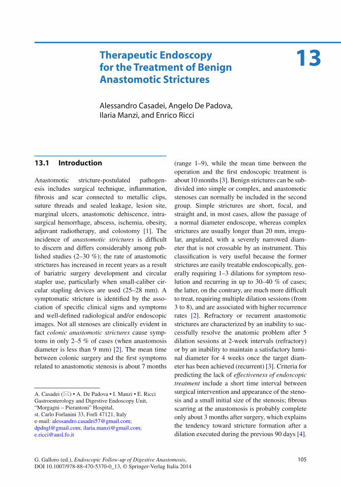
105G. Galloro (ed.), Endoscopic Follow-up of Digestive Anastomosis, DOI 10.1007/978-88-470-5370-0_13, © Springer-Verlag Italia 2014
13.1 Introduction
Anastomotic stricture-postulated pathogen-esis includes surgical technique, infl ammation, fi brosis and scar connected to metallic clips, suture threads and sealed leakage, lesion site, marginal ulcers, anastomotic dehiscence, intra-surgical hemorrhage, abscess, ischemia, obesity, adjuvant radiotherapy, and colostomy [ 1 ]. The incidence of anastomotic strictures is diffi cult to discern and differs considerably among pub-lished studies (2–30 %); the rate of anastomotic strictures has increased in recent years as a result of bariatric surgery development and circular stapler use, particularly when small-caliber cir-cular stapling devices are used (25–28 mm). A symptomatic stricture is identifi ed by the asso-ciation of specifi c clinical signs and symptoms and well- defi ned radiological and/or endoscopic images. Not all stenoses are clinically evident in fact colonic anastomotic strictures cause symp-toms in only 2–5 % of cases (when anastomosis diameter is less than 9 mm) [ 2 ]. The mean time between colonic surgery and the fi rst symptoms related to anastomotic stenosis is about 7 months
(range 1–9), while the mean time between the operation and the fi rst endoscopic treatment is about 10 months [ 3 ]. Benign strictures can be sub-divided into simple or complex, and anastomotic stenoses can normally be included in the second group. Simple strictures are short, focal, and straight and, in most cases, allow the passage of a normal diameter endoscope, whereas complex strictures are usually longer than 20 mm, irregu-lar, angulated, with a severely narrowed diam-eter that is not crossable by an instrument. This classifi cation is very useful because the former strictures are easily treatable endoscopically, gen-erally requiring 1–3 dilations for symptom reso-lution and recurring in up to 30–40 % of cases; the latter, on the contrary, are much more diffi cult to treat, requiring multiple dilation sessions (from 3 to 8), and are associated with higher recurrence rates [ 2 ]. Refractory or recurrent anastomotic strictures are characterized by an inability to suc-cessfully resolve the anatomic problem after 5 dilation sessions at 2-week intervals (refractory) or by an inability to maintain a satisfactory lumi-nal diameter for 4 weeks once the target diam-eter has been achieved (recurrent) [ 3 ]. Criteria for predicting the lack of effectiveness of endoscopic treatment include a short time interval between surgical intervention and appearance of the steno-sis and a small initial size of the stenosis; fi brous scarring at the anastomosis is probably complete only about 3 months after surgery, which explains the tendency toward stricture formation after a dilation executed during the previous 90 days [ 4 ].
A. Casadei (*) • A. De Padova • I. Manzi • E. Ricci Gastroenterology and Digestive Endoscopy Unit , “Morgagni – Pierantoni” Hospital , st. Carlo Forlanini 33 , Forlì 47121 , Italy e-mail: [email protected]; [email protected]; [email protected]; [email protected]
13 Therapeutic Endoscopy for the Treatment of Benign Anastomotic Strictures
Alessandro Casadei , Angelo De Padova , Ilaria Manzi , and Enrico Ricci
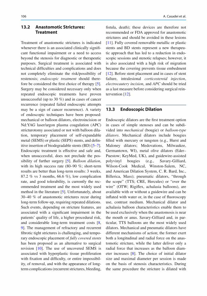
106
13.2 Anastomotic Strictures: Treatment
Treatment of anastomotic strictures is indicated whenever there is an associated clinically signifi -cant functional impairment or a need to access beyond the stenosis for diagnostic or therapeutic purposes. Surgical treatment is associated with technical diffi culties and complications and does not completely eliminate the risk/possibility of restenosis; endoscopic treatment should there-fore be considered the fi rst choice of therapy [ 5 ]. Surgery may be considered necessary only when repeated endoscopic treatments have proven unsuccessful (up to 30 %) and in cases of cancer recurrence (repeated failed endoscopic attempts may be a sign of cancer recurrence). A variety of endoscopic techniques have been proposed: mechanical or balloon dilators, electroincision or Nd:YAG laser/argon plasma coagulation (APC) stricturotomy associated or not with balloon dila-tion, temporary placement of self-expandable metal (SEMS) or plastic (SEPS) stents, and defi n-itive insertion of biodegradable stents (BD) [ 5 – 7 ]. Endoscopic treatment is effective and safe and, when unsuccessful, does not preclude the pos-sibility of further surgery [ 5 ]. Balloon dilation , with its high success rate (80–90 %; short- term results are better than long-term results: 3 weeks, 87.2 % vs 3 months, 66.6 %), low complication rate, and good tolerability, is currently the rec-ommended treatment and the most widely used method in the literature [ 5 ]. Unfortunately, about 30–40 % of anastomotic strictures recur during long-term follow-up, requiring repeated dilations. Such events, depending on stricture features, are associated with a signifi cant impairment in the patients’ quality of life, a higher procedural risk, and considerable long-term treatment costs [ 8 , 9 ]. The management of refractory and recurrent fi brotic tight strictures is challenging, and tempo-rary endoscopic placement of fully covered stents has been proposed as an alternative to surgical revision [ 10 ]. The use of uncovered SEMS is associated with hyperplastic tissue proliferation with fi xation and diffi culty, or entire impossibil-ity, of removal, and with the appearance of long-term complications ( recurrent strictures, bleeding,
fi stula, death); these devices are therefore not recommended or FDA approved for anastomotic strictures and should be avoided in these lesions [ 11 ]. Fully covered removable metallic or plastic stents and BD stents represent a new therapeu-tic approach that has led to a reduction in endo-scopic sessions and stenotic relapses; however, it is also associated with a high risk of migration because the covering prevents tissue embedment [ 12 ]. Before stent placement and in cases of stent failure, intralesional corticosteroid injection , electrocautery incision , and APC should be tried as a last measure before considering surgical rein-tervention [ 12 ].
13.3 Endoscopic Dilation
Endoscopic dilators are the fi rst treatment option in cases of simple stenoses and can be subdi-vided into mechanical (bougie ) or balloon - type dilators . Mechanical dilators include bougies fi lled with mercury or tungsten (e.g., Hurst and Maloney dilators; Medovations, Milwaukee, Germantown, WI), metal olive dilators (Eder–Puestow; KeyMed, UK), and guidewire-assisted polyvinyl bougies (e.g., Savary-Gilliard, Wilson- Cook Medical, Winston-Salem, NC, and American Dilation System, C. R. Bard, Inc., Billerica, Mass); pneumatic dilators, “through the scope” (TTS; CRE, Hercules) or “over the wire” (OTW; Rigifl ex, achalasia balloons), are available with or without a guidewire and can be infl ated with water or, in the case of fl uoroscopy use, contrast medium. Mechanical dilator and achalasia balloon characteristics enable them to be used exclusively when the anastomosis is near the mouth or anus. Savary-Gilliard and, in par-ticular, TTS balloons are the most widely used dilators. Mechanical and pneumatic dilators have different mechanisms of action; the former exert both a longitudinal and radial force on the anas-tomotic stricture, while the latter deliver only a radial force that increases as the balloon diam-eter increases [ 8 ]. The choice of initial dilator size and maximal diameter per session is made on the basis of stenosis characteristics. During the same procedure the stricture is dilated with
A. Casadei et al.
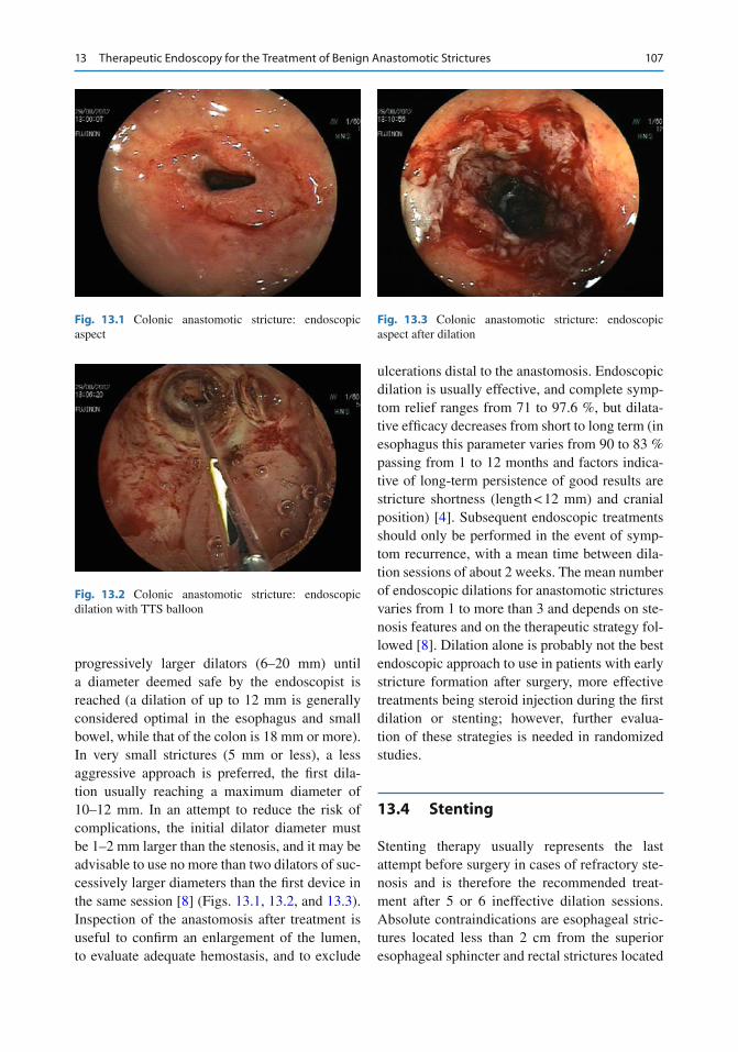
107
progressively larger dilators (6–20 mm) until a diameter deemed safe by the endoscopist is reached (a dilation of up to 12 mm is generally considered optimal in the esophagus and small bowel, while that of the colon is 18 mm or more). In very small strictures (5 mm or less), a less aggressive approach is preferred, the fi rst dila-tion usually reaching a maximum diameter of 10–12 mm. In an attempt to reduce the risk of complications, the initial dilator diameter must be 1–2 mm larger than the stenosis, and it may be advisable to use no more than two dilators of suc-cessively larger diameters than the fi rst device in the same session [ 8 ] (Figs. 13.1 , 13.2 , and 13.3 ). Inspection of the anastomosis after treatment is useful to confi rm an enlargement of the lumen, to evaluate adequate hemostasis, and to exclude
ulcerations distal to the anastomosis. Endoscopic dilation is usually effective, and complete symp-tom relief ranges from 71 to 97.6 %, but dilata-tive effi cacy decreases from short to long term (in esophagus this parameter varies from 90 to 83 % passing from 1 to 12 months and factors indica-tive of long-term persistence of good results are stricture shortness (length < 12 mm) and cranial position) [ 4 ]. Subsequent endoscopic treatments should only be performed in the event of symp-tom recurrence, with a mean time between dila-tion sessions of about 2 weeks. The mean number of endoscopic dilations for anastomotic strictures varies from 1 to more than 3 and depends on ste-nosis features and on the therapeutic strategy fol-lowed [ 8 ]. Dilation alone is probably not the best endoscopic approach to use in patients with early stricture formation after surgery, more effective treatments being steroid injection during the fi rst dilation or stenting; however, further evalua-tion of these strategies is needed in randomized studies.
13.4 Stenting
Stenting therapy usually represents the last attempt before surgery in cases of refractory ste-nosis and is therefore the recommended treat-ment after 5 or 6 ineffective dilation sessions. Absolute contraindications are esophageal stric-tures located less than 2 cm from the superior esophageal sphincter and rectal strictures located
Fig. 13.1 Colonic anastomotic stricture: endoscopic aspect
Fig. 13.2 Colonic anastomotic stricture: endoscopic dilation with TTS balloon
Fig. 13.3 Colonic anastomotic stricture: endoscopic aspect after dilation
13 Therapeutic Endoscopy for the Treatment of Benign Anastomotic Strictures
108
less than 5 cm from the anal sphincter [ 13 ]. Stent types include partially covered SEMS and fully covered SEMS and fully covered SEPS and BD stents . In benign diseases stents are not intended to be permanent and safe removal is an essential feature [ 14 ]. The technical success rate of stent placement ranges from 85 to 100 % [ 14 , 15 ]. Dilation prior to stenting should be avoided (high risk of perforation), and it is not recommended to cross the stent with the endoscope at the end of the procedure (high risk of device dislocation). Endoscopic follow-up after stent placement is not well defi ned: some authors perform endos-copy after 4–6 weeks or earlier if complications occur, while others propose surveillance with endoscopy or radiology every 2 weeks, with stent removal in the event of stricture [ 14 , 15 ]. The optimal duration of this treatment has not yet been established, and recommendations are based on data from small case series. Some authors report that 4–8 weeks after placement is the optimal time for removal [ 16 ]; during extrac-tion reactive tissue in- and overgrowth can deter-mine major and minor adverse events which subsequently require surgery [ 17 ]. Metal stents, long stenting duration, large-diameter devices, and high-expansion force are all associated with high rates of reactive tissue; this problem is infrequent with BD stents, and thus the develop-ment of new covered BD stents could further lower the rate of tissue ingrowth [ 18 , 19 ]. Stent migration is mainly related to device features but is also linked to stenosis peculiarities, and short anastomotic strictures are stent migration-pro-moting factors. Attempts to prevent stent migra-tion by clipping the device to the intestinal wall have shown poor results; in the large bowel, cov-ered stents almost uniformly migrate even when fi xation techniques are used, and so no dedicated covered colonic stents are currently commer-cially available in the USA [ 13 ]. Dilation or placement of a new stent after device explanta-tion is sometimes necessary (up to 21 % in some studies); in cases of ineffective endoscopic treat-ment surgery is required (up to 50 % in some case series) [ 14 ]. Stent placement provides immediate symptom relief and may obviate the need for surgery in selected patients, but data on
long-term patency and safety of this technique are lacking [ 14 ].
Metal stents . The main limitation of SEMS is tissue neoformation that causes recurrent symp-toms in >15 % of patients, a high rate of compli-cations, such as perforation and obstruction, and precludes easy stent removal; although hyper-plastic tissue reaction is lower with covered stents, it has also been reported to occur with these devices [ 14 ]. With regard to tissue reaction , partially covered SEMS show low migration (overall 12 %), can be diffi cult and traumatic to remove, and should be avoided because of the high risk of complications (recurrent stricture formation and mucosal damage, bleeding, or per-foration after removal); conversely, fully covered SEMS are easy to remove but have a higher migration rate than the former. SEMS are thus not recommended or approved for benign stric-tures by the US Food and Drug Administration [ 15 ]. Some recent studies have examined new coated or temperature-sensitive nitinol stents which show encouraging technical and clinical results and are easy to remove [ 16 ].
Plastic stents . SEPS were recently introduced to reduce the risk of hyperplastic tissue growth associated with metal stents. They are effective and safe compared to the metal devices and can be easily removed, but migration occurs more frequently, often when the stricture improves [ 18 ]. Polyfl ex stent (Boston Scientifi c Corp., Natick, Massachusetts, USA), a self-expanding plastic polyester silicone-coated stent, is particu-larly effective because the radial expansive force is similar to that of SEMS and the silicone coat-ing resists tissue ingrowth and hyperplasia [ 18 ]. The Polyfl ex esophageal stent is currently the only FDA-approved retrievable stent marketed in the USA. Esophageal stenosis such as that found in refractory benign strictures is one of the FDA- approved indications for Polyfl ex stents [ 18 ]. SEPS stenting time is not well defi ned: although some authors consider 6 weeks as the optimal duration (X), a longer period probably results in a higher success rate in cases of refractory stric-tures. It has therefore been hypothesized that the optimal stenting time depends on stricture fea-tures. The role of SEPS is still widely debated
A. Casadei et al.
109
because of confl icting results from various stud-ies, with success rates ranging from 0 to 94 % and an unexpected high rate of complications described in some cases series but not in others (in the majority of studies SEPS appear to be relatively safe) [ 20 ].
Biodegradable stents . BD stents would appear to eliminate the risk of mucosal hyperplastic reaction, have a shorter patency, show low migra-tion, and do not require subsequent removal: the prolonged dilatory effect before absorption and the progressive degradation could represent a favorable solution for refractory anastomotic strictures (stent degradation is about 3–4 months and the radial force remains around initial values for the fi rst 5 weeks, decreases to about 2/3 of the initial force after 7 weeks and further reduces to about 1/2 of the starting force at week 9) [ 21 ].
These stents often need an anastomotic stric-ture pre-dilation of up to 12–20 mm to facilitate the introduction of the delivery system and to ensure adequate device expansion; however, pre- dilation increases the risk of stent migration. Several authors advise endoscopy and fl uoros-copy to be repeated at 1-month intervals in order to monitor stent position, patency, and degrada-tion; in some studies clinical and radiographic follow-up is performed 1 week after insertion and again 1 month later, whereas endoscopic evaluation is only performed in the presence of symptoms [ 7 ]. Although mucosal hyperplasia after BD stenting can cause reobstruction, this problem responds well to single-balloon dilation and resolves completely after stent degradation [ 7 ]. Anastomotic stricture recurrence after com-plete stent degradation is easily resolved, treat-ment based on balloon dilation of up to 18–20 mm (usually only one session is needed to obtain an adequate lumen size) or on a new BD stent place-ment [ 22 ]. The main disadvantages of BD stents are that they can only be placed in areas close to the mouth or the anus [ 23 ], the devices tendency to migrate if they are not fi xed to the wall, and that a second stent after degradation in more long-standing strictures may become necessary. Even if anastomotic stricture long-term patency after BD stents placement is better than after dilation, there is a lack of published series; prob-
ably a longer BD stent duration would be associ-ated with a higher long - term success rate , and consequently new devices with delayed degrada-tion process could well give a more prolonged benefi t. The effi cacy of steroid injections in improving the effects of BD stents warrants investigation [ 24 , 25 ].
13.5 Incisional Therapy, Argon Plasma Coagulation, Intralesional Steroid Injection
There are no standard recommendations for the management of persistent or recurrent strictures resistant to dilation, and thus various techniques have been adopted: incisional therapy and Nd:YAG laser/APC, with or without dilation, intralesional steroid injection combined with dilation, stenting, and fi nally endoscopic surgery [ 6 ]. Incisional therapy , performed using a needle knife or polypectomy snare and evaluated in small cases series, is safe and particularly effec-tive in short strictures (<10 mm). In one study it was used in patients previously treated with dila-tion therapy or not and showed a success rate of 87.5 % after only one session [ 26 ]. This treat-ment has been successfully used together with balloon dilation or APC in uncontrolled studies, showing a good safety profi le [ 6 ]. A combination of APC and Savary dilation appears to be poten-tially useful in strictures that do not respond after three sessions of mechanical dilation [ 26 ]. In one study on 10 patients, neodymium: yttrium–alumi-num–garnet laser used in association with bal-loon dilation determined technical and clinical success without recurrence or complications in 90 % of patients with a median follow-up of 82 months. In various small, uncontrolled studies, some authors treated recurrent anastomotic stric-tures with steroid injection followed by dilation ( intralesional steroid injections increase the effect of dilation by reducing collagen formation through the local inhibition of the infl ammatory response) [ 27 ]; steroid treatment combined with dilation has an uncertain benefi t for the initial treatment of benign strictures [ 27 ].
13 Therapeutic Endoscopy for the Treatment of Benign Anastomotic Strictures
110
13.6 Endoscopic Treatment in Different Anastomotic Sites
13.6.1 Esophagus
Anastomotic strictures after esophageal resection occur in 5–46 % of patients; although there is no universal consensus, an esophageal anastomosis is normally considered stenotic when the diame-ter is less than 12 mm. Proximal strictures are particularly diffi cult to treat and are at a higher risk of complications [ 26 ]. The success rate of dilation therapy ranges from 70 to 90 %, but 40 % of patients require more than 3 dilation ses-sions; the risk of relapse can be reduced by per-forming a dilation of 18–20 mm, but a diameter of 12 mm may be suffi cient to mitigate dysphagia to solids [ 26 ]. Intralesional steroid injections associated with balloon dilations increase the maximum dilation diameter achieved and reduce the severity of symptoms (signifi cant improve-ment in the dysphagia score: 0.63 ± 0.59 vs 2.42 ± 0.5, P < 0.001), the number of dilation ses-sions required, and the rate of recurrence [ 27 ]. The clinical success rate of fully covered SEMS is less than 50 % [ 28 , 29 ]; a recent meta-analysis highlighted an improvement in dysphagia in 46 % of patients with benign strictures after placement of various removable stents, and another pooled analysis with Polyfl ex stent place-ment showed a clinical success rate of 52 % [ 18 , 28 ]. In the literature more than 15 papers have reported promising results for Polyfl ex stents placed for benign esophageal disorders: technical success 96 % (range 75–100 %), clinical success 89 % (range 69–100 %), and migration 27 % (range 7–57 %) [ 18 ]. A number of studies have tempered initial enthusiasm regarding SEPS by reporting high stent migration (62.1 %), a low rate of long-term improvement after stent removal (17 %), the need for new stent place-ment (55 %), and dysphagia relapse (70 %) [ 18 ]. A recent review evaluated a total of 10 studies on 130 patients with benign esophageal strictures treated with SEPS placement (49 anastomotic strictures, 39 %): technical success 98 %, clinical success 52 %, early migration with fully covered stents 24 %, need for endoscopic reinterventions
21 %, and major complications 9 % [ 18 ]. Only a small number of studies have focused on the use of BD stents , reporting complete relief from dys-phagia in 40–60 % of cases [ 19 ]; however, BD stents are “Conformité Européenne” marked, certifi ed to conform to European regulations for the treatment of patients with refractory benign esophageal stricture. One study investigated the effi cacy and safety of a BD stent (Ella-BD stent, Ella-CS, s.r.o., Hradec Kralove, Czech Republic) in 21 patients with refractory benign esophageal strictures, showing that this treatment is safe and effective: technical success 100 %, stent migra-tion 9.5 %, recurrent dysphagia caused by stent obstruction secondary to hyperplastic tissue in- and overgrowth 5 %, clinical success after a fol-low- up of 6 months 45 %, and major complications 0 % [ 29 ]. Another recent study evaluated safety and long-term effi cacy of the same stent in 28 patients with refractory benign esophageal stric-ture previously treated with multiple dilations associated or not with nonbiodegradable stent-ing; total technical success was 93 % and 13 patients were treated with sequential BD stent placement (median 3, range 2–8) showing that sequential stenting, in selected cases, may be a valid option to avoid serial dilations or surgery [ 30 ]. In cases of anastomotic strictures located near the upper esophageal sphincter, the use of stents has been associated with an extremely high rate of complications (almost 100 %), suggesting that dilation or incisional therapy may be better alternatives. Some esophageal stenoses are par-ticularly complex making it very diffi cult to iden-tify the true lumen of the anastomosis; in such cases an endoscopic “rendezvous” technique, i.e., combined antegrade and retrograde dilation, can be applied through a gastrostomy or jejunos-tomy to reduce the risk of a false route.
13.6.2 Stomach and Small Bowel
Anastomotic strictures associated with bariatric surgery are now the most frequent indications for endoscopic treatment (estimated rate of postop-erative stricture formation following gastroplasty is about 2 %, while that for Roux-en-Y gastric
A. Casadei et al.
111
bypass [RYGB] is around 3–27 %) [ 31 ]. The most common region of anastomotic stricture is the site of gastrojejunostomy, and the mean time to diagnosis after surgery is generally 3 months [ 31 ]. Gastrojejunostomy should have a diameter of 10–12 mm: in case of higher diameter surgical treatment executed is useless, while in case of lesser diameter problems of canalization may arise. Endoscopic stricture control is achieved in 95–100 % of cases; the best endoscopic tech-nique remains to be defi ned, but pneumatic dila-tion is the most widely used method, and many endoscopists prefer to use TTS balloons, dilating them to at least 15 mm in the fi rst session in order to decrease the risk of recurrence [ 32 ]. As the optimal diameter of the pylorus is approximately 12 mm, balloons with a smaller diameter, e.g., such as 10, 11, or 12 mm, may be suffi cient [ 32 ]. Pneumatic dilation is an effective and safe treat-ment strategy with a high overall success rate; the number of dilations required to successfully treat a gastrojejunal stricture is unknown, but many reviews have reported that the majority of patients require at least two dilation sessions [ 32 – 34 ]. Balloon dilation can produce desirable results in the short term (symptom improvement following the initial dilation is observed in 58–93 % of patients), but further research is needed to identify the endoscopic treatment modality that results in long-term success [ 33 , 34 ]. Savary dilation is also effective and safe: 100 % clinical success has been achieved using a maximum diameter of 12.8 mm and performing endoscopies 1–2 weeks after each session, the majority of patients requiring only one (46 %) or two dilations (50 %) and not experiencing com-plications [ 35 ]. Mechanical dilation may be use-ful in case of pneumatic failure; using Savary with size of 15–18 mm in anastomotic strictures treated unsuccessfully with initial pneumatic dilation, clinical success is 100 % and complica-tion rate is low (1.6 %) [ 33 ]. Diathermy com-bined with APC has been used in limited cases with good results (only one treatment session to gain long-term recanalization in 92 % of patients) [ 26 ]. The use of stenting in post-RYGB anasto-motic strictures is not particularly frequent or useful because of the high stent migration rate
(about 60 % of cases) whose occurrence is related to stenosis length and small bowel peristalsis [ 35 ]. A study has evaluated the clinical effi cacy and safety of balloon dilation and stent place-ment in 63 patients undergoing treatment for early benign anastomotic strictures after gastric surgery: clinical success was achieved in 89 % of patients after a single-balloon dilation (49 %), multiple-balloon dilations (32 %), and stent placement (8 %), highlighting that pneumatic dilation is safe and effective and that stenting can be effective in selected refractory patients [ 35 ]. The development of double - balloon endoscopy has rendered possible the endoscopic therapy of anastomoses located in sites which in the past could only be treated by surgeons; various endo-scopic treatments are currently possible for the small intestine, and stent placement is also feasi-ble, depending on the lesion site [ 36 , 37 ]. As the small intestine wall is very thin, great care must be taken during endoscopic therapy to avoid complications such as bleeding and perforation. Although endoscopic balloon dilation may be taken into consideration as the fi rst therapeutic option, a single session is generally not suffi cient and symptoms may recur after treatment [ 38 ]. Dilation diameter is determined on the basis of stricture size, and the fi rst dilation is usually per-formed up to a diameter of 12 mm (this diameter allows intake of a low-residue diet); sometimes, with a higher perforation risk, it is possible to dilate up to 15 mm. In cases of a tight stricture, dilation should initially be limited to a small diameter and gradually increased in size in mul-tiple therapeutic sessions [ 39 ].
13.6.3 Large Bowel
Colonic postoperative stricture, which usually occurs from 1 to 9 months after surgery in 5.8–30 % of cases, is more common after colorectal anastomosis and in cases of intraperitoneal sta-pled anastomoses rather than extraperitoneal ones [ 3 ]. Stricture occurs more frequently in cases of previous neoplastic resection ( p < 0.05), and anastomoses of previous oncologic surgery are more diffi cult to treat than those of benign
13 Therapeutic Endoscopy for the Treatment of Benign Anastomotic Strictures
112
resection (TTS balloons, technical success: benign resection 88–92.8 %, oncologic surgery 59–61.5 %) [ 2 , 8 ]. A colonic anastomosis is gen-erally defi ned as stenotic when it is not transit-able by a 13 mm endoscope [ 2 , 3 ]. Endoscopic dilation should be considered as the fi rst thera-peutic approach as it is immediately effective and repeatable and does not preclude surgery [ 2 , 5 ]. Balloon dilation is the treatment of choice, with clinical success rates ranging from 86 to 97 % (usually 2 or 3 dilations are needed for a good long-term result) [ 2 , 8 ]. Treatment end point is the easy transit of a standard colonoscope through the stricture after dilation. Ileocolic and colic anastomotic strictures can be dilated up to 15–20 mm without specifi c risks and sometimes, if the 20 mm diameter is easily reached, it is also possible to use a 30 mm balloon. In active Crohn’s disease, long and complex anastomotic strictures with marked infl ammation should not be dilated due to a high risk of perforation (11 %). When proximal to the anus, anastomotic stric-tures can be treated not only with TTS balloons but also with Savary dilators, Eder–Puestow metal olives, and achalasia balloons [ 40 ]. Treatment with Savary is clinically successful or partially successful in 80 % of cases; the majority of patients requiring 1–3 dilation sessions per-formed 14–21 days after the previous endoscopy [ 9 ]. Eder–Puestow metal olives can be used in rectal anastomoses; abundant scar tissue sur-rounding the stenosis could explain the low per-foration rate [ 9 ]. Achalasia balloons show a 94–100 % clinical success in patients and have a low complication rate (0–11 %); the mean num-ber of dilations ranges from 1 (29 %) to 4.5 (24 %), and the clinical success decreases from the short (87.2 %) to the long term (66.6 %) [ 2 ]. Several removable stents specifi cally designed for use in the colon are currently available [ 41 ]. SEMS show a technical success of over 90 % and a clinical success from 63 to 91 %. Covered stents with diameters of less than 25 mm are associated with a high risk of migration, and this complication, associated to obstruction, may limit the role of these devices in benign colonic strictures. Although plastic colonic stents are effective, there is still insuffi cient evidence for it
to be recommended for use in clinical practice [ 1 ]. Two studies that included the highest number of patients treated with covered SEMS or SEPS to date showed a technical success of 95–96 %, clinical success of 50–63 %, migration in 21.4–32 %, and major complications in 0–25 % [ 10 ]. Treatment with BD stents is feasible, and high migration rates can be resolved by appropriate improvements in the design of devices [ 7 ]. An important problem in the placement of these stents is the required proximity of the anastomo-sis to the anus because the positioner is relatively infl exible, and it may not be possible to place a stent more than 30 cm from the anus [ 7 ]. The role of stenting in the management of refractory ileo-colonic anastomotic strictures after resection for Crohn’s disease is widely debated with limited data and widely disparate outcomes. A recent review reported that stenting can provide lasting benefi t in select patients: technical success 100 %, clinical success 80 %, mean long-term luminal patency 34.8 months, and reobstruction rate 20 % with surgical intervention [ 42 ]. Another study evaluated the role of BD stents in patients with stenosing Crohn’s disease of the small and/or large intestine: technical success 90 %, early stent migration 27 %, clinical success after a fol-low- up of 16 months 63.6 %, and major compli-cations 0 % [ 42 ]. A higher number of stent options are available for rectosigmoid anastomo-ses, including the use of esophageal stents: in such patients treatment is possible with TTS and non-TTS stents. Treatment with stents in rectal anastomotic strictures is limited by the required distance of the stricture from the anal verge in order to avoid painful impingement of the device upon the sphincter muscles and by the rigid nature of the delivery devices. Colorectal anasto-mosis may occasionally close completely, and a variety of endoscopic techniques have been pro-posed to resolve the problem [ 43 ]. In these cases it is possible to use a suprapapillary biliary punc-ture catheter inserted into the center of the anas-tomosis; a 0.025 in. guidewire is passed through the catheter into the colon, and balloon dilation is performed up to a diameter of 2 cm. Although this treatment is useful and safe, it should only be performed by skilled endoscopists [ 43 ].
A. Casadei et al.
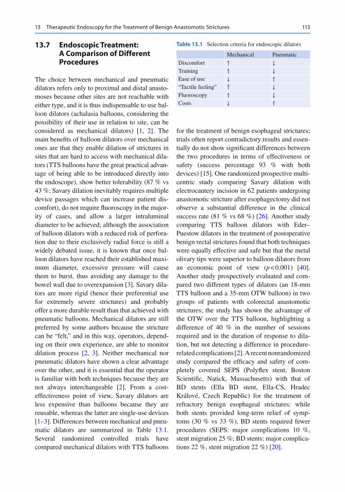
113
13.7 Endoscopic Treatment: A Comparison of Different Procedures
The choice between mechanical and pneumatic dilators refers only to proximal and distal anasto-moses because other sites are not reachable with either type, and it is thus indispensable to use bal-loon dilators (achalasia balloons, considering the possibility of their use in relation to site, can be considered as mechanical dilators) [ 1 , 2 ]. The main benefi ts of balloon dilators over mechanical ones are that they enable dilation of strictures in sites that are hard to access with mechanical dila-tors (TTS balloons have the great practical advan-tage of being able to be introduced directly into the endoscope), show better tolerability (87 % vs 43 %; Savary dilation inevitably requires multiple device passages which can increase patient dis-comfort), do not require fl uoroscopy in the major-ity of cases, and allow a larger intraluminal diameter to be achieved; although the association of balloon dilators with a reduced risk of perfora-tion due to their exclusively radial force is still a widely debated issue, it is known that once bal-loon dilators have reached their established maxi-mum diameter, excessive pressure will cause them to burst, thus avoiding any damage to the bowel wall due to overexpansion [ 3 ]. Savary dila-tors are more rigid (hence their preferential use for extremely severe strictures) and probably offer a more durable result than that achieved with pneumatic balloons. Mechanical dilators are still preferred by some authors because the stricture can be “felt,” and in this way, operators, depend-ing on their own experience, are able to monitor dilation process [ 2 , 3 ]. Neither mechanical nor pneumatic dilators have shown a clear advantage over the other, and it is essential that the operator is familiar with both techniques because they are not always interchangeable [ 2 ]. From a cost- effectiveness point of view, Savary dilators are less expensive than balloons because they are reusable, whereas the latter are single-use devices [ 1 – 3 ]. Differences between mechanical and pneu-matic dilators are summarized in Table 13.1 . Several randomized controlled trials have compared mechanical dilators with TTS balloons
for the treatment of benign esophageal strictures; trials often report contradictory results and essen-tially do not show signifi cant differences between the two procedures in terms of effectiveness or safety (success percentage 93 % with both devices) [ 15 ]. One randomized prospective multi-centric study comparing Savary dilation with electrocautery incision in 62 patients undergoing anastomotic stricture after esophagectomy did not observe a substantial difference in the clinical success rate (81 % vs 68 %) [ 26 ]. Another study comparing TTS balloon dilators with Eder–Puestow dilators in the treatment of postoperative benign rectal strictures found that both techniques were equally effective and safe but that the metal olivary tips were superior to balloon dilators from an economic point of view ( p < 0.001) [ 40 ]. Another study prospectively evaluated and com-pared two different types of dilators (an 18-mm TTS balloon and a 35-mm OTW balloon) in two groups of patients with colorectal anastomotic strictures; the study has shown the advantage of the OTW over the TTS balloon, highlighting a difference of 40 % in the number of sessions required and in the duration of response to dila-tion, but not detecting a difference in procedure-related complications [ 2 ]. A recent nonrandomized study compared the effi cacy and safety of com-pletely covered SEPS (Polyfl ex stent, Boston Scientifi c, Natick, Massachusetts) with that of BD stents (Ella BD stent, Ella-CS, Hradec Králové, Czech Republic) for the treatment of refractory benign esophageal strictures: while both stents provided long-term relief of symp-toms (30 % vs 33 %), BD stents required fewer procedures (SEPS: major complications 10 %, stent migration 25 %; BD stents: major complica-tions 22 %, stent migration 22 %) [ 20 ].
Table 13.1 Selection criteria for endoscopic dilators
Mechanical Pneumatic
Discomfort ↑ ↓ Training ↑ ↓ Ease of use ↓ ↑ “Tactile feeling” ↑ ↓ Fluoroscopy ↑ ↓ Costs ↓ ↑
13 Therapeutic Endoscopy for the Treatment of Benign Anastomotic Strictures
114
13.8 Complications
The complication rate of the treatment of benign anastomotic strictures differs considerably in the literature, ranging from 0.5 to 15 % (minimal events included). Stricture complexity is certainly one of the most important causes of complica-tions but also the skill of the endoscopist plays a determinant role [ 5 , 8 ]. Endoscopic dilation is a relatively safe technique, and the most serious complications include mortality (0.01 %), hemor-rhage (0.07 %), bacteremia (abscesses 0.2 %), and above all perforation (01–1.1 %) [ 5 ]. The risk of perforation signifi cantly decreases when “the rule of three” is applied, when dilation is per-formed gradually (the maximum dilation diame-ter should not increase by >3 mm per session), and when guidewire assistance or fl uoroscopic guidance are used (however, repeated endoscopic dilations may increase this risk) [ 1 , 2 , 5 , 8 ]. Previous radiotherapy and infl ammatory bowel diseases increase the possibility of perforation, as do specifi c locations, such as cervical esophagus, or anatomic variations, such as voluminous hiatal hernias. Perforation is usually successfully treated conservatively, and surgical examination is often unable to identify the leakage point of perforation in cases of small lesions [ 1 ]. As the event may also indicate the potential malignancy of the treated stenosis, histological evaluation of the anastomosis is required when the complication has been resolved [ 31 ]. Maloney dilators, passing blindly through strictures, show a higher perfora-tion rate with respect to balloons and Savary-Gilliard dilators, and it is thus advisable to use these devices only for simple stenoses. The over-all risk of complications from stent placement ranges from 1 to 15 %, and post- stenting compli-cations are subdivided into early (<30 days) and late (>30 days) events: the former include perfo-ration, major bleeding, pain, and dislocation, while the latter comprise migration, obstruction, mucosal erosion, pressure necrosis, tenesmus, dysphagia, intestinal occlusion, and occasionally perforation [ 19 ]. Perforation is undoubtedly the most dangerous complication (2–3 %); stenting
has a longer-lasting dilating effect than dilation and usually has a low risk of acute perforation. Stricture dilation before or immediately after stent placement results in a fi ve- to sixfold higher perforation rate (10–18 %) and generally should be avoided, as should stenting in anastomotic strictures with acute infl ammation [ 19 ]; perfora-tion is also favored by excessive air insuffl ation (reducible by using CO2 instead of air) and by treatment with bevacizumab. Post-stenting pain, which mainly depends on device diameter and radial expansive force, is common in patients with refractory strictures and is usually of short duration after SEMS and SEPS placement, per-sisting for longer when BD stents are used [ 19 ]. Common complications after BD stent placement for esophageal anastomotic stricture are vomiting and nausea caused by the material (polydioxa-none) from which the stent is made [ 13 ]. Stent migration is a common problem when covered SEMS and SEPS are used; in the most important studies published, SEMS and SEPS migration varies from 30 to 40 %, whereas migration of BD stents is much lower, ranging from 8 to 30 % [ 18 ]. This complication depends on multiple factors such as the location of the stenosis and device features [ 19 ]; in the esophagus BD stent migra-tion does not require endoscopic removal of the device because the gastric pH accelerates the material degradation process (in the stomach BD stent half-life is about 13 days) and the risk of obstruction or perforation is negligible [ 19 , 20 ]. Tissue overgrowth is linked to the material from which the device is made (nitinol stimulates hyperplastic tissue growth), radial force, and stent position inside the lumen (hyperplastic tissue grows more easily when the device is placed in an oblique position) [ 13 , 14 , 16 , 24 , 25 ]. Stent removal may cause adverse events, the most important being visceral disruption, perforation, intramural rupture, fi stula formation, and seg-mental amputation. A recent retrospective multi-center study examined the safety of esophageal stent removal in 214 patients with benign esopha-geal disease, taking into consideration fully cov-ered SEMS (52 %) and partially covered SEMS
A. Casadei et al.
115
(28.6 %) and SEPS (19.4 %). Results showed that endoscopic removal was feasible and relatively safe with a major adverse event rate of 2.1 %; fully covered SEMS were associated with a lower rate of adverse events than partially covered SEMS or SEPS [ 17 ]. In cases of steroid injection, there is a theoretical risk of Candida albicans intramural infection and perforation [ 27 ].
13.9 Cost Analysis and Final Considerations
Cost analysis includes costs pertaining to person-nel, material used during each procedure, each endoscopic examination required to establish diagnosis and performed during follow-up, and hospitalization. The only difference in cost between various endoscopic treatments is related to the equipment used to perform the procedure, especially when reusable devises are used. Multiple dilation procedures are usually neces-sary to achieve an adequate clinical outcome, which signifi cantly increases overall therapy costs. The indisputable advantage of mechanical dilators over balloon dilators stems from the low cost of equipment, which consequently lowers the cost of this therapy compared to that of pneu-matic dilation (mechanical dilators can be used for a long time, and the cost of a set of 16 Savary dilators is Euro 2,500; single-use balloon cathe-ters cost between Euro 207 and 680 depending on the model; the average cost of Eder–Puestow dilators for each procedure is Euro 22.30) [ 40 ]. The Savary-Gilliard method is thus a low-budget technique (bougies can be used almost indefi -nitely) that is effective, safe, relatively simple to perform, and inexpensive; it can also be carried out in any endoscopic intervention unit. Stents are obviously more expensive than dilators (SEMS cost from Euro 619 to 1,150 depending on device features and the country of purchase) but have the advantage of requiring only one endoscopic session, with consequent cost damp-ing. As stenting often follows an ineffective dila-tion treatment, with consequent additional costs,
it could be an appropriate fi rst-choice treatment in cases of long, tortuous, fi brous strictures. BD stents are more expensive than SEMS; however, in cases of stricture recurrence 4–6 months after dilation, the short-term patency of 4 months until stent degradation and the increased likelihood of long-term success make economic sense to con-sider BD stent insertion. In addition, these stents do not require removal within 4–6 weeks of insertion, thus avoiding the associated cost of removal and potential complications and conse-quently improving patient quality of life. Stent placement is cost-effective compared with sur-gery [ 10 , 20 , 22 , 25 ]. However, a surgical option should always be considered in the following cases: young patients who do not achieve essen-tial health benefi ts after 1–2 years of endoscopic sessions, patients who require very frequent dila-tions or with a previous complicated perforation after endoscopic treatment, individuals cannot tolerate continued dilations for technical or psy-chological reasons, and patients who have obtained no benefi t from adequately executed stenting treatments. It is very important to treat anastomotic strictures in a stepwise manner start-ing with the least invasive approach. Generally, dilation is the fi rst treatment modality used (usu-ally up to fi ve sessions), but if this approach is not suffi cient to relieve symptoms, an alternative treatment approach should be discussed with the patient to evaluate and balance out risks and ben-efi ts, especially in cases of refractory lesions (dilation combined with intralesional four- quadrant steroid injection, up to three sessions, and/or incisional therapy, with or without APC, up to three sessions). Stent placement is usually the last endoscopic choice (stenting should only be considered in carefully selected patients who have proven refractory to dilation); when BD stents are used but the stenosis persists or relapses after device degradation, it is possible to place more than one stent or to repeat the dilation pro-cedure. Surgical intervention is the fi nal step, but even after surgery the risk of recurrent stricture formation remains. The algorithm for anasto-motic stricture treatment is shown in Table 13.2 .
13 Therapeutic Endoscopy for the Treatment of Benign Anastomotic Strictures
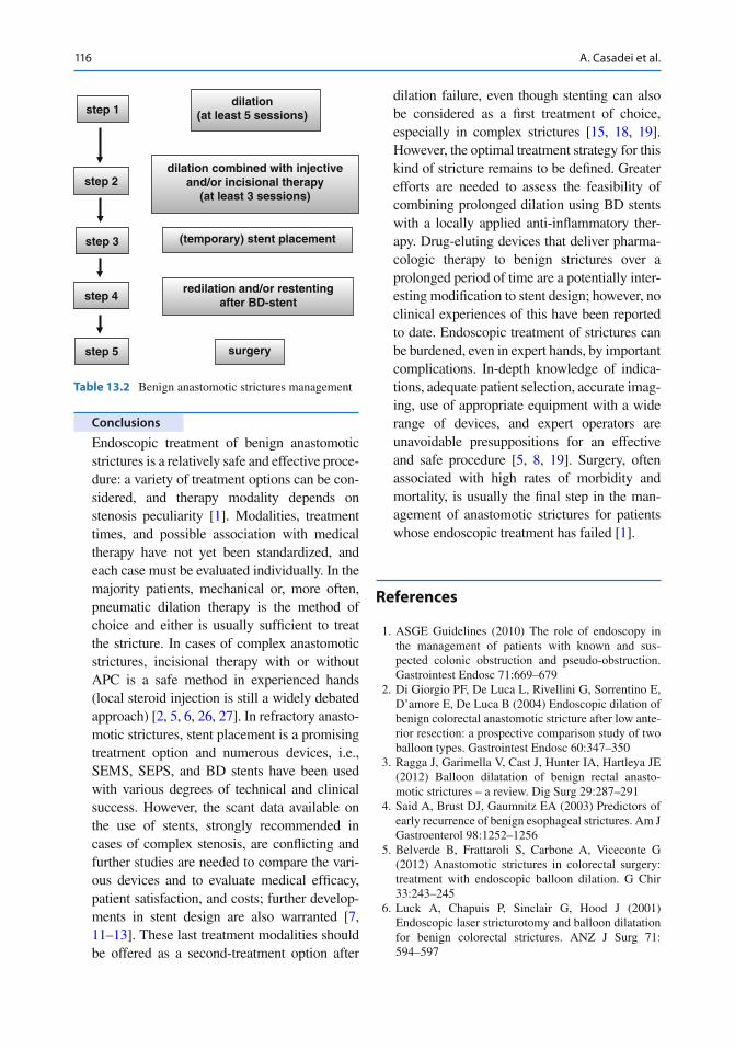
116
Conclusions
Endoscopic treatment of benign anastomotic strictures is a relatively safe and effective proce-dure: a variety of treatment options can be con-sidered, and therapy modality depends on stenosis peculiarity [ 1 ]. Modalities, treatment times, and possible association with medical therapy have not yet been standardized, and each case must be evaluated individually. In the majority patients, mechanical or, more often, pneumatic dilation therapy is the method of choice and either is usually suffi cient to treat the stricture. In cases of complex anastomotic strictures, incisional therapy with or without APC is a safe method in experienced hands (local steroid injection is still a widely debated approach) [ 2 , 5 , 6 , 26 , 27 ]. In refractory anasto-motic strictures, stent placement is a promising treatment option and numerous devices, i.e., SEMS, SEPS, and BD stents have been used with various degrees of technical and clinical success. However, the scant data available on the use of stents, strongly recommended in cases of complex stenosis, are confl icting and further studies are needed to compare the vari-ous devices and to evaluate medical effi cacy, patient satisfaction, and costs; further develop-ments in stent design are also warranted [ 7 , 11 – 13 ]. These last treatment modalities should be offered as a second-treatment option after
dilation failure, even though stenting can also be considered as a fi rst treatment of choice, especially in complex strictures [ 15 , 18 , 19 ]. However, the optimal treatment strategy for this kind of stricture remains to be defi ned. Greater efforts are needed to assess the feasibility of combining prolonged dilation using BD stents with a locally applied anti-infl ammatory ther-apy. Drug-eluting devices that deliver pharma-cologic therapy to benign strictures over a prolonged period of time are a potentially inter-esting modifi cation to stent design; however, no clinical experiences of this have been reported to date. Endoscopic treatment of strictures can be burdened, even in expert hands, by important complications. In-depth knowledge of indica-tions, adequate patient selection, accurate imag-ing, use of appropriate equipment with a wide range of devices, and expert operators are unavoidable presuppositions for an effective and safe procedure [ 5 , 8 , 19 ]. Surgery, often associated with high rates of morbidity and mortality, is usually the fi nal step in the man-agement of anastomotic strictures for patients whose endoscopic treatment has failed [ 1 ].
References
1. ASGE Guidelines (2010) The role of endoscopy in the management of patients with known and sus-pected colonic obstruction and pseudo-obstruction. Gastrointest Endosc 71:669–679
2. Di Giorgio PF, De Luca L, Rivellini G, Sorrentino E, D’amore E, De Luca B (2004) Endoscopic dilation of benign colorectal anastomotic stricture after low ante-rior resection: a prospective comparison study of two balloon types. Gastrointest Endosc 60:347–350
3. Ragga J, Garimella V, Cast J, Hunter IA, Hartleya JE (2012) Balloon dilatation of benign rectal anasto-motic strictures – a review. Dig Surg 29:287–291
4. Said A, Brust DJ, Gaumnitz EA (2003) Predictors of early recurrence of benign esophageal strictures. Am J Gastroenterol 98:1252–1256
5. Belverde B, Frattaroli S, Carbone A, Viceconte G (2012) Anastomotic strictures in colorectal surgery: treatment with endoscopic balloon dilation. G Chir 33:243–245
6. Luck A, Chapuis P, Sinclair G, Hood J (2001) Endoscopic laser stricturotomy and balloon dilatation for benign colorectal strictures. ANZ J Surg 71:594–597
step 1dilation
(at least 5 sessions)
dilation combined with injectiveand/or incisional therapy
(at least 3 sessions)step 2
(temporary) stent placementstep 3
step 4redilation and/or restenting
after BD-stent
step 5 surgery
Table 13.2 Benign anastomotic strictures management
A. Casadei et al.
117
7. Rejchrt S, Kopacova M, Brozik J, Bures J (2011) Biodegradable stents for the treatment of benign ste-noses of the small and large intestines. Endoscopy 43:911–917
8. Araujo SE, Costa AF (2008) Effi cacy and safety of endoscopic balloon dilation of benign anastomotic strictures after oncologic anterior rectal resection: report on 24 cases. Surg Laparosc Endosc Percutan Tech 18:565–568
9. Werre A, Mulder C, van Heteren C, Spillenaar Bilgen E (2000) Dilation of benign strictures following low anterior resection using Savary–Gilliard bougies. Endoscopy 32(5):385–388
10. Bonin EA, Baron TH (2010) Update on the indica-tions and use of colonic stents. Curr Gastroenterol Rep 12:374–382
11. Farrel JJ, Sack J (2008) Removable colonic stenting: time to expand the indications? Gastrointest Endosc 68(4):721–723
12. Marcotte E, Comeau E, Meziat-Burdin A, Ménard C, Rateb G (2012) Early migration of fully covered double- layered metallic stents for post-gastric bypass anastomotic strictures. Int J Surg Case Rep 3:283–286
13. Janik V, Horak L, Hnanicek J (2011) Biodegradable polydioxanone stents: a new option for therapy- resistant anastomotic strictures of the colon. Eur Radiol 21:1956–1961
14. Dai YY, Chopra SS, Wysocki WM, Hünerbein M (2010) Treatment of benign colorectal strictures by temporary stenting with self-expanding stents. Int J Colorectal Dis 25:1475–1479
15. de Wijkerslooth LRH, Vleggaar FP, Siersema PD (2011) Endoscopic management of diffi cult or recur-rent esophageal strictures. Am J Gastroenterol 106:2080–2091
16. Seo TS, Song HY, Sung KB, Ko GY, Yu CS (2003) A benign colorectal stricture: treatment with a retriev-able expandable nitinol stent. Cardiovasc Intervent Radiol 26:181–183
17. van Halsema EE, Wong Kee Song LM, Baron TH, Siersema PD, Vleggaar FP, Ginsberg GG, Shah PM, Fleischer DE, Ratuapli SK, Fockens P, Dijkgraaf MGV, Rando G, Repici A, van Hooft JE (2013) Safety of endoscopic removal of self-expandable stents after treatment of benign esophageal diseases. Gastrointest Endosc 77(1):18–28
18. Repici A, Hassan C, Sharma P (2010) Systematic review: the role of self-expanding plastic stents for benign oesophageal strictures. Aliment Pharmacol Ther 31:1268–1275
19. Repici A, Vleggaar FP, Hassan C (2010) Effi cacy and safety of biodegradable stents for refractory benign esophageal strictures: the BEST (Biodegradable Esophageal Stent) study. Gastrointest Endosc 72:927–934
20. van Boeckel PG, Vleggaar FP, Siersema PD (2011) A comparison of temporary self-expanding plastic and biodegradable stents for refractory benign esophageal strictures. Clin Gastroenterol Hepatol 9:653–659
21. Bhasin DK, Rana SS (2008) Biodegradable pancre-atic stents: are they a disappearing wonder? Gastrointest Endosc 67:1113–1115
22. Pérez Roldán F, González Carro P, Villafáñez García MC, Aoufi Rabih S, Legaz Huidobro ML, Sánchez- Manjavacas Múñoz N, Roncero García-Escribano O, Ynfante Ferrús M, Bernardos Martín E, Ruiz Carrillo F (2012) Usefulness of biodegradable polydioxanone stents in the treatment of postsurgical colorectal stric-tures and fi stulas. Endoscopy 44:297–300
23. Toth E, Nielsen J, Nemeth A, Wurm Johansson G, Syk I, Mangell P, Almqvist P, Thorlacius H (2011) Treatment of a benign colorectal anastomotic stric-ture with a biodegradable stent. Endoscopy 43:E252–E253
24. Saito Y, Tanaka T, Andoh A, Minematsu H et al (2007) Usefulness of biodegradable stents constructed of poly-l-lactic acid monofi laments in patients with benign esophageal stenosis. World J Gastroenterol 13:3977–3980
25. Hirdes MMC, Siersema PD, van Boeckel PGA, Vleggaar FP (2012) Single and sequential biodegrad-able stent placement for refractory benign esophageal strictures: a prospective follow-up study. Endoscopy 44:649–654
26. Hordijk ML, van Hooft JE, Hansen BE et al (2009) A randomized comparison of electrocautery incision with Savary bougienage for relief of anastomotic gas-troesophageal strictures. Gastrointest Endosc 70:849–855
27. Kochhar R, Poornachandra KS (2010) Intralesional steroid injection therapy in the management of resis-tant gastrointestinal strictures. World J Gastrointest Endosc 2(2):61–68
28. Hirdes MM, Vleggaar FP, Siersema PD (2011) Stent placement for esophageal strictures: an update. Expert Rev Med Devices 8(6):733–755
29. Wong RF, Adler DG, Hilden K, Fang JC (2008) Retrievable esophageal stents for benign indications. Dig Dis Sci 53:322–329
30. van Hooft JE, van Berge Henegouwen MI, Rauws EA et al (2011) Endoscopic treatment of benign anasto-motic esophagogastric strictures with a biodegradable stent. Gastrointest Endosc 73:1043–1047
31. Da Costa M, Mata A, Espinós J et al (2011) Endoscopic dilation of gastrojejunal anastomotic strictures after laparoscopic gastric bypass. Predictors of initial failure. Obes Surg 21:36–41
32. Kim JH, Song HY, Park SW et al (2008) Early symp-tomatic strictures after gastric surgery: palliation with balloon dilation and stent placement. J Vasc Interv Radiol 19(4):565–570
33. Gill R, Whitlock KA, Mohamed R et al (2012) Endoscopic treatment options in patients with gastro-jejunal anastomosis stricture following Roux-en-Y gastric bypass. Gastroenterol Res 5(1):1–5
34. Gill R, Whitlock KA, Mohamed R et al (2012) The role of upper gastrointestinal endoscopy in treating postoperative complications in bariatric surgery. J Interv Gastroenterol 2(1):37–41
13 Therapeutic Endoscopy for the Treatment of Benign Anastomotic Strictures
118
35. Fernández-Esparrach G, Bordas JM, Llach J et al (2008) Endoscopic dilation with Savary-Gilliard Bougies of stomal strictures after laparoscopic gastric bypass in morbidly obese patients. Obes Surg 18:155–161
36. Sunada K, Yamamoto H (2009) Double-balloon endoscopy: past, present, and future. J Gastroenterol 44:1–12
37. Yano T, Yamamoto H (2009) Current state of double balloon endoscopy: the latest approach to small intes-tinal diseases. J Gastroenterol Hepatol 24:185–192
38. May A, Nachbar L, Pohl J, Ell C (2007) Endoscopic interventions in the small bowel using double bal-loon enteroscopy: feasibility and limitations. Am J Gastroenterol 102(3):527–535
39. Fukumoto A, Tanaka S, Yamamoto H et al (2007) Diagnosis and treatment of small-bowel stricture by double balloon endoscopy. Gastrointest Endosc 66(3):S108–S112
40. Xinopoulos D, Kypreos D, Bassioukas SP et al (2011) Comparative study of balloon and metal olive dilators for endoscopic management of benign anastomotic rectal strictures: clinical and cost-effectiveness out-comes. Surg Endosc 25:756–763
41. Keränen I, Lepistö A, Udd M et al (2010) Outcome of patients after endoluminal stent placement for benign colorectal obstruction. Scand J Gastroenterol 45:725–731
42. Levine RA, Wasvary H, Kadro O (2012) Endoprosthetic management of refractory ileocolonic anastomotic strictures after resection for Crohn’s dis-ease: report of nine-year follow-up and review of the literature. Infl amm Bowel Dis 18(3):506–512
43. Curcio G, Spada M, di Francesco F et al (2010) Completely obstructed colorectal anastomosis: a new non-electrosurgical endoscopic approach before balloon dilatation. World J Gastroenterol 16(37):4751–4754
A. Casadei et al.
119G. Galloro (ed.), Endoscopic Follow-up of Digestive Anastomosis, DOI 10.1007/978-88-470-5370-0_14, © Springer-Verlag Italia 2014
14.1 Background
Anastomotic leakage is a relatively frequent post-operative complication after surgery owing to an increase morbidity and mortality.
There is no uniformity in defi ning anastomotic dehiscence . In 1991 the UK Surgical Infection Study Group proposed the defi nition of “leak of luminal contents from a surgical join between two hollow viscera” [ 1 ]. The majority of stud-ies defi ne an anastomotic leak as a combination of clinical signs (pain, peritonitis and feculent,
or purulent drainage), biochemical fi ndings (fever, tachycardia, leukocytosis), and radiologic fi ndings in addiction to any intraoperative fi nding.
As regards colorectal surgery, its incidence var-ies from 3 to 20 % (Table 14.1 ) [ 2 – 16 ]. The inci-dence of leak is signifi cantly associated with the distance of the anastomosis from the anal verge. The lowest leak rates are described after small bowel or ileocolic anastomosis (1–3 %), whereas the highest rates occur after colorectal and colo-anal anastomosis (10–20 %). Anastomotic leaks are detected in an extremely variable postopera-tive time interval, with two peaks of incidence on the third and the seventh postoperative day.
A. Arezzo (*) • M. Verra , MD • M. Morino Department of Surgical Sciences , University of Torino , Corso Dogliotti, 14 , Torino 10126 , Italy e-mail: [email protected]; [email protected]; [email protected]
G. Galloro Department of Clinical Medicine and Surgery, Digestive Surgical Endoscopy Unit , University of Naples Federico II, Italy – School of Medicine , via Pansini 5 , Naples 80131 , Italy e-mail: [email protected]
M. de Bellis Endoscopy Unit , National Cancer Institute and G. Pascale Foundation , Naples , Italy
A. Trecca Operative Endoscopy Unit , Usi Group , Rome , Italy
R. Manta Gastroenterology and Digestive Endoscopy Unit , New S. Agostino –Estense Hospital , Baggiovara , MO , Italy
14 Review : Therapeutic Endoscopy for the Treatment of Anastomotic Dehiscences
Alberto Arezzo , Mauro Verra , Giuseppe Galloro , Mario de Bellis , Antonello Trecca , Raffaele Manta , and Mario Morino
Table 14.1 Literature data for colorectal anastomotic dehiscence
First author No. of patients
Major leaks (%) <6 cm (%)
Biondo [ 7 ] 211 5.7 Docherty [ 12 ] 652 4.4 Alves [ 5 ] 707 6 Konishi [ 10 ] 391 2.8 Hyman [ 2 ] 1,223 2.7 Lipska [ 6 ] 541 6.5 Branagan [ 11 ] 1,834 3.9 Sorenson (1999 ) 333 15.9 Wong [ 15 ] 1,066 3.8 Platell [ 9 ] 1,639 2.4 Karanjia [ 14 ] 219 11 97 Law [ 3 ] 196 10.2 100 Gastinger [ 16 ] 2,729 14 100 Rullier [ 4 ] 131 19 100 Vignali [ 8 ] 284 7.8 100
120
Intrathoracic anastomotic leakage represents a much more life-threatening event, with high mor-tality rate. Its incidence is reported to range between 6 and 30 % [ 17 – 19 ].
14.2 Risk Factors
Many risk factors have been demonstrated to be signifi cantly related to anastomotic leak. Patient risk factors include malnutrition, steroids, tobacco and alcohol use, leukocytosis, cardio-vascular disease, ASA score, and diverticulitis; intraoperative risk factors include the distance of anastomosis from the anal verge, operative time >2 h, bowel obstruction, blood supply to anastomosis, perioperative blood transfusion, and intraoperative septic conditions; as regards extraperitoneal anastomoses, male gender and obesity are associated to an increased risk of dehiscence [ 16 , 20 – 22 ]. Defunctioning loop stoma has been described to reduce the rate of symptomatic anastomotic leakage in low anterior rectal resection [ 23 ].
In a large multicenter analysis of oncological and survival outcomes following anastomotic leakage after rectal cancer surgery, oncological outcome was not signifi cantly infl uenced by anastomotic leakage, even if overall survival was reduced [ 24 ].
14.3 Management
Anastomotic leakage management depends on clinical manifestation, on clinical stability of the patient, and on the distance of the anastomosis from the anal verge, particularly if the anasto-motic leak is cervical or extraperitoneal, rather than mediastinal or intraperitoneal.
Conventional operative management includes, as regards colorectal anastomosis, explorative laparotomy, peritoneal lavage, and, in at least 80 % of cases, the creation of a derivative stoma. This reduces complications such as peritonitis and sepsis and mortality rate [ 16 ], but surgical management leads to a relatively high morbidity rate. Morbidity rate with stoma creation is 30 %
and reversal rate in patient with anastomotic leakage is <50 % [ 25 , 26 ].
As regards intrathoracic dehiscence , surgical management is a highly demolishing surgery with high morbidity and mortality rate.
Technological improvements allowed intro-duction of new techniques in the management of anastomotic leaks. Endoscopic treatment of anas-tomotic dehiscence has been proposed as an alternative to operative management in clinically stable patient without generalized peritonitis. In general, patients who present with generalized peritonitis, free intraperitoneal leak or high-grade sepsis with hypotension should be fi rst resusci-tated and surgery is mandatory.
Distance of anastomotic dehiscence from anal verge influences subsequent management because of the different probability of free intra-peritoneal leak and subsequent generalized peri-tonitis. Extraperitoneal leaks (e.g., after anterior resection of rectum) often lead to extraperitoneal abscess, such as presacral collections that allow more likely a nonoperative management.
Three main endoscopic options in the man-agement of anastomotic dehiscence are nowa-days available:• Synthesis and suturing devices • EndoVac therapy • Covered self-expanding metal stents (SEMS)
14.4 Synthesis and Suturing Devices
Synthesis and suturing devices allow a closure by fi rst intention of the anastomotic defect. Many synthesis and suturing techniques have been described among which clipping is most frequently used. Different clipping devices have been described. Two-pronged endoclips (e.g., Quickclip II, Olympus, Tokyo, Japan, or Resolution Clip, Boston Scientifi c, MA, USA) with a maximum opening distance of 11 mm are marketed (Fig. 14.1a ). TriClip is characterized instead by a third equidistant prong and an open-ing distance of 12 mm, theoretically owing to the possibility to grasp a larger and a wider amount of tissue (Fig. 14.1b ).
A. Arezzo et al.
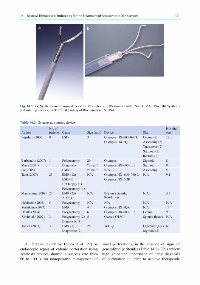
121
A literature review by Trecca et al. [ 27 ] on endoscopic repair of colonic perforation using synthesis devices showed a success rate from 60 to 100 % for nonoperative management of
small perforations, in the absence of signs of generalized peritonitis (Table 14.2 ). This review highlighted the importance of early diagnosis of perforation in order to achieve therapeutic
a b
Fig. 14.1 ( a ) Synthesis and suturing devices, the Resolution clip (Boston Scientifi c, Natick, MA, USA). ( b ) Synthesis and suturing devices, the TriClip (Courtesy of Bloomington, IN, USA)
Table 14.2 Systems ad suturing devices
Author No. of patients Cause Size (mm) Device Site
Hospital stay
Fujishiro (2006) 9 ESD 3 Olympus HX-600–090 L Cecum (2) 12.1 Olympus HX-5QR Ascending (2)
Transverse (2) Sigmoid (1) Rectum (2)
Barbagallo (2007) 1 Polypectomy 20 Olympus Sigmoid 8 Mana (2001) 1 Diagnostic “Small” Olympus HX-600–135 Sigmoid 8 Fu (2005) 1 EMR “Small” N/A Ascending 5 Taku (2007) 23 EMR (12) N/A Olympus HX–600–090 L N/A 9.1
ESD (6) Olympus HX–5QR Hot biopsy (1) Polypectomy (4)
Magdeburg (2008) 27 EMR (25) N/A Boston Scientifi c Resolution
N/A 3.5 APC (1)
Heldwein (2005) 5 Polypectomy N/A N/A N/A N/A Yoshikane (1997) 1 EMR 4 Olympus HX-5QR N/A 14 Dhalla (2004) 1 Polypectomy 6 Olympus HX-600–135 Cecum 9 Kirshniak (2007) 3 Polypectomy (2) 5 Ovesco OTSC Splenic fl exure N/A
Diagnostic (1) Trecca (2007) 3 EMR (1) 26 TriClip Descending (1) 6
Diagnostic (2) Sigmoid (2)
14 Review : Therapeutic Endoscopy for the Treatment of Anastomotic Dehiscences
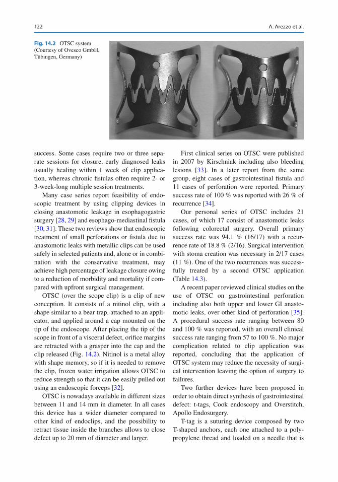
122
success. Some cases require two or three sepa-rate sessions for closure, early diagnosed leaks usually healing within 1 week of clip applica-tion, whereas chronic fi stulas often require 2- or 3-week-long multiple session treatments.
Many case series report feasibility of endo-scopic treatment by using clipping devices in closing anastomotic leakage in esophagogastric surgery [ 28 , 29 ] and esophago-mediastinal fi stula [ 30 , 31 ]. These two reviews show that endoscopic treatment of small perforations or fi stula due to anastomotic leaks with metallic clips can be used safely in selected patients and, alone or in combi-nation with the conservative treatment, may achieve high percentage of leakage closure owing to a reduction of morbidity and mortality if com-pared with upfront surgical management.
OTSC (over the scope clip ) is a clip of new conception. It consists of a nitinol clip, with a shape similar to a bear trap, attached to an appli-cator, and applied around a cap mounted on the tip of the endoscope. After placing the tip of the scope in front of a visceral defect, orifi ce margins are retracted with a grasper into the cap and the clip released (Fig. 14.2 ). Nitinol is a metal alloy with shape memory, so if it is needed to remove the clip, frozen water irrigation allows OTSC to reduce strength so that it can be easily pulled out using an endoscopic forceps [ 32 ].
OTSC is nowadays available in different sizes between 11 and 14 mm in diameter. In all cases this device has a wider diameter compared to other kind of endoclips, and the possibility to retract tissue inside the branches allows to close defect up to 20 mm of diameter and larger.
First clinical series on OTSC were published in 2007 by Kirschniak including also bleeding lesions [ 33 ]. In a later report from the same group, eight cases of gastrointestinal fi stula and 11 cases of perforation were reported. Primary success rate of 100 % was reported with 26 % of recurrence [ 34 ].
Our personal series of OTSC includes 21 cases, of which 17 consist of anastomotic leaks following colorectal surgery. Overall primary success rate was 94.1 % (16/17) with a recur-rence rate of 18.8 % (2/16). Surgical intervention with stoma creation was necessary in 2/17 cases (11 %). One of the two recurrences was success-fully treated by a second OTSC application (Table 14.3 ).
A recent paper reviewed clinical studies on the use of OTSC on gastrointestinal perforation including also both upper and lower GI anasto-motic leaks, over other kind of perforation [ 35 ]. A procedural success rate ranging between 80 and 100 % was reported, with an overall clinical success rate ranging from 57 to 100 %. No major complication related to clip application was reported, concluding that the application of OTSC system may reduce the necessity of surgi-cal intervention leaving the option of surgery to failures.
Two further devices have been proposed in order to obtain direct synthesis of gastrointestinal defect: t-tags, Cook endoscopy and Overstitch, Apollo Endosurgery.
T-tag is a suturing device composed by two T-shaped anchors, each one attached to a poly-propylene thread and loaded on a needle that is
Fig. 14.2 OTSC system (Courtesy of Ovesco GmbH, Tübingen, Germany)
A. Arezzo et al.

123
Tab
le 1
4.3
O
TSC
per
sona
l ser
ies
Age
Si
te
Etio
logy
T
ime
afte
r su
rger
y D
iam
eter
/m
argi
ns
OT
SC
mm
Su
cces
s C
ombi
ned
trea
tmen
t Fo
llow
-up
1.
73
Rec
tum
L
eak
colo
rect
al a
nast
omos
is
Post
radi
atio
n 24
mon
ths
10
Fibr
otic
12
Y
es
Prim
ary
succ
ess
2.
58
Rec
tum
L
eak
colo
rect
al a
nast
omos
is
Post
radi
atio
n 21
mon
ths
8/5
Fibr
otic
12
N
o Pe
rsis
tent
def
ect
3.
76
Rec
tum
L
eak
colo
rect
al a
nast
omos
is
Post
radi
atio
n 13
day
s 10
Fi
brot
ic
12
Yes
Pr
imar
y su
cces
s 4.
41
R
ectu
m
Lea
k co
lore
ctal
ana
stom
osis
Po
stra
diat
ion
16 m
onth
s 6
Fibr
otic
12
Y
es
Prim
ary
succ
ess
5.
59
Rec
tum
R
ecto
vagi
nal fi
stu
la/a
nast
omot
ic le
ak
Post
radi
atio
n 25
day
s 5
Fibr
otic
12
Y
es
Prim
ary
succ
ess
6.
65
Rec
tum
R
ecto
vagi
nal fi
stu
la/a
nast
omot
ic le
ak
6 da
ys
10
Tend
er
12
Yes
St
oma
Seco
ndar
y su
cces
s 7.
73
R
ectu
m
Lea
k co
lore
ctal
ana
stom
osis
Po
stra
diat
ion
15 d
ays
10
Fibr
otic
12
Y
es
Prim
ary
succ
ess
8.
56
Rec
tum
L
eak
colo
rect
al a
nast
omos
is
Post
radi
atio
n 20
day
s 10
Fi
brot
ic
12
Yes
Pr
imar
y su
cces
s 9.
74
R
ectu
m
Lea
k co
lore
ctal
ana
stom
osis
Po
stra
diat
ion
21 m
onth
s 12
Fi
brot
ic
12
Yes
Pr
imar
y su
cces
s 10
. 66
R
ectu
m
Lea
k co
lore
ctal
ana
stom
osis
Po
stra
diat
ion
13 d
ays
8 Fi
brot
ic
12
Yes
Pr
imar
y su
cces
s 11
. 69
R
ectu
m
Lea
k co
lore
ctal
ana
stom
osis
25
day
s 9
Fibr
otic
12
Y
es
Prim
ary
succ
ess
12.
76
Rec
tum
L
eak
colo
rect
al a
nast
omos
is
16 d
ays
12
Fibr
otic
12
Y
es
Prim
ary
succ
ess
13.
68
Col
on
Col
ocut
aneu
s/an
asto
mot
ic le
ak
6 m
onth
s 5
Fibr
otic
12
Y
es
Prim
ary
succ
ess
14.
82
Col
on
Col
ocut
aneu
s/an
asto
mot
ic le
ak
6 m
onth
s 2
Fibr
otic
12
Y
es
Rec
urre
nce
of d
efec
t 15
. 38
R
ectu
m
Rec
tove
sica
l fi s
tula
T
raum
atic
12
0 m
onth
s 5
Fibr
otic
14
Y
es
Prim
ary
succ
ess
16.
63
Rec
tum
L
eak
colo
rect
al a
nast
omos
is
Post
radi
atio
n 4
mon
ths
15
Fibr
otic
12
Y
es
Prim
ary
succ
ess
17.
57
Rec
tum
R
ecto
cuta
neou
s fi s
tula
/ana
stom
otic
leak
3
mon
ths
10
Fibr
otic
12
Y
es
Prim
ary
succ
ess
18.
74
Rec
tum
L
eak
colo
rect
al a
nast
omos
is
13 d
ays
12
Tend
er
14
Yes
St
oma
Prim
ary
succ
ess
19.
74
Rec
tum
R
ecto
vesi
cal fi
stu
la
Perf
orat
ion
duri
ng s
urge
ry
8 m
onth
s 10
Fi
brot
ic
12
Yes
Pe
rsis
tent
def
ect
20.
52
Rec
tum
R
ecto
vesi
cal fi
stu
la
Pouc
hitis
21
mon
ths
3 Fi
brot
ic
12
Yes
Fi
brin
se
alan
t Pe
rsis
tent
def
ect
21.
75
Rec
tum
R
ecto
vesi
cal fi
stu
la
PPH
72
mon
ths
5 Fi
brot
ic
12
Yes
Pe
rsis
tent
def
ect
14 Review : Therapeutic Endoscopy for the Treatment of Anastomotic Dehiscences
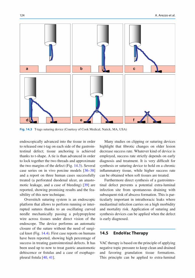
124
endoscopically advanced into the tissue in order to released one t-tag on each side of the gastroin-testinal defect; tissue anchoring is achieved thanks to t-shape. A tie is than advanced in order to lock together the two threads and approximate the two margins of the defect (Fig. 14.3 ). Several case series on in vivo porcine models [ 36 – 38 ] and a report on three human cases successfully treated (a perforated duodenal ulcer, an anasto-motic leakage, and a case of bleeding) [ 39 ] are reported, showing promising results and the fea-sibility of this new technique.
Overstitch suturing system is an endoscopic platform that allows to perform running or inter-rupted sutures thanks to an oscillating curved needle mechanically passing a polypropylene wire across tissues under direct vision of the endoscope. The device performs an automatic closure of the suture without the need of surgi-cal knot (Fig. 14.4 ). First case reports on humans have been reported, showing high percentage of success in treating gastrointestinal defects. It has been used up to now to treat gastric anastomotic dehiscence or fi stulas and a case of esophago- pleural fi stula [ 40 , 41 ].
Many studies on clipping or suturing devices highlight that fi brotic changes on older lesion decrease success rate. Whatever kind of device is employed, success rate strictly depends on early diagnosis and treatment. It is very diffi cult for synthesis or suturing device to hold on a chronic infl ammatory tissue, while higher success rate can be obtained when soft tissues are treated.
Furthermore direct synthesis of a gastrointes-tinal defect prevents a potential extra-luminal infection site from spontaneous draining with subsequent risk of abscess formation. This is par-ticularly important in intrathoracic leaks where mediastinal infection carries on a high morbidity and mortality risk. Application of suturing and synthesis devices can be applied when the defect is early diagnosed.
14.5 EndoVac Therapy
VAC therapy is based on the principle of applying negative topic pressure to keep clean and drained and favoring granulation tissue formations. This principle can be applied to extra- luminal
a b c
d e f
Fig. 14.3 T-tags suturing device (Courtesy of Cook Medical, Natick, MA, USA)
A. Arezzo et al.
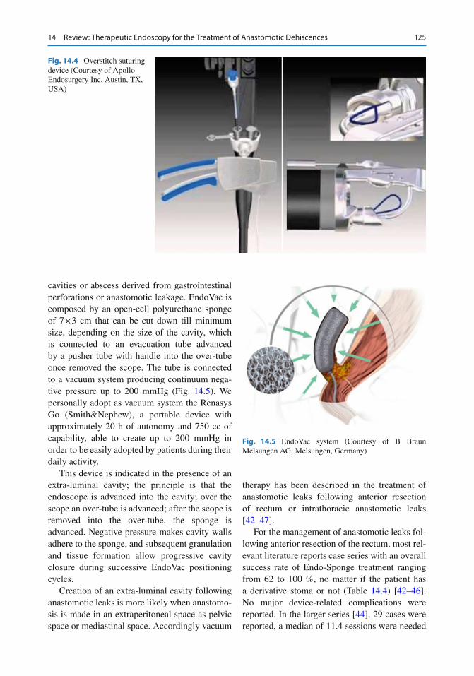
125
cavities or abscess derived from gastrointestinal perforations or anastomotic leakage. EndoVac is composed by an open-cell polyurethane sponge of 7 × 3 cm that can be cut down till minimum size, depending on the size of the cavity, which is connected to an evacuation tube advanced by a pusher tube with handle into the over-tube once removed the scope. The tube is connected to a vacuum system producing continuum nega-tive pressure up to 200 mmHg (Fig. 14.5 ). We personally adopt as vacuum system the Renasys Go (Smith&Nephew), a portable device with approximately 20 h of autonomy and 750 cc of capability, able to create up to 200 mmHg in order to be easily adopted by patients during their daily activity.
This device is indicated in the presence of an extra-luminal cavity; the principle is that the endoscope is advanced into the cavity; over the scope an over-tube is advanced; after the scope is removed into the over-tube, the sponge is advanced. Negative pressure makes cavity walls adhere to the sponge, and subsequent granulation and tissue formation allow progressive cavity closure during successive EndoVac positioning cycles.
Creation of an extra-luminal cavity following anastomotic leaks is more likely when anastomo-sis is made in an extraperitoneal space as pelvic space or mediastinal space. Accordingly vacuum
therapy has been described in the treatment of anastomotic leaks following anterior resection of rectum or intrathoracic anastomotic leaks [ 42 – 47 ].
For the management of anastomotic leaks fol-lowing anterior resection of the rectum, most rel-evant literature reports case series with an overall success rate of Endo-Sponge treatment ranging from 62 to 100 %, no matter if the patient has a derivative stoma or not (Table 14.4 ) [ 42 – 46 ]. No major device-related complications were reported. In the larger series [ 44 ], 29 cases were reported, a median of 11.4 sessions were needed
Fig. 14.4 Overstitch suturing device (Courtesy of Apollo Endosurgery Inc, Austin, TX, USA)
Fig. 14.5 EndoVac system (Courtesy of B Braun Melsungen AG, Melsungen, Germany)
14 Review : Therapeutic Endoscopy for the Treatment of Anastomotic Dehiscences
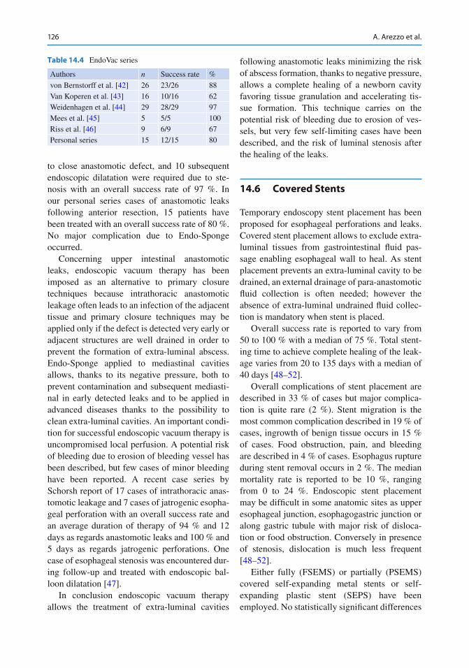
126
to close anastomotic defect, and 10 subsequent endoscopic dilatation were required due to ste-nosis with an overall success rate of 97 %. In our personal series cases of anastomotic leaks following anterior resection, 15 patients have been treated with an overall success rate of 80 %. No major complication due to Endo-Sponge occurred.
Concerning upper intestinal anastomotic leaks, endoscopic vacuum therapy has been imposed as an alternative to primary closure techniques because intrathoracic anastomotic leakage often leads to an infection of the adjacent tissue and primary closure techniques may be applied only if the defect is detected very early or adjacent structures are well drained in order to prevent the formation of extra-luminal abscess. Endo-Sponge applied to mediastinal cavities allows, thanks to its negative pressure, both to prevent contamination and subsequent mediasti-nal in early detected leaks and to be applied in advanced diseases thanks to the possibility to clean extra-luminal cavities. An important condi-tion for successful endoscopic vacuum therapy is uncompromised local perfusion. A potential risk of bleeding due to erosion of bleeding vessel has been described, but few cases of minor bleeding have been reported. A recent case series by Schorsh report of 17 cases of intrathoracic anas-tomotic leakage and 7 cases of jatrogenic esopha-geal perforation with an overall success rate and an average duration of therapy of 94 % and 12 days as regards anastomotic leaks and 100 % and 5 days as regards jatrogenic perforations. One case of esophageal stenosis was encountered dur-ing follow-up and treated with endoscopic bal-loon dilatation [ 47 ].
In conclusion endoscopic vacuum therapy allows the treatment of extra-luminal cavities
following anastomotic leaks minimizing the risk of abscess formation, thanks to negative pressure, allows a complete healing of a newborn cavity favoring tissue granulation and accelerating tis-sue formation. This technique carries on the potential risk of bleeding due to erosion of ves-sels, but very few self-limiting cases have been described, and the risk of luminal stenosis after the healing of the leaks.
14.6 Covered Stents
Temporary endoscopy stent placement has been proposed for esophageal perforations and leaks. Covered stent placement allows to exclude extra- luminal tissues from gastrointestinal fl uid pas-sage enabling esophageal wall to heal. As stent placement prevents an extra-luminal cavity to be drained, an external drainage of para-anastomotic fl uid collection is often needed; however the absence of extra-luminal undrained fl uid collec-tion is mandatory when stent is placed.
Overall success rate is reported to vary from 50 to 100 % with a median of 75 %. Total stent-ing time to achieve complete healing of the leak-age varies from 20 to 135 days with a median of 40 days [ 48 – 52 ].
Overall complications of stent placement are described in 33 % of cases but major complica-tion is quite rare (2 %). Stent migration is the most common complication described in 19 % of cases, ingrowth of benign tissue occurs in 15 % of cases. Food obstruction, pain, and bleeding are described in 4 % of cases. Esophagus rupture during stent removal occurs in 2 %. The median mortality rate is reported to be 10 %, ranging from 0 to 24 %. Endoscopic stent placement may be diffi cult in some anatomic sites as upper esophageal junction, esophagogastric junction or along gastric tubule with major risk of disloca-tion or food obstruction. Conversely in presence of stenosis, dislocation is much less frequent [ 48 – 52 ].
Either fully (FSEMS ) or partially (PSEMS ) covered self-expanding metal stents or self- expanding plastic stent (SEPS) have been employed. No statistically signifi cant differences
Table 14.4 EndoVac series
Authors n Success rate %
von Bernstorff et al. [ 42 ] 26 23/26 88 Van Koperen et al. [ 43 ] 16 10/16 62 Weidenhagen et al. [ 44 ] 29 28/29 97 Mees et al. [ 45 ] 5 5/5 100 Riss et al. [ 46 ] 9 6/9 67 Personal series 15 12/15 80
A. Arezzo et al.
127
on overall success rate have been reported among the kind of stents mentioned above [ 48 – 52 ]. A statistically signifi cant greater risk of tis-sue ingrowth has been described for partially covered metal stent, while a greater risk of stent migration has been reported for fully covered stent [ 53 , 54 ].
Partially covered stent should be removed or replaced after 2–4 weeks in order to avoid tissue ingrowth and subsequent complications at the time of stent removal. Fully covered stent may stay in place for a longer period (5–7 weeks).
Few cases of colorectal anastomotic leaks are reported with the use of covered stents. The principle is to place a covered stent across the colonic dehiscence in order to avoid leakage-related complication while it heals. Potential drawback is migration or expulsion of the stent in the absence of stenosis, the risk of colonic per-foration and the potential intolerance and tenes-mus from the patient if the stent is placed in the low rectum. A randomized controlled trial on porcine models has been published on 16 cases of colorectal anastomotic leaks. No stent-related major complication were reported, all the stents were expelled within ninth postoperative day. In the stent group there was no leakage-related complication, whereas in the control group 63 % developed intra-abdominal infection. On histol-ogy, lastly, the study group had statistically sig-nifi cant less visceral wall interruption than the control group [ 55 ].
Therefore, endoscopic covered stent as treat-ment of colorectal anastomotic leaks may be con-sidered in the presence of a stenosis owing to the high risk of migration in its absence.
14.7 Other Techniques
Fibrin glue or cyanoacrylate injection may be an option in the treatment of anastomotic leakage as an alternative option or in addiction to other techniques.
Fibrin glue is a biologic formulation made up of fi brinogen and thrombin, owing to create a fi brin clot, injected into the margin of a gastroin-testinal leak that acts as tissue adhesive.
Cyanoacrylate is an acrylic resin that rapidly polymerises in the presence of hydroxide ions forming long and strong chains that bond together two surfaces.
These techniques may be an option in the presence of little leaks or fi stulas. Overall suc-cess rate in healing anastomotic leakage is reported to range from 20 to 50 %. A large report by Lippert et al. [ 56 ] on 52 patients with gastrointestinal anastomotic leakage treated with fi brin glue injection showed an overall suc-cess rate of 36 % when glue is used alone and 55 % when combined with other endoscopic techniques. Surgery was necessary in 34 % of cases and mortality rate was 20 %. No major complications associated to endoscopic tech-niques were reported. The presence of major infection seemed to be a negative prognostic factor of the success of conservative manage-ment (50 % of these patients needed surgery versus 13 %) and seemed to defi ne a subgroup of patients with poorer outcome. Each treatment requires multiple session.
Overall success rate of tissue adhesive injec-tion in healing small gastrointestinal perforation seems to be inferior to the one reported for syn-thesis devices as endoclips, even if the need of surgery is however reduced. These techniques may be an option in the presence of small gastro-intestinal defects or fi stula in the absence of major infection or symptoms or when application of other clipping devices is not technically feasi-ble or combined with other synthesis or suturing devices.
Conclusion
Recent technology improvement allows today endoscopy to offer many alternative treatment options to surgery in the management of selected cases of anastomotic leakage. Urgent reintervention for anastomotic dehiscence car-ries on high morbidity rate and a signifi cant percentage of mortality. In selected cases the use of endoscopic devices allows to decrease the need of surgery and, thanks to the low complication rate of endoscopic management if compared to surgery, allows to decrease overall morbidity and mortality rate.
14 Review : Therapeutic Endoscopy for the Treatment of Anastomotic Dehiscences
128
An absolute contraindication to endoscopic management is considered hemodynamic instability, the presence of free intraperitoneal leak or high-grade sepsis with hypotension. In these cases, the patient has to be resuscitated and surgery is mandatory.
Early diagnosis is an important prognostic factor on the success of endoscopic treatment because overall success rate is signifi cantly higher when vital tissues are treated.
As regards colorectal anastomosis dehis-cence, conventional surgical management consists in the creation of a diverting stoma. This is necessary in about 80–90 % of cases and carries on up to 30 % stoma-related com-plication and a reversal rate of less than 50 %. A recent prospective report on 20 consecutive patients with anastomotic leaks after anterior resection of rectum compares upfront endo-scopic and surgical management. As showed in Table 14.5 , stoma creation was needed in 86 % versus 54 % in the endoscopic group. The two groups were comparable as regards clinical features, and no major complication due to endoscopic treatment was reported [ 57 ]. This signifi es improvements of quality of life due to the lower stoma rate creation, and as a consequence lower stoma-related complica-tions, in endoscopically treated patients.
In general a small leakage up to 20 mm of diameter may be managed with endoscopic clips, OTSC clips having showed the higher success rate. Fibrin glue injection may be used combined to other endoscopic techniques or alone if leakage or fi stula is not clinically sig-nifi cant or clipping is not technically feasible.
Bigger anastomotic dehiscence >30 % of the circumference can be treated with EndoVac therapy if an extra-luminal cavity is present. This happens most of the time in extra-cavital districts as extraperitoneal pelvis or mediastinum. EndoVac allows drainage of extra-luminal abscess as well as healing of the
defect thanks to tissue granulation preventing contamination from gastrointestinal contents.
Partially or fully covered stents may be used and temporally taken in place owing to exclude extra-luminal tissues from con-tamination while healing anastomotic leaks. Absence of extra- luminal infection or its good external drainage is necessary. It can be an option in intrathoracic leaks even if its success probability is lower at the level of esophagogastric junction, upper esophageal sphincter, or sometimes in esophagogastric anastomosis. The presence of concomitant stenosis improves success rate decreasing covered stent displacement.
It is desirable that future technological improvements, mainly in the fi eld of synthesis and suturing devices, will increase possibility to treat endoscopically gastrointestinal tract surgical complications.
References
1. Peel AL, Taylor EW (1991) Proposed defi nitions for the audit of postoperative infection: a discussion paper. Surgical Infection Study Group. Ann R Coll Surg Engl 73:385–388
2. Hyman N, Manchester TL, Osler T et al (2007) Anastomotic leaks after intestinal anastomosis: it’s later than you think. Ann Surg 245:254–258
3. Law WI, Chu KW, Ho JW, Chan CW (2000) Risk factors for anastomotic leakage after low anterior resection with total mesorectal excision. Am J Surg 179:92–96
4. Rullier E, Laurent C, Garrelon JL et al (1998) Risk factors for anastomotic leakage after resection of rec-tal cancer. Br J Surg 85:355–358
5. Alves A, Panis Y, Trancart D et al (2002) Factors associated with clinically signifi cant anastomotic leakage after large bowel resection: multivariate anal-ysis of 707 patients. World J Surg 26:499–502
6. Lipska MA, Bissett IP, Parry BR, Merrie AE (2006) Anastomotic leak- age after lower gastrointestinal anastomosis: men are at a higher risk. ANZ J Surg 76:579–585
7. Biondo S, Pares D, Kreisler E et al (2005) Anastomotic dehiscence after resection and primary anastomosis in
Table 14.5 Stoma creation rate on endoscopic vs surgical groups
Treatment Sepsis (%) Secondary stoma (%) Mortality Hospital stay
Surgery 43 86 0 26 Endoscopy 38.5 54 0 25
A. Arezzo et al.
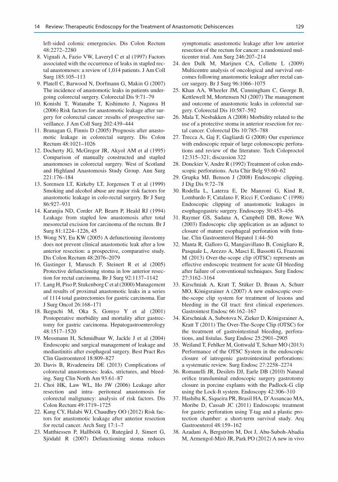
129
left-sided colonic emergencies. Dis Colon Rectum 48:2272–2280
8. Vignali A, Fazio VW, LaveryI C et al (1997) Factors associated with the occurrence of leaks in stapled rec-tal anastomoses: a review of 1,014 patients. J Am Coll Surg 185:105–113
9. Platell C, Barwood N, Dorfmann G, Makin G (2007) The incidence of anastomotic leaks in patients under-going colorectal surgery. Colorectal Dis 9:71–79
10. Konishi T, Watanabe T, Kishimoto J, Nagawa H (2006) Risk factors for anastomotic leakage after sur-gery for colorectal cancer :results of prospective sur-veillance. J Am Coll Surg 202:439–444
11. Branagan G, Finnis D (2005) Prognosis after anasto-motic leakage in colorectal surgery. Dis Colon Rectum 48:1021–1026
12. Docherty JG, McGregor JR, Akyol AM et al (1995) Comparison of manually constructed and stapled anastomoses in colorectal surgery. West of Scotland and Highland Anastomosis Study Group. Ann Surg 221:176–184
13. Sorensen LT, Kirkeby LT, Jorgensen T et al (1999) Smoking and alcohol abuse are major risk factors for anastomotic leakage in colo-rectal surgery. Br J Surg 86:927–931
14. Karanjia ND, Corder AP, Bearn P, Heald RJ (1994) Leakage from stapled low anastomosis after total mesorectal excision for carcinoma of the rectum. Br J Surg 81:1224–1226, 45
15. Wong NY, Eu KW (2005) A defunctioning ileostomy does not prevent clinical anastomotic leak after a low anterior resection: a prospective, comparative study. Dis Colon Rectum 48:2076–2079
16. Gastinger I, Marusch F, Steinert R et al (2005) Protective defunctioning stoma in low anterior resec-tion for rectal carcinoma. Br J Surg 92:1137–1142
17. Lang H, Piso P, Stukenborg C et al (2000) Management and results of proximal anastomotic leaks in a series of 1114 total gastrectomies for gastric carcinoma. Eur J Surg Oncol 26:168–171
18. Ikeguchi M, Oka S, Gomyo Y et al (2001) Postoperative morbidity and mortality after gastrec-tomy for gastric carcinoma. Hepatogastroenterology 48:1517–1520
19. Messmann H, Schmidbaur W, Jackle J et al (2004) Endoscopic and surgical management of leakage and mediastinitis after esophageal surgery. Best Pract Res Clin Gastroenterol 18:809–827
20. Davis B, Rivadeneira DE (2013) Complications of colorectal anastomoses: leaks, strictures, and bleed-ing. Surg Clin North Am 93:61–87
21. Choi HK, Law WL, Ho JW (2006) Leakage after resection and intra- peritoneal anastomosis for colorectal malignancy: analysis of risk factors. Dis Colon Rectum 49:1719–1725
22. Kang CY, Halabi WJ, Chaudhry OO (2012) Risk fac-tors for anastomotic leakage after anterior resection for rectal cancer. Arch Surg 17:1–7
23. Matthiessen P, Hallböök O, Rutegård J, Simert G, Sjödahl R (2007) Defunctioning stoma reduces
symptomatic anastomotic leakage after low anterior resection of the rectum for cancer: a randomized mul-ticenter trial. Ann Surg 246:207–214
24. den Dulk M, Marijnen CA, Collette L (2009) Multicentre analysis of oncological and survival out-comes following anastomotic leakage after rectal can-cer surgery. Br J Surg 96:1066–1075
25. Khan AA, Wheeler JM, Cunningham C, George B, Kettlewell M, Mortensen NJ (2007) The management and outcome of anastomotic leaks in colorectal sur-gery. Colorectal Dis 10:587–592
26. Mala T, Nesbakken A (2008) Morbidity related to the use of a protective stoma in anterior resection for rec-tal cancer. Colorectal Dis 10:785–788
27. Trecca A, Gaj F, Gagliardi G (2008) Our experience with endoscopic repair of large colonoscopic perfora-tions and review of the literature. Tech Coloproctol 12:315–321; discussion 322
28. Donckier V, Andre R (1992) Treatment of colon endo-scopic perforations. Acta Chir Belg 93:60–62
29. Grupka MJ, Benson J (2008) Endoscopic clipping. J Dig Dis 9:72–78
30. Rodella L, Laterza E, De Manzoni G, Kind R, Lombardo F, Catalano F, Ricci F, Cordiano C (1998) Endoscopic clipping of anastomotic leakages in esophagogastric surgery. Endoscopy 30:453–456
31. Raymer GS, Sadana A, Campbell DB, Rowe WA (2003) Endoscopic clip application as an adjunct to closure of mature esophageal perforation with fi stu-lae. Clin Gastroenterol Hepatol 1:44–50
32. Manta R, Galloro G, Mangiavillano B, Conigliaro R, Pasquale L, Arezzo A, Masci E, Bassotti G, Frazzoni M (2013) Over-the-scope clip (OTSC) represents an effective endoscopic treatment for acute GI bleeding after failure of conventional techniques. Surg Endosc 27:3162–3164
33. Kirschniak A, Kratt T, Stüker D, Braun A, Schurr MO, Königsrainer A (2007) A new endoscopic over-the- scope clip system for treatment of lesions and bleeding in the GI tract: fi rst clinical experiences. Gastrointest Endosc 66:162–167
34. Kirschniak A, Subotova N, Zieker D, Königsrainer A, Kratt T (2011) The Over-The-Scope Clip (OTSC) for the treatment of gastrointestinal bleeding, perfora-tions, and fi stulas. Surg Endosc 25:2901–2905
35. Weiland T, Fehlker M, Gottwald T, Schurr MO (2013) Performance of the OTSC System in the endoscopic closure of iatrogenic gastrointestinal perforations: a systematic review. Surg Endosc 27:2258–2274
36. Romanelli JR, Desilets DJ, Earle DB (2010) Natural orifi ce transluminal endoscopic surgery gastrotomy closure in porcine explants with the Padlock-G clip using the Lock-It system. Endoscopy 42:306–310
37. Hashiba K, Siqueira PR, Brasil HA, D’Assuncao MA, Moribe D, Cassab JC (2011) Endoscopic treatment for gastric perforation using T-tag and a plastic pro-tection chamber: a short-term survival study. Arq Gastroenterol 48:159–162
38. Azadani A, Bergström M, Dot J, Abu-Suboh-Abadia M, Armengol-Mirò JR, Park PO (2012) A new in vivo
14 Review : Therapeutic Endoscopy for the Treatment of Anastomotic Dehiscences
130
method for testing closures of gastric NOTES inci-sions using leak of the closure or gastric yield as end-points. J Laparoendosc Adv Surg Tech A 22:46–50
39. Bergström M, Swain P, Park PO (2008) Early clinical experience with a new fl exible endoscopic suturing method for natural orifi ce transluminal endoscopic surgery and intraluminal endosurgery. Gastrointest Endosc 67:528–533
40. Bonin EA, Wong Kee Song LM, Gostout ZS, Bingener J, Gostout CJ (2012) Closure of a per-sistent esophagopleural fi stula assisted by a novel endoscopic suturing system. Endoscopy 44(Suppl 2 UCTN):E8–E9
41. Armengol-Miro JR, Dot J, Abu-Suboh Abadia M, Masachs M, Salord JC, Armengol Bertroli J, Benages A, Kantsevoy SV (2011) New endoscopic suturing device for closure of chronic gastro-cutaneous fi s-tula in an immunocompromised patient. Endoscopy 43(Suppl 2 UCTN):E403–E404
42. von Bernstorff W, Glitsch A, Schreiber A, Partecke LI, Heidecke CD (2009) ETVARD (endoscopic trans-anal vacuum-assisted rectal drainage) leads to com-plete but delayed closure of extraperitoneal rectal anastomotic leakage cavities following neoadjuvant radiochemotherapy. Int J Colorectal Dis 24:819–825
43. van Koperen PJ, van Berge Henegouwen MI, Rosman C, Bakker CM, Heres P, Slors JF, Bemelman WA (2009) The Dutch multicenter experience of the endo- sponge treatment for anastomotic leakage after colorectal surgery. Surg Endosc 23:1379–1383
44. Weidenhagen R, Gruetzner KU, Wiecken T, Spelsberg F, Jauch KW (2008) Endoluminal vacuum therapy for the treatment of anastomotic leakage after anterior rectal resection. Rozhl Chir 87:397–402
45. Mees ST, Palmes D, Mennigen R, Senninger N, Haier J, Bruewer M (2008) Endo-vacuum assisted closure treatment for rectal anastomotic insuffi ciency. Dis Colon Rectum 51:404–410
46. Riss S, Stift A, Kienbacher C, Dauser B, Haunold I, Kriwanek S, Radlsboek W, Bergmann M (2010) Recurrent abscess after primary successful endo- sponge treatment of anastomotic leakage following rectal surgery. World J Gastroenterol 16:4570–4574
47. Schorsch T, Müller C, Loske G (2012) Endoscopic vacuum therapy of anastomotic leakage and iatro-genic perforation in the esophagus. Surg Endosc 27:2040–2045
48. Peters JH, Craanen ME, van der Peet DL et al (2006) Self-expanding metal stents for the treatment of intra-thoracic esophageal anastomotic leaks following esophagectomy. Am J Gastroenterol 101:1393–1395
49. Schubert D, Scheidbach H, Kuhn R et al (2005) Endoscopic treatment of thoracic esophageal anasto-motic leaks by using silicone-covered, self-expanding polyester stents. Gastrointest Endosc 61:891–896
50. Radecke K, Gerken G, Treichel U (2005) Impact of a self-expanding, plastic esophageal stent on various esophageal stenoses, fi stulas, and leak- ages: a single- center experience in 39 patients. Gastrointest Endosc 61:812–818
51. Kauer WK, Stein HJ, Dittler HJ et al (2008) Stent implantation as a treatment option in patients with thoracic anastomotic leaks after esophagectomy. Surg Endosc 22:50–53
52. Langer FB, Wenzl E, Prager G et al (2005) Management of postoperative esophageal leaks with the Polyfl ex self-expanding covered plastic stent. Ann Thorac Surg 79:398–403
53. Gelbmann CM, Ratiu NL, Rath HC et al (2004) Use of self-expandable plastic stents for the treatment of esophageal perforations and symptomatic anasto-motic leaks. Endoscopy 36:695–699
54. van Boeckel PG, Dua KS, Weusten BL, Schmits RJ, Surapaneni N, Timmer R, Vleggaar FP, Siersema PD (2012) Fully covered self-expandable metal stents (SEMS), partially covered SEMS and self- expandable plastic stents for the treatment of benign esophageal ruptures and anastomotic leaks. BMC Gastroenterol 12:19
55. Tsereteli Z, Sporn E, Geiger TM, Cleveland D, Frazier S, Rawlings A, Bachman SL, Miedema BW, Thaler K (2008) Placement of covered polyester stent prevents complications from a colorectal anastomotic leak and supports healing: randomized controlled trial in a large animal model. Surgery 144:786–792
56. Lippert E, Kleb FH, Schweller F, Ott C (2011) Fibrin glue in the endoscopic treatment of fi stulae and anas-tomotic leakages of the gastrointestinal tract. Int J Colorectal Dis 26:303–311
57. Chopra SS, Mrak K, Hünerbein M (2009) The effect of endoscopic treatment on healing of anastomotic leaks after anterior resection of rectal cancer. Surgery 145:182–188
A. Arezzo et al.

131G. Galloro (ed.), Endoscopic Follow-up of Digestive Anastomosis, DOI 10.1007/978-88-470-5370-0_15, © Springer-Verlag Italia 2014
15.1 Background
Ulceration and bleeding from a surgical anasto-mosis is a relatively unusual complication which has been reported to occur following 2–4 % [ 1 – 4 ] of upper GI surgical procedures.
The true incidence is diffi cult to ascertain as most reports are retrospective and prone to bias. The largest retrospective series consisting of data from 5,839 gastrectomies reported an overall com-plication rate of 10.5 % but included no reports of bleeding from a surgical anastomosis [ 5 ].
A retrospective review of 40 cases of inci-sional hernia repair following open gastric bypass surgery reported one case of anastomic ulceration and bleeding requiring emergency surgery [ 6 ]. A larger and more recent retrospective review of outcomes of 1,213 patients who had undergone laparoscopic gastric bypass for morbid obesity identifi ed ten patients with perforated ulcers at the gastrojejunal anastomosis [ 7 ]. However, this study only included perforated ulcers and the incidence of all anastomotic ulceration, follow-ing bariatric surgery, was found to be higher (2.4 %) in another large review of outcomes in 540 patients [ 8 ].
Perhaps the most likely anastomosis to ulcerate and bleed is following pancreatoduodenectomy. In a retrospective study [ 9 ], 18 % (9/53) of patients who had undergone a Whipple’s procedure with Roux-en-Y gastrojejunostomy developed an anas-tomotic ulcer and 6 of cases presented with bleed-ing. In contrast, in a group of 33 patients who had undergone Billroth-II type of reconstruction, there was only one reported anastomotic ulcer. Anastomotic ulceration has also been described after small bowel transplantation [ 10 ].
Less commonly, anastomotic ulceration and bleeding has also been described following lower gastrointestinal procedures. A retrospective search for anastomotic ulceration following ileo-colonic anastomosis was carried at the Mayo Clinic Arizona. Over a 5-year period, only 6 such cases were reported, giving a calculated inci-dence of 0.06 %. These patients all presented with anemia which resolved with iron supple-mentation and stopping NSAID therapy or treat-ing the underlying infl ammatory bowel disease [ 11 ]. This study may have missed cases of ulcer-ation as another, smaller but more detailed study of 1,316 laparoscopic colectomies, reported 29 cases of postoperative bleeding [ 12 ].
15.2 Etiology of Bleeding
The etiology of bleeding is unknown. Following Roux-en-Y gastrojejunostomy, lack of inactiva-tion of pepsin by bile acids, ischemia [ 13 ], or reverse peristalsis have all been proposed as an
B. Rembacken , MD Department of Gastroenterology, St. James University Hospital , Leeds , UK e-mail: [email protected]
15 Hemostatic Procedures in the Bleeding Anastomosis
Bjorn Rembacken
132
underlying mechanism. For this reason, prophylactic therapy with proton pump inhibitors and even bilateral truncal vagotomy has been advocated [ 14 – 16 ]. In a systematic review com-paring open versus laparoscopic Roux-en-Y gas-tric bypass, the risk of bleeding was found to be signifi cantly higher following the laparoscopic procedure, suggesting that ischemic injury may be an important etiological factor [ 3 ].
15.3 Management of Extra- Luminal Bleeding
Following surgery, patients may bleed into the abdominal cavity or into the lumen from the anastomosis [ 17 ]. Some have proposed that the primary role of placing drains at the site of anas-tomosis is to allow the early diagnosis of postop-erative hemorrhage and to distinguish this from an anastomotic or staple line leak [ 18 ]. However, drains are not always a reliable indicator, particu-larly if the bleeding is intraluminal. For this rea-son, monitoring of clinical signs is very important in the postoperative patient. A large quantity of bloody fl uid from the abdominal drains, hemo-peritoneum, tachycardia, a drop in the hemoglo-bin level, bright red blood per rectum, hematemesis, and melena have all been found to indicate postoperative hemorrhage [ 19 ].
Patients with evidence of extra-luminal hem-orrhage usually require abdominal re-exploration using either a laparoscopic or open approach. The operative goals are to evacuate the majority of the clots, attempt to identify and control the site of hemorrhage, or, if there is no obvious bleeding point, to oversew all staple lines.
In an interesting study [ 20 ], endoscopy was used intraoperatively to assess the anastomo-sis at the time of primary surgery. A total of 118 patients underwent resectional surgery and 5 were found to have bleeding at the site of the anastomosis at the time of surgery. Unfortunately, in the follow- up phase, a further 8 cases of anas-tomotic bleeding were reported. These cases had not bled at the time of the surgery; it is therefore possible that intraoperative endoscopy identifi es
early, self- limiting bleeding points but is unable to prevent cases of delayed bleeding perhaps developing as a consequence of ischemic injury. A smaller study, using a similar intraoperative endoscopic assessment of the surgical anasto-mosis, reported some benefi t during bariatric procedures [ 21 ]. However, it is still uncertain if this approach should become part of the standard intrasurgical assessment of the anastomosis.
15.4 Role of Endoscopy to Manage Anastomotic Bleeding
There is evidence that bleeding from anastomotic ulcers are diffi cult to treat endoscopically. A retro-spective analysis of 427 patients with bleeding from an anastomotic ulcer reported an odds ratio of 3.39 (95 % CI 1.37–7.29) for therapeutic failure [ 22 ].
In a smaller, retrospective review of 393 patients who had undergone laparoscopic- assisted gastrectomy, all cases of bleeding could be managed endoscopically [ 23 ]. I am only aware of a single report of fatal bleeding from an anas-tomotic ulcer [ 24 ].
Emergency gastroscopy is able to identify the site of bleeding in approximately 95 % of cases of peptic ulcer bleeding [ 25 , 26 ]. However, there is no corresponding data available on the success rate for bleeding anastomoses. Nevertheless, it seems reasonable to initially attempt endoscopy with the aim to (1) identify the source of hemor-rhage, (2) stop the bleeding, (3) assess the risk of rebreeding, and (4) outline a strategy of how the case should be managed if bleeding cannot be stopped or recurs.
Naturally, not every patient who develops bleeding following surgery suffers bleeding from the surgical anastomosis. Other causes to be con-sidered include ischemia, “stress” ulcers, refl ux esophagitis, coagulopathy, and aortoenteric fi s-tula. Patients with an intraluminal upper GI source of bleeding will usually present with hematemesis and/or melena. Intraluminal bleed-ing in the lower GI tract would normally present with rectal bleeding.
B. Rembacken
133
15.4.1 A Team Effort
Patients with a bleeding anastomosis are man-aged by a team approach involving the endosco-pist, surgeon, interventional radiologist, and hematologist in case of massive bleeding. Patients with major comorbidities or who are showing signs of decompensation should be sta-bilized and managed together with an intensivist in the intensive care unit [ 27 ]. Endoscopy after endotracheal intubation should be considered to protect the airway in patients with severe bleed-ing, patients in shock, and patients with respira-tory decompensation.
15.5 Endoscopic Therapies
The management of a patient with early postop-erative gastrointestinal bleeding does not differ signifi cantly from the management of common GI hemorrhage. After the bleeding site has been identifi ed, it should be risk stratifi ed depending on the stigmata of recent hemorrhage (SRH). Major SRH includes active arterial bleeding or a non-bleeding visible vessel [ 28 ]. Intermediate SRH includes an adherent clot on an ulcer or an ulcer that is oozing slightly without concomitant major SRH. Minor SRH includes ulcers with a pigmented fl at spot or a clean ulcer base. Ulcers with major SRH have the greatest risk of contin-ued bleeding or rebleeding without endoscopic therapy, whereas minor SRH have the lowest risk of rebleeding even without endoscopic therapy [ 29 – 32 ]. Ulcers with intermediate SRH have an intermediate risk of bleeding without endoscopic therapy [ 33 ]. Although this stratifi cation was developed for bleeding peptic ulcers, it would be reasonable to assume that the same is true for bleeding ulcers at the anastomotic site.
Bleeding sites should fi rst be treated with an initial injection of dilute adrenaline to provide tamponade. In bleeding peptic ulcers, adrenaline provides initial hemostasis in around 80 % of bleeding peptic ulcers [ 34 , 35 ]. Unfortunately, bleeding may recur after 20 min as the adrenaline is absorbed [ 26 ].
The use of a sclerosing agent such as alcohol or ethanolamine does cause tissue injury at the site of the anastomosis and should be avoided. Furthermore, there is no evidence that they pro-vide any advantage. In a study of 170 cases of bleeding peptic ulceration, injecting a sclerosant did not provide any better hemostasis than adren-aline [ 36 ]. The use of histoacryl tissue glue has been shown to be of benefi t in bleeding gastric varices but is associated with risks of emboliza-tion [ 37 ] and is unproven in the treatment of bleeding from surgical anastomoses.
Fibrin, when injected into the bleeding vessel, may aid the formation of a clot. Unfortunately, clinical studies have not demonstrated any clear advantage over adrenaline in bleeding peptic ulcers. In a European multicenter trial [ 38 ] of more than 800 patients, bleeding ulcers were ini-tially treated with adrenaline. The ulcers were then either injected with sclerosant (group A), fi brin glue (group B), or an intensive program of multiple fi brin glue injections over several days (group C). Initial hemostatic success was similar in the groups (92.5 % vs 95.1 % vs 96.4 %). However, patients in group C were less likely to rebleed than in group A (10.0 % vs 18.1 %). These results were corroborated in a subsequent, smaller study encompassing 135 patients. In this study, bleeding peptic ulcers treated with a single injection of adrenalin were just as likely to rebleed as ulcers treated with single injection fi brin glue (24 % vs 21 %) [ 39 ]. There has been one case report of the successful use of tissue glue to treat a bleeding varix at the site of a surgi-cal anastomosis [ 40 ].
Hemoclip s are short endoscopic clips only available in countries which do not solely rely on disposable equipment (such as the UK). Initial results were very promising with an Italian study reporting a rebleeding rate of 21 % following heater probe therapy versus only 1.8 % after the placement of hemoclips [ 41 ]. Unfortunately, a subsequent study [ 42 ] did not replicate the initial, promising results. Injection therapy achieved ini-tial hemostasis in 85 %, compared with 63 % for hemoclips, or 75 % when a combination of clips and injection therapy was used. At the site of a
15 Hemostatic Procedures in the Bleeding Anastomosis
134
small bowel anastomosis, clips have the theoreti-cal advantage that relatively little tissue injury is incurred by their use. Furthermore, the main shortcoming of hemoclips is that the main simply scrape the surface of a fi rm and sclerotic peptic ulcer. At a fresh, bleeding ulcer at a small bowel anastomosis, less fi brosis may be expected.
Proton pump inhibitor (PPI) therapy is recom-mended for patients with bleeding peptic ulcer-ation as the neutralization of gastric acid stabilizes clots [ 43 – 45 ], reduces the need for endoscopic therapy, and reduces the risk of rebleeding but has no proven effect on mortality [ 46 ]. However, when the bleeding is from an anastomotic ulcer, the effect of PPI therapy is unproven and cannot be recommended.
The largest series reporting on the outcomes of treating a bleeding anastomosis was published by Lee et al. [ 47 ] which included a series of 50 patients with anastomotic ulcer bleeding follow-ing Billroth-II procedures. The bleeding ulcers were usually found at the level of the anastomo-sis (64 %) or at the anastomotic bifurcation (20 %). The ulcers were classifi ed as Forrest Ia (32 %), Ib (38 %), IIa (20 %), or IIb (10 %). A total of 20 patients were treated with hemoclips and 30 with a combination of adrenaline and heater probe coagulation. In all cases, initial hemostasis was achieved. However, the risk of rebleeding was signifi cantly greater (33 % vs 5 %) following adrenaline + heater probe coagu-lation than after endoclip application. The likely reason for this is the combination of tissue isch-emia, a thin small bowel wall and tissue damage from the heater probe. Endoscopic therapy for non-anastomotic bleeding is associated with a 0.5–1.0 % risk of gastrointestinal perforation [ 48 , 49 ] and the risks when treating a bleeding ulcer at a surgical anastomosis are probably greater.
As previously outlined, bleeding from colorec-tal stapling anastomoses is uncommon. In study of 438 patients [ 50 ], only 6 patients were found to have ulceration at the site of the surgical anasto-mosis. Furthermore, the bleeding is less severe and usually settles with supportive care. In a study by Cirocco and Golub [ 51 ], only 1.8 % of patients developed an anastomotic hemorrhage and almost every case (82 %) settled with supportive care.
Conclusion
There is little evidence from published studies to guide the endoscopist when asked to help in the case of a postsurgical intraluminal bleed. Nevertheless, much of the training and tech-niques used in the management of peptic bleeding will also help at the anastomosis. The main caveat should be an awareness that the surgical anastomosis will be thin and less able to withstand thermal therapies. For this reason, injection therapy, followed by the application of clips, is preferable.
References
1. Callery CD, Filiciotto S, Neil KL (2012) Collagen matrix staple line reinforcement in gastric bypass. Surg Obes Relat Dis 8(2):185–189
2. Oida T, Kano H, Mimatsu K, Kawasaki A, Kuboi Y, Fukino N, Kida K, Amano S (2012) Gastric marginal ulcer after pancreaticoduodenectomy with pancreati-cogastrostomy due to delayed gastric emptying and Helicobacter pylori infection. Hepatogastroenterology 59(115):899–902
3. Podnos YD, Jimenez JC, Wilson SE et al (2003) Complications after laparoscopic gastric bypass: a review of 3464 cases. Arch Surg 138:957–961
4. Mehran A, Szomstein S, Zundel N et al (2003) Management of acute bleeding after laparoscopic Roux-en-Y gastric bypass. Obes Surg 13:842–847
5. Kim KM, An JY, Kim HI, Cheong JH, Hyung WJ, Noh SH (2012) Major early complications following open, laparoscopic and robotic gastrectomy. Br J Surg 99(12):1681–1687
6. Shermak MA (2006) Hernia repair and abdomino-plasty in gastric bypass patients. Plast Reconstr Surg 117:1145–1150
7. Kalaiselvan R, Exarchos G, Hamza N, Ammori BJ (2012) Incidence of perforated gastrojejunal anasto-motic ulcers after laparoscopic gastric bypass for morbid obesity and role of laparoscopy in their man-agement. Surg Obes Relat Dis 8(4):423–428
8. Crea N, Pata G, Di Betta E, Greco F, Casella C, Vilardi A, Mittempergher F (2011) Long-term results of biliopancreatic diversion with or without gastric preservation for morbid obesity. Obes Surg 21(2):139–145
9. Arlt G, Peiper C, Winkeltau G, Schumpelick V (1994) Anastomotic ulcers after duodenopancreatectomy for carcinoma of the head of the pancreas]. Wien Klin Wochenschr 106(22):713–717
10. Turner D, Martin S, Ngan BY, Grant D, Sherman PM (2006) Anastomotic ulceration following small bowel transplantation. Am J Transplant 6(1):236–240
B. Rembacken
135
11. Chari ST, Keate RF (2000) Ileocolonic anastomotic ulcers: a case series and review of the literature. Am J Gastroenterol 95(5):1239–1243
12. Kirchhoff P, Dincler S, Buchmann P (2008) A multi-variate analysis of potential risk factors for intra- and postoperative complications in 1316 elective laparo-scopic colorectal procedures. Ann Surg 248:259–265
13. Steinemann DC, Schiesser M, Clavien PA, Nocito A (2011) Laparoscopic gastric pouch and remnant resection: a novel approach to refractory anastomotic ulcers after Roux-en-Y gastric bypass: case report. BMC Surg 11:33
14. Kitagawa M, Ikoma H, Ochiai T, Ishii H, Shiozaki A, Kuriu Y, Nakanishi M, Ichikawa D, Okamoto K, Fujiwara H, Sakakura C, Kokuba Y, Sonoyama T, Otsuji E (2012) Total pancreatectomy for pancreatic carcinoma: evaluation of safety and effi cacy. Hepatogastroenterology 59(115):907–910
15. Piazza L, Ferrara F, Leanza S, Coco D, Sarva S, Bellia A, Di Stefano C, Basile F, Biondi A (2011) Laparoscopic mini-gastric bypass: short-term single- institute experience. Updates Surg 63(4):239–242
16. Safi oleas MC, Moulakakis KG, Andromanakos NP, Lygidakis NJ (2005) How necessary is vagotomy after pancreaticoduodenectomy and total pancreatec-tomy. Hepatogastroenterology 52(61):251–252
17. Nguyen NT, Rivers R, Wolfe BM (2003) Early gastro-intestinal hemorrhage after laparoscopic gastric bypass. Obes Surg 13:62–65
18. Rosenthal RJ, Szomstein S, Kennedy CI et al (2006) Laparoscopic surgery for morbid obesity: 1,001 con-secutive bariatric operations performed at the Bariatric Institute. Clevel Clin Fla Obes Surg 16:119–124
19. Bellorin O, Abdemur A et al (2011) Understanding the signifi cance, reasons and patterns of abnormal vital signs after gastric bypass for morbid obesity. Obes Surg 21:707–713
20. Lieto E, Orditura M, Castellano P, Pinto M, Zamboli A, De Vita F, Pignatelli C, Galizia G (2011) Endoscopic intraoperative anastomotic testing may avoid early gastrointestinal anastomotic complica-tions. A prospective study. J Gastrointest Surg 15(1):145–152
21. Cingi A, Yavuz Y (2011) Intraoperative endoscopic assessment of the pouch and anastomosis during lapa-roscopic Roux-en-Y gastric bypass. Obes Surg 21(10):1530–1534
22. Thomopoulos KC, Mitropoulos JA, Katsakoulis EC, Vagianos CE, Mimidis KP, Hatziargiriou MN, Nikolopoulou VN (2001) Factors associated with fail-ure of endoscopic injection haemostasis in bleeding peptic ulcers. Scand J Gastroenterol 36(6):664–668
23. Lim SG, Lee KM, Kim SS, Kim JS, Hwang JC, Shin SJ, Han SU, Kim JH, Cho SW (2012) Endoscopic approach for postoperative complications following laparoscopic-assisted gastrectomy in early gastric cancer. Hepatogastroenterology 59(116):1308–1312
24. Iannelli A, Amato D, Addeo P, Buratti MS, Damhan M, Ben Amor I, Sejor E, Facchiano E, Gugenheim J (2008) Laparoscopic conversion of vertical banded
gastroplasty (Mason MacLean) into Roux-en-Y gas-tric bypass. Obes Surg 18(1):43–46
25. Chak A et al (2001) Effectiveness of endoscopy in patients admitted to the intensive care unit with upper GI hemorrhage. Gastrointest Endosc 53:6–13
26. Cappell MS, Friedel D (2008) Acute nonvariceal upper gastrointestinal bleeding: endoscopic diagnosis and therapy. Med Clin North Am 92:511–550
27. Romagnuolo J, Barkun AN, Enns R, Armstrong D, Gregor J (2007) Simple clinical predictors may obvi-ate urgent endoscopy in patients with nonvariceal upper gastrointestinal bleeding. Arch Intern Med 167:265–270
28. Freeman ML (1997) Stigmata of hemorrhage in bleeding ulcers. Gastrointest Endosc Clin N Am 7:559–574
29. Kovacs TO, Jensen DM (2007) Endoscopic treatment of ulcer bleeding. Curr Treat Options Gastroenterol 10:143–148
30. Chung SC, Leung JW, Steele RJ, Crofts TJ, Li AK (1988) Endoscopic injection of adrenaline for actively bleeding ulcers: a randomized trial. Br Med J 296:1631–1633
31. Kovacs TO (2008) Management of upper gastrointes-tinal bleeding. Curr Gastroenterol Rep 10:535–542
32. Savides TJ, Jensen DM (2000) Therapeutic endos-copy for nonvariceal gastrointestinal bleeding. Gastroenterol Clin North Am 29:465–487
33. Kahi CJ et al (2005) Endoscopic therapy versus medi-cal therapy for bleeding peptic ulcer with adherent clot: a meta-analysis. Gastroenterology 129:855–862
34. Calvet X, Vergara M, Brullet E, Gisbert JP, Campo R (2004) Addition of a second endoscopic treatment fol-lowing epinephrine injection improves outcome in high-risk bleeding ulcers. Gastroenterology 126:441–450
35. Lin HJ et al (2002) A prospective, randomized trial of large- versus small-volume endoscopic injection of epinephrine for peptic ulcer bleeding. Gastrointest Endosc 55:615–619
36. Chung SCS, Seong HAT, Chan ACW et al (1996) Epinephrine or epinephrine plus alcohol for injection of bleeding ulcers. Gastrointest Endosc 43:591–595
37. Lee GH, Kim JH, Lee KJ et al (2000) Life-threatening intraabdominal arterial embolization after histoacryl. Injection for bleeding gastric ulcer. Endoscopy 32(5):422–424
38. Rutgeerts P, Rauws E, Wara P et al (1997) Randomised trial of single and repeated fi brin glue compares with injection of polidocanol in treatment of bleeding pep-tic ulcer. Lancet 350:662–696
39. Pescatore P, Jornod P, Borovicka J et al (2002) Epinephrine versus epinephrine plus fi brin glue injec-tion in peptic ulcer bleeding. Gastrointest Endosc 55:348–353
40. Chida T, Kageyama F, Yamada M, Yoshii S, Honjo Y et al (2010) Hemorrhage from varices of an interposed jejunum after choledochojejunostomy treated success-fully by endoscopic injection using α-cyanoacrylate monomer. Jpn J Gastroenterol 107(10):1661–1668
15 Hemostatic Procedures in the Bleeding Anastomosis
136
41. Cipolletta L, Bianco MA, Marmo R et al (2001) Endoclips versus heater probe in preventing early recurrent bleeding from peptic ulcer. Gastrointest Endosc 53:147–151
42. Gevers AM, Goede ED, Mimoens M et al (2002) A randomized trial comparing injection therapy with hemoclip and with injection combined with hemoclip for bleeding ulcers. Gastrointest Endosc 55:466–469
43. Andriulli A et al (2005) Proton-pump inhibitors and outcome of endoscopic hemostasis in bleeding peptic ulcers: a series of meta-analyses. Am J Gastroenterol 100:207–219
44. Green FW Jr, Kaplan MM, Curtis LE, Levine PH (1978) Effect of acid and pepsin on blood coagulation and platelet aggregation: a possible contributor to pro-longed gastroduodenal mucosal hemorrhage. Gastroenterology 74:38–43
45. Lau JY et al (2007) Omeprazole before endoscopy in patients with gastrointestinal bleeding. N Engl J Med 356:1631–1640
46. Leontiadis GI et al (2007) Systematic reviews of the clinical effectiveness and cost-effectiveness of proton
pump inhibitors in acute upper gastrointestinal bleed-ing. Health Technol Assess 11:1–164
47. Lee YC, Wang HP, Yang CS et al (2002) Endoscopic hemostasis of a bleeding marginal ulcer: hemoclip-ping or dual therapy with epinephrine injection and heater probe thermocoagulation. J Gastroenterol Hepatol 17:1220–1225
48. Laine L, Cook D (1995) Endoscopic ligation com-pared with sclerotherapy for treatment of esophageal variceal bleeding: a meta-analysis. Ann Intern Med 123:280–287
49. Olmos JA et al (2006) Long-term outcome of argon plasma ablation therapy for bleeding in 100 consecu-tive patients with colonic angiodysplasia. Dis Colon Rectum 49:1507–1516
50. Weinstock LB, Shatz BA (1994) Endoscopic abnormalities of the anastomosis following resec-tion of colonic neoplasm. Gastrointest Endosc 40:558–561
51. Cirocco WC, Golub RW (1995) Endoscopic treatment of postoperative hemorrhage from a staples colorectal anastomosis. Am Surg 61:460–463
B. Rembacken
137G. Galloro (ed.), Endoscopic Follow-up of Digestive Anastomosis, DOI 10.1007/978-88-470-5370-0_16, © Springer-Verlag Italia 2014
16.1 Introduction
With advances in technology and improved surgical techniques, the mortality following bariatric surgery is nowadays less than 1 % in centers of excellence. Approximately 9–25 % of patients have late complications following bariatric surgery [ 1 ]. Considering that bariat-ric surgery is increasing in number annually, endoscopists need to be familiar with the bariat-ric surgical operations and the new endoscopic anatomy in order to diagnose and treat early and late complications [ 2 , 3 ].
Nausea, vomiting, and abdominal pain are among the most commonly encountered symp-toms after bariatric surgery and may result from one or several structural and functional etiolo-gies. Symptoms are frequently associated with dietary noncompliance as to the volume and type of foods eaten, rapid ingestion, or inadequate chewing. Patients with persistent symptoms should be carefully evaluated, because these symptoms may indicate the development of mar-ginal ulcers, gastrogastric fi stulas, or partial or complete anastomotic obstruction.
Endoscopy is the preferred strategy, unless there is a suspicion of leaks or fi stulas, when
contrast radiography is more appropriate. Patient history may be helpful in differentiating the etiol-ogy of pain and in guiding the type of investiga-tion. Nausea, vomiting, abdominal distention, and bloating alone or in conjunction with abdom-inal pain can suggest an obstructive cause, such as strictures. Marginal ulcers are typically seen 1–6 months after surgery and may present with abdominal pain, bleeding, or nausea, although they may also be asymptomatic [ 4 ]. Clinical manifestations of leaks include tachycardia, fever, nausea, vomiting, and abdominal or chest pain.
General principles, when an endoscopy is considered in a patient who had bariatric surgery, involve the endoscopist to be aware of the opera-tive procedure performed and the fi ndings on pre-endoscopy imaging studies; he must under-stand the expected anatomy, including the extent of resection and the length of surgically created limbs. In patients who are in the early postopera-tive period, air insuffl ation may have potentially detrimental effects in the presence of leaks and/or tenuous anastomoses. If there is suspicion of a leak, then again the endoscopist should consider contrast radiography as an initial diagnostic test. Contrast studies are complementary to an endos-copy and are also helpful in delineating anatomy.
Accordingly to each bariatric surgical opera-tion, the next section provides an overview of the most common normal and pathological endoscopic fi ndings. Finally, major anastomotic complications are described, as well as their endoscopic therapy.
A. Genco (*) • R. Maselli • M. Cipriano G. Casella • A. Redler Department of Surgical Sciences , Umberto 1° General Hospital , Vial del Policlinico 155 , Rome , Italy e-mail: [email protected]
16 Endoscopic Treatment of Anastomotic Complications After Bariatric Surgery
Alfredo Genco , Roberta Maselli , Massimiliano Cipriano , Giovanni Casella , and Adriano Redler
138
16.2 Surgical Key Points and Normal Endoscopic Postsurgical Anatomy
Two types of surgical procedures are currently performed:• Restrictive procedures aiming to reduce gas-
tric capacity (vertical banded gastroplasty (VBG ) and laparoscopic adjustable gastric banding (LAGB ))
• Procedures combining a reduction in gastric capacity with maldigestion/malabsorption (Roux-en-Y gastrojejunal bypass, RYGB ), sleeve gastrectomy (SG), or duodenal switch and biliopancreatic diversion (BPD-DS ) Among these, VBG, RYGB, SG, and BPD-DS
involve gut anastomosis creation (Fig. 16.1 ). It is important for endoscopists to understand
the different types of operation, the resulting ana-tomical alterations, and the different complica-tions that can arise following these procedures.
16.2.1 VBG: Vertical Banded Gastroplasty
In this restrictive procedure, both band and staples are used to create a small stomach pouch. The surgery isolates a small section of the stomach for processing food, limiting the size of meals to approximately 1 oz, and slows digestion by forcing the food to pass through a restrictive ring and thence onto the remainder of the gastrointestinal tract. The isolated pouch is usually made using the lesser curvature of the stomach, virtually bypassing the gastric fundus; a trangastric window is made 6–8 cm below the His angle using a circular stapler, and a linear stapler is placed to create a pouch of 30 ml. The narrow outlet of 10–11 mm is surrounded by a non- distensible collar of polypropylene mesh of PTFE (polytetrafl uoroethylene) or a silicon ring, to avoid enlargement. The gastric pouch is small generally 15–30 ml in volume. The expected endoscopic fi ndings after VBG consist of a clean gastric channel 6–8 cm long, with a rosette at the distal end and snug passage of an 11-mm scope without diffi culty. Special care should be made to examine the pouch and suture line for
fi stulas and ulcerations. Retrofl exion of the tip of the endoscope in the distal stomach allows inspection of the caudal aspect of the staple-line partition and the remainder of the gastric fundus.
16.2.2 RYGB: Roux-en-Y Gastric Bypass
Roux-en-Y gastric bypass involves stapling the upper stomach into a small proximal 15–30-ml pouch along the lesser curvature, attached to the jejunum through a narrow (11 mm) anastomosis, bypassing a large part of the stomach and duode-num. There are two anastomoses: gastrojejunal and jejunojejunal.
The gastric pouch is small generally 15–30 ml in volume. The expected endoscopic fi ndings after RYGB include a normal esophagus and gastroesophageal junction. The size of the gas-tric pouch varies. The pouch may have several different confi gurations and may be long and narrow or shot and wide. Special care should be made to examine the pouch and suture line for fi stulas and ulcerations. The gastrojejunal stoma should be easily visible within several centime-ters of the gastroesophageal junction and should be carefully examined. The width of the anas-tomosis is generally 10–12 mm in diameter. Beyond the anastomosis, a short, blind limb is often visible alongside the efferent jejunal limb. The jejunojejunal anastomosis can sometimes be reached with an upper endoscope, depending on the length of the Roux limb. It should be noted that the length of the Roux limb after an RYGB can vary signifi cantly from standard Roux limbs created for nonbariatric procedures and can range from 50 to 150 cm. The distal or excluded stom-ach cannot be visualized in the absence of a fi s-tula with a regular gastroscope.
16.2.3 SG and BPD-DS: Sleeve Gastrectomy and Biliopancreatic Diversion with Duodenal Switch
Biliopancreatic diversion with duodenal switch (BPD-DS) includes both restrictive and malab-
A. Genco et al.
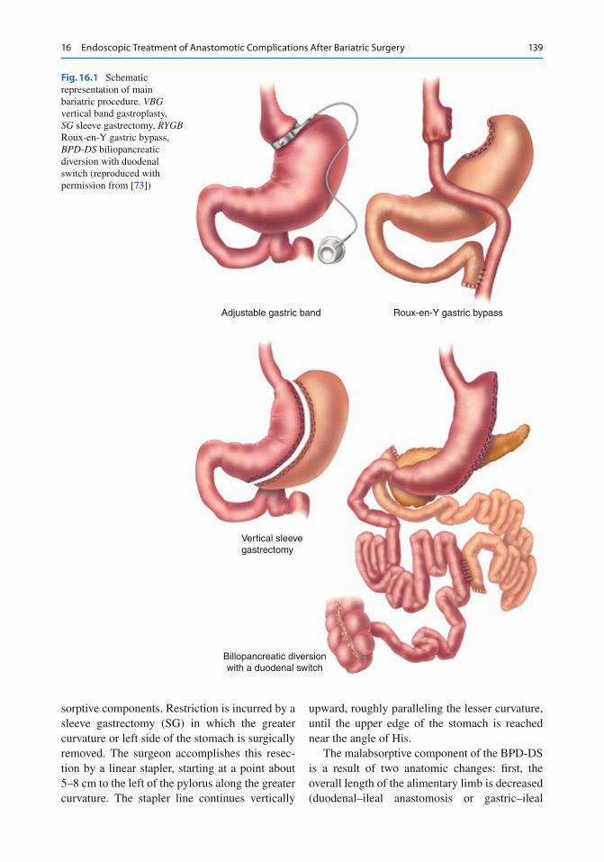
139
sorptive components. Restriction is incurred by a sleeve gastrectomy (SG) in which the greater curvature or left side of the stomach is surgically removed. The surgeon accomplishes this resec-tion by a linear stapler, starting at a point about 5–8 cm to the left of the pylorus along the greater curvature. The stapler line continues vertically
upward, roughly paralleling the lesser curvature, until the upper edge of the stomach is reached near the angle of His.
The malabsorptive component of the BPD-DS is a result of two anatomic changes: fi rst, the overall length of the alimentary limb is decreased (duodenal–ileal anastomosis or gastric–ileal
Adjustable gastric band Roux-en-Y gastric bypass
Vertical sleevegastrectomy
Billopancreatic diversionwith a duodenal switch
Fig. 16.1 Schematic representation of main bariatric procedure. VBG vertical band gastroplasty, SG sleeve gastrectomy, RYGB Roux-en-Y gastric bypass, BPD-DS biliopancreatic diversion with duodenal switch (reproduced with permission from [ 73 ])
16 Endoscopic Treatment of Anastomotic Complications After Bariatric Surgery
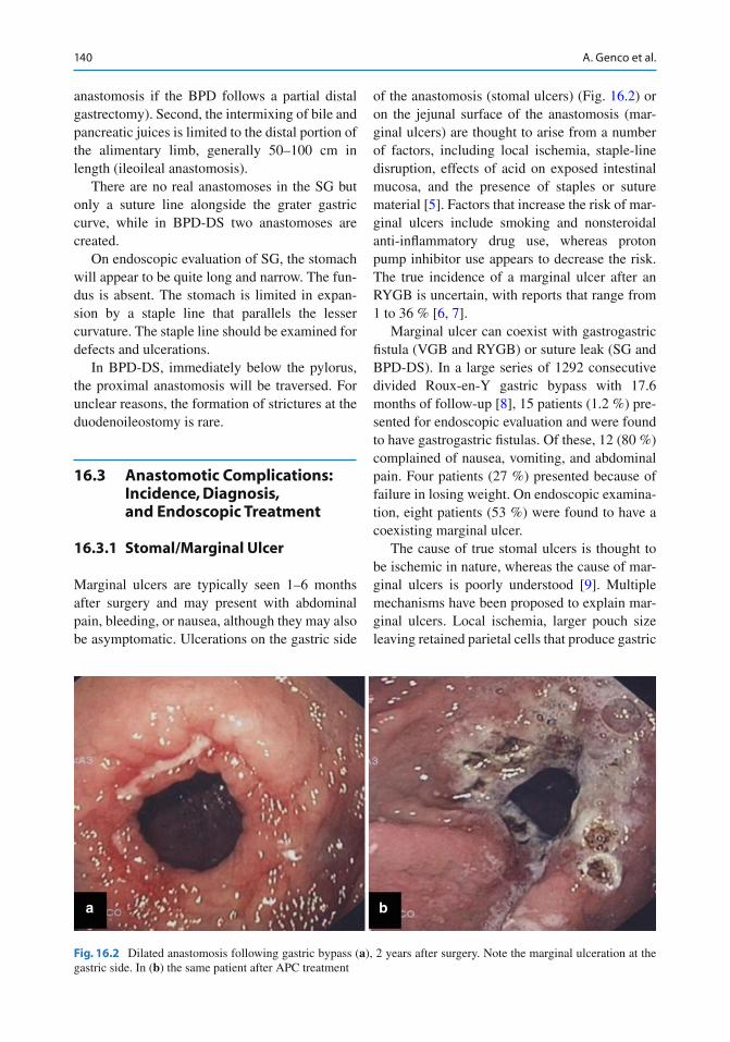
140
anastomosis if the BPD follows a partial distal gastrectomy). Second, the intermixing of bile and pancreatic juices is limited to the distal portion of the alimentary limb, generally 50–100 cm in length (ileoileal anastomosis).
There are no real anastomoses in the SG but only a suture line alongside the grater gastric curve, while in BPD-DS two anastomoses are created.
On endoscopic evaluation of SG, the stomach will appear to be quite long and narrow. The fun-dus is absent. The stomach is limited in expan-sion by a staple line that parallels the lesser curvature. The staple line should be examined for defects and ulcerations.
In BPD-DS, immediately below the pylorus, the proximal anastomosis will be traversed. For unclear reasons, the formation of strictures at the duodenoileostomy is rare.
16.3 Anastomotic Complications: Incidence, Diagnosis, and Endoscopic Treatment
16.3.1 Stomal/Marginal Ulcer
Marginal ulcers are typically seen 1–6 months after surgery and may present with abdominal pain, bleeding, or nausea, although they may also be asymptomatic. Ulcerations on the gastric side
of the anastomosis (stomal ulcers ) (Fig. 16.2 ) or on the jejunal surface of the anastomosis (mar-ginal ulcers) are thought to arise from a number of factors, including local ischemia, staple-line disruption, effects of acid on exposed intestinal mucosa, and the presence of staples or suture material [ 5 ]. Factors that increase the risk of mar-ginal ulcers include smoking and nonsteroidal anti-infl ammatory drug use, whereas proton pump inhibitor use appears to decrease the risk. The true incidence of a marginal ulcer after an RYGB is uncertain, with reports that range from 1 to 36 % [ 6 , 7 ].
Marginal ulcer can coexist with gastrogastric fi stula (VGB and RYGB) or suture leak (SG and BPD-DS). In a large series of 1292 consecutive divided Roux-en-Y gastric bypass with 17.6 months of follow-up [ 8 ], 15 patients (1.2 %) pre-sented for endoscopic evaluation and were found to have gastrogastric fi stulas. Of these, 12 (80 %) complained of nausea, vomiting, and abdominal pain. Four patients (27 %) presented because of failure in losing weight. On endoscopic examina-tion, eight patients (53 %) were found to have a coexisting marginal ulcer.
The cause of true stomal ulcers is thought to be ischemic in nature, whereas the cause of mar-ginal ulcers is poorly understood [ 9 ]. Multiple mechanisms have been proposed to explain mar-ginal ulcers. Local ischemia, larger pouch size leaving retained parietal cells that produce gastric
a b
Fig. 16.2 Dilated anastomosis following gastric bypass ( a ), 2 years after surgery. Note the marginal ulceration at the gastric side. In ( b ) the same patient after APC treatment
A. Genco et al.
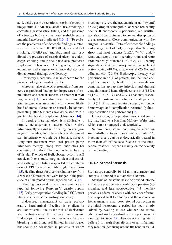
141
acid, acidic gastric secretions poorly tolerated in the jejunum, NSAID use, alcohol use, smoking, a coexisting gastrogastric fi stula, and the presence of a foreign body such as nonabsorbable suture material have been implicated [ 10 – 13 ]. To evalu-ate the predictors of endoscopic fi ndings, a retro-spective review of 1001 RYGB [ 4 ] showed that smoking, NSAID use, and abdominal pain pre-dicted the presence of marginal ulcers at endos-copy; smoking and NSAID use also predicted staple-line dehiscence. Age, gender, surgical technique, and surgeon experience did not pre-dict abnormal fi ndings at endoscopy.
Refractory ulcers should raise concern for the presence of a gastrogastric fi stula.
Moreover, also time of presentation from sur-gery can predicted fi ndings for the presence of sto-mal ulcers and stomal stenosis. In another RYGB large series [ 14 ], presenting more than 6 months after surgery was associated with a lower likeli-hood of stomal ulceration or stenosis. In contrast, presenting after 6 months was associated with a greater likelihood of staple-line dehiscence [ 14 ].
In treating marginal ulcer, it is advisable to remove nonabsorbable sutures when visible intraluminally to assist with healing, prevent gas-trogastric fi stulas, and relieve chronic abdominal pain in patients who underwent bariatric surgery. Long-term treatment with oral proton pump inhibitors therapy, along with antibiotics for coexisting H. pylori infection, has led to healing of fi stula. The role of Helicobacter pylori is still not clear. In one study, marginal ulcer and associ-ated gastrogastric fi stula responded to a combina-tion of PPI therapy and fi brin glue injections [ 15 ]. Healing times for ulcer resolution vary from 8 weeks to 6 months but were longer in the pres-ence of an untreated or undiagnosed fi stula [ 16 ].
Bleeding duodenal ulcers have been rarely reported following Roux-en-Y gastric bypass [ 17 ]. Early postoperative bleeding in RYGB most likely originates at the gastrojejunostomy.
Endoscopic management of early postop-erative intraluminal bleeding is challenging and controversial due to the risk of dehiscence and perforation at the surgical anastomosis. Endoscopy is usually not necessary because bleeding is mild and self-limited in most cases but should be considered in patients in whom
bleeding is severe (hemodynamic instability and/or ≥2 g drop in hemoglobin) or when rebleeding occurs. If endoscopy is performed, air insuffl a-tion should be minimized to prevent disruption of the anastomosis. Close communication with the surgeon is essential. Data of endoscopic fi ndings and management of early postoperative bleeding show that most patients (20/27; 74 %) under-went endoscopy in an operating room and were endotracheally intubated (19/27; 70 %). Bleeding stigmata seen at the gastrojejunostomy included active oozing (48 %), visible vessel (26 %), and adherent clot (26 %). Endoscopic therapy was performed in 85 % of patients and included epi-nephrine injection, heater probe coagulation, combination epinephrine injection and thermal coagulation, and hemoclip placement in 3 (13 %), 4 (17 %), 14 (61 %), and 2 (9 %) patients, respec-tively. Hemostasis was achieved in all patients, but 5 (17 %) patients required surgery to control hemorrhage and complication occurred (pulmo-nary aspiration and perforation) [ 18 ].
On occasion, postoperative nausea and vomit-ing may lead to a bleeding Mallory–Weiss tear, which can be managed endoscopically.
Summarizing, stomal and marginal ulcer can successfully be treated conservatively with PPI; bleeding ulcers can be endoscopically treated in more than 2/3 of the case. Success of the endo-scopic treatment depends mainly on the severity of the bleeding.
16.3.2 Stomal Stenosis
Stomas are generally 10–12 mm in diameter and stenosis is defi ned as a diameter <10 mm.
Stenosis of the stoma has to be divided into the immediate postoperative, early postoperative (<3 months), and late postoperative (>3 months) period, as edema or edema with early scar forma-tion respond well to dilation and the outcome in late scarring is rather poor. Stomal obstruction in the initial postoperative period has been simply solved by waiting to see whether the stomal edema and swelling subside after replacement of a nasogastric tube [ 19 ]. Stenosis occurring later is believed to result from fi brosis or an infl amma-tory reaction (occurring around the band in VGB).
16 Endoscopic Treatment of Anastomotic Complications After Bariatric Surgery
142
Stomal stenosis occurs in as many as 4.73–27 % of patients undergoing RYGB [ 20 , 21 ]. These patients typically present with dysphagia, nausea with vomiting, or early satiety as previ-ously noted. The primary endoscopic intervention is balloon dilation up to 15–18 mm, which has been associated with a greater than 93 % success rate in symptom resolution and subsequent weight loss [ 22 , 23 ] (Fig. 16.3 ). Dilation with Savary-Gilliard bougie (Cook Endoscopy; Winston-Salem, NC, USA) may be considered and is an effective intervention. In one review, both methods required 2–3 sessions of therapy, with a complica-tion rate of 3 % [ 24 ]. Endoscopic fl uoroscopy-guided balloon dilation has been demonstrated safe, effective, and durable [ 25 – 28 ].
Gradual dilation over a few sessions is likely the best; dilatation should not be performed to >15 mm, as this may be associated with future weight gain. For symptomatic patients presenting with refractory vomiting, thiamine repletion should be considered early and before exogenous glucose administration to prevent the precipita-tion of Wernicke encephalopathy [ 29 ].
16.3.3 Gastrogastric Fistula
In VBG and RYGB, the gastric suture line can present one or more dehiscences allowing an opening communication between the pouch and the excluded stomach, known as a gastrogastric fi stula .
Because of the communication, it is a particu-lar type of suture dehiscence, always presenting on endoscopy as fi stula completely or partially
re-epithelized and so far different from other anastomotic leaks.
Most large series report that gastrogastric fi s-tulas occur in 1.2–1.8 % of patients undergoing gastric bypass [ 30 ]. However, incidence rates from zero to as high as 46 % have been reported, with substantial improvements in recent years because of modifi cations in the surgical tech-nique [ 31 ].
Usually patients complain for weight regain or for not being able to lose weight.
Because of the high rate of morbidity and mor-tality associated with surgical revision of gastro-gastric fi stulas, initial treatment has evolved from surgical interventions to endoscopic management, with variable success. Reported endoscopic tech-niques (Fig. 16.4 ) include the use of fi brin glue sealants [ 32 , 33 ], insertion of a Surgisis fi stula plug (Cook Surgical, Inc, Bloomington, IN, USA) with or without a self-expanding stent [ 34 ], endoluminal stent placement [ 35 ], the use of mucosal suturing devices for tissue apposi-tion [ 36 ], and local debridement following argon plasma coagulation [ 37 ]. The optimal method of treatment is unknown, as comparison studies and randomized controlled trials are lacking.
16.3.4 Anastomotic Rupture/Dehiscence/Leaks
Published incidence rates for leaks following bar-iatric surgery range from 0.4 to 26 %, and leaks are associated with a mortality rate of 1.5 % [ 38 ].
Gastric leaks are potentially serious complica-tions of bariatric surgery and occur in 1–6 % of
a b c
Fig. 16.3 Stomal stenosis following gastric bypass ( a ). In ( b ) CRE balloon dilatation and in ( c ) the anastomosis after treatment
A. Genco et al.
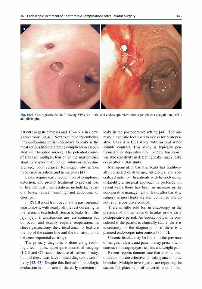
143
patients in gastric bypass and 0.7–4.6 % in sleeve gastrectomy [ 39 , 40 ]. Next to pulmonary embolus, intra-abdominal sepsis secondary to leaks is the most serious life-threatening complication associ-ated with bariatric surgery. The potential causes of leaks are multiple: tension on the anastomosis, staple or stapler malfunction, suture or staple-line seepage, poor surgical technique, obstruction, hypovascularization, and hematomas [ 41 ].
Leaks require early recognition of symptoms, detection, and prompt treatment to prevent loss of life. Clinical manifestations include tachycar-dia, fever, nausea, vomiting, and abdominal or chest pain.
In RYGB most leaks occur at the gastrojejunal anastomosis, with nearly all the rest occurring in the remnant (excluded) stomach; leaks from the jejunojejunal anastomosis are less common but do occur and usually require reoperation. In sleeve gastrectomy, the critical areas for leak are the top of the suture line and the transition point between sequential cartridge .
The primary diagnosis is done using radio-logic techniques: upper gastrointestinal imaging (UGI) and CT scans. Because of patient obesity, both of these tests have limited diagnostic sensi-tivity [ 42 , 43 ]. Despite this limitation, radiologic evaluation is important in the early detection of
leaks in the postoperative setting [ 44 ]. The pri-mary diagnostic tool used to assess for postoper-ative leaks is a UGI study with an oral water soluble contrast. This study is typically per-formed on postoperative day 1 or 2 and has shown variable sensitivity in detecting leaks (many leaks occur after a UGI study).
Management of bariatric leaks has tradition-ally consisted of drainage, antibiotics, and spe-cialized nutrition. In patients with hemodynamic instability, a surgical approach is preferred. In recent years there has been an increase in the nonoperative management of leaks after bariatric surgery as most leaks are well contained and do not require operative control.
There is little role for an endoscopy in the presence of known leaks or fi stulas in the early postoperative period. An endoscopy can be con-sidered if the patient is clinically stable, there is uncertainty of the diagnosis, or if there is a planned endoscopic intervention [ 35 , 45 ].
Chronic fi stulas may be found in the presence of marginal ulcers, and patients may present with nausea, vomiting, epigastric pain, and weight gain.
Recent reports demonstrate that endoluminal interventions are effective in healing anastomotic breeches. Multiple investigators are reporting the successful placement of covered endoluminal
a b
Fig. 16.4 Gastrogastric fi stula following VBG ( a ). In ( b ) and endoscopic view after argon plasma coagulation (APC) and fi brin glue
16 Endoscopic Treatment of Anastomotic Complications After Bariatric Surgery
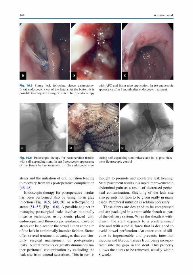
144
stents and the initiation of oral nutrition leading to recovery from this postoperative complication [ 46 – 48 ].
Endoscopic therapy for postoperative fi stulas has been performed also by using fi brin glue injection (Fig. 16.5 ) [ 49 , 50 ] or self-expanding stents [ 51 – 53 ] (Fig. 16.6 ). A possible adjunct in managing postsurgical leaks involves minimally invasive techniques using stents placed with endoscopic and fl uoroscopic guidance. Covered stents can be placed in the bowel lumen at the site of the leak in a minimally invasive fashion. Stents offer several treatment advantages that can sim-plify surgical management of postoperative leaks. A stent prevents or greatly diminishes fur-ther peritoneal contamination by excluding the leak site from enteral secretions. This in turn is
thought to promote and accelerate leak healing. Stent placement results in a rapid improvement in abdominal pain as a result of decreased perito-neal contamination. Shielding of the leak site also permits nutrition to be given orally in many cases. Parenteral nutrition is seldom necessary.
These stents are designed to be compressed and are packaged in a removable sheath as part of the delivery system. When the sheath is with-drawn, the stent expands to a predetermined size and with a radial force that is designed to avoid bowel perforation. An outer coat of sili-cone is impermeable and prevents intestinal mucosa and fi brotic tissues from being incorpo-rated into the gaps in the stent. This property allows the stents to be removed, usually within 8 weeks.
a b c
Fig. 16.5 Suture leak following sleeve gastrectomy. In ( a ) endoscopic view of the fi stula. At the bottom it is possible to recognize a surgical stitch. In ( b ) endotherapy
with APC and fi brin glue application. In ( c ) endoscopic appearance after 1 month after endoscopic treatment
a b c
Fig. 16.6 Endoscopic therapy for postoperative fi stulas with self-expanding stent. In ( a ) fl uoroscopic appearance of the fi stula before treatment. In ( b ) endoscopic view
during self-expanding stent release and in ( c ) post place-ment fl uoroscopic control
A. Genco et al.
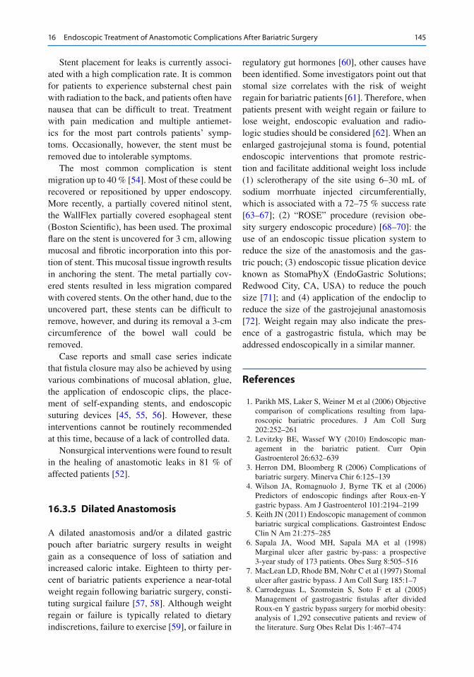
145
Stent placement for leaks is currently associ-ated with a high complication rate. It is common for patients to experience substernal chest pain with radiation to the back, and patients often have nausea that can be diffi cult to treat. Treatment with pain medication and multiple antiemet-ics for the most part controls patients’ symp-toms. Occasionally, however, the stent must be removed due to intolerable symptoms.
The most common complication is stent migration up to 40 % [ 54 ]. Most of these could be recovered or repositioned by upper endoscopy. More recently, a partially covered nitinol stent, the WallFlex partially covered esophageal stent (Boston Scientifi c), has been used. The proximal fl are on the stent is uncovered for 3 cm, allowing mucosal and fi brotic incorporation into this por-tion of stent. This mucosal tissue ingrowth results in anchoring the stent. The metal partially cov-ered stents resulted in less migration compared with covered stents. On the other hand, due to the uncovered part, these stents can be diffi cult to remove, however, and during its removal a 3-cm circumference of the bowel wall could be removed.
Case reports and small case series indicate that fi stula closure may also be achieved by using various combinations of mucosal ablation, glue, the application of endoscopic clips, the place-ment of self-expanding stents, and endoscopic suturing devices [ 45 , 55 , 56 ]. However, these interventions cannot be routinely recommended at this time, because of a lack of controlled data.
Nonsurgical interventions were found to result in the healing of anastomotic leaks in 81 % of affected patients [ 52 ].
16.3.5 Dilated Anastomosis
A dilated anastomosis and/or a dilated gastric pouch after bariatric surgery results in weight gain as a consequence of loss of satiation and increased caloric intake. Eighteen to thirty per-cent of bariatric patients experience a near-total weight regain following bariatric surgery, consti-tuting surgical failure [ 57 , 58 ]. Although weight regain or failure is typically related to dietary indiscretions, failure to exercise [ 59 ], or failure in
regulatory gut hormones [ 60 ], other causes have been identifi ed. Some investigators point out that stomal size correlates with the risk of weight regain for bariatric patients [ 61 ]. Therefore, when patients present with weight regain or failure to lose weight, endoscopic evaluation and radio-logic studies should be considered [ 62 ]. When an enlarged gastrojejunal stoma is found, potential endoscopic interventions that promote restric-tion and facilitate additional weight loss include (1) sclerotherapy of the site using 6–30 mL of sodium morrhuate injected circumferentially, which is associated with a 72–75 % success rate [ 63 – 67 ]; (2) “ROSE” procedure (revision obe-sity surgery endoscopic procedure) [ 68 – 70 ]: the use of an endoscopic tissue plication system to reduce the size of the anastomosis and the gas-tric pouch; (3) endoscopic tissue plication device known as StomaPhyX (EndoGastric Solutions; Redwood City, CA, USA) to reduce the pouch size [ 71 ]; and (4) application of the endoclip to reduce the size of the gastrojejunal anastomosis [ 72 ]. Weight regain may also indicate the pres-ence of a gastrogastric fi stula, which may be addressed endoscopically in a similar manner.
References
1. Parikh MS, Laker S, Weiner M et al (2006) Objective comparison of complications resulting from lapa-roscopic bariatric procedures. J Am Coll Surg 202:252–261
2. Levitzky BE, Wassef WY (2010) Endoscopic man-agement in the bariatric patient. Curr Opin Gastroenterol 26:632–639
3. Herron DM, Bloomberg R (2006) Complications of bariatric surgery. Minerva Chir 6:125–139
4. Wilson JA, Romagnuolo J, Byrne TK et al (2006) Predictors of endoscopic fi ndings after Roux-en-Y gastric bypass. Am J Gastroenterol 101:2194–2199
5. Keith JN (2011) Endoscopic management of common bariatric surgical complications. Gastrointest Endosc Clin N Am 21:275–285
6. Sapala JA, Wood MH, Sapala MA et al (1998) Marginal ulcer after gastric by-pass: a prospective 3-year study of 173 patients. Obes Surg 8:505–516
7. MacLean LD, Rhode BM, Nohr C et al (1997) Stomal ulcer after gastric bypass. J Am Coll Surg 185:1–7
8. Carrodeguas L, Szomstein S, Soto F et al (2005) Management of gastrogastric fi stulas after divided Roux-en Y gastric bypass surgery for morbid obesity: analysis of 1,292 consecutive patients and review of the literature. Surg Obes Relat Dis 1:467–474
16 Endoscopic Treatment of Anastomotic Complications After Bariatric Surgery
146
9. Dallal RM, Bailey LA (2006) Ulcer diseases after gas-tric bypass surgery. Surg Obes Relat Dis 2:455–459
10. Garrido AB Jr, Rossi M, Lima SE Jr et al (2010) Early marginal ulcer following Roux-en-Y gastric bypass under proton pump inhibitor treatment: prospective multicentric study. Arq Gastroenterol 47:130–134
11. Yu S, Jastrow K, Clapp B et al (2007) Foreign material erosion after laparoscopic Rouxen Y gastric bypass: fi ndings and treatment. Surg Endosc 21:1216–1220
12. Csendes A, Burgos AM, Altuve J et al (2009) Incidence of marginal ulcer 1 month and 1 to 2 years after gastric bypass: a prospective consecutive endo-scopic evaluation of 442 patients with morbid obesity. Obes Surg 19:135–138
13. Gumbs AA, Duffy AJ, Bell RL (2006) Incidence and management of marginal ulceration after laparo-scopic Roux-en Y gastric bypass. Surg Obes Relat Dis 2:460–463
14. Huang CS, Forse RA, Jacobsen BC et al (2003) Endoscopic fi ndings and their clinical correlation in patients with symptoms after gastric bypass surgery. Gastrointest Endosc 58:859–866
15. Gumbs AA, Duffy AJ, Bell RL (2006) Management of gastrogastric fi stula after laparoscopic Roux-en Y gastric bypass. Surg Obes Relat Dis 2:117–121
16. Schirmer BD (2006) Stricture and marginal ulcers in bariatric surgery. In: Buchwald H, Cowan GS, Pories W (eds) Surgical management of obesity. WB Saunders, Philadelphia, pp 297–303
17. Mittermair R, Renz O (2007) An unusual complica-tion of gastric bypass: perforated duodenal ulcer. Obes Surg 17:701–703
18. Jamil LH, Krause KR, Chengelis DL et al (2008) Endoscopic management of early upper gastrointesti-nal hemorrhage following laparoscopic Roux-en-Y gastric bypass. Am J Gastroenterol 103:86–91
19. Paulk SC (1983) Formal dilation after gastric parti-tioning. Surg Gynecol Obstet 156:502–504
20. Podnos YD, Jimenez JC, Wilson SE et al (2003) Complications after laparoscopic gastric bypass. Arch Surg 138:957–961
21. Matthews BD, Sing RF, Delegge MH et al (2000) Initial results with a stapled gastrojejunostomy for the laparoscopic isolated Roux-en Y gastric bypass. Am J Surg 179:476–481
22. Peifer KJ, Shiels AJ, Azar R et al (2007) Successful endoscopic management of gastrojejunal anastomotic strictures after Roux-en Y gastric bypass. Gastrointest Endosc 66:248–252
23. Ukleja A, Afonso BB, Pimental R et al (2008) Outcome of endoscopic balloon dilation of stric-tures after laparoscopic gastric bypass. Surg Endosc 22:1746–1750
24. Ellsmere JC, Thompson CC, Brugge WR et al (2009) Endoscopic interventions for weight loss surgery. Obesity (Silver Spring) 17:929–933
25. Sanyal AJ, Sugerman HJ, Kellum JM, Wolfe L (1992) Stomach complication of gastric bypass: inci-dence and outcome of therapy. Am J Gastroenterol 87:1165–1169
26. Barba C, Butensky MS, Lorenzo M, Newman R (2003) Endoscopic balloon dilation of gastroenteric anastomotic stricture after gastric bypass. Surg Endosc 17:416–420
27. Ahmad J, Martin J, Ikramuddin S, Schauer P, Slivka A (2003) Endoscopic balloon dilation of gastroenteric anastomotic stricture after laparoscopic gastric bypass. Endoscopy 35:725–728
28. Go MR, Muscarella P, Needleman BJ, Cook CH, Melvin WS (2004) Endoscopic management of sto-mal stenosis after Roux-en-Y gastric bypass. Surg Endosc 18:56–59
29. Boxbora A, Coskun H, Ozarmagan S et al (2000) A rare complication of adjustable gastric banding: Wernicke’s encephalopathy. Obes Surg 10:274–275
30. Filho AJ, Kondo W, Nassif LS et al (2006) Gastrogastric fi stula: a possible complication of Roux-en Y gastric bypass. JSLS 10:326–331
31. Cucchi SG, Pories WJ, MacDonald KG et al (1995) Gastrogastric fi stulas: a complication of divided gas-tric bypass surgery. Ann Surg 221:387–391
32. Papayramidis ST, Eleftheriadis EE, Papayramidis TS et al (2004) Endoscopic management of gastrocutane-ous fi stula after bariatric surgery by using a fi brin sealant. Gatrointest Endosc 59:296–300
33. Papayramidis TS, Kotzampassi K, Kotidis E et al (2008) Endoscopic fi brin sealing of gastrocutaneous fi stula after sleeve gastrectomy and biliopancreatic diversion with duodenal switch. J Gastroenterol Hepatol 23:1802–1805
34. Toussaint E, Eisendrath P, Kwan V et al (2009) Endoscopic treatment of postoperative enterocutane-ous fi stulas after surgery with the use of a fi stula plug: report of fi ve cases. Endoscopy 41:560–563
35. Eisendrath P, Cremer M, Himpens J et al (2007) Endotherapy including temporary stenting of fi stulas of the upper gastrointestinal tract after laparoscopic bariatric surgery. Endoscopy 39:625–630
36. Fernandez-Esparrach G, Lautz DB, Thompson CC (2010) Endoscopic repair of gastrogastric fi stula after Roux-en-Y gastric bypass: a less-invasive approach. Surg Obes Relat Dis 6:282–288
37. Bhardwaj A, Cooney RN, Wehrman A et al (2010) Endoscopic repair of small symptomatic gastrogastric fi stulas after gastric bypass surgery: a single center experience. Obes Surg 20:1090–1095
38. Fox SR, Srikanth MS (2006) Leaks and gastric dis-ruption in bariatric surgery. In: Buchwald H, Cowan GS, Pories WJ (eds) Surgical management of obesity. WB Saunders, Philadelphia, pp 304–312
39. Higa KD, Boone KB, Ho T (2000) Complications on the laparoscopic Roux-en-y gastric bypass: 1,040 patients-what have we learned? Obes Surg 10:509–513
40. Lalor PF, Tucker ON, Szomstein S, Rosenthal RJ (2008) Complications after laparoscopic sleeve gas-trectomy. Surg Obes Relat Dis 4(1):33–38, Epub 2007 Nov 5
41. Fernandez AZ (2004) Experience with over 3000 open and laparoscopic bariatric procedures: multivari-
A. Genco et al.
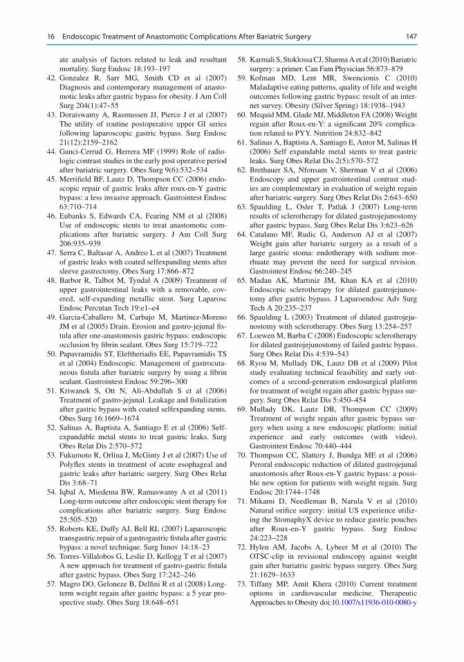
147
ate analysis of factors related to leak and resultant mortality. Surg Endosc 18:193–197
42. Gonzalez R, Sarr MG, Smith CD et al (2007) Diagnosis and contemporary management of anasto-motic leaks after gastric bypass for obesity. J Am Coll Surg 204(1):47–55
43. Doraiswamy A, Rasmussen JJ, Pierce J et al (2007) The utility of routine postoperative upper GI series following laparoscopic gastric bypass. Surg Endosc 21(12):2159–2162
44. Ganci-Cerrud G, Herrera MF (1999) Role of radio-logic contrast studies in the early post operative period after bariatric surgery. Obes Surg 9(6):532–534
45. Merrifi eld BF, Lautz D, Thompson CC (2006) endo-scopic repair of gastric leaks after roux-en-Y gastric bypass: a less invasive approach. Gastrointest Endosc 63:710–714
46. Eubanks S, Edwards CA, Fearing NM et al (2008) Use of endoscopic stents to treat anastomotic com-plications after bariatric surgery. J Am Coll Surg 206:935–939
47. Serra C, Baltasar A, Andreo L et al (2007) Treatment of gastric leaks with coated selfexpanding stents after sleeve gastrectomy. Obes Surg 17:866–872
48. Barbor R, Talbot M, Tyndal A (2009) Treatment of upper gastrointestinal leaks with a removable, cov-ered, self-expanding metallic stent. Surg Laparosc Endosc Percutan Tech 19:e1–e4
49. Garcia-Caballero M, Carbajo M, Martinez-Moreno JM et al (2005) Drain. Erosion and gastro-jejunal fi s-tula after one-anastomosis gastric bypass: endoscopic occlusion by fi brin sealant. Obes Surg 15:719–722
50. Papavramidis ST, Eleftheriadis EE, Papavramidis TS et al (2004) Endoscopic. Management of gastrocuta-neous fi stula after bariatric surgery by using a fi brin sealant. Gastrointest Endosc 59:296–300
51. Kriwanek S, Ott N, Ali-Abdullah S et al (2006) Treatment of gastro-jejunal. Leakage and fi stulization after gastric bypass with coated selfexpanding stents. Obes Surg 16:1669–1674
52. Salinas A, Baptista A, Santiago E et al (2006) Self- expandable metal stents to treat gastric leaks. Surg Obes Relat Dis 2:570–572
53. Fukumoto R, Orlina J, McGinty J et al (2007) Use of Polyfl ex stents in treatment of acute esophageal and gastric leaks after bariatric surgery. Surg Obes Relat Dis 3:68–71
54. Iqbal A, Miedema BW, Ramaswamy A et al (2011) Long-term outcome after endoscopic stent therapy for complications after bariatric surgery. Surg Endosc 25:505–520
55. Roberts KE, Duffy AJ, Bell RL (2007) Laparoscopic transgastric repair of a gastrogastric fi stula after gastric bypass: a novel technique. Surg Innov 14:18–23
56. Torres-Villalobos G, Leslie D, Kellogg T et al (2007) A new approach for treatment of gastro-gastric fi stula after gastric bypass. Obes Surg 17:242–246
57. Magro DO, Geloneze B, Delfi ni R et al (2008) Long- term weight regain after gastric bypass: a 5 year pro-spective study. Obes Surg 18:648–651
58. Karmali S, Stoklossa CJ, Sharma A et al (2010) Bariatric surgery: a primer. Can Fam Physician 56:873–879
59. Kofman MD, Lent MR, Swencionis C (2010) Maladaptive eating patterns, quality of life and weight outcomes following gastric bypass: result of an inter-net survey. Obesity (Silver Spring) 18:1938–1943
60. Mequid MM, Glade MJ, Middleton FA (2008) Weight regain after Roux-en-Y: a signifi cant 20% complica-tion related to PYY. Nutrition 24:832–842
61. Salinas A, Baptista A, Santiago E, Antor M, Salinas H (2006) Self expandable metal stents to treat gastric leaks. Surg Obes Relat Dis 2(5):570–572
62. Brethauer SA, Nfonsam V, Sherman V et al (2006) Endoscopy and upper gastrointestinal contrast stud-ies are complementary in evaluation of weight regain after bariatric surgery. Surg Obes Relat Dis 2:643–650
63. Spaulding L, Osler T, Patlak J (2007) Long-term results of sclerotherapy for dilated gastrojejunostomy after gastric bypass. Surg Obes Relat Dis 3:623–626
64. Catalano MF, Rudic G, Anderson AJ et al (2007) Weight gain after bariatric surgery as a result of a large gastric stoma: endotherapy with sodium mor-rhuate may prevent the need for surgical revision. Gastrointest Endosc 66:240–245
65. Madan AK, Martiniz JM, Khan KA et al (2010) Endoscopic sclerotherapy for dilated gastrojejunos-tomy after gastric bypass. J Laparoendosc Adv Surg Tech A 20:235–237
66. Spaulding L (2003) Treatment of dilated gastrojeju-nostomy with sclerotherapy. Obes Surg 13:254–257
67. Loewen M, Barba C (2008) Endoscopic sclerotherapy for dilated gastrojejunostomy of failed gastric bypass. Surg Obes Relat Dis 4:539–543
68. Ryou M, Mullady DK, Lautz DB et al (2009) Pilot study evaluating technical feasibility and early out-comes of a second-generation endosurgical platform for treatment of weight regain after gastric bypass sur-gery. Surg Obes Relat Dis 5:450–454
69. Mullady DK, Lautz DB, Thompson CC (2009) Treatment of weight regain after gastric bypass sur-gery when using a new endoscopic platform: initial experience and early outcomes (with video). Gastrointest Endosc 70:440–444
70. Thompson CC, Slattery J, Bundga ME et al (2006) Peroral endoscopic reduction of dilated gastrojejunal anastomosis after Roux-en-Y gastric bypass: a possi-ble new option for patients with weight regain. Surg Endosc 20:1744–1748
71. Mikami D, Needleman B, Narula V et al (2010) Natural orifi ce surgery: initial US experience utiliz-ing the StomaphyX device to reduce gastric pouches after Roux-en-Y gastric bypass. Surg Endosc 24:223–228
72. Hylen AM, Jacobs A, Lybeer M et al (2010) The OTSC-clip in revisional endoscopy against weight gain after bariatric gastric bypass surgery. Obes Surg 21:1629–1633
73. Tiffany MP, Amit Khera (2010) Current treatment options in cardiovascular medicine. Therapeutic Approaches to Obesity doi: 10.1007/s11936-010-0080-y
16 Endoscopic Treatment of Anastomotic Complications After Bariatric Surgery
149G. Galloro (ed.), Endoscopic Follow-up of Digestive Anastomosis, DOI 10.1007/978-88-470-5370-0, © Springer-Verlag Italia 2014
A Adenoma detection rate , 54 Adipose-tissue-derived stem cells , 90 Advanced features , 52 Afferent loop , 99 Air insuffl ation , 62 AJCC II or III disease , 74 Alloplastic meshes , 9 Anastomosis , 97, 131
complications , 63 dehiscence , 63, 119, 120, 127 fi stula , 28 leaks , 62, 120, 127 recurrence , 79 strictures , 72, 105 tightening , 28 ulcer , 85 ulcer complications , 87
Annual surveillance endoscopy , 36 Apparently negative examinations , 20 Appropriateness , 17 Appropriate timing of endoscopic
follow-up , 20 Argon plasma coagulator (APC) , 101
B Balloon dilation , 106, 112 Balloon-type dilators , 106 Bariatric leaks , 143 Bariatric surgery , 37–39, 137 Becaplermin gel , 89 Benign anastomotic stenosis , 28–29 Benign diseases , 108 Benign strictures , 108 Biliopancreatic diversion , 139 Biliopancreatic diversion with duodenal switch
(BPD-DS) , 138, 139 Biochemical differences , 8 Biodegradable (BD) stents , 108–110, 112 Biologic materials , 9 Bipolar needle knife , 81 Bleeding , 87 Bougie , 106 Bowel preparation , 54 Brachytherapy , 101
C Capillary pattern (CP) , 78 Capsule endoscopy (CE) , 41–42 Central recurrences , 70 Circumferential hypertrophy , 69 Clinical impact , 69 Clipping devices , 120 Colon , 79 Colonic anastomotic strictures , 105 Colonoscopy , 54 Colorectal cancer (CRC) , 49, 100 Colorectal surgery , 49 Comorbidities , 54 Complications , 9, 54, 108, 137 Contrast method , 77 Corticosteroid injection , 106 Cost/benefi t ratio , 54 Cost evaluation , 21 Covered self-expanding metal stents , 126 Covered stents , 128 CRC survivors , 54 Criteria for an accurate follow-up , 13 Crohn’s disease (CD) , 42–45 Cumulative risk , 53 Cyanoacrylate injection , 127
D Dehiscence , 120 Device-assisted enteroscopy (DAE) , 42–43 Diagnostic accuracy , 20 Dilated anastomosis , 145 Dilated gastric pouch , 145 Distant metastases , 50 Double-balloon endoscopy , 111 Duodenal switch , 139 Dysphagia , 93
E Echo-poor mass , 71 Effectiveness of endoscopic treatment , 105 Efferent loop , 99 Elderly patients , 54 Endocytoscopy , 79 Endoluminal interventions , 143
Index
150
Endoscopic follow-up (FU) , 49, 50, 54 examination , 18 timing of , 51
Endoscopic submucosal dissection (ESD) , 81 Endoscopic surveillance
examination, criteria , 18 post surgical complications , 43
Endoscopic ultrasound (EUS) , 50, 67 Endoscopy , 137
controls , 17 stent placement , 126 technique , 11 therapy , 35 treatment , 106, 141 vacuum therapy , 126
Equipment , 115 Esophagectomy , 97–98 Esophagitis , 29–30 Esophagojejunostomy , 98 EUS-guided FNAB , 73 Expensive , 115
F Familial adenomatous polyposis (FAP) , 53 Fibrin glue , 127 Fistula , 3, 24–28 Flexible endoscopy , 12 Focal wall thickening , 73 Follow-up protocols , 69 Fully covered SEMS (FSEMS) , 108, 126 Fully covered stents , 106
G Gastrectomy , 98–100 Gastric leaks , 142 Gastric pacemakers , 63 Gastric pouch , 88 Gastric surgery , 36 Gastroduodenostomy , 98 Gastrogastric fi stula , 140, 142 Gastrointestinal (GI)
bleeding , 62 tract surgery , 9
Gastrojejunal anastomosis , 85 Gastrojejunostomy , 98 Granulocyte–macrophage colony-stimulating factor , 90 Guidelines, endoscopic FU , 51 Guidewire , 114
H Hand-sewn , 89 Healing process , 8 Helicobacter pylori , 87 Hemoclips , 133 Hemorrhage , 114 Hemostasis , 133 Hereditary non Polyposis Colorectal Cancer (HNPCC) , 53
I Ileal pouch , 53 Incisional therapy , 109 Indications for endoscopic controls , 23 Insulation-tipped knife , 81 Intestinal anastomoses , 41 Intralesional steroid injections , 109 Intraoperative endoscopy , 62, 63
K Kudo’s classifi cation , 77
L Laparoscopic adjustable gastric banding
(LAGB) , 63, 138 Laterally spreading tumor , 81 Leaks , 3 Local recurrences , 50, 67 Locoregional staging , 69 Long-term success rate , 109 Lumen patency , 101
M Magnifying chromoendoscopy , 77–78 Marginal ulcer(s) , 85, 140–141 Mechanical dilation , 111 Medical therapy , 89 Meshed capillary vessels , 78 Metabolic energy , 8 Metachronous adenomas
high-risk , 52 low-risk , 52
Metachronous cancer , 79 Metachronous neoplasia , 50 Metal stents , 108 Methylene blue , 62 Miniprobes , 72 Monitoring neoplastic conditions , 17 Monitoring therapeutic response , 17 Morbidity , 4 Mortality , 4, 114 Mucosal damage , 30
N Narrow band imaging (NBI) , 78–79 Neodymium:yttrium–aluminum–garnet (Nd:YAG)
laser , 101, 109 Neoplastic recurrence , 30 Nonsteroidal anti-infl ammatory drug (NSAID)
therapy , 88
O Obstruction , 96 Outcomes, endoscopic FU , 54 Overall survival , 52, 54
Index
151
Overlooked synchronous cancers , 79 Over-stitch suturing , 124 Over the scope clip (OTSC) , 122
P Palliation , 93 Partial gastrectomy , 35–36 Partially covered SEMS (PSEMS) , 108, 126 Perforation , 87, 114 Pericardium bovine patch , 8 Periodic anastomotic evaluation , 74 PET , 75 Photodynamic therapy , 101 Pit pattern , 77 Plastic stents , 108 Platelet-derived factors , 89 Pneumatic dilation , 111 Postoperative gastrointestinal bleeding , 133 Postoperative leakage , 62 Postoperative surveillance , 54 Proctosigmoidoscopy , 53 Proton pump inhibitor (PPI) therapy , 88 Pulmonary insuffi ciency , 6
R Rectal cancer , 50 Rectal stump , 53 Recurrence, anastomotic , 93 Reinforcement of the intestinal anastomosis , 9 Reoperation , 73 Resective surgery , 6 Risk factors , 3, 87, 120 Risk of complications , 110 Risk of gastric stump cancer (GSC) , 36 Roux-en-Y gastric bypass (RYGB) , 85, 138, 139
S Sano’s classifi cation , 78 Selection of candidates , 19
Self-expandable metal stent (SEMS) , 93, 110, 112
Self-expanding plastic stent (SEPS) , 108, 110, 112
Sigmoidoscopy , 50 Sleeve gastrectomy (SG) , 62,
63, 138, 139 Small bowel diseases , 41 Small bowel tumors , 45–46 Stapler , 89 Stenosis , 87, 96 Stent migration , 96, 145 Stomal ulcers , 140–141 Stomas , 141–142 Strictures , 110 Surveillance colonoscopy , 51, 81 Survival rates , 51 Suture , 89 Suture leak , 140 Synchronous adenomas , 51 Synchronous CRC , 51 Synchronous tubular adenomas , 52
T Tissue reaction , 108 Tobacco use , 88 Total gastrectomy , 36–37 Total mesorectal excision , 73 Transrectal ultrasonography (TRUS) , 67
U Ulcer complication , 87 Upper esophageal sphincter (UES) , 97 Upper gastrointestinal neoplasms , 78
V VAC therapy , 124 Vertical banded gastroplasty (VBG) ,
62, 63, 138–139
Index