Embed Size (px)
Citation preview
TH
EJ
OU
RN
AL
OF
CE
LL
BIO
LO
GY
©
The Rockefeller University Press $8.00The Journal of Cell Biology, Vol. 169, No. 4, May 23, 2005 603–612http://www.jcb.org/cgi/doi/10.1083/jcb.200502086
JCB: ARTICLE
JCB 603
Endoplasmic reticulum stress modulates the response of myelinating oligodendrocytes to the immune cytokine interferon-
�
Wensheng Lin,
1
Heather P. Harding,
2
David Ron,
2
and Brian Popko
1
1
Jack Miller Center for Peripheral Neuropathy, Department of Neurology, University of Chicago, Chicago, IL 60637
2
The Skirball Institute, New York University School of Medicine, New York, NY 10016
nterferon-
�
(IFN-
�
) is believed to contribute to immune-mediated demyelinating disorders by targeting the my-elin-producing oligodendrocyte, a cell known to be
highly sensitive to the disruption of protein synthesis andto the perturbation of the secretory pathway. We foundthat apoptosis induced by IFN-
�
in cultured rat oligoden-drocytes was associated with endoplasmic reticulum (ER)stress. ER stress also accompanied oligodendrocyte ap-optosis and hypomyelination in transgenic mice that inap-propriately expressed IFN-
�
in the central nervous system(CNS). Compared with a wild-type genetic background,
I
the enforced expression of IFN-
�
in mice that were het-erozygous for a loss of function mutation in pancreatic ERkinase (PERK) dramatically reduced animal survival, pro-moted CNS hypomyelination, and enhanced oligoden-drocyte loss. PERK encodes an ER stress–inducible kinasethat phosphorylates eukaryotic translation initiation factor2
�
and specifically maintains client protein homeostasisin the stressed ER. Therefore, the hypersensitivity of
PERK
�
/
�
mice to IFN-
�
implicates ER stress in demyelin-ating disorders that are induced by CNS inflammation.
Introduction
The ER is a membranous labyrinthine network that extendsthroughout the cytoplasm of eukaryotic cells and is contiguouswith the nuclear envelope. Approximately one third of all cel-lular proteins, particularly membrane-spanning and secretedproteins, are translocated into the lumen of the ER, where post-translational modification, folding, and oligomerization occur.The ER is also the site for the biosynthesis of steroids, choles-terol, and other lipids, and it is the major signal-transducing or-ganelle in the cell that continuously responds to environmentalcues to release calcium (Kaufman, 1999). A number of cellstress conditions, such as perturbed calcium homeostasis or re-dox status, elevated secretory protein synthesis rates, alteredglycosylation levels, and cholesterol overloading, can interferewith oxidative protein folding. This can subsequently lead to
the accumulation of unfolded or misfolded proteins in the ERlumen, which has been referred to ER stress (Ma and Hender-shot, 2001; Ron, 2002; Rutkowski and Kaufman, 2004).
Three ER-resident transmembrane proteins have beenidentified as proximal sensors of ER stress: pancreatic ER ki-nase (PERK), the kinase encoded by the inositol requiring(IRE) 1 gene, and activating transcription factor (ATF) 6. Atthe onset of ER stress, the most rapidly activated pathway istranslational repression, which is mediated by PERK (Ron andHarding, 2000). PERK couples protein folding in the ER withprotein synthesis by phosphorylating the
�
subunit of the eu-karyotic translation initiation (eIF) factor 2, which attenuatesthe initiation of translation in response to ER stress (Harding etal., 1999; Okada et al., 2002). The cleavage of ATF6 and theactivation of IRE1 signaling follow fairly quickly; these pro-cesses promote the expression of ER-localized chaperones thatfacilitate the restoration of proper protein folding within the ER(Yoshida et al., 2001; Calfon et al., 2002; Okada et al., 2002).These protective responses act transiently to maintain homeo-stasis within the ER, but sustained ER stress ultimately leads tothe apoptotic death of the cell (Ma and Hendershot, 2001; Ron,2002; Rao et al., 2004; Rutkowski and Kaufman, 2004).
Oligodendrocytes produce vast amounts of myelin as anextension of their plasma membrane; a unique, lipid-rich,
Correspondence to Brian Popko: [email protected] used in this paper: ATF, activating transcription factor; BIP, bind-ing immunoglobulin protein; CGT, ceramide galactosyltransferase; CHOP,CAATT enhancer–binding protein homologous protein; CNP, 2
�
3
�
-cyclic nucle-otide 3
�
-phosphodiesterase; CNS, central nervous system; GFAP, glial fibrillaryacidic protein; E 14, embryonic day 14; eIF, eukaryotic translation initiationfactor; IFN-
�
, interferon-
�
; iNOS, inducible NO synthase;
IRE, inositol requir-ing; MBP, myelin basic protein; MHC, major histocompatibility complex; MS,multiple sclerosis; NO, nitric oxide; PERK, pancreatic ER kinase; PLP, proteo-lipid protein; PMD, Pelizaeus-Merzbacher disease; tTA, tetracycline-controlledtransactivator; VWM, leukoencephalopathy with vanishing white matter.
JCB • VOLUME 169 • NUMBER 4 • 2005604
multilamellar sheath that wraps axons in the central nervoussystem (CNS). On the basis of estimates from morphometricanalysis, the mean surface area of myelin membrane per ma-ture oligodendrocyte is 1–20
�
10
5
�
m
2
, compared with aperikaryal surface area of only 100–300
�
m
2
. During the activephase of myelination, each oligodendrocyte in the CNS mustproduce as much as
�
5,000
�
m
2
of myelin surface area per dayand
�
10
5
myelin protein molecules per minute (Pfeiffer et al.,1993). Perhaps not surprisingly, data from human hypomyeli-nating diseases and from dysmyelinating animal models suggestthat oligodendrocytes rank among the cells that are most sensi-tive to the disruption of protein translation and protein secretorypathways. Leukoencephalopathy with vanishing white matter(VWM) is caused by mutations in the gene encoding eIF-2B(Leegwater et al., 2001; Fogli et al., 2004). Moreover, the leu-kodystrophy Pelizaeus-Merzbacher disease (PMD), which re-sults from mutations in the myelin proteolipid protein (PLP)gene, is associated with perturbation of the protein secretorypathway, as are PMD animal models with point mutations in thePLP gene (Bauer et al., 2002; Southwood et al., 2002).
The pleotropic cytokine interferon-
�
(IFN-
�
), which issecreted by activated T lymphocytes and by natural killer cells,is believed to play a deleterious role in immune-mediated de-myelinating disorders such as multiple sclerosis (MS) and ex-perimental allergic encephalomyelitis (Popko et al., 1997;Popko and Baerwald, 1999; Steinman, 2001a). This cytokine,which is normally not present in the CNS, is detectable duringthe symptomatic phase of these disorders (Panitch, 1992). Theadministration of IFN-
�
to patients with MS leads to a worsen-ing disease course (Panitch et al., 1987), and treatment of suchpatients with an IFN-
�
antibody delays disability progression(Skurkovich et al., 2001). In vitro IFN-
�
is capable of promot-ing apoptosis in purified developing oligodendrocytes (An-drews et al., 1998; Baerwald and Popko, 1998; Feldhaus et al.,2004). Moreover, transgenic mice that ectopically express IFN-
�
in the CNS display a tremoring phenotype and myelin abnor-malities (Corbin et al., 1996; LaFerla et al., 2000). Nevertheless,the mechanisms by which the presence of IFN-
�
leads to oligo-dendroglial abnormalities and to alterations to the myelinsheath remain poorly understood. We provide evidence for theactivation of the ER stress response in oligodendrocytes thatare exposed to IFN-
�
in culture and in transgenic mice. More-over, we demonstrate that PERK, a kinase that responds specif-ically to ER stress, modulates disease severity in animals thatectopically express IFN-
�
and serves to protect oligodendro-cytes from apoptosis.
Results
IFN-
�
–induced apoptosis in rat oligodendrocytes is associated with ER stress
Our initial efforts to characterize the ER stress response inoligodendrocytes exposed to IFN-
�
were performed in vitro.Purified oligodendrocyte progenitor cells were allowed to dif-ferentiate for 5 d in defined media, at which point
�
40% ofthe cells expressed the myelin protein 2
�
3
�
-cyclic nucleotide
3
�
-phosphodiesterase (CNP) and extended branched processes.These cells did not extend the flat membrane sheets that arecharacteristic of more mature oligodendrocyte cultures. Whentreated with 70 U/ml IFN-
�
for 48 h, these cells showed ab-normal morphological changes, including cell shrinkage andaggregation of cell bodies, followed by detachment from the
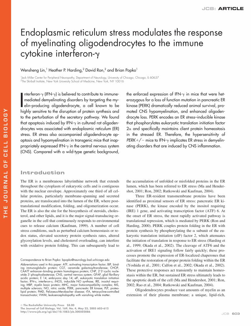
Figure 1. IFN-�–induced apoptosis in cultured rat oligodendrocytes isassociated with ER stress. (A) Untreated oligodendrocytes that underwentdifferentiation for 7 d. (B) Oligodendrocytes that underwent differentia-tion for 5 d and treatment with 70 U/ml IFN-� for 48 h, revealing cellshrinkage and aggregation of cell bodies (arrow). (C and D) TUNEL andCNP double labeling for untreated oligodendrocytes that underwent dif-ferentiation for 7 d (C) and for oligodendrocytes that underwent differenti-ation for 5 d and treatment with 70 U/ml IFN-� for 48 h (D). (E) Quantita-tion of TUNEL and CNPase double positive cells; *, P � 0.05. (F)Caspase-3 activity assay in the oligodendrocyte lysates; *, P � 0.01. (G)Real-time PCR analyses of the expression of BIP, CHOP, and caspase-12in oligodendrocytes treated with 70 U/ml IFN-�; *, P � 0.05. (E–G) Errorbars represent standard deviation. (H) Western blot analyses of total eIF-2�, p-eIF-2�, and caspase-12 in oligodendrocytes treated with 70 U/mlIFN-�. All experiments were repeated at least three times. Bars: (A and B)30 �M; (C and D) 20 �M.
ER STRESS, INTERFERON-
�
, AND OLIGODENDROCYTES • LIN ET AL.
605
culture plate (Fig. 1, A and B). TUNEL and CNP double label-ing revealed that IFN-
�
induced apoptosis in a significantnumber of oligodendrocytes (Fig. 1, C–E). Furthermore, thecaspase-3 activity in the cell lysates of IFN-
�
–treated oligo-dendrocytes was markedly increased (Fig. 1 F). Thus, 70 U/mlIFN-
�
is able to induce apoptosis in oligodendrocytes that areactively synthesizing myelin components.
To determine whether IFN-
�
interferes with ER func-tion, we monitored the expression of ER stress markers in cy-tokine-treated oligodendrocyte cultures. The levels of mRNAthat encode the binding immunoglobulin protein (BIP)/78-kD,glucose-regulated protein and the CAATT enhancer bindingprotein homologous protein (CHOP)/growth and DNA damageprotein 153, both of which are associated with the ER stress re-sponse, were increased
�
2–3 times in oligodendrocytes afterexposure to IFN-
�
(Fig. 1 G). The phosphorylation of eIF-2
�
,
which inhibits nucleotide exchange on the eIF-2 complex andattenuates most protein synthesis, occurs within minutes afterthe development of ER stress (Ron, 2002). Western blot analy-sis revealed that IFN-
�
significantly elevated the level of phos-phorylated eIF-2
�
(p-eIF-2
�
) in oligodendroglial cultures (Fig.1 H). Caspase-12, an ER-localized caspase, is activated by ERstress and can lead to the cleavage of caspase-3 (Nakagawa etal., 2000; Lamkanfi et al., 2004). The induction of caspase-12was observed after the treatment of oligodendrocytes with IFN-
�
(Fig. 1 G). Moreover, the level of the active fragment of cas-pase-12 was strongly elevated after 48 h of IFN-
�
treatment(Fig. 1 H). These results indicate that IFN-
�
–induced apoptosisin cultured oligodendrocytes is associated with the activationof the ER stress pathway.
Hypomyelination induced by ectopic expression of IFN-
�
is associated with ER stress
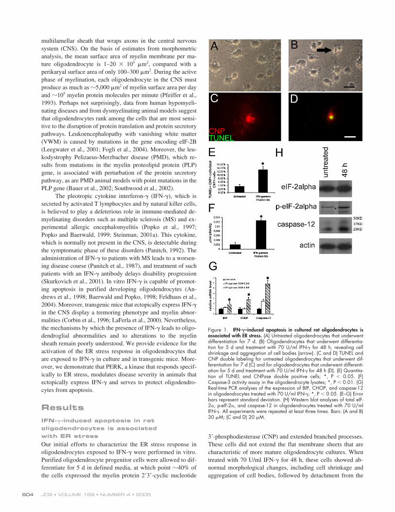
We have generated transgenic mice that allow for the temporallyregulated delivery of IFN-
�
to the CNS using the tetracycline-controllable system (Lin et al., 2004). To drive tetracycline-controlled transactivator (tTA) expression in astrocytes, wechose the transcriptional regulatory region of the glial fibrillaryacidic protein (GFAP) gene, which has been well characterizedin transgenic studies (Brenner et al., 1994).
GFAP/tTA
micewere mated with
TRE/IFN-
�
mice to produce animals that werehemizygous for both transgenes. When these mice are main-tained on doxycycline, the expression of the IFN-
�
transgene isrepressed (Fig. 2 A). When double transgenic mice were re-leased from doxycycline at embryonic day 14 (E 14), mRNAfor IFN-
�
could be detected as early as 10 d after birth (unpub-lished data). Real-time PCR analysis showed that these mice ex-pressed robust levels of IFN-
�
and major histocompatibilitycomplex (MHC) class I, a downstream target of IFN-
�
activity,in the CNS at postnatal day 14 (Fig. 2 A). The double transgenicmice that ectopically expressed IFN-
�
in the CNS during devel-opment are mildly hypomyelinated (see Figs. 4 and 5). This isconsistent with observations made on previously generatedtransgenic mice that expressed IFN-
�
constitutively in oligo-dendrocytes (Corbin et al., 1996). The diminished myelinationobserved in these mice is correlated with IFN-
�
–induced oligo-dendrocyte apoptosis (see Fig. 7, A and F). IFN-
�
up-regulatedBIP and CHOP expression
�
1.6 and 2 times those of the con-trol levels and strongly enhanced caspase-12 expression in theCNS of these animals (Fig. 2 A). More notable, the level of theactive fragment of caspase-12 was also increased in the CNS ofthese animals (Fig. 2 B). Furthermore, colocalization analysiswith the CC1 antibody revealed that oligodendrocytes increasedthe expression of BIP (Fig. 2, C and D), p-eIF-2
�
(Fig. 2, E andF), and caspase-12 (Fig. 2, G and H). These data support a linkbetween ER stress and IFN-
�
–induced oligodendrocyte apopto-sis and hypomyelination during development.
Hypersensitivity of
PERK
�
/
�
mice to the conditional misexpression of IFN-
�
We next pursued a genetic approach to examine the involve-ment of the ER stress response in the myelin perturbations that
Figure 2. Hypomyelination induced by ectopically expressed IFN-� isassociated with ER stress. (A) Real-time PCR analyses for detection ofmRNA in the brains of 14-d-old mice ectopically expressing IFN-� (n 3);*, P � 0.05; **, P � 0.01. Error bars represent standard deviation. (B)Western blot analyses for caspase-12 in the CNS of 14-d-old doubletransgenic mice released from doxyclycline at E 14. (C and D) BIP andCC1 double immunostaining in the spinal cord of 14-d-old double trans-genic mice that received doxycycline (C) or were released from doxycy-cline at E 14 (D). (E and F) p-eIF-2� and CC1 double immunostaining inthe spinal cord of 14-d-old double transgenic mice that received doxycy-cline (E) or were released from doxycycline at E 14 (F). (G and H) Cas-pase-12 and CC1 double immunostaining in the spinal cord of 14-d-olddouble transgenic mice that received doxycycline (G) or were releasedfrom doxycycline at E 14 (H). (C–H) n 3; bar, 30 �M.
JCB • VOLUME 169 • NUMBER 4 • 2005606
are elicited by IFN-
�. Three pathways are known to signal ERstress. The IRE1–X-box–binding protein-1 pathway is the old-est and most conserved; however, in mammals, it appears tohave been diverted to the control of genes involved in remodel-ing the ER to high capacity secretion (Ron and Hampton,2004). Furthermore, both IRE1 and X-box–binding protein-1mutant mice die at early embryonic stages, making their use tothe study of the myelination process ineffective (Ma and Hen-dershot, 2001). Currently, there are no genetic tools availableto explore the ATF6 pathway in mice. The PERK–eIF-2� path-way, on the other hand, contributes to the activation of mostER stress target genes (Harding et al., 2003; Lu et al., 2004).Moreover, although PERK�/� mice have a complex pheno-type that includes progressive diabetes mellitus, exocrine pan-creatic insufficiency, growth retardation, and high mortality(Harding et al., 2001; Zhang et al., 2002), PERK�/� mice, al-though healthy, display evidence of haploid insufficiency (Har-ding et al., 2000a). Thus, to examine the influence of the ERstress response on myelin and oligodendrocyte abnormalitiesthat are elicited by IFN-�, we exploited PERK mutant mice(Harding et al., 2001).
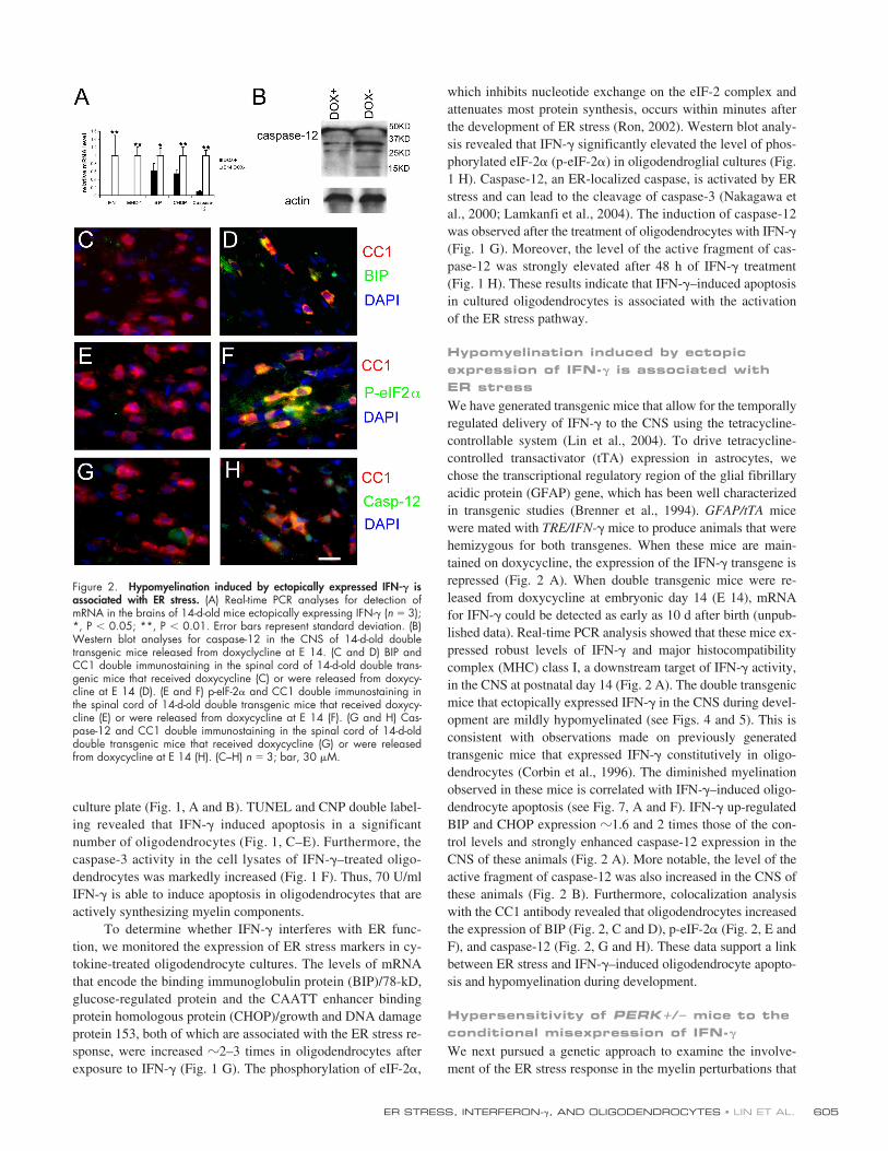
GFAP/tTA and TRE/IFN-� mice were crossed withPERK�/� mice, and the resulting progeny were intercrossed toobtain double transgenic mice that were homozygous or het-erozygous for the PERK mutation. As reported, the majority ofdouble transgenic mice with a PERK�/� background diedwithin 12 d after birth, regardless of whether they received doxy-cycline during the entire period or if doxycycline was switched
to water at E 14. Double transgenic GFAP/tTA; TRE/IFN-�mice on a PERK�/� background, released from doxycycline atE 14, showed the expected minor tremor and ataxia but exhib-ited good survival. In contrast, the double transgenic mice on aPERK�/� background had a much more severe phenotype.These animals were considerably smaller than their IFN-�–expressing PERK�/� littermates or PERK�/� animals thatdid not inherit the combination of GFAP/tTA and TRE/IFN-� al-leles and that showed severe tremor and ataxia. Approximatelytwo thirds of these mice experienced tonic seizures. Strikingly,90% of the double transgenic mice that were released fromdoxycycline at E 14 on a PERK�/� background died by post-natal day 27, whereas double transgenic mice on a wild-typebackground displayed normal survival (Fig. 3 A).
We next investigated the correlation between the severi-ties of the PERK�/� and PERK�/� animals’ phenotype withthe animals’ capacities to attenuate protein synthesis in re-sponse to IFN-� by examining the level of p-eIF-2�. Highlyelevated levels of p-eIF-2� in oligodendrocytes of the CNSwere observed in double transgenic mice on a PERK�/� back-ground (Fig. 2, E and F). In contrast, only a slight increase inp-eIF-2� immunoreactivity was observed in the CNS of doubletransgenic mice on a PERK�/� background (Fig. 3, B and C).Nevertheless, we did not find that the loss of function mutationin PERK significantly affected the RNA levels of BIP, CHOP,and caspase-12 in the CNS of mice misexpressing IFN-� (Fig.3 D). Collectively, these data indicate that the reduced capacityto elevate p-eIF-2� levels in response to IFN-� contributes tothe severe phenotype in mice misexpressing IFN-� on aPERK�/� background.
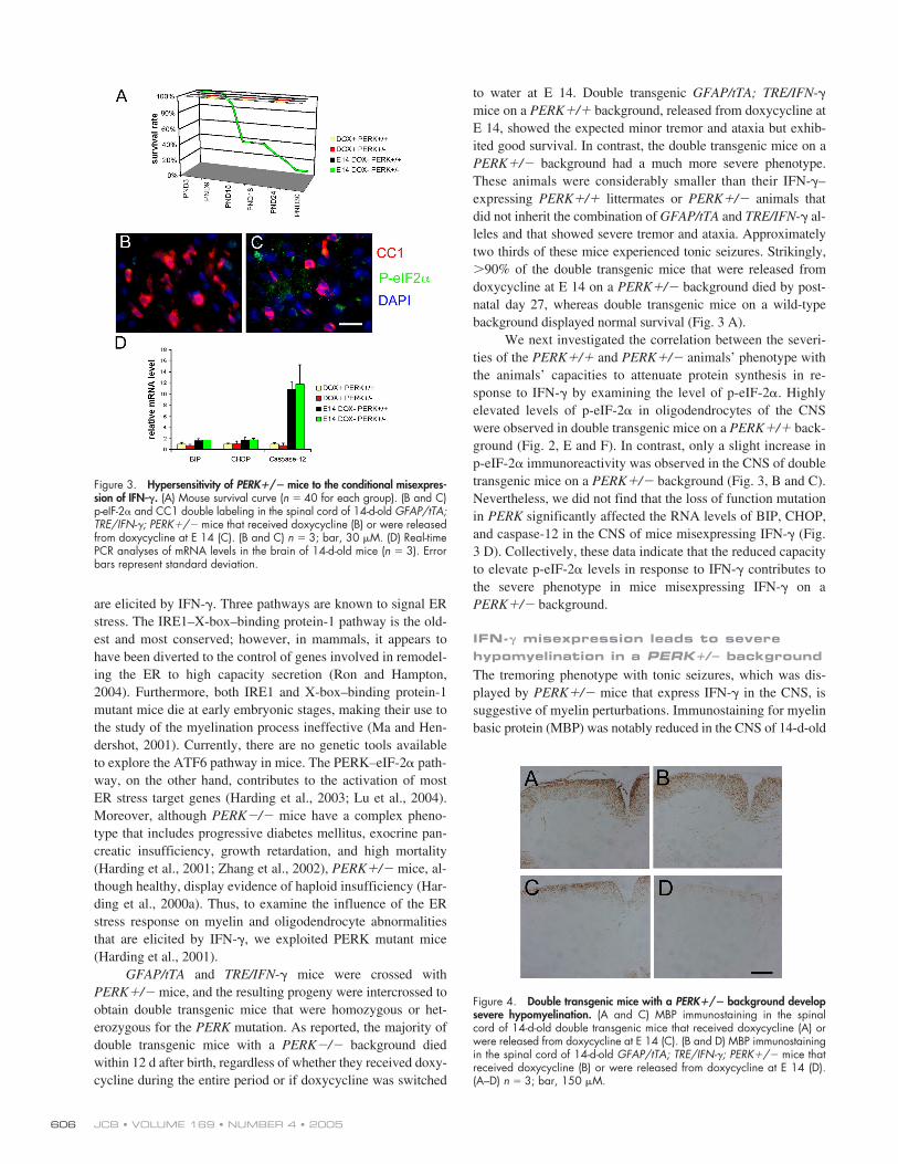
IFN-� misexpression leads to severe hypomyelination in a PERK�/� backgroundThe tremoring phenotype with tonic seizures, which was dis-played by PERK�/� mice that express IFN-� in the CNS, issuggestive of myelin perturbations. Immunostaining for myelinbasic protein (MBP) was notably reduced in the CNS of 14-d-old
Figure 3. Hypersensitivity of PERK�/� mice to the conditional misexpres-sion of IFN-�. (A) Mouse survival curve (n 40 for each group). (B and C)p-eIF-2� and CC1 double labeling in the spinal cord of 14-d-old GFAP/tTA;TRE/IFN-�; PERK�/� mice that received doxycycline (B) or were releasedfrom doxycycline at E 14 (C). (B and C) n 3; bar, 30 �M. (D) Real-timePCR analyses of mRNA levels in the brain of 14-d-old mice (n 3). Errorbars represent standard deviation.
Figure 4. Double transgenic mice with a PERK�/� background developsevere hypomyelination. (A and C) MBP immunostaining in the spinalcord of 14-d-old double transgenic mice that received doxycycline (A) orwere released from doxycycline at E 14 (C). (B and D) MBP immunostainingin the spinal cord of 14-d-old GFAP/tTA; TRE/IFN-�; PERK�/� mice thatreceived doxycycline (B) or were released from doxycycline at E 14 (D).(A–D) n 3; bar, 150 �M.
ER STRESS, INTERFERON-�, AND OLIGODENDROCYTES • LIN ET AL. 607
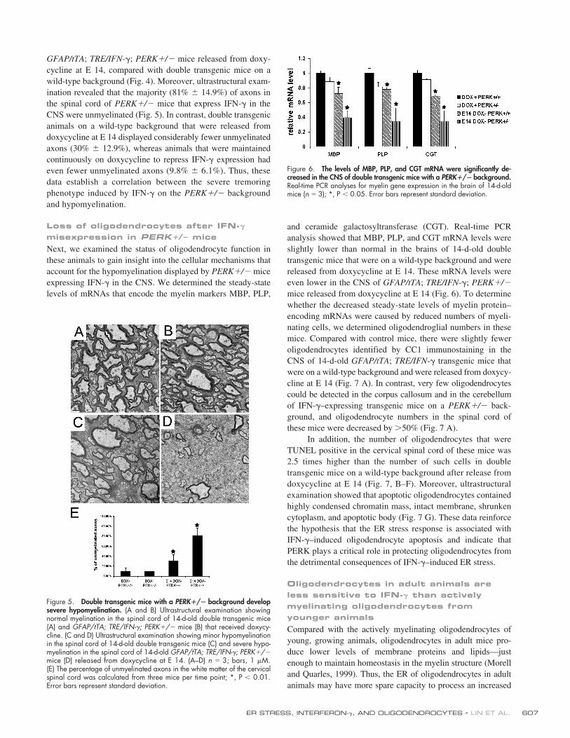
GFAP/tTA; TRE/IFN-�; PERK�/� mice released from doxy-cycline at E 14, compared with double transgenic mice on awild-type background (Fig. 4). Moreover, ultrastructural exam-ination revealed that the majority (81% � 14.9%) of axons inthe spinal cord of PERK�/� mice that express IFN-� in theCNS were unmyelinated (Fig. 5). In contrast, double transgenicanimals on a wild-type background that were released fromdoxycycline at E 14 displayed considerably fewer unmyelinatedaxons (30% � 12.9%), whereas animals that were maintainedcontinuously on doxycycline to repress IFN-� expression hadeven fewer unmyelinated axons (9.8% � 6.1%). Thus, thesedata establish a correlation between the severe tremoringphenotype induced by IFN-� on the PERK�/� backgroundand hypomyelination.
Loss of oligodendrocytes after IFN-� misexpression in PERK�/� miceNext, we examined the status of oligodendrocyte function inthese animals to gain insight into the cellular mechanisms thataccount for the hypomyelination displayed by PERK�/� miceexpressing IFN-� in the CNS. We determined the steady-statelevels of mRNAs that encode the myelin markers MBP, PLP,
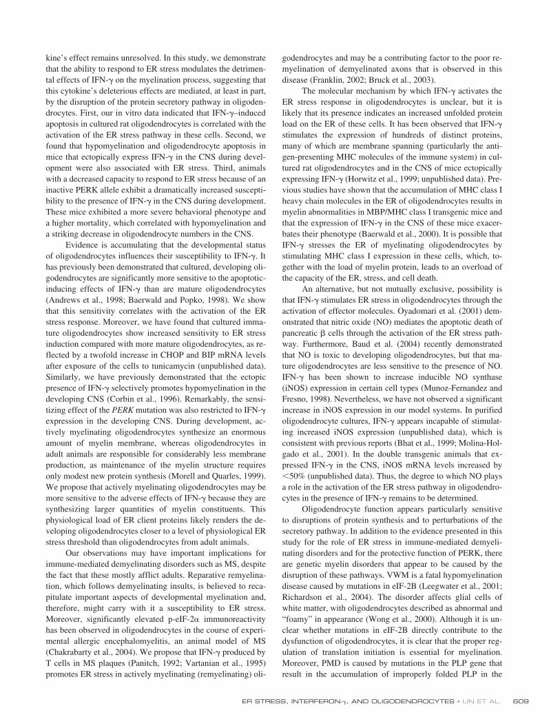
and ceramide galactosyltransferase (CGT). Real-time PCRanalysis showed that MBP, PLP, and CGT mRNA levels wereslightly lower than normal in the brains of 14-d-old doubletransgenic mice that were on a wild-type background and werereleased from doxycycline at E 14. These mRNA levels wereeven lower in the CNS of GFAP/tTA; TRE/IFN-�; PERK�/�mice released from doxycycline at E 14 (Fig. 6). To determinewhether the decreased steady-state levels of myelin protein–encoding mRNAs were caused by reduced numbers of myeli-nating cells, we determined oligodendroglial numbers in thesemice. Compared with control mice, there were slightly feweroligodendrocytes identified by CC1 immunostaining in theCNS of 14-d-old GFAP/tTA; TRE/IFN-� transgenic mice thatwere on a wild-type background and were released from doxycy-cline at E 14 (Fig. 7 A). In contrast, very few oligodendrocytescould be detected in the corpus callosum and in the cerebellumof IFN-�–expressing transgenic mice on a PERK�/� back-ground, and oligodendrocyte numbers in the spinal cord ofthese mice were decreased by 50% (Fig. 7 A).
In addition, the number of oligodendrocytes that wereTUNEL positive in the cervical spinal cord of these mice was2.5 times higher than the number of such cells in doubletransgenic mice on a wild-type background after release fromdoxycycline at E 14 (Fig. 7, B–F). Moreover, ultrastructuralexamination showed that apoptotic oligodendrocytes containedhighly condensed chromatin mass, intact membrane, shrunkencytoplasm, and apoptotic body (Fig. 7 G). These data reinforcethe hypothesis that the ER stress response is associated withIFN-�–induced oligodendrocyte apoptosis and indicate thatPERK plays a critical role in protecting oligodendrocytes fromthe detrimental consequences of IFN-�–induced ER stress.
Oligodendrocytes in adult animals are less sensitive to IFN-� than actively myelinating oligodendrocytes from younger animalsCompared with the actively myelinating oligodendrocytes ofyoung, growing animals, oligodendrocytes in adult mice pro-duce lower levels of membrane proteins and lipids—justenough to maintain homeostasis in the myelin structure (Morelland Quarles, 1999). Thus, the ER of oligodendrocytes in adultanimals may have more spare capacity to process an increased
Figure 5. Double transgenic mice with a PERK�/� background developsevere hypomyelination. (A and B) Ultrastructural examination showingnormal myelination in the spinal cord of 14-d-old double transgenic mice(A) and GFAP/tTA; TRE/IFN-�; PERK�/� mice (B) that received doxycy-cline. (C and D) Ultrastructural examination showing minor hypomyelinationin the spinal cord of 14-d-old double transgenic mice (C) and severe hypo-myelination in the spinal cord of 14-d-old GFAP/tTA; TRE/IFN-�; PERK�/�mice (D) released from doxycycline at E 14. (A–D) n 3; bars, 1 �M.(E) The percentage of unmyelinated axons in the white matter of the cervicalspinal cord was calculated from three mice per time point; *, P � 0.01.Error bars represent standard deviation.
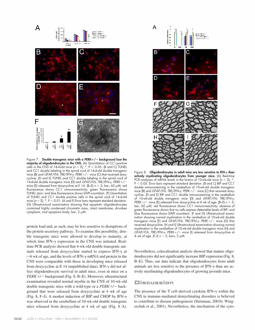
Figure 6. The levels of MBP, PLP, and CGT mRNA were significantly de-creased in the CNS of double transgenic mice with a PERK�/� background.Real-time PCR analyses for myelin gene expression in the brain of 14-d-oldmice (n 3); *, P � 0.05. Error bars represent standard deviation.
JCB • VOLUME 169 • NUMBER 4 • 2005608
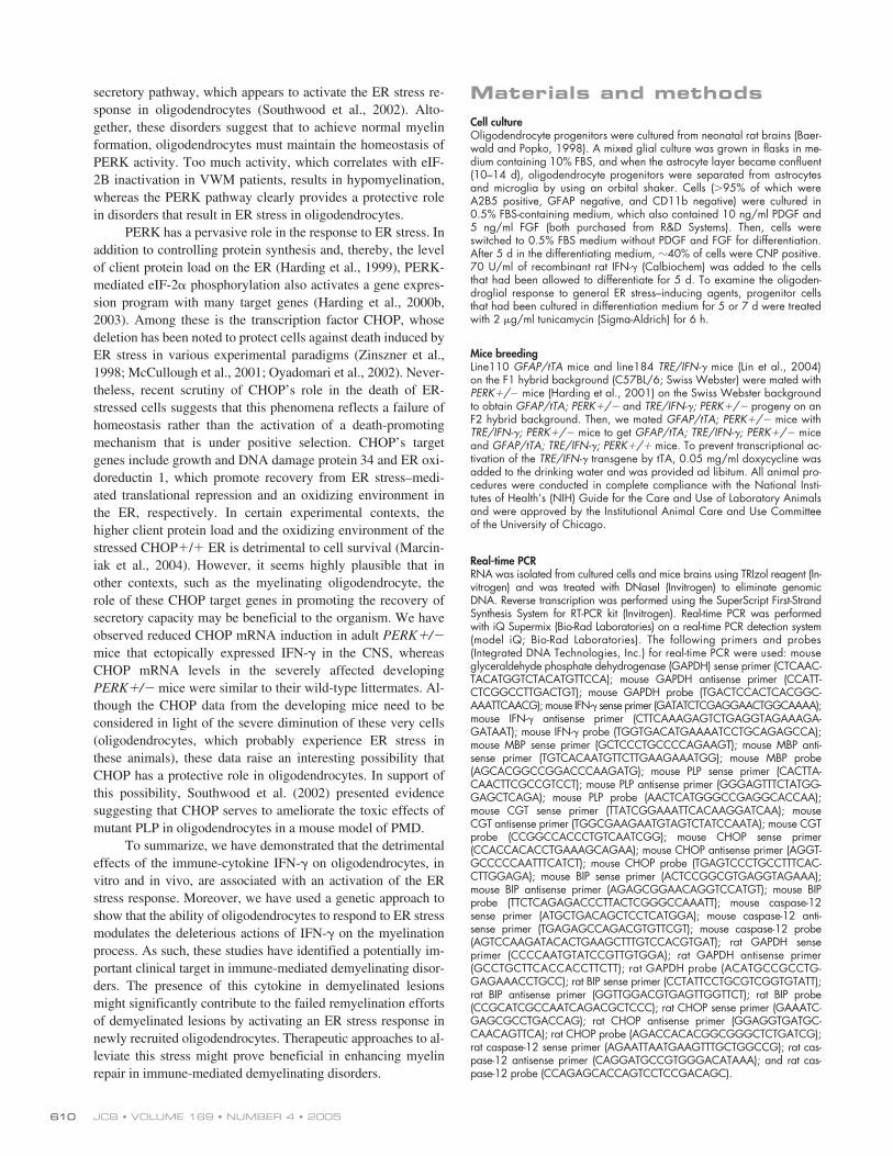
protein load and, as such, may be less sensitive to disruptions ofthe protein secretory pathway. To examine this possibility, dou-ble transgenic mice were allowed to develop to maturity, atwhich time IFN-� expression in the CNS was initiated. Real-time PCR analysis showed that 4-wk-old double transgenic ani-mals released from doxycycline started to express IFN-� at�6 wk of age, and the levels of IFN-� mRNA and protein in theCNS were comparable with those in developing mice releasedfrom doxycycline at E 14 (unpublished data). IFN-� did not af-fect oligodendrocyte survival in adult mice, even in mice on aPERK�/� background (Fig. 8, B–E). Moreover, ultrastructuralexamination revealed normal myelin in the CNS of 10-wk-olddouble transgenic mice with a wild-type or a PERK�/� back-ground that were released from doxycycline at 4 wk of age(Fig. 8, F–I). A modest induction of BIP and CHOP by IFN-�was observed in the cerebellum of 10-wk-old double transgenicmice released from doxycycline at 4 wk of age (Fig. 8 A).
Nevertheless, colocalization analysis showed that mature oligo-dendrocytes did not significantly increase BIP expression (Fig. 8,B–E). Thus, our data indicate that oligodendrocytes from adultanimals are less sensitive to the presence of IFN-� than are ac-tively myelinating oligodendrocytes of growing juvenile mice.
DiscussionThe presence of the T cell–derived cytokine IFN-� within theCNS in immune-mediated demyelinating disorders is believedto contribute to disease pathogenesis (Steinman, 2001b; Wing-erchuk et al., 2001). Nevertheless, the mechanism of the cyto-
Figure 7. Double transgenic mice with a PERK�/� background lose themajority of oligodendrocytes in the CNS. (A) Quantitation of CC1-positivecells in the CNS of 14-d-old mice (n 3); *, P � 0.05. (B and C) TUNELand CC1 double labeling in the spinal cord of 14-d-old double transgenicmice (B) and GFAP/tTA; TRE/IFN-�; PERK�/� mice (C) that received doxy-cycline. (D and E) TUNEL and CC1 double labeling in the spinal cord of14-d-old double transgenic mice (D) and GFAP/tTA; TRE/IFN-�; PERK�/�mice (E) released from doxycycline at E 14. (B–E) n 3; bar, 60 �M; redfluorescence shows CC1 immunoreactivity; green fluorescence showsTUNEL stain; and blue fluorescence shows DAPI countstain. (F) Quantitationof TUNEL and CC1 double positive cells in the spinal cord of 14-d-oldmice (n 3); *, P � 0.01. (A and F) Error bars represent standard deviation.(G) Ultrastructural examination showing that apoptotic oligodendrocytescontained highly condensed chromatin mass, intact membrane, shrunkencytoplasm, and apoptosis body; bar, 2 �M.
Figure 8. Oligodendrocytes in adult mice are less sensitive to IFN-� thanactively myelinating oligodendrocytes from younger mice. (A) Real-timePCR analyses of mRNA levels in the brains of 10-wk-old mice (n 3); *,P � 0.05. Error bars represent standard deviation. (B and C) BIP and CC1double immunostaining in the cerebellum of 10-wk-old double transgenicmice (B) and GFAP/tTA; TRE/IFN-�; PERK�/� mice (C) that received doxy-cycline. (D and E) BIP and CC1 double immunostaining in the cerebellumof 10-wk-old double transgenic mice (D) and GFAP/tTA; TRE/IFN-�;PERK�/� mice (E) released from doxycycline at 4 wk of age. (B–E) n 3;bar, 60 �M; red fluorescence shows CC1 immunoreactivity; absence ofgreen fluorescence shows that no cells express detectable levels of BIP; andblue fluorescence shows DAPI countstain. (F and G) Ultrastructural exami-nation showing normal myelination in the cerebellum of 10-wk-old doubletransgenic mice (F) and GFAP/tTA; TRE/IFN-�; PERK�/� mice (G) thatreceived doxycycline. (H and I) Ultrastructural examination showing normalmyelination in the cerebellum of 10-wk-old double transgenic mice (H) andGFAP/tTA; TRE/IFN-�; PERK�/� mice (I) released from doxycycline at4 wk of age. (F–I) n 3; bars, 2 �M.
ER STRESS, INTERFERON-�, AND OLIGODENDROCYTES • LIN ET AL. 609
kine’s effect remains unresolved. In this study, we demonstratethat the ability to respond to ER stress modulates the detrimen-tal effects of IFN-� on the myelination process, suggesting thatthis cytokine’s deleterious effects are mediated, at least in part,by the disruption of the protein secretory pathway in oligoden-drocytes. First, our in vitro data indicated that IFN-�–inducedapoptosis in cultured rat oligodendrocytes is correlated with theactivation of the ER stress pathway in these cells. Second, wefound that hypomyelination and oligodendrocyte apoptosis inmice that ectopically express IFN-� in the CNS during devel-opment were also associated with ER stress. Third, animalswith a decreased capacity to respond to ER stress because of aninactive PERK allele exhibit a dramatically increased suscepti-bility to the presence of IFN-� in the CNS during development.These mice exhibited a more severe behavioral phenotype anda higher mortality, which correlated with hypomyelination anda striking decrease in oligodendrocyte numbers in the CNS.
Evidence is accumulating that the developmental statusof oligodendrocytes influences their susceptibility to IFN-�. Ithas previously been demonstrated that cultured, developing oli-godendrocytes are significantly more sensitive to the apoptotic-inducing effects of IFN-� than are mature oligodendrocytes(Andrews et al., 1998; Baerwald and Popko, 1998). We showthat this sensitivity correlates with the activation of the ERstress response. Moreover, we have found that cultured imma-ture oligodendrocytes show increased sensitivity to ER stressinduction compared with more mature oligodendrocytes, as re-flected by a twofold increase in CHOP and BIP mRNA levelsafter exposure of the cells to tunicamycin (unpublished data).Similarly, we have previously demonstrated that the ectopicpresence of IFN-� selectively promotes hypomyelination in thedeveloping CNS (Corbin et al., 1996). Remarkably, the sensi-tizing effect of the PERK mutation was also restricted to IFN-�expression in the developing CNS. During development, ac-tively myelinating oligodendrocytes synthesize an enormousamount of myelin membrane, whereas oligodendrocytes inadult animals are responsible for considerably less membraneproduction, as maintenance of the myelin structure requiresonly modest new protein synthesis (Morell and Quarles, 1999).We propose that actively myelinating oligodendrocytes may bemore sensitive to the adverse effects of IFN-� because they aresynthesizing larger quantities of myelin constituents. Thisphysiological load of ER client proteins likely renders the de-veloping oligodendrocytes closer to a level of physiological ERstress threshold than oligodendrocytes from adult animals.
Our observations may have important implications forimmune-mediated demyelinating disorders such as MS, despitethe fact that these mostly afflict adults. Reparative remyelina-tion, which follows demyelinating insults, is believed to reca-pitulate important aspects of developmental myelination and,therefore, might carry with it a susceptibility to ER stress.Moreover, significantly elevated p-eIF-2� immunoreactivityhas been observed in oligodendrocytes in the course of experi-mental allergic encephalomyelitis, an animal model of MS(Chakrabarty et al., 2004). We propose that IFN-� produced byT cells in MS plaques (Panitch, 1992; Vartanian et al., 1995)promotes ER stress in actively myelinating (remyelinating) oli-
godendrocytes and may be a contributing factor to the poor re-myelination of demyelinated axons that is observed in thisdisease (Franklin, 2002; Bruck et al., 2003).
The molecular mechanism by which IFN-� activates theER stress response in oligodendrocytes is unclear, but it islikely that its presence indicates an increased unfolded proteinload on the ER of these cells. It has been observed that IFN-�stimulates the expression of hundreds of distinct proteins,many of which are membrane spanning (particularly the anti-gen-presenting MHC molecules of the immune system) in cul-tured rat oligodendrocytes and in the CNS of mice ectopicallyexpressing IFN-� (Horwitz et al., 1999; unpublished data). Pre-vious studies have shown that the accumulation of MHC class Iheavy chain molecules in the ER of oligodendrocytes results inmyelin abnormalities in MBP/MHC class I transgenic mice andthat the expression of IFN-� in the CNS of these mice exacer-bates their phenotype (Baerwald et al., 2000). It is possible thatIFN-� stresses the ER of myelinating oligodendrocytes bystimulating MHC class I expression in these cells, which, to-gether with the load of myelin protein, leads to an overload ofthe capacity of the ER, stress, and cell death.
An alternative, but not mutually exclusive, possibility isthat IFN-� stimulates ER stress in oligodendrocytes through theactivation of effector molecules. Oyadomari et al. (2001) dem-onstrated that nitric oxide (NO) mediates the apoptotic death ofpancreatic � cells through the activation of the ER stress path-way. Furthermore, Baud et al. (2004) recently demonstratedthat NO is toxic to developing oligodendrocytes, but that ma-ture oligodendrocytes are less sensitive to the presence of NO.IFN-� has been shown to increase inducible NO synthase(iNOS) expression in certain cell types (Munoz-Fernandez andFresno, 1998). Nevertheless, we have not observed a significantincrease in iNOS expression in our model systems. In purifiedoligodendrocyte cultures, IFN-� appears incapable of stimulat-ing increased iNOS expression (unpublished data), which isconsistent with previous reports (Bhat et al., 1999; Molina-Hol-gado et al., 2001). In the double transgenic animals that ex-pressed IFN-� in the CNS, iNOS mRNA levels increased by�50% (unpublished data). Thus, the degree to which NO playsa role in the activation of the ER stress pathway in oligodendro-cytes in the presence of IFN-� remains to be determined.
Oligodendrocyte function appears particularly sensitiveto disruptions of protein synthesis and to perturbations of thesecretory pathway. In addition to the evidence presented in thisstudy for the role of ER stress in immune-mediated demyeli-nating disorders and for the protective function of PERK, thereare genetic myelin disorders that appear to be caused by thedisruption of these pathways. VWM is a fatal hypomyelinationdisease caused by mutations in eIF-2B (Leegwater et al., 2001;Richardson et al., 2004). The disorder affects glial cells ofwhite matter, with oligodendrocytes described as abnormal and“foamy” in appearance (Wong et al., 2000). Although it is un-clear whether mutations in eIF-2B directly contribute to thedysfunction of oligodendrocytes, it is clear that the proper reg-ulation of translation initiation is essential for myelination.Moreover, PMD is caused by mutations in the PLP gene thatresult in the accumulation of improperly folded PLP in the
JCB • VOLUME 169 • NUMBER 4 • 2005610
secretory pathway, which appears to activate the ER stress re-sponse in oligodendrocytes (Southwood et al., 2002). Alto-gether, these disorders suggest that to achieve normal myelinformation, oligodendrocytes must maintain the homeostasis ofPERK activity. Too much activity, which correlates with eIF-2B inactivation in VWM patients, results in hypomyelination,whereas the PERK pathway clearly provides a protective rolein disorders that result in ER stress in oligodendrocytes.
PERK has a pervasive role in the response to ER stress. Inaddition to controlling protein synthesis and, thereby, the levelof client protein load on the ER (Harding et al., 1999), PERK-mediated eIF-2� phosphorylation also activates a gene expres-sion program with many target genes (Harding et al., 2000b,2003). Among these is the transcription factor CHOP, whosedeletion has been noted to protect cells against death induced byER stress in various experimental paradigms (Zinszner et al.,1998; McCullough et al., 2001; Oyadomari et al., 2002). Never-theless, recent scrutiny of CHOP’s role in the death of ER-stressed cells suggests that this phenomena reflects a failure ofhomeostasis rather than the activation of a death-promotingmechanism that is under positive selection. CHOP’s targetgenes include growth and DNA damage protein 34 and ER oxi-doreductin 1, which promote recovery from ER stress–medi-ated translational repression and an oxidizing environment inthe ER, respectively. In certain experimental contexts, thehigher client protein load and the oxidizing environment of thestressed CHOP�/� ER is detrimental to cell survival (Marcin-iak et al., 2004). However, it seems highly plausible that inother contexts, such as the myelinating oligodendrocyte, therole of these CHOP target genes in promoting the recovery ofsecretory capacity may be beneficial to the organism. We haveobserved reduced CHOP mRNA induction in adult PERK�/�mice that ectopically expressed IFN-� in the CNS, whereasCHOP mRNA levels in the severely affected developingPERK�/� mice were similar to their wild-type littermates. Al-though the CHOP data from the developing mice need to beconsidered in light of the severe diminution of these very cells(oligodendrocytes, which probably experience ER stress inthese animals), these data raise an interesting possibility thatCHOP has a protective role in oligodendrocytes. In support ofthis possibility, Southwood et al. (2002) presented evidencesuggesting that CHOP serves to ameliorate the toxic effects ofmutant PLP in oligodendrocytes in a mouse model of PMD.
To summarize, we have demonstrated that the detrimentaleffects of the immune-cytokine IFN-� on oligodendrocytes, invitro and in vivo, are associated with an activation of the ERstress response. Moreover, we have used a genetic approach toshow that the ability of oligodendrocytes to respond to ER stressmodulates the deleterious actions of IFN-� on the myelinationprocess. As such, these studies have identified a potentially im-portant clinical target in immune-mediated demyelinating disor-ders. The presence of this cytokine in demyelinated lesionsmight significantly contribute to the failed remyelination effortsof demyelinated lesions by activating an ER stress response innewly recruited oligodendrocytes. Therapeutic approaches to al-leviate this stress might prove beneficial in enhancing myelinrepair in immune-mediated demyelinating disorders.
Materials and methodsCell cultureOligodendrocyte progenitors were cultured from neonatal rat brains (Baer-wald and Popko, 1998). A mixed glial culture was grown in flasks in me-dium containing 10% FBS, and when the astrocyte layer became confluent(10–14 d), oligodendrocyte progenitors were separated from astrocytesand microglia by using an orbital shaker. Cells (95% of which wereA2B5 positive, GFAP negative, and CD11b negative) were cultured in0.5% FBS-containing medium, which also contained 10 ng/ml PDGF and5 ng/ml FGF (both purchased from R&D Systems). Then, cells wereswitched to 0.5% FBS medium without PDGF and FGF for differentiation.After 5 d in the differentiating medium, �40% of cells were CNP positive.70 U/ml of recombinant rat IFN-� (Calbiochem) was added to the cellsthat had been allowed to differentiate for 5 d. To examine the oligoden-droglial response to general ER stress–inducing agents, progenitor cellsthat had been cultured in differentiation medium for 5 or 7 d were treatedwith 2 �g/ml tunicamycin (Sigma-Aldrich) for 6 h.
Mice breedingLine110 GFAP/tTA mice and line184 TRE/IFN-� mice (Lin et al., 2004)on the F1 hybrid background (C57BL/6; Swiss Webster) were mated withPERK�/� mice (Harding et al., 2001) on the Swiss Webster backgroundto obtain GFAP/tTA; PERK�/� and TRE/IFN-�; PERK�/� progeny on anF2 hybrid background. Then, we mated GFAP/tTA; PERK�/� mice withTRE/IFN-�; PERK�/� mice to get GFAP/tTA; TRE/IFN-�; PERK�/� miceand GFAP/tTA; TRE/IFN-�; PERK�/� mice. To prevent transcriptional ac-tivation of the TRE/IFN-� transgene by tTA, 0.05 mg/ml doxycycline wasadded to the drinking water and was provided ad libitum. All animal pro-cedures were conducted in complete compliance with the National Insti-tutes of Health’s (NIH) Guide for the Care and Use of Laboratory Animalsand were approved by the Institutional Animal Care and Use Committeeof the University of Chicago.
Real-time PCRRNA was isolated from cultured cells and mice brains using TRIzol reagent (In-vitrogen) and was treated with DNaseI (Invitrogen) to eliminate genomicDNA. Reverse transcription was performed using the SuperScript First-StrandSynthesis System for RT-PCR kit (Invitrogen). Real-time PCR was performedwith iQ Supermix (Bio-Rad Laboratories) on a real-time PCR detection system(model iQ; Bio-Rad Laboratories). The following primers and probes(Integrated DNA Technologies, Inc.) for real-time PCR were used: mouseglyceraldehyde phosphate dehydrogenase (GAPDH) sense primer (CTCAAC-TACATGGTCTACATGTTCCA); mouse GAPDH antisense primer (CCATT-CTCGGCCTTGACTGT); mouse GAPDH probe (TGACTCCACTCACGGC-AAATTCAACG); mouse IFN-� sense primer (GATATCTCGAGGAACTGGCAAAA);mouse IFN-� antisense primer (CTTCAAAGAGTCTGAGGTAGAAAGA-GATAAT); mouse IFN-� probe (TGGTGACATGAAAATCCTGCAGAGCCA);mouse MBP sense primer (GCTCCCTGCCCCAGAAGT); mouse MBP anti-sense primer (TGTCACAATGTTCTTGAAGAAATGG); mouse MBP probe(AGCACGGCCGGACCCAAGATG); mouse PLP sense primer (CACTTA-CAACTTCGCCGTCCT); mouse PLP antisense primer (GGGAGTTTCTATGG-GAGCTCAGA); mouse PLP probe (AACTCATGGGCCGAGGCACCAA);mouse CGT sense primer (TTATCGGAAATTCACAAGGATCAA); mouseCGT antisense primer (TGGCGAAGAATGTAGTCTATCCAATA); mouse CGTprobe (CCGGCCACCCTGTCAATCGG); mouse CHOP sense primer(CCACCACACCTGAAAGCAGAA); mouse CHOP antisense primer (AGGT-GCCCCCAATTTCATCT); mouse CHOP probe (TGAGTCCCTGCCTTTCAC-CTTGGAGA); mouse BIP sense primer (ACTCCGGCGTGAGGTAGAAA);mouse BIP antisense primer (AGAGCGGAACAGGTCCATGT); mouse BIPprobe (TTCTCAGAGACCCTTACTCGGGCCAAATT); mouse caspase-12sense primer (ATGCTGACAGCTCCTCATGGA); mouse caspase-12 anti-sense primer (TGAGAGCCAGACGTGTTCGT); mouse caspase-12 probe(AGTCCAAGATACACTGAAGCTTTGTCCACGTGAT); rat GAPDH senseprimer (CCCCAATGTATCCGTTGTGGA); rat GAPDH antisense primer(GCCTGCTTCACCACCTTCTT); rat GAPDH probe (ACATGCCGCCTG-GAGAAACCTGCC); rat BIP sense primer (CCTATTCCTGCGTCGGTGTATT);rat BIP antisense primer (GGTTGGACGTGAGTTGGTTCT); rat BIP probe(CCGCATCGCCAATCAGACGCTCCC); rat CHOP sense primer (GAAATC-GAGCGCCTGACCAG); rat CHOP antisense primer (GGAGGTGATGC-CAACAGTTCA); rat CHOP probe (AGACCACACGGCGGGCTCTGATCG);rat caspase-12 sense primer (AGAATTAATGAAGTTTGCTGGCCG); rat cas-pase-12 antisense primer (CAGGATGCCGTGGGACATAAA); and rat cas-pase-12 probe (CCAGAGCACCAGTCCTCCGACAGC).
ER STRESS, INTERFERON-�, AND OLIGODENDROCYTES • LIN ET AL. 611
Western blot analysisTissues or cultured cells were rinsed in ice-cold PBS and were immedi-ately homogenized in 5 vol Triton X-100 buffer (20 mM Hepes, pH 7.5,150 mM NaCl, 1% Triton X-100, 10% glycerol, 1 mM EDTA, 10 mM tet-rasodium pyrophosphate, 100 mM NaF, 17.5 mM �-glycerophosphate,10 mM PMSF, 15 �g/ml aprotonin, and 6 �g/ml pepstatin A) using amotorized homogenizer. After incubating on ice for 15 min, the extractswere cleared by centrifugation at 14,000 rpm twice for 30 min each.The protein content of each extract was determined by protein assay (Bio-Rad Laboratories). The extracts (40 �g) were separated by SDS-PAGEand were transferred to nitrocellulose. The blots were incubated withprimary antibody (see below), and the signal was revealed by chemilumi-nescence after reacting with HRP-conjugated second antibody. The fol-lowing primary antibodies were used: anti–eIF-2� (1:500; Santa CruzBiotechnology, Inc.); anti–p-eIF-2� (1:1,000; Cell Signaling Technology);anti–caspase-12 (1:500; Santa Cruz Biotechnology, Inc.); and antiactin(1:1,000; Sigma-Aldrich).
Caspase-3 activity assayActivation of caspase-3 was assessed by using the fluorimetric Caspase-3Assay Kit (Sigma-Aldrich) according to the manufacturer’s instructions.
ImmunohistochemistryAnesthetized mice were perfused through the left cardiac ventricle with4% PFA in PBS. The half saggital brains and transverse cervical spinalcords were removed, postfixed with PFA, cryopreserved in 30% sucrose,embedded in optimal cutting temperature compound, and frozen on dryice. Frozen sections were cut in a cryostat at a thickness of 10 �m. For im-munohistochemistry, frozen sections were treated with �20 C acetone,blocked with PBS containing 10% NGS and 0.1% Triton X-100, and incu-bated overnight with the primary antibody diluted in blocking solution. Flu-orescein, Texas red, or enzyme-labeled secondary antibodies (Vector Lab-oratories) were used for detection. Immunohistochemistry for CC1 (APC7,1:50; EMD Biosciences, Inc.), BIP (1:50; Santa Cruz Biotechnology, Inc.),caspase-12 (1:50; Santa Cruz Biotechnology, Inc.), p-eIF-2� (1:50; CellSignaling Technology), and MBP (1:1,000; Sternberger Monoclonals)was performed. The fluorescent-stained sections were mounted withVectashield mounting medium with DAPI (Vector Laboratories) and werevisualized with a fluorescence microscope (model Axioplan; Carl ZeissMicroImaging, Inc.) using a 40� objective (1.3 oil, model Achrostigmat;Carl Zeiss MicroImaging, Inc.) or a 63� objective (1.4 oil, model Plan-Apochromat; Carl Zeiss MicroImaging, Inc.). Images were captured usinga camera (model PXL CCD; Photometrics) connected to a computer (Ap-ple) with the Open Lab software suite. We quantified immunopositive cellsby counting positive cells within the white matter of the spinal cord, cere-bellum, and corpus callosum. Only those cells with nuclei that were ob-servable by DAPI staining were counted.
TUNEL assayCNP (1:200; Sternberger Monoclonals) and TUNEL double staining in cul-tured cells and CC1 and TUNEL double staining in mice tissue were per-formed using the ApopTag Kit (Serologicals Corp.) following the manufac-turer’s instructions.
Electron microscopyMice were anesthetized and perfused with 4% PFA and 2.5% glutararal-dehyde. The cervical spinal cord and white matter of the cerebellum andcorpus callosum were processed. Thin sections were cut, stained with ura-nyl acetate and lead citrate, and analyzed as previously described (Coet-zee et al., 1996).
StatisticsData are expressed as mean � SEM. Multiple comparisons were statisti-cally evaluated by a one-way analysis of variance between groups test us-ing Sigmastat 3.1 software (Hearne Scientific Software). Differences wereconsidered statistically significant if P � 0.05.
We acknowledge the helpful contribution of discussions with colleagues atthe Myelin Repair Foundation.
This work was supported by grants to B. Popko from NIH(NS34939), the National Multiple Sclerosis Society (RG 3291 A4/T), andthe Myelin Repair Foundation, and by NIH grants (DK47119 andES08681) to D. Ron.
Submitted: 14 February 2005Accepted: 13 April 2005
ReferencesAndrews, T., P. Zhang, and N.R. Bhat. 1998. TNFalpha potentiates IFN-
gamma-induced cell death in oligodendrocyte progenitors. J. Neurosci.Res. 54:574–583.
Baerwald, K.D., and B. Popko. 1998. Developing and mature oligodendro-cytes respond differently to the immune cytokine interferon-gamma. J.Neurosci. Res. 52:230–239.
Baerwald, K.D., J.D. Corbin, and B. Popko. 2000. Major histocompatibility com-plex heavy chain accumulation in the endoplasmic reticulum of oligoden-drocytes results in myelin abnormalities. J. Neurosci. Res. 59:160–169.
Baud, O., J. Li, Y. Zhang, R.L. Neve, J.J. Volpe, and P.A. Rosenberg. 2004. Ni-tric oxide-induced cell death in developing oligodendrocytes is associ-ated with mitochondrial dysfunction and apoptosis-inducing factor trans-location. Eur. J. Neurosci. 20:1713–1726.
Bauer, J., M. Bradl, M. Klein, M. Leisser, T.L. Deckwerth, H. Wekerle, and H.Lassmann. 2002. Endoplasmic reticulum stress in PLP-overexpressingtransgenic rats: gray matter oligodendrocytes are more vulnerable thanwhite matter oligodendrocytes. J. Neuropathol. Exp. Neurol. 61:12–22.
Bhat, N.R., P. Zhang, and A.N. Bhat. 1999. Cytokine induction of inducible ni-tric oxide synthase in an oligodendrocyte cell line: role of p38 mitogen-activated protein kinase activation. J. Neurochem. 72:472–478.
Brenner, M., W.C. Kisseberth, Y. Su, F. Bernard, and A. Messing. 1994. GFAPpromoter directs astrocyte-specific expression in transgenic mice. J.Neurosci. 14:1030–1037.
Bruck, W., T. Kuhlmann, and C. Stadelmann. 2003. Remyelination in multiplesclerosis. J. Neurol. Sci. 206:181–185.
Calfon, M., H. Zeng, F. Urano, J.H. Till, S.R. Hubbard, H.P. Harding, S.G.Clark, and D. Ron. 2002. IRE1 couples endoplasmic reticulum load tosecretory capacity by processing the XBP-1 mRNA. Nature. 415:92–96.
Chakrabarty, A., M.M. Danley, and S.M. LeVine. 2004. Immunohistochemicallocalization of phosphorylated protein kinase R and phosphorylated eu-karyotic initiation factor-2 alpha in the central nervous system of SJLmice with experimental allergic encephalomyelitis. J. Neurosci. Res.76:822–833.
Coetzee, T., N. Fujita, J. Dupree, R. Shi, A. Blight, K. Suzuki, K. Suzuki, and B.Popko. 1996. Myelination in the absence of galactocerebroside and sul-fatide: normal structure with abnormal function and regional instability.Cell. 86:209–219.
Corbin, J.G., D. Kelly, E.M. Rath, K.D. Baerwald, K. Suzuki, and B. Popko.1996. Targeted CNS expression of interferon-gamma in transgenicmice leads to hypomyelination, reactive gliosis, and abnormal cerebellardevelopment. Mol. Cell. Neurosci. 7:354–370.
Feldhaus, B., I.D. Dietzel, R. Heumann, and R. Berger. 2004. Effects of inter-feron-gamma and tumor necrosis factor-alpha on survival and differentia-tion of oligodendrocyte progenitors. J. Soc. Gynecol. Investig. 11:89–96.
Fogli, A., R. Schiffmann, E. Bertini, S. Ughetto, P. Combes, E. Eymard-Pierre,C.R. Kaneski, M. Pineda, M. Troncoso, G. Uziel, et al. 2004. The effectof genotype on the natural history of eIF2B-related leukodystrophies.Neurology. 62:1509–1517.
Franklin, R.J. 2002. Why does remyelination fail in multiple sclerosis? Nat.Rev. Neurosci. 3:705–714.
Harding, H.P., Y. Zhang, and D. Ron. 1999. Protein translation and foldingare coupled by an endoplasmic-reticulum-resident kinase. Nature.397:271–274.
Harding, H.P., Y. Zhang, A. Bertolotti, H. Zeng, and D. Ron. 2000a. Perk is es-sential for translational regulation and cell survival during the unfoldedprotein response. Mol. Cell. 5:897–904.
Harding, H.P., I. Novoa, Y. Zhang, H. Zeng, R. Wek, M. Schapira, and D. Ron.2000b. Regulated translation initiation controls stress-induced gene ex-pression in mammalian cells. Mol. Cell. 6:1099–1108.
Harding, H.P., H. Zeng, Y. Zhang, R. Jungries, P. Chung, H. Plesken, D.D. Sa-batini, and D. Ron. 2001. Diabetes mellitus and exocrine pancreatic dys-function in perk�/� mice reveals a role for translational control insecretory cell survival. Mol. Cell. 7:1153–1163.
Harding, H.P., Y. Zhang, H. Zeng, I. Novoa, P.D. Lu, M. Calfon, N. Sadri, C.Yun, B. Popko, R. Paules, et al. 2003. An integrated stress response reg-ulates amino acid metabolism and resistance to oxidative stress. Mol.Cell. 11:619–633.
Horwitz, M.S., C.F. Evans, F.G. Klier, and M.B. Oldstone. 1999. Detailed invivo analysis of interferon-gamma induced major histocompatibilitycomplex expression in the central nervous system: astrocytes fail to ex-press major histocompatibility complex class I and II molecules. Lab.Invest. 79:235–242.
Kaufman, R.J. 1999. Stress signaling from the lumen of the endoplasmic reticu-lum: coordination of gene transcriptional and translational controls.Genes Dev. 13:1211–1233.
JCB • VOLUME 169 • NUMBER 4 • 2005612
LaFerla, F.M., M.C. Sugarman, T.E. Lane, and M.A. Leissring. 2000. Regionalhypomyelination and dysplasia in transgenic mice with astrocyte-directed expression of interferon-gamma. J. Mol. Neurosci. 15:45–59.
Lamkanfi, M., M. Kalai, and P. Vandenabeele. 2004. Caspase-12: an overview.Cell Death Differ. 11:365–368.
Leegwater, P.A., G. Vermeulen, A.A. Konst, S. Naidu, J. Mulders, A. Visser, P.Kersbergen, D. Mobach, D. Fonds, C.G. van Berkel, et al. 2001. Sub-units of the translation initiation factor eIF2B are mutant in leukoenceph-alopathy with vanishing white matter. Nat. Genet. 29:383–388.
Lin, W., A. Kemper, K. McCarthy, P. Pytel, J.P. Wang, I.L. Campbell, M.F. Ut-set, and B. Popko. 2004. Interferon-gamma induced medulloblastoma inthe developing cerebellum. J. Neurosci. 24:10074–10083.
Lu, P.D., C. Jousse, S.J. Marciniak, Y. Zhang, I. Novoa, D. Scheuner, R.J. Kauf-man, D. Ron, and H.P. Harding. 2004. Cytoprotection by pre-emptiveconditional phosphorylation of translation initiation factor 2. EMBO J.23:169–179.
Ma, Y., and L.M. Hendershot. 2001. The unfolding tale of the unfolded proteinresponse. Cell. 107:827–830.
Marciniak, S.J., C.Y. Yun, S. Oyadomari, I. Novoa, Y. Zhang, R. Jungreis, K.Nagata, H.P. Harding, and D. Ron. 2004. CHOP induces death by pro-moting protein synthesis and oxidation in the stressed endoplasmicreticulum. Genes Dev. 18:3066–3077.
McCullough, K.D., J.L. Martindale, L.O. Klotz, T.Y. Aw, and N.J. Holbrook.2001. Gadd153 sensitizes cells to endoplasmic reticulum stress by down-regulating Bcl2 and perturbing the cellular redox state. Mol. Cell. Biol.21:1249–1259.
Molina-Holgado, E., J.M. Vela, A. Arevalo-Martin, and C. Guaza. 2001. LPS/IFN-gamma cytotoxicity in oligodendroglial cells: role of nitric oxideand protection by the anti-inflammatory cytokine IL-10. Eur. J. Neurosci.13:493–502.
Morell, P., and R.H. Quarles. 1999. Myelin formation, structure and biochemis-try. In Basic Neurochemistry: Molecular, Cellular, and Medical Aspects.G.J. Siegel, B.W. Agranoff, R.W. Albers, S.K. Fisher, and M.D. Uhler,editors. Lippincott-Raven Publishers, Philadelphia. 69–93.
Munoz-Fernandez, M.A., and M. Fresno. 1998. The role of tumour necrosis fac-tor, interleukin 6, interferon-gamma and inducible nitric oxide synthasein the development and pathology of the nervous system. Prog. Neurobiol.56:307–340.
Nakagawa, T., H. Zhu, N. Morishima, E. Li, J. Xu, B.A. Yankner, and J. Yuan.2000. Caspase-12 mediates endoplasmic-reticulum-specific apoptosisand cytotoxicity by amyloid-beta. Nature. 403:98–103.
Okada, T., H. Yoshida, R. Akazawa, M. Negishi, and K. Mori. 2002. Distinctroles of activating transcription factor 6 (ATF6) and double-strandedRNA-activated protein kinase-like endoplasmic reticulum kinase (PERK)in transcription during the mammalian unfolded protein response. Bio-chem. J. 366:585–594.
Oyadomari, S., K. Takeda, M. Takiguchi, T. Gotoh, M. Matsumoto, I. Wada, S.Akira, E. Araki, and M. Mori. 2001. Nitric oxide-induced apoptosis inpancreatic beta cells is mediated by the endoplasmic reticulum stresspathway. Proc. Natl. Acad. Sci. USA. 98:10845–10850.
Oyadomari, S., A. Koizumi, K. Takeda, T. Gotoh, S. Akira, E. Araki, and M.Mori. 2002. Targeted disruption of the Chop gene delays endoplasmicreticulum stress-mediated diabetes. J. Clin. Invest. 109:525–532.
Panitch, H.S. 1992. Interferons in multiple sclerosis. A review of the evidence.Drugs. 44:946–962.
Panitch, H.S., R.L. Hirsch, J. Schindler, and K.P. Johnson. 1987. Treatment ofmultiple sclerosis with gamma interferon: exacerbations associated withactivation of the immune system. Neurology. 37:1097–1102.
Pfeiffer, S.E., A.E. Warrington, and R. Bansal. 1993. The oligodendrocyte andits many cellular processes. Trends Cell Biol. 3:191–197.
Popko, B., and K.D. Baerwald. 1999. Oligodendroglial response to the immunecytokine interferon gamma. Neurochem. Res. 24:331–338.
Popko, B., J.G. Corbin, K.D. Baerwald, J. Dupree, and A.M. Garcia. 1997. Theeffects of interferon-gamma on the central nervous system. Mol. Neurobiol.14:19–35.
Rao, R.V., H.M. Ellerby, and D.E. Bredesen. 2004. Coupling endoplasmic retic-ulum stress to the cell death program. Cell Death Differ. 11:372–380.
Richardson, J.P., S.S. Mohammad, and G.D. Pavitt. 2004. Mutations causingchildhood ataxia with central nervous system hypomyelination reduceeukaryotic initiation factor 2B complex formation and activity. Mol.Cell. Biol. 24:2352–2363.
Ron, D. 2002. Translational control in the endoplasmic reticulum stress response.J. Clin. Invest. 110:1383–1388.
Ron, D., and H. Harding. 2000. PERK and translational control by stress inthe endoplasmic reticulum. In Translational Control. J. Hershey, M.Mathews, and N. Sonenberg, editors. Cold Spring Harbor LaboratoryPress, Cold Spring Harbor. 547–560.
Ron, D., and R.Y. Hampton. 2004. Membrane biogenesis and the unfoldedprotein response. J. Cell Biol. 167:23–25.
Rutkowski, D.T., and R.J. Kaufman. 2004. A trip to the ER: coping with stress.Trends Cell Biol. 14:20–28.
Skurkovich, S., A. Boiko, I. Beliaeva, A. Buglak, T. Alekseeva, N. Smirnova,O. Kulakova, V. Tchechonin, O. Gurova, T. Deomina, et al. 2001. Ran-domized study of antibodies to IFN-gamma and TNF-alpha in secondaryprogressive multiple sclerosis. Mult. Scler. 7:277–284.
Southwood, C.M., J. Garbern, W. Jiang, and A. Gow. 2002. The unfolded proteinresponse modulates disease severity in Pelizaeus-Merzbacher disease.Neuron. 36:585–596.
Steinman, L. 2001a. Blockade of gamma interferon might be beneficial in MS.Mult. Scler. 7:275–276.
Steinman, L. 2001b. Multiple sclerosis: a two-stage disease. Nat. Immunol.2:762–764.
Vartanian, T., Y. Li, M. Zhao, and K. Stefansson. 1995. Interferon-gamma-induced oligodendrocyte cell death: implications for the pathogenesis ofmultiple sclerosis. Mol. Med. 1:732–743.
Wingerchuk, D.M., C.F. Lucchinetti, and J.H. Noseworthy. 2001. Multiple scle-rosis: current pathophysiological concepts. Lab. Invest. 81:263–281.
Wong, K., R.C. Armstrong, K.A. Gyure, A.L. Morrison, D. Rodriguez, R. Mata-lon, A.B. Johnson, R. Wollmann, E. Gilbert, T.Q. Le, et al. 2000. Foamycells with oligodendroglial phenotype in childhood ataxia with diffusecentral nervous system hypomyelination syndrome. Acta. Neuropathol.100:635–646.
Yoshida, H., T. Matsui, A. Yamamoto, T. Okada, and K. Mori. 2001. XBP1mRNA is induced by ATF6 and spliced by IRE1 in response to ER stressto produce a highly active transcription factor. Cell. 107:881–891.
Zhang, P., B. McGrath, S. Li, A. Frank, F. Zambito, J. Reinert, M. Gannon, K.Ma, K. McNaughton, and D.R. Cavener. 2002. The PERK eukaryoticinitiation factor 2 alpha kinase is required for the development of theskeletal system, postnatal growth, and the function and viability of thepancreas. Mol. Cell. Biol. 22:3864–3874.
Zinszner, H., M. Kuroda, X. Wang, N. Batchvarova, R.T. Lightfoot, H. Remotti,J.L. Stevens, and D. Ron. 1998. CHOP is implicated in programmed celldeath in response to impaired function of the endoplasmic reticulum.Genes Dev. 12:982–995.