Embed Size (px)
Citation preview

Clinical Endocrinology (1989, 22, 219-226
ENDOCRINE EFFECTS OF LOW DOSE AMINOGLUTETHIMIDE A S AN AROMATASE INHIBITOR
IN THE TREATMENT OF BREAST CANCER
R. STUART-HARRIS*, M. DOWSETT, A. D’SOUZA, A. DONALDSON, A. L. HARRIS, S. L. JEFFCOATE AND I. E. SMITH
Department ofMedicine and Medical Breast Unit, The Royal Marsden Hospital, Fulham Road, London S W3; Department ofBiochemica1 Endocrinology, Chelsea Hospitalfor
Women, Dovehouse Street, London S W3; and Department of Radiotherapy and Clinical Oncology, Newcastle General Hospital, Westgate Road, Newcastle-upon- Tyne, UK
(Received 18 July 1984; revised 24 September 1984; accepted 26 September 1984)
SUMMARY
The endocrine effects of low dose (62.5 mg, twice a day) aminoglutethimide (AG), without hydrocortisone (HC), escalating at monthly intervals to a conventional dose of AG (500 mg twice a day) combined with HC, were studied in 33 postmenopausal breast cancer patients. Pretreatment serum concentra- tions of oestrone (E 1) and oestradiol (E2) were significantly suppressed by 62.5 mg of AG twice a day. Although further suppression of El appeared to occur with 125 mg of AG twice a day, this was not statistically significant. For El and E2, higher doses of AG or combined AG and HC failed to cause further significant suppression compared with that obtained at 125 mg of AG twice a day. Significant rises in serum androstenedione were found with all doses of AG alone, although pretreatment concentrations of androstenedione were not significantly altered by combined AG and HC treatment. Mean pretreatment concentrations of dehydroepiandrosterone sulphate (DHA-S) were signifi- cantly suppressed by 62.5 mg of AG twice a day and further marked suppression occurred on combined AG and HC therapy. Serum cortisol, aldosterone and plasma ACTH concentrations showed no significant alterations throughout treatment. Aminoglutethimide is as effective at 125 mg twice a day without HC in its suppression of oestrogen levels as at 500 mg twice a day with HC, and its use in this form warrants clinical evaluation.
Aminoglutethimide was introduced as an anticonvulsant in 1960 but was later withdrawn from use after it was found to be associated with adrenal insufficiency in two children
* Current address: Ludwig Institute for Cancer Research (Sydney Branch), Blackburn Building, University of
Correspondence: I. E. Smith, Department of Medicine and Medical Breast Unit, The Royal Marsden
219
Sydney, Sydney, NSW 2006, Australia.
Hospital, Fulham Road, London SW3, UK.

220 R. Stuart-Harris et al.
(Camacho et al., 1966). Subsequently, aminoglutethimide was found to suppress a number of hydroxylase-mediated steps in adrenal steroidogenesis, including the conver- sion of cholesterol to pregnenolone by the 20,22 desmolase system (Kahnt & Neher, 1966; Cash et al., 1967; Gower, 1974; Touitouet al., 1975). On this basis aminoglutethimide has become widely used with hydrocortisone to produce what has become known as a ‘medical adrenalectomy’ in the treatment of patients with advanced breast cancer (Lipton & Santen, 1974; Santen et al., 1974; Newsome et ul., 1978).
More recently it has been shown that aminoglutethimide also inhibits the peripheral formation of oestrogen from androgen precursors, through inhibition of the aromatase enzyme (Santen et al., 1978; Brodie, 1982; Harris et al., 1983a). In one comparative study aromatase inhibition was accomplished by a lower concentration of aminoglutethimide than for desmolase inhibition (Graves & Salhanick, 1979). From other studies (Cohen & Fao, 1969; Chakraborty et al., 1972; Thompson & Siiteri, 1974) it has been suggested that aromatase inhibition may be accomplished by one tenth the concentration of aminoglu- tethimide required to inhibit desmolase (Santen & Misbin, 1981; Harris el al., 1983a), but differing methodologies make direct comparisons difficult. As the aromatase enzyme is thought to be responsible for nearly all oestrone synthesis in postmenopausal women (MacDonald et al., 1967; Grodin et al., 1973), aromatase inhibition may have been the underlying mechanism for suppression of oestrone and oestradiol synthesis in women previously treated with conventional doses of aminoglutethimide combined with hydrocortisone.
A previous pilot study (Harris et al., 1983a) has suggested that low doses of aminoglutethimide alone are as effective as conventional doses of aminoglutethimide combined with hydrocortisone in suppression of serum oestrone and oestradiol concentrations. However, the lowest dose of aminoglutethimide used in that study was 125 mg twice a day and dose increments occurred at weekly intervals. To investigate further the efficacy of low dose aminoglutethimide in producing oestrogen suppression in postmenopausal breast cancer patients, we have conducted a study of endocrine changes induced by low dose aminoglutethimide alone in a larger number of patients, using a lower initial dose of aminoglutethimide (62.5 mg twice a day) with dose increments at monthly intervals to ensure stabilization of hormone concentrations at each dosage.
MATERIALS A N D METHODS Patients
Thirty-six women with advanced breast cancer were entered into the study, after informed consent. Their mean age was 64 years (range 46-77 years). Thirty-five were postmenopausal (more than 2 years from the last menstrual period). One patient was classed as perimenopausal, having ceased to menstruate when started on tamoxifen therapy 17 months previously.
Dose and schedule The initial dose of aminoglutethimide was 62.5 mg twice a day. The dose of
aminoglutethimide was increased at monthly intervals; thus, during the second month patients received 125 mg twice a day, during the third month 250 mg twice a day and during the fourth month 500 mg twice a day. For the fifth and subsequent months of the study, patients received combined aminoglutethimide (500 mg twice a day) and
Am inoglu te t h im ide in breast cancer 22 1
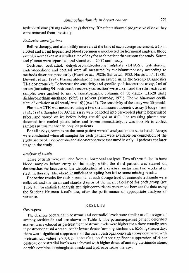
hydrocortisone (20 mg twice a day) therapy. If patients showed progressive disease they were removed from the study.
Endocrine investigations Before therapy, and at monthly intervals at the time of each dosage increment, a 10 ml
clotted and a 5 ml heparinized blood specimen was collected for hormonal analysis. Blood samples were taken at a similar time of day for each patient throughout the study. Serum and plasma were separated and stored at - 20°C until assay.
Oestrone, oestradiol, dehydroepiandrosterone sulphate (DHA-S), testosterone, androstenedione and cortisol were all measured by radioimmunoassay according to methods described previously (Harris et al., 1982b; Sufi et al., 1982; Harris et al., 1983b; Dowsett et al., 1984). Plasma aldosterone was measured using the Serono Diagnostics 3H-aldosterone kit. To increase the sensitivity and specificity of the oestrone assay, 2 ml of serum (including 3H-oestrone for recovery correction) were taken, and the ether-extracted samples were applied to mini-chromatographic columns of ‘Sephadex’ LH-20 using dichloromethane-methanol (95:5) as solvent (Murphy, 1971). The within-assay coeffi- cient of variation at 45 pmol/l was 16% (n = 15). The sensitivity of the assay was 30 pmol/l.
Plasma ACTH was measured using a two site immunoradiometric assay (Hodgkinson et al., 1984). Samples for ACTH assay were collected into pre-cooled plastic heparinized tubes, and stored on ice before being centrifuged at 4°C. The resulting plasma was decanted into cooled plastic tubes and frozen immediately. It was possible to collect samples in this manner in only 20 patients.
For all assays, samples on the same patient were all analysed in the same batch. Assays were conducted when all samples for each patient were available on completion of the study protocol. Testosterone and aldosterone were measured in only 13 patients at a later stage in the study.
Analysis of results Three patients were excluded from all hormonal analyses. Two of these failed to have
blood samples before entry to the study, whilst the third patient was started on dexamethasone because of the identification of a cerebral metastasis two weeks after starting therapy. Elsewhere, insufficient sampling has led to some missing results.
Endocrine results for each hormone, at each dosage level of aminoglutethimide were collected and the mean and standard error of the mean calculated for each group (see Table 1). For statistical analysis, multiple comparisons were made between the data using the Student Neuman Keul’s test, after the performance of appropriate analyses of variance.
RESULTS Oestrogens
The changes occurring in oestrone and oestradiol levels were similar at all dosages of aminoglutethimide and are shown in Table 1. The perimenopausal patient described earlier, was excluded as pretreatment oestrone levels were higher than those usually seen in postmenopausal women. At the lowest dose of aminoglutethimide, 62.5 mg twice a day, there was a significant suppression of the mean oestrogen concentrations compared with pretreatment values ( P < 0.0 1). Thereafter, no further significant suppression of either oestrone or oestradiol levels was achieved with higher doses of aminoglutethimide alone, or with combined aminoglutethimide and hydrocortisone therapy.

222 R. Stuart-Harris et al.
Table 1. Mean hormone concentrations ( f SEM) observed during increasing doses of aminoglutethimide ~
62 5 mg 125 mg 250 mg 500 mg 500 mg twice a day twice a twice a twice a twice a and hydrocortisone
Hormone Pretreatment day day day day 20 mg twice a day
Oestrone (pmoljl)
Oestradiol (pmol/l)
Androstenedione (nmolil)
Testosterone (nmol/l)
DHA-S (pmol/l)
Cortisol (nmolil)
Aldosterone (pmolil)
ACTH (ngjl)
162.2f 13.5
39.4 f 4 4 (30)
3.3 k 0.4 (33)
1 .4k0 .2
2.3 f 0.3 (33)
544+35 (33)
227 f 50
(32)
(13)
(13) 48.8 f 6.0
(20)
Numbers in parentheses refer to number of patients at each dosage
Androgens The mean androstenedione concentration at 62.5 mg twice a day of aminoglutethimide
was significantly higher than the mean pretreatment androstenedione concentration (P < 0.05, Table 1). No further significant alteration in the mean serum androstenedione concentration occurred until the dose of 500 mg twice a day of aminoglutethimide, when a further significant rise was observed (P < 0.01). However, the addition of hydrocortisone (20 mg twice a day) to this dosage caused significant suppression of mean serum androstenedione concentration (P < 0.0 1) back to a level not significantly different from the mean pretreatment concentration.
Mean levels of testosterone in 13 patients treated with aminoglutethimide alone were higher at all doses than pretreatment levels but this was significant only at 500 mg twice a day (P < 0.0 1). The levels when hydrocortisone was added to the regime were significantly lower than all other on-treatment values as well as pretreatment levels (P< 0.01).
The mean serum DHA-S concentration at the dose of 62.5 mg twice a day was significantly lower than the mean serum pretreatment concentration (P < 0.05). There- after, no further significant alteration in the mean serum DHA-S concentration occurred until hydrocortisone was combined with aminoglutethimide when a further significant fall was found (P<O.OI).
Cortisol Throughout the study, mean serum cortisol concentrations remained within normal
limits. Although mean cortisol concentrations at the doses of 125 mg twice a day and at 250 mg twice a day were lower than the mean pretreatment concentration, these alterations were not statistically significant. However, the mean serum cortisol concentra-

Am inoglu te t h im ide in breast cancer 223
tion on combined aminoglutethimide and hydrocortisone was significantly higher than at 125 or 250 mg twice a day (P<O.Ol).
Aldosterone
from the mean pretreatment level.
ACTH Plasma ACTH results for 20 patients at the various dosages of aminoglutethimide are
shown in Table 1. Throughout the study mean plasma ACTH concentrations were observed to be at the upper limit of normal (normal range 25 to 65 ng/l). However, no significant alterations in mean plasma ACTH concentrations were observed during aminoglutethimide therapy.
At no dose level was the mean on-treatment aldosterone level significantly different
DISCUSSION
AG in a dose approximately 1 g daily combined with a glucocorticoid is effective in the treatment of advanced breast cancer, inducing response in approximately one-third of postmenopausal women (Smith et al., 1978, 1982; Harris et al., 1982a). When used in this way, it causes suppression of circulating oestrone and oestradiol concentrations, and has been termed ‘medical adrenalectomy’ (Newsome et al., 1978; Lipton & Santen, 1974; Santen et al., 1974). We investigated the use of low dose AG alone to see whether equivalent oestrogen suppression can be achieved by aromatase inhibition alone as with aromatase plus adrenal inhibition.
There have only been two previous studies of low doses of AG used without HC (Harris et al., 1983a; Vermeulen et al., 1983). In both instances suppression of oestrogen levels and increases in androstenedione levels were found to occur, with no significant change of cortisol levels. However, in both cases the number of patients studied was relatively small (n= 13 and 6) , and the length of time on treatment was relatively short (7 and 10 days).
In the current study suppression of serum oestrone and oestradiol concentrations occurred even at the lowest dose of AG (62.5 mg twice a day). Suppression of oestrone appeared greater with 125 mg twice a day of AG, but the difference failed to reach statistical significance. Thereafter, higher doses of AG alone or combined AG and HC therapy failed to cause further significant suppression of serum oestrone or oestradiol concentrations. In contrast to its effect on serum oestrogens, low dose AG (125 mg twice a day) caused significantly less suppression of pretreatment DHA-S concentrations than conventional dose therapy combined with HC. These results suggest therefore that low dose AG alone might be as effective as conventional dose AG combined with HC in producing suppression of serum oestrogen concentrations. The lack of suppression by AG alone of cortisol or the adrenal oestrogen precursors, testosterone and androstene- dione, indicates that such treatment does not involve overall adrenal suppression, but rather the mechanism of suppression is likely to be entirely by aromatase inhibition.
Significant elevations of serum androstenedione were noted with all doses of AG alone, although serum androstenedione concentrations returned to pretreatment levels on combined AG and HC therapy. Although the rise in androstenedione might be attributed to aromatase inhibition, this is an unlikely mechanism as only 1-3% of circulating androstenedione is metabolically cleared via oestrone in postmenopausal women

224 R. Stuart-Harris et al.
(McDonald et al., 1967; Poortman et al., 1971, 1973; Grodin et al., 1973). The pattern of changes in testosterone levels was similar to that in androstenedione, although the degrees of change were much smaller. The increasing levels are probably due to inhibition of the 11 and/or 21 hydroxylase enzymes (Harris et al., 1983a; Vermeulen et al., 1983) possibly combined with stimulation of the 38-hydroxysteroid dehydrogenase isomerase enzyme (Samojlik & Santen, 1978).
In terms of the overall objective of oestrogen suppression the stimulation by AG of adrenal production of the oestrogen precursors, androstenedione and testosterone, is undesirable. It is probable that this effect of AG has attracted little attention, because AG is conventionally used in combination with glucocorticoid. Even in that situation androstenedione levels are higher with combined AG and HC than when glucocorticoid is given alone (Harris et al., 1984). It would therefore seem that the term ‘medical adrenalectomy’ to describe the use of AG with or without glucocorticoid is inappropriate. Indeed, the observation that serum oestrone concentrations fell in association with rising androstenedione concentrations suggest indirectly, that AG is an effective aromatase inhibitor even at the lowest dose of 62.5 mg twice a day, and that this is its mechanism of action.
The secretion rate of aldosterone has been found to fall markedly in four normal subjects during the first 2 days of treatment with 2 g of AG daily. Although in the present study levels of aldosterone appeared to fall in a dose-related manner on the higher dose levels of AG, the changes were not statistically significant. It may be that this is due to the high variability of aldosterone levels in non-recumbent subjects.
Throughout the study, serum cortisol and plasma ACTH concentrations remained within normal limits. This would appear suprising in the light of the initial withdrawal of AG from the market on the basis of induced adrenal insufficiency. However, it is notable that this problem occurred in two paediatric patients (Camacho et al., 1966) and it is possible that such patients may be particularly sensitive to the drug. This lack of effect on cortisol levels in the current study confirms other recent reports on the use of AG alone (Harris et al., 1983a; Vermeulen et al., 1983). Clearly inhibition by AG of the 20,22 desmolase, 1 1 -hydroxylase and 2 1 -hydroxylase enzyme systems does not result in suppression of cortisol levels. In such instances of incomplete enzyme blocks it would be expected that ACTH levels would have to be increased to maintain normal cortisol levels, and indeed in two small studies in three normal subjects (Fishman et al., 1967) and in 12 rats (Dexter et al., 1967) ACTH levels did rise on treatment with AG. We observed no such increase in ACTH levels. It is possible that any changes in ACTH concentrations during our study occurred shortly after starting therapy with AG, and were not present when samples were taken after the first month of treatment.
In summary, the suppression of circulating oestrone and oestradiol concentrations by low dose AG (125 mg twice a day) is not significantly different from that achieved by conventional dose AG combined with HC. AG when used alone appears to act through the mechanism of peripheral aromatase inhibition rather than adrenal suppression. Clinical studies of the use of low dose AG alone in the treatment of advanced breast cancer in postmenopausal women are now needed.
ACKNOWLEDGEMENTS We would like to thank our surgical and radiotherapy colleagues for allowing their patients to participate in this study, We would also like to thank Miss S. Beehnick for

Am inoglu te th im ide in breast cancer 225
assistance in collection of blood samples and Mrs L. Carr for the preparation of the manuscript. We are grateful to D. Easton for the statistical analyses. R.S-H. was in receipt of a Research Fellowship awarded by the Clinical Research Committee of the Royal Marsden Hospital during the course of these studies.
REFERENCES
BRODIE. A.M.H. (1962) Overview of recent development of aromatase inhibitors. Cancer Research. 42, (Suppl.), 33 12s-33 14s.
CAMACHO, A.M., BROUGH, A.J., CASH, R. & WILROY, R.S. (1966) Adrenal toxicity associated with the administration of an anticonvulsant drug. Journal of Pediatrics, 68, 852-853.
CASH. R., BROUGH, A.J., COHEN, M.N.P. & SATOH, P.S. (1967) Aminoglutethimide (Elipten-Ciba) as an inhibitor of adrenal steroidogenesis: mechanism of action and therapeutic trial. Journal of Clinical Endocrinology and Metabolism, 27, 1239-1248.
CHAKRABORTY, J., HOPKINS, R. & PARKE, D.V. (1972) Inhibition studies on the aromatization of androst-4- ene3,17-dione by human placental microsomal preparations. Biochemical Journal, 130, 19.
COHEN, M.P. & FOA, P.O. (1969) Aminoglutethimide inhibition of adrenal desmolase activity. Proceedings of the Society for Experimental Biology and Medicine, 127, 1086-1 090.
DEXTER, R.N., FISHMAN, L.M., NEY, R.L. & LIDDLE. G.W. (1967) Inhibition of adrenal corticosteroid synthesis by aminoglutethimide: studies of the mechanism of action. Journal of Clinical Endocrinology and Metabolism, 27,473480.
DOWSETT, M., HARRIS, A.L., SMITH, I.E. & JEFFCOATE, S.L. (1984) Endocrinechanges associated with relapse in advanced breast cancer patients on aminoglutethimide therapy. Journal of Clinical Endocrinology and Metabolism, 58, 99-104.
FISHMAN, L.M., LIDDLE, G.W., ISLAND, D.P., FLEISCHER, N. & KUCHEL, 0. (1967) Effects of aminoglutethimide on adrenal function in man. Journal of Clinical Endocrinology and Metabolism, 27, 481490.
GOWER, D.B. (1974). Modifiers of steroid-hormone metabolism: a review of their chemistry, biochemistry and clinical applications. Journal of Steroid Biochemistry, 5, 501-523.
GRAVES, P.E. & SALHANICK, H.A. (1979) Stereoselective inhibition of aromatase by enantiomers of aminoglutethirnide. Endocrinology, 105, 52-57.
GRODIN, J.M., SIITERI, P.K. & MACDONALD, P.C. (1973) Source of estrogen production in postmenopausal women. Journal of Clinical Endocrinology and Metabolism, 36, 201-214.
HARRIS, A.L.. POWLES, T.J. & SMITH, I.E. (1982a) Aminoglutethimide in the treatment of advanced postmenopausal breast cancer. Cancer Research, 42, (Suppl.), 3405~~3408s.
HARRIS, A.L., DOWSETT, M., JEFFCOATE, S.L., MCKINNA, J.A., MORGAN, M. & SMITH, I.E. (1 982b) Endocrine and therapeutic effects of aminoglutethimide in premenopausal patients with breast cancer. Journal of Clinical Endocrinology and Metabolism, 55, 7 18-722.
HARRIS, A.L., DOWSETT, M., SMITH, I.E. & JEFFCOATE, S.L. (1983a) Endocrine effects of low dose aminoglutethimide alone in advanced postmenopausal breast cancer. British Journal of Cancer, 47,
HARRIS, A.L., DOWSETT, M.. JEFFCOATE, S.L. & SMITH, I.E. (1983b) Aminoglutethimide dose and hormone suppression in advanced breast cancer. European Journal of Cancer and Clinical Oncology, 19,493-498.
HARRIS, A.L., DOWSETT, M., SMITH, I.E. & JEFFCOATE, S.L. (1984) Hydrocortisone alone vs. hydrocortisone plus aminoglutethimide: comparison of the adrenal effects in postmenopausal breast cancer. European Journal of Cancer and Clinical Oncology, 20,463-469.
HODGKINSON, S.C., ALLOLIO, B., LANDON, J LOWRY, P.J. (1984) Development of a non-extracted two site immunoradiometric assay for ACTH ut ng extreme amino and carboxyl terminally directed antibodies. Biochemical Journal (in press).
KAHNT, F.W. & NEHER, R. (1966) On the adrenal biosynthesis of steroids in titro. 111. Selective inhibition of adrenocortical function. Helvetica Chemica Acta, 49, 725-732.
LIPTON, A. & SANTEN, R.J. (1 974) Medical adrenalectomy using aminoglutethimide and dexamethasone in advanced breast cancer. Cancer, 33, 503-512.
MACDONALD, P.C., ROMBAUT, R.P. & SIITERI, P.K. (1967) Plasma precursors of estrogen. I. Extent of conversion of plasma androstenedione to estrone in normal males and nonpregnant, normal, castrate and adrenalectomized females. Journal of Clinical Endocrinology and Metabolism, 27, 1103-1 1 11.
621-627.
226 R . Stuart-Harris et al.
MURPHY, B.E.P. (1971) 'Sephadex' column chromatography as an adjunct to competitive protein binding assays of steroids. Nature New Biologj, 232, 21-24.
NEWSDME. H.H.. BROWN, P.W.. TERZ, J.J. & LAWRENCE, W. (1978) Medical adrenalectomy and plasma steroids in advanced breast carcinoma. Surgerj, 83, 83-89.
POORTMAN, J., THIJSSEN, J.H.H. & SCHWARZ, F. (1971) Production of androgens and oestrogens in postmenopausal women. Aria Endocrinologica, Suppl., 15, 79.
POORTMAN, J., THIJSSEN, J.H.H. & SCHWARZ. F. (1973) Androgen production and conversion to estrogens in normal postmenopausal women and selected breast cancer patients. Journal of Clincal Endocrinology and Metabolism, 37, 101-109.
SAMOJLIK, E. & SANTEN, R.J. (1978) Adrenal suppression with aminoglutethimide. 111. Comparison of plasma A4 and A5-steroids in postmenopausal women treated for breast carcinoma. Journal of Clinical Endocrinology and Metabolism, 47, 7 17-729.
SANTEN, R.J., LIPTON, A. & KENDALL, J. (1974) Successful medical adrenalectomy with aminoglutethimide. Role of altered drug metabolism. Journal of American Medical Association, 230, 1661-1 665.
SANTEN, R.J., SANTNER. S . , DAVIES, B., VELDHUIS, J., SAMOJLIK. E. & RUBY, E. (1978) Aminoglutethimide inhibits extraglandular estrogen production in postmenopausal women with breast carcinoma. Journal of Clinical Endocrinology and Metabolism. 41, 1257-1265.
SANTEN, R.J. & MISBIN, R.I. (1981) Aminoglutethimide: review of pharmacology and clinical use. Pharmaco- therapy, 1, 95-120.
SMITH, I.E.. FITZHARRIS, B.M., MCKINNA, J.A., FAHMY. D.R., NASH, A.G., NEVILLE, A.M., GAZET, J-C, FORD, H.T. & POWLES, T.J. (1978) Aminoglutethimide in the treatment of metastatic breast carcinoma. Lancet. ii, 646-649.
SMIW, I.E., HARRIS, A.L., MORGAN, M.. GAZET, J-C. & MCKINNA, J.A. (1982) Tamoxifen versus aminoglutethimide versus combined tamoxifen and aminoglutethimide in the treatment of advanced breast carcinoma. Cancer Research, 42 (Suppl.), 3430s-3433s.
SUFI, S.B., DONALDSON, A. & JEFFCOATE, S.L. ( I 982) Method Manual of the Matched Assay Rengent Programme. WHO Special Programme of Research in Human Reproduction. World Health Organization, Geneva.
THOMPSON, E.A. & SIITERI, P.K. (1974) The involvement of human placental microsomal cytochrome P-450 in aromatization. Journal of Biological Chemistry, 294, 5373-5378.
TOUITOU, Y., BOGDAN, A., LEGRAND, J.C. & DESGREZ, P. (1975) Aminoglutethimide and glutethimide: effects on 18-hydroxycorticosterone biosynthesis by human and sheep adrenals in iitro. Acta Endocrinologica, 80,
VERMEULEN, A,. PARIDAENS, R. & HEUSON. J.C. (1983) Eflects of aminoglutethimide on adrenal steroid 517-526.
secretion. Clinicnl Endocrinologj, 19, 673-682.
![Research Paper: Gerotarget (Focus on Aging) Estrogen ... · joint pain [21]. In women with breast cancer, the arthralgia related to aromatase inhibitor therapy has been noted [37]](https://img.pdfslide.us/doc/110x75/600ae8640d2b053be97248d7/research-paper-gerotarget-focus-on-aging-estrogen-joint-pain-21-in-women.jpg)