Embed Size (px)
Citation preview

Copyright Pearson Prentice Hall
End Show
Slide 1 of 47
39–4 Fertilization and Development

Copyright Pearson Prentice Hall
End Show
Slide 2 of 47
Development is . .
Cell division
Growth
Differentiation
From embryonic tissues to
Specialized tissues & organs

Copyright Pearson Prentice Hall
End Show
Slide 3 of 47
39–4 Fertilization and Development
Fertilization
??

Copyright Pearson Prentice Hall
End Show
Slide 4 of 47
39–4 Fertilization and Development
Blastocyst Formation
The First Two Weeks

Copyright Pearson Prentice Hall
End Show
39–4 Fertilization and Development
Slide 5 of 47
Early Development
The stages of early development include implantation, gastrulation, and neurulation.

Copyright Pearson Prentice Hall
End Show
39–4 Fertilization and Development
Slide 6 of 47
Early Development
Implantation & Differentiation
Implantation
Copyright Pearson Prentice Hall
End Show
39–4 Fertilization and Development
Slide 7 of 47
Early Development
Gastrulation
The inner cell mass of the blastocyst gradually sorts itself into two layers, which then give rise to a third layer.
Early Gastrulation
Amniotic cavity
Primitive streak
Ectoderm
Endoderm
Mesoderm

Copyright Pearson Prentice Hall
End Show
39–4 Fertilization and Development
Slide 8 of 47
Early Development
3 Germ Layers
The ectoderm develops into the skin and nervous system.
The endoderm forms the digestive lining and organs.
Mesoderm cells differentiate into internal tissues and organs.

Copyright Pearson Prentice Hall
End Show
39–4 Fertilization and Development
Slide 9 of 47
Early Development
Neurulation the development of the nervous system.

Copyright Pearson Prentice Hall
End Show
Slide 10 of 47
39–4 Fertilization and Development
Neural crest Neural fold
Notochord
Early Development

Copyright Pearson Prentice Hall
End Show
Slide 11 of 47
39–4 Fertilization and Development
Neural crest Neural tube
Ectoderm
Notochord
Early Development
Gradually, these folds move together to create a neural tube from which the spinal cord and the nervous system develop.
Nova - Gastrulation

Copyright Pearson Prentice Hall
End Show
Slide 12 of 47
39–4 Fertilization and Development
Morphing Embryos

Copyright Pearson Prentice Hall
End Show
39–4 Fertilization and Development
Slide 13 of 47
Early Development
Extraembryonic Membranes
As the embryo develops, membranes form to protect and nourish the embryo.
Two of these membranes are the amnion and the chorion.

Copyright Pearson Prentice Hall
End Show
39–4 Fertilization and Development
Slide 14 of 47
Early Development
The amnion develops into a fluid-filled amniotic sac, which cushions and protects the developing embryo.
Uterus
Amnion
Fetus
Amniotic sac
Placenta
Umbilical cord
Copyright Pearson Prentice Hall
End Show
39–4 Fertilization and Development
Slide 15 of 47
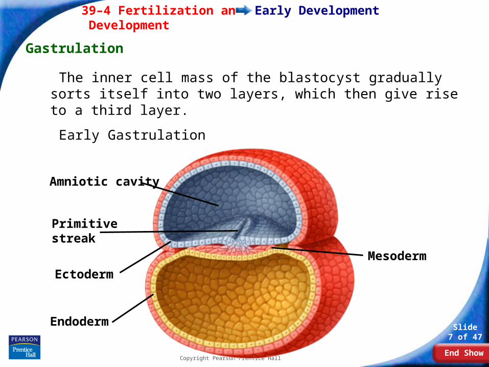
Fingerlike projections called chorionic villi form on the outer surface of the chorion and extend into the uterine lining.
Early Development
Fetal portion of placenta
Maternal portion of placenta
Maternal artery
Maternal vein
Umbilical vein
Umbilical arteries
Umbilical cord
Amnion
Chorionic villus

Copyright Pearson Prentice Hall
End Show
39–4 Fertilization and Development
Slide 16 of 47

Copyright Pearson Prentice Hall
End Show
39–4 Fertilization and Development
Slide 17 of 47
Early Development
Fetal Development (after 8+ weeks).
After three months, most major organs and tissues are formed. During this time, the umbilical cord also forms.
The umbilical cord connects the fetus to the placenta.
Placenta & Fetal Development
Copyright Pearson Prentice Hall
End Show
39–4 Fertilization and Development
Slide 18 of 47
Later Development
Later Development
4–6 months after fertilization:
• The heart can be heard with a stethoscope.
• Bone replaces cartilage that forms the early skeleton.
• A layer of soft hair grows over the fetus’s skin.
• The fetus grows and the mother can feel it moving.
Copyright Pearson Prentice Hall
End Show
39–4 Fertilization and Development
Slide 19 of 47
Later Development
During the last three months, the organ systems mature.
• The fetus doubles in mass.
• It can now regulate its body temperature.
• The central nervous system and lungs completely develop.

Copyright Pearson Prentice Hall
End Show
39–4 Fertilization and Development
Slide 20 of 47
Childbirth
Childbirth
About nine months after fertilization, the fetus is ready for birth.
A complex set of factors affects the onset of childbirth.

Copyright Pearson Prentice Hall
End Show
39–4 Fertilization and Development
Slide 21 of 47
Childbirth
The mother’s posterior pituitary gland releases the hormone oxytocin, which affects involuntary muscles in the uterine wall.
These muscles begin rhythmic contractions known as labor.
The contractions become more frequent and more powerful.

Copyright Pearson Prentice Hall
End Show
39–4 Fertilization and Development
Slide 22 of 47
Childbirth
The opening of the cervix expands until it is large enough for the head of the baby to pass through it.
At some point, the amniotic sac breaks, and the fluid it contains rushes out of the vagina.
Contractions force the baby out through the vagina.

Copyright Pearson Prentice Hall
End Show
39–4 Fertilization and Development
Slide 23 of 47
Childbirth
The baby now begins an independent existence.
Its systems quickly adapt to life outside the uterus, supplying its own oxygen, excreting waste on its own, and maintaining its own body temperature.