Embed Size (px)
Citation preview

Encephalopathy?
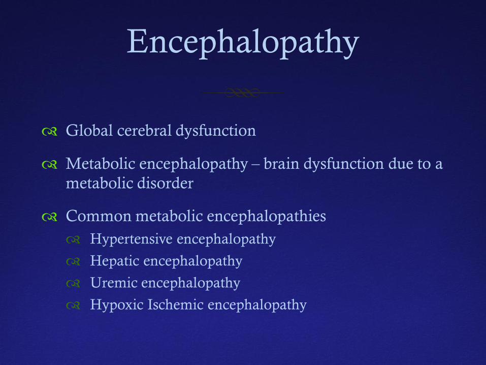
Encephalopathy
Global cerebral dysfunction
Metabolic encephalopathy – brain dysfunction due to a metabolic disorder
Common metabolic encephalopathies Hypertensive encephalopathy
Hepatic encephalopathy
Uremic encephalopathy
Hypoxic Ischemic encephalopathy
Encephalopathy
Metabolic encephalopathy – important but less common causes Mitochondrial disorders
Seretonin syndrome
Neuroleptic malignant syndrome
Paraneoplastic disorder associated with teratoma (anti glutamate receptor AB)
Hypertensive Encephalopathy
Hypertensive Encephalopathy
1% of patients with hypertension will present with hypertensive crisis (end-organ damage)
If hypertension remains untreated mortality is >50% over next 12 months
Hypertensive encephalopathy is among most serious presentations of uncontrolled hypertension
May be due to acute crisis in setting of medication non-compliance
May be part of drug intoxication (cocaine, amphetamines)
Hypertensive Encephalopathy
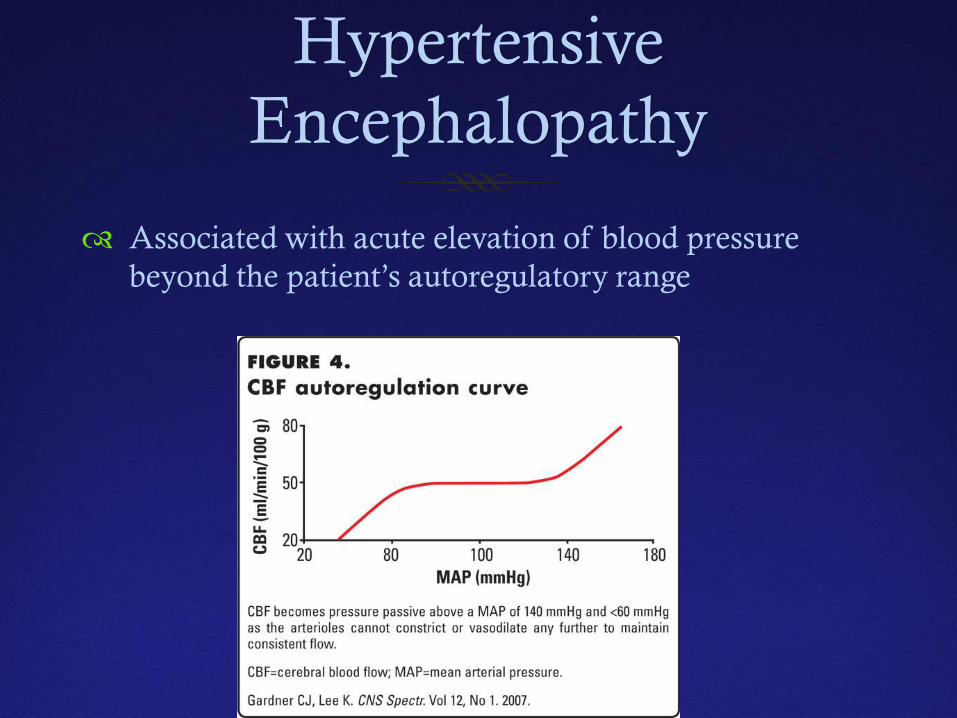
Associated with acute elevation of blood pressure beyond the patient’s autoregulatory range
Hypertensive Encephalopathy
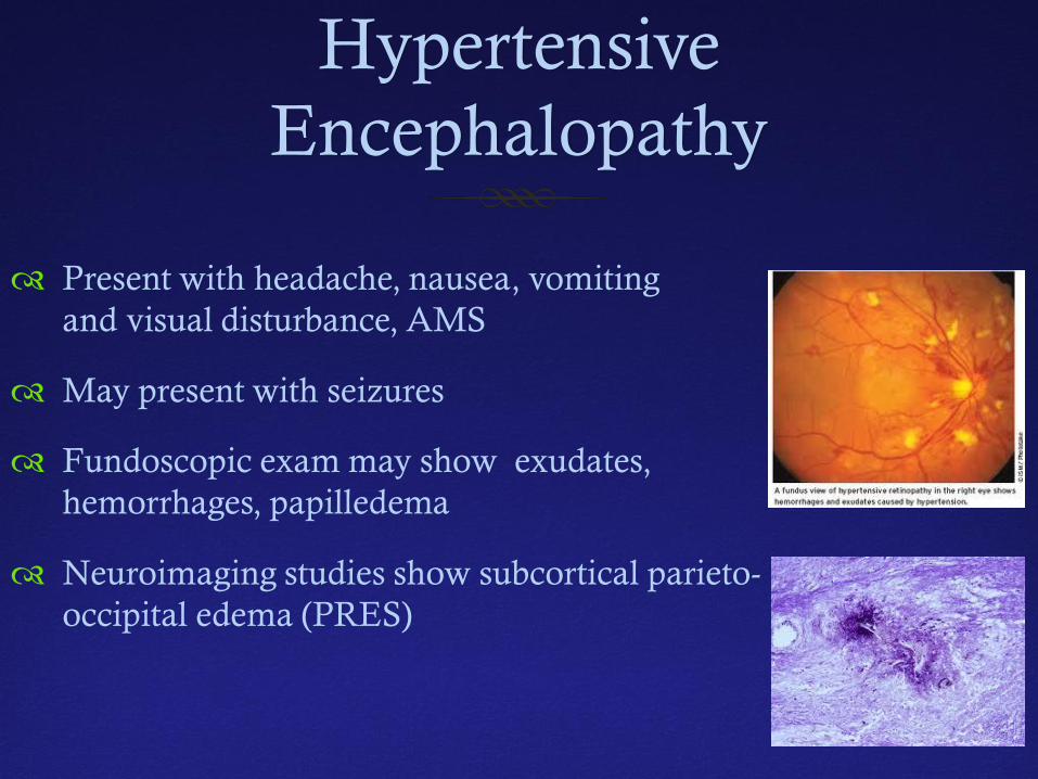
Present with headache, nausea, vomiting and visual disturbance, AMS
May present with seizures
Fundoscopic exam may show exudates, hemorrhages, papilledema
Neuroimaging studies show subcortical parieto-occipital edema (PRES)
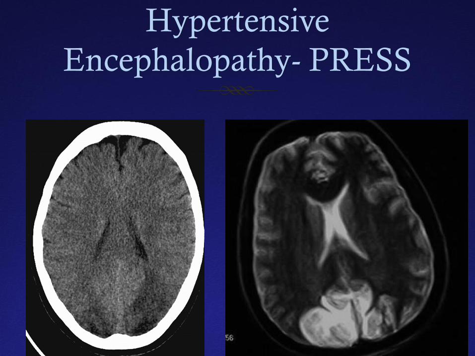
Hypertensive Encephalopathy- PRESS
Hypertensive Encephalopathy
Immediate treatment of hypertension is the key
Reduce MAP by 25%
Use parenteral antihypertensive agents and monitor BP in ICU setting with real time arterial pressure monitoring
Calcium channel blockers or beta blockers reduce BP without cerebral vasodilatation
Seizures not typically treated with AEDs
Hypertensive Encephalopathy- Future Treatment Possibilities
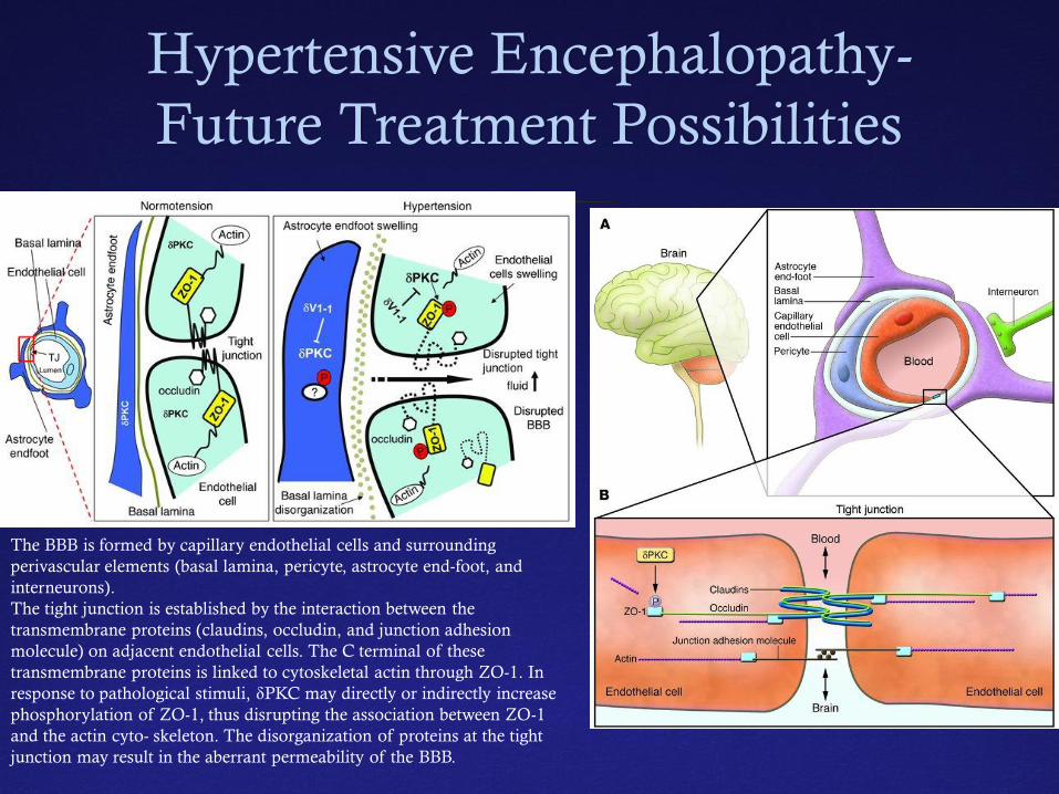
The BBB is formed by capillary endothelial cells and surrounding perivascular elements (basal lamina, pericyte, astrocyte end-foot, and interneurons). The tight junction is established by the interaction between the transmembrane proteins (claudins, occludin, and junction adhesion molecule) on adjacent endothelial cells. The C terminal of these transmembrane proteins is linked to cytoskeletal actin through ZO-1. In response to pathological stimuli, δPKC may directly or indirectly increase phosphorylation of ZO-1, thus disrupting the association between ZO-1 and the actin cyto- skeleton. The disorganization of proteins at the tight junction may result in the aberrant permeability of the BBB.
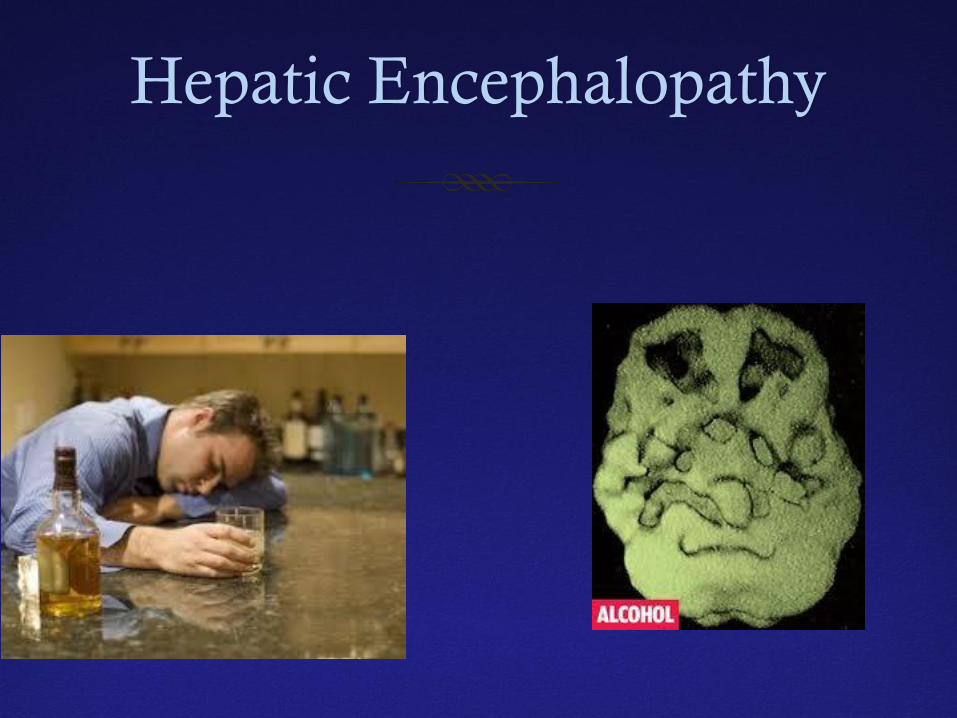
Hepatic Encephalopathy
Hepatic Encephalopathy
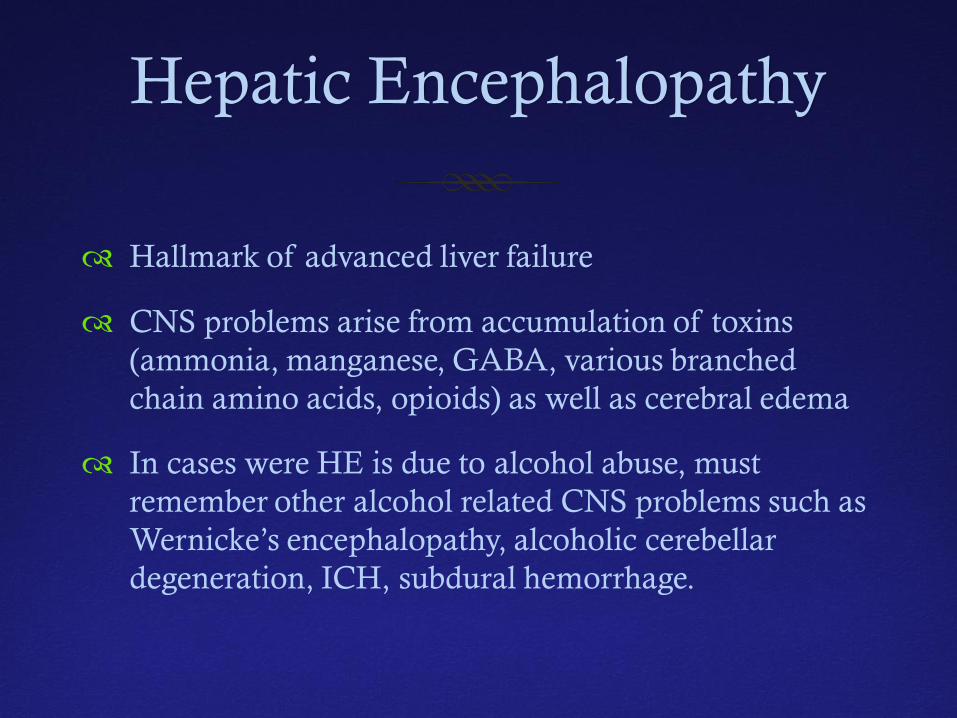
Hallmark of advanced liver failure
CNS problems arise from accumulation of toxins (ammonia, manganese, GABA, various branched chain amino acids, opioids) as well as cerebral edema
In cases were HE is due to alcohol abuse, must remember other alcohol related CNS problems such as Wernicke’s encephalopathy, alcoholic cerebellar degeneration, ICH, subdural hemorrhage.
Hepatic Encephalopathy
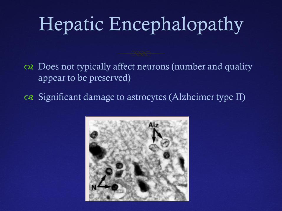
Does not typically affect neurons (number and quality appear to be preserved)
Significant damage to astrocytes (Alzheimer type II)
Hepatic Encephalopathy
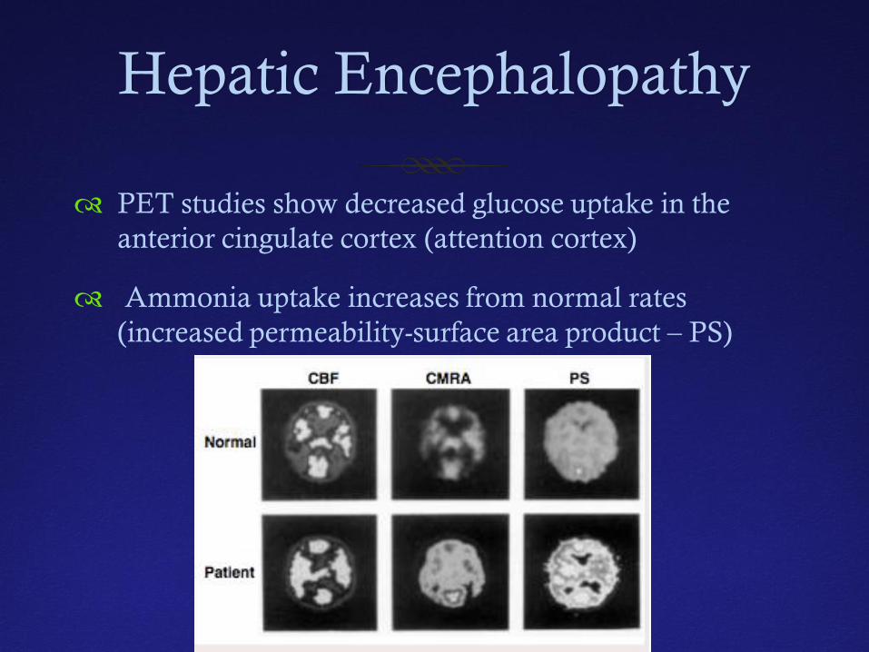
PET studies show decreased glucose uptake in the anterior cingulate cortex (attention cortex)
Ammonia uptake increases from normal rates (increased permeability-surface area product – PS)
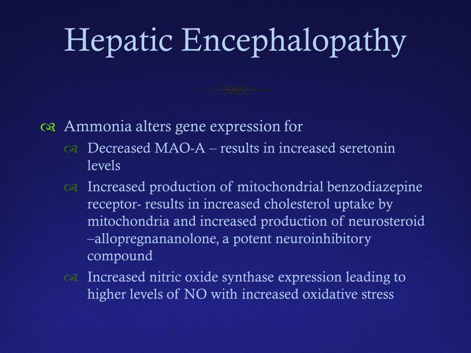
Hepatic Encephalopathy
Ammonia alters gene expression for Decreased MAO-A – results in increased seretonin
levels
Increased production of mitochondrial benzodiazepine receptor- results in increased cholesterol uptake by mitochondria and increased production of neurosteroid –allopregnananolone, a potent neuroinhibitory compound
Increased nitric oxide synthase expression leading to higher levels of NO with increased oxidative stress
Hepatic Encephalopathy
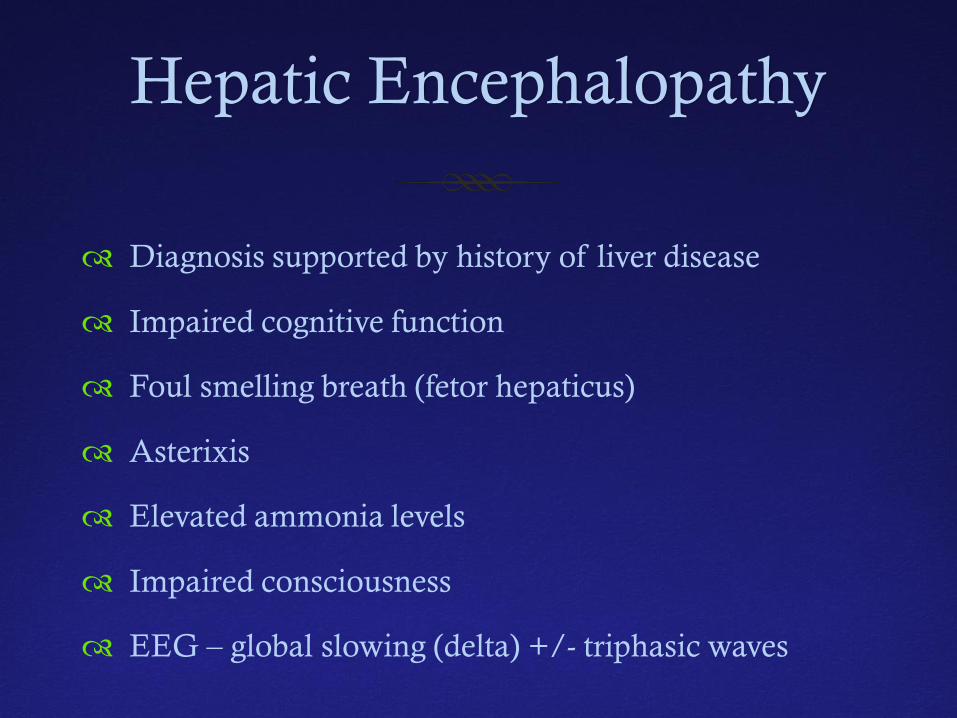
Diagnosis supported by history of liver disease
Impaired cognitive function
Foul smelling breath (fetor hepaticus)
Asterixis
Elevated ammonia levels
Impaired consciousness
EEG – global slowing (delta) +/- triphasic waves
Hepatic Encephalopathy
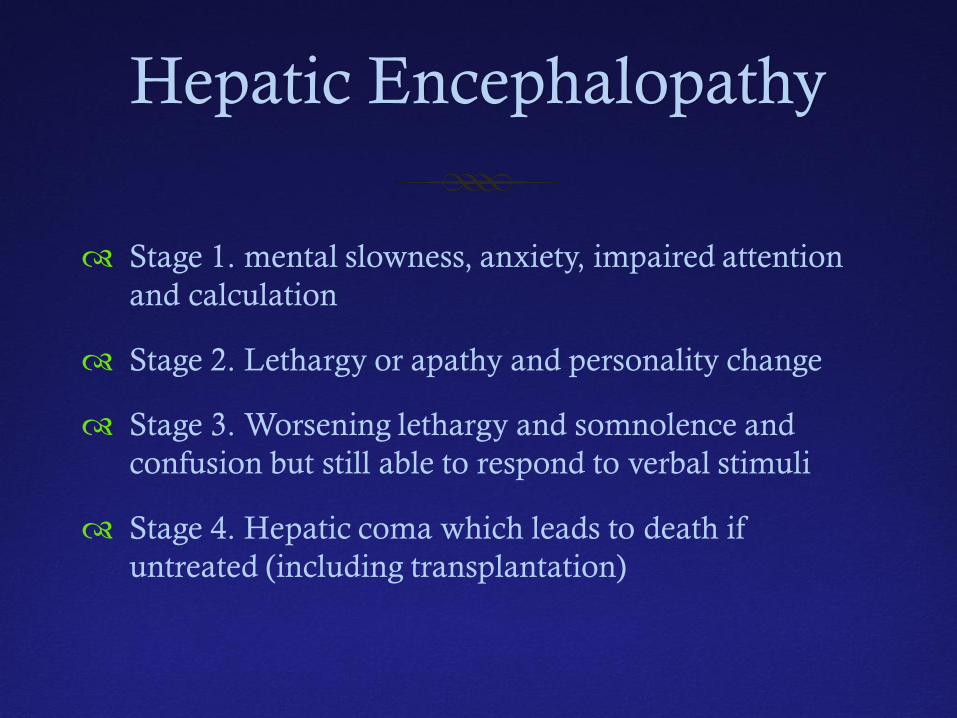
Stage 1. mental slowness, anxiety, impaired attention and calculation
Stage 2. Lethargy or apathy and personality change
Stage 3. Worsening lethargy and somnolence and confusion but still able to respond to verbal stimuli
Stage 4. Hepatic coma which leads to death if untreated (including transplantation)
Hepatic Encephalopathy
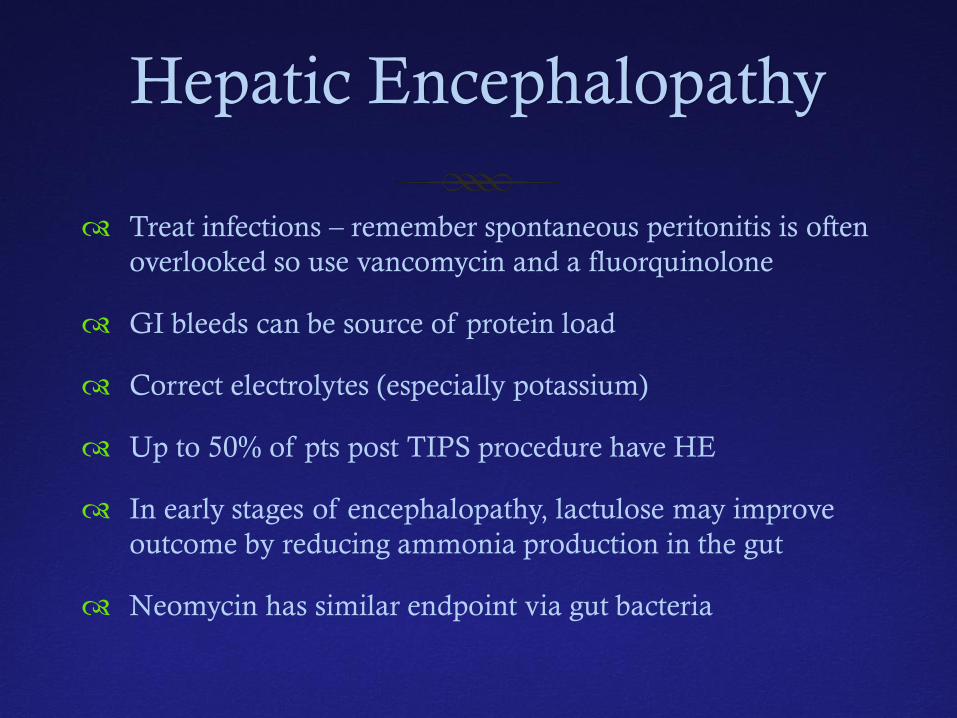
Treat infections – remember spontaneous peritonitis is often overlooked so use vancomycin and a fluorquinolone
GI bleeds can be source of protein load
Correct electrolytes (especially potassium)
Up to 50% of pts post TIPS procedure have HE
In early stages of encephalopathy, lactulose may improve outcome by reducing ammonia production in the gut
Neomycin has similar endpoint via gut bacteria
Hepatic Encephalopathy
Late stage liver failure associated with severe coagulopathy and failure of gluconeogenesis
Correction of coagulopathy in setting of active bleeding
Glucose infusions often necessary
Flumazenil only shown to be helpful in setting of patients given benzodiazepines
Cerebral edema and coma are pre-terminal events
Transplantation is ultimate therapeutic intervention
Hepatic Encephalopathy –ICP Management
Stage 1 and 2 not associated with cerebral edema
Stage 3 and 4 associated with cerebral edema and coma
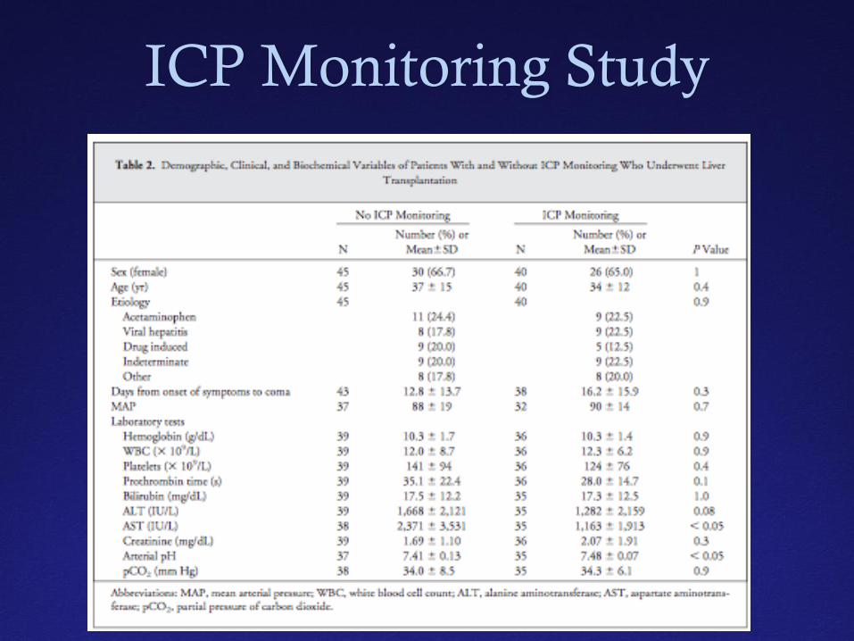
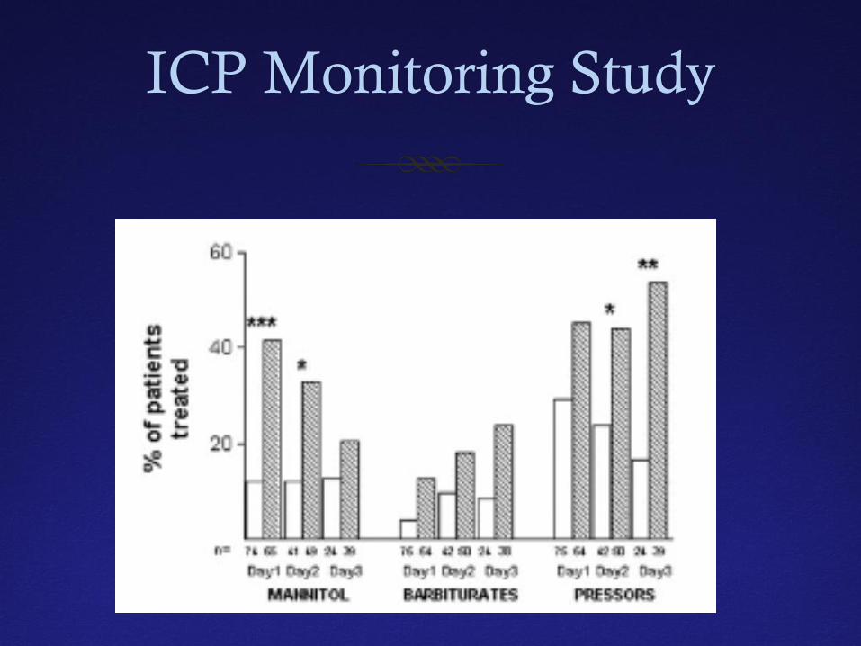
Vaquero et al looked at 332 pts with liver failure and severe encephalopathy – 92 had ICP monitoring (28%)
58 pts were reviewed closely
10.3% had ICH (half were incidental findings)
ICP Monitoring Study
ICP Monitoring Study
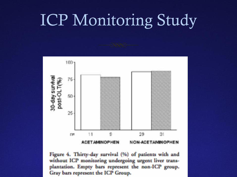
ICP Monitoring Study
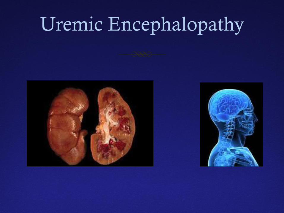
Uremic Encephalopathy
Uremic Encephalopathy
Characterized by slowly progressive dementia, psychiatric changes, speech disorders, involuntary movements, seizures, coma and death
EEG usually shows synchronous, symmetrical complexes lasting 2-4 seconds, high-amplitude slow waves, frontotemporal sharp waves, spikes and triphasic waves.
Progression associated with intermittent rhythmic delta activity, random spikes and sharp waves
Sleep is disorganized and fails to show REM
As uremia progresses, the paroxysmal bursts tend to decrease and ultimately disappear
Uremic Encephalopathy
Treatment of uremic encephalopathy revolves around management of the renal failure
AKI – minimize secondary worsening
Hemodialysis in setting of uremic symptoms
Remember to modify drug doses based on estimated creatinine clearance of various drugs
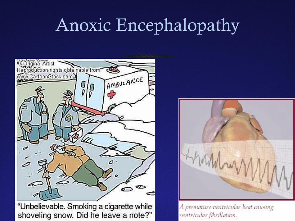
Anoxic Encephalopathy
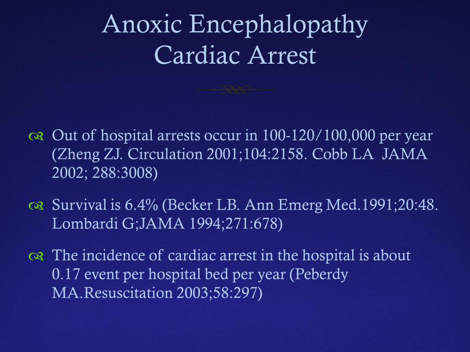
Anoxic Encephalopathy Cardiac Arrest
Out of hospital arrests occur in 100-120/100,000 per year (Zheng ZJ. Circulation 2001;104:2158. Cobb LA JAMA 2002; 288:3008)
Survival is 6.4% (Becker LB. Ann Emerg Med.1991;20:48. Lombardi G;JAMA 1994;271:678)
The incidence of cardiac arrest in the hospital is about 0.17 event per hospital bed per year (Peberdy MA.Resuscitation 2003;58:297)
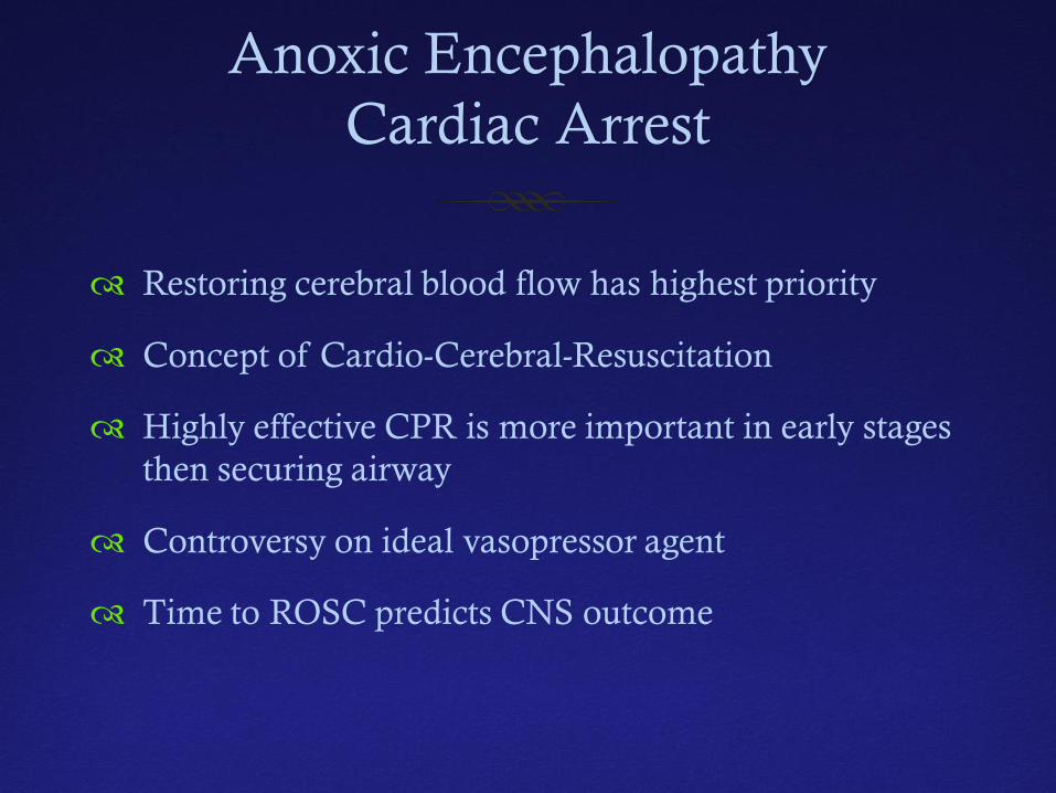
Anoxic Encephalopathy Cardiac Arrest
Restoring cerebral blood flow has highest priority
Concept of Cardio-Cerebral-Resuscitation
Highly effective CPR is more important in early stages then securing airway
Controversy on ideal vasopressor agent
Time to ROSC predicts CNS outcome
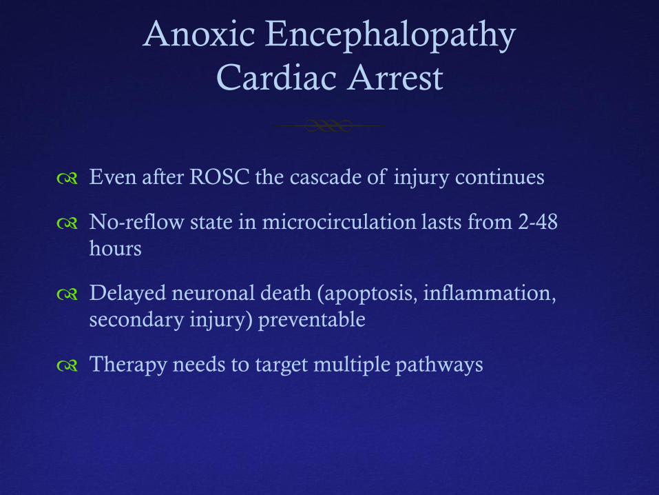
Anoxic Encephalopathy Cardiac Arrest
Even after ROSC the cascade of injury continues
No-reflow state in microcirculation lasts from 2-48 hours
Delayed neuronal death (apoptosis, inflammation, secondary injury) preventable
Therapy needs to target multiple pathways
Therapeutic Hypothermia
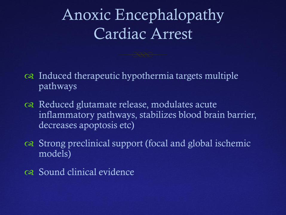
Anoxic Encephalopathy Cardiac Arrest
Induced therapeutic hypothermia targets multiple pathways
Reduced glutamate release, modulates acute inflammatory pathways, stabilizes blood brain barrier, decreases apoptosis etc)
Strong preclinical support (focal and global ischemic models)
Sound clinical evidence
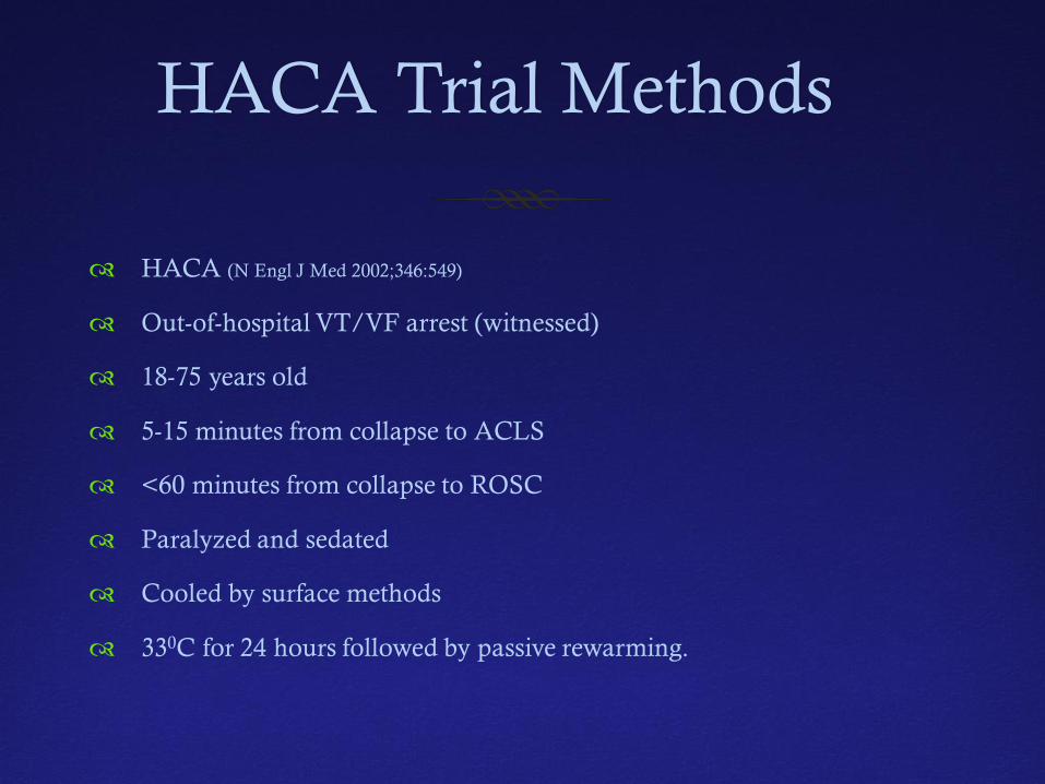
HACA Trial Methods
HACA (N Engl J Med 2002;346:549)
Out-of-hospital VT/VF arrest (witnessed)
18-75 years old
5-15 minutes from collapse to ACLS
<60 minutes from collapse to ROSC
Paralyzed and sedated
Cooled by surface methods
330C for 24 hours followed by passive rewarming.
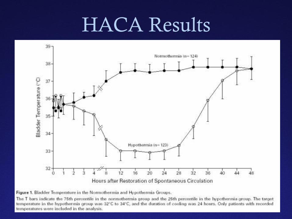
HACA Results
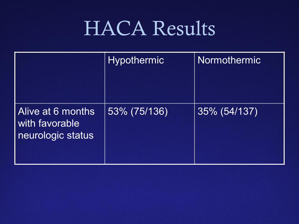
HACA Results Hypothermic Normothermic
Alive at 6 months with favorable neurologic status
53% (75/136) 35% (54/137)
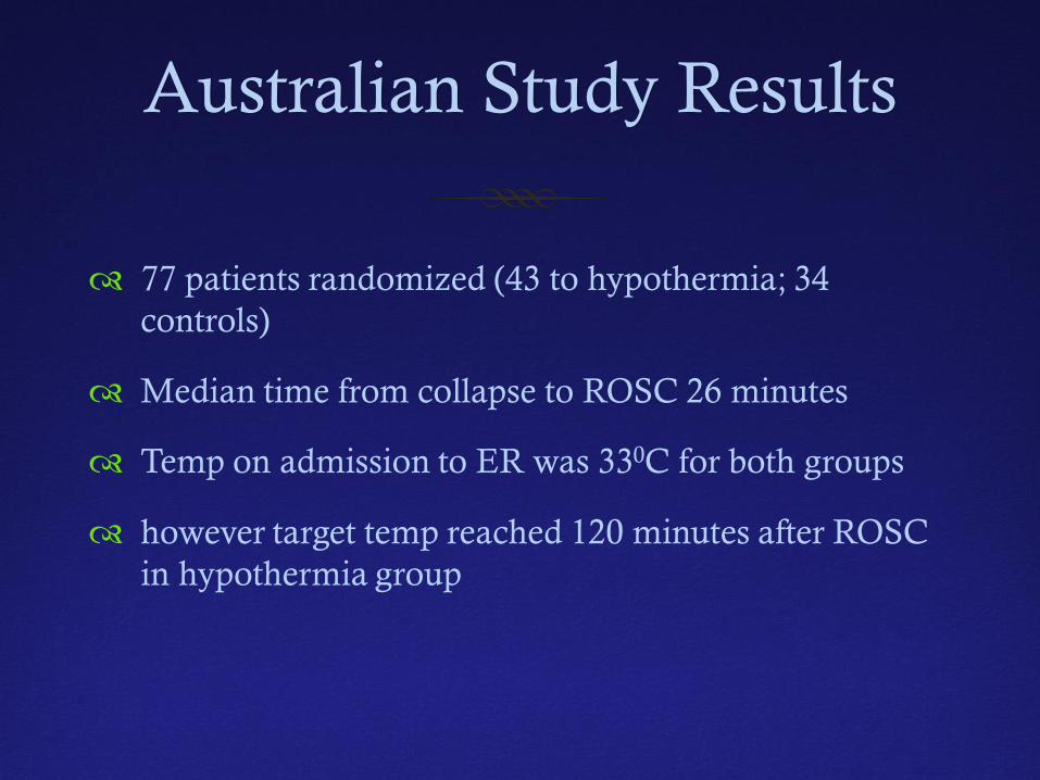
Australian Study Results
77 patients randomized (43 to hypothermia; 34 controls)
Median time from collapse to ROSC 26 minutes
Temp on admission to ER was 330C for both groups
however target temp reached 120 minutes after ROSC in hypothermia group
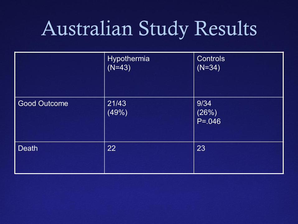
Australian Study Results Hypothermia (N=43)
Controls (N=34)
Good Outcome 21/43 (49%)
9/34 (26%) P=.046
Death 22 23
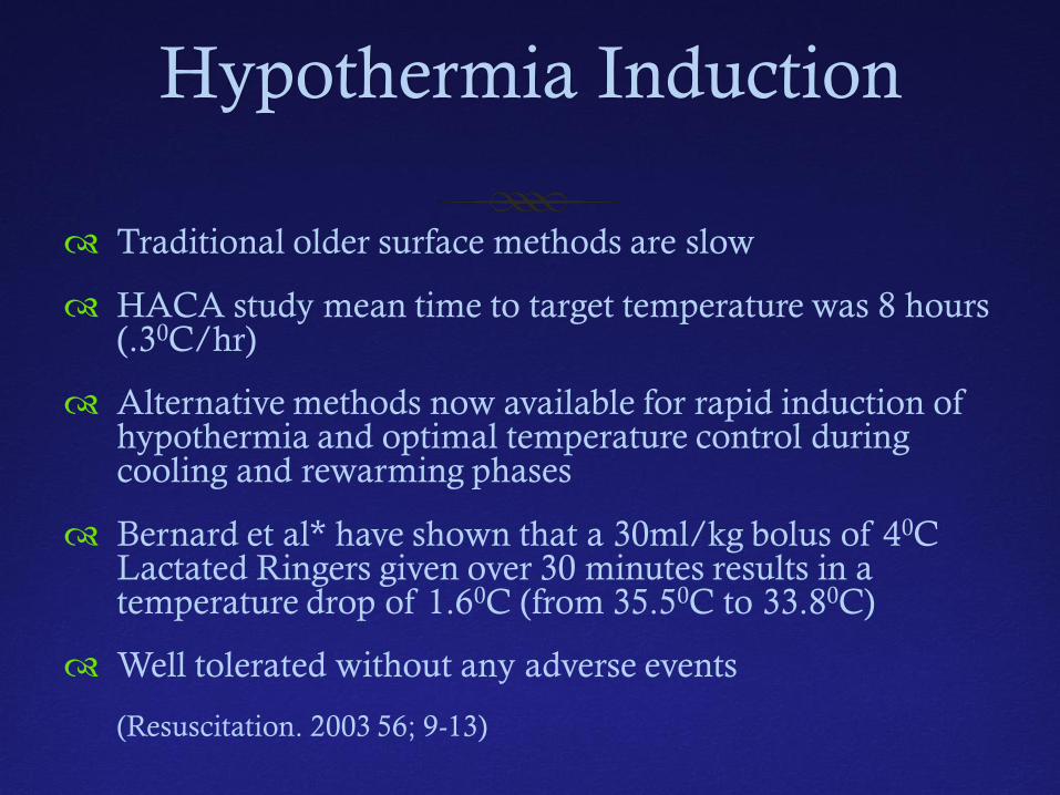
Hypothermia Induction
Traditional older surface methods are slow
HACA study mean time to target temperature was 8 hours (.30C/hr)
Alternative methods now available for rapid induction of hypothermia and optimal temperature control during cooling and rewarming phases
Bernard et al* have shown that a 30ml/kg bolus of 40C Lactated Ringers given over 30 minutes results in a temperature drop of 1.60C (from 35.50C to 33.80C)
Well tolerated without any adverse events
(Resuscitation. 2003 56; 9-13)
Maintenance of Hypothermia Phase
Hypothermia patients should be paralyzed sufficiently to prevent shivering during induction (may not be needed once cold)
Sedation advised during the hypothermia period to and maintain comfort; If paralyzed, should consider EEG or BIS monitoring.
Seizures are common following cardiac arrest (up to 20%) – EEG monitoring may help diagnose occult seizures especially during rewarming
EEG may be useful in prognosticating
Prophylactic AEDs not advised
Maintenance of Hypothermia Phase
Airway/Ventilator – Keep pO2 /pCO2 optimized (temp controlling may result in mild hyperventilation which is not optimal)
Avoid hypovolemia – associated with increased mortality in hypothermic patients in NABISH study
Bradycardia is most common rhythm
Maintenance of Hypothermia Phase
Avoid hyperglycemia
Do not overcorrect K+ (goal >3.0-3.5 mmol/L) as hyperkalemia occurs during rewarming if overcorrected
Start Lovenox early (recent study showed that in immobile stroke patients Lovenox superior to unfractionated heparin
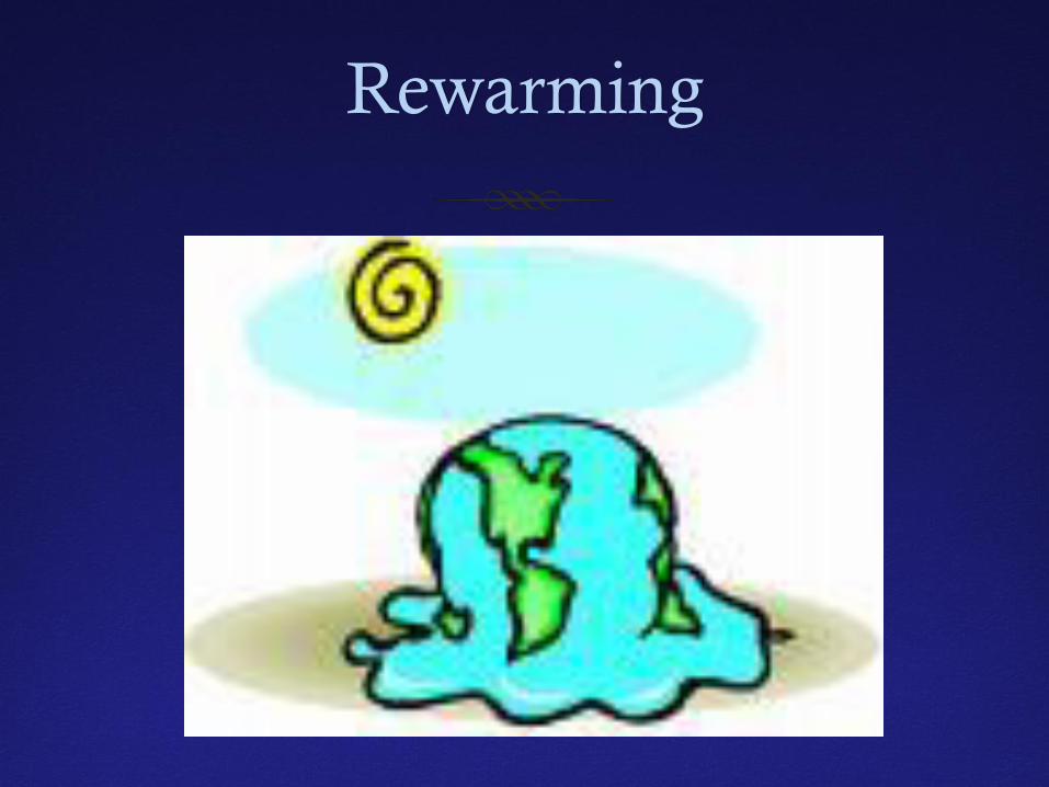
Rewarming
Rewarming Phase
Controlled slow rewarming is critical
Suggest a rate of 0.25-.300C per hour
Keep sedation and analgesic on while patient actively cooled
Monitor for seizures and cardiac arrhythmia
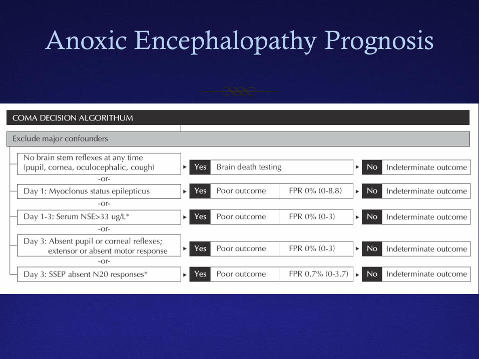
Anoxic Encephalopathy Prognosis
Mitochondrial Disorders
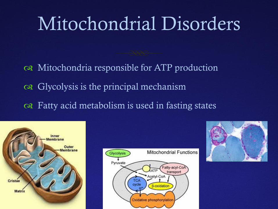
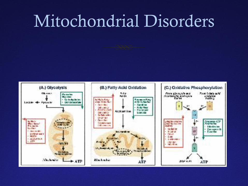
Mitochondria responsible for ATP production
Glycolysis is the principal mechanism
Fatty acid metabolism is used in fasting states
Mitochondrial Disorders
Mitochondrial Disorders
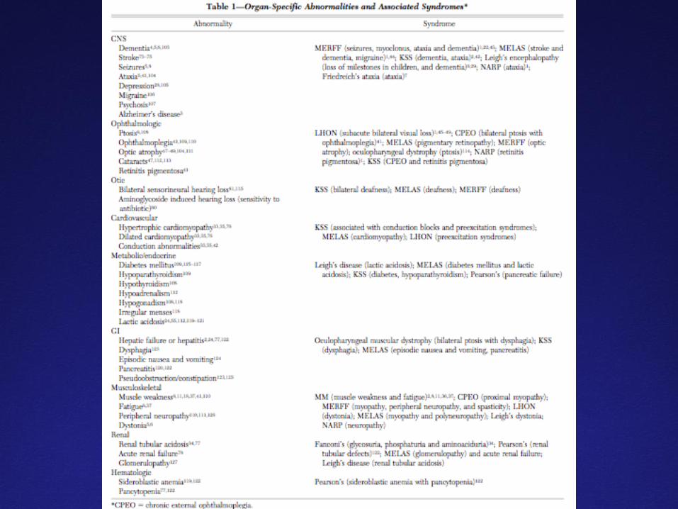
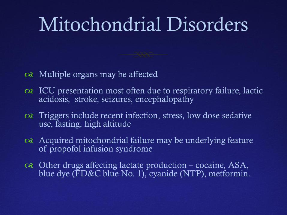
Multiple organs may be affected
ICU presentation most often due to respiratory failure, lactic acidosis, stroke, seizures, encephalopathy
Triggers include recent infection, stress, low dose sedative use, fasting, high altitude
Acquired mitochondrial failure may be underlying feature of propofol infusion syndrome
Other drugs affecting lactate production – cocaine, ASA, blue dye (FD&C blue No. 1), cyanide (NTP), metformin.
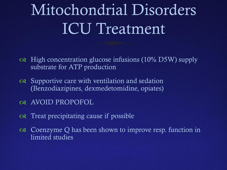
Mitochondrial Disorders ICU Treatment
High concentration glucose infusions (10% D5W) supply substrate for ATP production
Supportive care with ventilation and sedation (Benzodiazipines, dexmedetomidine, opiates)
AVOID PROPOFOL
Treat precipitating cause if possible
Coenzyme Q has been shown to improve resp. function in limited studies
Serotonin Syndrome
Seretonin Syndrome
Potentially life threatening adverse drug reaction
Not ideiosyncratic drug reaction as a consequence of excess serotonergic activity at CNS and PNS receptors (serotonin toxicity)
May be mistaken for neuroleptic malignant syndrome
No laboratory tests confirm diagnosis
Based on history and physical findings
Seretonin Syndrome
Antidepressants- MAOIs, TCAs, SSRIs, SNRIs, buproprion
Opioids – tramadol, fentanyl, oxycontin, methadone, dilaudid
CNS stimulants- amphetamine, methamphetamine, phentermine,
5-HT1 agonists – tryptans
Psychedelics – NMDA, MDA, LSD
Herbs – St John’s Wort, Syrian rue, Panax ginseng, Nutmeg
Others- tryptophan, L-Dopa, valproate, buspirone, lithium, linezolid, ritonavir
Seretonin Syndrome
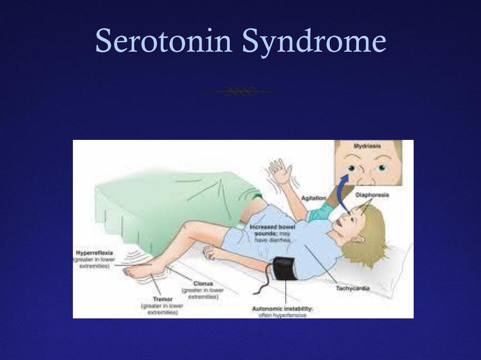
Mild cases – tachycardia, sweating, dilated pupils, myoclonus, hyperreflexia
Moderate cases- add hyperactive bowel, hypertension, hyperthermia (400C is common), clonus, hypervigilance and agitation
Severe cases- malignant hyperthermia (>41.10C), metabolic acidosis, rhabdomyolysis, seizures, renal failure, DIC
Seretonin Syndrome
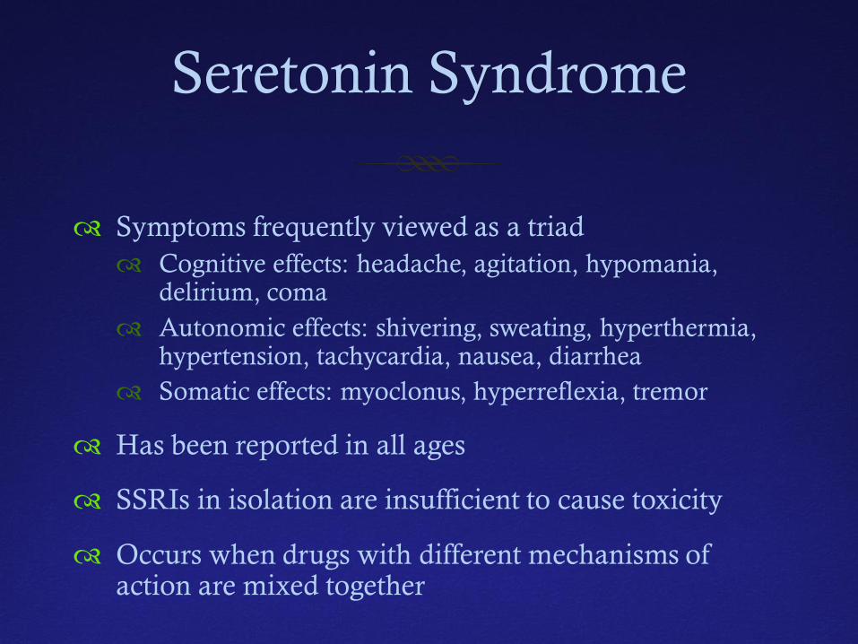
Symptoms frequently viewed as a triad Cognitive effects: headache, agitation, hypomania,
delirium, coma Autonomic effects: shivering, sweating, hyperthermia,
hypertension, tachycardia, nausea, diarrhea Somatic effects: myoclonus, hyperreflexia, tremor
Has been reported in all ages
SSRIs in isolation are insufficient to cause toxicity
Occurs when drugs with different mechanisms of action are mixed together
Serotonin Syndrome
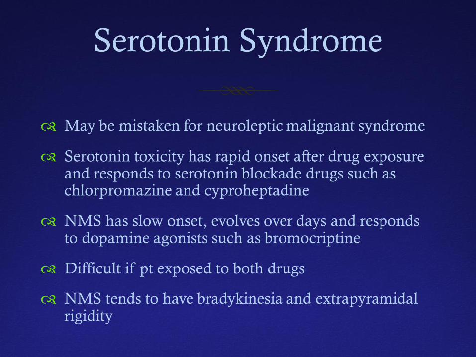
May be mistaken for neuroleptic malignant syndrome
Serotonin toxicity has rapid onset after drug exposure and responds to serotonin blockade drugs such as chlorpromazine and cyproheptadine
NMS has slow onset, evolves over days and responds to dopamine agonists such as bromocriptine
Difficult if pt exposed to both drugs
NMS tends to have bradykinesia and extrapyramidal rigidity
Serotonin Syndrome Management
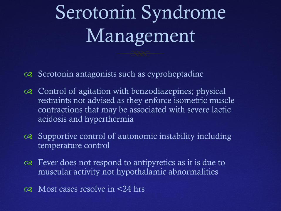
Serotonin antagonists such as cyproheptadine
Control of agitation with benzodiazepines; physical restraints not advised as they enforce isometric muscle contractions that may be associated with severe lactic acidosis and hyperthermia
Supportive control of autonomic instability including temperature control
Fever does not respond to antipyretics as it is due to muscular activity not hypothalamic abnormalities
Most cases resolve in <24 hrs
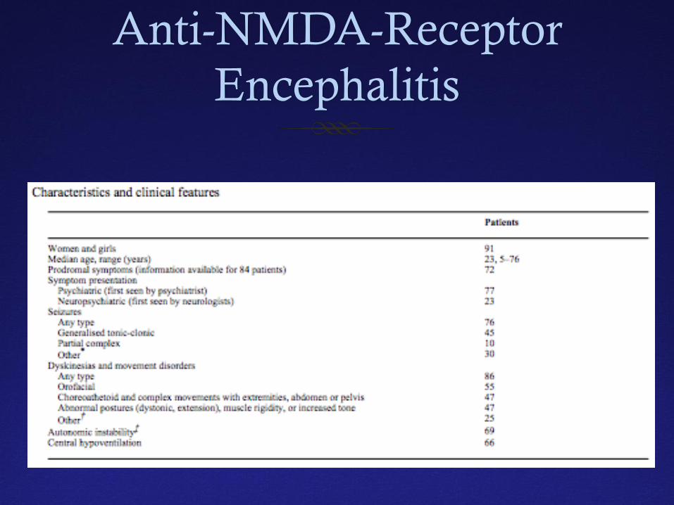
Anti-NMDA-Receptor Encephalitis
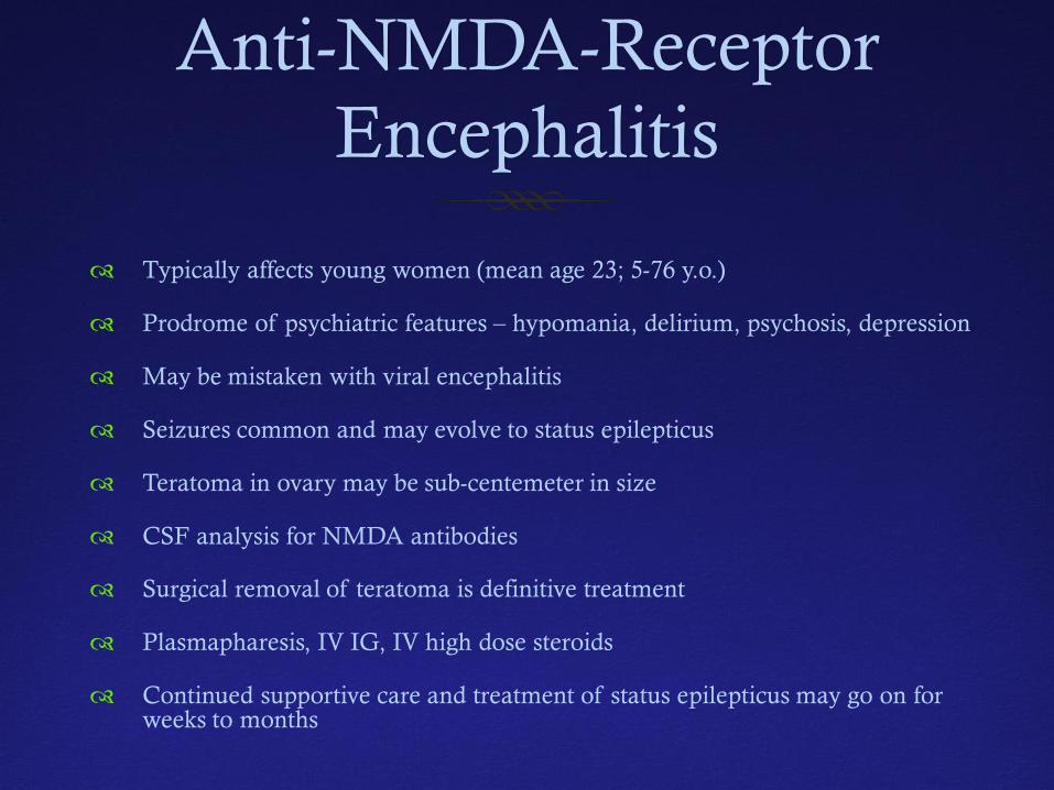
Typically affects young women (mean age 23; 5-76 y.o.)
Prodrome of psychiatric features – hypomania, delirium, psychosis, depression
May be mistaken with viral encephalitis
Seizures common and may evolve to status epilepticus
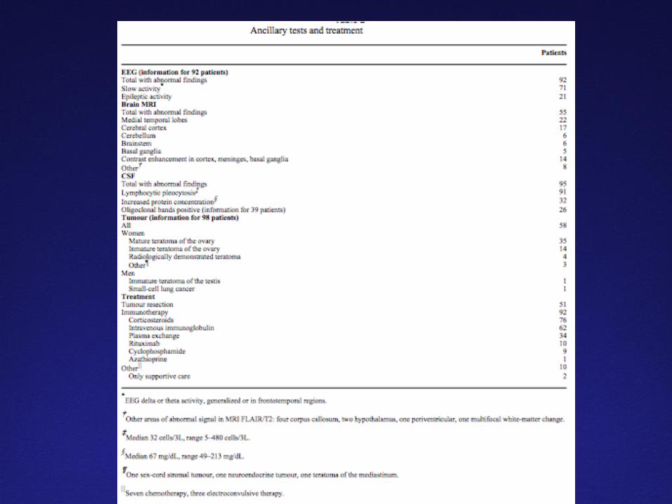
Teratoma in ovary may be sub-centemeter in size
CSF analysis for NMDA antibodies
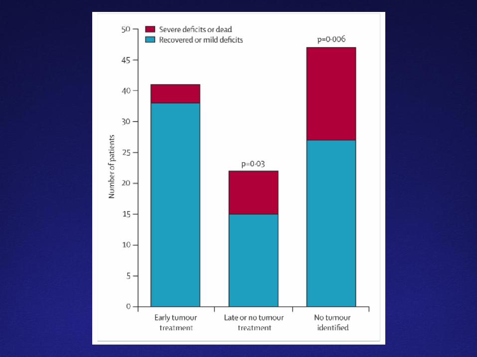
Surgical removal of teratoma is definitive treatment
Plasmapharesis, IV IG, IV high dose steroids
Continued supportive care and treatment of status epilepticus may go on for weeks to months
Anti-NMDA-Receptor Encephalitis
Selected References
Journal of Clinical Invest. 118: 17-20, 2008
Alcohol Research and Health. 27: 240-246, 2003
Liver Transplantation. 11: 1581-1589, 2005
N Engl J Med. 346 :549, 2002
N. Engl. J. Med. 352 (11): 1112–20, 2005
Chest. 120. 634-648, 2001
Lancet Neurology. 7: 1091-1098: 2008