Embed Size (px)
Citation preview
ACUTE ENCEPHALITIS
&ENCEPHALOPATHY
DR AFREEN KHAN
ASSISTANT PROFESSOR PEDIATRICS
HIMSR & HAHC
DEFINITIONS
Encephalopathy: Clinical syndrome of altered
mental status, manifesting as reduced
consciousness or altered behaviour
Encephalitis: An inflammatory process of the
brain parenchyma
ACUTE ENCEPHALITIS SYNDROME
A person of any age, presenting at any time of
year, with acute onset of fever and altered mental
status manifesting with symptoms such as
confusion, disorientation, coma, or inability to
talk and/or new onset of seizures (excluding
simple febrile seizures)
WHO. Acute encephalitis syndrome. Japanese encephalitis surveillance
standards. January 2006. from WHO recommended standards of surveillance of
vaccine preventable diseases
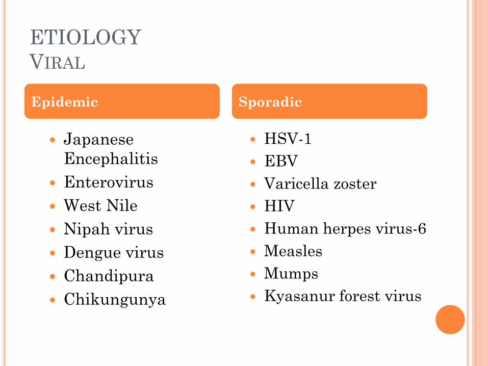
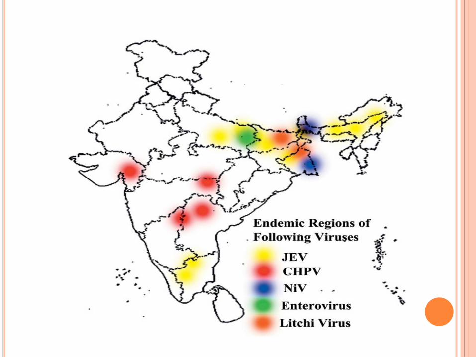
ETIOLOGY
VIRAL
Japanese
Encephalitis
Enterovirus
West Nile
Nipah virus
Dengue virus
Chandipura
Chikungunya
HSV-1
EBV
Varicella zoster
HIV
Human herpes virus-6
Measles
Mumps
Kyasanur forest virus
Epidemic Sporadic
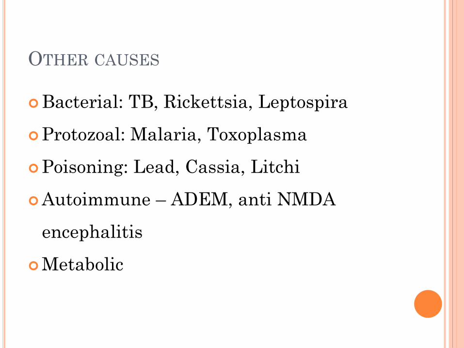
OTHER CAUSES
Bacterial: TB, Rickettsia, Leptospira
Protozoal: Malaria, Toxoplasma
Poisoning: Lead, Cassia, Litchi
Autoimmune – ADEM, anti NMDA
encephalitis
Metabolic
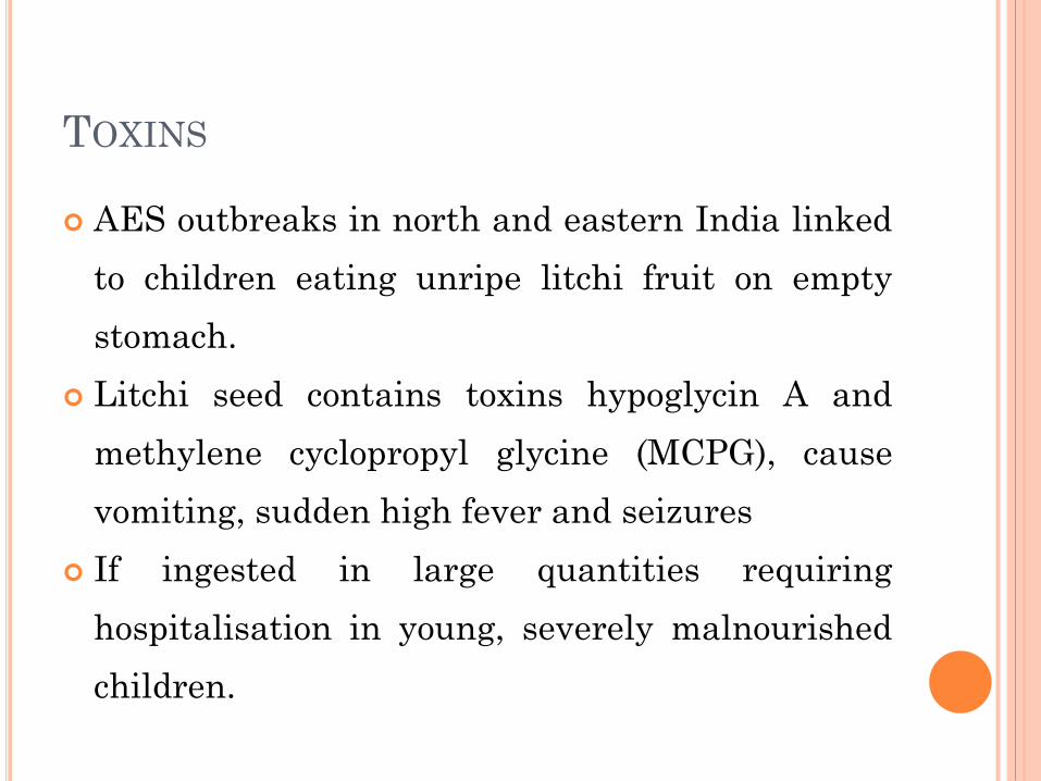
TOXINS
AES outbreaks in north and eastern India linked
to children eating unripe litchi fruit on empty
stomach.
Litchi seed contains toxins hypoglycin A and
methylene cyclopropyl glycine (MCPG), cause
vomiting, sudden high fever and seizures
If ingested in large quantities requiring
hospitalisation in young, severely malnourished
children.
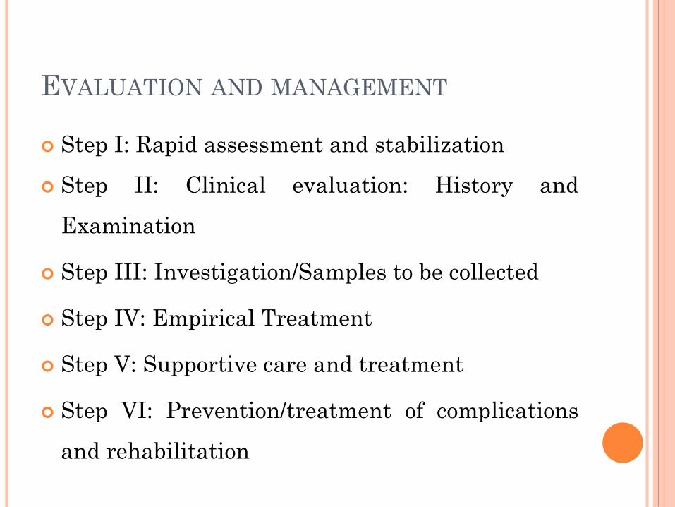
EVALUATION AND MANAGEMENT
Step I: Rapid assessment and stabilization
Step II: Clinical evaluation: History and
Examination
Step III: Investigation/Samples to be collected
Step IV: Empirical Treatment
Step V: Supportive care and treatment
Step VI: Prevention/treatment of complications
and rehabilitation
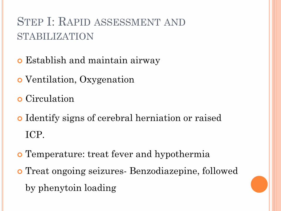
STEP I: RAPID ASSESSMENT AND
STABILIZATION
Establish and maintain airway
Ventilation, Oxygenation
Circulation
Identify signs of cerebral herniation or raised
ICP.
Temperature: treat fever and hypothermia
Treat ongoing seizures- Benzodiazepine, followed
by phenytoin loading
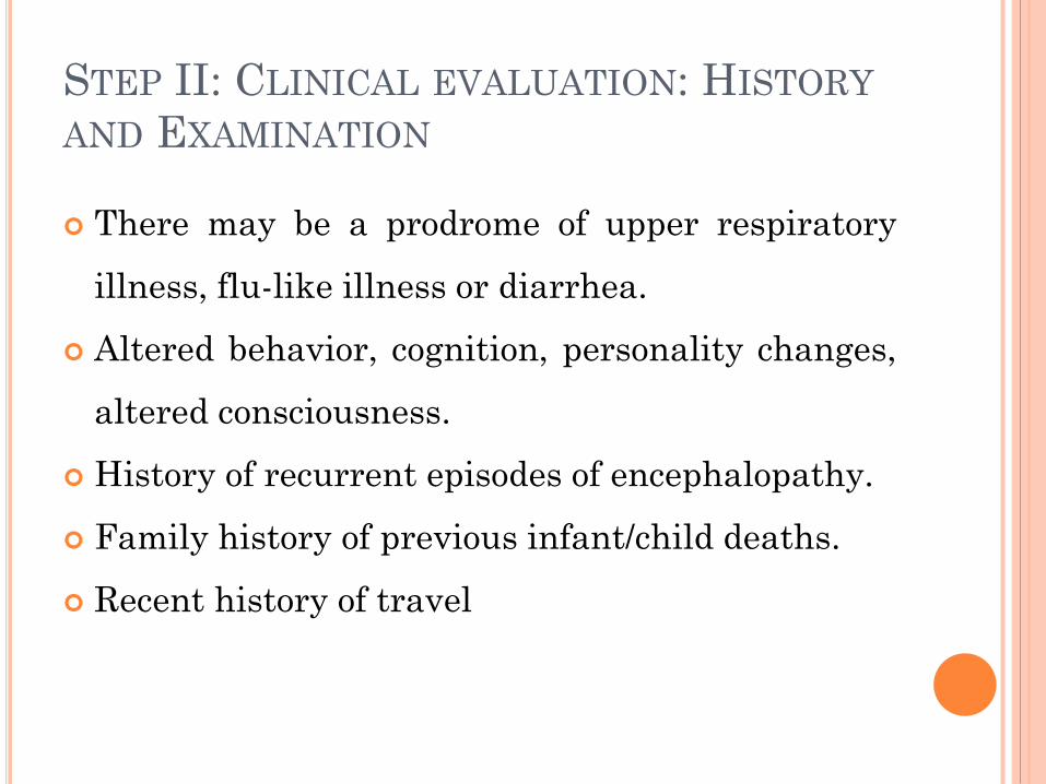
STEP II: CLINICAL EVALUATION: HISTORY
AND EXAMINATION
There may be a prodrome of upper respiratory
illness, flu-like illness or diarrhea.
Altered behavior, cognition, personality changes,
altered consciousness.
History of recurrent episodes of encephalopathy.
Family history of previous infant/child deaths.
Recent history of travel
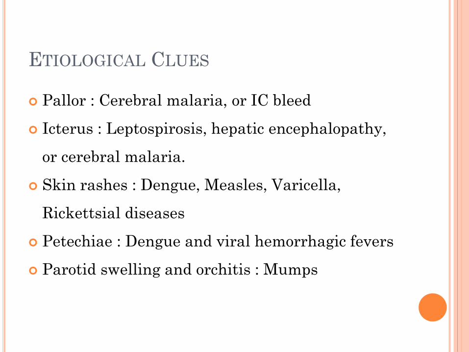
ETIOLOGICAL CLUES
Pallor : Cerebral malaria, or IC bleed
Icterus : Leptospirosis, hepatic encephalopathy,
or cerebral malaria.
Skin rashes : Dengue, Measles, Varicella,
Rickettsial diseases
Petechiae : Dengue and viral hemorrhagic fevers
Parotid swelling and orchitis : Mumps
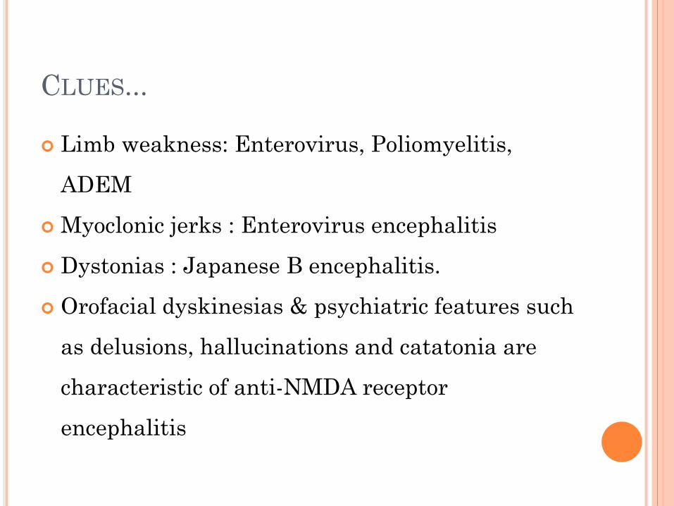
CLUES...
Limb weakness: Enterovirus, Poliomyelitis,
ADEM
Myoclonic jerks : Enterovirus encephalitis
Dystonias : Japanese B encephalitis.
Orofacial dyskinesias & psychiatric features such
as delusions, hallucinations and catatonia are
characteristic of anti-NMDA receptor
encephalitis
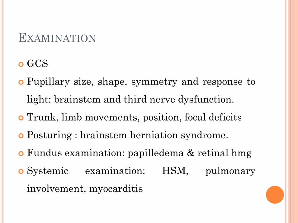
EXAMINATION
GCS
Pupillary size, shape, symmetry and response to
light: brainstem and third nerve dysfunction.
Trunk, limb movements, position, focal deficits
Posturing : brainstem herniation syndrome.
Fundus examination: papilledema & retinal hmg
Systemic examination: HSM, pulmonary
involvement, myocarditis
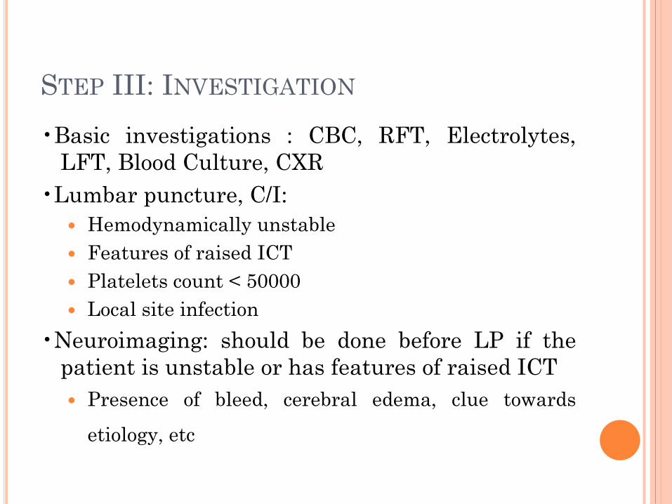
STEP III: INVESTIGATION
•Basic investigations : CBC, RFT, Electrolytes,
LFT, Blood Culture, CXR
•Lumbar puncture, C/I:
Hemodynamically unstable
Features of raised ICT
Platelets count < 50000
Local site infection
•Neuroimaging: should be done before LP if the
patient is unstable or has features of raised ICT
Presence of bleed, cerebral edema, clue towards
etiology, etc
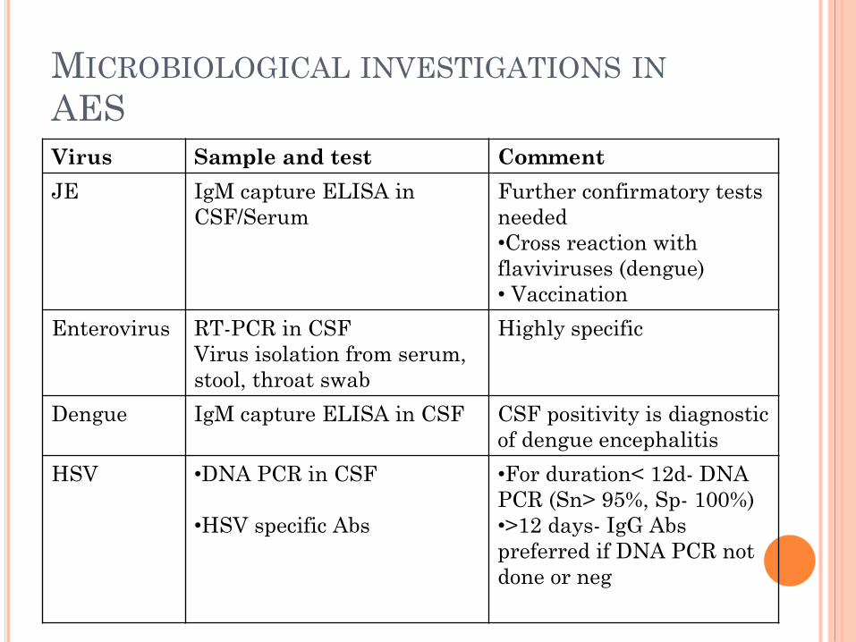
MICROBIOLOGICAL INVESTIGATIONS IN
AES
Virus Sample and test Comment
JE IgM capture ELISA in
CSF/Serum
Further confirmatory tests
needed
•Cross reaction with
flaviviruses (dengue)
• Vaccination
Enterovirus RT-PCR in CSF
Virus isolation from serum,
stool, throat swab
Highly specific
Dengue IgM capture ELISA in CSF CSF positivity is diagnostic
of dengue encephalitis
HSV •DNA PCR in CSF
•HSV specific Abs
•For duration< 12d- DNA
PCR (Sn> 95%, Sp- 100%)
•>12 days- IgG Abs
preferred if DNA PCR not
done or neg
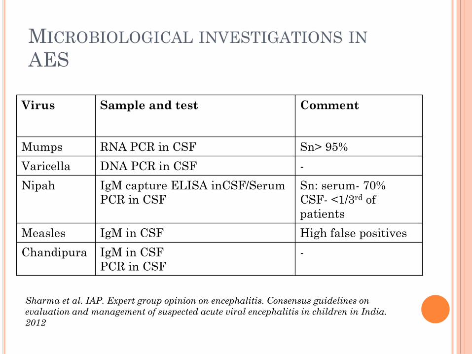
MICROBIOLOGICAL INVESTIGATIONS IN
AES
Virus Sample and test Comment
Mumps RNA PCR in CSF Sn> 95%
Varicella DNA PCR in CSF -
Nipah IgM capture ELISA inCSF/Serum
PCR in CSF
Sn: serum- 70%
CSF- <1/3rd of
patients
Measles IgM in CSF High false positives
Chandipura IgM in CSF
PCR in CSF
-
Sharma et al. IAP. Expert group opinion on encephalitis. Consensus guidelines on
evaluation and management of suspected acute viral encephalitis in children in India.
2012
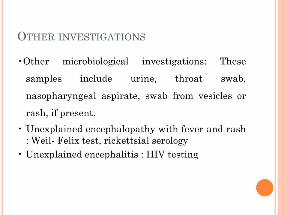
OTHER INVESTIGATIONS
•Other microbiological investigations: These
samples include urine, throat swab,
nasopharyngeal aspirate, swab from vesicles or
rash, if present.
• Unexplained encephalopathy with fever and rash
: Weil- Felix test, rickettsial serology
• Unexplained encephalitis : HIV testing
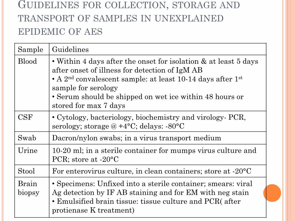
GUIDELINES FOR COLLECTION, STORAGE AND
TRANSPORT OF SAMPLES IN UNEXPLAINED
EPIDEMIC OF AES
Sample Guidelines
Blood • Within 4 days after the onset for isolation & at least 5 days
after onset of illness for detection of IgM AB
• A 2nd convalescent sample: at least 10-14 days after 1st
sample for serology
• Serum should be shipped on wet ice within 48 hours or
stored for max 7 days
CSF • Cytology, bacteriology, biochemistry and virology- PCR,
serology; storage @ +4°C; delays: -80°C
Swab Dacron/nylon swabs; in a virus transport medium
Urine 10-20 ml; in a sterile container for mumps virus culture and
PCR; store at -20°C
Stool For enterovirus culture, in clean containers; store at -20°C
Brain
biopsy
• Specimens: Unfixed into a sterile container; smears: viral
Ag detection by IF AB staining and for EM with neg stain
• Emulsified brain tissue: tissue culture and PCR( after
protienase K treatment)
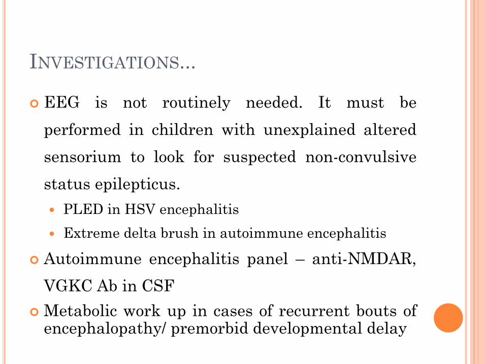
INVESTIGATIONS...
EEG is not routinely needed. It must be
performed in children with unexplained altered
sensorium to look for suspected non-convulsive
status epilepticus.
PLED in HSV encephalitis
Extreme delta brush in autoimmune encephalitis
Autoimmune encephalitis panel – anti-NMDAR,
VGKC Ab in CSF
Metabolic work up in cases of recurrent bouts ofencephalopathy/ premorbid developmental delay
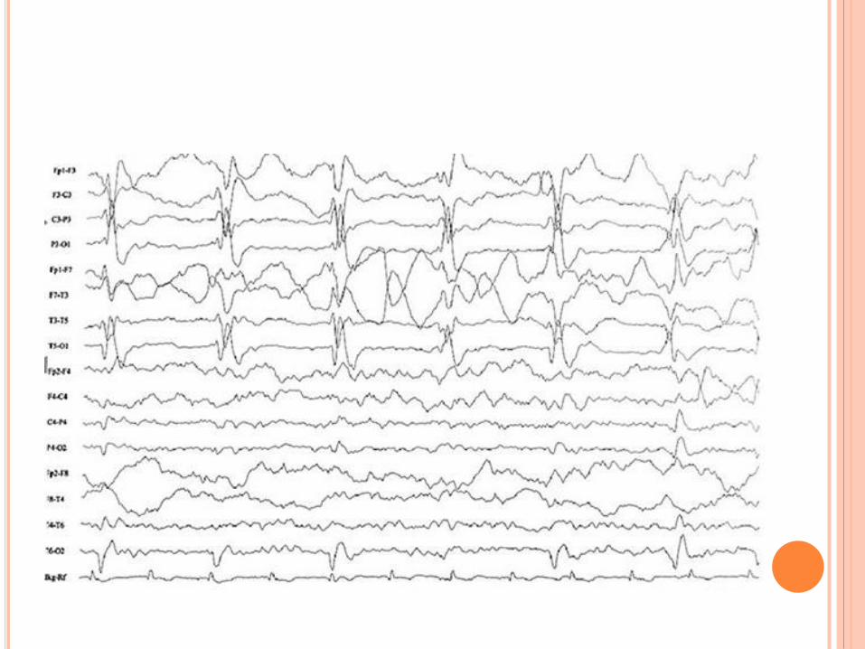
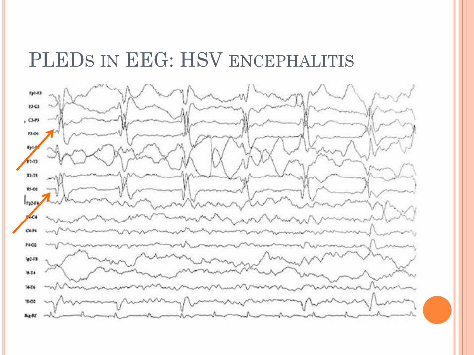
PLEDS IN EEG: HSV ENCEPHALITIS
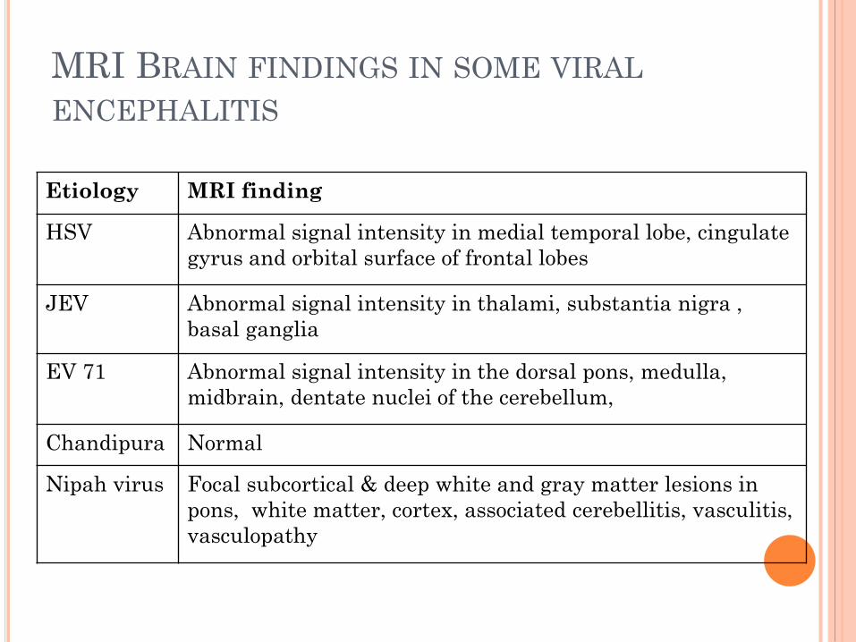
MRI BRAIN FINDINGS IN SOME VIRAL
ENCEPHALITIS
Etiology MRI finding
HSV Abnormal signal intensity in medial temporal lobe, cingulate
gyrus and orbital surface of frontal lobes
JEV Abnormal signal intensity in thalami, substantia nigra ,
basal ganglia
EV 71 Abnormal signal intensity in the dorsal pons, medulla,
midbrain, dentate nuclei of the cerebellum,
Chandipura Normal
Nipah virus Focal subcortical & deep white and gray matter lesions in
pons, white matter, cortex, associated cerebellitis, vasculitis,
vasculopathy
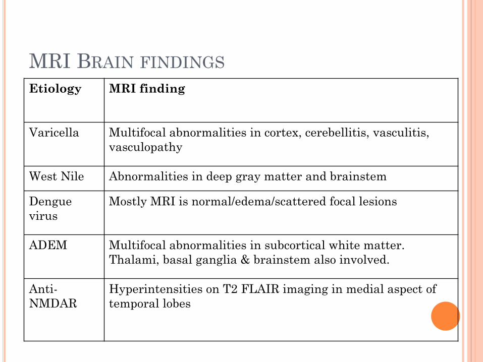
MRI BRAIN FINDINGS
Etiology MRI finding
Varicella Multifocal abnormalities in cortex, cerebellitis, vasculitis,
vasculopathy
West Nile Abnormalities in deep gray matter and brainstem
Dengue
virus
Mostly MRI is normal/edema/scattered focal lesions
ADEM Multifocal abnormalities in subcortical white matter.
Thalami, basal ganglia & brainstem also involved.
Anti-
NMDAR
Hyperintensities on T2 FLAIR imaging in medial aspect of
temporal lobes
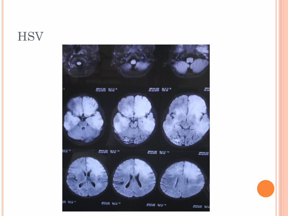
HSV
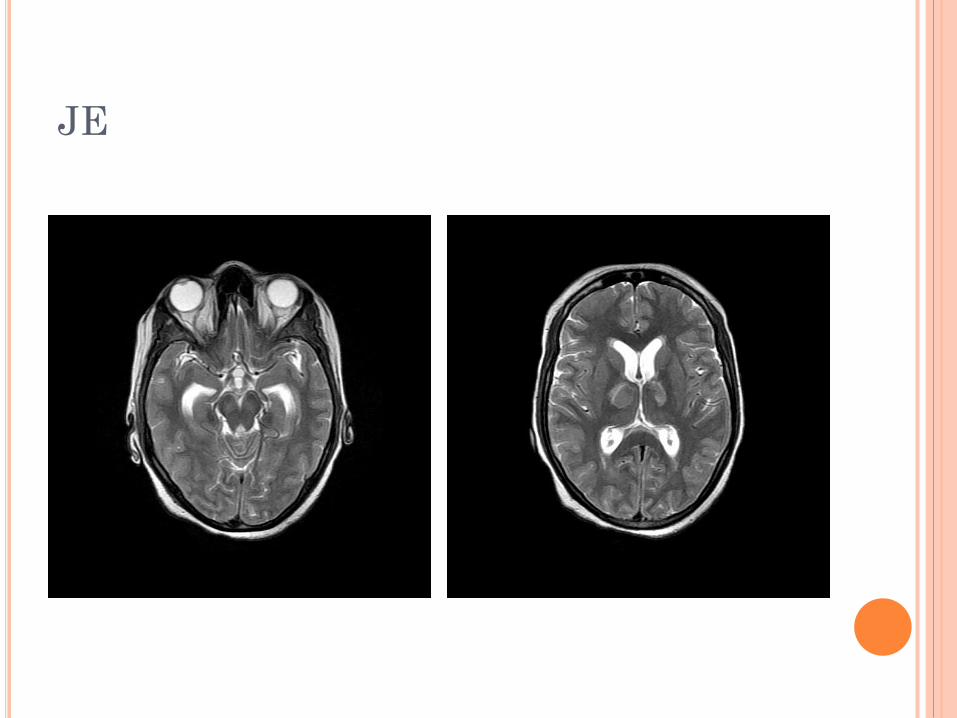
JE
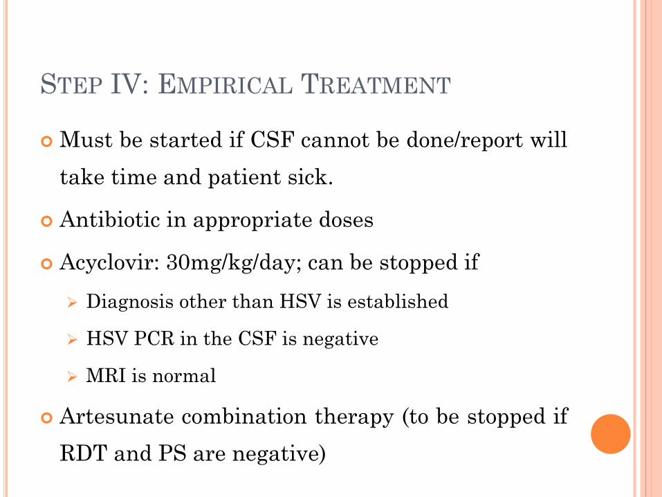
STEP IV: EMPIRICAL TREATMENT
Must be started if CSF cannot be done/report will
take time and patient sick.
Antibiotic in appropriate doses
Acyclovir: 30mg/kg/day; can be stopped if
➢ Diagnosis other than HSV is established
➢ HSV PCR in the CSF is negative
➢ MRI is normal
Artesunate combination therapy (to be stopped if
RDT and PS are negative)
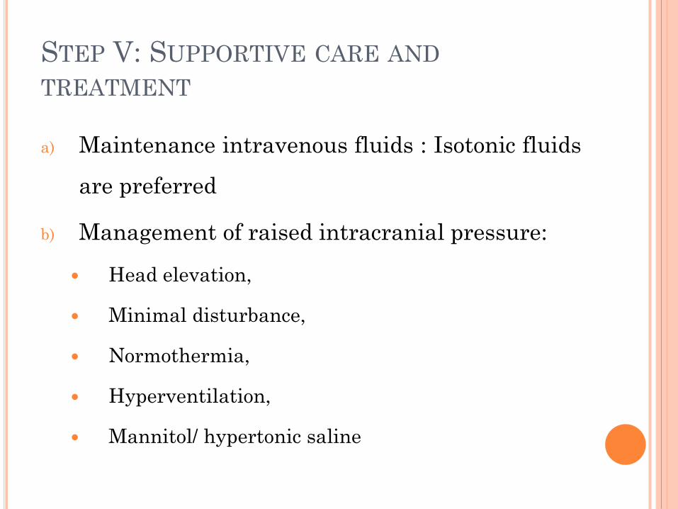
STEP V: SUPPORTIVE CARE AND
TREATMENT
a) Maintenance intravenous fluids : Isotonic fluids
are preferred
b) Management of raised intracranial pressure:
Head elevation,
Minimal disturbance,
Normothermia,
Hyperventilation,
Mannitol/ hypertonic saline
SUPPORTIVE CARE AND TREATMENT
(c) Maintain euglycemia
(d) Treatment and prevention of seizures:
If child is having seizures or has history of seizures
Even in absence of seizures, if GCS <8 and features
of raised ICT
(e) Other drugs : Corticosteroids, IVIG
(f) Other measures: Acid-base and electrolyte
abnormalities should be corrected.
NEW DRUGS
Pleconaril: enterovirus encephalitis and aseptic
meningitis
Minocycline : neuroprotective and antiviral
STEP VI: PREVENTION/TREATMENT OF
COMPLICATIONS AND REHABILITATION
• Physiotherapy, posture change, Prevent bed sores
and exposure keratitis.
• Complications: aspiration pneumonia, nosocomial
infections, coagulation disturbances
• Nutrition: early feeding
• Psychological support to patient and family
PREVENTIVE STRATEGIES
(i) Surveillance for cases of AES;
(ii) Vector control;
(iii) Reduction in man-vector contact;
(iv) Vaccination
CASE
14 yr girl with abnormal fluctuating behaviour &
focal seizures over 6 weeks, abnormal posturing of
right hand with involuntary perioral movements,
refractory status epilepticus, mutism for 4 weeks
No h/o fever, headache, myoclonic jerks, visual
complaints, weight loss or any systemic complaints.
At the time of admission, she was in encephalopathy
with status epilepticus, and right upper limb dystonic
posturing and perioral dyskinetic movements.
During hospital stay: autonomic disturbances,
respiratory distress
Routine investigations were within normal
limits. MRI brain and CSF studies were also
normal.
Her EEG showed generalised beta-delta range
slowing with “delta brush” and focal epileptiform
discharges as well.
CSF anti-NMDA receptor antibody was positive.
She received a course of intravenous
methylprednisolone f/b IVIg
She improved significantly. Presently she is on
low dosage of steroids
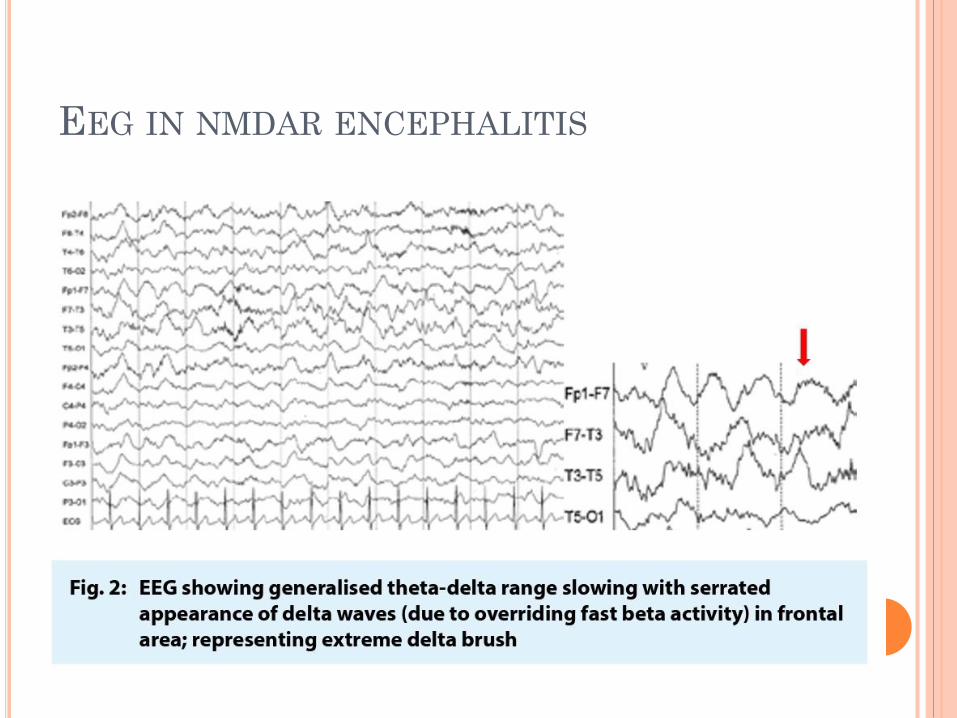
EEG IN NMDAR ENCEPHALITIS
AUTOIMMUNE ENCEPHALITIS
Group of neuropsychiatric disorders, presenting
acutely or subacutely with alteration of
consciousness, cognitive decline
Associated with antibodies against neuronal cell
surface proteins and synaptic receptors involved
in synaptic transmission, plasticity or neuronal
excitability
More common in younger adults and children
Severe and fatal but responds to immunotherapy
Often associated with an underlying tumor
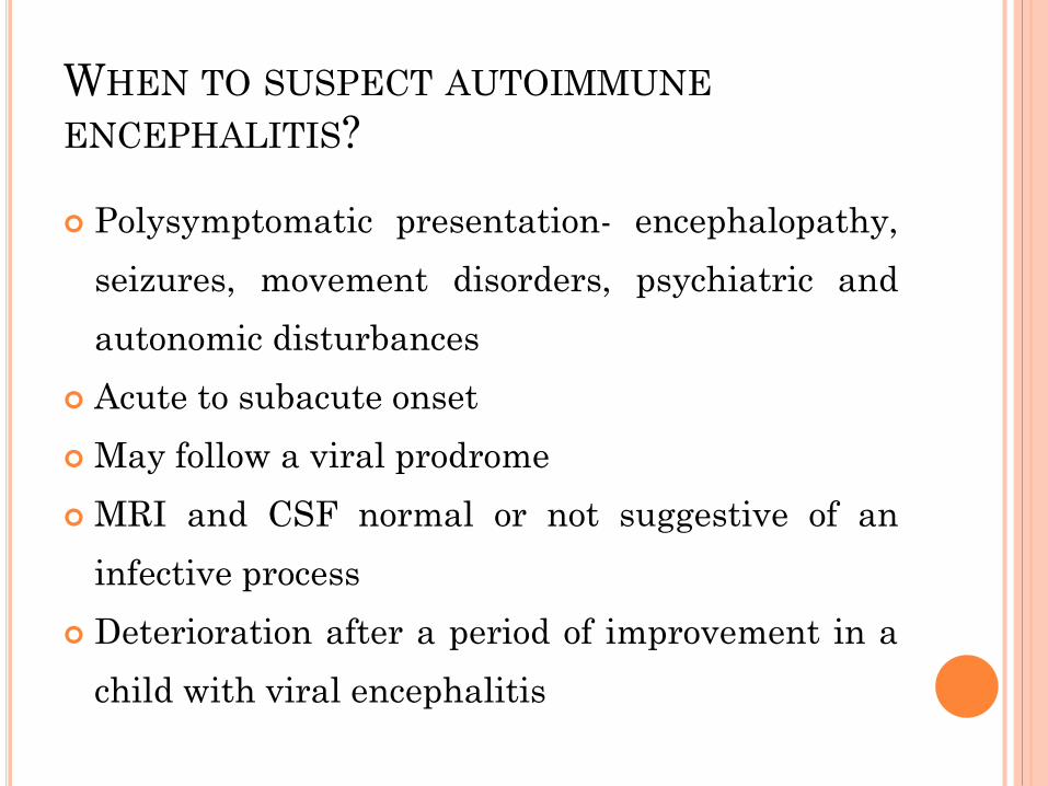
WHEN TO SUSPECT AUTOIMMUNE
ENCEPHALITIS?
Polysymptomatic presentation- encephalopathy,
seizures, movement disorders, psychiatric and
autonomic disturbances
Acute to subacute onset
May follow a viral prodrome
MRI and CSF normal or not suggestive of an
infective process
Deterioration after a period of improvement in a
child with viral encephalitis
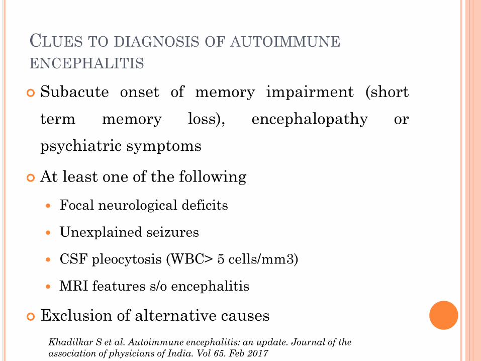
CLUES TO DIAGNOSIS OF AUTOIMMUNE
ENCEPHALITIS
Subacute onset of memory impairment (short
term memory loss), encephalopathy or
psychiatric symptoms
At least one of the following
Focal neurological deficits
Unexplained seizures
CSF pleocytosis (WBC> 5 cells/mm3)
MRI features s/o encephalitis
Exclusion of alternative causes
Khadilkar S et al. Autoimmune encephalitis: an update. Journal of the
association of physicians of India. Vol 65. Feb 2017
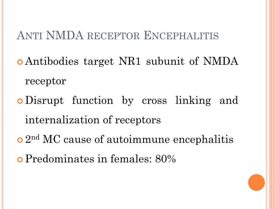
ANTI NMDA RECEPTOR ENCEPHALITIS
Antibodies target NR1 subunit of NMDA
receptor
Disrupt function by cross linking and
internalization of receptors
2nd MC cause of autoimmune encephalitis
Predominates in females: 80%
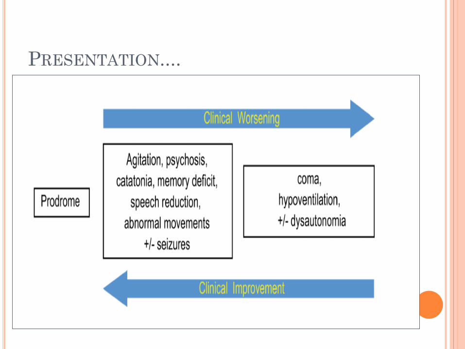
PRESENTATION....
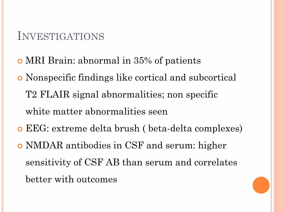
INVESTIGATIONS
MRI Brain: abnormal in 35% of patients
Nonspecific findings like cortical and subcortical
T2 FLAIR signal abnormalities; non specific
white matter abnormalities seen
EEG: extreme delta brush ( beta-delta complexes)
NMDAR antibodies in CSF and serum: higher
sensitivity of CSF AB than serum and correlates
better with outcomes
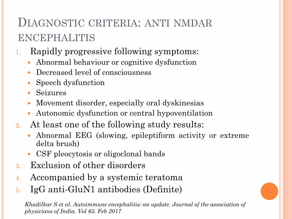
DIAGNOSTIC CRITERIA: ANTI NMDAR
ENCEPHALITIS
1. Rapidly progressive following symptoms:
Abnormal behaviour or cognitive dysfunction
Decreased level of consciousness
Speech dysfunction
Seizures
Movement disorder, especially oral dyskinesias
Autonomic dysfunction or central hypoventilation
2. At least one of the following study results:
Abnormal EEG (slowing, epileptiform activity or extremedelta brush)
CSF pleocytosis or oligoclonal bands
3. Exclusion of other disorders
4. Accompanied by a systemic teratoma
5. IgG anti-GluN1 antibodies (Definite)
Khadilkar S et al. Autoimmune encephalitis: an update. Journal of the association of
physicians of India. Vol 65. Feb 2017
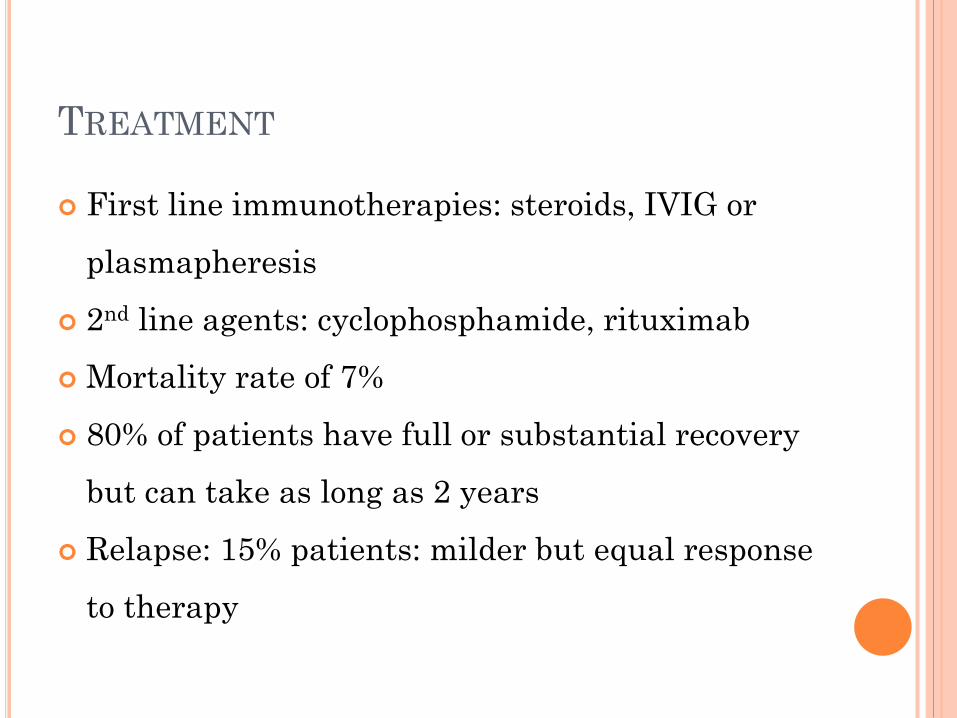
TREATMENT
First line immunotherapies: steroids, IVIG or
plasmapheresis
2nd line agents: cyclophosphamide, rituximab
Mortality rate of 7%
80% of patients have full or substantial recovery
but can take as long as 2 years
Relapse: 15% patients: milder but equal response
to therapy
CASE
A 4 year old girl came to OPD with C/O:
Sudden onset weakness of left side of body- 5 days
Deviation of left side of mouth
Irritability
No c/o fever, seizure, bladder/bowel incontinence, no deviation of eyes or change in voice or nasal regurtitation or blurred vision
No h/o recent vaccination, or any viral illness
Developmentally normal child premorbidly
On examination: child was irritable
Right upper motor neuron type facial nerve palsy present
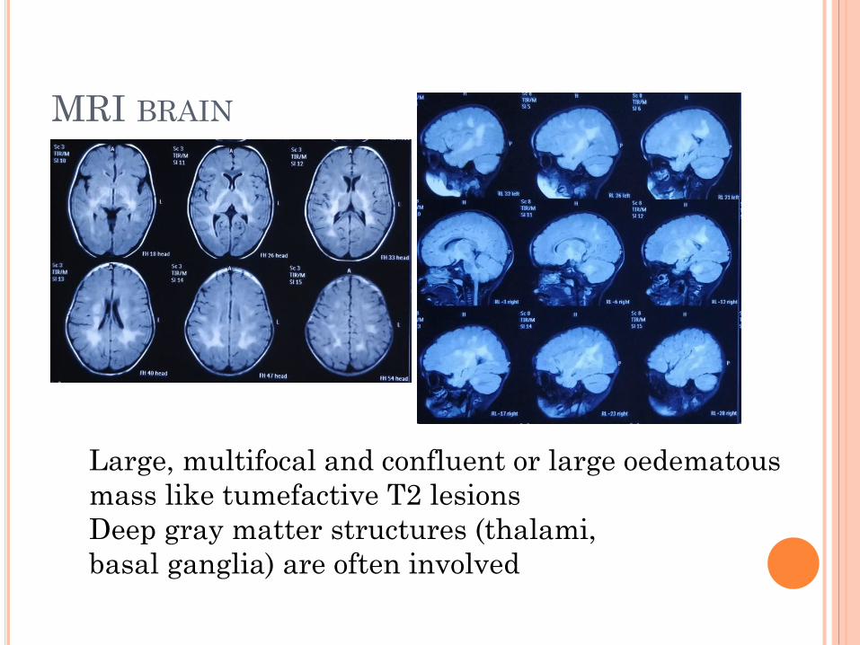
MRI BRAIN
Large, multifocal and confluent or large oedematous
mass like tumefactive T2 lesions
Deep gray matter structures (thalami,
basal ganglia) are often involved
ADEM
Inflammatory, demyelinating event with
multifocal neurological deficits
accompanied by encephalopathy
Pathogenesis: molecular mimicry
Influenza, EBV, CMV, varicella,
enterovirus, measles, mumps, rubella,
HSV and mycoplasma pneumoniae
Post vaccination: rabies, smallpox,
measles, mumps, rubella, JE B vaccine,
DPT
ADEM
Non specific neurological features
CSF: lymphocytic pleocytosis, protein-elevated
EEG: generalized slowing (polyregionaldemyelination- focal slowing orepileptoform discharges)
Treatment
High dose corticosteroids f/b oral steroids
IVIG/ plasmapheresis
Rituximab/ cyclophosphamide: severe cases
KEY MESSAGES
Most important part of managing a patient of
AES is to stabilize the patient
Managing the ongoing seizures and features of
raised ICT
Empirical treatment has to be started as soon as
possible without delay and after appropriate
investigations, antivirals/ antimalarials can be
stopped.
READING MATERAIL
GHAI’S TEXTBOOK OF PEDIATRICS
NELSON’S TEXTBOOK OF PEDIATRICS

1
JAPANESE B ENCEPHALITIS
2
HISTORY
• Epidemics of encephalitis ‐ Japan from the late 1800s.
• First isolated in Japan during an epidemic in 1935.
• In India first recognized in 1955 in Vellore.
• JE is a positive sense single stranded RNA virus.
• Family of Flaviviridae.

EPIDEMIOLOGY
• Annual incidence in endemic areas 1‐10/10,000 population.
• Children <15 yr of age are principally affected.
• Highly endemic areas ‐ A P, T N, Karnataka & UP.
• Peak starts after rains‐ July to December.
TRANSMISSION
4
• Transmitted as zoonotic cycle
– Mosquito Vertebrates like‐
– Culex tritaeniorhynchus Pigs & wading birds
– Culex vishnui Pigeon, Sparrow,
Duck, Horses, Swine,
Cattle & Buffalo
• Humans are dead end host.
• Pigs serve as amplifying host.
PATHOLOGY
5
• Areas of brain most commonly thalamus, substantia
nigra, anterior horns of spinal cord, Cerebral cortex and
cerebellum
• Other organs affected are: Lymph nodes, spleen,
myocardium, lungs and kidney
• After transmission virus multiplies locally and in
regional nodes‐ transient viremia‐ invasion of CNS‐ in
the neurons virus multiplies in the neuronal secretary
system
PATHOLOGY CONT.
6
• Affect endoplasmic reticulam & Golgi apparatus and
destroy them.
• After primary infection IgM response in serum and
CSF usually within 7 days.
• Immunization with inactivated JE vaccine induces T
cell activation in vivo.
CLINICAL FEATURES
7
• Incubation period 1 to 14 days.
• Onset is abrupt, acute, sub‐acute or gradual.
• Typically progress through 4 stages
Prodromal stage: 2 to 3 days
Acute stage (3‐4 days)
Sub acute stage (‐10 days)
Convalescence (4‐ 7 wk)
PRODROMAL STAGE
8
Abrupt onset of high grade fever
Head ache
Malaise
Abdominal pain
Nausea & vomiting
Sensory changes and psychotic episodes.
ACUTE STAGE
9
• Neurological symptoms 3 to 5 days
• Altered sensorium, Convulsions
• Neck stiffness, muscular rigidity
• Mask like facies, ICT
• Characteristic are rapidly changing central nervous
system signs .
• Gastric hemorrhage, pulmonary edema
CONVALESCENCE STAGE
10
• Stage of recovery
• Slowly regain neurological function over several
weeks
• Speech defects
• Paresis
• Intellectual deficit
DIAGNOSIS
11
• CSF: pleocytosis (100‐1000 leukocytes/mm3)
Increased protein
Normal glucose
• CT: involvement of thalamus, basal ganglia,
mid brain, pons & medulla
• EEG: diffuse theta & delta waves
ETIOLOGICAL DIAGNOSIS
12
• Four fold rise or greater in serum antibody.
• Isolation of virus/ demonstration of viral antigen or
genomic sequences .
• IgM capture Elisa
STANDARD CASE DEFINITION
13
• Suspect case of JE‐ clinical description
• Probable JE‐ presumptive lab results
• Confirmed JE‐ confirmatory lab results
• Antigen or genome in tissues or blood by immune
chemistry or immune fluorescence or by PCR
• JE virus specific IgM in CSF
• 4 fold or greater rise in JE virus specific antibody in
paired sera
PRESUMPTIVE LAB DIAGNOSIS
14
• Detection of acute phase antiviral antibody response by any one of the following
– Increased and stable serum antibody titres of JEV by ELISA.
– Hemagglutination or virus neutralization assay
IgM antibody to the virus in serum.
DIFFERENTIAL DIAGNOSIS
• West Nile virus
• Entero virus
• Herpes virus
• CNS tumors
• SLE
• Enteric encephalopathy
TREATMENT
16
• No specific treatment.
• Symptomatic & supportive aimed at prevention of
Pulmonary aspiration, hypoxia
Hypoglycemia, hyper pyrexia
Uncontrolled seizures, raised ICT
Pulmonary edema, SIADH
Secondary infection, brainstem involvement
TREATMENT CONT..
17
• Airway, breathing & circulation
• Seizures: Diazepam, Valproate
• Fluid, electrolyte & blood sugar maintained
• Raised ICT: Hyperosmolar therapy
• Coma ‐ prevent aspiration, bedsores, nosocomial
infection, malnutrition & contractures
• Extra pyramidal symptoms: Haloperidol, Diazepam,
chloral hydrate
SPECIFIC THERAPY
• Monoclonal antibodies
• Recombinant Interferon alpha
PROGNOSIS
19
• Patient fatality rates are 24‐42%.
• Frequency of sequelae is 5‐70% and is directly related
to the age of the patient and severity of the disease.
• Most common sequelae are mental retardation,
severe emotional instability, personality changes,
motor abnormalities and speech disturbances.
PROGNOSIS
20
• Poor prognostic signs are:
Younger age
Hyponatremia
Shock
Low GCS
Presence of immune complexes in CSF
Increased levels of tumor necrosis factor
• Good prognostic sign: high levels of neutralizing
antibodies ( IgG) in CSF
PREVENTION
21
• Control of mosquito: insecticide, fogging, larvicidal
measures & pyrethrum.
• Prevention of bites: Avoid out door activities, clothing,
mosquito repellants, bed nets or house screening.
• Control/ protection of reservoirs, piggeries,
vaccination in pigs‐ decrease viral amplification.
• Vaccination in high risk areas, susceptible population
VACCINATION
22
• Travelers to endemic countries who plan to be in rural
areas of the endemic region during the expected
period of seasonal transmission.
• Travelers in rural areas experiencing endemic
transmission should receive JE vaccine.
• In humans, prior dengue virus infection provides
partial protection from clinical JE.
VACCINATION
23
• Inactivated mouse brain vaccine‐ Nakayama strain‐
dose: 0.5‐ l ml SC – 1 to 3 years‐ 3 doses‐ 0‐7‐10 days,
booster every 3 years‐ till 10‐15 years
• Inactivated primary hamster kidney cells‐ China‐ SC
0.5ml‐ 1to2 years‐ booster 6 yrs ‐cheap
• Live attenuated primary hamster kidney cells‐ cheap‐
single dose‐ not approved by WHO
VACCINATION CONT.
24
• Vaccination of travelers‐ 0‐7‐30
• The final dose should be completed at least 1 wk prior
to the patient's expected arrival in a JE endemic area.
Newer vaccines:‐
• Recombinant JE vaccine
• DNA multivalent vaccine
• Chimeric vaccine
25
Q
READINGS : Nelson’s Textbook of Pediatrics 20th Edition
Ghai’s Textbook of Pediatrics
26
Q
QUESTIONS :
What is the vaccination schedule for Japanese encephalitis?
What are the MRI findings in Japanese encephalitis and HSV encephalitis?
Define Encephalitis and Encephalopathy?
Enumerate various causes of Acute Encephalopathy Syndrome(AES)?
How we should manage AES?

27
Q


![The Pediatric Altered Brain · appropriate code from subcategory P91.6, Hypoxic ischemic encephalopathy [HIE]. Current science has demonstrated that a newborn may meet the diagnostic](https://img.pdfslide.us/doc/110x75/600040f8c65a6e2dbc213c45/the-pediatric-altered-brain-appropriate-code-from-subcategory-p916-hypoxic-ischemic.jpg)