Embed Size (px)
Citation preview
Page 1/21
Effect of Carvacrol on In�ammatory Factors andMyelin Repair in Experimental AutoimmuneEncephalomyelitis on Female Lewis RatsMahdie Ahmadi
Islamic Azad UniversityAkram Eidi
Islamic Azad UniversityMojtaba Khaksarian ( [email protected] )
Lorestan University of Medical SciencesHassan Ahmadvand
Lorestan University of Medical SciencesFattah Sotoodehnejadnematalahi
Islamic Azad University
Research Article
Keywords: Remyelination, EAE, Multiple Scelrosis, Carvacrol, In�ammation
Posted Date: April 27th, 2021
DOI: https://doi.org/10.21203/rs.3.rs-430600/v1
License: This work is licensed under a Creative Commons Attribution 4.0 International License. Read Full License
Page 2/21
AbstractCarvacrol is a phenolic monoterpene constituent of essential oils produced by plants such as Nigellasativa. This study examined the therapeutic effects of carvacrol in a model of experimental autoimmuneencephalomyelitis (EAE). EAE induction was performed on female Lewis rats and, after appearance of the�rst clinical signs, a second EAE group received carvacrol intraperitoneally each day for 17 days. Clinicalsymptoms and body weight were assessed daily. All animals sacri�ced and histological and geneexpression analysis of the spinal cord were performed. The carvacrol treated group scored lower fordisease after therapeutic administration than the control group. Gene expression analysis on thecarvacrol treated group showed a decrease in TNF-α, NF-ҚB and IL-17 (pro-in�ammatory) geneexpression and an increase in MBP and OLIG2 remyelination markers. In the carvacrol treated group, H&Estain of spinal cord sections revealed a remarkable decrease in the amount of in�ammatory cellin�ltration. Immunostaining of lumbar spinal cord sections demonstrated a remarkable increase in MBPand a decrease in IL-17 secretion in the carvacrol treated group. These results demonstrate that carvacrolhad a bene�cial therapeutic effect and anti-in�ammatory properties that signi�cantly reduced the clinicalscores and promoted myelin repair.
IntroductionCarvacrol is a monoterpenoid phenol that has special antioxidant effects against free radicals. Carvacrolhas antioxidant, anti-in�ammatory and antimicrobial properties. It has been shown to reduce theexpression of EGF, TNF-α and MCP-1 in patients exposed to mustard gas(Aristatile, Al-Assaf et al. 2013)In hepatotoxicity induced by D-galactosamine, carvacrol was found to reduce mRNA expression of IL-6,inducible nitric oxide synthase (iNOS), TNF-αand NF-ҚB(Aristatile, Al-Assaf et al. 2013). In IL-1β-in�amedchondrocytes, carvacrol has been shown to inhibit the production of nitric oxide and prostaglandin E2(PGE2) and reduce the iNOS and COX-2 expression. Inhibition of matrix metalloproteinase-13 (MMP-3)and MMP-3 genes and inhibition of NF-ҚB signaling pathway activation are other effects of carvacroltreatment(Yu, Zhang et al. 2012).
The neuroprotective effects of carvacrol in neurological disorders such as ischemia,epilepsy(Baluchnejadmojarad and Roghani 2016) and Parkinson's disease(Baluchnejadmojarad,Hassanshahi et al. 2014; Haddadi, Rajaei et al. 2018) have been demonstrated. In a Parkinson's model,carvacrol-treated mice showed improved memory de�cits and decreased caspase(Dati, Ulrich et al. 2017)levels(Baluchnejadmojarad, Hassanshahi et al. 2014; Haddadi, Rajaei et al. 2018). In a cerebral ischemicmodel, carvacrol improved learning ability and memory in mice and decreased lipid peroxide and celldeath biomarkers in the ischemic brain tissue. It also inhibited ferroptosis by increasing the expression ofglutathione peroxidase 4 (GFX 4)(Guan, Li et al. 2019).
Multiple sclerosis (MS) is a neurodegenerative disease of the central nervous system (CNS), Which isknown for features such as in�ammation, altered axonal function and demyelination (Yoshida, Kimura etal. 2012). Experimental Autoimmune Encephalomyelitis (EAE) is a common model used to study MS. EAE
Page 3/21
is a complex condition in which the interaction of a variety of neuronal-damaging mechanisms and theimmune system shows striking resemblance to key pathogenic features of MS. These features includein�ammation, demyelination, axonal removal and gliosis. Regulatory mechanisms of in�ammation andremyelination also occur in EAE, so they act as a model for these processes.
EAE has a complex pharmacology and many drugs that are commonly used or are in development forMS have been tested and approved using an EAE model. However, a limited number of studies haveevaluated the effect of carvacrol on MS patients or using the EAE model. An examination of the effect ofcarvacrol in an EAE model and by culturing spleen cells found that it can decrease clinical scores and IL-6, IFN-γ and IL-17secretion and increase IL-10 and TGF-β. The carvacrol-treated mice also experiencedless weight loss(Mahmoodi, Amiri et al. 2019).
This current study evaluated the in�ammatory factors of IL-17, MCP, TNF-α, NF-ҚB and the genesinvolved in myelin repair (MBP, PDGFR and OLIG2) to determine the effects of carvacrol on the process ofmyelination and in reducing demyelination and in�ammation by means of molecular and histologicalexaminations of the spinal cords of Lewis rats.
Materials And Methods
Induction, treatment of EAE and Clinical scoreAdult female Lewis rats weighting 180 to 210 g were used in this experiment. The animals were kept inclean conditions under a 12:12 h dark/light cycle and the room temperature was controled. Theimmunization protocol from Beeton et al(Beeton, Garcia et al. 2007) was used to prepare guinea pigspinal cord homogenate in distilled water, which then was mixed with complete Freund’s adjuvant (CFA;Sigma-Aldrich; USA) containing Mycobacterium lyophilized cell powder.
At �rst, the rats were injected subcutaneously with 0.5 ml of the mentioned emulsion at the tail. After 2 hand 48 h, each rat received an intraperitoneal injection of pertusis toxin (Sigma Aldrich; USA) in distilledwater.
After immunization, the rats were weighed daily and the clinical signs of EAE were monitored daily. Thescores were as follows: 0 = without clinical signs; 1 = full tail paralysis; 2 = moderate paralysis of hindlimbs; 3 = hind limb paralysis; 4 = bilateral hind limb paralysis; 5 = complete palsy, dying or death. Aftermonitoring for signs of disease on day 10 of post-immunization, the animals in the carvacrol treatedgroup received 20 mg/kg carvacrol once daily for 17 days, from day 12 to day 28 post-immunization. Thehealthy control group received phosphate buffered saline (PBS). The EAE group of immunized ratsreceived no treatment.
Histological assessmentAfter anesthetization, the rats were perfused intracardially with PBS and paraformaldehyde. Their lumbarspinal cords were removed and �xed overnight at 4°C in 4% paraformaldehyde. For in�ammatory cell
Page 4/21
in�ltration evaluation with H&E stain, dehydration of �xed tissues was performed in an alcohol series,incubated in xylene until clear, embedded in para�n and sectioned in 5-µm thicknesses using amicrotome. The para�n-embedded sections were rehydrated in an alcohol series, stained withhematoxylin and then cleared in two baths of xylene. Then, the slides were washed and stained witheosin. These slides then were cover-slipped and photographed through a light microscope.
Immunohisto�uorescenceAs in previous histological studies, the lumbar spinal cords were isolated, �xed in paraformaldehyde,dehydrated in alcohol, incubated in xylene and embedded in para�n. Sections of 6.8 µm in size wereprepared using a microtome. These were blocked with normal serum and stained with primary antibodiesagainst myelin basic protein (MBP) and IL-17 (Abcam; UK; dilution 1:50). The slides were washed withPBS and incubated with goat anti-mouse rhodamine conjugate (Invitrogen; 1:100) and goat anti-rabbitFITC (1:100). The nuclei were stained with DAPI and images were obtained using a BX-51 �uorescentmicroscope (Olympus DP72).
RNA extraction and quantitative RT- PCRThe total RNA was isolated from the lumbar spinal cord with Trizol reagent (Parstous Company, Iran).The integrity of the isolated RNA was con�rmed by agarose gel electrophoresis. To determine the purityand concentration of the RNA, a spectrophotometer (Pharmacia Biotech, Cambridge, United Kingdom)was used. The cDNA was synthesized using a cDNA synthesis kit (Ana Cell; Iran). RT- PCR was carried outusing RT-PCR master mix (Amplicon). The thermal conditions were as follows: preheating for 3 min at95°C; denaturation for 30 s at 95°C; annealing for 30 s at 60°C; extension for 20 s at 72°C.
The primers were designed and synthesized (Bioneer; Korea) to assessthe expression of GAPDH(housekeeping gene), Interleukin-10 (IL-10), IL-4, IL-1, IL-17, tumor necrosis factor α (TNFα), forkhead boxp3 (FoxP3), tumor growth factor β (TGFβ), iNOS, nuclear factor-kappa B (NF-ĸB), platelet-derived growthfactor receptor α (PDGFRα), Nrf2, MCP-1, MBP, nerve growth factor (NGF), brain-derived neurotropic factor(BDNF), OLIG2 and heme oxygenase 1( HO 1). The primer pairs used in this study are shown in table 1.For analysis of the relative gene expression level, 2−ΔΔCt formula was used.
Statistical analysisThe gene expression and histological data were analyzed by one-way ANOVA or t-student testing andcompared using the Tukey post-test at p < 0.05 as the signi�cant difference.
Results
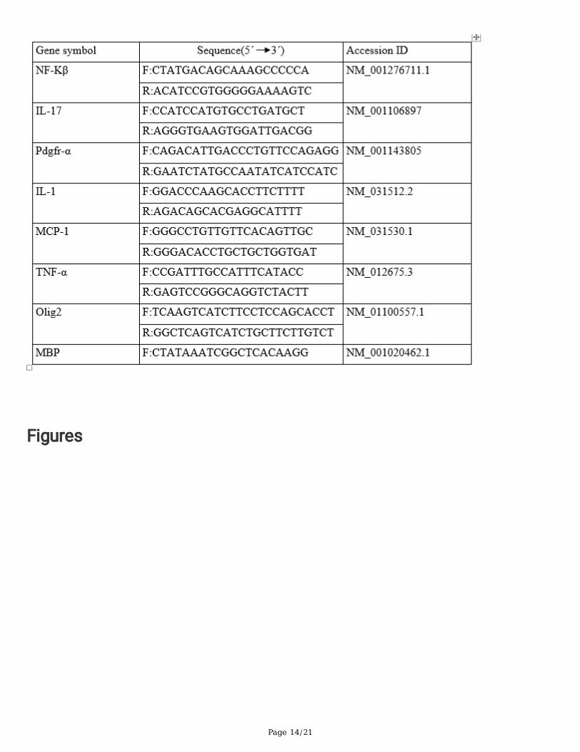
Histopathological �ndingsThe carvacrol treated group received carvacrol from day 12 (peak disease score) to day28. The resultsindicate that daily administration of carvacrol during this period led to signi�cant recovery (Fig. 1a) and
Page 5/21
signi�cant recovery of all rats at day 24. After treatment with carvacrol, relapse of EAE severity wasdiminished, compared to EAE group.
Up to day 9, no signi�cant differences were observed between weight of groups. After day 9, signi�cantweight loss was observed in both the EAE and carvacrol treated groups. Weight loss continued at aslower rate in the EAE group until day 28. In the carvacrol treated group, on days 21 to 23, the weightbegan showing an upward trend.
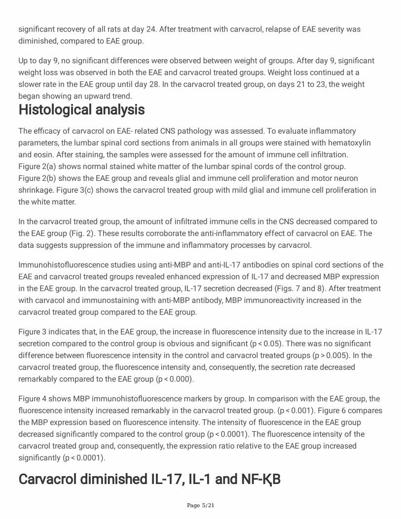
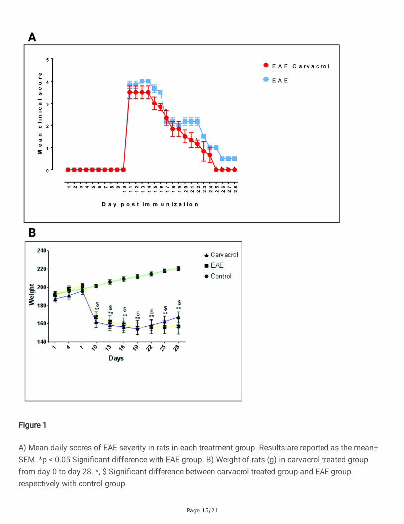
Histological analysisThe e�cacy of carvacrol on EAE- related CNS pathology was assessed. To evaluate in�ammatoryparameters, the lumbar spinal cord sections from animals in all groups were stained with hematoxylinand eosin. After staining, the samples were assessed for the amount of immune cell in�ltration.Figure 2(a) shows normal stained white matter of the lumbar spinal cords of the control group.Figure 2(b) shows the EAE group and reveals glial and immune cell proliferation and motor neuronshrinkage. Figure 3(c) shows the carvacrol treated group with mild glial and immune cell proliferation inthe white matter.
In the carvacrol treated group, the amount of in�ltrated immune cells in the CNS decreased compared tothe EAE group (Fig. 2). These results corroborate the anti-in�ammatory effect of carvacrol on EAE. Thedata suggests suppression of the immune and in�ammatory processes by carvacrol.
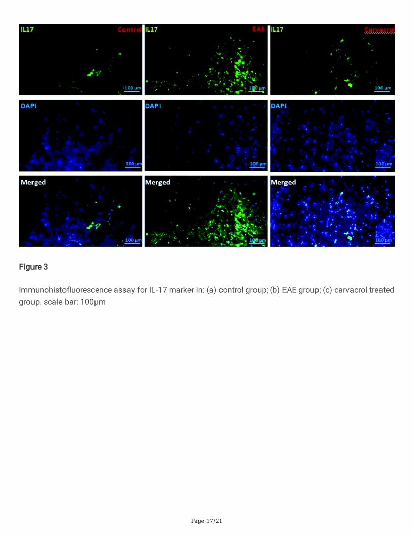
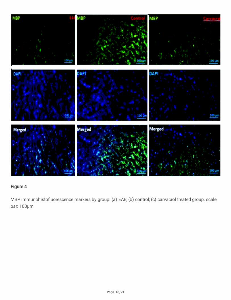
Immunohisto�uorescence studies using anti-MBP and anti-IL-17 antibodies on spinal cord sections of theEAE and carvacrol treated groups revealed enhanced expression of IL-17 and decreased MBP expressionin the EAE group. In the carvacrol treated group, IL-17 secretion decreased (Figs. 7 and 8). After treatmentwith carvacol and immunostaining with anti-MBP antibody, MBP immunoreactivity increased in thecarvacrol treated group compared to the EAE group.
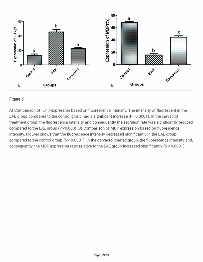
Figure 3 indicates that, in the EAE group, the increase in �uorescence intensity due to the increase in IL-17secretion compared to the control group is obvious and signi�cant (p < 0.05). There was no signi�cantdifference between �uorescence intensity in the control and carvacrol treated groups (p > 0.005). In thecarvacrol treated group, the �uorescence intensity and, consequently, the secretion rate decreasedremarkably compared to the EAE group (p < 0.000).
Figure 4 shows MBP immunohisto�uorescence markers by group. In comparison with the EAE group, the�uorescence intensity increased remarkably in the carvacrol treated group. (p < 0.001). Figure 6 comparesthe MBP expression based on �uorescence intensity. The intensity of �uorescence in the EAE groupdecreased signi�cantly compared to the control group (p < 0.0001). The �uorescence intensity of thecarvacrol treated group and, consequently, the expression ratio relative to the EAE group increasedsigni�cantly (p < 0.0001).
Carvacrol diminished IL-17, IL-1 and NF-ҚB
Page 6/21
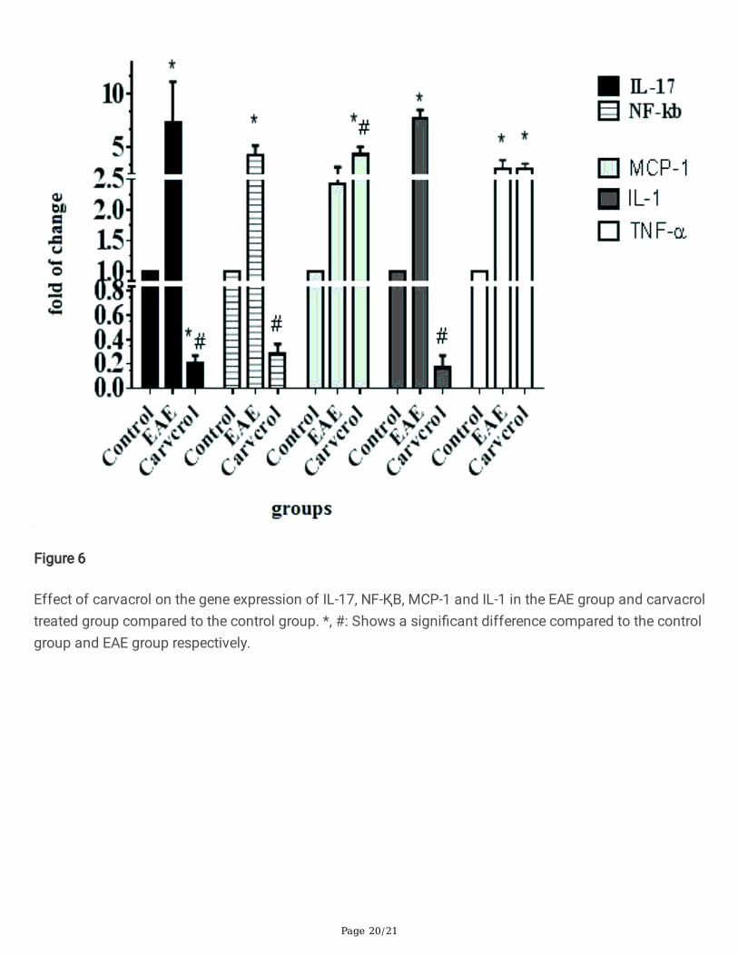
The carvacrol effect on expression of the in�ammatory gene was evaluated using RT- PCR and the resultsare shown in Fig. 6. EAE induction caused a signi�cant increase in IL-17 compared to the control group (p < 0.0001), but this decreased in the carvacrol treated group to even below that of the control group (p < 0.0001). The same pattern of change was observed for IL-1 and NF-ҚB (p < 0.0001). Carvacroladministration reversed the mRNA level of IL-1 and NF-ҚB to below that of the control group.
EAE increased the expression of MCP at the mRNA level compared to the control group. The mRNA levelof MCP in the carvacrol treated group was even higher than for the EAE group (p < 0.0001). Theexpression of TNF-α in the EAE group was higher than in the control group (p < 0.05), but did not changein the carvacrol treated group. The expression of TNF in the EAE and carvacrol treated group was similar.
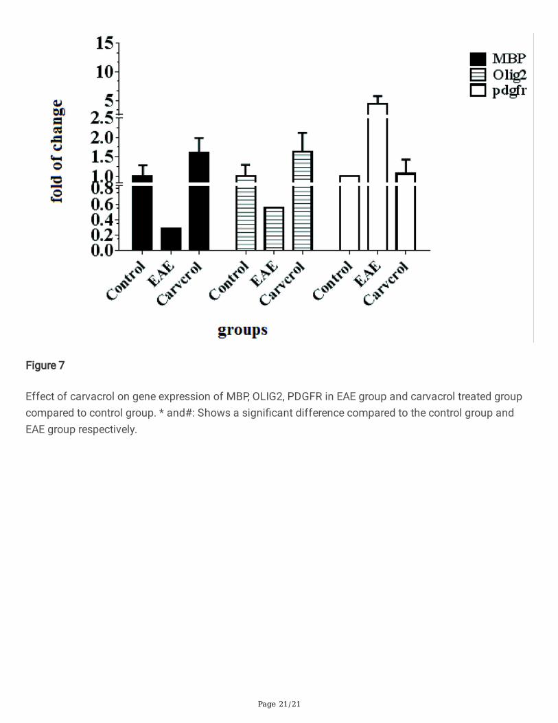
Effect of carvacrol on MBP, PDGFR and OLIG2The expression pattern of MBP, OLIG2 and PDGFR were assayed following EAE induction and in thecarvacrol treated group. A decrease in MBP gene expression was observed in the EAE group (p < 0.05), butcarvacrol signi�cantly increased this level in the carvacrol treated group compared to the EAE and controlgroups (p < 0.0001).
The tissues were evaluated for expression of OLIG2 and it was found that the level of OLIG2 mRNAdecreased in the EAE group and signi�cantly increased in the carvacrol treated group compared to theEAE group. This level was even higher than for the control group.
Compared with the control group, PDGFR gene expression increased in the EAE group (p < 0.0001) andthe PDGFR mRNA level decreased in the carvacrol treated group compared to the EAE group (p < 0.0001).No signi�cant difference was observed between the carvacrol treated group and the control group (p = 0.652; Fig. 7).
DiscussionCarvacrol has been shown to be effective on some in�ammatory and neurodegenerative diseases of thebrain, which makes it a good option for the treatment of MS. The characteristics of the EAE model werecon�rmed by the observed increase in neurological defects and clinical scoring, the presence of CNS-in�ltrated immune cells and axonal degradation, which are consistent with results of previousstudies(Mohajeri, Sadeghizadeh et al. 2015; Yang, Zheng et al. 2017).
Carvacrol has neuroprotective effects that are demonstrated in neurological models such as ischemiaand Parkinson's disease(Baluchnejadmojarad, Hassanshahi et al. 2014; Dati, Ulrich et al. 2017). Thecarvacrol treatment results in the present study showed a decrease in clinical scores and histologicalchanges. It also resulted in decreased expression of some proin�ammatory genes and increasedexpression of genes involved in myelination.
CNS repair is the goal of treatment for CNS and autoimmune disorders. Different neuroprotectivetherapeutic strategies have been used in clinical trials but, as yet, no comprehensive and successful
Page 7/21
examination of remyelination has been proposed. In the case of MS, remyelination not only betterprotects from axonal damage, but also can halt progression of the disease.
Oligodendrocytes have been a focus of attention for the development of therapeutic approaches andtreatments for neurodegenerative disease because of their myelination potential(Kotter, Stadelmann et al.2011). One of the most important features of MS is demyelination, in which the axon-oligodendrocyterelationship in the CNS is disrupted(Shakhbazau, Schenk et al. 2016). Evaluation of the therapeuticeffects of such research methods requires the identi�cation of oligodendrocytes. For this purpose, thepresent study used MBP, PDGFR and OLIG2 markers.
The results of molecular analysis for the carvacrol treated group showed that the remyelination rateincreased and demonstrated that carvacrol increased OLIG2 mRNA expression. It was found that clinicalscoring in the carvacrol treated group was lower than in the EAE group, indicating that the clinicaldamage improved with carvacrol treatment. OLIG2 expression increased in the carvacrol treated group asthe clinical scores decreased. OLIG2 encodes the transcription factor that is necessary for remyelinationand is obtained through proliferation and differentiation of neuronal progenitors and oligodendrogenesisprogenitor cells (OPCs). This factor is necessary for oligodendrogenesis in the spinal cord. OLIG2-expressing progenitor cells differentiate into oligodendrocytes in damaged lesions, leading to prematuremyelination of the CNS(Wegener, Deboux et al. 2015) .
In addition to OLIG2, PDGF is also involved in activating the oligodendrogenesis pathway and is involvedin their proliferation. An increase in PDGF has been shown to increase the number of OPCs indemyelinated lesions. However, although PDGF infusion induces SVZ type B cell proliferation which iseffective for remyelination, it can have side effects such as tumor formation(Rivera and Aigner 2012).
OPCs express various markers, the most important of which are NG2 and PDGFRα. Because NG2 can bepresent in other glia precursor cells, the PDGFRα marker is more speci�c for immature oligodendrocytes.
In this study PDGFRα was used to assess the amount of immature oligodendrocyte formation. Precursoroligodendrocytes were observed to attach to neurons and, after two to three days, myelinatedthem(Blakemore and Irvine 2008). However, contrary to the results of previous studies, PDGFR showedincreased expression in the EAE group and decreased expression in the carvacrol treated group.
In agreement with the results of the present study, Qiang Zho et al. reported that transgenic mice showedthat deletion of PDGFR caused precocious differentiation of OPCs. Their �ndings demonstrate thatPDGFR is an important negative regulator of OPC maturation in the developing CNS. During OPCdifferentiation, PDGFR expression gradually decreases and is completely silent in adult oligodendrocytes.It is not clear how cessation of PDGFR expression or OPC signaling triggers oligodendrocytedifferentiation. One possibility is that PDGFR signaling may inhibit the expression or function ofoligodendrocyte maturation activators. These activators include myelin regulatory factor (MRF), sip1 andSOX10, each of which is su�cient to differentiate oligodendrocytes. If this hypothesis is correct, Nkx2.2transcriptional inhibitor initiates internal differentiation by removing the inhibition of these regulatory
Page 8/21
factors and responding to axon signals or other environmental messages. It may initiate internaldifferentiation by removing the inhibition of these regulatory factors in response to axon signals.
Nkx2.2 is a transcriptional inhibitor in cell-fate control during CNS development and determines the timeof OPC differentiation. Overexpression of Nkx2.2 in transgenics will extinguish PDGFR expression. It ispossible that, when Nkx2.2 binds to the binding site on the PDGFR promoter, it inhibits PDGFRtranscription. By repressing the expression of the membrane receptor PDGFR, the OPCs will no longerrespond to mitogen PDGFR, which will cause the proliferation and activation of cell differentiation tostop.
On the other hand, an increase in Nkx2.2 expression and inhibition of PDGFR expression has beenobserved to increase MBP expression in primary OPCs. Therefore, the possibility that Nkx2.2 mayin�uence other factors simultaneously to affect differentiation and maturation of OPCs cannot beignored (Zhu, Zhao et al. 2014). After demyelination, OPCs switch from neutral to active and mitotic cellsby upregulating OLIG2 and Nkx2.2 (Göttle and Küry 2015). In general, the reduction in PDGFR expressionby carvacrol in this study could provide a pathway for the differentiation of OPCs and help to myelinateand repair damage caused by the model induction.
MBP plays the most important role in the formation of the three-dimensional (3D) structure of the axialmembrane of myelin. This protein binds to membrane lipids as it enters the oligodendrocyte membraneand strengthens the 3D structure of the myelin membrane(Czepiel, Boddeke et al. 2015). MBP was usedin the present study as an adult oligodendrocyte marker to evaluate the �nal remyelination rate. The goalwas to determine which changes occur during the process of remyelination from immatureoligodendrocytes to adults. MBP mRNAs are located at one point in the cytoplasm of oligodendrocytesbefore myelination and then disperse into the cytoplasm at the beginning of myelination. This spatialseparation differentiates oligodendrocytes into complete myelinating cells (Musse and Harauz 2007) .
It has been shown in MS patients and animal models that MBP decreased about three-fold compared tonormal levels. In glial cells and macrophages, the group of enzymes called protein arginine deiminase(PAD) becomes uncontrolled for unknown reasons in in�ammatory diseases such as MS and producesimmunogenic citrullinated epitopes. PAD2 and PAD4 levels increase in MS. It is possible that these twoenzymes alter the structure of the third MBP, and predispose them to proteolysis so that MBP cannotproperly form the multilayer structure of a myelin sheath. Eventually, the accumulation of nerve cells willdecrease(Jones, Causey et al. 2009).
In our study induction of the EAE model reduced MBP expression in the EAE group and further studyshowed that carvacrol administration increased MBP expression in the carvacrol treated group. Theseresults were con�rmed by both molecular methods and immunohisto�uorescence analysis. The increasein OLIG2 expression increased the production of differentiated oligodendrocytes as well as MBPexpression, which indicates the activity of oligodendrocytes in the myelination pathway. The increase ofthese two factors demonstrates the process of myelination. However, molecular processes are so
Page 9/21
complex that these observations cannot easily be attributed to an increase or decrease in the expressionof a gene. In such cases, many factors must be considered.
Studies have shown that carvacrol has an effect on in�ammatory factors and oxidative stress. Gholijaniet al. (2016) caused in�ammation in mice and cultured their spleen cells to show that carvacrol reducedthe formation of IL-17 as well as the expression of T-bet, GATA-3 and ROR c transcription factors thatnecessary for the maturation and function of Th1, Th2 and Th17 cells, respectively)(Gholijani andAmirghofran 2016). The Th17 lymphocyte has multiple pathogenic roles, most of which are attributed toIL-17 secretion. These include neutrophil recall, activation of innate immune cells, increased Blymphocyte function and stimulation of the release of in�ammatory cytokines, including IL-1β and TNF-α,which are the cause of many autoimmune diseases and the induction of EAE and are present in manyMS lesions. IL-1 acts directly on T lymphocytes and causes the secretion of IL-17; therefore, it plays apivotal role in the progress of autoimmunity in EAE and initiates the differentiation of pathogenic Th17cells(Sutton, Brereton et al. 2006). Th17 causes BBB disruption and promotes CNS immune cell tra�cand tissue in�ammation through IL-17 and IL-22. Il-17 exerts its pathological effects through chemokinestimulation and the adhesin molecules involved in the neutrophils penetration from the surroundingenvironment to the CNS(Raphael, Nalawade et al. 2015) .
Due to the decrease in IL-17 expression caused by carvacrol both in RT and immunohistochemicalhistological analysis in the present study, it appears that the effectiveness of carvacrol on reducing IL-17production may relate, at least partially, to the modulation of auto-reactive Th17 and Th1. Carvacrol alsodiminish the �ow of leukocytes to the CNS by reducing the in�ammatory cytokines that are participatedin the production of chemokines and adhesion molecules(Yang, Zheng et al. 2017). Studies have shownthat carvacrol reduces IL-1 and TNF-α and inhibits dendritic cell maturation and function as well as theselection of T cell responses.
The results of this research on the NF-ҚB and IL-1 gene expression are consistent with the results ofGholijani et al. and indicated that carvacrol reduced the expression of these genes(Raphael, Nalawade etal. 2015). NF-ҚB plays an important role in in�ammatory processes. Aristatile et al. Induced in�ammationin rat liver cells and showed that treatment of these cells with carvacrol reduced NF-Қβ and TNF-αexpression(Aristatile, Al-Assaf et al. 2013). The TNF-α gene promoter contains the NF-Қβ responseelement. Under normal condition, NF-β is present in the cytoplasm of cells and binds to IҚBα and IҚBβ,which prevents it from entering the nucleus. Separation of NF-ҚB from IҚB leads to NF-ҚB entry into thenucleus and its binding to speci�c sequences in the promoters of speci�c genes and triggers geneexpression. Aristatile et al. showed that carvacrol inhibits NF-ҚB expression by inhibiting IҚB degradation,which inhibits TNF-α gene expression(Gimenez, Sim et al. 2006).
When EAE mice are immunized with myelin peptides, increased TNF-α expression exacerbatesdemyelination [27]. However, the results of analysis of TNF-α gene in the present study was not consistentwith the results of previous studies. In the carvacrol treated group, the carvacrol did not change theexpression of this gene compared to the EAE group.
Page 10/21
Despite the proin�ammatory effect of TNF-α, which has been the subject of numerous articles, Arnet etal.,(Arnett, Mason et al. 2001) got unexpected results.They induced a demyelinated model in mice usingcuprizone and deliberately inhibited TNF-α using XPro1595. Contrary to expectations, they found that thelack of TNFα caused signi�cant delays in remyelination. These results were con�rmed by histologicaland immunohistochemical analysis for myelin proteins. The reason for this failure in repair was shown tobe the reduction of the pool of proliferating oligodendrocyte precursors, which resulted in a decrease inthe number of adult oligodendrocytes.
TNF-α actually initiates remyelination through the TNFR2 signaling pathway and TNFR2 is necessary forthe maturation of oligodendrocytes(Kircik and Del Rosso 2009). MCP in�ammatory factor also isinvolved in the development of MS. It is broadly expressed in brain of MS patients, but not in the whitematter of CNS tissue of healthy individuals. MCP is a β-chemokine that is a potent monocyte and T-cellchemoattractant and is secreted by active hypertrophic astrocytes(Van Der Voorn, Tekstra et al. 1999).Altered astrocyte functions, such as loss of normal activity or gaining abnormal functions could beinvolved in the onset and progression of MS(Mostafavi, Khaksarian et al. 2014). MCP activates andinvokes myelin-destroying macrophages and is directly involved in the progression of MS.
MCP causes the secretion of lysosomal enzymes in monocytes and provides the signal needed toactivate T lymphocytes, thereby facilitating antigen presentation. On the other hand, it also increases thesecretion of MMP-9 by T-cells. MMP-9 has the ability to destroy the basal membrane and other matrixcompounds, which will cause in�ammatory cells to migrate to such tissue. MCP-1 can activate microgliacells in the tissue and in the newly arrived lymphocytes in MS wounds, causing tissue damage anddemyelination. MCP-1 also increases in�ammation, stimulates the secretion of enzymes, which is likelyinvolved in BBB degradation and activates monocytes(Mostafavi, Khaksarian et al. 2014).
The effect of carvacrol on MCP as related to in�ammatory factors in patients exposed to mustard gashas been shown to decrease. The antioxidant effect of carvacrol on MCP-1 also has been demonstratedin mice infected with Campylobacter jejuni. However, the results of this study did not agree with thoseresults. On the contrary, carvacrol not only failed to reduce this factor in the carvacrol treated group, butactually increased it compared to the EAE group. Little study has been done thus far on the e�cacy ofcarvacrol on MCP-1 in an EAE model and this �nding requires further investigation. The favorable effectsof carvacrol observed on some in�ammatory factors and myelin repair factors show promise for thetherapeutic effects of carvacrol. In this regard, additional clinical and molecular evidence must begathered.
DeclarationsFunding
This study is sponsored by Science and Research Branch, Islamic Azad University, Tehran, Iran with agrant no. of 488
Page 11/21
Declaration of Con�icting Interests
The author declares no con�ict of interest, �nancial or otherwise.
Ethics approval and consent to participate
All stages of this experiment were in accordance with the ethical standards of the Ethics Committee ofLorestan University of Medical Sciences, Khorammabad, Iran (Approval ID: IR.LUMS.REC.1397.201).
Acknowledement
The authors gratefully thank Department of Biology, School of Basic Science, Science and ResearchBranch, Islamic Azad University, Tehran, Iran.
ReferencesAristatile B, Al-Assaf AH, et al. (2013). "Carvacrol suppresses the expression of in�ammatory markergenes in D-galactosamine-hepatotoxic rats." Asian Paci�c Journal of Tropical Medicine 6(3): 205–211
Arnett, H. A., J. Mason, et al. (2001). "TNFα promotes proliferation of oligodendrocyte progenitors andremyelination." Nature neuroscience 4(11): 1116–1122.
Baluchnejadmojarad, T., J. Hassanshahi, et al. (2014). "Protective effect of carvacrol in 6-hydroxydopamine hemi-parkinsonian rat model." Journal of Basic and Clinical Pathophysiology 2(2): 29–34.
Baluchnejadmojarad T, Roghani M (2016). "The protective effect of carvacrol on kainic acid-inducedmodel of temporal lobe epilepsy in male rat." Journal of Basic and Clinical Pathophysiology 4(2): 11–16
Beeton C, Garcia A, et al. (2007). "Induction and clinical scoring of chronic-relapsing experimentalautoimmune encephalomyelitis." JoVE (Journal of Visualized Experiments)(5): e224
Blakemore W and K. Irvine (2008). "Endogenous or exogenous oligodendrocytes for remyelination."Journal of the neurological sciences 265(1–2): 43–46
Czepiel, M., E. Boddeke, et al. (2015). "Human oligodendrocytes in remyelination research." Glia 63(4):513–530.
Dati, L., H. Ulrich, et al. (2017). "Carvacrol promotes neuroprotection in the mouse hemiparkinsonianmodel." Neuroscience 356: 176–181.
Gholijani N, Amirghofran Z (2016). "Effects of thymol and carvacrol on T-helper cell subset cytokines andtheir main transcription factors in ovalbumin-immunized mice." Journal of immunotoxicology 13(5):729–737
Page 12/21
Gimenez MA, Sim J, et al. (2006). "A tumor necrosis factor receptor 1-dependent conversation betweencentral nervous system-speci�c T cells and the central nervous system is required for in�ammatoryin�ltration of the spinal cord." The American journal of pathology 168(4): 1200–1209
Göttle P and P. Küry (2015). "Intracellular protein shuttling: a mechanism relevant for myelin repair inmultiple sclerosis?" International journal of molecular sciences 16(7): 15057–15085
Guan X, Li X, et al. (2019). "The neuroprotective effects of carvacrol on ischemia/reperfusion-inducedhippocampal neuronal impairment by ferroptosis mitigation." Life sciences 235: 116795
Haddadi H, Rajaei Z, et al. (2018). "Chronic treatment with carvacrol improves passive avoidance memoryin a rat model of Parkinson's disease." Arquivos de neuro-psiquiatria 76(2): 71–77
Jones J, Causey C, et al. (2009). "Protein arginine deiminase 4 (PAD4): Current understanding and futuretherapeutic potential." Current opinion in drug discovery & development 12(5): 616
Kircik, L. H. and J. Q. Del Rosso (2009). "Anti-TNF agents for the treatment of psoriasis." Journal of drugsin dermatology: JDD 8(6): 546–559.
Kotter MR, Stadelmann C, et al. (2011). "Enhancing remyelination in disease—can we wrap it up?" Brain134(7): 1882–1900
Mahmoodi, M., H. Amiri, et al. (2019). "Carvacrol ameliorates experimental autoimmuneencephalomyelitis through modulating pro-and anti-in�ammatory cytokines." Life sciences 219: 257–263.
Mohajeri, M., M. Sadeghizadeh, et al. (2015). "Polymerized nano-curcumin attenuates neurologicalsymptoms in EAE model of multiple sclerosis through down regulation of in�ammatory and oxidativeprocesses and enhancing neuroprotection and myelin repair." Neuropharmacology 99: 156–167.
Mostafavi H, Khaksarian M, et al. (2014). "Fluoxetin upregulates connexin 43 expression in astrocyte."Basic and clinical neuroscience 5(1): 74
Musse, A. A. and G. Harauz (2007). "Molecular “negativity” may underlie multiple sclerosis: role of themyelin basic protein family in the pathogenesis of MS." International review of neurobiology 79: 149–172.
Raphael I, Nalawade S, et al. (2015). "T cell subsets and their signature cytokines in autoimmune andin�ammatory diseases." Cytokine 74(1): 5–17
Rivera, F. J. and L. Aigner (2012). "Adult mesenchymal stem cell therapy for myelin repair in multiplesclerosis." Biological research 45(3): 257–268.
Page 13/21
Shakhbazau A, Schenk GJ, et al. (2016). "Demyelination induces transport of ribosome-containingvesicles from glia to axons: evidence from animal models and MS patient brains." Molecular biologyreports 43(6): 495–507
Sutton C, Brereton C, et al. (2006). "A crucial role for interleukin (IL)-1 in the induction of IL-17–producingT cells that mediate autoimmune encephalomyelitis." The Journal of experimental medicine 203(7):1685–1691
Van Der Voorn P, Tekstra J, et al. (1999). "Expression of MCP-1 by reactive astrocytes in demyelinatingmultiple sclerosis lesions." The American journal of pathology 154(1): 45–51
Wegener, A., C. Deboux, et al. (2015). "Gain of Olig2 function in oligodendrocyte progenitors promotesremyelination." Brain 138(1): 120–135.
Yang T, Zheng Q, et al. (2017). "Effect of catalpol on remyelination through experimental autoimmuneencephalomyelitis acting to promote Olig1 and Olig2 expressions in mice." BMC complementary andalternative medicine 17(1): 1–15
Yoshida, H., A. Kimura, et al. (2012). "Low dose CP-690,550 (tofacitinib), a pan-JAK inhibitor, acceleratesthe onset of experimental autoimmune encephalomyelitis by potentiating Th17 differentiation."Biochemical and biophysical research communications 418(2): 234–240.
Yu H, Zhang Z-L, et al. (2012). "Carvacrol, a food-additive, provides neuroprotection on focal cerebralischemia/reperfusion injury in mice." PloS one 7(3): e33584
Zhu Q, Zhao X, et al. (2014). "Genetic evidence that Nkx2. 2 and Pdgfra are major determinants of thetiming of oligodendrocyte differentiation in the developing CNS." Development 141(3): 548–555
TablesTable-1.List of RT-PCR primers
Page 14/21
Figures
Page 15/21
Figure 1
A) Mean daily scores of EAE severity in rats in each treatment group. Results are reported as the mean±SEM. *p < 0.05 Signi�cant difference with EAE group. B) Weight of rats (g) in carvacrol treated groupfrom day 0 to day 28. *, $ Signi�cant difference between carvacrol treated group and EAE grouprespectively with control group
Page 16/21
Figure 2
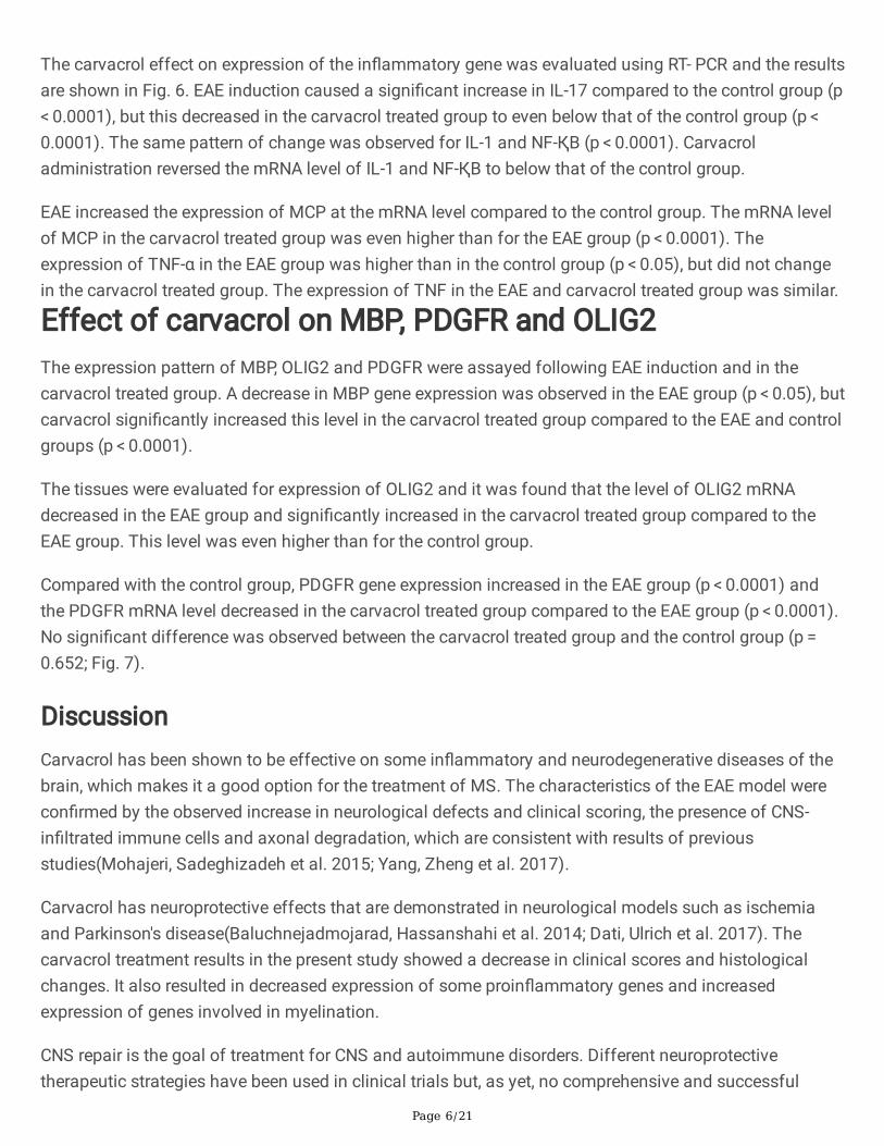
In�ltration of immune cells in lumbar spinal cord of animals in all groups. The arrows indicate the statusof immune cells:. (1) control group; (2) EAE group; (3) carvacrol treated group group. (4) This imageshows in�ammatory cell in�ux into the CNS after carvacrol treatment. The amount of in�ltration in thecarvacrol treated group group was signi�cantly lower than in the EAE group (p < 0.05). #Signi�cantdifferences with the EAE group(P<0.05)
Page 17/21
Figure 3
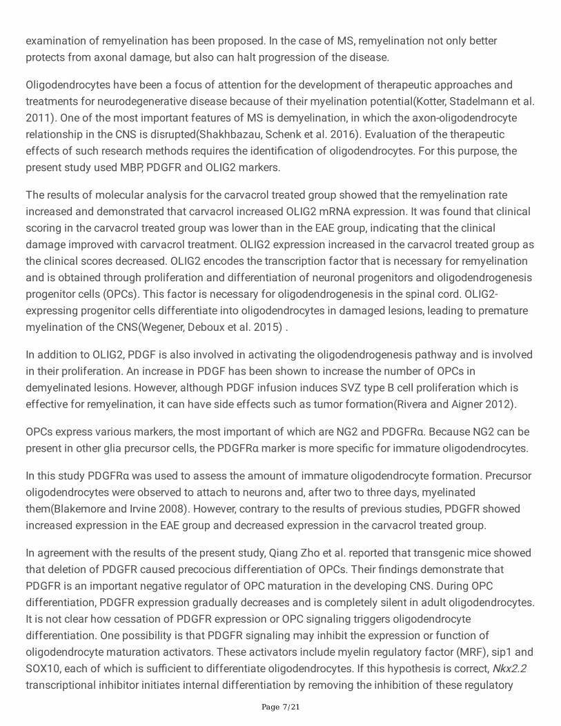
Immunohisto�uorescence assay for IL-17 marker in: (a) control group; (b) EAE group; (c) carvacrol treatedgroup. scale bar: 100µm
Page 18/21
Figure 4
MBP immunohisto�uorescence markers by group: (a) EAE; (b) control; (c) carvacrol treated group. scalebar: 100µm
Page 19/21
Figure 5
A) Comparison of IL-17 expression based on �uorescence intensity. The intensity of �uorescent in theEAE group compared to the control group had a signi�cant increase (P <0.0001). In the carvacroltreatment group, the �uorescence intensity and consequently the secretion rate was signi�cantly reducedcompared to the EAE group (P <0.000). B) Comparison of MBP expression based on �uorescenceintensity. Figures shows that the �uorescence intensity decreased signi�cantly in the EAE groupcompared to the control group (p < 0.0001). In the carvacrol treated group, the �uorescence intensity and,consequently, the MBP expression ratio relative to the EAE group increased signi�cantly (p < 0.0001).
Page 20/21
Figure 6
Effect of carvacrol on the gene expression of IL-17, NF-ҚB, MCP-1 and IL-1 in the EAE group and carvacroltreated group compared to the control group. *, #: Shows a signi�cant difference compared to the controlgroup and EAE group respectively.
Page 21/21
Figure 7
Effect of carvacrol on gene expression of MBP, OLIG2, PDGFR in EAE group and carvacrol treated groupcompared to control group. * and#: Shows a signi�cant difference compared to the control group andEAE group respectively.


![Myelin oligodendrocyte glycoprotein-specific antibodies from ......protein (MBP)] used to induce experimental autoimmune encephalomyelitis (EAE) in rodent models through induction](https://img.pdfslide.us/doc/110x75/60ff0b7639f1f130b4007123/myelin-oligodendrocyte-glycoprotein-specific-antibodies-from-protein-mbp.jpg)