Embed Size (px)
Citation preview
7/28/2019 En Radioisotopes in Medicine Final
http://slidepdf.com/reader/full/en-radioisotopes-in-medicine-final 1/40
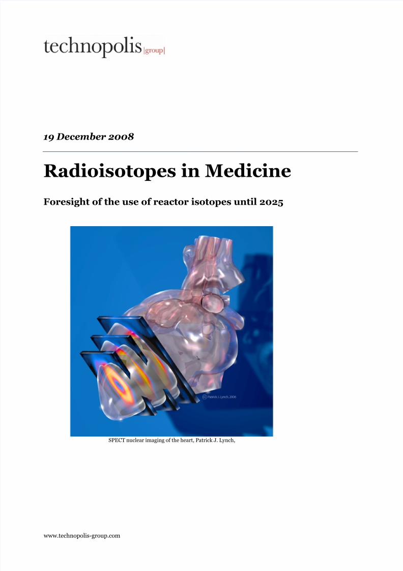
19 December 2008
Radioisotopes in Medicine
Foresight of the use of reactor isotopes until 2025
www.technopolis-group.com
SPECT nuclear imaging of the heart, Patrick J. Lynch,

7/28/2019 En Radioisotopes in Medicine Final
http://slidepdf.com/reader/full/en-radioisotopes-in-medicine-final 2/40
Radioisotopes in Medicine i
Table of contents1. Introduction 1
1.1 Commission VROM 1 1.2 Background HFR 1 1.3 Request 2 1.4 Delineation 3 1.5 Structure of the report 3
2. Method of investigation 4 2.1 Foresight studies 4 2.2 Expert Committee 4 2.3 Exploratory interviews 4 2.4 Survey of a broad-based group of high-level experts 5
3. Current system 7 3.1 Description of the technologies 7 3.2 Production of radiopharmaceuticals 11 3.3 Use of radiopharmaceuticals by the respondents 13
4. Explorations into the future use of radiopharmaceuticals in medical practice 21
4.1 Modalities 21 4.2 Future use of technetium 28 4.3 Therapy 33
5. Conclusion 34
7/28/2019 En Radioisotopes in Medicine Final
http://slidepdf.com/reader/full/en-radioisotopes-in-medicine-final 3/40
Radioisotopes in Medicine 1
1. Introduction
1.1 Commission VROM1
In April 2008, Technopolis received a request from the Ministry of VROM to conducta foresight study of the use of reactor isotopes, as part of the decision-making processregarding the construction of a new research reactor. Expectations are that the HighFlux Reactor (HFR) in Petten, the Netherlands, will reach its end of life around 2016.In the HFR in Petten, one of the materials that is irradiated with neutrons is uranium.In this process, molybdenum is generated, which decays into technetium. Technetiumis used for imaging techniques in nuclear medicine on a large scale.
In 2003, a report of the official steering group of the Ministry of VROM indicated that
at present, there is no alternative for the medical uses of technetium2. Furthermore,the use of the products generated by the HFR in medicine for imaging techniques
contributes substantially to the operating results of the HFR. Vital medical uses and aneconomic right of existence are important factors in the justification of theconstruction of a new research reactor.
The Dutch government, including the Ministry of VROM, will have to decide onpermission for a new research reactor and its justification. That is why for the Ministry of VROM it is not only relevant to know the need for the products of the HFR (and inparticular Technetium), but also what alternative technologies have to offer in thelonger term, i.e. after 2015. The request from VROM therefore concerns a foresightstudy into the possibilities of future, alternative technologies to gain an understandingof the relevance of a new research reactor. This involves both a study into alternativeproduction methods and a study into alternative (imaging) technologies. This reportonly addresses the latter. The Reactor Institute Delft performs a study into alternativeproduction methods – also commissioned by VROM.
1.2 Background HFR
Since 1962, the HFR is owned by the Institute for Energy (IE) of the Joint ResearchCenter (JRC) of the European Committee (EC). The Nuclear Research andConsultancy Group (NRG) is the licensee of the reactor. The reactor is operated andmaintained by NRG-staff.
The HFR is Europe's largest manufacturer of radiopharmaceuticals. The vast majority of the radiopharmaceuticals are used for the imaging of biological processes in the body. The most widely used isotope for medical imaging is technetium. For imagingprocedures with technetium, proteins are labelled with this isotope. Subsequently,
these proteins are administered to patients. In the body, these proteins adhere totargeted areas, thereby enabling visualisation by means of external scans with animaging device. This application is used for different diagnostic purposes, in particularin oncology, neurology, cardiology, for bone scans, and for imaging the function of organs such as kidneys, liver, brain and lungs. Technetium is of great importance tothe medical world: Around 80-85% of all nuclear-medicine procedures performed usetechnetium; there are about 40 million procedures performed each year, 50% of which
1 VROM: Dutch Ministry of Housing, Spatial Planning and the Environment2 Medical isotopes and the high flux reactor. Letter to the Dutch Lower House, no. 25422-27
with annex, March 2003.

7/28/2019 En Radioisotopes in Medicine Final
http://slidepdf.com/reader/full/en-radioisotopes-in-medicine-final 4/40
Radioisotopes in Medicine 2
in North America3 and 30-40% in Europe4. In the Netherlands, in 2007 approximately
393,000 nuclear medical procedures were performed5.
Apart from technetium, other products of the HFR are used for medical purposes (seeTable 1). Next to technetium, the most important isotopes in terms of market volume
are iodine-131 and iridum-192. These are highly important to certain groups of patients. In addition, there are other isotopes that are increasingly used, such aslutetium-177 and ytrrium-90. This is connected with new developments in therapeuticuses of radioisotopes.
Table 1 Overview of the medical uses of HFR products
HFR Product Use
Molybdenum-900/Technetium-99m
Diagnostic imaging for oncology, bone scans, cardiology, and theimaging of the function of organs such as kidneys, liver, brainand lungs.
Iodine-131 Therapy for thyroid disorders, cancer therapy
Xenon-133 Diagnostic imaging of lung function
Strontium-89 Therapy for metastatic bone pain
Iridium-192 Cancer therapy, e.g. lung cancer, head and neck cancer, tongueand mouth cancer, cancer of the throat and treatment of stenosis
Samarium-153 Therapy for metastatic bone pain and bone cancer
Rhenium-186 Therapy for metastatic bone pain
Iodine-125 Therapy for prostate and eye cancer
Yttrium-90 Therapy for arthritis
Erbium-169 Therapy for arthritis of smaller jointsLutetium-177 Therapy for tumours
Holmium-166 Development of therapy for liver cancer and blood cancer.
NRG, 2008: www.nrg-nl.com
1.3 Request
The request from the Ministry of VROM concerns a foresight study into thepossibilities of future, alternative technologies for medical imaging and the futureneed in medicine for radioisotopes in general and technetium in particular. At present,HFR is of great importance for nuclear imaging in the medical world at national,
European and worldwide level. How this will be in the period from 2015-2025 has to be answered with the following research questions:
1. What is the expected volume of imaging technologies for medical purposes inthe future, i.e. from now till 2025 and what will be the relative share of technetium-based imaging in these technologies?"
3 Triumf, 2008. Making Medical Isotopes.4 NRG: http://www.nrg-nl.com/product/fuel/isotopes/index.html , visited in Augustus 2008.5 RIVM, http://www.rivm.nl/ims/object_document/o5n1150.html , visited in November 2008.

7/28/2019 En Radioisotopes in Medicine Final
http://slidepdf.com/reader/full/en-radioisotopes-in-medicine-final 5/40

7/28/2019 En Radioisotopes in Medicine Final
http://slidepdf.com/reader/full/en-radioisotopes-in-medicine-final 6/40
Radioisotopes in Medicine 4
2. Method of investigation
The research questions have been answered through a combination of methods toidentify future developments. A large number of experts were involved in the entireprocess, enabling a sound picture of future developments in imaging technologies ingeneral and the use of technetium in particular. Technopolis itself did not make any technological choices: an expert committee guarantees the independence and scientificaccuracy.
2.1 Foresight studies
The importance of foresight is that it retrieves knowledge that can be used toanticipate and respond timely to developments that have yet to take place. This isparticularly important for problems that may have far-reaching consequences and a
large time span before solutions may be realised. Shortage of radiopharmaceuticalscould cause great difficulties that would take years to remedy. That is why today weneed to know the problems of tomorrow. There are different ways to study the future,in which experts often play an important part. More and more, the influence of theuser is also seen as a determining factor in this process. Any future study is plagued by uncertainty; the further ahead you look, the higher the level of uncertainty and thegreater the variation in the results. In this study, we use a combination of methods toarrive at a maximally reliable result:
• Expert committee;
• Structured and exploratory interviews with the industry, physicians andspecialists;
• A survey of a broad-based group of experts.Below, the steps taken are explained in more detail.
2.2 Expert Committee
An Expert Committee (EC) is important to safeguard the quality of the study. Thecommittee is composed of high-level experts with a collective insight in the differentareas that are relevant to medical imaging and diagnostics and medical, scientificresearch (see Appendix A.1). The Expert Committee made important contributions atthree stages in the entire process:
• Identification of experts, both for the interviews and for the survey;
• Providing an outline of the present state of the art;
• Validation of the survey results and generation of consensus.
2.3 Exploratory interviews
The first step in explorations is to identify the domains in which radioisotopes areused and for what medical indications, the availability of any other imaging techniquesand what these are used for, the prospects, and the present and future issues anduncertainties. What changes in the environment affect these domains?
To this end, interviews were conducted with 10 experts (researchers and users) in thefield of imaging and nuclear medicine as well as with industry representatives (see Appendix A.2). These exploratory interviews provided greater insight into the most

7/28/2019 En Radioisotopes in Medicine Final
http://slidepdf.com/reader/full/en-radioisotopes-in-medicine-final 7/40
Radioisotopes in Medicine 5
recent technological developments, as well as in the possible role of non-technologicalaspects in future developments.
2.4 Survey of a broad-based group of high-level experts
The purpose of a survey is the identification of the views held by a certain target group,and the development of a joint image of the future. Often, the studies involve thegeneration of a design orientation in relation to research, which is predominantly technological in nature. In this survey, a large group of experts and specialists areconsulted and asked to give their opinion. This contributes to an understanding of future developments that are important for the different stakeholders in the targetgroup and their expectations regarding these developments. Preferably therefore, theparticipants are involved stakeholders and experts.
In the present case, it means that experts from various fields share their expert viewsof future developments of medical uses of radiopharmaceuticals and imagingtechniques that might be an alternative to technetium. The survey used an online
questionnaire7 to guarantee efficiency and efficacy.
The Expert Committee validated the survey questions. Subsequently, these questionshave been presented online to a group of 102 mainly Dutch experts. These expertshave received two reminders, which resulted in a response rate of 45%. In total, 44persons completed the survey. Given the fact that for this explorative study involvement of high-level experts was preferred, the majority of the respondents are working in a university or leading hospital. Figure 1 presents an overview of thegeographic distribution of the respondents.
Figure 1 Geographic distribution of the respondents in the Netherlands
7 SurveyMonkey

7/28/2019 En Radioisotopes in Medicine Final
http://slidepdf.com/reader/full/en-radioisotopes-in-medicine-final 8/40
Radioisotopes in Medicine 6
The majority of the respondents are nuclear physicians (42%). This group of respondents are the ones with the best understanding of the use of reactor isotopes inclinical practice. The other respondents are radiologists, internists, oncologists,(clinical) physicists, (radio)chemists and pharmacists. Approximately 70% of therespondents are physicians; the others mainly have a technical background (see Figure
2).
Figure 2 Expertise of the respondents
Technopolis Group

7/28/2019 En Radioisotopes in Medicine Final
http://slidepdf.com/reader/full/en-radioisotopes-in-medicine-final 9/40
Radioisotopes in Medicine 7
3. Current system
3.1 Description of the technologies
Since the discovery of X-rays in 1895, many kinds of imaging techniques have beendeveloped in the course of the 2oth century to afford, as it were, a look inside thehuman body.
It is impossible to imagine present-day medicine without medical imaging, a processin which the computer is indispensable. In 2006, as many as 8.5 million X-rays weretaken. More advanced technologies such as CT and nuclear imaging was also usedfrequently and the number of procedures is increasing rapidly. For example, in 1991,360,000 CT scans were made; in 2006 this number increased to 890,000. Thenumber of nuclear medicine procedures increased from 200,000 in 1991 to 380,000
per year in 2006.8
Medical imaging is a domain that is still rapidly changing: several aspects, such as therefinement of imaging techniques, increase of the resolution and specificity,improvement of the efficiency, moderation of radiation, etc., still demand a great dealof research involving physicians, engineers, physicists, chemists, informationscientists, biologists, and mathematicians. Finally, ICT systems also continue toevolve, enabling image processing of ever-increasing quality.
3.1.1 Current imaging techniques
The following technologies are important in medical imaging.
• CT scan
CT is short for 'computed tomography'. It is a method to examine the human body using X-rays. The principle is as follows: On one side of the patient the X-ray source islocated, on the other side an X-ray detector. The source emits a small radiation beamthat passes through the patient in a straight line and is weakened by all tissuesthrough which it passes. The detector measures the strength of the remainingradiation. CT produces a large amount of data that can be manipulated through aprocess called 'windowing', visualising various structures based on their ability to block the X-ray beam. With the aid of digital geometric processing, a three-dimensional image is generated from a large series of two-dimensional X-rays takenaround an axis of rotation. Although in the beginning mainly axial or transversalimages were made, nowadays there are a number of modern CT scanners on the
market (dynamic volume, electron beam (EBCT), helix, multi-slice, peripheralQuantitative (pQCT)), enabling 3D images and structures at different angles. Theadvantage is the excellent visualisation of all sorts of defects and abnormalities with aresolution of 0.3 millimetres, not only of bones but also of all kinds of tissues. Themain disadvantage is the higher exposure to radiation compared to conventional X-rays (1 CT scan is roughly the equivalent of 200 X-rays).
8 RIVM: Diagnostiek: trends in aantal verrichtingen (Diagnostics: trends in number of
procedures). http://www.rivm.nl/ims/object_document/o19n1101.html , visited november
2008.

7/28/2019 En Radioisotopes in Medicine Final
http://slidepdf.com/reader/full/en-radioisotopes-in-medicine-final 10/40
Radioisotopes in Medicine 8
• MRI scan
MRI is short for 'magnetic resonance imaging'. The technique is based on the magneticproperties of atoms, especially hydrogen atoms, which are affected by electromagneticradiation. The technique is based on the fact that protons (especially hydrogen, but
also phosphorus protons) have 'spin', i.e. a small magnetic field. When the spins areplaced in a magnetic field, they align either with or against the field. There is adifference of energy between these two states, depending on the strength of theexternal magnetic field. When the hydrogen nucleus is exposed to a pulseelectromagnetic radiation with exactly the right energy (radio waves in the case of MR scanners), this causes the spin status to flip. Shortly after the flip, the nucleus returnsto its original position while emitting a photon. By applying a gradient to the force of the magnetic field, flipping the hydrogen nuclei and subsequently measuring theamount of radiation of different wavelengths that is re-transmitted by the nuclei thatreturn to their natural state, you can determine the number of hydrogen nuclei atcertain locations. By means of receivers and computers, a 3-dimensional image isformed. An MR scanner consists of a movable table on which the patient has to liedown and which can be moved into a hollow, cylindrical magnet, the magnetic field of
which (with a strength of 0.5 to 7 Tesla) is induced by superconductive coils. Withmodern MR scanners, the resolution is approximately 0.3 millimetres. To increase thecontrast of the MR scans, a contrast agent may be applied (gadolinium compounds). With MRI, tissues with many hydrogen atoms can be distinguished from tissues withfew hydrogen atoms, as a result of which details of the anatomy can be obtained. MRIscans are particularly useful in producing images of soft tissues. One advantage is thatno use is made of X-rays or radioactivity, although little research has been done intothe consequences of electromagnetic radiation. Its main disadvantage is that due tothe very strong magnetic field, it is not suitable for patients with electronic devices intheir body (such as a pacemaker).
• Echography
Echography, also called echoscopy, is a technique that applies ultrasonic sound wavesthat move through the body and reflect at interfaces between tissues with differentdensity. The ultrasonic waves are directed into the body by a transducer. Theultrasonic sound waves reflected by the body are received by the same transducer(which alternates between transmitting and receiving) and converted into smallelectrical impulses. A scanning converter converts the echo signals into video imagesmade visible on a monitor. The potential of digitally processing the signal hasexpanded enormously in the last decades. Based on the echo, the computer cangenerate an image of the interior of the body. This technique can be used to make apicture of organs to determine their size, structure, and pathological abnormalities, if any. The most common use of echoscopy is during pregnancy check-ups, but it canalso be used to measure the blood flow, for instance. The resolution of ultrasonography is approx. 1 mm. The main disadvantage of the technique is its
limited range of applications; it is suitable in particular for soft tissues. The advantageof ultrasonography is it does not involve any ionising radiation or radioactivity.
• Optical imaging
Optical imaging is a technique based on the inference and bending of light emittedfrom a laser or infrared light source to the body or tissue. Proteins are introduced inthe body that are labelled with a fluorescent marker, for instance, after which thefluorescence is detected. Optical imaging can be further classified into diffusive and ballistic imaging systems. Since light cannot penetrate the body that well, opticalimaging is not suitable for examining organs, for instance. It is used, however, forneurological research. The resolution (10 micrometer) is much better than the other
modalities, but the importance of optical imaging is marginal compared to the othertechniques.

7/28/2019 En Radioisotopes in Medicine Final
http://slidepdf.com/reader/full/en-radioisotopes-in-medicine-final 11/40
Radioisotopes in Medicine 9
• Nuclear medicine
Nuclear medicine is a mainly diagnostic discipline of medicine for imaging
metabolism and other functional processes in the human body. To this end, patientsare administered a radioactively labelled tracer. The strength of the technique lies inthe fact that use can be made of substances, which move to organ systems in very selective ways. The link to radioactive substances (especially technetium) enablesimaging of the distribution of the tracer in the human body with a gamma camera orPET scanner. A major advantage is the high sensitivity that enables measuring(several) molecular processes at picomolar level. Three different modalities areavailable for this process: planar scintigraphy, SPECT (single photon emissioncomputed tomography) and PET (positron emission tomography).
! Planar scintigraphy is the simplest available technique yielding a 2-dimensional projection image of the activity distribution of the tracer in thehuman body. The technique is based on the gamma radiation that is emittedon decay of the nucleus of a radioactive isotope.
! SPECT (single photon emission computed tomography) provides three-dimensional images of the distribution of nuclear activity with a resolution of 0.5 to 1 cm, which is not very high compared to other techniques. Isotopessuitable for SPECT typically have a half-life of a few hours to a number of days. Isotopes such as technetium (Tc-99m) with a half-life of 6 hours areoften applied. With longer half-lives, the radiation exposure of the patient istoo high; with shorter half-lives, the necessary connection cannot be madequick enough and preserved long enough. SPECT has been developed fromplanar techniques and is frequently combined with them. Thus, a gammacamera rotates around the patient and acquires a series of planar projections.SPECT subsequently processes them into three-dimensional imagesrepresenting the distribution of nuclear activity and enables the physician to
view the activity distribution in cross sections of the human body. The gammarays are detected with a photon detector. The detector consists of ascintillating crystal (a sodium iodide crystal for instance) in which a smallflash of light is created when hit by a gamma quantum. A series of horizontaland vertical photo detectors along the crystal record the position of this flashof light in the crystal. In front of the detection crystal, a collimator is placed, asheet of lead with many tiny holes in it so that a gamma quant incomingdiagonally won’t get through, but one that is incoming perpendicularly to thesurface of the crystal usually is. The result is that in the detector crystal,gamma quanta are detected that is known to be coming from a part of thepatient vertically below that place on the detector. It cannot be measured,however, from what depth in the patient's body the gamma photon wasemitted.
! PET (positron emission tomography) is an imaging technique with radioactiveisotopes (radionuclides) that emits positrons during their decay. When apositron encounters an electron annihilation takes place, producing energy inthe form of 2 gamma photons that are detected by a PET camera. When twophotons are detected simultaneously by two detectors that are placed oppositeto each other (at 180 degrees), they have been generated by the decay of thesame positron, which must have been situated on a straight line between thedetection points. From the time difference between the detection of bothgamma photons, it can be calculated where the annihilation took place on thisstraight line; however, light travels at such high speed that even moderndetector rings have much better angle than distance accuracy. A computer canreconstruct a three-dimensional image from a large number of such decay occurrences together, observed from different directions by a ring of detectors.
The three-dimensional image has a resolution of several millimetres. Most

7/28/2019 En Radioisotopes in Medicine Final
http://slidepdf.com/reader/full/en-radioisotopes-in-medicine-final 12/40
Radioisotopes in Medicine 10
PET radionuclides have a very short half-life and are produced in a cyclotron just before they are used. The most common example of such radionuclide is18F. This radionuclide is used to label FDG (18F-fluordeoxyglucose) forinstance, which is used to image the glucose metabolism in the body. Othersuitable radionuclides are carbon (11C), oxygen (15O) and nitrogen (13N). The
use of PET depends on the nature of the substance that is chosen. That choiceis determined by the process and/or tissue of which an image must be made.Compared to SPECT, PET offers a better sensitivity, higher resolution and thepossibility of accurately quantifying the examined processes. The applicationof the PET technique is in particular limited by the infrastructural and logisticpreconditions: because of the extremely short half-life (less than two hours toseveral minutes are not unusual), a cyclotron has to be present on site or at ashort distance.
The table below (Table 2) gives an overview of the different imaging technologies, theirresolution and their advantages and disadvantages.
Table 2 Overview of the characteristics of imaging techniques
Imaging Resolution Based on Advantage Disadvantage
CT 0.3 mm X-rays Visualisation of abnormalities in bonesand organs
Radiation exposure
MRI 0.3 mm MagnetismGood image of softtissues
Costs and magnetisminterference (e.g. withpacemakers)
SPECT 7 mm Gamma radiationMetabolism andfunctional processes
Low spatial resolution
PET 4 mm Gamma radiationMetabolism andfunctional processes
Costs and availability
Ultrasound 1 mm Sound Safe, soft tissues Limited applicability Optical 0.01 mm Light
Measurement of activity in the course of time
Very limitedpenetration depth
Technopolis Group
3.1.2 Medical indication of current modalities
The choice of one of the different imaging technologies depends in the first instance onthe medical indication that requires diagnosis. In some cases, that medical indicationcan only be shown by one technology. However, given uncertainty about a particularcondition, several techniques are available; most of the time, more than onetechnology may be used to confirm diagnosis. That is why the overview below containssome overlap. A neurologist, for instance, may use several imaging technologies toidentify a defect; the modality chosen usually depends on the expertise of theneurologist himself, the expertise of the hospital's staff and the presence or absence of specific modalities. In addition, the fact that nuclear medicine and radiology are twoseparate medical disciplines, responsible for SPECT/PET (nuclear medicine) and CT,MR and conventional X-ray and ultrasonography (radiology) respectively is alsoimportant. In most hospitals, these departments operate separately, while in a numberof hospitals these departments are integrated. In some hospitals, one discipline isstronger than the other, which also changes in time when new staff is hired. The list below of indications must be regarded in the light of these observations.
• Indications for CT (non-exhaustive)
Oncological indications of various sorts, both for staging and follow-up procedures,lung diseases, urinary tract disorders, complex skeleton disorders, bone tumours,check-up of liver shunts, neck disorders, suspect abdominal swelling.

7/28/2019 En Radioisotopes in Medicine Final
http://slidepdf.com/reader/full/en-radioisotopes-in-medicine-final 13/40
Radioisotopes in Medicine 11
• Indications for MRI (non-exhaustive)
Brain diagnostics, among others in case of tumours, ischemia (lack of oxygen),arteriography (vessels), demyelination (of the nerves), trauma, dementia, infection.Hernia, spinal disorders and bone infections, myocardial infarction, cardiomyopathy
(heart muscle disease), heart valve disease, kidney, adrenal gland, pelvis (prostate anduterus), soft tissue swelling and tumours, disorders of soft bone tissue, optic nerve block.
• Indications for SPECT (non-exhaustive)
Localisation and extensiveness of specific tumours, thyroid disorders, skeletondisorders, inflammations or infections, blood flow in the heart muscle, heart function, brain disorders, including Parkinson's disease.
• Indications for PET (non-exhaustive)
Particularly within the context of staging and follow-up of lung cancer and malignantlymphatic disorders, intestinal carcinoma, head-neck tumours, unexplainedpulmonary affections, melanoma, oesophagus carcinoma, ovarian tumours, thyroid
tumours, cardiology (vitality of cardiac muscle tissue), neurology (dementia).
3.2 Production of radiopharmaceuticals
3.2.1 Current situation
Worldwide, there are approximately 100 research reactors that produce isotopes;however, the majority of these isotopes are not suitable or used for medicalapplications. Only a handful of reactors dominate the entire molybdenum/technetiumsupply for the medical market. Table 3 presents an overview of the reactors thatproduce molybdenum used for medical use (the grey-highlighted lines indicate theEuropean share). The table includes two estimates; one by NRG dated 2002 and onedated 2008 by Nuclear Engineering International.
In the European context, the HFR is of essential importance, given that it provides forapproximately two thirds of the European demand and more than a quarter of thedemand worldwide for technetium (see Table 3). The HFR is a large producer due tothe fact that it meets two key conditions. A proper supply system of reactor isotopesfor the provision of medical use requires a high number of operational hours and agood infrastructure. Operational hours are important because a fairly constant supply of isotopes is needed, also in connection with the decay rate of molybdenum.Moreover, the infrastructure around the reactor has to be well developed, so that theisotopes can be processed as quickly as possible (according to Good Laboratory Practice guidelines) and transported to hospitals. Molybdenum has a half-life of 66hours. This requires that the isotopes be delivered to the hospitals within a few days.
The HFR, taking into consideration both aspects, is one of the most suitable in the world. Relatively speaking, the HFR has a high rate of operational hours and theNetherlands is a densely populated country with a well-developed infrastructure sothat all hospitals are within reach. From an international perspective, the location of the Netherlands in general and that of the HFR in particular (i.e near Schiphol Amsterdam Airport) is an important aspect in the prominent position of the HFR onthe European market.
Apart from importance of the HFR’s production share in absolute terms, it isconsidered important to have a substantial production capacity in Europe. Firstly, because the isotopes must be within reach considering the decay rates of the isotopes.Secondly, to be self-sufficient as EU in the production of radiopharmaceuticals.Complete dependency on countries outside the EU is deemed undesirable.

7/28/2019 En Radioisotopes in Medicine Final
http://slidepdf.com/reader/full/en-radioisotopes-in-medicine-final 14/40
Radioisotopes in Medicine 12
Table 3 Overview of the production capacity worldwide of molybdenum-99.European reactors are highlighted in grey.
NRG, 2002 NEI, 2008
Reactor Share Reactor Share
NRU (Ca) 45 % NRU (Ca) 38%
HFR (EU/NL) 27 % HFR (NL) 26%
Safari-1 (SA) 9% Safari-1 (SA) 16%
BR2 (Be) 8% BR-2 (Be) 16%
HIFAR (Aus) 2% Rest of the world 4%
OSIRIS (F) 2%
FRJ2 (D) 9 2%
Others 5%
NRG, 2002 & L. Kid, Nuclear Engineering International, 2008
Currently, the HFR is of great importance to both the Netherlands and the rest of Europe for maintaining the current production capacity of isotopes for medicalpurposes and safeguarding the availability of sufficient reactor isotopes for medical
imaging10.
3.2.2 The future
In the last ten years, the use of technetium has increased by 50% and is expected to
increase in the years to come11. NRG expects a moderate increase in the European
sales for the coming years. Outside Europe, NRG also anticipates an increase in theuse of Technetium, especially outside the United States. The growing prosperity indeveloping countries will result in a higher demand for nuclear imaging and thereforein a higher pressure on the world market for technetium. Although NRG expects therelevant countries to become self-supporting in time, they anticipate an extra highpurchase in the transitional phases (between increasing use and the set-up of production facilities).
In this light there is a risk of a shortage of medical isotope-producing reactors in thelong term. This is already clear at present now that the existing reactors are (more andmore) shut down due to maintenance operations. The current top 4 of reactors thataccount for 96% of the molybdenum production are old. They came into operation in
the fifties or sixties of the previous century 12. In all probability, these reactors will
reach their end of life in the near future. Safety considerations require regularmaintenance, and these reactors will eventually be closed down.
9 The Forschungsreactor Jülich is now closed.10 Report on the consequences of the (longer) shut down of the high flux reactor in Putten for
the supply of radioisotopes for medical applications. Dutch Healthcare Inspectorate, 2002.11 L. Kid, 2008. Curies for Patients. Nuclear Engineering International.12 NRU: http://www.nrureactor.ca/html/index.html
Safari-1: http://www.igorr.com/home/liblocal/docs/Proceeding/Meeting%208/ouo_06.pdf
BR2:
http://www.sckcen.be/SCKCEN_Information_Package_2007/CDROM_files/NL/Info_NL/p
dfs/2_Installaties_De_BR2_Reactor.pdf

7/28/2019 En Radioisotopes in Medicine Final
http://slidepdf.com/reader/full/en-radioisotopes-in-medicine-final 15/40
Radioisotopes in Medicine 13
Furthermore, there is no great increase in suitable new reactors. There is no new construction planned in North America. In the United States, exploratory research iscarried out into the possibility of building a few reactors for the production of small
amounts of technetium13. In France, the construction of the Jules Horowitz reactor
has started; commissioning is planned for 2014. The reactor will be able to produceapproximately a quarter of the amount of technetium currently used in Europe, andthus will not produce enough to meet demands and counterbalance the loss of thecurrent reactors. For more information about the production of radiopharmaceuticals,please refer to the research report of the Reactor Institute Delft, commissioned by VROM and carried out at the same time as this report, or international
publications14,15,16.
3.3 Use of radiopharmaceuticals by the respondents
Paragraph 3.1.2 indicated the suitability of the different modalities for medicalapplications. It is not clear from this overview, however, what are the key modalities
for specific disorders. By means of the survey, respondents were asked what imagingtechnique they used for certain disorders. Respondents were asked to distribute 100%over the modalities for use within the following categories: cardiology, oncology,neurology, bone scanning and other organ imaging.
The modalities that use reactor isotopes are planar nuclear technology, SPECT and
multi-modal17 imaging techniques that combine SPECT with another modality. Toestimate the relevance of reactor isotopes in each disorder domain, the relativecontribution of planar nuclear technology and SPECT modalities have to be summed.They will be singled out in the following paragraphs.
It is important to note in respect of the data presented in the following paragraphs thatthe picture drawn does not necessarily is representative for the use of reactor isotopesin the total Dutch medical practice. Our group of respondents mainly consists of
prominent physicians/researchers from university or top hospitals. In general, theserespondents have more modalities at their disposal than an average peripheralhospital. Thus, they can dispose of a PET scanner with personnel that can operate thisscanner more often than in the average Dutch hospital. In addition, there is moreexpertise in these hospitals regarding imaging techniques, which will affect the choicefor certain modalities. Furthermore, the group of respondents is made up of arelatively high number of nuclear medicine physicians, as a result of which a highshare will be assigned to these technologies in particular. The expert committeeassesses that technologies that are already in use for a longer time and require lessexpertise, will be better represented when a picture is drawn of the total use in theNetherlands. These particular technologies are echoscopy, CT, MRI, planartechnologies and SPECT. However, we have chosen the respondents precisely becauseof their understanding of and experience with imaging technologies in clinical practice
and research.
13 Advanced Molecular Imaging and Therapy, 2008 Preliminary Draft Report of the SNM
Isotope Availability Task Group14 Advanced Molecular Imaging and Therapy, 2008. Preliminary Draft Report of the SNM
Isotope Availability Task Group15 Triumf, 2008. Making Medical Isotopes.16 L. Kid, 2008. Curies for Patients. Nuclear Engineering International.17 Multi-modal scanners are imaging devices that combine different modalities in one device.
7/28/2019 En Radioisotopes in Medicine Final
http://slidepdf.com/reader/full/en-radioisotopes-in-medicine-final 16/40
Radioisotopes in Medicine 14
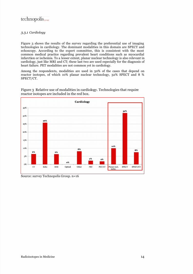
3.3.1 Cardiology
Figure 3 shows the results of the survey regarding the preferential use of imaging
technologies in cardiology. The dominant modalities in this domain are SPECT andechoscopy. According to the expert committee, this is consistent with the mostcommon medical practice regarding prevalent heart conditions such as myocardialinfarction or ischemia. To a lesser extent, planar nuclear technology is also relevant incardiology, just like MRI and CT; these last two are used especially for the diagnosis of heart failure. PET modalities are not common yet in cardiology.
Among the respondents, modalities are used in 50% of the cases that depend onreactor isotopes, of which 10% planar nuclear technology, 32% SPECT and 8 %SPECT/CT.
Figure 3 Relative use of modalities in cardiology. Technologies that requirereactor isotopes are included in the red box.
Source: survey Technopolis Group. n=16

7/28/2019 En Radioisotopes in Medicine Final
http://slidepdf.com/reader/full/en-radioisotopes-in-medicine-final 17/40
Radioisotopes in Medicine 15
3.3.2 Oncology
Figure 4 shows the relative use of preferential modalities in oncology. The mostdominant modality is by far the CT scan. MRI, PET, PET/CT and nuclear planar
technology are also important. CT is still preferred regarding a first diagnosis in casecancer is suspected and in the procedures to follow up treatment. Subsequently, forfurther research, a nuclear medicine technique is used to determine the activity of atumour. This is possible with SPECT, but our group of respondents often opt for a PETmodality already in this stage. Planar nuclear technology is the preferred choice in thedetermining bone metastases.
Modalities that use reactor isotopes represent approximately 23% of cases for ourrespondents: 14% planar nuclear technology, 8% SPECT and 1 % SPECT/CT.
Figure 4 Relative use of modalities in oncology. Technologies that use reactorisotopes are included in the red box.
Source: survey Technopolis Group. n=16

7/28/2019 En Radioisotopes in Medicine Final
http://slidepdf.com/reader/full/en-radioisotopes-in-medicine-final 18/40
Radioisotopes in Medicine 16
3.3.3 Neurology
Figure 5 shows the relative preferential use of modalities in neurology. MRI is thedominant modality here, and to a slightly lesser extent, CT. Of the other modalities,only SPECT is applied regularly as well. This picture corresponds with the usual
preference in indications for brain diagnostics. The fact that MRI plays an importantrole is consistent with the fact that brains are soft tissues. The contribution of SPECTis explained by the use of this modality in diagnosis of Parkinson's and relateddiseases, and also of dementia.
The respondents use reactor isotopes in 22% of the cases with the followingdistribution over the modalities: SPECT (15%), planar nuclear technology (6%) andSPECT/CT (1%).
Figure 5 Relative use of modalities in neurology. Technologies that requirereactor isotopes are included in the red box.
Source: survey Technopolis Group. n=19

7/28/2019 En Radioisotopes in Medicine Final
http://slidepdf.com/reader/full/en-radioisotopes-in-medicine-final 19/40
Radioisotopes in Medicine 17
3.3.4 Bone scans
For bone scans, planar nuclear technology is the dominant modality; the respondentsuse it in 43% of the cases (see Figure 6). Other relevant modalities are MRI, CT and
SPECT. SPECT is used mainly to determine the exact location of bone metastases andfor applications in orthopaedics and sports medicine. The results may overlap with theresult for planar technology in the oncology domain, thus leading to an overestimationof the total share of planar nuclear technology. MRI is used for diagnostics of joints,the soft tissues between the bones. CT is the present-day technology, like x-ray was inthe past.
Reactor isotopes are used in 55% of the cases, especially in planar nuclear technology and to a much lesser extent in SPECT (10%) and SPECT/CT (2%).
Figure 6 Relative use of modalities for bone scans. Technologies that usereactor isotopes are included in the red box.
3.3.5 Source: survey Technopolis Group. n=15

7/28/2019 En Radioisotopes in Medicine Final
http://slidepdf.com/reader/full/en-radioisotopes-in-medicine-final 20/40
Radioisotopes in Medicine 18
Scans of other organs
For the rest of the organs, planar nuclear technology is the preferred technology as well; it is applied in 57% of the cases (see Figure 7) involving mainly determination of functional aspects of organs such as kidneys and liver. Other important modalities are
echoscopy and SPECT. There may be a certain amount of overlap with SPECT resultsin the cardiology domain, thus probably leading to an overestimation of SPECT’s totalshare. Reactor isotopes are also important for the other organs. In 74% of the cases,the respondents use modalities that require reactor isotopes.
Figure 7 Relative use of modalities for the other organs; the technologies thatuse reactor isotopes are included in the red box.
Source: survey Technopolis Group. n=14

7/28/2019 En Radioisotopes in Medicine Final
http://slidepdf.com/reader/full/en-radioisotopes-in-medicine-final 21/40
Radioisotopes in Medicine 19
3.3.6 Therapy
It is estimated that more than 90% of the medical reactor isotopes are used forimaging procedures. The remaining part is used for therapy (see Figure 8 for a list of
isotopes for therapeutic use). Although this domain is relatively unimportant inquantitative terms, it is of vital importance to the quality of medical care in generaland to the quality of life of a smaller group of patients in particular. There is noalternative, for instance, for iodine-131 treatment of patients with thyroid cancer.Reactor isotopes are also very important in palliative therapy (therapy directed atsymptom relief): they are used for pain relief in patients with bone metastases wheremorphine is the only alternative.
Figure 8 Reactor isotopes for therapeutic use
Isotope
Iodine-131
Strontium-89
Iridium-192
Samarium-153
Rhenium-186
Iodine-125
Yttrium-90
Lutetium-177
Holmium-166
Source: NRG, 2002; adapted by Technopolis Group
3.3.7 Findings
Reactor isotopes are of great importance in the current medical imaging practice of therespondents in cardiology (50%), for bone scans (55%) and for the other organs (74%).In oncology (23%) and neurology (22%), reactor isotopes are of less importance. Asalready mentioned above, these results might not be entirely representative for theaverage Dutch situation, but provide a picture of the current situation in university
and top hospitals (STZ hospitals18
). However, many patients visit these hospitals. Inthe general Dutch situation, conventional technologies (including CT and planarnuclear technologies) are bound to have a greater share. The results presented in thisreport describe the situation in the clinical practice in the hospitals that fulfil a leading
role19.
18 STZ hospitals: Hospitals affiliated with the Dutch association of tertiary medical teaching
hospitals.19 A complete picture of the use of the different modalities at present (and in the past) can only
be obtained by asking all Dutch hospitals for the production figures of the departments of
radiology and nuclear medicine, subdivided in figures per modality.

7/28/2019 En Radioisotopes in Medicine Final
http://slidepdf.com/reader/full/en-radioisotopes-in-medicine-final 22/40
Radioisotopes in Medicine 20
Although therapy only accounts for a small share in the total use of reactor isotopes, itplays an important role in terms of quality.
The expert committee endorses the above picture of the use of reactor isotopes. CT iscommon in oncology, and PET is increasing; MRI is frequently applied in neurology,
SPECT is used to determine the functionality of tissues, where PET is emerging as well, and planar nuclear technology is commonly used for bone scans. CT and MRI aregenerally used at every radiology department, while in nuclear medicine planartechniques and to a lesser extent SPECT are dominant. Reactor isotopes areconsequently of great importance to medical imaging at present, especially in thefields mentioned above. Moreover, they are even more important in hospitals that donot dispose of a PET scanner. In a limited number of medical centres (including VUmc, UMCG, ErasmusMC, UMC St. Radboud in Nijmegen and St. Antonius inNieuwegein), a lot of research is done currently into PET applications. PET use isfrequent in 10 hospitals. Apart from that, another 35 hospitals in principle have accessto PET technology.
The distribution of the use of various modalities for certain conditions already showsthat more than one modality can be used for the same disorder. As already mentioned
in 3.1.2, within the imaging domain further choices for a particular modality are madeon medical grounds, or dependent on the preference or expertise of the physician andthe radiological technologists. In paragraph 4.1.2, we will address the decisive factorsthat determine the preference for a specific modality in more detail.

7/28/2019 En Radioisotopes in Medicine Final
http://slidepdf.com/reader/full/en-radioisotopes-in-medicine-final 23/40
Radioisotopes in Medicine 21
4. Explorations into the future use of radiopharmaceuticals inmedical practice
Radiopharmaceuticals are currently of great importance in the medical practice,especially in diagnostic imaging. This chapter will deal in greater detail with theexperts’ future expectations in this area. The central question here is to what extentreactor isotopes will be important in the future; in other words: will the modalitiesthat use reactor isotopes (SPECT, planar, and multi-modalities) still be used in 2015and 2025?
4.1 Modalities
4.1.1 TrendsBased on the interviews, a few important trends were identified that in relation to therange of modalities and that might bring about some shifts. The following trends areimportant for substitution effects:
• Improvement of current modalities. The current modalities are stillimproved incrementally. These are in particular technological improvements, suchas the increase in sensitivity and resolution, the further development of softwareto process the data and combine parameters. Eventually, the quality of the scans will increase as a result of these improvements and better diagnoses can be made,or the use of a specific scanner can be expanded. PET is a good example of atechnology that is still developing rapidly. Experts still expect a considerableadvance in this area, for instance through discovering, testing or experimenting
with new tracers, as a result of which the PET scanner can be applied for a widerrange of procedures. But also 'older' modalities, such as SPECT, are stilldeveloping. Currently, new crystals are being tested and used, which will improvethe resolution of SPECT. Some experts anticipate that as a result SPECT will equalthe resolution of PET in time; others take the opposite view.
Our interviews show that PET is one of the fastest growing modalities. Some of the
interviewees in this study, as well as a Canadian expert panel 20 expect that PET will expand more rapidly than, for instance, SPECT. It remains to be seen,however, if this will lead to large shifts in applications that are now conducted with planar nuclear technology and SPECT to PET. Many developments will befocused on extending the possibilities for medical science, as a result of which thediagnosis in specific cases is improved and PET will function as an add-on. Theinterviewees also point out that so far, no modality has ever disappeared from the
total range of technologies.
• Combinations of modalities. Parallel to the improvement of the modalities,one of the most important developments is the combination of modalities into onedevice. The biggest advantage of the so-called multi-modal scanners is that they can generate information about metabolism as well as spatial information(location). When measuring metabolism with a SPECT scan for instance, it is lessclear where the metabolic abnormalities are located. This information can be better obtained with a CT scan. Combining these modalities is combining the 'bestof both worlds'. At present, the PET/CT scanner is a well-selling multi-modal
20 See Triumf, 2008.

7/28/2019 En Radioisotopes in Medicine Final
http://slidepdf.com/reader/full/en-radioisotopes-in-medicine-final 24/40
Radioisotopes in Medicine 22
scanner. Other combinations are still under development, such as the SPECT/CT,PET/MR, and SPECT/MR scanners.
Combinations of modalities require further technological developments, inparticular in the area of integrating two devices. Apart from developments in
physics, mechanical engineering, and the like, significant developments insoftware are also required given the fact that two datasets have to be combined with each other.
• Development of new tracers. The detection of certain processes (metabolism)or substances (e.g., particular proteins) requires tracers which attach themselvesto the right place in the body and which can subsequently be represented by means of a modality. New tracer’s function as 'enablers' for a modality, they expand its possibilities. At present, many tracers are developed, in particular forPET; however, also for other modalities expectations for new tracers are high.Nanotechnology and bionanotechnology hold promises for new tracers andmarkers, for instance for optical, MR or other modalities. Although new tracerscould lead to shifts between modalities, it is not yet clear what modalities will bestimulated as a result. In the recently started Center for Translational Molecular
Medicine (CTMM), the research into biomarkers and imaging technologies iscombined.
• Development of new therapies. Although reactor isotopes are mainly used forimaging purposes, they are also of qualitative therapeutic importance. Reactorisotopes already play an important role in thyroid and prostate cancer therapy andpalliative therapy for bone metastases. Therapeutic applications seem to begaining importance rapidly. For instance, a few years ago, Lutetium-octreotate was administered to patients for the first time. This isotope is given to patients with neuroendocrine tumours (they are mainly located in the stomach, intestinesand pancreas and may produce spreading harmful amounts of hormones). The beauty of such applications lies in the fact that the administered substances can betargeted to the process under treatment; this is not possible with external
radiotherapy, where the surrounding tissue is also exposed to radiation.Furthermore, treatment with lutetium results in an increased life expectancy of
patients with 4 years with a relatively good quality of life. 21
• Development of new devices/modalities. Although at present there is noprospect of a totally new modality, this scenario of 'unforeseen circumstances' hasto be taken into account in a foresight. In the survey, we investigated the length of time required for a new finding to develop from the laboratory to preferred clinicaluse (see Figure 9). According to the respondents, the average duration of theinitial research phase to obtain clinical evidence is approximately 8 years. Fromthat point onwards preferential use takes at least another ten years. Therefore, if options are overlooked in this explorative study because they are only in an early stage of development, it will last at least 18 years before they become a preferredmodality in clinical practice. Although the estimates of the experts differ greatly,
the averages are confirmed in the interviews with the experts from the industry and medicine, as well as by the expert committee.
21 http://www.nrg-nl.com/general/nieuws_nl/cms/2008/200801161635.html

7/28/2019 En Radioisotopes in Medicine Final
http://slidepdf.com/reader/full/en-radioisotopes-in-medicine-final 25/40
Radioisotopes in Medicine 23
Figure 9 Expert estimates of the length of the innovation process in yearsfrom initiation of research to clinical proof incl. clinical trials (red) and thelength from clinical evidence to preferential use in years (black). Average
values: research phase: 8 years; implementation: 10 years
Source: survey Technopolis Group. n=32
4.1.2 Expectations regarding the use of modalities
In the survey, we asked the respondents about their expectations regarding the use of the various modalities in clinical practice. By means of the interviews we identified allthe possible modalities that might play a more prominent role in future. Subsequently,the respondents were asked to indicate what share they expected a particular modality to have in the number of scans in their clinical practice in 2008, 2015, and 2025. Therespondents could assign 100% in total to all modalities; Figure 10 shows the results.Note that the figures express the share of one modality relative to the other modalities,rather than absolute numbers.

7/28/2019 En Radioisotopes in Medicine Final
http://slidepdf.com/reader/full/en-radioisotopes-in-medicine-final 26/40
Radioisotopes in Medicine 24
Figure 10 Expert estimation of the relative use of modalities in 2008, 2015and 2025.
Source: survey Technopolis Group. n=23
Based on these expectations for the future, the following conclusions can be drawnregarding the proportion between the modalities. Our respondents expect:
• A relative decrease in the CT in the procedures they perform, which is significant(more than 10% in the period from 2008-2025)
• A slight decrease in the number of single MRI procedures (less than 3%)
• A slight decrease in the number of ultrasounds (approximately 4%)
• An equal number of PET procedures
• A fairly substantial decrease in the share of SPECT (approximately 7%)
• A fairly substantial decrease in the other modalities (in particular planar, optic).These are the modalities that were expected to have a share of a few percent only (approximately 4%)
• A fairly substantial increase in the share of PET/CT (approximately 7%)
• A fairly substantial increase in the share of SPECT/CT (approximately 7%)
• Sharp increase in PET/MRI (more than 10%)
• After 2015: growth of the SPECT/MRI (approximately 2.5% from 2015 to 2025)

7/28/2019 En Radioisotopes in Medicine Final
http://slidepdf.com/reader/full/en-radioisotopes-in-medicine-final 27/40
Radioisotopes in Medicine 25
To single out the trends in modalities that use reactor isotopes, in Figure 11 thepercentages of the multi-modalities have been added up to the single modalities. Theshare of PET/CT has been added up to both PET and CT and the share of SPECT/MRIto both SPECT and MRI. As a result, a picture is created of the total use of the single
base modalities, even though the total number of scans exceeds one hundred percent.
Figure 11 Share of the number of scans according to base modalities
Source: survey Technopolis Group. n=23
Figure 11 shows that the respondents expect a strong increase in the combined use of PET, as well as MRI,. The decrease in the number of PET and MRI is substituted by the use of multi-modalities. The combined use of CT will increase fairly substantially at first, followed by a slight decrease at the expense of MRI multi-modalities. Theshare of SPECT modalities will remain more or less unchanged. The considerabledecline in the share of SPECT (see Figure 10) is substituted by the use of multi-modalities: initially by SPECT/CT and after 2015 by SPECT/MRI. Since SPECT is themodality that uses reactor isotopes, the share of SPECT is broken down in Figure 12.
Figure 12 is a break-down of figure 11; it shows the substitution effects of the SPECTmulti-modalities: the use of SPECT will decline strongly, the use of multi-modalitiesincluding SPECT will increase strongly.

7/28/2019 En Radioisotopes in Medicine Final
http://slidepdf.com/reader/full/en-radioisotopes-in-medicine-final 28/40
Radioisotopes in Medicine 26
Figure 12 Break-down of the shares of SPECT modalities
Source: survey Technopolis Group. n=23
4.1.3 Innovation dynamics
The implementation of innovative technologies is driven by technological and non-technological factors. One of the technological factors in this case is the resolution.
Non-technological factors that are related to nuclear technology are the logistic andinfrastructural problems around isotopes. As for reactor isotopes, they have to berefined and purified according to GLP and transported to hospitals; as for PETradionuclides, the infrastructure has to meet certain requirements for production inthe cyclotron and transport to hospitals, and well-trained staff is required who can work with these radionuclides. Furthermore, in the medical domain and in hospitals, various human aspects play an important role, such as the skills and expertise of theradiological technologist and convenience of use, but also costs, contracts and
efficiency 22.
To determine what technological or non-technological factors are decisive in choosinga particular modality in the clinic, the interviewees were asked to indicate which of those factors are (very) important or unimportant. Figure 13 shows that to a greater or
lesser extent, the respondents find nearly all factors important.
22 PET Gepast gebruik(t), ZonMW Doelmatigheidsonderzoek, januari 2007 (Efficiency study
regarding the use of PET scanners, carried out by the Netherlands Organisation for Health
Reserach and Development)

7/28/2019 En Radioisotopes in Medicine Final
http://slidepdf.com/reader/full/en-radioisotopes-in-medicine-final 29/40
Radioisotopes in Medicine 27
Figure 13 Factors that determine the choice for a certain modality (1= very unimportant, 2= unimportant, 3=neutral, 4=important, 5= very important)
Source: survey Technopolis Group. n=21
The only factor that is considered less decisive is dependency on contracts. Of the 4factors that on average score higher than 4 (important), only higher resolution is atechnological driver. Two factors are related to human aspects: the skills of theradiological technologist (this is connected in part with the expertise of theradiological technologist) and human resources in general. The latter mainly refersto the highly specialised staff required for PET modalities. Finally, infrastructuralproblems are regarded as important. This can refer to the problems with the delivery of technetium as well as to the infrastructural organisation of cyclotrons in hospitals.In summary, we may conclude that a higher resolution is a technology driver, but thatthe human factors are the ones that largely determine the success of that technology.In this case it also means that the pace at which the PET applications will be expanded
mainly depends on sufficient qualified personnel and infrastructure. Incidentally, thedecisive factor 'costs' has not been further defined; it may refer to the costs of a singleprocedure as well as to investment costs of a hospital when acquiring the technology.
4.1.4 Summary of findings
Based on the identified trends (4.1.1) and the proportions of modalities (4.1.2) it may be concluded that the experts anticipate a significant increase in PET modalities. Theinterviewees from both the industry and the group of users indicate that this modality,at present, is showing the biggest growth. The multi-modal developments seem tostimulate the growth of PET.
The share of SPECT modalities will remain more or less unchanged. The number of SPECT scans will decrease in the years to come; however, the share of multi-

7/28/2019 En Radioisotopes in Medicine Final
http://slidepdf.com/reader/full/en-radioisotopes-in-medicine-final 30/40
Radioisotopes in Medicine 28
modalities that combine SPECT with other modalities will increase. The industrialinterviewees indicated that many efforts are put into the development of a SPECT/CTscanner, and the survey shows that the clinical practice foresees a substantial share forthis scanner.
As for the mix of modalities, it has become evident that reactor isotopes will continueto be of great importance. Based on the unchanged share of SPECT scans, it can beconcluded that the relative demand for reactor isotopes (in proportion to the totalnumber of scans) will remain more or less the same.
The implementation of a technology is determined by both technological and non-technological factors. When choosing a particular modality in the clinic, it appears thatalthough higher resolution is shown to be a technology driver, human factors are theones that determine the success of that technology to a large extent. In this case itmeans that the pace at which the PET applications will be expanded mainly dependson sufficient qualified personnel and infrastructure.
In paragraph 4.2 we will discuss the expectations of the experts regarding the use of technetium in the clinic in more detail.
4.2 Future use of technetium
4.2.1 Total number of scans
The total number of scans in medicine is expected to grow in the coming years.Increasing prosperity results in a higher standard of living, better medical science anda higher life expectancy. As people are getting older and the ageing populationincreases, the number of medical procedures will increase as well. Moreover, medicaltechnology will be used more frequently as a result of a higher standard of living.Combined with a growing population figure, this will lead to an increasing number of imaging procedures. This trend has already started, and the total number of scans willcontinue to increase especially due to the ageing population and population growth.This picture is supported by the estimates of the experts. They almost unanimously anticipate an increase, and even a significant one, in the total number of scans overtime. Figure 14 shows the experts' expectations for the total number of scans in 2008-2010, 2010-2015 and 2015-2025.

7/28/2019 En Radioisotopes in Medicine Final
http://slidepdf.com/reader/full/en-radioisotopes-in-medicine-final 31/40
Radioisotopes in Medicine 29
Figure 14 Expert expectations of the total number of scans for medic use
Source: survey Technopolis Group. n=33
Figure 15 shows the same results as Figure 14, however represented as the weightedaveraged of the respondents' answers. Nearly all experts foresee an increasingly certain growth of the total number of scans in the future. On average, the expertsanticipate an increase; for 2015-2025, 60% of the respondents anticipates a significantincrease.
Figure 15 Weighted average of the expert expectations on the total number of scans in 2008-2010, 2010-2015, 2015-2025. (1= large decline, 2= decline, 3=unchanged, 4= increase, 5= large increase)
Source: survey Technopolis Group. n=33

7/28/2019 En Radioisotopes in Medicine Final
http://slidepdf.com/reader/full/en-radioisotopes-in-medicine-final 32/40
Radioisotopes in Medicine 30
4.2.2 Probability that scans using technetium will be substituted by other
technologies
The survey also asked for the probability of technetium-based procedures beingreplaced by modalities that do not require the use of technetium. Figure 16 shows how
likely it seems to the respondents that technetium will be substituted in the period till2010 (red), from 2010-2015 (black) and from 2015-2025 (grey). Figure 17 shows the weighted average of these results.
Figure 16 Expert estimations on the probability of the substitution of technetium
Source: survey Technopolis Group. n=33

7/28/2019 En Radioisotopes in Medicine Final
http://slidepdf.com/reader/full/en-radioisotopes-in-medicine-final 33/40
Radioisotopes in Medicine 31
Figure 17 Weighted average of the expert estimates of the substitution of technetium. (1= certainly not, 2= probably not, 3= neutral, 4= probably,5=certainly)
Source: survey Technopolis Group. n=33
Sixty-five per cent of respondents do not consider it probable that technetium will bepartly replaced in the period up to 2010. For the period from 2010-2015, still 30% of the respondents consider this unlikely, although the same percentage gives a neutralanswer. Further into the future, the respondents expect an increasing chance of
technetium replacement. On the one hand, this can be explained by an increasinguncertainty: in future forecasts there are always unknowns and uncertainties. This isshown by the increasingly diverging responses to the question of time: the spread forthe period 2015 - 2025 is markedly greater than for earlier periods (see Figure 16).However, the weighted average of the substitution shifts from 'probably not' for 2008-2010 to 'neutral' for 2010-2015, with a slight tendency towards 'probably' for theperiod 2015-2025. The average value shifts from 2.0 for 2008-2010 to 3.2 for 2015-2025 (see Figure 17). From the interviews it has become clear that this shift can beexplained in particular by the estimation that other modalities will have higherfunctionalities in this period (see also Figure 10). Nuclear physicians anticipateimportant developments especially for PET. Experts from other fields of medicalimaging expect breakthroughs regarding MRI, as a result of which this technology may also be used for the imaging of metabolisms. There is no consensus on these matters.
In general, experts do not have a total view of all these future developments. Theaverage estimate shows, however, that the use of technetium will slightly decline astime goes by.
4.2.3 Future use of technetium
The probability of substitution was examined in greater detail in the survey by meansof a quantitative estimate of the future use of technetium. The experts were asked inthe survey to give an estimate of the use of technetium in the future compared to2008. Figure 18 shows the distribution of the estimates of the experts in percentagepoints.

7/28/2019 En Radioisotopes in Medicine Final
http://slidepdf.com/reader/full/en-radioisotopes-in-medicine-final 34/40
Radioisotopes in Medicine 32
Figure 18 Expert estimates on the use of technetium in 2010, 2015 and 2025,as a percentage of the current use (2008).
Source: survey Technopolis Group. n=29
Figure 19 shows the weighted average of these answers. This indicates the totalestimated use of technetium in 2010, 1015 and 2025, compared to 2008.
Figure 19 Weighted average of the expert estimates on the use of technetiumuse 2010, 2015 and 2025 (reference year: 2008).
Source: survey Technopolis Group. n=29
In the short term (2008-2010, red), 90% of the respondents expect the use of technetium to slightly increase or remain unchanged (Figure 18). Overall, the experts
expect the use of technetium to grow with 5% by 2010 (see Figure 19). After that, in

7/28/2019 En Radioisotopes in Medicine Final
http://slidepdf.com/reader/full/en-radioisotopes-in-medicine-final 35/40
Radioisotopes in Medicine 33
the period from 2010-2015 (Figure 18, black), the number of respondents that think of a decrease in use increases, but the use, on average, is the same as the use now. Figure19 shows that the weighted average of the use in 2015 is estimated to be 99% of the useof 2008. For the period from 2015-2025 (Figure 18, grey), the spread in responses is
significant. However, the weighted average results in a slight decline in the total use of technetium till 92% of its current level.
The answers to this question match the expectations regarding technetiumsubstitution.
4.2.4 Summary of findings
All experts unanimously anticipate a significant to very significant increase in the totalnumber of diagnostic imaging scans in the future. This outcome is connected to theageing population and population growth.
As for the expectations regarding the substitution of imaging modalities that usetechnetium, major changes (substitution) are not expected for the time being. For theperiod after 2015, the experts are divided in their opinion; the average, however, shifts
from 'probably not' to more or less neutral, with a slight tendency towards 'probably'in 2025.
The same trend can be observed in the total use of technetium: in the coming years,the use of technetium will certainly not decrease, but rather slightly increase. For theperiod from 2015-2025, the experts foresee that the use of technetium will slightly decrease (<10%), but the spread in responses is high.
4.3 Therapy
As for the use of reactor isotopes for therapy, the results of the survey are unequivocal(see Figure 20). The current use of iodine and iridium is not expected to increase a
great deal (indicated with o). However, the experts do expect an increase, in the use of lutetium-177 and ytrrium-90 and this increase will start now and continue until past2015 (indicated with +). The use of holmium-166 and samarium-153 will also increase, but that will start after 2010. In this area, the expectations of the experts coincide withthe results of the interviews and the literature, which without exception point out thedevelopments in radiopharmaceuticals for therapy.
Figure 20 Expert expectations for the application of therapy with reactorisotopes. ( -- = large decrease, - = decrease, o = unchanged, + = increase, ++ =large increase)
2008-2010 2010-2015 2015-2025
Iodine-131 o o o
Strontium-89 o o o
Iridium-192 o o o
Samarium-153 o + o
Rhenium-186 o o o
Iodine-125 o o o
Yttrium-90 + + +
Lutetium-177 + + +
Holmium-166 o + 0
Source: survey Technopolis Group. n=22

7/28/2019 En Radioisotopes in Medicine Final
http://slidepdf.com/reader/full/en-radioisotopes-in-medicine-final 36/40
Radioisotopes in Medicine 34
5. Conclusion
The Ministry of VROM commissioned this study. It sets out to answer the followingresearch questions:
1. What is the expected volume of imaging technologies for medical purposes inthe future, i.e. from now till 2025 and what will be the relative share of technetium-based imaging in these technologies?"
2. "What new or upcoming imaging technologies in medicine may affect ordisplace the technetium-based imaging technology in the period till 2025,both in terms of quality and quantity?"
The following conclusions may be drawn on the basis of interviews, the results of anonline survey and validation by an expert committee.
Current developments
• There is a range of imaging modalities available at present (CT, MRI, SPECT andPET) each of which has a specific application in the medical domain. Technetiumis used for SPECT and planar technology; these modalities are preferentially applied in bone scans (including bone metastases in oncology) and organ scans(including measuring of blood flow and heart muscle function in cardiology).
• Multi-modalities that combine nuclear and radiological techniques in one deviceare emerging. For the future, shifts are anticipated in the use of the modalities,foreseeing a decrease in the single modalities in favour of the multi-modalities.
• At the moment, there is no new technology that might affect the use of technetium. If there were one, the experts think that it would take at least 18 years before its use would be preferred in clinical practice. Furthermore, 'old' techniquesusually do not disappear.
• Although an important technological driver for imaging modalities is a highresolution, the human factors determine the success of a technology to asubstantial extent.
Expectations
• A significant increase in PET modalities is expected, especially in combination with CT or MRI. Partly due to the high resolution, the current applications of PET will be expanded, but probably not at the expense of the total share of SPECTmodalities. The rate of the developments related to PET also depends on thedevelopment of new radiopharmaceuticals, required infrastructure and expertise.
• The relative share of SPECT modalities probably remains unchanged, but thesingle SPECT will be replaced in time by SPECT/CT and later on by SPECT/MRI(not available yet).
• The experts are convinced that the total number of scans will show a (significant)increase in the future. This increase already started in the last few years.
• Furthermore, the experts consider it unlikely that technetium-based imaging will be replaced by other technologies in the medium term (till 2015); it might slightly decrease in the period from 2015-2025. This also becomes clear from theexpectations for the total use of technetium: It remains unchanged for the time being, but will slightly decrease (<10%) in the period from 2015-2025. A number
of experts also indicate that to date, no imaging modality has ever been replaced,

7/28/2019 En Radioisotopes in Medicine Final
http://slidepdf.com/reader/full/en-radioisotopes-in-medicine-final 37/40
Radioisotopes in Medicine 35
even the conventional x-rays are still used; the total range of possibilities has only been further increased in the course of time.
Summarising, the conclusion can be drawn that the demand for radiopharmaceuticalsthat are produced in nuclear reactors will continue to exist till 2025. In the field of nuclear medicine, the experts anticipate that the current accelerated development of PET will continue, which will cause a relative decline in the use of reactor isotopes.However, due to the low costs and relative simplicity of SPECT and planar nuclearimaging, these technologies will continue to exist and, in absolute terms, will be used just as much.

7/28/2019 En Radioisotopes in Medicine Final
http://slidepdf.com/reader/full/en-radioisotopes-in-medicine-final 38/40

7/28/2019 En Radioisotopes in Medicine Final
http://slidepdf.com/reader/full/en-radioisotopes-in-medicine-final 39/40
Radioisotopes in Medicine 37
Appendix A Consulted individuals
A.1. Expert committee
Expert Institute
Prof. Dr. H.G.M. RooijmansProfessor emeritus Psychiatry, former chairman of the
Raad voor Gezondheidsonderzoek (RGO – HealthResearch Council)
-
Prof. Dr. Ir. M.A. ViergeverProfessor medical imaging
Image Sciences Institute (ISI), UMCUtrecht
Prof. Dr. A.A. LammertsmaProfessor clinical physics
Nuclear medicine & PET research, VUMc Amsterdam
Dr. A. VerzijlbergenChairman Dutch Society of Nuclear Medicine
Department of Nuclear Medicine, Sint Antonius Hospital, Nieuwegein
A.2. Interviews
Name Institute
H.G.M Rooijmans Former chairman of the RGO – Health Research Council
M.A. Viergever UMCU
A.A. Lammertsma VUMc
H. Hofstraat CTMM, UMC
F. Gerritsen Philips and TU Eindhoven
P. Luijten CTMM, UU, Philips
R. A. Diericx RUG
A.M. Verbruggen KU Leuven
B. Van der Schaaf NRG
A. Paans UMCG

7/28/2019 En Radioisotopes in Medicine Final
http://slidepdf.com/reader/full/en-radioisotopes-in-medicine-final 40/40
Technopolis Group The NetherlandsHerengracht 141


![Tb radioisotopes for medical applicationsTb offers four radioisotopes that are interesting for applications in nuclear medicine, i.e. 149 Tb, 152 Tb, 155 Tb and 161 Tb [4]. By giving](https://img.pdfslide.us/doc/110x75/600d41328dddfa5d101bdfd1/tb-radioisotopes-for-medical-applications-tb-oiers-four-radioisotopes-that-are.jpg)