Embed Size (px)
Citation preview
REVIEW Open Access
Emerging roles of telomeric chromatinalterations in cancerStefano Cacchione1*, Annamaria Biroccio2 and Angela Rizzo2*
Abstract
Telomeres, the nucleoprotein structures that cap the ends of eukaryotic chromosomes, play important andmultiple roles in tumorigenesis. Functional telomeres need the establishment of a protective chromatinstructure based on the interplay between the specific complex named shelterin and a tight nucleosomalorganization. Telomere shortening in duplicating somatic cells leads eventually to the destabilization of thetelomere capping structure and to the activation of a DNA damage response (DDR) signaling. The finaloutcome of this process is cell replicative senescence, which constitute a protective barrier against unlimitedproliferation. Cells that can bypass senescence checkpoint continue to divide until a second replicativecheckpoint, crisis, characterized by chromosome fusions and rearrangements leading to massive cell death byapoptosis. During crisis telomere dysfunctions can either inhibit cell replication or favor tumorigenesis by theaccumulation of chromosomal rearrangements and neoplastic mutations. The acquirement of a telomeremaintenance mechanism allows fixing the aberrant phenotype, and gives the neoplastic cell unlimitedreplicative potential, one of the main hallmarks of cancer.Despite the crucial role that telomeres play in cancer development, little is known about the epigeneticalterations of telomeric chromatin that affect telomere protection and are associated with tumorigenesis. Herewe discuss the current knowledge on the role of telomeric chromatin in neoplastic transformation, with aparticular focus on H3.3 mutations in alternative lengthening of telomeres (ALT) cancers and sirtuindeacetylases dysfunctions.
Keywords: Telomere, Cancer, Chromatin, Epigenetics, Sirtuins, Heterochromatin, ALT
BackgroundThe presence of a mechanism to maintain telomeres - thenucleoprotein structures at the end of human chromo-somes - is essential to allow the indefinite proliferation cap-acity of cancer cells. Due to the inability of DNApolymerases to completely replicate the ends of linear DNAmolecules, known as the end-replication problem,eukaryotic chromosomes shorten at each duplication cycle.At birth, human telomeres typically consist of 10–15 kilo-bases (kb) of double-stranded TTAGGG repeats ending ina 50–400 nt long 3′-extension of the G-rich strand. Linearends need also to be protected from being recognized asDNA breaks and being incorrectly repaired by fusion withother chromosomes. End-protection is assured by a
six-protein complex, shelterin, which binds and cap telo-meres (see ref. [1] for an extensive and complete review).Human shelterin is anchored to double-stranded telomericDNA by the binding of TRF1 and TRF2; TIN2 connectsTRF1, TRF2, and TPP1, which in turn binds POT1, whichrecognizes the single-stranded protrusion. The sixth pro-tein, Rap1, interacts with TRF2. Shelterin caps human telo-meres by forming t-loops, lariat-like structures in which thesingle-stranded 3′-overhang invades the upstreamdouble-stranded telomeric DNA [2].Telomere length maintenance and telomere protection
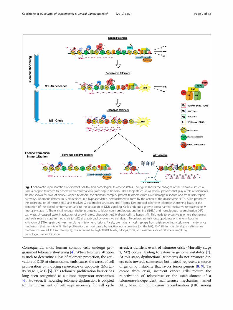
are interdependent, since telomere shortening induces telo-mere deprotection and chromosome instability (see Fig. 1for a schematic description). In most eukaryotes,end-erosion is counteracted by the action of the ribonu-cleoproteic enzyme telomerase, which adds short repeats tothe 3′ ends of chromosomes, the telomeres [3]. In humans,telomerase is active only in germinal and in stem cells.
* Correspondence: [email protected]; [email protected] of Biology and Biotechnology “Charles Darwin”, SapienzaUniversity of Roma, Piazzale Aldo Moro 5, 00185 Rome, Italy2Oncogenomic and Epigenetic Unit, IRCCS-Regina Elena National CancerInstitute, Via Elio Chianesi 53, 00144 Rome, Italy
© The Author(s). 2019 Open Access This article is distributed under the terms of the Creative Commons Attribution 4.0International License (http://creativecommons.org/licenses/by/4.0/), which permits unrestricted use, distribution, andreproduction in any medium, provided you give appropriate credit to the original author(s) and the source, provide a link tothe Creative Commons license, and indicate if changes were made. The Creative Commons Public Domain Dedication waiver(http://creativecommons.org/publicdomain/zero/1.0/) applies to the data made available in this article, unless otherwise stated.
Cacchione et al. Journal of Experimental & Clinical Cancer Research (2019) 38:21 https://doi.org/10.1186/s13046-019-1030-5
Consequently, most human somatic cells undergo pro-grammed telomere shortening [4]. When telomere attritionis such to determine a loss of telomere protection, the acti-vation of DDR at chromosome ends causes the arrest of cellproliferation by inducing senescence or apoptosis (Mortal-ity stage 1, M1) [5]. This telomere proliferation barrier haslong been recognized as a tumor suppressor mechanism[6]. However, if mounting telomere dysfunction is coupledto the impairment of pathways necessary for cell cycle
arrest, a transient event of telomere crisis (Mortality stage2, M2) occurs, leading to extensive genome instability [7].At this stage, dysfunctional telomeres do not anymore dir-ect cells towards senescence but instead represent a sourceof genomic instability that favors tumorigenesis [8, 9]. Toescape from crisis, incipient cancer cells require there-activation of telomerase or the establishment of atelomerase-independent maintenance mechanism namedALT, based on homologous recombination (HR) among
Fig. 1 Schematic representation of different healthy and pathological telomeric states. The figure shows the changes of the telomere structurefrom a capped telomere to neoplastic transformations (from top to bottom). The t-loop structure, as several proteins that play a role at telomeres,are not shown for sake of clarity. Capped telomere: the shelterin complex protect telomeres from DNA damage response and from DNA repairpathways. Telomeric chromatin is maintained in a hypoacetylated, heterochromatic form by the action of the deacetylase SIRT6, ATRX promotesthe incorporation of histone H3.3 and resolves G-quadruplex structures and R-loops. Deprotected telomere: telomere shortening leads to thedisruption of the closed conformation and to the activation of DDR signaling. Cells undergo a growth arrest named replicative senescence or M1(mortality stage 1). There is still enough shelterin proteins to block non-homologous end joining (NHEJ) and homologous recombination (HR)pathways. Uncapped state: Inactivation of growth arrest checkpoint (p53) allows cells to bypass M1. This leads to excessive telomere shortening,until cells reach a state termed crisis (or M2) characterized by extensive cell death. Telomeres are fully uncapped, loss of shelterin leads toactivation of DNA repair pathways, resulting in telomeric fusions. Rarely, premalignant cells escape from crisis acquiring a telomere maintenancemechanism that permits unlimited proliferation. In most cases, by reactivating telomerase (on the left); 10–15% tumors develop an alternativemechanism named ALT (on the right), characterized by high TERRA levels, R-loops, DDR, and maintenance of telomere length byhomologous recombination
Cacchione et al. Journal of Experimental & Clinical Cancer Research (2019) 38:21 Page 2 of 12
telomeres [10]. Telomere maintenance confers unlimitedproliferative potential to pre-neoplastic cells, allowingalso the stabilization of a heavily rearranged genomethat has acquired new and potentially tumorigenicgenetic mutations. In most cancers immortalizationderives from telomerase reactivation [11]; theremaining 10–15% of tumors are telomerase-negativeand utilize the ALT mechanism of telomere mainten-ance [12, 13]. Reactivation of telomere maintenanceprograms also enables the transmission of abnormalchromosomal structures (i.e., amplifications, deletions,translocations, inversions) that arise as a result of it-erative chromosomal breakage-fusion bridge cycles[7].Given the crucial role telomeres play in cancer devel-
opment, studying the mechanisms of telomere protec-tion and the changes in telomere structure duringtumorigenesis is essential to understand the biology ofcancer and develop effective therapeutic strategies. Herewe review the modifications of the structure and the epi-genetic state of telomere chromatin that occur uponcancer establishment, with a particular emphasis on therole of H3.3 mutations in pediatric ALT tumors and ontelomere dysfunctions derived by altered expression ofsirtuin deacetylases.
Structure of human telomeric chromatinShelterin complexes bind telomeric DNA as independentunits [14], in a chromatin environment characterized byan atypical nucleosomal organization (see ref. [15] for areview on the argument). Telomeric nucleosomes inhuman cells have a repeat length of 160 bp, about 40 bpshorter than in the rest of chromatin [16]. Moreover, invitro studies showed that telomeric nucleosomes are lessstable than average nucleosomes [17] and can slide alongtelomeric DNA [18]. The telomeric nucleosomalorganization seems to persist until the very end of thechromosome [19], limiting and affecting shelterin accessto telomeric DNA. Furthermore, studies on mouse celllines show that shelterin removal has no effect on thenucleosomal organization at telomeres [19, 20]. Theseresults suggest that shelterin and the other proteins in-volved in telomere function have to interplay with astable nucleosomal scaffold and not with naked DNA.Kinetic studies showed that nucleosomes have a verylow turnover [21], whereas the proteins that composethe shelterin complex have a very rapid exchange at telo-meres [22], mainly by 3D diffusive search of telomericsequences [14]. Telomerase also accesses telomeres inS-phase with high frequency [23]. In vitro studiesshowed that the presence of nucleosomes modulatesbinding of TRF1 and TRF2 to telomeric double-strandedrepeats [24, 25], indicating that TRF1 has a much higheraffinity than TRF2 both to nucleosomal binding sites
and to linker DNA. Other studies suggest that TRF2 caninduce compaction of telomeric chromatin [26] and thatTRF2 overexpression can alter nucleosomal spacing in acancer cell line [27].Whether nucleosomal organization plays a role in
human telomere protection is still an open matter.Recently, it was proposed that access of DDR factors todeprotected telomeres might depend on decompactionof telomeric chromatin upon loss of TRF1 and TRF2[28]. Contrary to these findings, other recent works sug-gest that DDR response at telomeres as a consequenceof shelterin depletion does not significantly change telo-mere compaction and accessibility [29, 30]. Mammaliantelomeric chromatin is generally considered heterochro-matic [31–33], enriched in heterochromatic marks suchas trimethylation of Lys9 of histone H3 (H3K9me3) andLys20 of histone H4 (H4K20me3) (Fig. 1). However, thisconcept is based mainly on data obtained on mouse telo-meres [34]. The epigenetic state at human telomeres isless typically heterochromatic [15]. ChIP and ChIP-seqexperiments show unexpected low levels of H3K9me3 attelomeres in human fibroblasts [35], in human CD4CT-cells [36], and in nine human cell lines of differentorigin [37, 38]. Clear heterochromatic marks such asH3K9me3 and DNA hypermethylation characterizeinstead subtelomeric regions [36, 38]. However, otherdirect and indirect evidences support the importance ofa heterochromatic state for healthy human telomeres.Specifically, hypoacetylation of lysines 9 and 56 of his-tone H3 – a typical heterochromatic pattern - is essen-tial for a correct telomere capping [39, 40]. In addition,the heterochromatin protein HP1-γ interacts with theshelterin protein TIN2 and is required for telomere co-hesion during S-phase [41]. Another peculiar feature oftelomeric chromatin is the enrichment for the H3 his-tone variant H3.3 [42]. H3.3 is expressed throughout thecell cycle by two genes, H3F3A and H3F3B, located onchromosomes 1 and 17, respectively. Enrichment forH3.3 was first found within actively transcribed genes,via a replication-independent deposition mechanism cat-alyzed by the histone chaperone Histone Regulator A(HIRA) [42, 43]. More recent studies showed that his-tone H3.3 is also incorporated in telomeres by a com-plex comprising the α-thalassemia/mental retardationsyndrome X-linked protein (ATRX) in cooperation withthe histone chaperone death domain-associated protein6 (DAXX) [42, 44, 45], also involved in H3.3 depositionat imprinted genes and interstitial heterochromatic sites[46]. The HIRA complex and the ATRX-DAXX complexcontrol replication-independent deposition of H3.3 atdistinct sites on the genome [42, 45]. These specificdeposition mechanisms indicate that H3.3 has multipleand distinct functions. The role played by H3.3 in telo-mere homeostasis is still unknown.
Cacchione et al. Journal of Experimental & Clinical Cancer Research (2019) 38:21 Page 3 of 12

However, heterochromatin formation does not impedethat telomeres are actively transcribed to generate longnon-coding UUAGGG-repeated RNAs named TERRA(telomeric repeat–containing RNA) [47, 48]. Even if themechanisms of TERRA functions have to be fully eluci-dated, it is now commonly recognized that TERRAs areimplicated in important telomere functions [49], includ-ing telomere homeostasis [50], and telomere protection[51, 52]. Importantly, several evidences show thatTERRA interacts with TRF1 and TRF2 and is involvedin heterochromatin formation [53]. Moreover, it hasbeen shown that TERRA interacts with heterochromatinprotein 1 (HP1) and with telomeric chromatin contain-ing H3K9me3 [53–55]. Upon TRF2 depletion, TERRAtranscription is upregulated and TERRA interacts withthe histone methyltransferase SUV39H1, promotingmethylation of histone H3K9 [56].
Shelterin alterations and cancerSeveral mutations and/or altered expression in shelterincomponents at telomeres have been described in cancer,but how these components are regulated during differ-ent stages of cancer development is not well understood.Patients with early-stage chronic lymphocytic leukemia(CLL) have an increased frequency of dysfunctional telo-meres and telomere-to-telomere fusions are observed inadvanced stages of the disease [57, 58]. In agreementwith a role of telomere-dysfunction in CLL, reducedexpression levels of TRF1, RAP1 and POT1 [59], as wellas TIN2 and TPP1 [58] have been detected. Further-more, somatic mutations in POT1 account for 5% ofCLL cases [60]. Of note, in addition to leukemia, muta-tions in POT1 or RAP1 have been found to be mainlyassociated with familial melanoma [61, 62], familial gli-oma [63], Li-Fraumeni-like syndrome [64], mantle celllymphoma [65] and parathyroid adenoma [66]. Themalignant-predisposing mutations in the POT1 gene,which alter the ability of the shelterin protein to bind tosingle-stranded telomeric DNA, lead to the fusion of sis-ter telomeres and are associated to increased telomerelength, owing to the loss of POT1-mediated inhibition oftelomerase [67]. These findings provide novel insightsinto how genomic instability induced by dysfunctionaltelomeres contributes to tumorigenesis. On one side,POT1 inhibition may result in defective telomere repli-cation caused by impaired CST (CTC1-STN1-TEN1)function at telomeres, thus promoting a telomere-drivengenome instability [68]. On the other, the presence oflonger telomeres may reduce the tumor-suppressiveeffects of telomere attrition as consequence of a delayedsenescence onset in precancerous dividing cells.Additionally, POT1 and RAP1 expression appearedderegulated in hepatocellular carcinoma (HCC) [69].Finally, TRF1 and TRF2 were reported to be
up-regulated in several cancer types such as lung,gastric, breast, colon and renal tumors [70–74]. The roleof the shelterin gene mutations in cancer rely mainly onthe perturbation of their telomere-related activitiesimpacting on telomere integrity. However, the putativeroles of TRF2 in tumorigenesis, as well as of RAP1, havebeen ascribed also to extra-telomeric functions. Bycombining chromatin immunoprecipitation withhigh-throughput DNA sequencing (ChIP-Seq), it hasbeen shown that TRF2 and RAP1 occupy both telomericand extratelomeric TTAGGG repeats throughout thehuman genome, referred to as interstitial telomericsequences (ITSs), where they can affect gene transcrip-tion [75–77]. Specifically, RAP1 associates to both subte-lomeric related genes and genes linked to metabolicregulation, cell adhesion, and cancer [75]. Additionally,RAP1 can translocate to the cytoplasm, where it acts asa modulator of the NF-kB signaling pathway by interact-ing with IKK complex. The RAP1-IKK interaction is re-quired for the phosphorylation of the p65 subunit ofNF-kB, enabling it to perform gene transcriptional acti-vation [78]. By binding ITSs, TRF2 modulates theHS3ST4 gene, encoding heparan sulfate (glucosamine)3-O-sulphotransferase 4, which is involved in regulatingNK cell recruitment/activation at the tumor site with animpact on tumor take/growth [79]. By localizing directlyto specific promoter regions, TRF2 regulates the expres-sion of the platelet-derived growth factor receptor-β(PDGFRβ; [80]), thus promoting angiogenesis; further-more, TRF2 represses the cyclin-dependent kinase p21(CDKN1A/CIP1/WAF1) through the REST-LSD1 re-pressor complex recruitment [81].Collectively, these findings implicate that an altered
expression of shelterin genes, besides impacting on telo-mere homeostasis, may have substantial consequenceson extra-telomeric loci, thus integrating telomeric chro-matin alterations with aberrant gene transcription pro-files. Consistently, looping of telomeres to interstitialsites, referred to as interstitial t-loops, mediated throughTRF2 and lamin associations has been reported [82].More recently, Mukherjee et al. [83] have shown thatbinding of TRF2 at promoters about 60 Mbp fromchromosome ends depends on telomere length in hu-man cells. Promoter TRF2 occupancy was affected incells with elongated telomeres producing an alteredTRF2-mediated transcription of distal genes.
Epigenetic alterations of telomeric chromatin incancerIt is still not clear whether telomerase-positive cancercells are characterized by a specific epigenetic pattern.Roles for epigenetic regulation of telomere maintenancehave been reported in mouse. Knockout of various chro-matin remodeling factors (CRFs), such as histone
Cacchione et al. Journal of Experimental & Clinical Cancer Research (2019) 38:21 Page 4 of 12

methyltransferases SUV39H1/2, SUV4-20H1/2 result indefective telomere function, aberrantly increased telo-mere length, and chromosomal instability (see ref. [84]for a review). In humans, SIRT1 and SIRT6, both mem-bers of the mammalian sirtuin family of Nad +−dependent histone deacetylases, are among the mostextensively studied CRFs interacting withtelomere-repeats implicated in telomere integrity [39,85–90]. Specific epigenetic changes have been associatedwith ALT cancers, such as the increase of TERRA tran-scription and enrichment of heterochromatic marks[52]. Importantly, high frequency of H3.3 point muta-tions and/or ATRX/DAXX mutations have beenassociated with pediatric cancers [91–94] and with theestablishment of a ALT mechanism of telomere main-tenance [95].
SirtuinsDeacetylation activity of SIRT1 is directed against bothhistone and non-histone targets, implying the involve-ment of SIRT1 in several cellular functions includingenergy metabolism, cellular stress resistance, genomicstability, aging and tumorigenesis (reviewed in [96]).SIRT1 was firstly demonstrated to be recruited to telo-meres in murine pluripotent stem cells (iPSCs) and topositively regulate telomere length in both mouseembryonic fibroblasts and tissues [86]. Chen et al. [88]have reported that SIRT1-silencing causes nuclearabnormalities, telomere dysfunction induced foci and in-duced cellular senescence in HCC cells by inhibiting theshelterin TPP1 expression. Indeed, up-regulated expres-sion of TPP1 in SIRT1-depleted HCC cells improvedcellular senescence, strongly suggesting that TPP1 wasclosely involved in the SIRT1-mediated anti-senescenceeffects in HCC cells [88]. Another study showed thatSIRT1 is necessary for telomere elongation after repro-gramming of murine and human somatic cells, and it isrequired to maintain genomic stability, telomeric tran-scription and remodeling of telomeric chromatin [90].SIRT6 is a complex enzyme with multiple substrates
and catalytic activities, as deacetylation of both histonesand non-histone proteins, deacetylation of long-chainfatty acyl groups and mono-ADP-ribosylation activity[97]. At chromatin level, SIRT6 deacetylates the histoneH3 on acetylated K9, K56 [39, 98] and the more recentlyidentified K18 and K27 residues [98–100], causing therepression of many genes differently involved in inflam-mation, aging, genome stability, metabolic pathways andtelomere integrity [101, 102]. Upon DNA damage, SIRT6is recruited to double strand breaks (DSBs) ensuring theproper activation of downstream DDR factors leading toan efficient repair [87]. In 2008, Michishita et al. [39]showed that SIRT6-mediated deacetylation of histoneH3 on acetylated lysine 9 (H3K9ac) modulated telomeric
chromatin structure. Specifically, SIRT6 can localize tothe telomeric chromatin and its loss leads to the dys-function of telomeres resembling a phenotype of telo-mere abnormality similar to that of Werner syndrome[39, 40, 98], with chromosome end fusions and cellularsenescence. The Werner syndrome ATP-dependent heli-case (WRN) is a well-known RecQ-like helicase thatplays a major role in genome stability, particularlyduring DNA replication and telomere metabolism [103].In detail, SIRT6 deacetylates H3K9 at telomeric chroma-tin and is required for the stable association of WRN.Additionally, SIRT6 is required for proper replication oftelomeres by deacetylating H3K9 and H3K56 duringS-phase [40]. Thus, depletion of SIRT6 from humancells resulted in abnormal telomere structures and sto-chastic replication-associated telomere sequence loss, ul-timately leading to chromosomal end-to-end fusions andconsequent genomic instability [87]. A very recent paperattributes to SIRT6 the ability to facilitate directionaltelomere movement upon oxidative damage by recruit-ing SNF2H (an ATP-dependent chromatin-remodelingfactor) with resulting local chromatin decondensation attelomeres [104]. Another important function of SIRT6 attelomeres is the ability to prevent impaired telomereposition effect (TPE), the epigenetic silencing oftelomere-proximal genes [87]. Indeed, RNAi-mediated de-pletion of SIRT6 abrogated silencing of both an integratedtelomeric transgene and an endogenous telomere-prox-imal gene. Moreover, enhanced telomeric silencing in re-sponse to telomere elongation is associated with increasedrepressive chromatin marks, and this heterochromatic mi-lieu is lost in SIRT6-deficient cells. These findings may berelevant in suggesting an additional mechanism by whichtelomeric chromatin may contribute to tumorigenesis.Since aberrant expression of silent chromatin has been in-creasingly recognized to have a role in cancer [105], itwould be interesting to understand if telomere erosion, aswell as SIRT6 inhibition —and consequent de-repressionof telomere-proximal genes—may impact oncancer-related changes in gene expression [106, 107].Interestingly, in line with this notion, recently pub-lished data suggest that histone modifications typicalof chromatin compaction (H3K27me3) or access(H3K4me1 and H3K4me3) to regulatory factors, atsites distant from telomere ends depend on telomerelength [83]. Moreover, loss of silencing factors fromshortening and/or dysfunctional telomeres might leadto a relocalization of these factors from chromosomeends to other genomic loci, triggering aberrant silen-cing of non-telomeric genes [108].The role of SIRT6 in cancer is controversial. In some
tumors, high levels of SIRT6 are associated with pooreroutcomes [109, 110]. In other tumors, including colorec-tal cancer (CRC), SIRT6 functions are associated with its
Cacchione et al. Journal of Experimental & Clinical Cancer Research (2019) 38:21 Page 5 of 12

tumor suppressive activity [111–113]. Of note, the telo-meric protein TRF2 has been newly identified as a novelsubstrate of SIRT6. Upon exposure to a DNA damagingagent, SIRT6-dependent lysine deacetylation of TRF2leads to the ubiquitin-dependent proteolysis of the shel-terin protein, resulting in downstream proper activationof DDR machinery [114]. An inverse correlation betweenSIRT6 and TRF2 protein expression levels have beenalso found in a cohort of CRC patients [114], suggestingthat an impairment of TRF2 degradation, as a conse-quence of SIRT6 loss, could be one of the mechanismsunderlying the increased dosages of TRF2 observed inmany human malignancies. Whether SIRT6 could alsoimpact on the binding affinity to DNA of TRF2 (andeventually of other shelterin factors) through histonedeacetylation remains to be fully elucidated.
ATRX/DAXX mutations in ALT tumorsSeveral immortalized cell lines and 10–15% of tumorsare telomerase-negative and maintain functional telo-meres by utilizing an ALT mechanism (for a review, seerefs. [10, 115, 116]). ALT activity has been detectedprevalently in cancers from mesenchymal tissues such asbone, soft tissues, neuroendocrine systems, peripheraland central nervous systems [12, 117]. ALT cells showseveral unusual features, such as highly heterogeneoustelomere length [118]. Other markers for ALT includeabundant extra-chromosomal double-stranded telomericDNA prevalently in circular form (t-circles), partiallysingle-stranded telomeric C-rich circles (C-circles), hightelomere-specific DDR, telomere sister chromatidexchanges (tSCEs) and formation of APBs (ALT-asso-ciated promyelocytic leukemia (PML) nuclear bodies),containing chromosomal or extra-chromosomal telo-meric DNA, telomere-associated proteins, and proteinsinvolved in homologous recombination (reviewed in[10]). Several evidences indicate that ALT maintenanceis based on DNA recombination [10, 115]. For example,a DNA tag inserted in a single telomere was copied todifferent telomeres in human ALT cells, but not intelomerase-positive cells [119]. Since HR at telomeres isrepressed in normal cells and in telomerase-positiveimmortalized cells, ALT activation likely requires the in-activation of factors repressing HR. The protein ATRX(a chromatin remodeler of the SWI/SNF family) not onlydoes inhibit HR, but is also able to repress ALT activityif transiently expressed in ALT-positive/ATRX-negativecells [120]. ATRX also binds telomeric repeats andG-quadruplex structures in vitro [121], suggesting that itmight play a role in resolving G-quadruplex structuresforming at telomeres during replication, thus inhibitingreplication fork stalling. Through its ADD domain,ATRX interacts with H3K9me3 [122] and its localizationat telomeres is antagonized by TERRA [51]. TERRA also
plays a role in ALT that remains to be fully defined. InALT cancer cells, TERRA levels are higher than intelomerase-positive cancer cells and TERRA transcriptsconstitutively associate with telomeres [123]. Moreover,a recent finding shows that TERRA directs the enrich-ment of HP1, H3K9me3, H3K27me3, H4K20me3 in theALT cell line U2OS, through the recruitment of Poly-comb repressive complex 2 (PRC2) [52], typical of facul-tative heterochromatin. Importantly, at chromosomeends TERRA molecules form RNA-DNA hybrids(R-loops), three-stranded nucleic acid structures consist-ing of a DNA:RNA hybrid and a displaced DNA strand.The displaced G-rich DNA strand is thought to formG-quadruplex structures, which may cause stalling ofreplication and DNA damage at telomeres [124], thus in-creasing homologous recombination among telomeres[125]. Suppression of R-loop formation is one of themultiple functions of ATRX [124], consistent with itsALT suppressing role. However, the main role of ATRXis the deposition - together with the histone chaperoneDAXX - of the histone variant H3.3 at pericentricheterochromatic regions and at telomeres [42, 45]. Atthe moment, it is unknown which role H3.3 depositionat telomeres plays in the development of ALT pathway.However, the importance of the ALT/DAXX/H3.3 path-way is supported by recent surveys of ALT-positivetumors showing a high frequency of mutations inATRX/DAXX and/or H3.3 [92–95].
H3.3 mutations in pediatric tumorsRecent studies reported high frequencies of H3.3 muta-tions in pediatric cancers, often associated with ALT (fora review see [91, 126]). Three residues are involved,respectively Lys27, Gly34, and Lys36. MutatedH3.3K27M (from Lysine to Methionine) and H3.3G34R/V (from Glycine to Arginine or Valine) are frequent inpediatric high-grade glioma (pHGG) or in diffuse intrin-sic pontine gliomas (DIPG) [94, 127, 128]. Other twomutations, H3.3K36M and H3.3G34W/L (from Glycineto Tryptophan or Leucine), have been found at high fre-quency in two juvenile bone tumors, chondroblastomasand giant cell tumors of the bone (GCTBs) [129]. A raremutation, H3.3K27I (from Lysine to Isoleucine) has beenalso described in DIPG [130]; moreover, K27M mutationcan affect also the canonical histones H3.1 and H3.2[127, 128, 130]. Although both genes express the sameprotein product, mutations occur either in H3F3A or inH3F3B gene. Mutations regarding residues K27 and G34affect preferentially H3F3A gene, whereas K36M muta-tions occur mostly in H3F3B [91]. These missense muta-tions act in heterozygosis, indicating a “gain of function”role of the mutated histone in cancer development. Re-markably, mutant histones - termed as “oncohistones”[91] due to their dominant nature - are found in
Cacchione et al. Journal of Experimental & Clinical Cancer Research (2019) 38:21 Page 6 of 12
pediatric and juvenile tumors but rarely in their adultcounterparts. Another peculiar feature is that the ana-tomical location, the average age at diagnosis, and theoverall survival are highly mutation-specific [127, 128,131]. H3.3G34R/V cancers are found almost exclusivelyin the cerebral hemispheres, accounting for 16.2% oftotal cases, and show a longer overall survival comparedwith other H3.3 mutant groups (median 18 months).H3.1/H3.2 K27M are restricted to the pons (21.4%) andshow a median survival of 15 months. H3.3K27M muta-tions are abundant in the midline and pons, accountingfor 63.0% DIPG and 59.7% non-brainstem midline tu-mors. This group is characterized by a shorter overallsurvival (median 11 months). The reason for these speci-ficities and the molecular mechanisms at the basis ofoncohistones are mostly unknown. The amino acids thatare mutated in tumors are sites of possible methylationor acetylation (K27 and K36), or can interfere withpost-translational modifications of close lysines (G34).However, the most striking feature of oncohistones isthat they act globally, despite the fact that they areexpressed by a single allele. Pediatric glioblastomas har-boring H3.3K27M mutation show a global reduction ofH3K27me3 [132–134]; to a lesser extent, also K27Ireduces the global levels of H3K27me3 [132]. Trimethy-lation of H3K27 is a mark of facultative heterochroma-tin, catalyzed by PRC2 [135, 136]. In vitro analysis ofPRC2 methyltransferase activity and crystal structurestudies show that H3K27M inhibits K27 methylationthrough specific binding to EZH2, the enzymatic subunitof PRC2 [132, 137], leading to a general reprogrammingof H3K27me3 and EZH2 on the genome [138]. Recentdata suggests that in vivo H3K27M does not bind orsequester PRC2 but instead forms heterotypicH3K27M-K27 ac nucleosomes that interact with bromo-domain proteins [139]; in agreement with these results,a recent study shows no increased Ezh2 affinity fornucleosomes containing H3K27M [140].Similarly to H3K27M mutations, H3.3K36M expres-
sion in chondroblastoma correlates with global reduc-tion in H3K36 methylation [141], due to inhibition ofNSD2/MMSET, a methyltransferase that catalyzesmono- and di-methylation of H3K36, and SETD2, whichcatalyzes trimethylation of H3K36me2 [141, 142]. Analo-gously to H3K36M, it has been proposed thatH3.3K36M might act by sequestering NSD2 and SETD2;support to this hypothesis comes from the crystal struc-ture showing a strong binding of H3K36M to the cata-lytic site of SET2D [143, 144].The last H3 residue mutated in a subset of pediatric
cancers, H3.3G34, is not a site for post-translationalmodifications, but is in close proximity of H3K36.Indeed, structural analysis showed that H3.3G34R/V/Dmutations result in a steric hindrance to the catalytic
activity of SETD2 on H3K36 [145]. As a consequence,H3K36 methylation is inhibited also by mutations ofH3.3G34 [132, 146], but only in cis on the mutant nucleo-somes, whereas nucleosomes containing wild-type H3 arenot affected by the mutations [132, 146]. Very recently, ithas been shown that targeted G34R mutations on oneallele of H3f3a in mouse embryonic stem (ES) cellsresulted in a global epigenetic change [147], namely theinhibition of the KDM4 family of histone demethylases,which target H3 residues K27 and K36. Further analysesare necessary to assess the importance of KDM4 demethy-lases inhibition in H3.3G34R/V tumors.
Therapeutic strategiesTherapeutic strategies targeting chromatin modificationsare defined as epigenetic therapy. Currently, epigenetictherapy has been proven to be a successful approach forthe treatment of hematological malignancies, but littlesuccess has been achieved in the treatment of solidtumors (for a recent review see [148]). However, accu-mulating data on the role of epigenetic alterations occur-ring at telomeres of cancer cells provides an intriguingand challenging chance for potential targeted therapeuticinterventions.The essential dependence of cancer cells on a telo-
mere maintenance mechanism for replicativeimmortalization led researchers to investigate thesemechanisms as potential cancer-specific therapeutictargets. Given the majority of carcinomas and soft tis-sue cancers present telomerase activity, whereastelomerase is absent in most normal tissues [11, 149],several efforts have been made to inhibit telomeraseby pursuing different strategies: small-molecule inhibi-tors, antisense oligonucleotides, G-quadruplex stabi-lizers, immunotherapy, telomerase-driven suicide genetherapy, and chemicals blocking telomerase biogenesis(see ref. [150] for an extensive review). Unfortunately,anti-telomerase approaches have showed effectivenessin only some myeloid tumors but have largely failedin solid tumors (reviewed in [151]). The limitations oftargeting telomerase, and the fact that telomeraseinhibition would not affect cancer cells using the ALTpathway, encouraged researchers to investigate alter-native therapeutic approaches targeting telomeres in atelomerase- and telomere length-independent manner.In agreement with growing findings about the alteredtelomeric chromatin composition of cancer cells, andconsidering the pivotal role of shelterin componentsin telomere protection, targeting telomeric bindingfactors has been developing as an emerging antitumorapproach. Indeed, chemical inhibition of TRF2 [152]or TRF1 [153, 154] were reported to induce rapidDDR activation and growth arrest both in in vitroand in vivo tumor models, respectively.
Cacchione et al. Journal of Experimental & Clinical Cancer Research (2019) 38:21 Page 7 of 12

Until now, telomeric chromatin alterations in cancerhave not yet been considered in the design of effect-ive epigenetic therapy, however they can be indirectlytargeted by novel identified epigenetic drugs. Due tothe broad range of activities and substrates, Sirtuinsare involved in several cellular processes, includingtelomere integrity, but their role in cancer is contro-versial. These reasons led to the identification ofmany sirtuin modulators over recent years, both in-hibitors and activators, mainly through chemical li-brary screening and catalytic mechanism-based designapproaches (reviewed in [155]). Very recently, newchemical activators of SIRT6 have been identified. Ithas been shown that UBCS039 and MDL-800 are ableto inhibit the proliferation of various cell lines regard-less of tumor histotype [156–158]. Moreover,MDL-800 compound showed efficacy in a xenograftmodel of hepatocellular carcinoma [158]. Given thedescribed ability of SIRT6 to affect the protein stabil-ity of TRF2 [114], as well as telomere capping, it isreasonable to ask whether the antitumor activitiescaused by the exposure to SIRT6 activators can bepartially attributable to telomere-driven effects. Toaddress this issue, further studies will be needed.Importantly, there is mounting evidence showing that
epigenetic cancer therapy could target ALT-positivegliomas harboring H3.3 mutations [159]. Specifically,recent preclinical studies showed that GSKJ4, a smallmolecule inhibitor of the histone H3K27 demethylasesJMJD3 (KDM6B) and UTX (KDM6A), decreased tumorcell viability and increased H3K27me3 levels in gliomacell lines harboring the mutation of lysine to methioninesubstitution at codon 27 (K27M), and significantlyextended survival of mice with K27M mutant glioma xe-nografts [160]. In contrast, GSKJ4 has not shown activityin an H3.3G34V mutant glioma cell line [160]. Panobi-nostat, a histone deacetylase inhibitor, resulted in de-creased tumor cell viability in both K27M mutantglioma cell lines and in mice with K27M mutant gliomaxenografts [161, 162]. Panobinostat treatment demon-strated a dose dependent increase in histone acetylationand in H3K27me3 [161, 162]. Combined use of GSKJ4and panobinostat produced a synergistic reduction oftumor cell viability in K27M mutant glioma cell lines[161]. Other strategies to modulate histone methylationare under study, such as targeting EZH2, the histonedemethylases KDM1 and KDM5 (see refs. [91, 126, 163]for a review). Strategies that modulate DNA methylationat subtelomeres in ALT are expected to affect cell sur-vival of ALT cells. Additionally, inhibitors of the proteinkinase ATR, a regulator of homologous recombinationwith prolonged recruitment to telomere ends in the set-ting of ATRX mutation, have been found to selectivelyinduce death of ALT-positive cancer cells [123].
ConclusionsTelomeres and telomerase have become a main target indeveloping anticancer strategies, due to their crucial rolein cancer development. Many efforts have been focusedon telomerase inhibition, however this strategy hastherapeutic limits. New anticancer targets could emergefrom a clearer comprehension of telomere structure anddynamics. Several aspects of telomere biology need adeeper investigation: the epigenetic pattern of humantelomeres is still controversial [38], the role played bythe histone H3.3 at telomeres is largely unknown, howtelomeric chromatin changes during neoplastic trans-formation is an issue mostly unexplored.Effective anticancer strategies require an accurate
mapping of the mutations causing the disease, with theultimate goal to precisely tailor the therapy to thepatient. Besides genetic mutations, it is now generallyrecognized that epigenetic changes play an importantrole in cancer development [164, 165]. Even if stillpoorly defined, strategies directed against epigenetic tar-gets have features that can potentially complement clas-sical anticancer approaches, like the possibility toaddress different pathways at the same time. Character-izing the telomeric epigenome is therefore an importantissue, both for a deeper understanding of the telomereprotective structure and because it might lead to theemergence of new anti-cancer targets.
AbbreviationsALT: Alternative lengthening of telomeres; APB: ALT-associated promyelociticleukemia (PML) nuclear body; ATRX: α-thalassemia/mental retardationsyndrome X-linked; ChIP-seq: Chromatin immunoprecipitation followed bynext-generation sequencing; CLL: Chronic lymphocytic leukemia;CRF: Chromatin remodeling factors; DAXX: Death domain-associated protein6; DDR: DNA damage response; DIPG: Diffuse intrinsic pontine glioma;DSB: Double-strand DNA break; GCTB: Giant cell tumors of the bone;HCC: Hepatocellular carcinoma; HP1: Heterochromatin protein 1;HR: Homologous recombination; iPSC: Induced pluripotent stem cells;ITS: Interstitial telomeric sequences; NHEJ: Non-homologous end-joining;PDGFRβ: Platelet-derived growth factor receptor-β; pHGG: Pediatric high-grade glioma; PML: Promyelocytic leukaemia; TERRA: Telomeric repeat-containing RNA; TPE: Telomere-position effect; TRF1: Telomeric repeat-binding factor 1; TRF2: Telomeric repeat-binding factor 2; tSCE: Telomeresister chromatid exchange; WRN: Werner syndrome ATP-dependent helicase
AcknowledgementsThanks are due to Emanuela Micheli for critical reading and help fith thefigure.
FundingThis review article was supported by the Italian Association for CancerResearch (AIRC, #16910 A.B.) and by Fondi di Ateneo 2016, 2017 (S.C.)
Availability of data and materialsNot applicable.
Authors’ contributionsAll the authors contributed for the preparation of this manuscript. S.C. andA.R. wrote the review article, were responsible for the figure and legend, finalediting, and preparation of the manuscript for submission. A.B. contributedto defining the topic, analyzing article and drafting the first copy.
Cacchione et al. Journal of Experimental & Clinical Cancer Research (2019) 38:21 Page 8 of 12

Ethics approval and consent to participateNot applicable.
Consent for publicationNot applicable.
Competing interestsAll the authors declare that there are not any competing financial interestsin relation to this work.
Publisher’s NoteSpringer Nature remains neutral with regard to jurisdictional claims inpublished maps and institutional affiliations.
Received: 22 November 2018 Accepted: 7 January 2019
References1. de Lange T. Shelterin-mediated telomere protection. Annu Rev Genet. 2018;
52:223–47.2. Doksani Y, Wu JY, de Lange T, Zhuang X. Super-resolution fluorescence
imaging of telomeres reveals TRF2-dependent T-loop formation. Cell. 2013;155(2):345–56.
3. Blackburn EH. Telomerases. Annu Rev Biochem. 1992;61:113–29.4. Harley CB, Futcher AB, Greider CW. Telomeres shorten during ageing of
human fibroblasts. Nature. 1990;345(6274):458–60.5. von Zglinicki T, Saretzki G, Ladhoff J, d'Adda di Fagagna F, Jackson SP.
Human cell senescence as a DNA damage response. Mech Ageing Dev.2005;126(1):111–7.
6. Campisi J. Cellular senescence as a tumor-suppressor mechanism. TrendsCell Biol. 2001;11(11):S27–31.
7. Maciejowski J, de Lange T. Telomeres in cancer: tumour suppression andgenome instability. Nat Rev Mol Cell Biol. 2017;18(3):175–86.
8. Blackburn EH, Epel ES, Lin J. Human telomere biology: a contributory andinteractive factor in aging, disease risks, and protection. Science. 2015;350(6265):1193–8.
9. Robinson NJ, Schiemann WP. Means to the ends: the role of telomeres andtelomere processing machinery in metastasis. Biochim Biophys Acta. 2016;1866(2):320–9.
10. Cesare AJ, Reddel RR. Alternative lengthening of telomeres: models,mechanisms and implications. Nat Rev Genet. 2010;11(5):319–30.
11. Shay JW, Bacchetti S. A survey of telomerase activity in human cancer. Eur JCancer. 1997;33(5):787–91.
12. Dilley RL, Greenberg RA. ALTernative telomere maintenance and Cancer.Trends Cancer. 2015;1(2):145–56.
13. De Vitis M, Berardinelli F, Sgura A. Telomere Length Maintenance in Cancer:At the Crossroad between Telomerase and Alternative Lengthening ofTelomeres (ALT). Int J Mol Sci. 2018;19(2):606.
14. Erdel F, Kratz K, Willcox S, Griffith JD, Greene EC, de Lange T. Telomererecognition and assembly mechanism of mammalian Shelterin. Cell Rep.2017;18(1):41–53.
15. Galati A, Micheli E, Cacchione S. Chromatin structure in telomere dynamics.Front Oncol. 2013;3:46.
16. Tommerup H, Dousmanis A, de Lange T. Unusual chromatin in humantelomeres. Mol Cell Biol. 1994;14(9):5777–85.
17. Filesi I, Cacchione S, De Santis P, Rossetti L, Savino M. The main role of thesequence-dependent DNA elasticity in determining the free energy ofnucleosome formation on telomeric DNAs. Biophys Chem. 2000;83(3):223–37.
18. Pisano S, Marchioni E, Galati A, Mechelli R, Savino M, Cacchione S. Telomericnucleosomes are intrinsically mobile. J Mol Biol. 2007;369(5):1153–62.
19. Wu P, de Lange T. No overt nucleosome eviction at deprotected telomeres.Mol Cell Biol. 2008;28(18):5724–35.
20. Sfeir A, de Lange T. Removal of shelterin reveals the telomere end-protection problem. Science. 2012;336(6081):593–7.
21. Phair RD, Scaffidi P, Elbi C, Vecerova J, Dey A, Ozato K, Brown DT, Hager G,Bustin M, Misteli T. Global nature of dynamic protein-chromatin interactionsin vivo: three-dimensional genome scanning and dynamic interactionnetworks of chromatin proteins. Mol Cell Biol. 2004;24(14):6393–402.
22. Mattern KA, Swiggers SJ, Nigg AL, Lowenberg B, Houtsmuller AB, ZijlmansJM. Dynamics of protein binding to telomeres in living cells: implications fortelomere structure and function. Mol Cell Biol. 2004;24(12):5587–94.
23. Schmidt JC, Zaug AJ, Cech TR. Live cell imaging reveals the dynamics oftelomerase recruitment to telomeres. Cell. 2016;166(5):1188–97 e1189.
24. Galati A, Micheli E, Alicata C, Ingegnere T, Cicconi A, Pusch MC, Giraud-PanisMJ, Gilson E, Cacchione S. TRF1 and TRF2 binding to telomeres is modulatedby nucleosomal organization. Nucleic Acids Res. 2015;43(12):5824–37.
25. Galati A, Rossetti L, Pisano S, Chapman L, Rhodes D, Savino M, Cacchione S.The human telomeric protein TRF1 specifically recognizes nucleosomalbinding sites and alters nucleosome structure. J Mol Biol. 2006;360(2):377–85.
26. Baker AM, Fu Q, Hayward W, Victoria S, Pedroso IM, Lindsay SM, FletcherTM. The telomere binding protein TRF2 induces chromatin compaction.PLoS One. 2011;6(4):e19124.
27. Galati A, Magdinier F, Colasanti V, Bauwens S, Pinte S, Ricordy R, Giraud-Panis MJ, Pusch MC, Savino M, Cacchione S, et al. TRF2 controls telomericnucleosome organization in a cell cycle phase-dependent manner. PLoSOne. 2012;7(4):e34386.
28. Bandaria JN, Qin P, Berk V, Chu S, Yildiz A. Shelterin protects chromosomeends by compacting Telomeric chromatin. Cell. 2016;164(4):735–46.
29. Timashev LA, Babcock H, Zhuang X, de Lange T. The DDR at telomereslacking intact shelterin does not require substantial chromatindecompaction. Genes Dev. 2017;31(6):578–89.
30. Vancevska A, Douglass KM, Pfeiffer V, Manley S, Lingner J. The telomericDNA damage response occurs in the absence of chromatin decompaction.Genes Dev. 2017;31(6):567–77.
31. Tardat M, Dejardin J. Telomere chromatin establishment and its maintenanceduring mammalian development. Chromosoma. 2018;127(1):3–18.
32. Janssen A, Colmenares SU, Karpen GH. Heterochromatin: Guardian of thegenome. Annu Rev Cell Dev Biol. 2018;34:265–88.
33. Schoeftner S, Blasco MA. A 'higher order' of telomere regulation: telomereheterochromatin and telomeric RNAs. EMBO J. 2009;28(16):2323–36.
34. Schoeftner S, Blasco MA. Chromatin regulation and non-coding RNAs atmammalian telomeres. Semin Cell Dev Biol. 2010;21(2):186–93.
35. O'Sullivan RJ, Kubicek S, Schreiber SL, Karlseder J. Reduced histonebiosynthesis and chromatin changes arising from a damage signal attelomeres. Nat Struct Mol Biol. 2010;17(10):1218–25.
36. Rosenfeld JA, Wang Z, Schones DE, Zhao K, DeSalle R, Zhang MQ.Determination of enriched histone modifications in non-genic portions ofthe human genome. BMC Genomics. 2009;10:143.
37. Ernst J, Kheradpour P, Mikkelsen TS, Shoresh N, Ward LD, Epstein CB, ZhangX, Wang L, Issner R, Coyne M, et al. Mapping and analysis of chromatinstate dynamics in nine human cell types. Nature. 2011;473(7345):43–9.
38. Cubiles MD, Barroso S, Vaquero-Sedas MI, Enguix A, Aguilera A, Vega-PalasMA. Epigenetic features of human telomeres. Nucleic Acids Res. 2018;46(5):2347–55.
39. Michishita E, McCord RA, Berber E, Kioi M, Padilla-Nash H, Damian M, CheungP, Kusumoto R, Kawahara TL, Barrett JC, et al. SIRT6 is a histone H3 lysine 9deacetylase that modulates telomeric chromatin. Nature. 2008;452(7186):492–6.
40. Michishita E, McCord RA, Boxer LD, Barber MF, Hong T, Gozani O, Chua KF.Cell cycle-dependent deacetylation of telomeric histone H3 lysine K56 byhuman SIRT6. Cell Cycle. 2009;8(16):2664–6.
41. Canudas S, Houghtaling BR, Bhanot M, Sasa G, Savage SA, Bertuch AA,Smith S. A role for heterochromatin protein 1gamma at human telomeres.Genes Dev. 2011;25(17):1807–19.
42. Goldberg AD, Banaszynski LA, Noh KM, Lewis PW, Elsaesser SJ, Stadler S,Dewell S, Law M, Guo X, Li X, et al. Distinct factors control histone variantH3.3 localization at specific genomic regions. Cell. 2010;140(5):678–91.
43. Tagami H, Ray-Gallet D, Almouzni G, Nakatani Y. Histone H3.1 and H3.3complexes mediate nucleosome assembly pathways dependent orindependent of DNA synthesis. Cell. 2004;116(1):51–61.
44. Drane P, Ouararhni K, Depaux A, Shuaib M, Hamiche A. The death-associated protein DAXX is a novel histone chaperone involved in thereplication-independent deposition of H3.3. Genes Dev. 2010;24(12):1253–65.
45. Lewis PW, Elsaesser SJ, Noh KM, Stadler SC, Allis CD. Daxx is an H3.3-specific histone chaperone and cooperates with ATRX in replication-independent chromatin assembly at telomeres. Proc Natl Acad Sci U SA. 2010;107(32):14075–80.
46. Voon HP, Hughes JR, Rode C, De La Rosa-Velazquez IA, Jenuwein T, Feil R,Higgs DR, Gibbons RJ. ATRX plays a key role in maintaining silencing at interstitialheterochromatic loci and imprinted genes. Cell Rep. 2015;11(3):405–18.
47. Azzalin CM, Reichenbach P, Khoriauli L, Giulotto E, Lingner J. Telomericrepeat containing RNA and RNA surveillance factors at mammalianchromosome ends. Science. 2007;318(5851):798–801.
Cacchione et al. Journal of Experimental & Clinical Cancer Research (2019) 38:21 Page 9 of 12

48. Schoeftner S, Blasco MA. Developmentally regulated transcription ofmammalian telomeres by DNA-dependent RNA polymerase II. Nat Cell Biol.2008;10(2):228–36.
49. Cusanelli E, Chartrand P. Telomeric repeat-containing RNA TERRA: a noncodingRNA connecting telomere biology to genome integrity. Front Genet. 2015;6:143.
50. Flynn RL, Centore RC, O'Sullivan RJ, Rai R, Tse A, Songyang Z, Chang S,Karlseder J, Zou L. TERRA and hnRNPA1 orchestrate an RPA-to-POT1 switchon telomeric single-stranded DNA. Nature. 2011;471(7339):532–6.
51. Chu HP, Cifuentes-Rojas C, Kesner B, Aeby E, Lee HG, Wei C, Oh HJ, BoukhaliM, Haas W, Lee JT. TERRA RNA antagonizes ATRX and protects telomeres.Cell. 2017;170(1):86–101 e116.
52. Montero JJ, Lopez-Silanes I, Megias D, F Fraga M, Castells-Garcia A, BlascoMA. TERRA recruitment of polycomb to telomeres is essential for histonetrymethylation marks at telomeric heterochromatin. Nat Commun. 2018;9(1):1548.
53. Deng Z, Norseen J, Wiedmer A, Riethman H, Lieberman PM. TERRA RNAbinding to TRF2 facilitates heterochromatin formation and ORC recruitmentat telomeres. Mol Cell. 2009;35(4):403–13.
54. Arnoult N, Van Beneden A, Decottignies A. Telomere length regulatesTERRA levels through increased trimethylation of telomeric H3K9 andHP1alpha. Nat Struct Mol Biol. 2012;19(9):948–56.
55. Episkopou H, Draskovic I, Van Beneden A, Tilman G, Mattiussi M, Gobin M,Arnoult N, Londono-Vallejo A, Decottignies A. Alternative lengthening oftelomeres is characterized by reduced compaction of telomeric chromatin.Nucleic Acids Res. 2014;42(7):4391–405.
56. Porro A, Feuerhahn S, Delafontaine J, Riethman H, Rougemont J, Lingner J.Functional characterization of the TERRA transcriptome at damagedtelomeres. Nat Commun. 2014;5:5379.
57. Lin TT, Letsolo BT, Jones RE, Rowson J, Pratt G, Hewamana S, Fegan C,Pepper C, Baird DM. Telomere dysfunction and fusion during theprogression of chronic lymphocytic leukemia: evidence for a telomere crisis.Blood. 2010;116(11):1899–907.
58. Augereau A, T'Kint de Roodenbeke C, Simonet T, Bauwens S, Horard B,Callanan M, Leroux D, Jallades L, Salles G, Gilson E, et al. Telomeric damagein early stage of chronic lymphocytic leukemia correlates with shelterindysregulation. Blood. 2011;118(5):1316–22.
59. Poncet D, Belleville A, T'kint de Roodenbeke C, Roborel de Climens A, BenSimon E, Merle-Beral H, Callet-Bauchu E, Salles G, Sabatier L, Delic J, et al.Changes in the expression of telomere maintenance genes suggest globaltelomere dysfunction in B-chronic lymphocytic leukemia. Blood. 2008;111(4):2388–91.
60. Ramsay AJ, Quesada V, Foronda M, Conde L, Martinez-Trillos A, Villamor N,Rodriguez D, Kwarciak A, Garabaya C, Gallardo M, et al. POT1 mutationscause telomere dysfunction in chronic lymphocytic leukemia. Nat Genet.2013;45(5):526–30.
61. Robles-Espinoza CD, Harland M, Ramsay AJ, Aoude LG, Quesada V, Ding Z,Pooley KA, Pritchard AL, Tiffen JC, Petljak M, et al. POT1 loss-of-functionvariants predispose to familial melanoma. Nat Genet. 2014;46(5):478–81.
62. Aoude LG, Pritchard AL, Robles-Espinoza CD, Wadt K, Harland M, Choi J,Gartside M, Quesada V, Johansson P, Palmer JM, et al. Nonsense mutationsin the shelterin complex genes ACD and TERF2IP in familial melanoma. JNatl Cancer Inst. 2015;107(2):dju408.
63. Bainbridge MN, Armstrong GN, Gramatges MM, Bertuch AA, Jhangiani SN,Doddapaneni H, Lewis L, Tombrello J, Tsavachidis S, Liu Y, et al. Germlinemutations in shelterin complex genes are associated with familial glioma. JNatl Cancer Inst. 2015;107(1):384.
64. Calvete O, Martinez P, Garcia-Pavia P, Benitez-Buelga C, Paumard-HernandezB, Fernandez V, Dominguez F, Salas C, Romero-Laorden N, Garcia-Donas J,et al. A mutation in the POT1 gene is responsible for cardiac angiosarcomain TP53-negative Li-Fraumeni-like families. Nat Commun. 2015;6:8383.
65. Zhang J, Jima D, Moffitt AB, Liu Q, Czader M, Hsi ED, Fedoriw Y, Dunphy CH,Richards KL, Gill JI, et al. The genomic landscape of mantle cell lymphoma isrelated to the epigenetically determined chromatin state of normal B cells.Blood. 2014;123(19):2988–96.
66. Newey PJ, Nesbit MA, Rimmer AJ, Attar M, Head RT, Christie PT, Gorvin CM,Stechman M, Gregory L, Mihai R, et al. Whole-exome sequencing studies ofnonhereditary (sporadic) parathyroid adenomas. J Clin Endocrinol Metab.2012;97(10):E1995–2005.
67. Chang S. Cancer chromosomes going to POT1. Nat Genet. 2013;45(5):473–5.68. Pinzaru AM, Hom RA, Beal A, Phillips AF, Ni E, Cardozo T, Nair N, Choi J,
Wuttke DS, Sfeir A, et al. Telomere replication stress induced by POT1inactivation accelerates tumorigenesis. Cell Rep. 2016;15(10):2170–84.
69. El Idrissi M, Hervieu V, Merle P, Mortreux F, Wattel E. Cause-specific telomerefactors deregulation in hepatocellular carcinoma. J Exp Clin Cancer Res. 2013;32:64.
70. Miyachi K, Fujita M, Tanaka N, Sasaki K, Sunagawa M. Correlation betweentelomerase activity and telomeric-repeat binding factors in gastric cancer. JExp Clin Cancer Res. 2002;21(2):269–75.
71. Saito K, Yagihashi A, Nasu S, Izawa Y, Nakamura M, Kobayashi D, Tsuji N,Watanabe N. Gene expression for suppressors of telomerase activity(telomeric-repeat binding factors) in breast cancer. Jpn J Cancer Res. 2002;93(3):253–8.
72. Nakanishi K, Kawai T, Kumaki F, Hiroi S, Mukai M, Ikeda E, Koering CE, GilsonE. Expression of mRNAs for telomeric repeat binding factor (TRF)-1 and TRF2in atypical adenomatous hyperplasia and adenocarcinoma of the lung. ClinCancer Res. 2003;9(3):1105–11.
73. Diehl MC, Idowu MO, Kimmelshue KN, York TP, Jackson-Cook CK, Turner KC,Holt SE, Elmore LW. Elevated TRF2 in advanced breast cancers with shorttelomeres. Breast Cancer Res Treat. 2011;127(3):623–30.
74. Pal D, Sharma U, Singh SK, Kakkar N, Prasad R. Over-expression of telomerebinding factors (TRF1 & TRF2) in renal cell carcinoma and their inhibition byusing SiRNA induce apoptosis, reduce cell proliferation and migrationinvitro. PLoS One. 2015;10(3):e0115651.
75. Martinez P, Thanasoula M, Carlos AR, Gomez-Lopez G, Tejera AM, SchoeftnerS, Dominguez O, Pisano DG, Tarsounas M, Blasco MA. Mammalian Rap1controls telomere function and gene expression through binding totelomeric and extratelomeric sites. Nat Cell Biol. 2010;12(8):768–80.
76. Yang D, Xiong Y, Kim H, He Q, Li Y, Chen R, Songyang Z. Human telomericproteins occupy selective interstitial sites. Cell Res. 2011;21(7):1013–27.
77. Simonet T, Zaragosi LE, Philippe C, Lebrigand K, Schouteden C, Augereau A,Bauwens S, Ye J, Santagostino M, Giulotto E, et al. The human TTAGGGrepeat factors 1 and 2 bind to a subset of interstitial telomeric sequencesand satellite repeats. Cell Res. 2011;21(7):1028–38.
78. Yeung F, Ramirez CM, Mateos-Gomez PA, Pinzaru A, Ceccarini G, Kabir S,Fernandez-Hernando C, Sfeir A. Nontelomeric role for Rap1 in regulatingmetabolism and protecting against obesity. Cell Rep. 2013;3(6):1847–56.
79. Biroccio A, Cherfils-Vicini J, Augereau A, Pinte S, Bauwens S, Ye J, Simonet T,Horard B, Jamet K, Cervera L, et al. TRF2 inhibits a cell-extrinsic pathwaythrough which natural killer cells eliminate cancer cells. Nat Cell Biol. 2013;15(7):818–28.
80. El Mai M, Wagner KD, Michiels JF, Ambrosetti D, Borderie A, Destree S,Renault V, Djerbi N, Giraud-Panis MJ, Gilson E, et al. The Telomeric proteinTRF2 regulates angiogenesis by binding and activating the PDGFRbetapromoter. Cell Rep. 2014;9(3):1047–60.
81. Hussain T, Saha D, Purohit G, Kar A, Mukherjee AK, Sharma S, Sengupta S,Dhapola P, Maji B, Vedagopuram S, et al. Transcription regulation ofCDKN1A (p21/CIP1/WAF1) by TRF2 is epigenetically controlled through theREST repressor complex. Sci Rep. 2017;7:11541.
82. Wood AM, Danielsen JMR, Lucas CA, Rice EL, Scalzo D, Shimi T, GoldmanRD, Smith ED, Le Beau MM, Kosak ST. TRF2 and Lamin a/C interact to facilitatethe functional organization of chromosome ends. Nat Commun. 2014;5:5467.
83. Mukherjee AK, Sharma S, Sengupta S, Saha D, Kumar P, Hussain T, SrivastavaV, Roy SD, Shay JW, Chowdhury S. Telomere length-dependent transcriptionand epigenetic modifications in promoters remote from telomere ends.PLoS Genet. 2018;14(11):e1007782.
84. Blasco MA. The epigenetic regulation of mammalian telomeres. Nat RevGenet. 2007;8(4):299–309.
85. Jung-Hynes B, Ahmad N. SIRT1 controls circadian clock circuitry andpromotes cell survival: a connection with age-related neoplasms. FASEB J.2009;23(9):2803–9.
86. Palacios JA, Herranz D, De Bonis ML, Velasco S, Serrano M, Blasco MA. SIRT1contributes to telomere maintenance and augments global homologousrecombination. J Cell Biol. 2010;191(7):1299–313.
87. Tennen RI, Bua DJ, Wright WE, Chua KF. SIRT6 is required for maintenanceof telomere position effect in human cells. Nat Commun. 2011;2:433.
88. Chen J, Zhang B, Wong N, Lo AW, To KF, Chan AW, Ng MH, Ho CY, ChengSH, Lai PB, et al. Sirtuin 1 is upregulated in a subset of hepatocellularcarcinomas where it is essential for telomere maintenance and tumor cellgrowth. Cancer Res. 2011;71(12):4138–49.
89. Kagawa Y. From clock genes to telomeres in the regulation of thehealthspan. Nutr Rev. 2012;70(8):459–71.
90. De Bonis ML, Ortega S, Blasco MA. SIRT1 is necessary for proficient telomereelongation and genomic stability of induced pluripotent stem cells. StemCell Reports. 2014;2(5):690–706.
Cacchione et al. Journal of Experimental & Clinical Cancer Research (2019) 38:21 Page 10 of 12

91. Mohammad F, Helin K. Oncohistones: drivers of pediatric cancers. GenesDev. 2017;31(23–24):2313–24.
92. Heaphy CM, de Wilde RF, Jiao Y, Klein AP, Edil BH, Shi C, Bettegowda C,Rodriguez FJ, Eberhart CG, Hebbar S, et al. Altered telomeres in tumors withATRX and DAXX mutations. Science. 2011;333(6041):425.
93. Jiao Y, Shi C, Edil BH, de Wilde RF, Klimstra DS, Maitra A, Schulick RD, TangLH, Wolfgang CL, Choti MA, et al. DAXX/ATRX, MEN1, and mTOR pathwaygenes are frequently altered in pancreatic neuroendocrine tumors. Science.2011;331(6021):1199–203.
94. Schwartzentruber J, Korshunov A, Liu XY, Jones DT, Pfaff E, Jacob K, SturmD, Fontebasso AM, Quang DA, Tonjes M, et al. Driver mutations in histoneH3.3 and chromatin remodelling genes in paediatric glioblastoma. Nature.2012;482(7384):226–31.
95. Lovejoy CA, Li W, Reisenweber S, Thongthip S, Bruno J, de Lange T, De S,Petrini JH, Sung PA, Jasin M, et al. Loss of ATRX, genome instability, and analtered DNA damage response are hallmarks of the alternative lengtheningof telomeres pathway. PLoS Genet. 2012;8(7):e1002772.
96. O'Callaghan C, Vassilopoulos A. Sirtuins at the crossroads of stemness,aging, and cancer. Aging Cell. 2017;16(6):1208–18.
97. Kugel S, Mostoslavsky R. Chromatin and beyond: the multitasking roles forSIRT6. Trends Biochem Sci. 2014;39(2):72–81.
98. Yang B, Zwaans BM, Eckersdorff M, Lombard DB. The sirtuin SIRT6 deacetylatesH3 K56Ac in vivo to promote genomic stability. Cell Cycle. 2009;8(16):2662–3.
99. Tasselli L, Xi Y, Zheng W, Tennen RI, Odrowaz Z, Simeoni F, Li W, ChuaKF. SIRT6 deacetylates H3K18ac at pericentric chromatin to preventmitotic errors and cellular senescence. Nat Struct Mol Biol. 2016;23(5):434–40.
100. Wang WW, Zeng Y, Wu B, Deiters A, Liu WR. A chemical biology approachto reveal Sirt6-targeted histone H3 sites in nucleosomes. ACS Chem Biol.2016;11(7):1973–81.
101. Jia G, Su L, Singhal S, Liu X. Emerging roles of SIRT6 on telomeremaintenance, DNA repair, metabolism and mammalian aging. Mol CellBiochem. 2012;364(1–2):345–50.
102. Mei Z, Zhang X, Yi J, Huang J, He J, Tao Y. Sirtuins in metabolism, DNArepair and cancer. J Exp Clin Cancer Res. 2016;35(1):182.
103. Chen LS, Huang SR, Lee L, Davalos A, Schiestl RH, Campisi J, Oshima J. WRN,the protein deficient in Werner syndrome, plays a critical structural role inoptimizing DNA repair. Aging Cell. 2003;2(4):191–9.
104. Gao Y, Tan J, Jin JY, Ma HQ, Chen XK, Leger B, Xu JQ, Spagnol ST, Dahl KN,Levine AS, et al. SIRT6 facilitates directional telomere movement uponoxidative damage. Sci Rep. 2018;8(1):5407.
105. Ting DT, Lipson D, Paul S, Brannigan BW, Akhavanfard S, Coffman EJ,Contino G, Deshpande V, Iafrate AJ, Letovsky S, et al. Aberrantoverexpression of satellite repeats in pancreatic and other epithelial cancers.Science. 2011;331(6017):593–6.
106. Ottaviani A, Gilson E, Magdinier F. Telomeric position effect: from the yeastparadigm to human pathologies? Biochimie. 2008;90(1):93–107.
107. Villeponteau B. The heterochromatin loss model of aging. Exp Gerontol.1997;32(4–5):383–94.
108. Oberdoerffer P, Michan S, Mcvay M, Mostoslavsky R, Vann J, Park SK,Hartlerode A, Stegmuller J, Hafner A, Loerch P, et al. SIRT1 redistribution onchromatin promotes genomic stability but alters gene expression duringaging. Cell. 2008;135(5):907–18.
109. Huang N, Liu ZW, Zhu JB, Cui ZQ, Li YG, Yu YC, Sun FY, Pan QH, Yang QY.Sirtuin 6 plays an oncogenic role and induces cell autophagy in esophagealcancer cells. Tumor Biol. 2017;39(6):1–13.
110. Khongkow M, Olmos Y, Gong C, Gomes AR, Monteiro LJ, Yague E, CavacoTB, Khongkow P, Man EPS, Laohasinnarong S, et al. SIRT6 modulatespaclitaxel and epirubicin resistance and survival in breast cancer.Carcinogenesis. 2013;34(7):1476–86.
111. Kugel S, Sebastian C, Fitamant J, Ross KN, Saha SK, Jain E, Gladden A, AroraKS, Kato Y, Rivera MN, et al. SIRT6 suppresses pancreatic Cancer throughcontrol of Lin28b. Cell. 2016;165(6):1401–15.
112. Liu WG, Wu MW, Du HC, Shi XL, Zhang T, Li J. SIRT6 inhibits colorectal cancerstem cell proliferation by targeting CDC25A. Oncol Lett. 2018;15(4):5368–74.
113. Marquardt JU, Fischer K, Baus K, Kashyap A, Ma SY, Krupp M, Linke M, TeufelA, Zechner U, Strand D, et al. Sirtuin-6-dependent genetic and epigeneticalterations are associated with poor clinical outcome in hepatocellularcarcinoma patients. Hepatology. 2013;58(3):1054–64.
114. Rizzo A, Iachettini S, Salvati E, Zizza P, Maresca C, D'Angelo C, Benarroch-Popivker D, Capolupo A, Del Gaudio F, Cosconati S, et al. SIRT6 interacts
with TRF2 and promotes its degradation in response to DNA damage.Nucleic Acids Res. 2017;45(4):1820–34.
115. Conomos D, Pickett HA, Reddel RR. Alternative lengthening of telomeres:remodeling the telomere architecture. Front Oncol. 2013;3:27.
116. Pompili L, Leonetti C, Biroccio A, Salvati E. Diagnosis and treatment of ALTtumors: is Trabectedin a new therapeutic option? J Exp Clin Canc Res. 2017;36(1):189.
117. Henson JD, Reddel RR. Assaying and investigating alternative lengtheningof telomeres activity in human cells and cancers. FEBS Lett. 2010;584(17):3800–11.
118. Bryan TM, Englezou A, Gupta J, Bacchetti S, Reddel RR. Telomere elongationin immortal human cells without detectable telomerase activity. EMBO J.1995;14(17):4240–8.
119. Dunham MA, Neumann AA, Fasching CL, Reddel RR. Telomere maintenanceby recombination in human cells. Nat Genet. 2000;26(4):447–50.
120. Napier CE, Huschtscha LI, Harvey A, Bower K, Noble JR, Hendrickson EA,Reddel RR. ATRX represses alternative lengthening of telomeres.Oncotarget. 2015;6(18):16543-58.
121. Law MJ, Lower KM, Voon HP, Hughes JR, Garrick D, Viprakasit V, Mitson M,De Gobbi M, Marra M, Morris A, et al. ATR-X syndrome protein targetstandem repeats and influences allele-specific expression in a size-dependent manner. Cell. 2010;143(3):367–78.
122. Iwase S, Xiang B, Ghosh S, Ren T, Lewis PW, Cochrane JC, Allis CD, PickettsDJ, Patel DJ, Li H, et al. ATRX ADD domain links an atypical histonemethylation recognition mechanism to human mental-retardationsyndrome. Nat Struct Mol Biol. 2011;18(7):769–76.
123. Flynn RL, Cox KE, Jeitany M, Wakimoto H, Bryll AR, Ganem NJ, Bersani F,Pineda JR, Suva ML, Benes CH, et al. Alternative lengthening of telomeresrenders cancer cells hypersensitive to ATR inhibitors. Science. 2015;347(6219):273–7.
124. Nguyen DT, Voon HPJ, Xella B, Scott C, Clynes D, Babbs C, Ayyub H, Kerry J,Sharpe JA, Sloane-Stanley JA, et al. The chromatin remodelling factor ATRXsuppresses R-loops in transcribed telomeric repeats. EMBO Rep. 2017;18(6):914–28.
125. Arora R, Lee Y, Wischnewski H, Brun CM, Schwarz T, Azzalin CM. RNaseH1regulates TERRA-telomeric DNA hybrids and telomere maintenance in ALTtumour cells. Nat Commun. 2014;5:5220.
126. Shi L, Wen H, Shi X. The histone variant H3.3 in transcriptional regulationand human disease. J Mol Biol. 2017;429(13):1934–45.
127. Fontebasso AM, Papillon-Cavanagh S, Schwartzentruber J, Nikbakht H,Gerges N, Fiset PO, Bechet D, Faury D, De Jay N, Ramkissoon LA, et al.Recurrent somatic mutations in ACVR1 in pediatric midline high-gradeastrocytoma. Nat Genet. 2014;46(5):462–6.
128. Mackay A, Burford A, Carvalho D, Izquierdo E, Fazal-Salom J, Taylor KR,Bjerke L, Clarke M, Vinci M, Nandhabalan M, et al. Integrated molecularmeta-analysis of 1,000 pediatric high-grade and diffuse intrinsic pontineglioma. Cancer Cell. 2017;32(4):520–37 e525.
129. Behjati S, Tarpey PS, Presneau N, Scheipl S, Pillay N, Van Loo P, Wedge DC,Cooke SL, Gundem G, Davies H, et al. Distinct H3F3A and H3F3B drivermutations define chondroblastoma and giant cell tumor of bone. NatGenet. 2013;45(12):1479–82.
130. Castel D, Philippe C, Calmon R, Le Dret L, Truffaux N, Boddaert N, Pages M,Taylor KR, Saulnier P, Lacroix L, et al. Histone H3F3A and HIST1H3B K27Mmutations define two subgroups of diffuse intrinsic pontine gliomas withdifferent prognosis and phenotypes. Acta Neuropathol. 2015;130(6):815–27.
131. Cohen KJ, Jabado N, Grill J. Diffuse intrinsic pontine gliomas-currentmanagement and new biologic insights. Is there a glimmer of hope?Neuro-Oncology. 2017;19(8):1025–34.
132. Lewis PW, Muller MM, Koletsky MS, Cordero F, Lin S, Banaszynski LA, GarciaBA, Muir TW, Becher OJ, Allis CD. Inhibition of PRC2 activity by a gain-of-function H3 mutation found in pediatric glioblastoma. Science. 2013;340(6134):857–61.
133. Bender S, Tang Y, Lindroth AM, Hovestadt V, Jones DT, Kool M, Zapatka M,Northcott PA, Sturm D, Wang W, et al. Reduced H3K27me3 and DNAhypomethylation are major drivers of gene expression in K27M mutantpediatric high-grade gliomas. Cancer Cell. 2013;24(5):660–72.
134. Venneti S, Garimella MT, Sullivan LM, Martinez D, Huse JT, Heguy A, Santi M,Thompson CB, Judkins AR. Evaluation of histone 3 lysine 27 trimethylation(H3K27me3) and enhancer of zest 2 (EZH2) in pediatric glial andglioneuronal tumors shows decreased H3K27me3 in H3F3A K27M mutantglioblastomas. Brain Pathol. 2013;23(5):558–64.
Cacchione et al. Journal of Experimental & Clinical Cancer Research (2019) 38:21 Page 11 of 12

135. Wiles ET, Selker EU. H3K27 methylation: a promiscuous repressive chromatinmark. Curr Opin Genet Dev. 2017;43:31–7.
136. Allshire RC, Madhani HD. Ten principles of heterochromatin formation andfunction. Nat Rev Mol Cell Biol. 2018;19(4):229–44.
137. Jiao L, Liu X. Structural basis of histone H3K27 trimethylation by an activepolycomb repressive complex 2. Science. 2015;350(6258):aac4383.
138. Chan KM, Fang D, Gan H, Hashizume R, Yu C, Schroeder M, Gupta N,Mueller S, James CD, Jenkins R, et al. The histone H3.3K27M mutation inpediatric glioma reprograms H3K27 methylation and gene expression.Genes Dev. 2013;27(9):985–90.
139. Piunti A, Hashizume R, Morgan MA, Bartom ET, Horbinski CM, Marshall SA,Rendleman EJ, Ma Q, Takahashi YH, Woodfin AR, et al. Therapeutic targetingof polycomb and BET bromodomain proteins in diffuse intrinsic pontinegliomas. Nat Med. 2017;23(4):493–500.
140. Wang X, Paucek RD, Gooding AR, Brown ZZ, Ge EJ, Muir TW, Cech TR.Molecular analysis of PRC2 recruitment to DNA in chromatin and itsinhibition by RNA. Nat Struct Mol Biol. 2017;24(12):1028–38.
141. Fang D, Gan H, Lee JH, Han J, Wang Z, Riester SM, Jin L, Chen J, Zhou H,Wang J, et al. The histone H3.3K36M mutation reprograms the epigenomeof chondroblastomas. Science. 2016;352(6291):1344–8.
142. Lu C, Jain SU, Hoelper D, Bechet D, Molden RC, Ran L, Murphy D, Venneti S,Hameed M, Pawel BR, et al. Histone H3K36 mutations promotesarcomagenesis through altered histone methylation landscape. Science.2016;352(6287):844–9.
143. Zhang Y, Shan CM, Wang J, Bao K, Tong L, Jia S. Molecular basis for the roleof oncogenic histone mutations in modulating H3K36 methylation. Sci Rep.2017;7:43906.
144. Yang S, Zheng X, Lu C, Li GM, Allis CD, Li H. Molecular basis for oncohistoneH3 recognition by SETD2 methyltransferase. Genes Dev. 2016;30(14):1611–6.
145. Fang J, Huang Y, Mao G, Yang S, Rennert G, Gu L, Li H, Li GM. Cancer-driving H3G34V/R/D mutations block H3K36 methylation and H3K36me3-MutSalpha interaction. Proc Natl Acad Sci U S A. 2018;115(38):9598–603.
146. Shi L, Shi J, Shi X, Li W, Wen H. Histone H3.3 G34 mutations Alter histoneH3K36 and H3K27 methylation in Cis. J Mol Biol. 2018;430(11):1562–5.
147. Voon HPJ, Udugama M, Lin W, Hii L, Law RHP, Steer DL, Das PP, Mann JR,Wong LH. Inhibition of a K9/K36 demethylase by an H3.3 point mutationfound in paediatric glioblastoma. Nat Commun. 2018;9(1):3142.
148. Fardi M, Solali S, Hagh MF. Epigenetic mechanisms as a new approach incancer treatment: an updated review. Genes Dis. 2018;5(4):304–11.
149. Shay JW. Role of telomeres and telomerase in aging and Cancer. CancerDiscov. 2016;6(6):584–93.
150. Arndt GM, MacKenzie KL. New prospects for targeting telomerase beyondthe telomere. Nat Rev Cancer. 2016;16(8):508–24.
151. Schrank Z, Khan N, Osude C, Singh S, Miller RJ, Merrick C, Mabel A, KuckovicA, Puri N. Oligonucleotides Targeting Telomeres and Telomerase in Cancer.Molecules. 2018;23(9):2267.
152. Di Maro S, Zizza P, Salvati E, De Luca V, Capasso C, Fotticchia I, Pagano B,Marinelli L, Gilson E, Novellino E, et al. Shading the TRF2 recruiting function:a new horizon in drug development. J Am Chem Soc. 2014;136(48):16708–11.
153. Garcia-Beccaria M, Martinez P, Mendez-Pertuz M, Martinez S, Blanco-AparicioC, Canamero M, Mulero F, Ambrogio C, Flores JM, Megias D, et al.Therapeutic inhibition of TRF1 impairs the growth of p53-deficient K-Ras(G12V)-induced lung cancer by induction of telomeric DNA damage.Embo Mol Med. 2015;7(7):930–49.
154. Mendez-Pertuz M, Martinez P, Blanco-Aparicio C, Gomez-Casero E, GarciaAB, Martinez-Torrecuadrada J, Palafox M, Cortes J, Serra V, Pastor J, et al.Modulation of telomere protection by the PI3K/AKT pathway. Nat Commun.2017;8.
155. Wang Y, He J, Liao M, Hu M, Li W, Ouyang H, Wang X, Ye T, Zhang Y,Ouyang L. An overview of Sirtuins as potential therapeutic target: structure,function and modulators. Eur J Med Chem. 2019;161:48–77.
156. You W, Rotili D, Li TM, Kambach C, Meleshin M, Schutkowski M, Chua KF,Mai A, Steegborn C. Structural basis of Sirtuin 6 activation by synthetic smallmolecules. Angew Chem. 2017;56(4):1007–11.
157. Iachettini S, Trisciuoglio D, Rotili D, Lucidi A, Salvati E, Zizza P, Di Leo L, DelBufalo D, Ciriolo MR, Leonetti C, et al. Pharmacological activation of SIRT6triggers lethal autophagy in human cancer cells. Cell Death Dis. 2018;9(10):996.
158. Huang Z, Zhao J, Deng W, Chen Y, Shang J, Song K, Zhang L, Wang C, Lu S,Yang X, et al. Identification of a cellularly active SIRT6 allosteric activator. NatChem Biol. 2018;14(12):1118–26.
159. Lee J, Solomon DA, Tihan T. The role of histone modifications and telomerealterations in the pathogenesis of diffuse gliomas in adults and children. JNeuro-Oncol. 2017;132(1):1–11.
160. Hashizume R, Andor N, Ihara Y, Lerner R, Gan HY, Chen XY, Fang D, HuangX, Tom MW, Ngo V, et al. Pharmacologic inhibition of histonedemethylation as a therapy for pediatric brainstem glioma. Nat Med. 2014;20(12):1394–6.
161. Grasso CS, Tang YJ, Truffaux N, Berlow NE, Liu LN, Debily MA, Quist MJ,Davis LE, Huang EC, Woo PJ, et al. Functionally defined therapeutic targetsin diffuse intrinsic pontine glioma. Nat Med. 2015;21(6):555–9.
162. Brown ZZ, Muller MM, Jain SU, Allis CD, Lewis PW, Muir TW. Strategy for“detoxification” of a Cancer-derived histone mutant based on mapping itsinteraction with the methyltransferase PRC2. J Am Chem Soc. 2014;136(39):13498–501.
163. Romani M, Pistillo MP, Banelli B. Epigenetic targeting of glioblastoma. FrontOncol. 2018;8:448.
164. Feinberg AP, Koldobskiy MA, Gondor A. Epigenetic modulators, modifiersand mediators in cancer aetiology and progression. Nat Rev Genet. 2016;17(5):284–99.
165. Pfister SX, Ashworth A. Marked for death: targeting epigenetic changes incancer. Nat Rev Drug Discov. 2017;16(4):241–63.
Cacchione et al. Journal of Experimental & Clinical Cancer Research (2019) 38:21 Page 12 of 12