Embed Size (px)
Citation preview
Emerging parasite zoonoses associated with water and food
Theresa R. Slifkoa, Huw V. Smithb,*, Joan B. Rosea
aCollege of Marine Science, University of South Florida, 140 7th Avenue South, St. Petersburg, FL 33701, USAbScottish Parasite Diagnostic Laboratory, Stobhill Hospital, Springburn, Glasgow G21 3UW, UK
Received 7 August 2000; received in revised form 22 August 2000; accepted 1 September 2000
Abstract
The environmental route of transmission is important for many protozoan and helminth parasites, with water, soil and food being
particularly signi®cant. Both the potential for producing large numbers of transmissive stages and their environmental robustness, being
able to survive in moist microclimates for prolonged periods of time, pose a persistent threat to public and veterinary health. The increased
demands on natural resources increase the likelihood of encountering environments and produce contaminated with parasites. For waterborne
diseases, the protozoa, Cryptosporidium, Giardia and Toxoplasma, are the most signi®cant causes, yet, with the exception of Toxoplasma, the
contribution of zoonotic transmission remains unclear due to the absence of `standardised' methods. The microsporidia have been docu-
mented in one waterborne outbreak, but the role of animals as the cause of contamination was not elucidated. In foods, surface contamination
is associated with the faecal±oral pathogens, and some data are available to indicate that animal wastes remain an important source of
contamination (e.g. cattle faeces and apple cider outbreaks), however, further work should focus on examining the source of contamination
on fruit and vegetables. Increasing recognition of the burden of human fascioliasis has occurred; it is now recognised as an emerging zoonosis
by the WHO. Toxoplasma, Trichinella and Taenia spp. remain important meatborne parasites, however, others, including Pleistophora-like
microsporidians may be acquired from raw or lightly cooked ®sh or crustaceans. With increased international travel, the public health
importance of the foodborne trematodiases must also be realised. Global sourcing of food, coupled with changing consumer vogues,
including the consumption of raw vegetables and undercooking to retain the natural taste and preserve heat-labile nutrients, can increase
the risk of foodborne transmission. A greater awareness of parasite contamination of our environment and its impact on health has
precipitated the development of better detection methods. Robust, ef®cient detection, viability and typing methods are required to assess
risks and to further epidemiological understanding. q 2000 Published by Elsevier Science Ltd. on behalf of the Australian Society for
Parasitology Inc.
Keywords: Zoonoses; Cryptosporidium; Giardia; Microsporidia; Toxoplasma; Fasciola; Taenia; Trichinella; Waterborne; Foodborne
1. Introduction
Zoonotic diseases are described as those diseases trans-
mitted from animals to humans. Zoonotic parasitic diseases
are transmitted to humans either by ingesting environmen-
tally robust transmissive stages (spores, cysts, oocysts, ova,
larval and encysted stages) or by eating raw or undercooked
`meat' containing infective tissue stages. Humans can be
®nal, intermediate or paratenic (maintenance) or accidental
hosts. While the transmissive stages of some of these
zoonoses can be transmitted directly (e.g. by animal
human contact or through contact with contaminated faeces,
soil and herbage), they can also be transmitted through
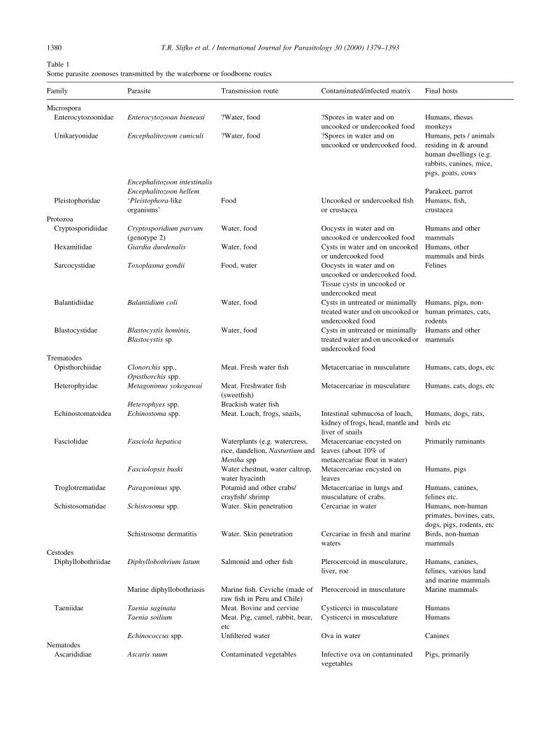
contaminated water and food. Some parasite zoonoses trans-
mitted by the waterborne and foodborne routes are
presented in Table 1.
2. Water and food as sources of infection
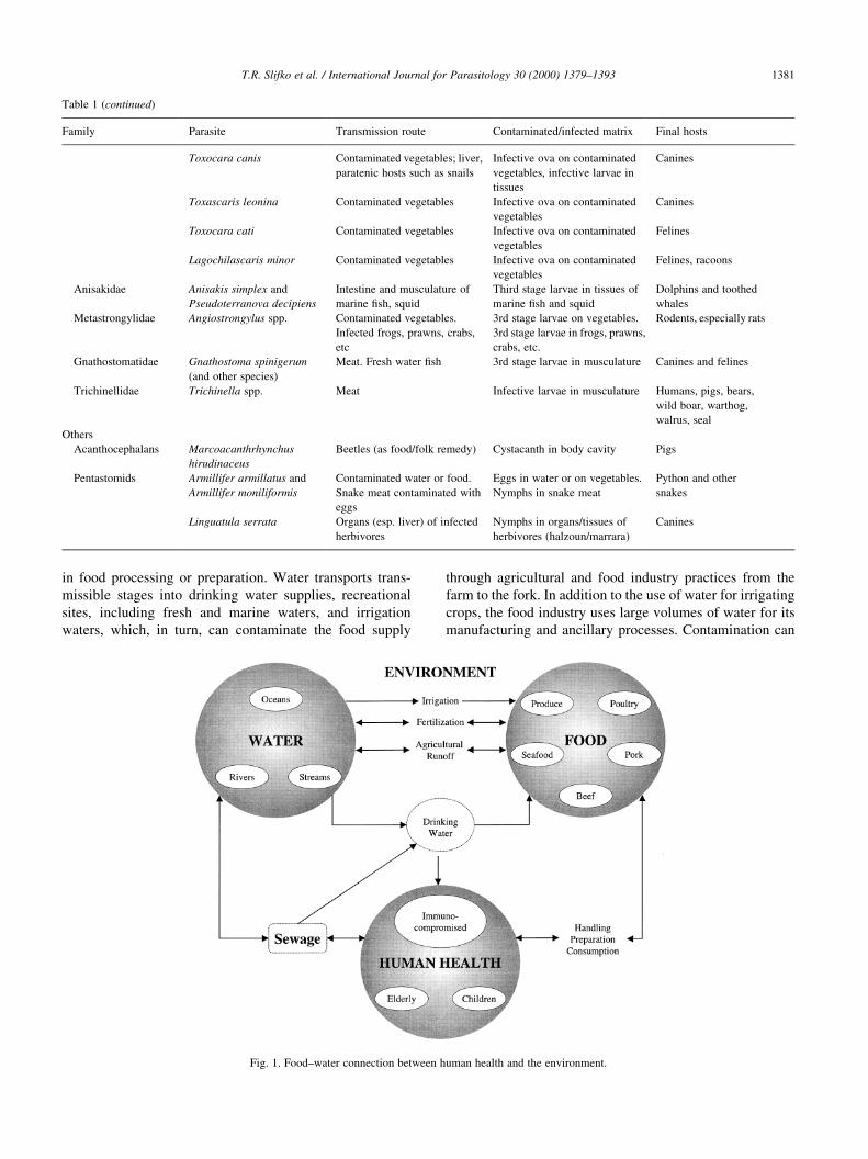
The water±food connection for parasite zoonoses is
complex (Fig. 1), with faeces as a major vehicle for many
environmental transmissive stages. However, the spores of
some microsporidia (e.g. Encephalitozoon cuniculi) and the
ova of Schistosoma haematobium contaminate the environ-
ment through urine. The transmissible stages can contami-
nate water or foods directly, voided in faeces, or indirectly.
The disposal of animal (and human) wastes remains a
signi®cant public health issue that has yet to be assessed
or controlled in most countries.
Water is a major conduit for these parasites, and contami-
nated water is an important source of human infection either
by direct consumption or by the use of contaminated water
International Journal for Parasitology 30 (2000) 1379±1393
0020-7519/00/$20.00 q 2000 Published by Elsevier Science Ltd. on behalf of the Australian Society for Parasitology Inc.
PII: S0020-7519(00)00128-4
www.parasitology-online.com
* Corresponding author. Tel.: 144-141-201-3028; fax: 144-141-558-
5508.
E-mail address: [email protected] (H.V. Smith).
T.R. Slifko et al. / International Journal for Parasitology 30 (2000) 1379±13931380
Table 1
Some parasite zoonoses transmitted by the waterborne or foodborne routes
Family Parasite Transmission route Contaminated/infected matrix Final hosts
Microspora
Enterocytozoonidae Enterocytozooan bieneusi ?Water, food ?Spores in water and on
uncooked or undercooked food
Humans, rhesus
monkeys
Unikaryonidae Encephalitozoon cuniculi ?Water, food ?Spores in water and on
uncooked or undercooked food.
Humans, pets / animals
residing in & around
human dwellings (e.g.
rabbits, canines, mice,
pigs, goats, cows
Encephalitozoon intestinalis
Encephalitozoon hellem Parakeet, parrot
Pleistophoridae `Pleistophora-like
organisms'
Food Uncooked or undercooked ®sh
or crustacea
Humans, ®sh,
crustacea
Protozoa
Cryptosporidiidae Cryptosporidium parvum
(genotype 2)
Water, food Oocysts in water and on
uncooked or undercooked food
Humans and other
mammals
Hexamitidae Giardia duodenalis Water, food Cysts in water and on uncooked
or undercooked food
Humans, other
mammals and birds
Sarcocystidae Toxoplasma gondii Food, water Oocysts in water and on
uncooked or undercooked food.
Tissue cysts in uncooked or
undercooked meat
Felines
Balantidiidae Balantidium coli Water, food Cysts in untreated or minimally
treated water and on uncooked or
undercooked food
Humans, pigs, non-
human primates, cats,
rodents
Blastocystidae Blastocystis hominis,
Blastocystis sp.
Water, food Cysts in untreated or minimally
treated water and on uncooked or
undercooked food
Humans and other
mammals
Trematodes
Opisthorchiidae Clonorchis spp.,
Opisthorchis spp.
Meat. Fresh water ®sh Metacercariae in musculature Humans, cats, dogs, etc
Heterophyidae Metagonimus yokogawai Meat. Freshwater ®sh
(sweet®sh)
Metacercariae in musculature Humans, cats, dogs, etc
Heterophyes spp. Brackish water ®sh
Echinostomatoidea Echinostoma spp. Meat. Loach, frogs, snails, Intestinal submucosa of loach,
kidney of frogs, head, mantle and
liver of snails
Humans, dogs, rats,
birds etc
Fasciolidae Fasciola hepatica Waterplants (e.g. watercress,
rice, dandelion, Nasturtium and
Mentha spp
Metacercariae encysted on
leaves (about 10% of
metacercariae ¯oat in water)
Primarily ruminants
Fasciolopsis buski Water chestnut, water caltrop,
water hyacinth
Metacercariae encysted on
leaves
Humans, pigs
Troglotrematidae Paragonimus spp. Potamid and other crabs/
cray®sh/ shrimp
Metacercariae in lungs and
musculature of crabs.
Humans, canines,
felines etc.
Schistosomatidae Schistosoma spp. Water. Skin penetration Cercariae in water Humans, non-human
primates, bovines, cats,
dogs, pigs, rodents, etc
Schistosome dermatitis Water. Skin penetration Cercariae in fresh and marine
waters
Birds, non-human
mammals
Cestodes
Diphyllobothriidae Diphyllobothrium latum Salmonid and other ®sh Plerocercoid in musculature,
liver, roe
Humans, canines,
felines, various land
and marine mammals
Marine diphyllobothriasis Marine ®sh. Ceviche (made of
raw ®sh in Peru and Chile)
Plerocercoid in musculature Marine mammals
Taeniidae Taenia saginata Meat. Bovine and cervine Cysticerci in musculature Humans
Taenia soilium Meat. Pig, camel, rabbit, bear,
etc
Cysticerci in musculature Humans
Echinococcus spp. Un®ltered water Ova in water Canines
Nematodes
Ascarididiae Ascaris suum Contaminated vegetables Infective ova on contaminated
vegetables
Pigs, primarily
in food processing or preparation. Water transports trans-
missible stages into drinking water supplies, recreational
sites, including fresh and marine waters, and irrigation
waters, which, in turn, can contaminate the food supply
through agricultural and food industry practices from the
farm to the fork. In addition to the use of water for irrigating
crops, the food industry uses large volumes of water for its
manufacturing and ancillary processes. Contamination can
T.R. Slifko et al. / International Journal for Parasitology 30 (2000) 1379±1393 1381
Table 1 (continued)
Family Parasite Transmission route Contaminated/infected matrix Final hosts
Toxocara canis Contaminated vegetables; liver,
paratenic hosts such as snails
Infective ova on contaminated
vegetables, infective larvae in
tissues
Canines
Toxascaris leonina Contaminated vegetables Infective ova on contaminated
vegetables
Canines
Toxocara cati Contaminated vegetables Infective ova on contaminated
vegetables
Felines
Lagochilascaris minor Contaminated vegetables Infective ova on contaminated
vegetables
Felines, racoons
Anisakidae Anisakis simplex and
Pseudoterranova decipiens
Intestine and musculature of
marine ®sh, squid
Third stage larvae in tissues of
marine ®sh and squid
Dolphins and toothed
whales
Metastrongylidae Angiostrongylus spp. Contaminated vegetables.
Infected frogs, prawns, crabs,
etc
3rd stage larvae on vegetables.
3rd stage larvae in frogs, prawns,
crabs, etc.
Rodents, especially rats
Gnathostomatidae Gnathostoma spinigerum
(and other species)
Meat. Fresh water ®sh 3rd stage larvae in musculature Canines and felines
Trichinellidae Trichinella spp. Meat Infective larvae in musculature Humans, pigs, bears,
wild boar, warthog,
walrus, seal
Others
Acanthocephalans Marcoacanthrhynchus
hirudinaceus
Beetles (as food/folk remedy) Cystacanth in body cavity Pigs
Pentastomids Armillifer armillatus and
Armillifer moniliformis
Contaminated water or food.
Snake meat contaminated with
eggs
Eggs in water or on vegetables.
Nymphs in snake meat
Python and other
snakes
Linguatula serrata Organs (esp. liver) of infected
herbivores
Nymphs in organs/tissues of
herbivores (halzoun/marrara)
Canines
Fig. 1. Food±water connection between human health and the environment.
also occur when foods, particularly salad vegetables and
fruit, are rinsed in parasite-contaminated potable water in
the household. Furthermore, consumer vogues, such as the
consumption of raw vegetables and undercooking to retain
the natural taste and preserve heat-labile nutrients, can
increase the risk of foodborne transmission.
In addition to the foodborne parasite zoonoses trans-
mitted by `meat', those transmitted through the surface
contamination of produce (often fruit and vegetables) either
at source or during food processing (see above) must also be
included. Both source contamination of produce and
contamination from water used in food preparation are
transmission routes that are signi®cant to the food industry.
Surface contamination can be direct, following contamina-
tion by the infected host, or indirect, following contamina-
tion by transport (birds, ¯ies, etc.) hosts, the use of manure
and contaminated water for irrigation, fumigation and pesti-
cide application, etc. Whether seasonal variation occurs in
the surface contamination of foods requires further investi-
gation, however, seasonal peaks in parasitism will in¯uence
when water and foods become surface-contaminated. The
rapid transportation of foods acquired from global markets
and their chilling and wetting can enhance parasite survival.
Water and food enhance the survival of environmental
stages by preventing their desiccation.
A variety of infective tissue parasite stages are responsi-
ble for transmitting meat and ®shborne zoonoses (Table 1).
Here, the preparation of the foodstuff is the key to the risk of
transmission. Eating raw, undercooked, cured, smoked,
salted, pickled or air-dried meat and offal can increase the
risk of contracting foodborne parasite zoonoses, especially
when the preservation treatment is inadequate. As well as
transmitting infective stages, some ®lter feeders also act as
transport hosts. For example, bivalves act as transport hosts
by concentrating viable Cryptosporidium oocysts and Giar-
dia cysts (and probably other zoonotic transmissive stages
found in faecally-contaminated fresh, estuarine and marine
waters) from their environment and have been suggested as
reservoirs for zoonotic transmission [1±5].
Previously, the consumption of raw or undercooked meat
and ®sh was associated with speci®c cultures and practices,
but with shifting consumer vogues, increased international
travel, globalisation of food supply and cosmopolitan eating
habits, what were once regarded as rare diseases are now
becoming increasingly more recognised. While there are no
adequate estimates of the numbers of foodborne and water-
borne disease world-wide, agencies, such as the United
States Centers for Disease Control and Prevention (CDC),
collect and report the incidence of noti®able diseases such
as giardiasis and cryptosporidiosis. A recent review of food
related illness and death in the United States reported that an
estimated 2.5 million (7%) foodborne illnesses were caused
by parasitic diseases (300 000; 2 000 000; 225 000; and 52
for Cryptosporidium parvum, Giardia lamblia, Toxoplasma
gondii and Trichinella spiralis, respectively), all with
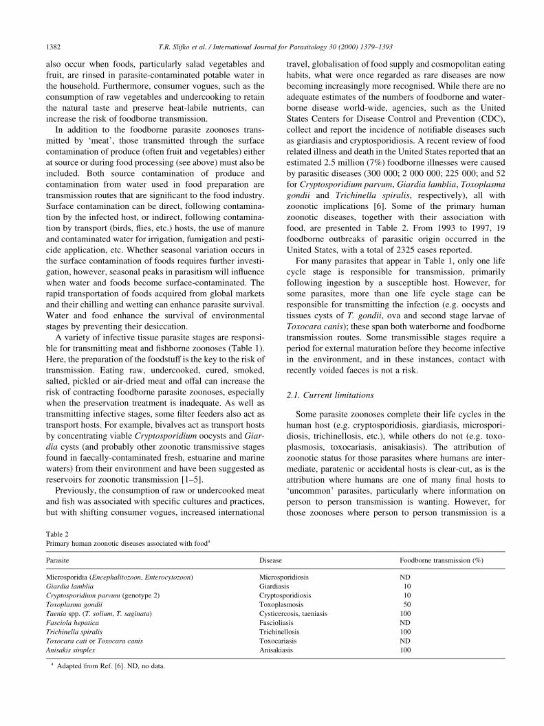
zoonotic implications [6]. Some of the primary human
zoonotic diseases, together with their association with
food, are presented in Table 2. From 1993 to 1997, 19
foodborne outbreaks of parasitic origin occurred in the
United States, with a total of 2325 cases reported.
For many parasites that appear in Table 1, only one life
cycle stage is responsible for transmission, primarily
following ingestion by a susceptible host. However, for
some parasites, more than one life cycle stage can be
responsible for transmitting the infection (e.g. oocysts and
tissues cysts of T. gondii, ova and second stage larvae of
Toxocara canis); these span both waterborne and foodborne
transmission routes. Some transmissible stages require a
period for external maturation before they become infective
in the environment, and in these instances, contact with
recently voided faeces is not a risk.
2.1. Current limitations
Some parasite zoonoses complete their life cycles in the
human host (e.g. cryptosporidiosis, giardiasis, microspori-
diosis, trichinellosis, etc.), while others do not (e.g. toxo-
plasmosis, toxocariasis, anisakiasis). The attribution of
zoonotic status for those parasites where humans are inter-
mediate, paratenic or accidental hosts is clear-cut, as is the
attribution where humans are one of many ®nal hosts to
`uncommon' parasites, particularly where information on
person to person transmission is wanting. However, for
those zoonoses where person to person transmission is a
T.R. Slifko et al. / International Journal for Parasitology 30 (2000) 1379±13931382
Table 2
Primary human zoonotic diseases associated with fooda
Parasite Disease Foodborne transmission (%)
Microsporidia (Encephalitozoon, Enterocytozoon) Microsporidiosis ND
Giardia lamblia Giardiasis 10
Cryptosporidium parvum (genotype 2) Cryptosporidiosis 10
Toxoplasma gondii Toxoplasmosis 50
Taenia spp. (T. solium, T. saginata) Cysticercosis, taeniasis 100
Fasciola hepatica Fascioliasis ND
Trichinella spiralis Trichinellosis 100
Toxocara cati or Toxocara canis Toxocariasis ND
Anisakis simplex Anisakiasis 100
a Adapted from Ref. [6]. ND, no data.
major route, the lack of effective typing and subtyping
systems limits our knowledge of the signi®cance of zoonotic
waterborne and foodborne transmission, although descrip-
tive epidemiology incriminates these routes. Currently, this
is a particular problem with C. parvum and Giardia duode-
nalis. While direct zoonotic transmission has been docu-
mented for Cryptosporidium and Giardia, it remains in
doubt whether zoonotic waterborne and foodborne trans-
missions of Giardia occur commonly. The widespread
distribution of infection in a variety of livestock, wild
animals and household pets indicates the potential for this
route of transmission. That outbreaks of waterborne giardia-
sis do occur with relative frequency from supplies consid-
ered to be pristine (i.e. not receiving contributions from
Giardia-infected humans) is support that the zoonotic
contribution may be important. Increased demands made
on natural resources increase the likelihood of encountering
both environments and produce contaminated with these
parasites.
3. Environmental contamination
The potential for environmental contamination depends
upon a variety of factors, including the number of infected
non-human hosts, the number of transmissive stages
excreted, agricultural practices, host behaviour and activity,
socio-economic and ethnic differences in human behaviour,
geographic distribution, sanitation, safety of drinking water
and food sources and supplies, and the climate and hydro-
geology of the area.
3.1. Sources, contributions and survival
Some aspects of the biology of the intestinal parasites C.
parvum, G. duodenalis and T. canis can be used to demon-
strate the potential for environmental contamination. For
Cryptosporidium and Giardia, the contribution from live-
stock and farming practices is dif®cult to assess. Infection
can be clinical in calves, but subclinical in adult cattle. A
clinically ill neonate can excrete #109 oocysts daily during
the course of infection, whereas a clinically-well, infected
cow can excrete between 7.6 £ 105 and 7.2 £ 108 oocysts
daily [7]. The sum total of oocysts contributed into the
environment over a year is similar for both ill and well
animals, given that immunity prevents the acquisition of
further infection in the neonatal host. Enumerating contri-
butions from agricultural practices, such as the storage and
spread of farmyard manure and slurry, on-farm discharge of
oocyst-contaminated dirty water to land or to water courses,
pasturing of livestock in land adjoining water sources, and
from the the disposal of faecally-contaminated waste from
abattoirs provides data on the potential for livestock to
contribute to C. parvum oocysts present in water courses.
On an UK dairy farm, with a history of cryptosporidiosis,
over 550 oocysts l21 were discharged into watercourses.
The practices which contributed high densities of oocysts
into water courses included hosing down calf rearing pens
and sluices (180 oocysts l21) and the contamination of farm
drains with slurry and farm yard manure applied onto land
(c. 370 oocysts l21) [7]. Practices such as hosing down calf
rearing pens and sluices release recently excreted oocysts
into an aquatic environment where survival is prolonged.
Such oocysts are likely to have a higher viability than
those excreted onto grazing land, which take time to perco-
late through substrata into watercourses. Overall contami-
nation rates with Cryptosporidium, for a pristine watershed,
have been estimated to be between 0.5±32 £ 105 oocysts/ha
per day and 0.12±2 £ 105 cysts/ha per day for Giardia [8]
from different reaches of another watershed, with uses
ranging from recreation only to dairy farming [9].
T. canis has a high prevalence rate in adult dogs and
foxes, with approximately 20% of adult dogs having patent
intestinal infections [10], and is particularly abundant (90%
plus) in puppies [11]. Gravid females produce up to 200 000
fertile eggs/day [12], which are voided in faeces, and
embryonate to infectivity in the environment. Kennelled
dogs excrete between 100 and 2000 eggs/g [13], and adult
foxes up to 2145 eggs/g [14]. Environmental contamination
with T. canis ova can be high: 66% of parks [15], 38% of
gardens [16] and 56% of sandpits [17], with 11% of parks
containing viable ova [18]. Ova can leach through soils or
be washed into combined sewer over¯ows during periods of
heavy rain. T. canis ova survive composting for at least 1
year [19] and survive in the environment for up to 4 years
[20]. The ova remain dormant but viable if covered by snow
at 211.5 to 108C or protected in faeces [21], but are killed
when unprotected at 2158C [22].
The transmissible stages can also be redistributed to other
uncontaminated matrices by coprophagous transport hosts,
including pigs, dogs, chickens, gulls and ¯ies [23±29]. Flies
ingest 1±3 mg faeces over 2±3 h [27], and can transmit
Giardia cysts [30], Cryptosporidium oocysts [23±24] and
Toxocara ova [28]. Filth ¯ies transmit C. parvum oocysts
both in excreta and on their external surfaces (experimen-
tally, up to eight oocysts in the adult digestive tract; 150±
320 oocysts on maggot and pupal surfaces) and wild-caught
¯ies harboured a mean of 73 oocysts/¯y [23]. Toxocara ova
were detected on 2.4 and in 2.1% of wild-caught naturally
infected ¯ies in Nigeria [29]. Toxocara ova require a period
of embryonation before being infective, although ¯ies could
deposit ova in/on food which could be ingested later.
4. Waterborne parasite zoonoses
Waterborne outbreaks of protozoan parasites are far more
common than outbreaks due to helminths because of the
smaller sizes of their transmissible stages. Giardia and
Cryptosporidium have become signi®cant waterborne
pathogens in the developed world for three reasons. Firstly,
giardiasis and cryptosporidiosis are indigenous infections in
many animals; secondly, the densities of environmental
T.R. Slifko et al. / International Journal for Parasitology 30 (2000) 1379±1393 1383
contamination with infective cysts and oocysts are suf®cient
to pollute the aquatic environment; and thirdly, the cysts and
oocysts which penetrate water treatment processes are
insensitive to the disinfectants commonly used in water
treatment. Giardia cysts and Cryptosporidium oocysts are
also small enough to pose a threat to groundwaters [31,32].
Toxoplasma and microsporidia have been associated with
waterborne diseases on rare occasions. T. gondii oocysts are
resistant to disinfection. The spores of the microsporidia are
small (1±5 mm) and less is known regarding their resistance
to water treatment. Community water systems are not
regarded as a major route of transmission for the helminth
zoonoses. Filtration, as a minimum, is an effective barrier to
helminth ova (.20 mm) and the larger protozoan cysts,
although ova can be found in the air, in dust and soil, and
can be transferred to uncovered water sources.
4.1. Giardia and Cryptosporidium
The waterborne transmission of the intestinal protozoan
parasites G. duodenalis and C. parvum has been well docu-
mented [33±37]; over 160 waterborne outbreaks of giardia-
sis and cryptosporidiosis have been reported, with the
greatest documentation in the US and UK. Within the last
12 years, 39 documented outbreaks of waterborne cryptos-
poridiosis have occurred in the USA, Canada, UK and Japan
[38]. Activities associated with cattle farming, particularly
muck spreading, slurry spraying and run off from contami-
nated grazing land, have been proposed as causes of many
of these outbreaks, but, in the absence of de®nitive informa-
tion in many instances, the number attributed to the zoonotic
route has to remain speculative. The search for both the
contributors and causes has driven method development
(Table 3) [26,39±41].
Developments in molecular and genetic analyses of
waterborne protozoan parasites, including the determination
of species identity and subtyping species, will help in deter-
mining the contributors of environmental contamination.
Considerable research is currently ongoing in this area,
and several genotyping techniques have been developed
for Cryptosporidium, Giardia, microsporidia spp. and Toxo-
plasma [42±46]. In British Columbia, a waterborne outbreak
of giardiasis was considered to have originated from an
infected beaver, not only because beavers were epidemio-
logically-linked to the outbreak and were found to inhabit
lodges close to the water supply, but also because typing
studies (isoenzyme analysis and pulsed-®eld gel electro-
phoresis) indicated that Giardia isolated from the indivi-
duals affected by the outbreak were found to be of the
same zymodeme and karyotype as Giardia isolated from
the epidemiologically-linked beavers [47,48]. A compari-
son of 11 previously described species differentiation and
genotyping protocols for Cryptosporidium determined that
two were not Cryptosporidium speci®c [42]. While these
molecular methods have great potential for tracking the
source of contamination [49], comprehensive comparisons
are necessary to validate the ef®ciency of the protocols,
particularly with respect to environmental contamination
[26].
4.2. Microsporidia
The microsporidia are obligate, intracellular spore-form-
ing protozoa that belong to the phylum Microspora. About
1000 species of microsporidia are recognised [50], being,
primarily, ubiquitous parasites of invertebrates and ®sh
[50,51]. Largely unknown as causes of human disease
before the HIV pandemic [51,52], human microsporidial
infections have been found predominantly in HIV-infected
immunocompromised individuals, although some infections
in immunocompetent individuals have also been identi®ed
[53]. Currently, their role as emerging pathogens is being
increasingly recognised. The prevalence of microsporidiosis
in studies of patients with chronic diarrhoea ranges from 7 to
50%, world-wide [54], although it is unclear whether this
broad range represents a geographic variation, differences in
diagnostic capabilities or differences in risk factors for
exposure to microsporidia.
In the summer of 1995, a waterborne outbreak of micro-
sporidiosis occurred in France, with approximately 200
cases, primarily in the immunocompromised (chronic diar-
rhoea, dehydration and signi®cant weight loss (.10% body
weight), and low CD4 counts) [55]. While faecal contam-
ination of the drinking water was never demonstrated,
contamination from a nearby lake was suspected, but the
source of that contamination (animal or human) was not
suggested.
Microsporidial spores are stable in the environment and
remain infective for days to weeks outside their hosts [56±
58]. Their small size (1±5 mm) makes them dif®cult to
remove using conventional water ®ltration techniques and
there is concern that they may possess increased resistance
to chlorine disinfection; similar to Cryptosporidium. Initial
studies using cell culture suggest that the spores may be
susceptible to disinfection [59].
4.3. Toxoplasma
Two outbreaks of toxoplasmosis, associated with the
consumption of oocyst-contaminated water, have also
been documented [60,61]. The ®rst outbreak occurred in
Panama in British troops, and epidemiological evidence
indicated that the most likely vehicle for transmission was
the ingestion of creek water, contaminated with oocysts
excreted by jungle cats, consumed during manoeuvres in
the jungle. The second outbreak occurred in British Colum-
bia, Canada in 1995, and 110 acute Toxoplasma infections
were identi®ed. Fifty-®ve were in non-pregnant individuals
and 42 women were pregnant at the time of infection.
Eleven infants became infected. The epidemiological
evidence was consistent with a waterborne source and
implicated the municipal drinking water [61] whose raw
T.R. Slifko et al. / International Journal for Parasitology 30 (2000) 1379±13931384
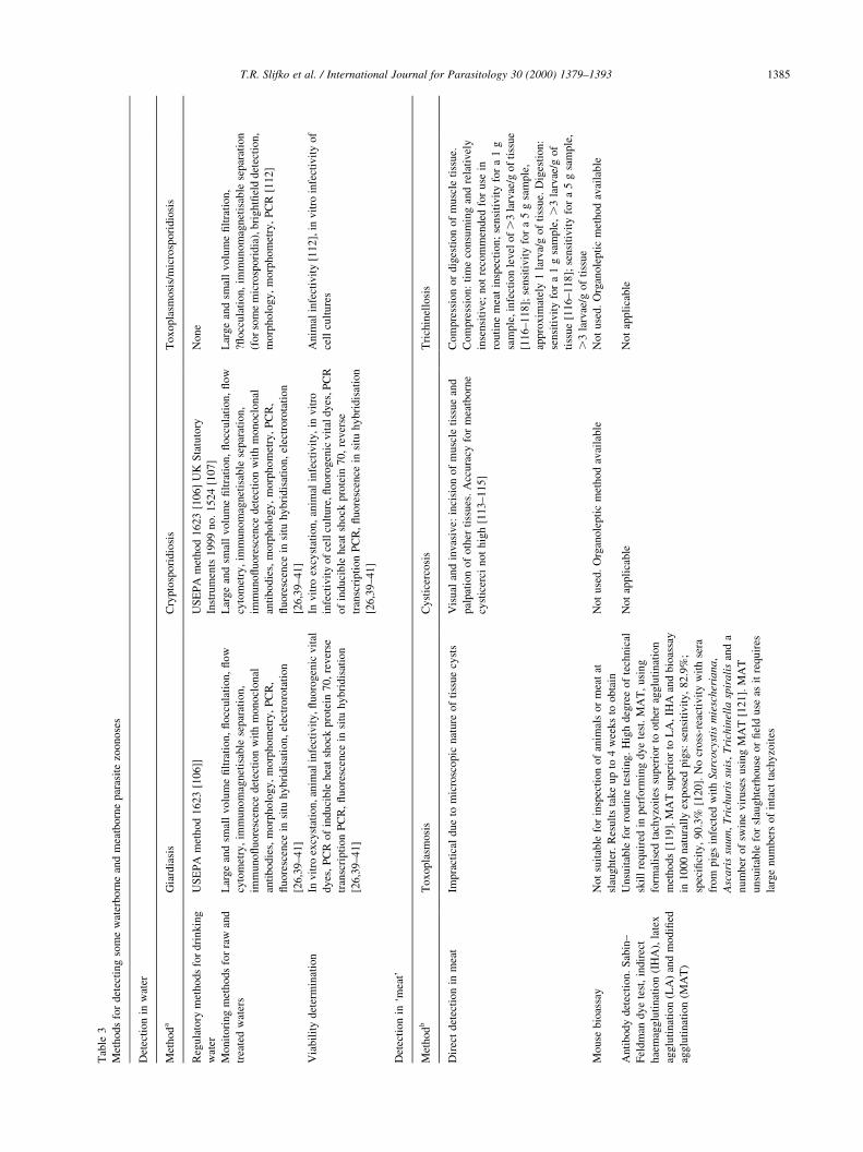
T.R. Slifko et al. / International Journal for Parasitology 30 (2000) 1379±1393 1385
Tab
le3
Met
ho
ds
for
det
ecti
ng
som
ew
ater
bo
rne
and
mea
tbo
rne
par
asit
ezo
onose
s
Det
ecti
on
inw
ater
Met
ho
da
Gia
rdia
sis
Cry
pto
spori
dio
sis
Toxopla
smosi
s/m
icro
spori
dio
sis
Reg
ula
tory
met
ho
ds
for
dri
nk
ing
wat
er
US
EP
Am
eth
od
16
23
[106]]
US
EP
Am
ethod
1623
[106]
UK
Sta
tuto
ry
Inst
rum
ents
1999
no.
1524
[107]
None
Mo
nit
ori
ng
met
ho
ds
for
raw
and
trea
ted
wat
ers
Lar
ge
and
smal
lv
olu
me
®lt
rati
on,
¯occ
ula
tion,
¯ow
cyto
met
ry,
imm
un
om
agnet
isab
lese
par
atio
n,
imm
un
o¯
uo
resc
ence
det
ecti
on
wit
hm
onocl
onal
anti
bo
die
s,m
orp
ho
log
y,
morp
hom
etry
,P
CR
,
¯u
ore
scen
cein
situ
hy
bri
dis
atio
n,
elec
troro
tati
on
[26
,39
±4
1]
Lar
ge
and
smal
lvolu
me
®lt
rati
on,
¯occ
ula
tion,
¯ow
cyto
met
ry,
imm
unom
agnet
isab
lese
par
atio
n,
imm
uno¯
uore
scen
cedet
ecti
on
wit
hm
onocl
onal
anti
bodie
s,m
orp
holo
gy,
morp
hom
etry
,P
CR
,
¯uore
scen
cein
situ
hybri
dis
atio
n,
elec
troro
tati
on
[26,3
9±41]
Lar
ge
and
smal
lvolu
me
®lt
rati
on,
?¯occ
ula
tion,
imm
unom
agnet
isab
lese
par
atio
n
(for
som
em
icro
spori
dia
),bri
ght®
eld
det
ecti
on,
morp
holo
gy,
morp
hom
etry
,P
CR
[112]
Via
bil
ity
det
erm
inat
ion
Inv
itro
excy
stat
ion
,an
imal
infe
ctiv
ity,
¯uoro
gen
icvit
al
dy
es,
PC
Ro
fin
du
cible
hea
tsh
ock
pro
tein
70,
rever
se
tran
scri
pti
on
PC
R,
¯u
ore
scen
cein
situ
hybri
dis
atio
n
[26
,39
±4
1]
Invit
roex
cyst
atio
n,
anim
alin
fect
ivit
y,
invit
ro
infe
ctiv
ity
of
cell
cult
ure
,¯uoro
gen
icvit
aldyes
,PC
R
of
induci
ble
hea
tsh
ock
pro
tein
70,
rever
se
tran
scri
pti
on
PC
R,
¯uore
scen
cein
situ
hybri
dis
atio
n
[26,3
9±41]
Anim
alin
fect
ivit
y[1
12],
invit
roin
fect
ivit
yof
cell
cult
ure
s
Det
ecti
on
in`m
eat'
Met
ho
db
To
xop
lasm
osi
sC
yst
icer
cosi
sT
rich
inel
losi
s
Dir
ect
det
ecti
on
inm
eat
Imp
ract
ical
du
eto
mic
rosc
opic
nat
ure
of
tiss
ue
cyst
sV
isual
and
invas
ive:
inci
sion
of
musc
leti
ssu
ean
d
pal
pat
ion
of
oth
erti
ssues
.A
ccura
cyfo
rm
eatb
orn
e
cyst
icer
cinot
hig
h[1
13±115]
Com
pre
ssio
nor
dig
esti
on
of
musc
leti
ssue.
Com
pre
ssio
n:
tim
eco
nsu
min
gan
dre
lati
vel
y
inse
nsi
tive;
not
reco
mm
ended
for
use
in
routi
ne
mea
tin
spec
tion;
sensi
tivit
yfo
ra
1g
sam
ple
,in
fect
ion
level
of
.3
larv
ae/g
of
tiss
ue
[116±118];
sensi
tivit
yfo
ra
5g
sam
ple
,
appro
xim
atel
y1
larv
a/g
of
tiss
ue.
Dig
esti
on:
sensi
tivit
yfo
ra
1g
sam
ple
,.
3la
rvae
/gof
tiss
ue
[116±118];
sensi
tivit
yfo
ra
5g
sam
ple
,
.3
larv
ae/g
of
tiss
ue
Mo
use
bio
assa
yN
ot
suit
able
for
insp
ecti
on
of
anim
als
or
mea
tat
slau
gh
ter.
Res
ult
sta
ke
up
to4
wee
ks
toobta
in
Not
use
d.
Org
anole
pti
cm
ethod
avai
lable
Not
use
d.
Org
anole
pti
cm
ethod
avai
lable
An
tib
od
yd
etec
tio
n.
Sab
in±
Fel
dm
and
ye
test
,in
dir
ect
hae
mag
glu
tin
atio
n(I
HA
),la
tex
agg
luti
nat
ion
(LA
)an
dm
od
i®ed
agg
luti
nat
ion
(MA
T)
Un
suit
able
for
rou
tin
ete
stin
g.
Hig
hdeg
ree
of
tech
nic
al
skil
lre
qu
ired
inp
erfo
rmin
gdye
test
.M
AT
,usi
ng
form
alis
edta
chy
zoit
essu
per
ior
tooth
erag
glu
tinat
ion
met
ho
ds
[11
9].
MA
Tsu
per
ior
toL
A,
IHA
and
bio
assa
y
in1
00
0n
atura
lly
exp
ose
dpig
s:se
nsi
tivit
y,
82.9
%;
spec
i®ci
ty,
90
.3%
[12
0].
No
cross
-rea
ctiv
ity
wit
hse
ra
from
pig
sin
fect
edw
ith
Sarc
ocy
stis
mie
scher
iana,
Asc
ari
ssu
um
,T
rich
uri
ssu
is,
Tri
chin
ella
spir
ali
san
da
nu
mb
ero
fsw
ine
vir
use
susi
ng
MA
T[1
21].
MA
T
un
suit
able
for
slau
gh
terh
ouse
or
®el
duse
asit
requir
es
larg
en
um
ber
so
fin
tact
tach
yzo
ites
Not
appli
cable
Not
appli
cable
T.R. Slifko et al. / International Journal for Parasitology 30 (2000) 1379±13931386
Tab
le3
(con
tin
ued
)
Det
ecti
on
in`m
eat'
Met
ho
db
To
xo
pla
smo
sis
Cyst
icer
cosi
sT
rich
inel
losi
s
An
tib
od
yd
etec
tio
n.
EL
ISA
/
Wes
tern
blo
ttin
g
EL
ISA
inp
igs
has
spec
i®ci
tyof
85.9
%an
dse
nsi
tivit
yof
72
.9%
,co
mp
ared
wit
hbio
assa
y[1
20].
Anti
-Toxo
pla
sma
IgG
EL
ISA
,u
sin
ga
cru
de
tach
yzo
ite
lysa
te,
isre
liab
le
for
iden
tify
ing
infe
cted
anim
als
and
corr
elat
esw
ell
wit
h
dy
ete
stre
sult
sin
exp
erim
enta
lly
infe
cted
pig
s.C
ross
-
reac
tiv
ity
wit
hp
igs
har
bouri
ng
Sarc
ocy
stis
infe
ctio
n
[12
2].
Use
ful
for
det
ecti
ng
both
acute
and
chro
nic
infe
ctio
ns
inh
um
ans
[12
3].
Rec
om
bin
ant
anti
gen
s:
com
par
edw
ith
the
nat
ive
anti
gen
EL
ISA
,th
e
reco
mbin
ant
anti
gen
s(H
4an
dH
11)
hav
ea
sensi
tivit
yof
79
%an
da
spec
i®ci
tyo
f100%
usi
ng
sera
from
nat
ura
lly
exp
ose
dsh
eep
.D
on
ot
reco
gnis
ean
tibodie
sse
rafr
om
chro
nic
ally
infe
cted
pig
s[1
24]
Wes
tern
blo
ttin
g(W
B)
more
sensi
tive
than
EL
ISA
and,
wit
haf
®nit
ypuri
®ed
gly
copro
tein
anti
gen
s,is
the
met
hod
of
choic
efo
rth
ese
rodia
gnosi
s.A
nti
gen
stri
ps
com
mer
cial
lyav
aila
ble
.W
Bse
nsi
tivit
yfo
r
pig
sw
ith
one
det
ecta
ble
cyst
,60±80%
[125].
Het
erolo
gous
anti
gen
s:an
tibodie
sfr
om
Taen
ia
sagin
ata
-infe
cted
catt
lere
act
wit
hli
popro
tein
anti
gen
sfr
om
Taen
iahyd
ati
gen
acy
st¯
uid
(ThF
AS
)
[126].
ThF
AS
has
low
reac
tivit
yw
ith
anti
-Fasc
iola
hep
ati
caan
tibodie
s.T
hF
AS
can
det
ect
`Tai
wan
Taen
ia'
pig
infe
ctio
ns
[127].
Inpig
snat
ura
lly
infe
cted
wit
hT
aen
iaso
lium
,T
aen
iacr
ass
icep
s
anti
gen
sac
hie
ved
97%
spec
i®ci
tyan
d100%
sensi
tivit
y[1
28].
Rec
om
bin
antan
tigen
s:T
cA
2-M
BP
(rec
om
bin
ant
from
aT
.cr
ass
icep
scD
NA
sequen
ce)
issp
eci®
cfo
rT
.sa
gin
ata
and
can
det
ect
infe
ctio
ns
in
catt
le[1
29]
EL
ISA
isth
ebes
tm
ethod
for
ante
-mort
em
dia
gnosi
s.C
om
par
able
inse
nsi
tivit
yto
bes
t
dir
ectm
ethods
wit
hin
fect
ion
level
sas
low
as1
larv
a/100
gof
tiss
ue
det
ecte
d[1
17,1
30].
Only
short
-ter
mE
San
tigen
or
bio
chem
ical
ly
puri
®ed
anti
gen
sca
nbe
use
dcu
rren
tly
[131±
133].
Over
all
esti
mat
esof
EL
ISA
ef®
cacy
,
93.1
±99.3
%se
nsi
tivit
yan
d90.6
±99.0
%
spec
i®ci
ty[1
34±138].
Rec
om
bin
ant
anti
gen
s:
the
maj
or
pro
ble
mis
inab
ilit
yto
repro
duce
the
gly
copro
tein
stru
cture
of
imm
unodom
inan
t
anti
gen
s.S
ynth
esis
edneo
gly
can
[139]
wit
hth
e
anti
gen
icst
ruct
ure
found
on
Tri
chin
ella
gly
copro
tein
suse
din
EL
ISA
[138]
and
per
form
edas
wel
las
nat
ive
ES
anti
gen
sw
hen
test
ing
sera
from
exper
imen
tall
yan
dnat
ura
lly
infe
cted
pig
s
Cir
cula
tin
gan
tig
end
etec
tio
n.
EL
ISA
Su
f®ci
ent
anti
gen
avai
lab
lefo
rdet
ecti
on
only
for
ash
ort
tim
ein
hu
man
sera
and
mouse
tiss
ues
,duri
ng
acute
ph
ase
of
infe
ctio
n
Monocl
onal
anti
bodie
sag
ainst
met
aces
tode
extr
acts
can
det
ect
circ
ula
ting
anti
gen
sin
the
sera
of
79%
infe
cted
pig
s;sp
eci®
city
,97%
[140]
Anti
gen
det
ecti
on
isunre
liab
lefo
rro
uti
ne
dia
gnosi
sas
anti
gen
aem
iadet
ecte
din
only
56%
of
anim
als
test
ed[1
41]
Mo
lecu
lar
met
ho
ds
(PC
Ran
d
DN
Ap
rob
es)
PC
Ro
fre
pet
itiv
eg
ene
frag
men
t(B
1)
has
sensi
tivit
yof
10
tach
yzo
ites
/10
5le
ukocy
tes
Burg
etal
.[1
42].
PC
Rof
rib
oso
mal
DN
Afo
rsp
eci®
cid
enti
®ca
tion
of
To
xop
lasm
a[1
43
].H
ighly
sensi
tive
and
spec
i®c
when
com
bin
edw
ith
P3
0g
ene
PC
R
Com
bin
eduse
of
DN
Apro
bes
for
T.
sagin
ata
and
T.
soli
um
pro
vid
esposi
tive
iden
ti®
cati
on
of
T.
sagin
ata
pro
glo
ttid
sfr
om
faec
alsa
mple
s[1
44]
Import
ant
toid
enti
fyin
fect
ing
spec
ies
and
types
asth
isca
nas
sist
inid
enti
fyin
gth
eso
urc
e
of
infe
ctio
n.
Dif
fere
nt
spec
ies/
types
pro
duce
dif
feri
ng
pat
holo
gy
inhum
ans.
Ran
dom
ly
pri
med
PC
Rre
acti
ons
(RA
PD
)use
dto
dif
fere
nti
ate
the
eight
acce
pte
dgro
ups
of
Tri
chin
ella
[145,1
46]
aA
dap
ted
fro
mR
ef.
[26
].b
Ad
apte
dfr
om
Ref
.[9
5].
water source was probably contaminated with oocysts from
domestic and feral cats and cougars.
5. Recreational water
Giardia and Cryptosporidium are the most commonly
recognised cause of recreational waterborne disease. Most
recreational water outbreaks are the result of faecal acci-
dents or cross-connections in swimming pools, and the
contamination of recreational waters with animal wastes is
not well documented or recognised [62], although defeca-
tion by infected livestock and feral animals into lakes,
canals, other outdoor recreational water bodies or receiving
waters must be borne in mind. A statistically signi®cant
association was identi®ed between the drinking of untreated
surface water and illness in New Mexico [63]. An increased
risk of infection was also related to swimming in surface
water, as well as attending a day-care centre, camping and
having a pet that was ill or young. In 1997 and 1998, in the
most recent reports on recreational outbreaks in the USA,
Cryptosporidium was responsible for nine of 18, and all but
one occurred in swimming pools, with one occurring in an
interactive fountain [64]. Giardia was not reported. The
source of the contamination that occurred in a lake at a
State park was not identi®ed. In 1999, the second interactive
fountain outbreak of gastroenteritis occurred at a beachside
park [65], however, no source of contamination was deter-
mined.
While outbreaks can be seen as extreme consequences of
zoonotic transmission, it is likely that numerous cases of
disease associated with recreational exposure go unrecog-
nised, and hence, are not reported. The swimming in waters
in¯uenced by the wastes of animals is of concern, yet
assessment of the risk requires further quanti®cation.
Increased utilisation of outdoor recreational waters for
immersion water sports is likely to precipitate increased
reporting of water-associated zoonoses. Of interest is the
increased reporting of periodic clusters of swimmers' itch,
a dermatitis caused by cercariae of avian schistosomes that
penetrate into human skin, but which are unable to complete
their life cycles in the human host.
6. Foodborne parasite zoonoses
6.1. Surface contamination
The increased demand, global sourcing and rapid trans-
port of foods, especially soft fruit and salad vegetables,
enhance both the likelihood of surface contamination and
survival of the transmissive stages of parasites pathogenic to
man. Food normally becomes a potential source of human
infection by contamination, during production, collection,
transport and preparation (e.g. milk, fruit, vegetables, soft
drinks, etc.) or during processing, and the sources of zoono-
tic contamination are usually faeces, faecally-contaminated
soil or water. The number of contaminating organisms will
vary depending upon the route or vehicle of contamination,
and therefore, the sensitivity of the methods developed will
have to address the detection of the smallest numbers of
contaminants, practicable (1±100). Given the low infectious
doses of many parasites, surface contamination with low
numbers of viable parasites, in produce that receives mini-
mal washing prior to ingestion, poses a threat to public
health. It is often dif®cult to associate an outbreak with a
particular food item and furthermore, if the foodborne route
is suspected, to identify how the food implicated became
contaminated. Due to these dif®culties, the acquisition of
parasitic infections via the foodborne route is almost
certainly under-detected. Casemore [66], in reviewing food-
borne protozoal infection, suggested that the degree of
under-detection might be by a factor of 10 or more.
With these current limitations, it not surprising to realise
that documented zoonotic foodborne outbreaks are rare,
although some foods can be important vehicles of transmis-
sion, especially in situations of poor hygiene and endemni-
city of infection [28,67]. Currently, foodborne giardiasis
and cryptosporidiosis are of signi®cance because of both
the low infectious doses and the robustness and disinfection
insensitivity of their transmissive stages [68±72], and modi-
®cations of the methods based on their detection in water are
being developed [71,72].
6.1.1. Giardia and Cryptosporidium
The foodborne transmission of giardiasis was suggested
in the 1920s [73,74], and anecdotal evidence from other
outbreaks has frequently implicated food handlers and
contaminated fruit and vegetables [75]. The ®rst foodborne
outbreak of giardiasis in the US was described in 1979
[75,76]. Of eight outbreaks of foodborne giardiasis docu-
mented, only one reports the possibility of food (i.e. tripe)
being intrinsically infected. The other outbreaks, affecting
217 individuals, between 1979 and 1990, are associated
with contamination by food handlers, and include foods
such as salmon, fruit salad, raw vegetables, lettuce, onions
and tomatoes. In two outbreaks, the original source of infec-
tion was traced to the infected infant of the food handler
[72].
Suspected outbreaks of foodborne cryptosporidiosis have
been reported from travellers visiting Mexico, in the UK and
Australia, the suspect foods including salads, raw milk,
sausages and tripe [76]. An outbreak following the
consumption of apple cider was the ®rst associated with
the zoonotic transmission route [77]. The fresh pressed
cider was squeezed from apples collected from an orchard
in which an infected calf grazed. Some apples had fallen
onto the ground and had probably been contaminated with
infectious oocysts [77]. While three other outbreaks have
been reported since 1993, none implicated zoonotic trans-
mission [78,79]. Again, the absence of standardised detec-
tion and subtyping methods limits our understanding of the
zoonotic route of infection.
T.R. Slifko et al. / International Journal for Parasitology 30 (2000) 1379±1393 1387
6.1.2. Fasciola
For many, the perception of human fascioliasis, caused by
Fasciola hepatica or Fasciola gigantica, is that it is a spora-
dic disease of low economic importance, but Chen and Mott
[80] highlighted the importance of this zoonosis, identifying
2594 cases from 42 different countries between 1970 and
1990. Current estimations indicate between 2.4 and 17
million human infections world-wide [81,82] and the
WHO recognises fascioliasis as an emerging disease of
humans [83]. Estimates based on faecal egg counts, will
be underestimates, as they will not include those individuals
with prepatent or ectopic infections, or those with low grade
infections excreting intermittently or at very low egg densi-
ties.
The distribution of the disease is predominantly rural,
being associated with cattle and sheep breeding [84±86],
although high prevalences in humans are not necessarily
associated with areas where fascioliasis is a signi®cant
veterinary problem [86]. The incidence appears to be
concentrated within families, as they are all likely to eat
the same contaminated product(s) [87]. Interestingly, Mas-
Coma et al. [86] state that in hyperendemic areas, the para-
site is better adapted to the human host, presumably leading
to reduced liver pathology, increased adult numbers and egg
production. Further information on human infection can be
found in the reviews of Chen and Mott [80], Mas-Coma et
al. [86] and Esteban et al. [88].
The most common transmission route is the ingestion of
watercress (Nasturtium and Roripa species; Table 2)
contaminated with encysted metacercariae, although,
depending upon the geographical location, a variety of
edible aquatic plants can be vehicles of transmission.
Water containing ¯oating metacercariae has also been
implicated in disease transmission [89], as have salads
contaminated with metacercaria-contaminated irrigation
water [90]. In Iran, the risk factors include the use of animal
manure as fertiliser and wastewater ef̄ uent for irrigating
aquatic or semi-aquatic vegetable crops [83]. Recently,
transmission following consumption of fresh, raw liver
dishes containing immature ¯ukes was suggested [91].
6.2. Meatborne infection
Meatborne parasitic zoonoses remain an important cause
of illness and economic loss, globally [92±95]. Of known
importance are toxoplasmosis, cysticercosis and trichinosis,
while ®shborne parasites remain a problem in certain
regions of the world. Foodborne trematode infections also
exert a signi®cant economic impact, with more than 40
million people infected with one or more different species
[82,83]. Efforts to control these zoonoses continue, yet the
overall progress is unsatisfactory [96]. In addition to eating
infected meat bought over the counter, eating inadequately
cooked game (e.g. bear, boar) and ®sh, during or after hunt-
ing, ®shing and shooting expeditions also contributes to the
increased reporting of zoonotic meatborne infections.
6.2.1. Toxoplasma, Taenia spp. and Trichinella
In terms of illness and death, Toxoplasma, Listeria and
Salmonella are the three most important pathogens trans-
mitted by food in the USA, and possibly Europe [97]. Pork,
lamb and mutton are the most important sources of meat-
borne infections of Toxoplasma, together with game such as
bear and feral swine meat [6]. In a European multicentre
case-controlled study, Cook et al. [98] identi®ed that eating
undercooked lamb, beef or game, contact with soil, and
travel outside Europe, the USA and Canada were the risk
factors most strongly predictive of acute Toxoplasma infec-
tion in pregnant women. The infection of livestock with
larval stages (cysticercosis) of Taenia saginata (beef tape-
worm) and Taenia solium (pork tapeworm), which develop
into adults in the human intestine, is also of great public
health concern [93,99,100]. Clinically, T. solium is of
greater concern because, unlike T. saginata, humans also
serve as the intermediate host for the cysticercus stage,
following autoinfection or if ova are ingested accidentally
from the environment or in folk remedies. Human trichinel-
losis, contracted by eating raw or undercooked meat
containing infective larvae (trichinae) of the nematode para-
site Trichinella spp., is most commonly associated with
eating pork, bear meat and horse meat [95]. T. spiralis is
the species of greater concern, as it the species most
commonly found in pigs. High priority is placed on the
inspection of swine and horse carcasses for trichinae at
slaughter in many countries: the European Union spends
$570 million each year on Trichinella testing [101]. Of
the anisakine parasites, Anisakis simplex and Pseudoterra-
nova decipiens are of major signi®cance [83], with more
than 80% of Paci®c salmon and red snapper infected with
larvae of these species [102].
6.2.2. Microsporidia
In addition to its waterborne route of transmission, micro-
sporidiosis is also a potential emerging meatborne zoonosis,
given that natural hosts of human infective microsporidia
can be part of the human food chain. Pleistophora-like
microsporidians, initially found in muscle, may be acquired
from raw or lightly cooked ®sh or crustaceans. Some
evidence for the foodborne route comes from the incidental
®nding of microsporidial spores in a human stool sample
from an AIDS patient with diarrhoea [103], which also
contained muscle ®bres (meat) infected with microsporidia.
The suggested transmission route was as follows: after
eating the ®sh, spores from the infected musculature
remained largely intact during passage through the patient's
gut, with some of these viable spores initiating the infection.
The relationship between microsporidial parasites of ®sh
and crustacean muscle and those found in human cases
requires further elucidation.
6.2.3. Foodborne trematodes
Foodborne trematode infections, acquired through eating
raw, improperly cooked or processed freshwater ®sh, shell-
T.R. Slifko et al. / International Journal for Parasitology 30 (2000) 1379±13931388
®sh, crabs, or unwashed or inadequately washed vegetables
(Table 1), were recognised as a public health problem in
1991 by the Southeast Asian Ministries of Education Orga-
nisation Regional Tropical Medicine and Public Health
Project (SEAMEO-TROPMED). Clonorchiasis, paragoni-
miasis, fascioliasis, fasciolopsiasis and other intestinal
trematodiases are the most important diseases contracted,
and strategies to control foodborne trematode infections
were identi®ed in 1995 [83]. Among these strategies, health
education programmes featured greatly; the awareness of
both hazards and risks being fundamental goals, as was
the generation of baseline epidemiological data and the
development of food safety programmes and hazard assess-
ment at critical control points (HACCP) approaches. The
application of international codes of practice (e.g. FAO/
WHO Codex for ®sh and ®sheries products; legislation
controlling disposal of excreta) could also reduce environ-
mental contamination, as could speci®c legislation for agri-
culture and aquaculture.
The identi®cation of strategies to control foodborne trans-
mission in 1995 indicates that the trematode zoonoses are
well recognised, yet, for many, particularly in developed
countries, these are emerging zoonoses. Have the foodborne
zoonoses been ignored, and if so, can they be addressed
currently? The World Health Organisation/Pan American
Health Organisation informal consultation document on
intestinal protozoa [104,105] offers a way ahead. In addition
to the strategies identi®ed above, new immunological and
molecular technologies were deemed to have applications in
the environment, especially where waterborne (and food-
borne) transmissions are known to occur. It was concluded
that the development of molecular biological tools for diag-
nostic and epidemiological purposes should be encouraged
[104].
7. Trends: current and future
Recent advances in immunology and molecular biology
have enabled us to develop more sensitive, speci®c and
rapid tests that could supersede current methods. For water-
borne zoonoses, particularly Giardia and Cryptosporidium,
great interest exists in developing both effective detection
(Table 3) and typing systems which have public health perti-
nence. Immunomagnetisable separation, followed by anti-
body detection or PCR (for intact cysts and oocysts) appear
to be effective test formats (Table 3), while for some meat-
borne zoonoses, antibody detection, in serum or meat juices,
as a re¯ection of exposure, and PCR or nucleic acid probes
for determining the presence of the parasite (Toxoplasma,
cysticercosis and Trichinella) appear effective (Table 3).
Similarly, the development of new chemotherapeutic agents
and alternative vaccine strategies in livestock offer new
opportunities to improve the control of some waterborne
and meatborne zoonoses, yet, for meatborne zoonoses
infecting livestock, testing at slaughter or prior to proces-
sing remains necessary to protect public health.
The sample matrix plays a signi®cant role in test devel-
opment, and once optimised for the matrices and parasites of
current signi®cance, the same formats should prove useful
for detecting other emerging and re-emerging zoonoses in
similar matrices. For example, given the levels of environ-
mental contamination described for T. canis ova, our close
association with dogs, the large range of intermediate and
paratenic hosts which form part of the human food chain,
and the recognised foodborne outbreaks (e.g. surface
contamination of vegetables with ova; infective larvae in
raw liver, edible snails and raw or undercooked meat),
might foodborne transmission of toxocariasis be more
prevalent that we think?
Two issues will determine the adoption of new methods:
whether they can be adapted to, and will be suitable for on
line testing; and whether suf®cient testing has been under-
taken to provide con®dence in their use.
8. Epilogue
The under-diagnoses and reporting of these important
zoonotic parasites undermines our ability to bring the
diseases to the attention of industries, governments and
communities, and to implement controls. The multiple
routes of transmission complicate the understanding and
the ability to estimate the magnitude of contaminated
water and food in the overall burden of disease for many
of these pathogens. Environmental monitoring of water and
food utilising new technologies, along with molecular
epidemiology will be one of the best approaches for the
identi®cation of the risks in the future, and new approaches
for recovery of the parasites from water and food are now
available.
Many countries have implemented regulations addressing
the control of the spread of waterborne and foodborne
diseases, particularly through water reuse programmes.
Despite these efforts, the potential for contaminated food
and water to cause extensive outbreaks still remains, due
to the breadth of populations served. Several problems exist
with identifying outbreaks associated with parasitic
zoonoses. Often, the foremost problem occurs with detect-
ing and reporting the contaminated water or food. Some
countries have adopted regulations to minimise protozoan
parasite contamination of potable water [106,107] and some
of the meatborne zoonoses, e.g. [83,108±111], however,
regulations for other foodborne parasitic zoonoses are
fewer, although the livestock and food industries have
adopted the best practice by developing effective HACCP
programmes. Interestingly, the rise in general public
concern over food safety has helped to focus more attention
on zoonotic parasites. For many of the zoonotic parasites,
the system for routine monitoring or reporting is inadequate,
thus the incidence of human disease and parasite occurrence
T.R. Slifko et al. / International Journal for Parasitology 30 (2000) 1379±1393 1389
in water and food is undoubtedly underestimated. Particu-
larly for the foodborne zoonoses, parasitic infections have a
lower impact than prokaryotic pathogens, which, again,
contributes to an underestimation of the number of identi-
®ed cases/outbreaks.
With the new international codes regarding food safety,
risk assessment methodologies are being seen as the scien-
ti®c process to address public health, globally. Both epide-
miological and risk assessment approaches are dependent
upon an evaluation of the occurrence and survival of the
transmissive stage(s) of the parasite in question. For exam-
ple, for many parasitic infections transmitted through the
environment, exposure assessment is perhaps the most dif®-
cult parameter to measure. The understanding of this vari-
able is dependent not only upon the detection of organisms
in the environment, but also on an understanding of the
occurrence, transport, survival and fate through various
matrices [28]. Here, sensitive, robust and reproducible
detection, viability, typing and subtyping methods, with
suitability for the matrix in question, will be the arbiters.
Only by developing such methods can we attempt to deter-
mine the impact of the parasite zoonoses transmitted by
water and food, and their public health signi®cance.
References
[1] Fayer R, Lewis EJ, Trout JM, et al. Cryptosporidium parvum in
oysters from commercial harvesting sites in the Chesapeake Bay.
Emerg Infect Dis 1999;5:706±10.
[2] Graczyk TK, Fayer R, Cran®eld MR, Conn DB. In vitro interactions
of Asian freshwater clam (Corbicula ¯uminea) hemocytes and Cryp-
tosporidium parvum oocysts. Appl Environ Microbiol
1997;63:2910±2.
[3] Graczyk TK, Fayer R, Lewis EJ, Farley CA, Trout JM. In vitro
interactions between hemocytes of the eastern oyster, Crassostrea
virginica Gmelin, 1791 and Cryptosporidium parvum oocysts. J
Parasitol 1997;83:949±52.
[4] Graczyk TK, Fayer R, Lewis EJ, Farley CA, Trout JM. Detection of
Cryptosporidium oocysts and Giardia cysts in the tissues of eastern
oysters (Crassostrea virginica) carrying principal oyster infectious
diseases. J Parasitol 1998;84:1039±42.
[5] Tamburrini A, Pozio E. Long-term survival of Cryptosporidium
parvum oocysts in seawater and in experimentally infected mussels
(Mytilus galloprovincialis). Int J Parasitol 1999;29:711±5.
[6] Mead PS, Slutsker L, Dietz V, et al. Food-related illness and death in
the United States. Emerg Infect Dis 1999;5:607±25.
[7] Smith HV, Nichols RN. Case study of health effects of Cryptospor-
idium in drinking water. Article 4.12.4.8. UNESCO-EOLSS Ency-
clopaedia of Life Support Systems ± Theme ± Environmental
Toxicology and Human Health, 2000; in press.
[8] Ongerth JE. Workshop on Cryptosporidium and cryptosporidiosis.
Session IV. Control of Cryptosporidium (Logsdon GS, moderator).
In: Craun GF, Sykora JL, editors. The taxonomy, detection, epide-
miology and waterborne control of Cryptosporidium, 1989.
[9] Hansen JS, Ongerth JE. Effects of time and watershed characteristics
on the concentration of Cryptosporidium oocysts in river water. Appl
Environ Microbiol 1991;57:2790±5.
[10] Glickman LT, Schantz PM. Epidemiology and pathogenesis of
zoonotic toxocariasis. Epidemiol Rev 1981;3:230±50.
[11] Misra SC. Experimental prenatal infection of Toxocara canis in dogs
and effective chemotherapeutic measures. Indian J Anim Sci
1972;42:608±12.
[12] Glickman LT, Schantz PM, Cypess RH. Canine and human toxocar-
iasis: review of transmission, pathogenesis and clinical disease. J
Am Vet Med Assoc 1979;175:1265±9.
[13] Vanparijs O, Hermans L, van der Flaes L. Helminth and protozoan
parasites in dogs and cats in Belgium. Vet Parasitol 1991;38:67±73.
[14] Richards DT, Lewis JW. Epidemiology of Toxocara canis in the fox.
In: Lewis JW, Maizels RM, editors. Toxocara and toxocariasis:
clinical, epidemiological and molecular perspectives. British Society
for Parasitology/Institute of Biology, 1993. pp. 25±37.
[15] Snow KR, Ball SJ, Berwick JA. Prevalence of Toxocara species
eggs in the soil of ®ve east London parks. Vet Rec 1987;121:66±67.
[16] Holland C, O'Connor P, Taylor MRH, et al. Families, parks, gardens
and Toxocariasis. Scand J Infect Dis 1991;23:225±31.
[17] Horn K, Schneider T, Stoye M. Contamination of public childrens'
playgrounds with helminth eggs in Hanover. DTW Dtsch TieraÈrztl
Wochenschr 1990;97:124±5.
[18] Quinn R, Smith HV, Girdwood RWA, Bruce RG. Studies on the
incidence of Toxocara and Toxascaris spp. ova in the environment.
1. A comparison of ¯otation procedures for recovering Toxocara
spp. ova from soil. J Hyg 1980;84:83±89.
[19] Pegg EJ, Donald CR. The effects of composting on the eggs of
Toxocara canis and Toxascaris leonina. J Inst Anim Technol
1978;29:29±30.
[20] Lloyd S. Toxocara canis in the dog. In: Lewis J, Maizels RM,
editors. Toxocara and toxocariasis: clinical, epidemiological and
molecular perspectives. British Society for Parasitology/Institute
of Biology, 1993. pp. 11±24.
[21] Velichkin PA, Radun FL. The epizootiology and prophylaxis of
Toxocara infections in dogs and fur bearing animals. In: Other
AN, editor. Antropozoogel' mintosy i perspektivy ikh likvidatsii,
1975. pp. 12±15 (Cited in Ref. [20]).
[22] Okoshi S, Usui M. Experimental studies on Toxascaris leonina: IV.
Development of eggs of three ascarids, T. leonina, Toxocara canis
and Toxocara cati in dogs and cats. Jpn J Vet Sci 1968;30:29±38.
[23] Graczyk TK, Fayer R, Cran®eld MR, et al. Filth ¯ies are transport
hosts of Cryptosporidium parvum. Emerg Infect Dis 1999;5:726±7.
[24] Graczyk TK, Cran®eld MR, Fayer R, Bixler H. House ¯ies (Musca
domestica) as transport hosts of Cryptosporidium parvum. Am J
Trop Med Hyg 1999;61:500±4.
[25] Smith HV, Brown J, Coulson JC, Morris GP, Girdwood RWA.
Occurrence of Cryptosporidium sp. oocysts in Larus spp. gulls.
Epidemiol Infect 1993;110:135±43.
[26] Smith HV. Detection of parasites in the environment. In: Smith HV,
Stimson WH, editors; co-ordinating editor Chappel LH). Infectious
diseases diagnosis: current status and future trends. Parasitology
1998;117:S113-S141.
[27] Greenberg B. Flies and disease. Vol. II. Biology and disease trans-
mission. Princeton, NJ: Princeton University Press, 1973.
[28] Pegg EJ. Infection of dogs with Toxocara canis carried by ¯ies.
Parasitology 1971;62:409±14.
[29] Umeche N, Mandah LE. Musca domestica as carrier of intestinal
helminths in Calabor, Nigeria. East Afr Med J 1989;66:349±52.
[30] Sterling CR, Miranda E, Gilman RH. The potential role of ¯ies
(Musca domestica) in the mechanical transmission of Giardia and
Cryptosporidium in a Pueblo Joven community of Lima, Peru. Am
Soc Trop Med Hyg 1987;233:349.
[31] The National Cryptosporidium Survey Group. A survey of Cryptos-
poridium oocysts in surface and groundwaters in the UK. J Inst
Water Environ Management 1992;6:697±703.
[32] Hancock CM, Rose JB, Callahan MC. Cryptosporidium and Giardia
in US groundwater. J Am Water Works Assoc 1998;90:58±61.
[33] Craun GF. Waterborne giardiasis. In: Meyer EA, Ruitenberg EJ,
MacInnes AJ, editors. Giardiasis, Series in human parasitic diseases,
Vol. 3. New York: Elsevier, 1990. pp. 267±93.
T.R. Slifko et al. / International Journal for Parasitology 30 (2000) 1379±13931390
[34] Smith HV, Rose JB. Waterborne cryptosporidiosis. Parasitol Today
1990;6:8±12.
[35] Anonymous. Cryptosporidium in water supplies. Report of the
Group of Experts; Chairman, Sir John Badenoch. Dept of the Envir-
onment/Dept of Health. London, HMSO, 1990. pp. 230.
[36] Anonymous. Cryptosporidium in water supplies. Third Report of the
Group of Experts; Chairman, Professor Ian Bouchier. Dept of the
Environment, Transport and the Regions/Dept of Health. London,
HMSO, 1998. pp. 171.
[37] Solo-Gabrielle H, Neumeister S. US outbreaks of cryptosporidiosis.
J Am Water Works Assoc 1996;88:76±86.
[38] Rose JB. Environmental ecology of Cryptosporidium and public
health implications. Annu Rev Public Health 1997;18:135±61.
[39] Smith HV, Robertson LJ, Campbell AT. Cryptosporidium and cryp-
tosporidiosis. Part 2. Future technologies and state of the art
research. Eur Microbiol 1993;2:22±29.
[40] Jakubowski W, Boutros S, Faber W, et al. Environmental methods
for Cryptosporidium. J Am Water Works Assoc 1996;88:107±21.
[41] Smith HV, Rose JB. Waterborne cryptosporidiosis: current status.
Parasitol Today 1998;14:14±22.
[42] Suliaman IM, Xiao L, Lal A. Evaluation of Cryptosporidium parvum
genotyping techniques. Appl Environ Microbiol 1999;65:4431±5.
[43] Adam RD. The Giardia lamblia genome. Int J Parasitol
2000;30:475±84.
[44] Mathis A. Microsporidia: emerging advances in understanding the
basic biology of these unique organisms. Int J Parasitol
2000;30:795±804.
[45] Orlandi PA, Lampel KA. Extraction free, ®lter based template
preparation for rapid and sensitive PCR detection of pathogenic
protozoa. J Clin Microbiol 2000;38:2271±7.
[46] Howe DK, Honore S, Derouin F, Sibley LD. Determination of geno-
types of Toxoplasma gondii strains isolated from patients with toxo-
plasmosis. J Clin Microbiol 1997;35:1411±4.
[47] Isaac-Renton JL, Cordeiro C, Sara®s K, Shahriari H. Characterisa-
tion of Giardia duodenalis isolates from a waterborne outbreak. J
Infect Dis 1993;167:431±40.
[48] Sara®s K, Isaac-Renton J. Pulsed-®eld gel electrophoresis as a
method of biotyping of Giardia duodenalis. Am J Trop Med Hyg
1993;48:134±44.
[49] McIntyre L, Hoang L, Ong CSL, Lee P, Issac-Renton JL. Evaluation
of molecular techniques to biotype Giardia duodenalis collected
during an outbreak. J Parasitol 2000;86:172±7.
[50] Canning EU. Microsporidia. In: Kreier JP, editor. Parasitic protozoa,
vol. 6. London: Academic Press, 1993.
[51] Canning EU, Lom J. The Microsporidia of vertebrates. London:
Academic Press, 1986.
[52] Canning EU, Hollister WS, Colbourn NI, Curry A, Gobel UB.
Human microsporidioses: site speci®city, prevalence and species
identi®cation. AIDS 1983;7(Suppl 3):S3±S7.
[53] Curry A, Smith HV. Emerging pathogens, Isospora, Cyclospora, and
microsporidia. Parasitology 1998;117:S143±59.
[54] Bryan RT. Microsporidiosis as an AIDS-related opportunistic infec-
tion. Clin Infect Dis 1995;21:62±65.
[55] Cotte L, Rabodonirina M, Chapuis F, et al. Waterborne outbreak of
intestinal microsporidiosis in persons with and without human
immunode®ciency virus infection. J Infect Dis 1999;180:2003±8.
[56] Shadduck JA. Human microsporidiosis and AIDS. Rev Infect Dis
1989;11:203±7.
[57] Waller T. Sensitivity of Encephalitozoon cuniculi to various
temperatures, disinfectants and drugs. Lab Anim 1979;13:227±30.
[58] Shadduck JA, Polley MB. Some factors in¯uencing the in vitro
infectivity and replication of Encephalitozoon cuniculi. J Protozool
1978;25:491±6.
[59] Wolk DM, Johnson CH, Rice EW, et al. A spore counting method
and cell culture model for chlorine disinfection studies of Encepha-
litozoon syn. Septata intestinalis. Appl Environ Microbiol
2000;66:1266±73.
[60] Benenson MW, Takafuji ET, Lemon SM, Greenup RL, Sulzer AJ.
Oocyst-transmitted toxoplasmosis associated with the ingestion of
contaminated water. N Engl J Med 1982;307:666±9.
[61] Bowie WR, King AE, Werker DH, et al. Outbreak of toxoplasmosis
associated with municipal drinking water. Lancet 1997;350:173±7.
[62] Gerba CP, Gerba P. Outbreaks caused by Giardia and Cryptospor-
idium associated with swimming pools. J Swim Pool Spa Ind
1995;1:9±18.
[63] Gallaher MM, Herndon JL, Nims LJ, Sterling CR, Grabowski DJ,
Hull HH. Cryptosporidiosis and surface water. Am J Public Health
1989;79:39±42.
[64] Centers for Disease Control and Prevention. MMWR Morb Mortal
Wkly Rep 2000;49 SS-4.
[65] Centers for Disease Control and Prevention. MMWR Morb Mortal
Wkly Rep 2000;49 SS-1.
[66] Casemore DP. Foodborne protozoal infection. Lancet
1990;336:1427±32.
[67] Feacham RG, Bradley DJ, Garelick H, Mara DD. Chapter 23. Sani-
tation and disease. Health aspects of excreta and wastewater
management. Chichester: Wiley, 1982. pp. 375±99.
[68] Smith JL. Cryptosporidium and Giardia as agents of foodborne
disease. J Food Prot 1993;56:451±61.
[69] Smith HV, Robertson LJ, Campbell AT, Girdwood RWA. Giardia
and giardiasis, what's in a name? Microbiol Eur 1995;3:22±29.
[70] Laberge I, Grif®ths MW, Grif®ths MW. Prevalence, detection and
control of Cryptosporidium parvum in food. Int J Food Microbiol
1996;31:1±26.
[71] Girdwood RWA, Smith HV. Cryptosporidium. In: Robinson R, Batt
C, Patel P, editors. Encyclopaedia of food microbiology. London:
Academic Press, 1999. pp. 487±97.
[72] Girdwood RWA, Smith HV. Giardia. In: Robinson R, Batt C, Patel
P, editors. Encyclopaedia of food microbiology. London: Academic
Press, 1999. pp. 946±54.
[73] Musgrave WE. Flagellate infestations and infections. J Am Med
Assoc 1922;79:2219±20.
[74] Lyon BBV, Swalm WA. Giardiasis, its frequency, recognition and
certain clinical factors. Am J Med Sci 1925;170:348±64.
[75] Barnard RJ, Jackson GJ. Giardia lamblia. The transfer of human
infections by foods. In: Erlandsen SL, Meyer EA, editors. Giardia
and giardiasis. New York: Plenum Press, 1984. pp. 365±77.
[76] Rose JB, Slifko TR. Giardia, Cyclospora, and Cryptosporidium and
their impact on foods ± a review. J Food Prot 1999;62:1059±70.
[77] Millard PS, Gensheimer KF, Addiss DG, et al. An outbreak of cryp-
tosporidiosis from fresh-pressed apple cider. J Am Med Assoc
1994;272:592±1596.
[78] Centers for Disease Control and Prevention. Foodborne outbreak of
diarrhoeal illness associated with Cryptosporidium parvum, Minne-
sota, 1995. MMWR Morb Mortal Wkly Rep 1996;45:783.
[79] Centers for Disease Control and Prevention. Foodborne outbreak of
cryptosporidiosis ± Spokane, Washington, 1997. MMWR Morb
Mortal Wkly Rep 1998;47:27.
[80] Chen MG, Mott KE. Progress in assessment of morbidity due to
Fasciola hepatica infection: a review of recent literature. Trop Dis
Bull 1990;87:R1±R38.
[81] Hopkins DR. Homing in on helminths. Am J Trop Med Hyg
1992;46:624±34.
[82] Rim HJ, Farag HF, Sornmani S, Cross JH. Food-borne trematodes:
ignored or emerging? Parasitol Today 1994;10:207±9.
[83] World Health Organisation. Control of foodborne trematode infec-
tions. WHO technical report 849. WHO technical report series.
Geneva, Switzerland, 1995. pp. 1±157.
[84] Stork MG, Venables GS, Jennings SMF, Beesley JR, Bendezu P,
Capron A. An investigation of fascioliasis in Peruvian village chil-
dren. J Trop Med Hyg 1973;76:231±5.
[85] BoureÂe P, Thiebault M. Fasciolose aÁ Fasciola hepatica en Basse
Normandie de 1980 aÁ 1990. Bull Soc FrancËaise Parasitol
1993;11:79±84.
T.R. Slifko et al. / International Journal for Parasitology 30 (2000) 1379±1393 1391
[86] Mas-Coma S, Bargues MD, Esteban JG. Human fascioliasis. In:
Dalton J, editor. Fascioliasis. Wallingford, Oxon: CABI Publishing,
CAB International, 1999. pp. 411±34.
[87] Bechtel U, Feucht HE, Held E, Vogl T, Nothdurft HD. Fasciola
hepatica. Infektion einer familie. Diagnostik und therapie. Dtsch
Med Wochenschr 1992;117:978±82.
[88] Esteban JG, Bargues MD, Mas-Coma S. Geographical distribution,
diagnosis and treatment of human fascioliasis: a review. Res Rev
Parasitol 1998;58:13±42.
[89] Vareille-Morel V, Dreyfuss G, Rondelaud D. PremieÁres donneÂes sur
la dispersion et le devenir des meÂtacercaires ¯ottantes de Fasciola
hepatica L. Bull Soc FrancËaise Parasitol 1993;11:63±69.
[90] Cadel S, Barbier D, Duhamel C, Georges P. A propos de 18 cas de
fasciole humaine recenseÂs en Basse±Normandie. AnneeÂs 1994±
1995. Bull Soc FrancËaise Parasitol 1996;14:39±43.
[91] Taira N, Yoshifuji H, Boray JC. Zoonotic potential of infection with
Fasciola spp. by consumption of freshly prepared raw liver contain-
ing immature ¯ukes. Int J Parasitol 1997;27:775±9.
[92] World Health Organisation. The role of food safety in health and
development. WHO Technical Reports. No. 705. World Health
Organisation, Geneva, Switzerland, 1984.
[93] Roberts T, Murrell KD, Marks S. Economic losses caused by food-
borne parasitic disease. Parasitol Today 1994;10:419±23.
[94] Murrell KD. Foodborne parasites. Int J Environ Health Res
1995;5:63±85.
[95] Gamble HR, Murrell KD. Diagnosis of parasites in food. In: Smith
HV, Stimson WH, editors. Chappel LH, co-ordinating editor. Infec-
tious diseases diagnosis: current status and future trends. Parasitol-
ogy 1998;117:S97-S112.
[96] World Health Organisation. Global health situation and projection
estimates. World Health Organisation, Geneva, Switzerland, 1992.
[97] Dubey JB. Sources of Toxoplasma gondii infection in pregnancy
editorial. Br Med J 2000;321:127±8.
[98] Cook AJC, Gilbert RE, Buffolano W, et al. Sources of Toxoplasma
infection in pregnant women: European multicentre case-controlled
study On behalf of the European Research Network on Congenital
Toxoplasmosis. Br Med J 2000;321:142±7.
[99] Pawlowski ZS. Taeniasis and cysticercosis Chapter 11. In: Hui YH,
Gorham JR, Murrell KD, Diver DO, editors. Foodborne diseases
handbook, Vol. 2, New York: Marcel Dekker, 1994. pp. 177±99.
[100] Tsang VCW, Wilson M. Taenia solium cysticercosis: an unrecog-
nised but serious public health problem. Parasitol Today
1995;11:124±6.
[101] Pozio E. Trichinellosis in the European Union: epidemiology, ecol-
ogy and economic impact. Parasitol Today 1998;14:25±38.
[102] Makerrow JH, Sakanari JA, Deardorff TL. Revenge of the sushi
parasite. N Engl J Med 1988;319:1228±9.
[103] McDougall RJ, Tandy MW, Boreham RE, Stenzel DJ, O'Donoghue
PJ. Incidental ®nding of a microsporidian parasite from an AIDS
patient. J Clin Microbiol 1993;31:436±9.
[104] World Health Organisation. WHO/PAHO informal consultation on
intestinal protozoal infections. WHO/CDS/IPI/92.2. World Health
Organisation, Geneva, Switzerland, 1992.
[105] Warhurst DC, Smith HV. Getting to the guts of the problem. Para-
sitol Today 1992;8:292±3.
[106] United States Environmental Protection Agency. Consumer con®-
dence reports ®nal rule. Federal Register 1998;63:160.
[107] UK Statutory Instruments 1999 No. 1524. The water supply (water
quality) (amendment) regulations, 1999. The Stationery Of®ce, Ltd.
5 pp.
[108] European Economic Community. Commission directive 77/96/EEC.
Off J Eur Commun 1977;26:67±77.
[109] European Economic Community. Commission directive 84/319/
EEC. Off J Eur Commun 1984;167:34±43.
[110] Code of Federal Regulations, Title 9, Ch. 3, Part 318.10(D). Animals
and animal products. Of®ce of the Federal Register, US Government
Printing Of®ce, Washington, DC, USA, 1994. pp. 212±220.
[111] Deardorff TL. Epidemiology of marine ®shborne parasitic zoonoses.
Southeast Asian J Trop Med Pub Health 1991;22:146±9.
[112] Issac-Renton J, Bowie WR, King A, et al. Detection of Toxoplasma
gondii oocysts in drinking water. Appl Environ Microbiol
1998;64:2270±80.
[113] Dewhurst LW, Cramer JD, Sheldon JJ. An analysis of current
inspection procedures for detecting bovine cysticercosis. J Am Vet
Med Assoc 1967;150:412±7.
[114] Kyvsgaard NC, Ilsoe SA, Henrikson SA, Nansen P. Distribution of
Taenia saginata cysts in carcasses of experimentally infected calves
and its signi®cance for routine meat inspection. Res Vet Sci
1990;49:29±33.
[115] Viljoen NF. Cysticercosis in swines and bovines, with special refer-
ence to South African conditions. Onderstepoort J Vet Res
1937;9:337±70.
[116] Ruitenberg EJ, van Knapen F, Vermeulen CJ. Enzyme-linked immu-
nosorbent assay (ELISA) in Trichinella spiralis infections in pigs.
In: Kim CW, Pawlowski ZS, editors. Trichinellosis. Hanover, NH:
University Press of New England, 1998. pp. 487±99.
[117] Gamble HR. Detection of trichinellosis in pigs by arti®cial digestion
and enzyme immunoassay. J Food Prot 1996;59:295±8.
[118] Gamble HR. Trichinellosis. In: OIE Manual of Standards for Diag-
nostic Tests and Vaccines, Of®ce Internationale des Epizooties.
Chapter 3.5.3, Paris, France, 1996. pp. 477±480.
[119] Dubey JP, Desmonts G, McDonald C, Walls KW. Serologic evalua-
tion of cattle inoculated with Toxoplasma gondii: comparison of
Sabin±Feldman dye test and other agglutination tests. Am J Vet
Res 1985;46:1085±8.
[120] Dubey JP, Thulliez P, Weigel RM, Andrews CD, Lind P, Powell EC.
Sensitivity and speci®city of various serologic tests for detection of
Toxoplasma gondii infection in naturally infected sows. Am J Vet
Res 1995;56:1030±6.
[121] Dubey JP. Validation of the speci®city of the modi®ed agglutination
test for toxoplasmosis in pigs. Vet Parasitol 1997;71:307±10.
[122] Lind P, Haugegaard J, Wingstrand A, Henriksen SA. The time
course of the speci®c antibody response by various ELISAs in
pigs experimentally infected with Toxoplasma gondii. Vet Parasitol
1997;71:1±15.
[123] Santoro F, Afchain D, Pierce R, Cesbron JY, Oviaque G, Capron A.
Serodiagnosis of Toxoplasma infection using a puri®ed parasite
protein (P30). Clin Exp Immunol 1985;62:262±9.
[124] Andrews CD, Dubey JP, Tenter AM, Webert DW. Toxoplasma
gondii recombinant antigens H4 and Hil. Use in ELISAs for detec-
tion of toxoplasmosis in swine. Vet Parasitol 1997;70:1±11.
[125] Tsang VCW, Brand JA, Boyer AE. An enzyme-linked immunoelec-
trotransfer blot assay and glycoprotein antigens for diagnosing
human cysticercosis (Taenia solium). J Infect Dis 1989;159:50±58.
[126] Rhoads ML, Murrell KD, Dilling GW, Wong MM, Baker NF. A
potential diagnostic: reagent for bovine cysticercosis. J Parasitol
1985;71:779±87.
[127] Rhoads ML, Murrell KD, Cross JR, Fan PL. The serological
response of pigs experimentally infected with a species of Taenia
from Taiwan. Vet Parasitol 1989;20:279±85.
[128] Biondi GF, Mucciolo RG, Nunes CM, Richtzenhain LJ. Immuno-
diagnosis of swine cysticercosis by indirect ELISA employing a
heterologous antigen from Taenia crassiceps metacestode. Vet Para-
sitol 1996;64:261±6.
[129] Zarlenga US, Rhoads ML, Al-Yarnan FM. A Taenia crassiceps
cDNA sequence encoding a putative immunodiagnostic antigen
for bovine cysticercosis. Mol Biochem Parasitol 1994;67:215±23.
[130] Smith HJ, Snowdon KE. Comparative assessment of a double anti-
body enzyme immunoassay test kit and a triple antibody enzyme
immunoassay for the diagnosis of Trichinella spiralis spiralis and
Trichinella spiralis nativa infections in swine. Can J Vet Res
1989;53:497±9.
[131] Gamble HR, Anderson WR, Graham CE, Murrell KD. Serodiagnosis
T.R. Slifko et al. / International Journal for Parasitology 30 (2000) 1379±13931392
of swine trichinosis using an excretory±secretory antigen. Vet Para-
sitol 1983;13:349±61.
[132] Seawright GL, Despommier DD, Zimmerman W, Isenstein RS.
Enzyme immunoassay for swine trichinellosis using antigens puri-
®ed by immunoaf®nity chromatography. Am J Trop Med Hyg
1983;32:1275±84.
[133] Arriaga C, Yepez-Mulia L, Morilla A, Ortega-Pierres MG. Detection
of circulating Trichinella spiralis muscle larva antigen in serum
samples of experimentally and naturally infected swine. In: Camp-
bell WC, Pozio E, Bruschi F, editors. Trichinellosis. Rome: Istituto
Superiore di Sanita Press, 1993. pp. 301±6.
[134] Murrell KD, Anderson WR, Schad GA, et al. Field evaluation of the
enzyme-linked immunosorbent assay for swine trichinosis: ef®cacy
of the excretory±secretory antigen. Am J Vet Res 1986;47:1046±9.
[135] Oliver DC, Singh P, Allison DE. Trichina detection techniques as
applied to a high-speed slaughterhouse environment. Agri-Practice
1988;9:45±48.
[136] Oliver DG, Singh P, Allison DE, Murrell KD, Gamble HR. Field
evaluation of an enzyme immunoassay for detection of hogs in a
high volume North Carolina abattoir. In: Tanner CE, editor. Trichi-
nellosis. Madrid: Consejo Superior de Investigaciones Press, 1989.
pp. 439±44.
[137] van der Leek NL, Dame JB, Adams CL, Gillis KD, Littell RC.
Evaluation of an enzyme-linked immunosorbent assay for diagnosis
of trichinellosis in swine. Am J Vet Res 1992;53:877±82.
[138] Gamble HR, Wisnewski N, Wassom D. Detection of trichinellosis in
swine by enzyme immunoassay using a synthetic glycan antigen.
Am J Vet Res 1997;58:417±21.
[139] Wisnewski N, McNeil M, Grieve RB, Wassom DL. Characterisation
of novel fucosyl- and tyvelosyl-containing glycoconjugates from
Trichinella spiralis muscle stage larvae. Mol Biochem Parasitol
1993;61:25±36.
[140] Rodriguez-del-Rosal E, Correa D, Flisser A. Swine cysticercosis:
detection of parasite products in serum. Vet Rec 1989;124:488.
[141] Arriaga C, Yepez-Mulia A, Morilla A, Ortega-Pierres G. Detection
of circulating Trichinella spiralis muscle larva antigens in serum
samples of experimentally and naturally infected swine. Vet Para-
sitol 1995;58:319±26.
[142] Burg JL, Grover CM, Pouletty P, Boothroyd JC. Direct and sensitive
detection of a pathogenic protozoan, Toxoplasma gondii by poly-
merase chain reaction. J Clin Microbiol 1989;27:1787.
[143] Cazenave J, Cheyrou A, Bloum P, Johnson AM, Begueret J. Use of
polymerase chain reaction to detect Toxoplasma. J Clin Pathol
1991;44:1137.
[144] Harrison LJS, Delgado J, Parkhouse RME. Differential diagnosis of
Taenia saginata and Taenia solium with DNA probes. Parasitology
1990;100:459±61.
[145] Dupouy-Carnet J, Guillou RF, Vallet JP, Perret C, Soule C. Genetic
analysis of Trichinella isolates with random ampli®ed polymorphic
DNA markers. In: Campbell WC, Pozio E, Bruschi F, editors.
Trichinellosis. Rome: Instituto Superiore di Sanita Press, 1993. pp.
83±88.
[146] Bandi C, La Rosa G, Bardin MG, Damiani G, De Carneri I, Pozio E.
Arbitrarily primed polymerase chain reaction of individual Trichi-
nella specimens. J Parasitol 1993;79:437±40.
T.R. Slifko et al. / International Journal for Parasitology 30 (2000) 1379±1393 1393


![16 zoonoses [zoʊ'ɒnəsɪs] pathogens](https://img.pdfslide.us/doc/110x75/55d16fd0bb61eb16718b4672/16-zoonoses-zonss-pathogens.jpg)