Embed Size (px)
Citation preview
microorganisms
Review
Emerging Human Babesiosis with “Ground Zero” inNorth America
Yi Yang 1, Jevan Christie 2, Liza Köster 3 , Aifang Du 1,* and Chaoqun Yao 4,*
�����������������
Citation: Yang, Y.; Christie, J.; Köster,
L.; Du, A.; Yao, C. Emerging Human
Babesiosis with “Ground Zero” in
North America. Microorganisms 2021,
9, 440. https://doi.org/10.3390/
microorganisms9020440
Academic Editor:
Antonio Rivero-Juárez
Received: 27 January 2021
Accepted: 16 February 2021
Published: 20 February 2021
Publisher’s Note: MDPI stays neutral
with regard to jurisdictional claims in
published maps and institutional affil-
iations.
Copyright: © 2021 by the authors.
Licensee MDPI, Basel, Switzerland.
This article is an open access article
distributed under the terms and
conditions of the Creative Commons
Attribution (CC BY) license (https://
creativecommons.org/licenses/by/
4.0/).
1 Department of Veterinary Medicine, College of Animal Sciences, Zhejiang Provincial Key Laboratory ofPreventive Veterinary Medicine, Zhejiang University, Hangzhou 310058, China; [email protected]
2 The Animal Hospital, Murdoch University, 90 South Street, Murdoch, WA 6150, Australia;[email protected]
3 Department of Small Animal Clinical Sciences, College of Veterinary Medicine, University of Tennessee,2407 River Drive, Knoxville, TN 37996, USA; [email protected]
4 Department of Biomedical Sciences and One Health Center for Zoonoses and Tropical Veterinary Medicine,Ross University School of Veterinary Medicine, Basseterre 00334, Saint Kitts and Nevis
* Correspondence: [email protected] (A.D.); [email protected] (C.Y.)
Abstract: The first case of human babesiosis was reported in the literature in 1957. The clinical diseasehas sporadically occurred as rare case reports in North America and Europe in the subsequent decades.Since the new millennium, especially in the last decade, many more cases have apparently appearednot only in these regions but also in Asia, South America, and Africa. More than 20,000 cases ofhuman babesiosis have been reported in North America alone. In several cross-sectional surveys,exposure to Babesia spp. has been demonstrated within urban and rural human populations withclinical babesiosis reported in both immunocompromised and immunocompetent humans. Thisreview serves to highlight the widespread distribution of these tick-borne pathogens in humans,their tick vectors in readily accessible environments such as parks and recreational areas, and theirphylogenetic relationships.
Keywords: human babesiosis; Babesia spp.; Babesia microti; Babesia divergens; Babesia venatorum;Babesia duncani; Babesia crassa
1. Introduction
Babesia spp. are piroplasm parasites of various vertebrate animals with host specificity.Their infections may cause clinical manifestations such as fever, anemia, or even deathalthough asymptomatic infections are not unusual. Some Babesia spp. are zoonotic, causinghuman infections that result in babesiosis. The very first case of human babesiosis appearedin the literature in 1957. This fatal disease was diagnosed in a 33-year-old asplenic man fromZagreb [1]. The disease was so rare back then that by 1968 only three human cases werereported [2]. Nevertheless, human babesiosis has been emerging in recent years in manygeographical regions around the world, particularly in the United States of America (USA),Canada, and China. In the last decade, reviews have been published on the Babesia spp.life cycle, pathogenesis, immunity, diagnosis, and treatment as well as human babesiosisin Europe and China [3–6]. We reviewed in this manuscript; (a) human babesiosis casesthat had been diagnosed as Babesia species by molecular confirmation with attention tocross-sectional surveys, (b) vectors for these Babesia spp., (c) Babesia spp. in tick vectorscollected in the recreational areas readily accessible to humans and possible roles by thedomestic dog in human babesiosis. We further performed a phylogenetic analysis of theBabesia spp. in human cases and those harbored by ticks recovered in the areas easilyaccessible to humans. We aimed to initiate a debate on this emerging disease and call theattention of both the medical and veterinary professionals.
Microorganisms 2021, 9, 440. https://doi.org/10.3390/microorganisms9020440 https://www.mdpi.com/journal/microorganisms
Microorganisms 2021, 9, 440 2 of 13
2. Human Babesiosis
2.1. Challenge in Diagnosis of Babesia spp. in Humans
Individual Babesia spp. usually have a rather narrow spectrum of hosts, i.e., each withstrict host specificity. For instance, Babesia canis causes canine babesiosis in domestic dog(Canis lupus familiaris). So far, no Babesia spp. are found exclusively using only humansas a host although several species have been found infecting humans. Due to technicalchallenges in the diagnosis of Babesia spp. in humans that have similar morphologyand cross-reaction of antibody and antigen, molecular techniques such as PCR and DNAsequencing are often required for species identification. So far, Babesia spp. that havebeen confirmed infecting humans by molecular methods include B. microti, B. divergens,B. venatorum (Babesia sp., EU1), B. duncani (Babesia sp., WA1), B. crassa, and two yet to benamed species Babesia sp. KO1 and Babesia sp. CN1 (Babesia sp. XXB/HangZhou).
2.2. Geographical Distribution
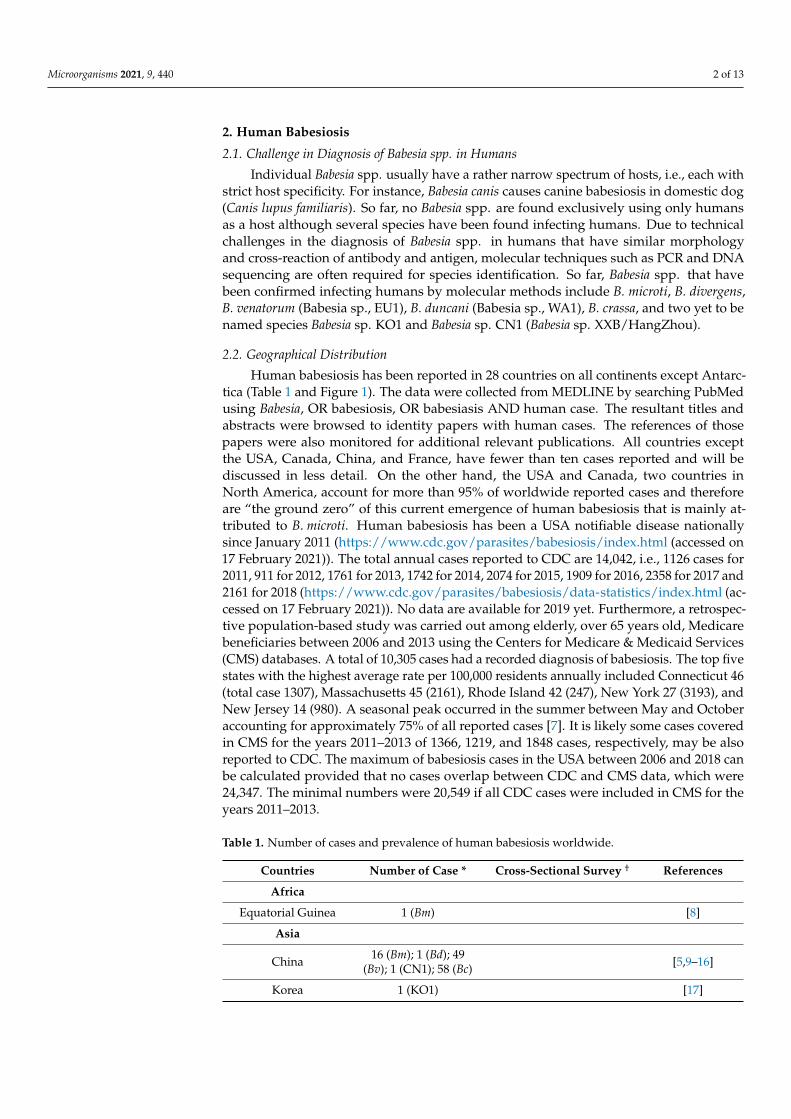
Human babesiosis has been reported in 28 countries on all continents except Antarc-tica (Table 1 and Figure 1). The data were collected from MEDLINE by searching PubMedusing Babesia, OR babesiosis, OR babesiasis AND human case. The resultant titles andabstracts were browsed to identity papers with human cases. The references of thosepapers were also monitored for additional relevant publications. All countries exceptthe USA, Canada, China, and France, have fewer than ten cases reported and will bediscussed in less detail. On the other hand, the USA and Canada, two countries inNorth America, account for more than 95% of worldwide reported cases and thereforeare “the ground zero” of this current emergence of human babesiosis that is mainly at-tributed to B. microti. Human babesiosis has been a USA notifiable disease nationallysince January 2011 (https://www.cdc.gov/parasites/babesiosis/index.html (accessed on17 February 2021)). The total annual cases reported to CDC are 14,042, i.e., 1126 cases for2011, 911 for 2012, 1761 for 2013, 1742 for 2014, 2074 for 2015, 1909 for 2016, 2358 for 2017 and2161 for 2018 (https://www.cdc.gov/parasites/babesiosis/data-statistics/index.html (ac-cessed on 17 February 2021)). No data are available for 2019 yet. Furthermore, a retrospec-tive population-based study was carried out among elderly, over 65 years old, Medicarebeneficiaries between 2006 and 2013 using the Centers for Medicare & Medicaid Services(CMS) databases. A total of 10,305 cases had a recorded diagnosis of babesiosis. The top fivestates with the highest average rate per 100,000 residents annually included Connecticut 46(total case 1307), Massachusetts 45 (2161), Rhode Island 42 (247), New York 27 (3193), andNew Jersey 14 (980). A seasonal peak occurred in the summer between May and Octoberaccounting for approximately 75% of all reported cases [7]. It is likely some cases coveredin CMS for the years 2011–2013 of 1366, 1219, and 1848 cases, respectively, may be alsoreported to CDC. The maximum of babesiosis cases in the USA between 2006 and 2018 canbe calculated provided that no cases overlap between CDC and CMS data, which were24,347. The minimal numbers were 20,549 if all CDC cases were included in CMS for theyears 2011–2013.
Table 1. Number of cases and prevalence of human babesiosis worldwide.
Countries Number of Case * Cross-Sectional Survey † References
Africa
Equatorial Guinea 1 (Bm) [8]
Asia
China 16 (Bm); 1 (Bd); 49(Bv); 1 (CN1); 58 (Bc) [5,9–16]
Korea 1 (KO1) [17]
Microorganisms 2021, 9, 440 3 of 13
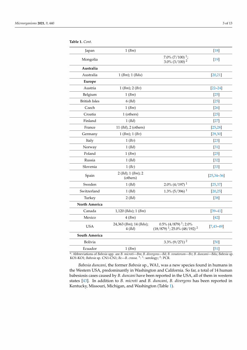
Table 1. Cont.
Japan 1 (Bm) [18]
Mongolia 7.0% (7/100) 1;3.0% (3/100) 2 [19]
Australia
Australia 1 (Bm); 1 (Bdu) [20,21]
Europe
Austria 1 (Bm); 2 (Bv) [22–24]
Belgium 1 (Bm) [25]
British Isles 6 (Bd) [25]
Czech 1 (Bm) [26]
Croatia 1 (others) [25]
Finland 1 (Bd) [27]
France 11 (Bd), 2 (others) [25,28]
Germany 1 (Bm); 1 (Bv) [29,30]
Italy 1 (Bv) [23]
Norway 1 (Bd) [31]
Poland 1 (Bm) [25]
Russia 1 (Bd) [32]
Slovenia 1 (Bc) [33]
Spain 2 (Bd); 1 (Bm); 2(others) [25,34–36]
Sweden 1 (Bd) 2.0% (4/197) 1 [25,37]
Switzerland 1 (Bd) 1.3% (5/396) 1 [20,25]
Turkey 2 (Bd) [38]
North America
Canada 1,120 (Bdu); 1 (Bm) [39–41]
Mexico 4 (Bm) [42]
USA 24,363 (Bm); 14 (Bdu);4 (Bd)
0.5% (4/879) 1; 2.0%(18/879) 1; 25.0% (48/192) 2 [7,43–49]
South America
Bolivia 3.3% (9/271) 2 [50]
Ecuador 1 (Bm) [51]*: Abbreviations of Babesia spp. are B. microti—Bm; B. divergens—Bd; B. venatorum—Bv; B. duncani—Bdu; Babesia sp.KO1-KO1; Babesia sp. CN1-CN1; Bc—B. crassa. †: 1: serology; 2: PCR.
Babesia duncani, the former Babesia sp., WA1, was a new species found in humans inthe Western USA, predominantly in Washington and California. So far, a total of 14 humanbabesiosis cases caused by B. duncani have been reported in the USA, all of them in westernstates [43]. In addition to B. microti and B. duncani, B. divergens has been reported inKentucky, Missouri, Michigan, and Washington (Table 1).
Microorganisms 2021, 9, 440 4 of 13
Microorganisms 2021, 9, x FOR PEER REVIEW 3 of 14
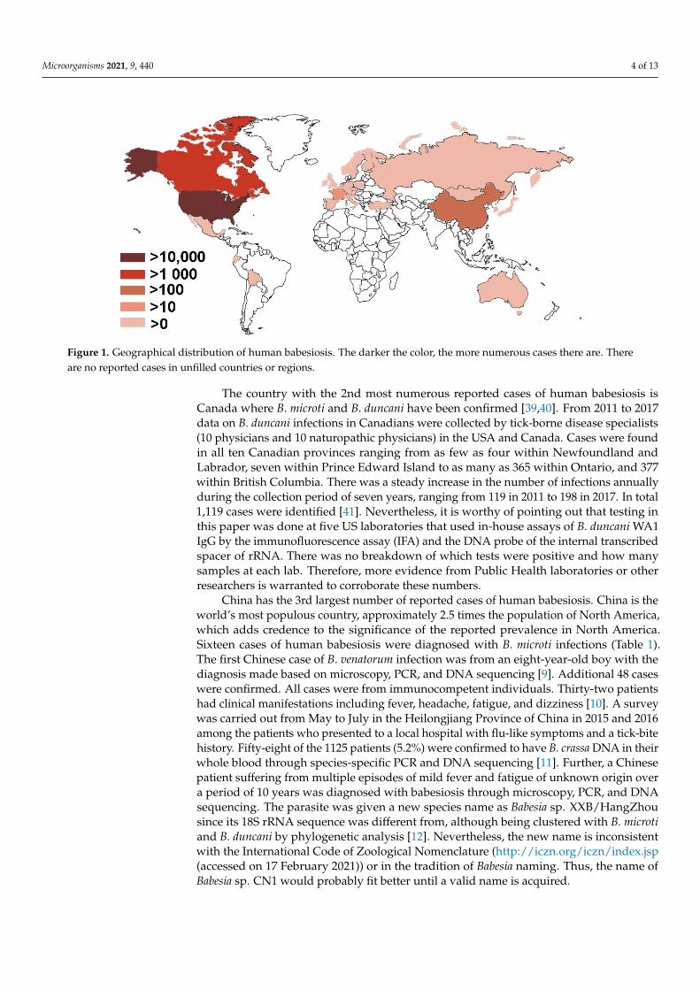
Figure 1. Geographical distribution of human babesiosis. The darker the color, the more numerous cases there are. There are no reported cases in unfilled countries or regions.
Table 1. Number of cases and prevalence of human babesiosis worldwide.
Countries Number of Case * Cross-Sectional Survey † References Africa
Equatorial Guinea
1 (Bm); [8]
Asia
China 16 (Bm); 1 (Bd); 49 (Bv); 1 (CN1);
58 (Bc) [5,9–16]
Korea 1 (KO1) Japan 1 (Bm); [17]
Mongolia 7.0% (7/100) 1 [18] 3.0% (3/100) 2 [19]
Australia Australia 1 (Bm); 1 (Bdu) [20,21] Europe Austria 1 (Bm); 2 (Bv) [22–24] Belgium 1 (Bm) [25]
British Isles 6 (Bd) [25] Czech 1 (Bm) [26]
Croatia 1 (others) [25] Finland 1 (Bd) [27] France 11 (Bd), 2 (others) [25,28]
Germany 1 (Bm); 1 (Bv) [29,30] Italy 1 (Bv) [23]
Norway 1 (Bd) [31] Poland 1 (Bm) [25] Russia 1 (Bd) [32]
Slovenia 1 (Bc) [33] Spain 2 (Bd); 1 (Bm); 2 (others) [25,34–36]
Sweden 1 (Bd) 2.0% (4/197) 1 [25,37] Switzerland 1 (Bd) 1.3% (5/396) 1 [20,25]
Turkey 2 (Bd) [38]
Figure 1. Geographical distribution of human babesiosis. The darker the color, the more numerous cases there are. Thereare no reported cases in unfilled countries or regions.
The country with the 2nd most numerous reported cases of human babesiosis isCanada where B. microti and B. duncani have been confirmed [39,40]. From 2011 to 2017data on B. duncani infections in Canadians were collected by tick-borne disease specialists(10 physicians and 10 naturopathic physicians) in the USA and Canada. Cases were foundin all ten Canadian provinces ranging from as few as four within Newfoundland andLabrador, seven within Prince Edward Island to as many as 365 within Ontario, and 377within British Columbia. There was a steady increase in the number of infections annuallyduring the collection period of seven years, ranging from 119 in 2011 to 198 in 2017. In total1,119 cases were identified [41]. Nevertheless, it is worthy of pointing out that testing inthis paper was done at five US laboratories that used in-house assays of B. duncani WA1IgG by the immunofluorescence assay (IFA) and the DNA probe of the internal transcribedspacer of rRNA. There was no breakdown of which tests were positive and how manysamples at each lab. Therefore, more evidence from Public Health laboratories or otherresearchers is warranted to corroborate these numbers.
China has the 3rd largest number of reported cases of human babesiosis. China is theworld’s most populous country, approximately 2.5 times the population of North America,which adds credence to the significance of the reported prevalence in North America.Sixteen cases of human babesiosis were diagnosed with B. microti infections (Table 1).The first Chinese case of B. venatorum infection was from an eight-year-old boy with thediagnosis made based on microscopy, PCR, and DNA sequencing [9]. Additional 48 caseswere confirmed. All cases were from immunocompetent individuals. Thirty-two patientshad clinical manifestations including fever, headache, fatigue, and dizziness [10]. A surveywas carried out from May to July in the Heilongjiang Province of China in 2015 and 2016among the patients who presented to a local hospital with flu-like symptoms and a tick-bitehistory. Fifty-eight of the 1125 patients (5.2%) were confirmed to have B. crassa DNA in theirwhole blood through species-specific PCR and DNA sequencing [11]. Further, a Chinesepatient suffering from multiple episodes of mild fever and fatigue of unknown origin overa period of 10 years was diagnosed with babesiosis through microscopy, PCR, and DNAsequencing. The parasite was given a new species name as Babesia sp. XXB/HangZhousince its 18S rRNA sequence was different from, although being clustered with B. microtiand B. duncani by phylogenetic analysis [12]. Nevertheless, the new name is inconsistentwith the International Code of Zoological Nomenclature (http://iczn.org/iczn/index.jsp(accessed on 17 February 2021)) or in the tradition of Babesia naming. Thus, the name ofBabesia sp. CN1 would probably fit better until a valid name is acquired.
Microorganisms 2021, 9, 440 5 of 13
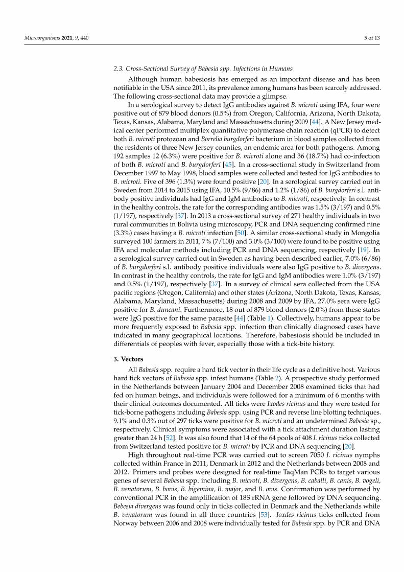
2.3. Cross-Sectional Survey of Babesia spp. Infections in Humans
Although human babesiosis has emerged as an important disease and has beennotifiable in the USA since 2011, its prevalence among humans has been scarcely addressed.The following cross-sectional data may provide a glimpse.
In a serological survey to detect IgG antibodies against B. microti using IFA, four werepositive out of 879 blood donors (0.5%) from Oregon, California, Arizona, North Dakota,Texas, Kansas, Alabama, Maryland and Massachusetts during 2009 [44]. A New Jersey med-ical center performed multiplex quantitative polymerase chain reaction (qPCR) to detectboth B. microti protozoan and Borrelia burgdorferi bacterium in blood samples collected fromthe residents of three New Jersey counties, an endemic area for both pathogens. Among192 samples 12 (6.3%) were positive for B. microti alone and 36 (18.7%) had co-infectionof both B. microti and B. burgdorferi [45]. In a cross-sectional study in Switzerland fromDecember 1997 to May 1998, blood samples were collected and tested for IgG antibodies toB. microti. Five of 396 (1.3%) were found positive [20]. In a serological survey carried out inSweden from 2014 to 2015 using IFA, 10.5% (9/86) and 1.2% (1/86) of B. burgdorferi s.l. anti-body positive individuals had IgG and IgM antibodies to B. microti, respectively. In contrastin the healthy controls, the rate for the corresponding antibodies was 1.5% (3/197) and 0.5%(1/197), respectively [37]. In 2013 a cross-sectional survey of 271 healthy individuals in tworural communities in Bolivia using microscopy, PCR and DNA sequencing confirmed nine(3.3%) cases having a B. microti infection [50]. A similar cross-sectional study in Mongoliasurveyed 100 farmers in 2011, 7% (7/100) and 3.0% (3/100) were found to be positive usingIFA and molecular methods including PCR and DNA sequencing, respectively [19]. Ina serological survey carried out in Sweden as having been described earlier, 7.0% (6/86)of B. burgdorferi s.l. antibody positive individuals were also IgG positive to B. divergens.In contrast in the healthy controls, the rate for IgG and IgM antibodies were 1.0% (3/197)and 0.5% (1/197), respectively [37]. In a survey of clinical sera collected from the USApacific regions (Oregon, California) and other states (Arizona, North Dakota, Texas, Kansas,Alabama, Maryland, Massachusetts) during 2008 and 2009 by IFA, 27.0% sera were IgGpositive for B. duncani. Furthermore, 18 out of 879 blood donors (2.0%) from these stateswere IgG positive for the same parasite [44] (Table 1). Collectively, humans appear to bemore frequently exposed to Babesia spp. infection than clinically diagnosed cases haveindicated in many geographical locations. Therefore, babesiosis should be included indifferentials of peoples with fever, especially those with a tick-bite history.
3. Vectors
All Babesia spp. require a hard tick vector in their life cycle as a definitive host. Varioushard tick vectors of Babesia spp. infest humans (Table 2). A prospective study performedin the Netherlands between January 2004 and December 2008 examined ticks that hadfed on human beings, and individuals were followed for a minimum of 6 months withtheir clinical outcomes documented. All ticks were Ixodes ricinus and they were tested fortick-borne pathogens including Babesia spp. using PCR and reverse line blotting techniques.9.1% and 0.3% out of 297 ticks were positive for B. microti and an undetermined Babesia sp.,respectively. Clinical symptoms were associated with a tick attachment duration lastinggreater than 24 h [52]. It was also found that 14 of the 64 pools of 408 I. ricinus ticks collectedfrom Switzerland tested positive for B. microti by PCR and DNA sequencing [20].
High throughout real-time PCR was carried out to screen 7050 I. ricinus nymphscollected within France in 2011, Denmark in 2012 and the Netherlands between 2008 and2012. Primers and probes were designed for real-time TaqMan PCRs to target variousgenes of several Babesia spp. including B. microti, B. divergens, B. caballi, B. canis, B. vogeli,B. venatorum, B. bovis, B. bigemina, B. major, and B. ovis. Confirmation was performed byconventional PCR in the amplification of 18S rRNA gene followed by DNA sequencing.Bebesia divergens was found only in ticks collected in Denmark and the Netherlands whileB. venatorum was found in all three countries [53]. Ioxdes ricinus ticks collected fromNorway between 2006 and 2008 were individually tested for Babesia spp. by PCR and DNA
Microorganisms 2021, 9, 440 6 of 13
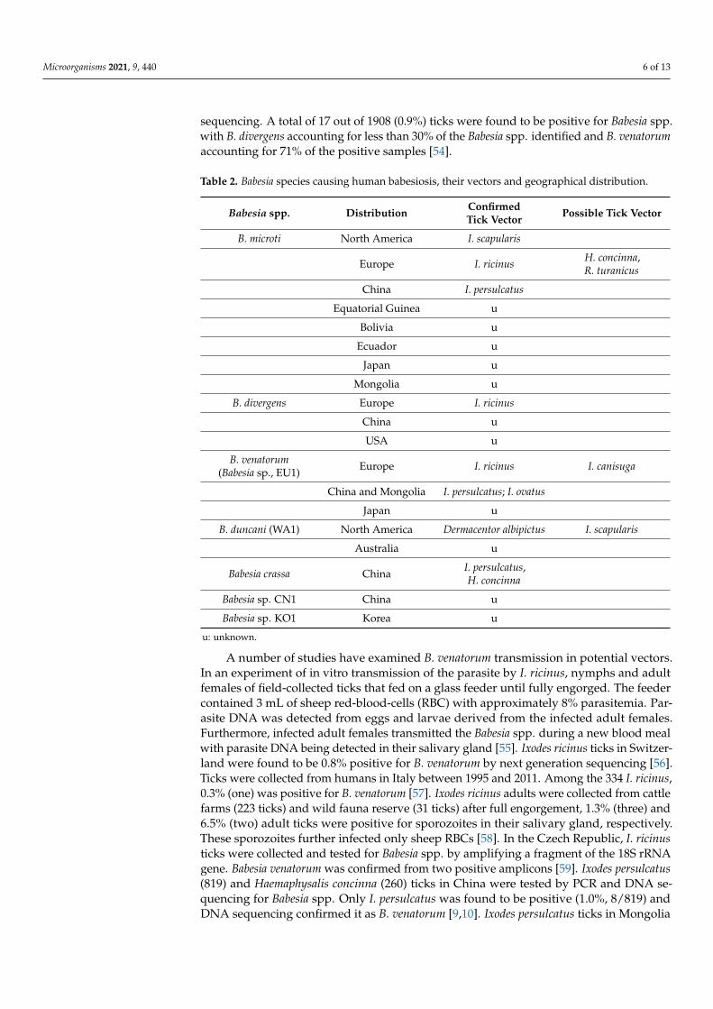
sequencing. A total of 17 out of 1908 (0.9%) ticks were found to be positive for Babesia spp.with B. divergens accounting for less than 30% of the Babesia spp. identified and B. venatorumaccounting for 71% of the positive samples [54].
Table 2. Babesia species causing human babesiosis, their vectors and geographical distribution.
Babesia spp. Distribution ConfirmedTick Vector Possible Tick Vector
B. microti North America I. scapularis
Europe I. ricinus H. concinna,R. turanicus
China I. persulcatus
Equatorial Guinea u
Bolivia u
Ecuador u
Japan u
Mongolia u
B. divergens Europe I. ricinus
China u
USA u
B. venatorum(Babesia sp., EU1) Europe I. ricinus I. canisuga
China and Mongolia I. persulcatus; I. ovatus
Japan u
B. duncani (WA1) North America Dermacentor albipictus I. scapularis
Australia u
Babesia crassa China I. persulcatus,H. concinna
Babesia sp. CN1 China u
Babesia sp. KO1 Korea u
u: unknown.
A number of studies have examined B. venatorum transmission in potential vectors.In an experiment of in vitro transmission of the parasite by I. ricinus, nymphs and adultfemales of field-collected ticks that fed on a glass feeder until fully engorged. The feedercontained 3 mL of sheep red-blood-cells (RBC) with approximately 8% parasitemia. Par-asite DNA was detected from eggs and larvae derived from the infected adult females.Furthermore, infected adult females transmitted the Babesia spp. during a new blood mealwith parasite DNA being detected in their salivary gland [55]. Ixodes ricinus ticks in Switzer-land were found to be 0.8% positive for B. venatorum by next generation sequencing [56].Ticks were collected from humans in Italy between 1995 and 2011. Among the 334 I. ricinus,0.3% (one) was positive for B. venatorum [57]. Ixodes ricinus adults were collected from cattlefarms (223 ticks) and wild fauna reserve (31 ticks) after full engorgement, 1.3% (three) and6.5% (two) adult ticks were positive for sporozoites in their salivary gland, respectively.These sporozoites further infected only sheep RBCs [58]. In the Czech Republic, I. ricinusticks were collected and tested for Babesia spp. by amplifying a fragment of the 18S rRNAgene. Babesia venatorum was confirmed from two positive amplicons [59]. Ixodes persulcatus(819) and Haemaphysalis concinna (260) ticks in China were tested by PCR and DNA se-quencing for Babesia spp. Only I. persulcatus was found to be positive (1.0%, 8/819) andDNA sequencing confirmed it as B. venatorum [9,10]. Ixodes persulcatus ticks in Mongolia
Microorganisms 2021, 9, 440 7 of 13
were also found to be positive for B. venatorum in two studies, one found a prevalence of3.2% (2/63) [60] and the other found a prevalence of 3.3% (9/275) [61].
The vector and reservoir of B. duncani had been elusive for over two decades sincethe parasite was first discovered in a patient in Washington State, USA. Lately, DNA ofB. duncani was found in the larval and adult stages of the winter tick, Dermacentor albipictus.Overall, a minimum infection prevalence in larvae was 7.2% with the highest rate of 20.7%,whereas rate in adults was 2.1% [43]. The same study also confirmed a primary reservoirwas mule deer (Odocoileus henionus) [43].
Ticks were collected from May to July in 2014 in the Heilongjiang Province of Chinaand were analyzed through species-specific PCR and DNA sequencing for B. crassa DNA.Eight of the 1296 (0.6%) I. persulcatus ticks and one of 252 (0.4%) H. concinna ticks werefound to be positive [11].
Collectively, I. ricinus has been confirmed a vector for B. microti, B. venatorum andB. divergens in Europe. Ixodes persulcatus is a vector for B. venatorum and B. crassa in Asia.Haemaphysalis concinna is a vector for B. crassa in Asia. Dermacenter albipictus is the vectorfor B. duncani in North America.
4. Tick Vectors Found in the Recreational Areas Readily Accessible to Humans
Ticks were collected all-year-round in 2011 in the Insugherata Natural Reserve locatedin northwestern Rome. The ticks that were PCR positive for the 18S rRNA gene of B. microtiwere Rhipicephalus turanicus, 1.2% (1/85, 29 males and 56 females) and I. ricinus, 12.1% (4/33,11 males and 22 females) while D. marginatus (1 male and 6 females) and H. punctate, (1 maleand 3 females) were found to be negative [62]. Three parks in the Emilia-Romagna Regionof Northern Italy were surveyed for ticks from April to October 2010 every 15 days. Picnicareas and footpaths that were frequented by people were selected. DNA was extractedfrom individual adult ticks or pools of 5 nymphs or 10 larvae from the same area at thesame time point followed by PCR and DNA sequencing of piroplasm’s 18S rRNA. In total,6.4% of the male ticks (2/31), 4.8% of the female ticks (1/21), 9.9% of the nymph tick pools(25/232), and 0.0% of the larvae (0/32) were PCR positive. Eleven including nine poolsof nymphs and two adults were positive with B. venatorum. Two pools of nymphs werepositive with B. divergens and B. capreoli [63]. Ticks were collected in the Tri-City LandscapePark in northern Poland during 2009–2010. The park was a destination for tourism andleisure among residents of the cities of Gdansk, Sopot, and Gdynia. 4.5% (34/757) of theI. ricinus ticks were found to be Babesia sp. positive through PCR with B. venatorum beingthe predominant species found [64]. In a survey in Bratislava, Slovakia, a total of 2799I. ricinus ticks were collected and this represented an urban/suburban habitat and wascharacterized by significant human development between 2011 and 2013. Thirty-threeticks (1.2%) were positive for Babesia spp. which included B. microti, B. venatorum, andB. divergens [65]. Ticks were also collected between April and June 2008 from a suburbanforest, Sénart Forest, in the southern Paris metropolitan area. This forest has three millionvisitors annually. Five hundred and fifty-eight out of the 574 ticks identified were I. ricinusticks. Babesia sp. was tested by PCR and DNA sequencing and among these I. ricinus ticks,an estimated overall prevalence of 1.6% was established. All parasites were identified byDNA sequencing as B. venatorum [66].
In short, ticks collected in areas with high human activities such as parks and picnicareas include confirmed vectors for human babesiosis such as I. ricinus. Further, PCRand DNA sequencing have detected the DNA of many Babesia spp. including B. microti,B. venatorum, and B. divergens in these ticks.
5. Phylogenetic Analysis of Babesia spp. Harbored by Humans, and Ticks on These Hosts
To understand the relationship between Babesia spp. that had been identified fromhumans, and tick vectors on humans in different geographic regions a phylogenetic anal-ysis was carried out using 18S rRNA sequences of Babesia spp. The trees were rootedwith Toxoplasma gondii. Two piroplasm species, one each in the closely related genera of
Microorganisms 2021, 9, 440 8 of 13
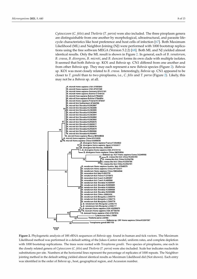
Cytauxzoon (C. felis) and Theileria (T. parva) were also included. The three piroplasm generaare distinguishable from one another by morphological, ultrastructural, and parasite life-cycle characteristics like host preference and host cells of infection [67]. Both MaximumLikelihood (ML) and Neighbor-Joining (NJ) were performed with 1000 bootstrap replica-tions using the free software MEGA (Version 5.2.2) [68]. Both ML and NJ yielded almostidentical results. Only the ML result is shown in Figure 2. In general, each of B. venatorum,B. crassa, B. divergens, B. microti, and B. duncani forms its own clade with multiple isolates.It seemed that both Babesia sp. KO1 and Babesia sp. CN1 differed from one another andfrom other Babesia spp. They may each represent a new Babesia species (Figure 2). Babesiasp. KO1 was most closely related to B. crassa. Interestingly, Babesia sp. CN1 appeared to becloser to T. gondii than to two piroplasms, i.e., C. felis and T. parva (Figure 2). Likely, thismay not be a Babesia sp. at all.
Microorganisms 2021, 9, x FOR PEER REVIEW 9 of 14
Figure 2. Phylogenetic analysis of 18S rRNA sequences of Babesia spp. found in human and tick vectors. The MaximumLikelihood method was performed in a default setting of the Jukes–Cantor model, uniform rates, and complete depletionwith 1000 bootstrap replications. The trees were rooted with Toxoplasma gondii. Two species of piroplasms, one each inthe closely related genera of Cytauxzoon (C. felis) and Theileria (T. parva) were also included. Scale bar indicates nucleotidesubstitutions per site. Numbers at the horizontal lines represent the percentage of replicates of 1000 repeats. The Neighbor-jointing method in the default setting yielded almost identical results as Maximum Likelihood did (Not shown). Each entrywas identified in the order of Babesia sp., host, geographical region, and Accession number.
Microorganisms 2021, 9, 440 9 of 13
6. Possible Roles of the Domestic Dog in Human Babesiosis
May it even be possible that pet dogs play a role in human babesiosis? Firstly, dogsin China and Russia were found to be infected with B. microti by PCR and DNA sequenc-ing [69,70]. Secondly, ticks collected from pet dogs carry B. microti, B. venatorum, andB. divergens [71–79]. These ticks might have obtained Babesia spp. pathogens from otherhosts prior to their attachment to the pet dogs. Nevertheless, these pet dogs bring theseBabesia infected ticks to the human households, making tick infestation and Babesia spp.transmission to humans a much higher possibility. Lastly, pet dogs living within thesame household were incriminated for at least two cases of human babesiosis in the litera-ture [80,81]. One needs to be cautious in interpreting this finding as no evidence presentedlinked those dogs directly to human infections. Collectively, there is no unequivocal evi-dence so far in the literature suggesting dogs contribute to human babesiosis. They mayplay a marginal role. Further experimental data are required to ascertain their roles.
7. Conclusions
This review aimed to address the geographic distribution of the human-infectingBabesia spp., their phylogenetic relationship, and their tick vector worldwide. Humanbabesiosis is caused by several Babesia spp. that includes but are not limited to, B. microti,B. divergens, B. venatorum, B. duncani, B. crassa, and two Babesia spp. strains i.e., Babesia sp.KO1 and Babesia sp. CN1. The latter two may represent a new Babesia species, which as ofyet needs to be further defined and properly named.
The number of human cases that appeared in the literature has been exponentiallyincreased in the last decade. Two countries in North America, USA, and Canada haveover twenty thousand and one thousand cases, respectively. China is in distant third placewith over one hundred cases. Cases have been reported from all continents with humanresidence. Several factors may contribute to this rapid increase in confirmed cases ofhuman babesiosis. First is the awareness of the disease in medical professionals, resultingin the correct diagnosis of the disease which would have been mistakenly diagnosed asother infections. The second is through active monitoring and survey studies. Babesiosisis a notifiable disease in the USA. Cross-sectional surveys have been carried out in manyregions including Canada and China. Third is global warming, which has expandedtick vector habitats to areas that can be readily accessed by humans such as parks andrecreational areas. The fourth factor is transfusion transmission. From 2009 to 2016 inMassachusetts alone 45 of 2578 (1.7%) were transmitted by transfusion [82]. The last, butnot least, factor is vertical transmission from an infected mother to her offspring [83].
Author Contributions: A.D. and C.Y. conceived the review. C.Y. did the literature research, per-formed the phylogenetic analysis, and drafted the manuscript. Y.Y., J.C., and L.K. wrote themanuscript. All authors have read and agreed to the published version of the manuscript.
Funding: This work was partially supported by the National Key Basic Research Program (973 Pro-gram) of China (2015CB150300), National Key Research and Development Program of China(2017YFD0501200), and Ross University School of Veterinary Medicine (41002-2021). APC waspaid by the Center one of Ross University School of Verterinary Medicine. The funding source playedno role in the design of the study and collection, analysis, and interpretation of data and in writingthe manuscript.
Institutional Review Board Statement: Not applicable.
Informed Consent Statement: Not applicable.
Data Availability Statement: All data generated or analyzed during this study are included in thispublished article.
Conflicts of Interest: The authors declare that they have no competing interests.
Microorganisms 2021, 9, 440 10 of 13
References1. Skrabalo, Z.; Deanovic, Z. Piroplasmosis in man; report of a case. Doc. Med. Geogr. Trop 1957, 9, 11–16.2. Fitzpatrick, J.E.P.; Kennedy, C.C.; McGeown, M.G.; Oreopoulos, D.G.; Robertson, J.H.; Soyannwo, M.A.O. Human Case of
Piroplasmosis (Babesiosis). Nature 1968, 217, 861. [CrossRef] [PubMed]3. Hildebrandt, A.; Gray, J.S.; Hunfeld, K.-P. Human Babesiosis in Europe: What clinicians need to know. Infection
2013, 41, 1057–1072. [CrossRef] [PubMed]4. Ord, R.L.; Lobo, C.A. Human Babesiosis: Pathogens, Prevalence, Diagnosis and Treatment. Curr. Clin. Microbiol. Rep. 2015, 2, 173–181.
[CrossRef] [PubMed]5. Zhou, X.; Xia, S.; Huang, J.-L.; Tambo, E.; Zhuge, H.-X.; Zhou, X.-N. Human babesiosis, an emerging tick-borne disease in the
People’s Republic of China. Parasit. Vctors. 2014, 7, 509. [CrossRef] [PubMed]6. Krause, P.J. Human babesiosis. Int. J. Parasitol. 2019. [CrossRef]7. Menis, M.; Forshee, R.A.; Kumar, S.; McKean, S.; Warnock, R.; Izurieta, H.S.; Gondalia, R.; Johnson, C.; Mintz, P.D.;
Walderhaug, M.O.; et al. Babesiosis Occurrence among the Elderly in the United States, as Recorded in Large Medicare Databasesduring 2006–2013. PLoS ONE 2015, 10, e0140332. [CrossRef]
8. Arsuaga, M.; González, L.M.; Padial, E.S.; Dinkessa, A.W.; Sevilla, E.; Trigo, E.; Puente, S.; Gray, J.; Montero, E. Misdiagnosis ofBabesiosis as Malaria, Equatorial Guinea, 2014. Emerg. Infect. Dis. 2018, 24, 1588. [CrossRef] [PubMed]
9. Sun, Y.; Li, S.-G.; Jiang, J.-F.; Wang, X.; Zhang, Y.; Wang, H.; Cao, W.-C. Babesia venatorum Infection in Child, China.Emerg. Infect. Dis. 2014, 20, 896–897. [CrossRef]
10. Jiang, J.-F.; Zheng, Y.-C.; Jiang, R.-R.; Li, H.; Huo, Q.-B.; Jiang, B.-G.; Sun, Y.; Jia, N.; Wang, Y.-W.; Ma, L.; et al. Epidemiological,clinical, and laboratory characteristics of 48 cases of “Babesia venatorum” infection in China: A descriptive study. Lancet. Inf. Dis.2015, 15, 196–203. [CrossRef]
11. Jia, N.; Zheng, Y.-C.; Jiang, J.-F.; Jiang, R.-R.; Jiang, B.-G.; Wei, R.; Liu, H.-B.; Huo, Q.-B.; Sun, Y.; Chu, Y.-L.; et al. HumanBabesiosis Caused by a Babesia crassa-like Pathogen: A Case Series. Clin. Infect. Dis. 2018, ciy212. [CrossRef] [PubMed]
12. Man, S.-Q.; Qiao, K.; Cui, J.; Feng, M.; Fu, Y.-F.; Cheng, X.-J. A case of human infection with a novel Babesia species in China.Infect. Dis. Poverty 2016, 5, 28. [CrossRef]
13. Zhou, X.; Li, S.-G.; Wang, J.-Z.; Huang, J.-l.; Zhou, H.-J.; Chen, J.-H.; Zhou, X.-N. Emergence of human babesiosis along the border ofChina with Myanmar: Detection by PCR and confirmation by sequencing. Emerg. Microbes Infect. 2014, 3, e55. [CrossRef] [PubMed]
14. Huang, S.; Zhang, L.; Yao, L.; Li, J.; Chen, H.; Ni, Q.; Pan, C.; Jin, L. Human babesiosis in Southeast China: A case report. Int. J.Inf. Dis. 2018, 68, 36–38. [CrossRef] [PubMed]
15. Wang, H.; Huang, F. Babesia Infection in the Southwest of China, A Case Report. Jundishapur J. Microbiol 2014, 7, e13504. [CrossRef]16. Qi, C.; Zhou, D.; Liu, J.; Cheng, Z.; Zhang, L.; Wang, L.; Wang, Z.; Yang, D.; Wang, S.; Chai, T. Detection of Babesia divergens using
molecular methods in anemic patients in Shandong Province, China. Parasitol Res. 2011, 109, 241–245. [CrossRef]17. Kim, J.-Y.; Cho, S.-H.; Joo, H.-N.; Tsuji, M.; Cho, S.-R.; Park, I.-J.; Chung, G.-T.; Ju, J.-W.; Cheun, H.-I.; Lee, H.-W.; et al. First Case
of Human Babesiosis in Korea: Detection and Characterization of a Novel Type of Babesia sp. (KO1) Similar to Ovine Babesia.J. Clin. Microbiol. 2007, 45, 2084–2087. [CrossRef]
18. Wei, Q.; Tsuji, M.; Zamoto, A.; Kohsaki, M.; Matsui, T.; Shiota, T.; Telford, S.R., 3rd; Ishihara, C. Human babesiosis in Japan:Isolation of Babesia microti-like parasites from an asymptomatic transfusion donor and from a rodent from an area where babesiosisis endemic. J. Clin. Microbiol. 2001, 39, 2178–2183. [CrossRef]
19. Hong, S.H.; Anu, D.; Jeong, Y.I.; Abmed, D.; Cho, S.H.; Lee, W.J.; Lee, S.E. Molecular detection and seroprevalence of Babesiamicroti among stock farmers in Khutul City, Selenge Province, Mongolia. Korean J. Parasitol. 2014, 52, 443–447. [CrossRef]
20. Foppa, I.M.; Krause, P.J.; Spielman, A.; Goethert, H.; Gern, L.; Brand, B.; Telford, S.R., 3rd. Entomologic and serologic evidence ofzoonotic transmission of Babesia microti, eastern Switzerland. Emerg. Infect. Dis. 2002, 8, 722–726. [CrossRef]
21. Paparini, A.; Senanayake, S.N.; Ryan, U.M.; Irwin, P.J. Molecular confirmation of the first autochthonous case of human babesiosisin Australia using a novel primer set for the beta-tubulin gene. Exp. Parasitol. 2014, 141, 93–97. [CrossRef] [PubMed]
22. Ramharter, M.; Walochnik, J.; Lagler, H.; Winkler, S.; Wernsdorfer, W.H.; Stoiser, B.; Graninger, W. Clinical and MolecularCharacterization of a Near Fatal Case of Human Babesiosis in Austria. J. Travel Med. 2010, 17, 416–418. [CrossRef] [PubMed]
23. Herwaldt, B.L.; Caccio, S.; Gherlinzoni, F.; Aspock, H.; Slemenda, S.B.; Piccaluga, P.; Martinelli, G.; Edelhofer, R.; Hollenstein, U.;Poletti, G.; et al. Molecular characterization of a non-Babesia divergens organism causing zoonotic babesiosis in Europe.Emerg Infect. Dis 2003, 9, 942–948. [CrossRef]
24. Blum, S.; Gattringer, R.; Haschke, E.; Walochnik, J.; Tschurtschenthaler, G.; Lang, F.; Oberbauer, R. The Case | Hemolysis andacute renal failure. Kidney Int. 2011, 80, 681–683. [CrossRef]
25. Gorenflot, A.; Moubri, K.; Precigout, E.; Carcy, B.; Schetters, T.P. Human babesiosis. Ann. Trop Med. Parasitol. 1998, 92, 489–501.[CrossRef] [PubMed]
26. Strizova, Z.; Havlova, K.; Patek, O.; Smrz, D.; Bartunkova, J. The first human case of babesiosis mimicking Reiter’s syndrome.Folia Parasitol. 2020, 67, 031. [CrossRef] [PubMed]
27. Karita, H.; Pekka, S.; Antti, S.; Heli, S.; Jokiranta, T.S. Fatal Babesiosis in Man, Finland, 2004. Emerg. Infect. Dis. 2010, 16, 1116. [CrossRef]28. Martinot, M.; Zadeh, M.M.; Hansmann, Y.; Grawey, I.; Christmann, D.; Aguillon, S.; Jouglin, M.; Chauvin, A.; Briel, D.D.
Babesiosis in Immunocompetent Patients, Europe. Emerg. Infect. Dis 2011, 17, 114. [CrossRef]
Microorganisms 2021, 9, 440 11 of 13
29. Hildebrandt, A.; Hunfeld, K.P.; Baier, M.; Krumbholz, A.; Sachse, S.; Lorenzen, T.; Kiehntopf, M.; Fricke, H.J.; Straube, E. First con-firmed autochthonous case of human Babesia microti infection in Europe. Eur. J. Clin. Microbiol. Infect. Dis. 2007, 26, 595–601.[CrossRef]
30. Häselbarth, K.; Tenter, A.M.; Brade, V.; Krieger, G.; Hunfeld, K.-P. First case of human babesiosis in Germany–Clinical presentationand molecular characterisation of the pathogen. Int. J. Med. Microbiol. 2007, 297, 197–204. [CrossRef]
31. Mørch, K.; Holmaas, G.; Frolander, P.S.; Kristoffersen, E.K. Severe human Babesia divergens infection in Norway. Int. J. Inf. Dis.2015, 33, 37–38. [CrossRef]
32. Kukina, I.V.; Guzeeva, T.M.; Zelya, O.P.; Ganushkina, L.A. Fatal human babesiosis caused by Babesia divergens in an asplenic host.IDCases 2018, 13, e00414. [CrossRef] [PubMed]
33. Strasek-Smrdel, K.; Korva, M.; Pal, E.; Rajter, M.; Skvarc, M.; Avsic-Zupanc, T. Case of Babesia crassa-Like Infection, Slovenia, 2014.Emerg. Infect. Dis. 2020, 26, 1038–1040. [CrossRef] [PubMed]
34. Arsuaga, M.; Gonzalez, L.M.; Lobo, C.A.; de la Calle, F.; Bautista, J.M.; Azcarate, I.G.; Puente, S.; Montero, E. First Report ofBabesia microti-Caused Babesiosis in Spain. Vector Borne Zoonotic Dis. 2016, 16, 677–679. [CrossRef]
35. González, L.M.; Castro, E.; Lobo, C.A.; Richart, A.; Ramiro, R.; González-Camacho, F.; Luque, D.; Velasco, A.C.; Montero, E. Firstreport of Babesia divergens infection in an HIV patient. Int. J. Inf. Dis. 2015, 33, 202–204. [CrossRef] [PubMed]
36. Gonzalez, L.M.; Rojo, S.; Gonzalez-Camacho, F.; Luque, D.; Lobo, C.A.; Montero, E. Severe Babesiosis in Immunocompetent Man,Spain, 2011. Emerg. Infect. Dis. 2014, 20, 724. [CrossRef] [PubMed]
37. Svensson, J.; Hunfeld, K.-P.; Persson, K.E.M. High seroprevalence of Babesia antibodies among Borrelia burgdorferi-infected humansin Sweden. Ticks Tick-Borne Dis. 2018. [CrossRef]
38. Tanyel, E.; Guler, N.; Hokelek, M.; Ulger, F.; Sunbul, M. A case of severe babesiosis treated successfully with exchange transfusion.Int. J. Inf. Dis. 2015, 38, 83–85. [CrossRef] [PubMed]
39. Bullard, J.M.P.; Ahsanuddin, A.N.; Perry, A.M.; Lindsay, L.R.; Iranpour, M.; Dibernardo, A.; Van Caeseele, P.G. The first case oflocally acquired tick-borne Babesia microti infection in Canada. Can. J. Inf. Dis. Med. Microbiol. 2014, 25, e87–e89. [CrossRef]
40. Scott, J.D. First record of locally acquired human babesiosis in Canada caused by Babesia duncani: A case report. SAGE Open Med.Case Rep. 2017, 5, 2050313×17725645. [CrossRef]
41. Scott, J.; Scott, C. Human Babesiosis Caused by Babesia duncani Has Widespread Distribution across Canada. Healthcare 2018, 6, 49.[CrossRef] [PubMed]
42. Gaspar, P.-L.; Lucero, B.; Carlos, P.-O.; Claudia, M.-Z. Human Babesiosis, Yucatán State, Mexico, 2015. Emerg. Infect. Dis. 2018, 24, 2061.[CrossRef]
43. Swei, A.; O’Connor, K.E.; Couper, L.I.; Thekkiniath, J.; Conrad, P.A.; Padgett, K.A.; Burns, J.; Yoshimizu, M.H.; Gonzales, B.;Munk, B.; et al. Evidence for transmission of the zoonotic apicomplexan parasite Babesia duncani by the tick Dermacentoralbipictus. Int. J. Parasitol. 2018. [CrossRef] [PubMed]
44. Prince, H.E.; Lapé-Nixon, M.; Patel, H.; Yeh, C. Comparison of the Babesia duncani (WA1) IgG Detection Rates among Clinical SeraSubmitted to a Reference Laboratory for WA1 IgG Testing and Blood Donor Specimens from Diverse Geographic Areas of theUnited States. Clin. Vaccine Immunol. 2010, 17, 1729–1733. [CrossRef] [PubMed]
45. Primus, S.; Akoolo, L.; Schlachter, S.; Gedroic, K.; Rojtman, A.D.; Parveen, N. Efficient detection of symptomatic and asymptomaticpatient samples for Babesia microti and Borrelia burgdorferi infection by multiplex qPCR. PLoS ONE 2018, 13, e0196748. [CrossRef][PubMed]
46. Herwaldt, B.L.; de Bruyn, G.; Pieniazek, N.J.; Homer, M.; Lofy, K.H.; Slemenda, S.B.; Fritsche, T.R.; Persing, D.H.; Limaye, A.P.Babesia divergens-like infection, Washington State. Emerg. Infect. Dis. 2004, 10, 622–629. [CrossRef]
47. Beattie, J.F.; Michelson, M.L.; Holman, P.J. Acute Babesiosis Caused by Babesia divergens in a Resident of Kentucky. N. Engl. J. Med.2002, 347, 697–698. [CrossRef] [PubMed]
48. Herc, E.; Pritt, B.; Huizenga, T.; Douce, T.; Hysell, M.; Newton, D.; Sidge, J.; Losman, E.; Sherbeck, J.; Kaul, D.R. Probable LocallyAcquired Babesia divergens–Like Infection in Woman, Michigan, USA. Emerg. Infect. Dis. 2018, 24, 1558. [CrossRef] [PubMed]
49. Herwaldt, B.; Persing, D.H.; Precigout, E.A.; Goff, W.L.; Mathiesen, D.A.; Taylor, P.W.; Eberhard, M.L.; Gorenflot, A.F. A fatalcase of babesiosis in Missouri: Identification of another piroplasm that infects humans. Ann. Intern. Med. 1996, 124, 643–650.[CrossRef]
50. Gabrielli, S.; Totino, V.; Macchioni, F.; Zuniga, F.; Rojas, P.; Lara, Y.; Roselli, M.; Bartoloni, A.; Cancrini, G. Human Babesiosis,Bolivia, 2013. Emerg. Infect. Dis. 2016, 22, 1445–1447. [CrossRef] [PubMed]
51. Al Zoubi, M.; Kwak, T.; Patel, J.; Kulkarni, M.; Kallal, C.A. Atypical challenging and first case report of babesiosis in Ecuador.IDCases 2016, 4, 15–17. [CrossRef]
52. Tijsse-Klasen, E.; Jacobs, J.J.; Swart, A.; Fonville, M.; Reimerink, J.H.; Brandenburg, A.H.; van der Giessen, J.W.; Hofhuis, A.;Sprong, H. Small risk of developing symptomatic tick-borne diseases following a tick bite in The Netherlands. Parasit Vectors2011, 4, 17. [CrossRef] [PubMed]
53. Michelet, L.; Delannoy, S.; Devillers, E.; Umhang, G.; Aspan, A.; Juremalm, M.; Chirico, J.; van der Wal, F.J.; Sprong, H.; Boye Pihl,T.P.; et al. High-throughput screening of tick-borne pathogens in Europe. Front. Cell Infect. Microbiol. 2014, 4, 103. [CrossRef][PubMed]
54. Øines, Ø.; Radzijevskaja, J.; Paulauskas, A.; Rosef, O. Prevalence and diversity of Babesia spp. in questing Ixodes ricinus ticks fromNorway. Parasit. Vectors 2012, 5, 156. [CrossRef]
Microorganisms 2021, 9, 440 12 of 13
55. Bonnet, S.; Brisseau, N.; Hermouet, A.; Jouglin, M.; Chauvin, A. Experimental in vitro transmission of Babesia sp. (EU1) by Ixodesricinus. Vet. Res. 2009, 40, 21. [CrossRef] [PubMed]
56. Oechslin, C.P.; Heutschi, D.; Lenz, N.; Tischhauser, W.; Péter, O.; Rais, O.; Beuret, C.M.; Leib, S.L.; Bankoul, S.;Ackermann-Gäumann, R. Prevalence of tick-borne pathogens in questing Ixodes ricinus ticks in urban and suburban ar-eas of Switzerland. Parasit. Vectors 2017, 10, 558. [CrossRef] [PubMed]
57. Otranto, D.; Dantas-Torres, F.; Giannelli, A.; Latrofa, M.S.; Cascio, A.; Cazzin, S.; Ravagnan, S.; Montarsi, F.; Zanzani, S.A.;Manfredi, M.T.; et al. Ticks infesting humans in Italy and associated pathogens. Parasit. Vectors 2014, 7, 328. [CrossRef] [PubMed]
58. Becker, C.A.M.; Bouju-Albert, A.; Jouglin, M.; Chauvin, A.; Malandrin, L. Natural Transmission of Zoonotic Babesia spp. by Ixodesricinus Ticks. Emerg. Infect. Dis. 2009, 15, 320–322. [CrossRef]
59. Venclikova, K.; Mendel, J.; Betasova, L.; Hubalek, Z.; Rudolf, I. First evidence of Babesia venatorum and Babesia capreoli in questingIxodes ricinus ticks in the Czech Republic. Ann. Agric. Environ. Med. 2015, 22, 212–214. [CrossRef]
60. Tuvshintulga, B.; Battsetseg, B.; Battur, B.; Myagmarsuren, P.; Narantsatsral, S.; Sivakumar, T.; Takemae, H.; Igarashi, I.; Inoue, N.;Yokoyama, N. First detection of Babesia venatorum (EU1) in Ixodes persulcatus ticks in Mongolia. J. Protozool. Res. 2015, 25, 29–37.
61. Karnath, C.; Obiegala, A.; Speck, S.; Essbauer, S.; Derschum, H.; Scholz, H.; Kiefer, D.; Tserennorov, D.; Dashdavaa, O.;Tsogbadrakh, N.; et al. Detection of Babesia venatorum, Anaplasma phagocytophilum and Candidatus Neoehrlichia mikurensis inIxodes persulcatus ticks from Mongolia. Ticks Tick-Borne Dis. 2016, 7, 357–360. [CrossRef] [PubMed]
62. Mancini, F.; Di Luca, M.; Toma, L.; Vescio, F.; Bianchi, R.; Khoury, C.; Marini, L.; Rezza, G.; Ciervo, A. Prevalence of tick-bornepathogens in an urban park in Rome, Italy. Ann. Agric. Environ. Med. 2014, 21, 723–727. [CrossRef]
63. Aureli, S.; Galuppi, R.; Ostanello, F.; Foley, J.E.; Bonoli, C.; Rejmanek, D.; Rocchi, G.; Orlandi, E.; Tampieri, M.P. Abundanceofquesting ticks and molecular evidence for pathogens in ticks in three parks of Emilia-Romagnaregion of Northern Italy. Ann. Agric.Environ. Med. 2015, 22, 459–466. [CrossRef]
64. Stanczak, J.; Cieniuch, S.; Lass, A.; Biernat, B.; Racewicz, M. Detection and quantification of Anaplasma phagocytophilum andBabesia spp. in Ixodes ricinus ticks from urban and rural environment, northern Poland, by real-time polymerase chain reaction.Exp. Appl. Acarol. 2015, 66, 63–81. [CrossRef]
65. Hamšíková, Z.; Kazimírová, M.; Haruštiaková, D.; Mahríková, L.; Slovák, M.; Berthová, L.; Kocianová, E.; Schnittger, L. Babesiaspp. in ticks and wildlife in different habitat types of Slovakia. Parasit. Vectors 2016, 9, 292. [CrossRef]
66. Reis, C.; Cote, M.; Paul, R.E.; Bonnet, S. Questing ticks in suburban forest are infected by at least six tick-borne pathogens.Vector Borne Zoonotic Dis. 2011, 11, 907–916. [CrossRef] [PubMed]
67. Wang, J.L.; Li, T.T.; Liu, G.H.; Zhu, X.Q.; Yao, C. Two Tales of Cytauxzoon felis Infections in Domestic Cats. Clin. Microbiol. Rev.2017, 30, 861–885. [CrossRef]
68. Tamura, K.; Peterson, D.; Peterson, N.; Stecher, G.; Nei, M.; Kumar, S. MEGA5: Molecular evolutionary genetics analysis usingmaximum likelihood, evolutionary distance, and maximum parsimony methods. Mol. Biol. Evol. 2011, 28, 2731–2739. [CrossRef][PubMed]
69. Bloch, E.M.; Yang, Y.; He, M.; Tonnetti, L.; Liu, Y.; Wang, J.; Guo, Y.; Li, H.; Leiby, D.A.; Shan, H.; et al. A pilot serosurvey ofBabesia microti in Chinese blood donors. Vox Sang. 2018. [CrossRef]
70. Gabrielli, S.; Otasevic, S.; Ignjatovic, A.; Savic, S.; Fraulo, M.; Arsic-Arsenijevic, V.; Momcilovic, S.; Cancrini, G. Canine Babesiosesin Noninvestigated Areas of Serbia. Vector Borne Zoonotic Dis. 2015, 15, 535–538. [CrossRef] [PubMed]
71. Abdullah, S.; Helps, C.; Tasker, S.; Newbury, H.; Wall, R. Prevalence and distribution of Borrelia and Babesia species in ticksfeeding on dogs in the U.K. Med. Vet. Entomol. 2018, 32, 14–22. [CrossRef]
72. Inokuma, H.; Yoshizaki, Y.; Shimada, Y.; Sakata, Y.; Okuda, M.; Onishi, T. Epidemiological Survey of Babesia Species in JapanPerformed with Specimens from Ticks Collected from Dogs and Detection of New Babesia DNA Closely Related to Babesia odocoileiand Babesia divergens DNA. J. Clin. Microbiol. 2003, 41, 3494–3498. [CrossRef]
73. Król, N.; Obiegala, A.; Pfeffer, M.; Lonc, E.; Kiewra, D. Detection of selected pathogens in ticks collected from cats and dogs in theWrocław Agglomeration, South-West Poland. Parasit. Vectors 2016, 9, 351. [CrossRef]
74. Lempereur, L.; De Cat, A.; Caron, Y.; Madder, M.; Claerebout, E.; Saegerman, C.; Losson, B. First molecular evidence of potentiallyzoonotic Babesia microti and Babesia sp. EU1 in Ixodes ricinus ticks in Belgium. Vector. Borne Zoonotic Dis. 2011, 11, 125–130.[CrossRef] [PubMed]
75. Potkonjak, A.; Gutiérrez, R.; Savic, S.; Vracar, V.; Nachum-Biala, Y.; Jurišic, A.; Kleinerman, G.; Rojas, A.; Petrovic, A.; Baneth, G.;et al. Molecular detection of emerging tick-borne pathogens in Vojvodina, Serbia. Ticks Tick-Borne Dis. 2016, 7, 199–203. [CrossRef][PubMed]
76. Schreiber, C.; Krücken, J.; Beck, S.; Maaz, D.; Pachnicke, S.; Krieger, K.; Gross, M.; Kohn, B.; von Samson-Himmelstjerna, G.Pathogens in ticks collected from dogs in Berlin/Brandenburg, Germany. Parasit Vectors 2014, 7, 535. [CrossRef]
77. Smith, F.D.; Wall, L.E. Prevalence of Babesia and Anaplasma in ticks infesting dogs in Great Britain. Vet. Parasitol. 2013, 198, 18–23.[CrossRef]
78. Stensvold, C.R.; Al Marai, D.; Andersen, L.O.B.; Krogfelt, K.A.; Jensen, J.S.; Larsen, K.S.; Nielsen, H.V. Babesia spp. and otherpathogens in ticks recovered from domestic dogs in Denmark. Parasit. Vectors 2015, 8, 262. [CrossRef]
79. Zygner, W.; Baska, P.; Winsniewski, M.; Halina, W. The Molecular Evidence of Babesia microti in Hard Ticks Removed from Dogsin Warsaw (central Poland). Pol. J. Microbiol. 2010, 59, 95–97. [CrossRef] [PubMed]

Microorganisms 2021, 9, 440 13 of 13
80. Chan, J.M.; Tsang, K.Y.; Chik, T.; Leung, W.S.; Tsang, O.T. Babesiosis acquired from a pet dog: A second reported case in HongKong. Hong Kong Med. J. 2016, 22, 393–395. [CrossRef]
81. El-Bahnasawy, M.M.; Khalil, H.H.; Morsy, T.A. Babesiosis in an Egyptian boy aquired from pet dog, and a general review. J. EgyptSoc. Parasitol. 2011, 41, 99–108.
82. Klevens, R.M.; Cumming, M.A.; Caten, E.; Stramer, S.L.; Townsend, R.L.; Tonnetti, L.; Rios, J.; Young, C.T.; Soliva, S.; DeMaria, A.Transfusion-transmitted babesiosis: One state’s experience. Transfusion 2018, 58, 2611–2616. [CrossRef]
83. Iyer, S.; Goodman, K. Congenital Babesiosis from Maternal Exposure: A Case Report. J. Emerg. Med. 2019. [CrossRef]