Embed Size (px)
Citation preview
Emergency Medical Response
Table of Contents vii
Table of ContentsUNIT 1: PREPARATORY
1 The Emergency Medical Responder 2
2 The Well-Being of the Emergency Medical Responder 14
3 Medical, Legal and Ethical Issues 41
4 The Human Body 58
5 Lifting and Moving Patients 81
UNIT 2: ASSESSMENT
6 Scene Size-Up 114
7 Primary Assessment 135
8 History Taking and Secondary Assessment 163
9 Communication and Documentation 199
UNIT 3: AIRWAY
10 Airway and Ventilation 213
11 Airway Management 251
12 Emergency Oxygen 277
UNIT 4: CIRCULATION
13 Circulation and Cardiac Emergencies 290
UNIT 5: MEDICAL EMERGENCIES
14 Medical Emergencies 324
15 Poisoning 348
16 Environmental Emergencies 372
17 Behavioral Emergencies 404
UNIT 6: TRAUMA EMERGENCIES
18 Shock 418
19 Bleeding and Trauma 425
20 Soft Tissue Injuries 444
21 Injuries to the Chest, Abdomen and Genitalia 461
22 Injuries to Muscles, Bones and Joints 475
23 Injuries to the Head, Neck and Spine 505
UNIT 7: SPECIAL POPULATIONS
24 Childbirth 530
25 Pediatrics 550
26 Geriatrics and Special Needs Patients 569
UNIT 8: EMS OPERATIONS
27 EMS Support and Operations 584
28 Access and Extrication 601
29 Hazardous Materials Emergencies 611
30 Incident Command and Multiple-Casualty Incidents 623
31 Response to Disasters and Terrorism 638
32 Special Operations 659
GLOSSARY 671
SOURCES 698
INDEX 702
viii Emergency Medical Response
Detailed Table of ContentsUNIT 1: PREPARATORY
1 The Emergency Medical Responder 2
You Are the Emergency Medical Responder 2
Key Terms 3
Learning Objectives 3
Introduction 4
The EMS System 4
Emergency Medical Responder 8
Putting It All Together 13
2 The Well-Being of the Emergency Medical Responder 14
You Are the Emergency Medical Responder 14
Key Terms 15
Learning Objectives 16
Introduction 17
Preventing Disease Transmission 17
Emotional Aspects of Emergency Care 30
Stress Management 32
Incident Stress Management 33
Putting It All Together 35
You Are the Emergency Medical Responder 36
Skill Sheet: Removing Disposable Gloves 37
Enrichment: Health of the Emergency Medical Responder 38
3 Medical, Legal and Ethical Issues 41
You Are the Emergency Medical Responder 41
Key Terms 42
Learning Objectives 43
Introduction 44
Legal Duties 44
Patient Consent and Refusal of Care 46
Other Legal Issues 52
Confi dentiality and Privacy 53
Special Situations 55
Putting It All Together 56
You Are the Emergency Medical Responder 57
4 The Human Body 58
You Are the Emergency Medical Responder 58
Key Terms 59
Learning Objectives 59
Introduction 60
Medical Terminology 60
Anatomical Terms 60
Body Systems 64
Putting It All Together 79
You Are the Emergency Medical Responder 80
5 Lifting and Moving Patients 81
You Are the Emergency Medical Responder 81
Key Terms 82
Learning Objectives 83
Skill Objectives 83
Detailed Table of Contents ix
Introduction 84
Role of the Emergency Medical Responder 84
Principles of Moving Patients 85
Emergency Moves 87
Non-Emergency Moves 90
Equipment 92
Patient Positioning and Packaging for Transport 94
Medical Restraint 97
Putting It All Together 98
You Are the Emergency Medical Responder 99
Skill Sheet: Clothes Drag 100
Skill Sheet: Blanket Drag 101
Skill Sheet: Shoulder Drag 102
Skill Sheet: Ankle Drag 103
Skill Sheet: Firefighter’s Drag 104
Skill Sheet: Firefighter’s Carry 105
Skill Sheet: Pack-Strap Carry 106
Skill Sheet: Walking Assist 107
Skill Sheet: Two-Person Seat Carry 108
Skill Sheet: Direct Ground Lift 109
Skill Sheet: Extremity Lift 111
UNIT 2: ASSESSMENT
6 Scene Size-Up 114
You Are the Emergency Medical Responder 114
Key Terms 115
Learning Objectives 115
Introduction 116
Dispatch Information 116
Safety 116
Mechanism of Injury and Nature of Illness 120
Additional Resources 127
Putting It All Together 130
You Are the Emergency Medical Responder 130
Enrichment: Dealing with Hazards at the Scene 131
7 Primary Assessment 135
You Are the Emergency Medical Responder 135
Key Terms 136
Learning Objectives 137
Skill Objectives 137
Introduction 138
The Importance of Scene Size-Up 138
General Impression of the Patient 139
Responsiveness 140
Airway Status 142
Breathing Status 144
Circulatory Status 147
Identifying Life Threats 151
Shock 151
Putting It All Together 152
You Are the Emergency Medical Responder 152
Skill Sheet: Jaw-Thrust (Without Head Extension) Maneuver 153
Skill Sheet: Using a Resuscitation Mask—Adult, Child and Infant 154
Skill Sheet: Using a Resuscitation Mask—Head, Neck or Spinal Injury Suspected (Jaw-Thrust [Without Head Extension] Maneuver—Adult or Child) 156
x Emergency Medical Response
Skill Sheet: Taking and Recording a Patient’s Blood Pressure (by Auscultation) 193
Skill Sheet: Taking and Recording a Patient’s Blood Pressure (by Palpation) 195
Enrichment: Pulse Oximetry 197
9 Communication and Documentation 199
You Are the Emergency Medical Responder 199
Key Terms 200
Learning Objectives 200
Introduction 201
Communicating Within the Emergency Communications System 201
Interpersonal Communication 204
The Importance of Documentation 206
Prehospital Care Report 206
Transfer of Care 209
Special Situations 210
Putting It All Together 210
You Are the Emergency Medical Responder 210
UNIT 3: AIRWAY
10 Airway and Ventilation 213
You Are the Emergency Medical Responder 213
Key Terms 214
Learning Objectives 215
Skill Objectives 215
Introduction 216
The Respiratory System 216
Respiratory Emergencies 217
Airway 221
Skill Sheet: Primary Assessment 158
Enrichment: Glasgow Coma Scale 161
8 History Taking and Secondary Assessment 163
You Are the Emergency Medical Responder 163
Key Terms 164
Learning Objectives 165
Skill Objectives 165
Introduction 166
Obtaining the Focused/Medical History 166
Components of a Patient History 167
SAMPLE History 168
The Secondary Assessment 169
Detailed Physical Exam 172
Obtaining Baseline Vital Signs 175
Ongoing Assessment 182
The Need for More Advanced Medical Personnel 183
Putting It All Together 183
You Are the Emergency Medical Responder 185
Skill Sheet: How to Obtain a SAMPLE History 186
Skill Sheet: How to Perform a Secondary Assessment for a Responsive Medical Patient 187
Skill Sheet: How to Perform a Secondary Assessment for an Unresponsive Medical Patient 188
Skill Sheet: Physical Exam and Patient History 189
Skill Sheet: How to Obtain Baseline Vital Signs 191
Detailed Table of Contents xi
You Are the Emergency Medical Responder 260
Skill Sheet: Using a Mechanical Suctioning Device 261
Skill Sheet: Using a Manual Suctioning Device 263
Skill Sheet: Inserting an Oral Airway 265
Skill Sheet: Conscious Choking—Adult and Child 267
Skill Sheet: Conscious Choking—Infant 269
Skill Sheet: Unconscious Choking—Adult and Child 271
Skill Sheet: Unconscious Choking—Infant 273
Enrichment: Nasopharyngeal Airway 275
Skill Sheet: Inserting a Nasal Airway 275
12 Emergency Oxygen 277
You Are the Emergency Medical Responder 277
Key Terms 278
Learning Objectives 278
Skill Objectives 278
Introduction 279
Administering Emergency Oxygen 279
Safety Precautions 285
Putting It All Together 285
You Are the Emergency Medical Responder 285
Skill Sheet: Oxygen Delivery 286
UNIT 4: CIRCULATION
13 Circulation and Cardiac Emergencies 290
You Are the Emergency Medical Responder 290
Assessing Breathing 223
Artifi cial Ventilation 226
Putting It All Together 231
You Are the Emergency Medical Responder 232
Skill Sheet: Giving Ventilations—Adult and Child 233
Skill Sheet: Giving Ventilations—Infant 236
Skill Sheet: Giving Ventilations—Head, Neck or Spinal Injury Suspected: Jaw-Thrust (Without Head Extension) Maneuver—Adult and Child 238
Skill Sheet: Giving Ventilations Using a Bag-Valve-Mask Resuscitator—Two Rescuers 241
Enrichment: Assessing Breath Sounds 244
Enrichment: Sellick’s Maneuver (Cricoid Pressure) 245
Skill Sheet: Perfoming the Sellick’s Maneuver (Cricoid Pressure) 246
Enrichment: Assisting the Patient with Asthma 247
Skill Sheet: Assisting with an Asthma Inhaler 249
11 Airway Management 251
You Are the Emergency Medical Responder 251
Key Terms 252
Learning Objectives 252
Skill Objectives 252
Introduction 253
Suctioning 253
Breathing Devices 254
Airway Adjuncts 254
Airway Obstruction 256
Putting It All Together 260
xii Emergency Medical Response
Diabetic Emergencies 332
Stroke 334
Abdominal Pain 336
Hemodialysis 338
Putting It All Together 339
You Are the Emergency Medical Responder 339
Enrichment: Basic Pharmacology 340
Enrichment: Blood Glucose Monitoring 346
15 Poisoning 348
You Are the Emergency Medical Responder 348
Key Terms 349
Learning Objectives 350
Introduction 351
Poison Control Centers 351
How Poison Enters the Body 353
Substance Abuse and Misuse 357
Adolescents and Substance Abuse 364
Putting It All Together 366
You Are the Emergency Medical Responder 366
Enrichment: Administering Activated Charcoal 367
Enrichment: Carbon Monoxide and Cyanide Poisoning 368
16 Environmental Emergencies 372
You Are the Emergency Medical Responder 372
Key Terms 373
Learning Objectives 374
Skill Objectives 374
Introduction 375
Key Terms 291
Learning Objectives 292
Skill Objectives 292
Introduction 293
The Circulatory System 293
Cardiac Arrest 298
CPR 299
Automated External Defi brillation 305
Automated External Defi brillators 306
AED Precautions 310
AED Maintenance 310
Putting It All Together 310
You Are the Emergency Medical Responder 311
Skill Sheet: CPR—Adult and Child 312
Skill Sheet: CPR—Infant 314
Skill Sheet: Two-Rescuer CPR—Adult and Child 315
Skill Sheet: Two-Rescuer CPR—Infant 317
Skill Sheet: AED—Adult, Child and Infant 319
Enrichment: Preventing Coronary Heart Disease 322
UNIT 5: MEDICAL EMERGENCIES
14 Medical Emergencies 324
You Are the Emergency Medical Responder 324
Key Terms 325
Learning Objectives 326
Introduction 327
General Medical Complaints 327
Altered Mental Status 327
Seizures 329
Detailed Table of Contents xiii
UNIT 6: TRAUMA EMERGENCIES
18 Shock 418
You Are the Emergency Medical Responder 418
Key Terms 419
Learning Objectives 419
Introduction 420
What is Shock? 420
Why Shock Occurs 420
Types of Shock 421
Signs and Symptoms of Shock 421
Providing Care 423
Putting It All Together 424
You Are the Emergency Medical Responder 424
19 Bleeding and Trauma 425
You Are the Emergency Medical Responder 425
Key Terms 426
Learning Objectives 427
Skill Objectives 427
Introduction 428
Incidence/Signifi cance of Trauma 428
Trauma System 428
Multi-System Trauma 429
Perfusion 429
Bleeding 429
Dressings and Bandages 430
External Bleeding 432
Putting It All Together 437
You Are the Emergency Medical Responder 437
Skill Sheet: Controlling External Bleeding 438
Body Temperature 375
People at Risk for Heat-Related Illnesses and Cold-Related Emergencies 376
Heat-Related Illnesses 377
Cold-Related Emergencies 381
Preventing Heat-Related Illnesses and Cold-Related Emergencies 384
Bites and Stings 385
Water-Related Emergencies 392
Putting It All Together 396
You Are the Emergency Medical Responder 396
Enrichment: Assisting with an Epinephrine Auto-Injector 397
Skill Sheet: Assisting with an Epinephrine Auto-injector 398
Enrichment: Lightning 400
Enrichment: SCUBA and Free Diving Emergencies 402
17 Behavioral Emergencies 404
You Are the Emergency Medical Responder 404
Key Terms 405
Learning Objectives 405
Introduction 406
Behavioral Emergencies 406
Psychological Emergencies 407
Violence 409
Providing Care for Behavioral Emergencies 414
Putting It All Together 415
You Are the Emergency Medical Responder 416
xiv Emergency Medical Response
22 Injuries to Muscles, Bones and Joints 475
You Are the Emergency Medical Responder 475
Key Terms 476
Learning Objectives 477
Skill Objectives 477
Introduction 478
Musculoskeletal System 478
Injuries to Muscles, Bones and Joints 479
Splinting 484
Putting It All Together 491
You Are the Emergency Medical Responder 491
Skill Sheet: Applying a Rigid Splint 492
Skill Sheet: Applying a Sling and Binder 494
Skill Sheet: Applying an Anatomic Splint 496
Skill Sheet: Applying a Soft Splint 498
Enrichment: Agricultural and Industrial Emergencies 500
23 Injuries to the Head, Neck and Spine 505
You Are the Emergency Medical Responder 505
Key Terms 506
Learning Objectives 506
Skill Objectives 506
Introduction 507
Anatomy of the Head, the Neck and the Spine 507
Injuries to the Head 507
Injuries to the Neck and Spine 514
Enrichment: Mechanisms of Injury—The Kinematics of Trauma 439
Enrichment: Tourniquets 442
Skill Sheet: Using a Manufactured Tourniquet 442
20 Soft Tissue Injuries 444
You Are the Emergency Medical Responder 444
Key Terms 445
Learning Objectives 445
Introduction 446
Skin and Soft Tissue Injuries 446
Closed Wounds 447
Open Wounds 448
Burns 452
Putting It All Together 460
You Are the Emergency Medical Responder 460
21 Injuries to the Chest, Abdomen and Genitalia 461
You Are the Emergency Medical Responder 461
Key Terms 462
Learning Objectives 462
Introduction 463
Anatomy of the Chest, Abdomen and Genitalia 463
Chest Injuries 464
Abdominal Injuries 470
Genital Injuries 473
Putting It All Together 474
You Are the Emergency Medical Responder 474
Detailed Table of Contents xv
Putting It All Together 547
You Are the Emergency Medical Responder 547
Enrichment: More Complications During Pregnancy and Delivery 548
25 Pediatrics 550
You Are the Emergency Medical Responder 550
Key Terms 551
Learning Objectives 551
Introduction 552
Anatomical Differences 552
Child Development 553
Assessing Pediatrics 554
Common Problems in Pediatric Patients 558
The Emergency Medical Responder’s Needs 567
Putting It All Together 568
You Are the Emergency Medical Responder 568
26 Geriatrics and Special Needs Patients 569
You Are the Emergency Medical Responder 569
Key Terms 570
Learning Objectives 570
Introduction 571
Geriatric Patients 571
Patients with Special Needs 577
Putting It All Together 582
You Are the Emergency Medical Responder 582
Putting It All Together 517
You Are the Emergency Medical Responder 517
Skill Sheet: Manual Stabilization 518
Skill Sheet: Controlling Bleeding from an Open Head Wound 519
Skill Sheet: Bandaging an Eye with an Injury from an Impaled Object 520
Skill Sheet: Caring for Foreign Bodies in the Eye 521
Enrichment: Removing Helmets and Other Equipment 522
Enrichment: Cervical Collars and Backboarding 525
Skill Sheet: Immobilizing a Head, Neck or Spinal Injury 526
UNIT 7: SPECIAL POPULATIONS
24 Childbirth 530
You Are the Emergency Medical Responder 530
Key Terms 531
Learning Objectives 532
Introduction 533
Anatomy and Physiology of Pregnancy 533
Normal Pregnancy 533
Birth and Labor Process 534
Preparing for Delivery 536
Delivery 538
Caring for the Newborn and Mother 539
Complications During Pregnancy 542
Complications During Delivery 544
xvi Emergency Medical Response
29 Hazardous Materials Emergencies 611
You Are the Emergency Medical Responder 611
Key Terms 612
Learning Objectives 612
Introduction 613
Hazardous Materials 613
HAZMAT Incidents 615
Scene Safety and Personal Protective Equipment 618
Contamination and Decontamination 620
Putting It All Together 622
You Are the Emergency Medical Responder 622
30 Incident Command and Multiple-Casualty Incidents 623
You Are the Emergency Medical Responder 623
Key Terms 624
Learning Objectives 624
Introduction 625
National Incident Management System 625
Multiple-Casualty Incidents 627
Triage 628
Stress at an MCI 635
Communication 637
Putting It All Together 637
You Are the Emergency Medical Responder 637
31 Response to Disasters and Terrorism 638
You Are the Emergency Medical Responder 638
Key Terms 639
Learning Objectives 639
UNIT 8: EMS OPERATIONS
27 EMS Support and Operations 584
You Are the Emergency Medical Responder 584
Key Terms 585
Learning Objectives 585
Introduction 586
Roles of the EMR in the EMS System 586
Phases of a Response 586
Air Medical Transport Considerations 590
Emergency Vehicle Safety 593
Leaving the Scene 598
EMS Equipment 598
Putting It All Together 599
You Are the Emergency Medical Responder 599
Enrichment: Operational Safety and Security Measures 600
28 Access and Extrication 601
You Are the Emergency Medical Responder 601
Key Terms 602
Learning Objectives 602
Introduction 603
Fundamentals of Extrication and Rescue Operations 603
Vehicle Stabilization 606
Gaining Access 608
Extrication 609
Putting It All Together 610
You Are the Emergency Medical Responder 610
Detailed Table of Contents xvii
32 Special Operations 659
You Are the Emergency Medical Responder 659
Key Terms 660
Learning Objectives 660
Introduction 661
Hazardous Terrain 665
Confined Space 665
Crime Scene 667
Fireground Operations 667
Special Events and Standby 668
Putting It All Together 669
You Are the Emergency Medical Responder 670
Glossary 671
Sources 698
Index 702
Introduction 640
Preparing for Disasters and Terrorist Incidents 640
Incident Management 640
The Role of the Emergency Medical Responder 643
Disaster Response 643
WMDs (Chemical, Biological, Radiological/Nuclear and Explosive Incidents) 646
Response to a CBRNE WMD Incident 651
Providing Self-Care and Peer Care for Nerve Agents 653
Putting It All Together 655
You Are the Emergency Medical Responder 656
Enrichment: Preparing for a Public Health Disaster—Pandemic Flu 657
Enrichment: Personal Preparedness 658
Circulation and Cardiac Emergencies
YOU ARE THE EMERGENCY MEDICAL RESPONDER
An elderly man suddenly collapses while working in the offi ce. He is lying on
the fl oor and does not appear to be moving. You, as a member of the medical
emergency response team (MERT), recognize the emergency, activate the
emergency response plan and perform a primary assessment. The emergency
medical services (EMS) system has been activated. You determine that the man is
unconscious, not breathing and does not have a pulse. The offi ce building has an
automated external defi brillator (AED). How would you respond?
)))
13
13 : Circulation and Cardiac Emergencies 291
Acute coronary syndrome (ACS): Term that describes a range of clinical conditions, including unstable angina, that are due to insuffi cient blood supply to the heart muscle resulting from coronary heart disease (CHD).
Acute myocardial ischemia: An episode of chest pain due to reduced blood fl ow to the heart muscle.
Angina pectoris: Pain in the chest that comes and goes at different times; caused by a lack of oxygen reaching the heart; can be stable (occurring under exertion or stress) or unstable (occurring at rest, without reason).
Arrhythmia: Disturbance in the regular rhythmic beating of the heart.
Asystole: A condition where the heart has stopped generating electrical activity.
Atherosclerosis: A condition in which deposits of plaque, including cholesterol (a fatty substance made by the liver and found in foods containing animal or animal products) build up on the inner walls of the arteries, causing them to harden and narrow, reducing the amount of blood that can fl ow through; develops gradually and can go undetected for many years.
Atrial fi brillation: Irregular and fast electrical discharges of the heart that lead to an irregular heartbeat; the most common type of abnormal cardiac rhythm.
Atrioventricular (AV) node: A cluster of cells in the center of the heart, between the atria and ventricles; serves as a relay to slow down the signal received from the sinoatrial (SA) node before it passes through to the ventricles.
Automated external defi brillator (AED): A portable electronic device that analyzes the heart’s electrical rhythm and, if necessary, can deliver an electrical shock to a person in cardiac arrest.
Cardiac arrest: A condition in which the heart has stopped or beats too irregularly or weakly to pump blood effectively.
Cardiac chain of survival: A set of four critical steps in responding to a cardiac emergency: early recognition and access to the EMS system, early cardiopulmonary resuscitation (CPR), early defi brillation and early advanced medical care.
Cardiopulmonary resuscitation (CPR): A technique that combines chest compressions and ventilations to circulate blood containing oxygen to the brain and other vital organs for a person whose heart and breathing have stopped.
Key TermsCardiovascular disease: A disease affecting the
heart and blood vessels.Chest compressions: A technique used in CPR,
in which external pressure is placed on the chest to increase the level of pressure in the chest cavity and cause the blood to circulate through the arteries.
Cholesterol: A fatty substance made by the liver and found in foods containing animal or animal products; diets high in cholesterol contribute to the risk of heart disease.
Commotio cordis: Sudden cardiac arrest from a blunt, non-penetrating blow to the chest, of which the basis is ventricular fi brillation (V-fi b) triggered by chest wall impact immediately over the heart.
Congestive heart failure: A chronic condition in which the heart no longer pumps blood effectively throughout the body.
Coronary heart disease (CHD): A disease in which cholesterol and plaque build up on the inner walls of the arteries that supply blood to the heart; also called coronary artery disease (CAD).
Defi brillation: An electrical shock that disrupts the electrical activity of the heart long enough to allow the heart to spontaneously develop an effective rhythm on its own.
Electrocardiogram (ECG or EKG): A test that measures and records the electrical activity of the heart.
Heart: A fi st-sized muscular organ that pumps blood throughout the body.
Hypertension: Another term for high blood pressure.
Implantable cardioverter-defi brillator (ICD): A miniature version of an AED, implanted under the skin, that acts to automatically recognize and help correct abnormal heart rhythms.
Myocardial infarction (MI): The death of cardiac muscle tissue due to a sudden deprivation of circulating blood; also called a heart attack.
Normal sinus rhythm (NSR): The normal, regular rhythm of the heart, set by the SA node in the right atrium of the heart.
Pacemaker: A device implanted under the skin, sometimes below the right collarbone, to help regulate heartbeat in someone with a weak heart, a heart that skips beats or one that beats too fast or too slow.
Risk factors: Conditions or behaviors that increase the chance that a person will develop a disease.
292 Unit 4 : Circulation
rhythm is restored, death follows within a matter of minutes.
Transdermal medication patch: A patch on the skin that delivers medication; commonly contains nitroglycerin, nicotine or other medications; should be removed prior to defi brillation.
Ventricular fi brillation (V-fi b): A life-threatening heart rhythm in which the heart is in a state of totally disorganized electrical activity.
Ventricular tachycardia (V-tach): A life-threatening heart rhythm in which there is very rapid contraction of the ventricles.
Silent heart attack: A heart attack during which the patient has either no symptoms or very mild symptoms that the person does not associate with heart attacks; mild symptoms include indigestion or sweating.
Sinoatrial (SA) node: A cluster of cells in the right atrium that generates the electrical impulses that set the pace of the heart’s natural rhythm.
Sudden cardiac arrest: A condition where the heart’s pumping action stops abruptly, usually due to abnormal heart rhythms called arrhythmias, most commonly V-fi b; unless an effective heart
• Describe how to recognize and care for a victim who may be experiencing a heart attack.
• Describe how to care for a patient who may be experiencing cardiac arrest.
• List the reasons for the heart to stop beating.• Describe the skill components of CPR.• List the steps of one-rescuer CPR for an adult, a
child and an infant.• Explain when it is appropriate to stop
performing CPR.• Describe how to perform two-rescuer CPR for an
adult, a child and an infant.
• Defi ne defi brillation and describe how it works.• Identify the abnormal heart rhythms commonly
present during cardiac arrest.• Describe the role and importance of early
defi brillation in cardiac arrest.• List the general steps for using an automated
external defi brillator (AED).• Identify precautions for using an AED.• Identify special situations that may arise when
using an AED.• Identify controllable risk factors for cardiovascular
disease (Enrichment).
Learning ObjectivesAfter reading this chapter, and completing the class activities, you will have the information needed to—
• Demonstrate one-rescuer CPR for an adult, a child and an infant.
• Demonstrate two-rescuer CPR for an adult, a child and an infant.
• Demonstrate how to use an AED for adult and pediatric patients in cardiac arrest.
Skill ObjectivesAfter reading this chapter, and completing the class activities, you should be able to—
13 : Circulation and Cardiac Emergencies 293
to the lungs where waste products are removed and oxygen is absorbed.
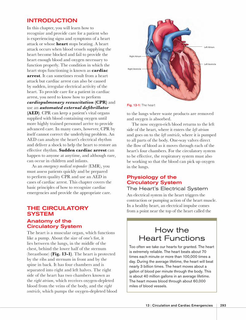
The now oxygen-rich blood returns to the left side of the heart, where it enters the left atrium and goes on to the left ventricle, where it is pumped to all parts of the body. One-way valves direct the fl ow of blood as it moves through each of the heart’s four chambers. For the circulatory system to be effective, the respiratory system must also be working so that the blood can pick up oxygen in the lungs.
Physiology of the Circulatory SystemThe Heart’s Electrical SystemAn electrical system in the heart triggers the contraction or pumping action of the heart muscle. In a healthy heart, an electrical impulse comes from a point near the top of the heart called the
INTRODUCTIONIn this chapter, you will learn how to recognize and provide care for a patient who is experiencing signs and symptoms of a heart attack or whose heart stops beating. A heart attack occurs when blood vessels supplying the heart become blocked and fail to provide the heart enough blood and oxygen necessary to function properly. The condition in which the heart stops functioning is known as cardiac arrest. It can sometimes result from a heart attack but cardiac arrest can also be caused by sudden, irregular electrical activity of the heart. To provide care for a patient in cardiac arrest, you need to know how to perform cardiopulmonary resuscitation (CPR) and use an automated external defi brillator (AED). CPR can keep a patient’s vital organs supplied with blood containing oxygen until more highly trained personnel arrive to provide advanced care. In many cases, however, CPR by itself cannot correct the underlying problem. An AED can analyze the heart’s electrical rhythm and deliver a shock to help the heart to restore an effective rhythm. Sudden cardiac arrest can happen to anyone at anytime, and although rare, can occur in children and infants.
As an emergency medical responder (EMR), you must assess patients quickly and be prepared to perform quality CPR and use an AED in cases of cardiac arrest. This chapter covers the basic principles of how to recognize cardiac emergencies and provide the appropriate care.
THE CIRCULATORY SYSTEMAnatomy of the Circulatory System The heart is a muscular organ, which functions like a pump. About the size of one’s fi st, it lies between the lungs, in the middle of the chest, behind the lower half of the sternum (breastbone) (Fig. 13-1). The heart is protected by the ribs and sternum in front and by the spine in back. It has four chambers and is separated into right and left halves. The right side of the heart has two chambers known as the right atrium, which receives oxygen-depleted blood from the veins of the body, and the right ventricle, which pumps the oxygen-depleted blood
Left Atrium
Left Ventricle
Right Atrium
Right Ventricle
Fig. 13-1: The heart
How the Heart Functions
Too often we take our hearts for granted. The heart is extremely reliable. The heart beats about 70 times each minute or more than 100,000 times a day. During the average lifetime, the heart will beat nearly 3 billion times. The heart moves about a gallon of blood per minute through the body. This is about 40 million gallons in an average lifetime. The heart moves blood through about 60,000 miles of blood vessels.
294 Unit 4 : Circulation
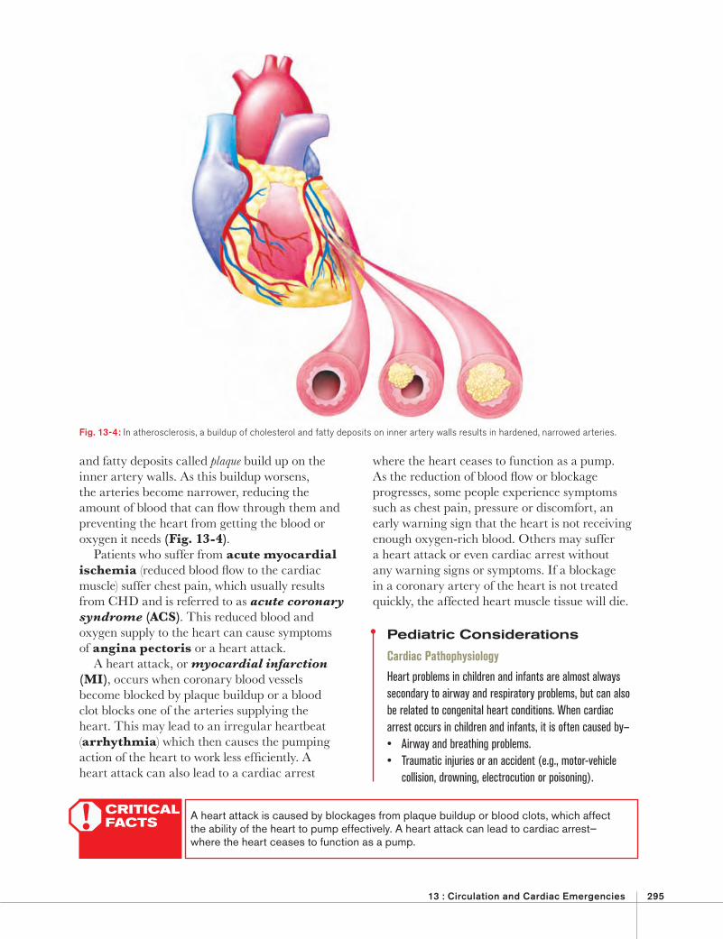
cells throughout the body, and as blood fl ows through the veins, carbon dioxide and other wastes are taken away. This continuous process is called perfusion (Fig. 13-3).
The primary gases exchanged are oxygen and carbon dioxide. All cells require oxygen to function. Cells also require energy to function. Glucose, a simple sugar molecule, is the main source of energy inside the cell.
Pathophysiology of the Circulatory SystemCardiovascular disease is an abnormal condition that affects the heart and blood vessels. An estimated 80 million Americans suffer from some form of the disease. It remains the number-one killer in the United States, and a major cause of disability. The most common conditions caused by cardiovascular disease include coronary heart disease (CHD), also known as coronary artery disease (CAD), and stroke, also called a brain attack. (See Chapter 14 for more information on stroke.)
CHD occurs when the arteries that supply blood to the heart muscle become hardened and narrowed, a process called atherosclerosis. This damage occurs gradually, as cholesterol
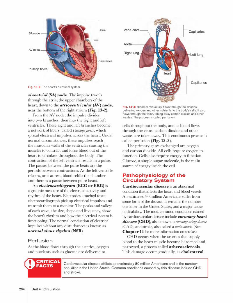
sinoatrial (SA) node. The impulse travels through the atria, the upper chambers of the heart, down to the atrioventricular (AV) node, near the bottom of the right atrium (Fig. 13-2).
From the AV node, the impulse divides into two branches, then into the right and left ventricles. These right and left branches become a network of fi bers, called Purkinje fi bers, which spread electrical impulses across the heart. Under normal circumstances, these impulses reach the muscular walls of the ventricles causing the muscles to contract and force blood out of the heart to circulate throughout the body. The contraction of the left ventricle results in a pulse. The pauses between the pulse beats are the periods between contractions. As the left ventricle relaxes, or is at rest, blood refi lls the chamber and there is a pause between pulse beats.
An electrocardiogram (ECG or EKG) is a graphic measure of the electrical activity and rhythm of the heart. Electrodes attached to an electrocardiograph pick up electrical impulses and transmit them to a monitor. The peaks and valleys of each wave, the size, shape and frequency, show the heart’s rhythm and how the electrical system is functioning. The normal conduction of electrical impulses without any disturbances is known as normal sinus rhythm (NSR).
PerfusionAs the blood fl ows through the arteries, oxygen and nutrients such as glucose are delivered to
Aorta
Vena cava
Capillaries
Heart
Left lungRight lung
Capillaries
Fig. 13-3: Blood continuously fl ows through the arteries delivering oxygen and other nutrients to the body’s cells. It also fl ows through the veins, taking away carbon dioxide and other wastes. The process is called perfusion.
SA node
AV node
Atria
Ventricles
Purkinje fibers
Fig. 13-2: The heart’s electrical system
CRITICAL FACTS! Cardiovascular disease affl icts approximately 80 million Americans and is the number-
one killer in the United States. Common conditions caused by this disease include CHD and stroke.
13 : Circulation and Cardiac Emergencies 295
where the heart ceases to function as a pump. As the reduction of blood fl ow or blockage progresses, some people experience symptoms such as chest pain, pressure or discomfort, an early warning sign that the heart is not receiving enough oxygen-rich blood. Others may suffer a heart attack or even cardiac arrest without any warning signs or symptoms. If a blockage in a coronary artery of the heart is not treated quickly, the affected heart muscle tissue will die.
Pediatric Considerations
Cardiac Pathophysiology
Heart problems in children and infants are almost always secondary to airway and respiratory problems, but can also be related to congenital heart conditions. When cardiac arrest occurs in children and infants, it is often caused by—• Airway and breathing problems. • Traumatic injuries or an accident (e.g., motor-vehicle
collision, drowning, electrocution or poisoning).
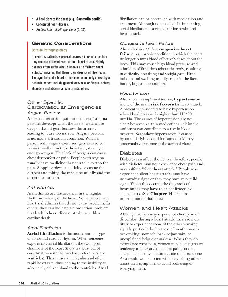
and fatty deposits called plaque build up on the inner artery walls. As this buildup worsens, the arteries become narrower, reducing the amount of blood that can fl ow through them and preventing the heart from getting the blood or oxygen it needs (Fig. 13-4).
Patients who suffer from acute myocardial ischemia (reduced blood fl ow to the cardiac muscle) suffer chest pain, which usually results from CHD and is referred to as acute coronary syndrome (ACS). This reduced blood and oxygen supply to the heart can cause symptoms of angina pectoris or a heart attack.
A heart attack, or myocardial infarction (MI), occurs when coronary blood vessels become blocked by plaque buildup or a blood clot blocks one of the arteries supplying the heart. This may lead to an irregular heartbeat (arrhythmia) which then causes the pumping action of the heart to work less effi ciently. A heart attack can also lead to a cardiac arrest
Fig. 13-4: In atherosclerosis, a buildup of cholesterol and fatty deposits on inner artery walls results in hardened, narrowed arteries.
CRITICAL FACTS! A heart attack is caused by blockages from plaque buildup or blood clots, which affect
the ability of the heart to pump effectively. A heart attack can lead to cardiac arrest—where the heart ceases to function as a pump.
296 Unit 4 : Circulation
fi brillation can be controlled with medication and treatment. Although not usually life threatening, atrial fi brillation is a risk factor for stroke and heart attack.
Congestive Heart FailureAlso called heart failure, congestive heart failure is a chronic condition in which the heart no longer pumps blood effectively throughout the body. This may cause high blood pressure and a buildup of fl uid throughout the body, resulting in diffi culty breathing and weight gain. Fluid buildup and swelling usually occur in the face, hands, legs, ankles and feet.
HypertensionAlso known as high blood pressure, hypertension is one of the main risk factors for heart attack. A patient is considered to have hypertension when blood pressure is higher than 140/90 mmHg. The causes of hypertension are not clear; however, certain medications, salt intake and stress can contribute to a rise in blood pressure. Secondary hypertension is caused by an underlying condition such as a kidney abnormality or tumor of the adrenal gland.
DiabetesDiabetes can affect the nerves; therefore, people with diabetes may not experience chest pain and may suffer a “silent heart attack.” People who experience silent heart attacks may have no warning signs or they may have very mild signs. When this occurs, the diagnosis of a heart attack may have to be confi rmed by special tests. (See Chapter 14 for more information on diabetes.)
Women and Heart AttacksAlthough women may experience chest pain or discomfort during a heart attack, they are more likely to experience some of the other warning signals, particularly shortness of breath; nausea or vomiting; stomach, back or jaw pain; or unexplained fatigue or malaise. When they do experience chest pain, women may have a greater tendency to have atypical chest pain: sudden, sharp but short-lived pain outside the breastbone. As a result, women often will delay telling others about their symptoms to avoid bothering or worrying them.
• A hard blow to the chest (e.g., Commotio cordis). • Congenital heart disease.• Sudden infant death syndrome (SIDS).
Geriatric Considerations
Cardiac Pathophysiology
In geriatric patients, a general decrease in pain perception may cause a different reaction to a heart attack. Elderly patients often suffer what is known as a “silent heart attack,” meaning that there is an absence of chest pain. The symptoms of a heart attack most commonly shown by a geriatric patient include general weakness or fatigue, aching shoulders and abdominal pain or indigestion.
Other Specifi c Cardiovascular EmergenciesAngina PectorisA medical term for “pain in the chest,” angina pectoris develops when the heart needs more oxygen than it gets, because the arteries leading to it are too narrow. Angina pectoris is normally a transient condition. When a person with angina exercises, gets excited or is emotionally upset, the heart might not get enough oxygen. This lack of oxygen can cause chest discomfort or pain. People with angina usually have medicine they can take to stop the pain. Stopping physical activity or easing the distress and taking the medicine usually end the discomfort or pain.
ArrhythmiasArrhythmias are disturbances in the regular rhythmic beating of the heart. Some people have heart arrhythmias that do not cause problems. In others, they can indicate a more serious problem that leads to heart disease, stroke or sudden cardiac death.
Atrial FibrillationAtrial fi brillation is the most common type of abnormal cardiac rhythm. When someone experiences atrial fi brillation, the two upper chambers of the heart (the atria) beat out of coordination with the two lower chambers (the ventricles). This causes an irregular and often rapid heart rate, thus leading to the inability to adequately deliver blood to the ventricles. Atrial
13 : Circulation and Cardiac Emergencies 297
is usually treated with a nitroglycerin pill or patches. This medication reduces the workload of the heart by dilating the coronary arteries. Diffi culty breathing is another sign of a heart
attack. The patient may be breathing faster than normal because the body tries to get much-needed oxygen to the heart. A patient who is sitting upright and learning forward with hands on knees in the tripod position is struggling to breathe. Diffi culty breathing also includes noisy breathing and shortness of breath. Other signs and symptoms include pale or
ashen skin, especially around the face. The face also may be damp with sweat. Some people suffering from a heart attack sweat heavily, feel dizzy or lightheaded and/or may lose consciousness. Nausea is also a sign and symptom of a heart attack.
Assessment of Cardiac Emergencies The sooner you recognize the signs and symptoms of a heart attack and act, the better chance you have to save a life. Many people will deny they are having a heart attack. Summon more advanced medical personnel if the patient shows some or all of the following signs and symptoms: Discomfort, pressure or pain. The major sign
is persistent discomfort, pressure or pain in the chest that does not go away. Unfortunately, it is not always easy to distinguish heart attack pain from the pain of indigestion, muscle spasms or other conditions. This often causes people to delay getting medical care. Brief, stabbing pain or pain that gets worse when you bend or breathe deeply is not usually caused by a heart problem. The pain associated with a heart attack can
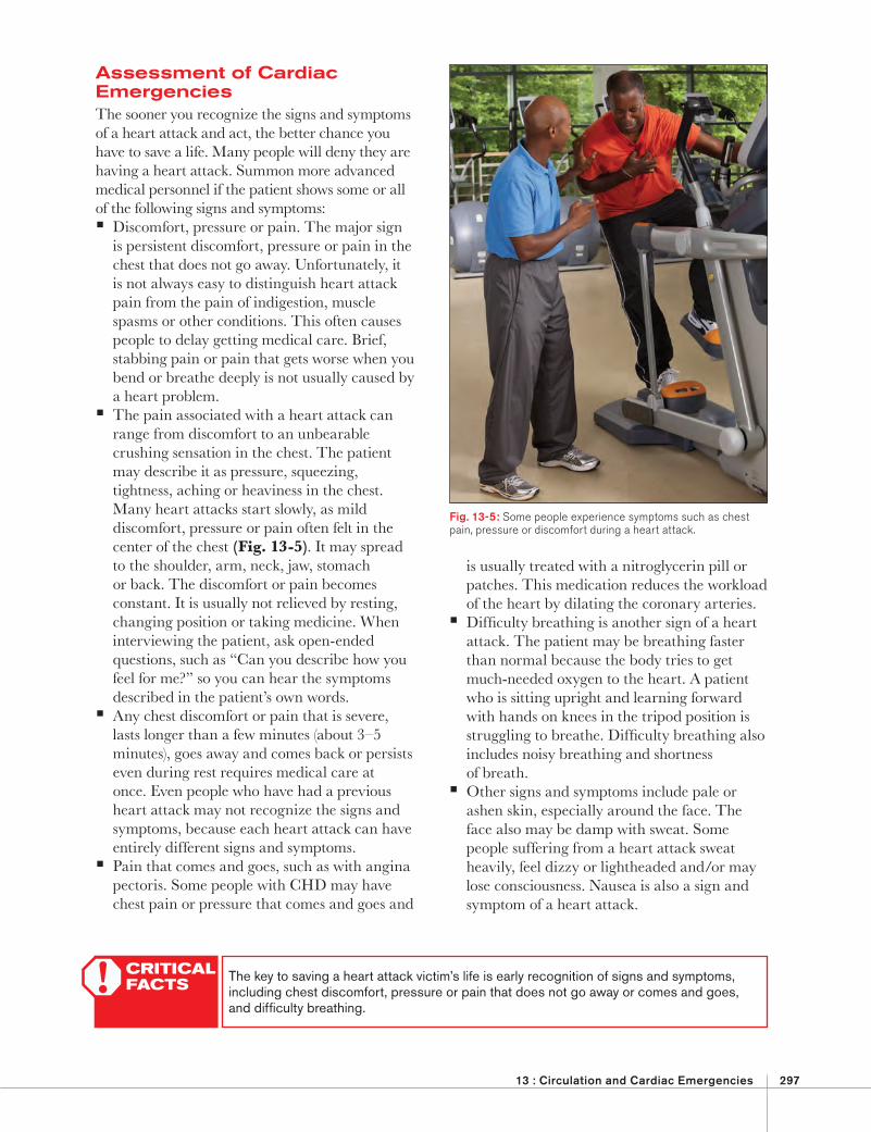
range from discomfort to an unbearable crushing sensation in the chest. The patient may describe it as pressure, squeezing, tightness, aching or heaviness in the chest. Many heart attacks start slowly, as mild discomfort, pressure or pain often felt in the center of the chest (Fig. 13-5). It may spread to the shoulder, arm, neck, jaw, stomach or back. The discomfort or pain becomes constant. It is usually not relieved by resting, changing position or taking medicine. When interviewing the patient, ask open-ended questions, such as “Can you describe how you feel for me?” so you can hear the symptoms described in the patient’s own words. Any chest discomfort or pain that is severe,
lasts longer than a few minutes (about 3–5 minutes), goes away and comes back or persists even during rest requires medical care at once. Even people who have had a previous heart attack may not recognize the signs and symptoms, because each heart attack can have entirely different signs and symptoms. Pain that comes and goes, such as with angina
pectoris. Some people with CHD may have chest pain or pressure that comes and goes and
Fig. 13-5: Some people experience symptoms such as chest pain, pressure or discomfort during a heart attack.
CRITICAL FACTS! The key to saving a heart attack victim’s life is early recognition of signs and symptoms,
including chest discomfort, pressure or pain that does not go away or comes and goes, and diffi culty breathing.
298 Unit 4 : Circulation
Is taking any blood thinners, such as warfarin (Coumadin®). Has been told by a physician to not take
aspirin.
If the patient answers no to all of these questions, administration of two chewable (162 mg) baby aspirins, or one 5-grain (325 mg) adult aspirin tablet with a small amount of water should be considered.
Be sure that only aspirin is given and not acetaminophen (e.g., Tylenol®) or nonsteroidal anti-infl ammatory drugs (NSAIDs) such as ibuprofen (e.g., Motrin® or Advil®) and naproxen (e.g., Aleve®). Likewise, coated aspirin products or products meant for multiple symptoms/uses such as cold, fever and headache, should not be used. Coated aspirin takes too long to dissolve to be effective.
CARDIAC ARRESTWhen the heart stops beating, or beats too ineffectively to circulate blood to the brain and other vital organs, this is called cardiac arrest. The beats or contractions of the heart become ineffective if they are weak, irregular or uncoordinated, because, at that point, the blood no longer fl ows through the arteries to the rest of the body.
When the heart stops beating properly, the body cannot survive. Breathing will stop soon after, and the body’s organs will no longer receive the oxygen they need to function. Without oxygen, brain damage can begin in about 4–6 minutes, and the damage can become irreversible after about 10 minutes.
A person in cardiac arrest is not breathing and has no pulse. The heart has either stopped beating or is beating weakly and irregularly so that a pulse cannot be detected.
Cardiovascular disease is the primary cause of cardiac arrest. About 900,000 people in the United States die each year from all forms of the disease. Other causes of cardiac arrest include drowning, choking, drugs, severe injury, brain damage and electrocution.
Cardiac arrest can happen suddenly, without any of the warning signs usually seen in a heart attack. This is known as sudden cardiac arrest or sudden cardiac death and accounts for more than 300,000 deaths annually in the United States. Sudden cardiac arrest is caused by abnormal,
Providing Care for Cardiac EmergenciesIf you think someone is having a heart attack— Take immediate action and summon more
advanced medical personnel. Have the patient stop any activity and rest
(Fig. 13-6). Loosen any tight or uncomfortable clothing. Closely monitor the patient until more
advanced medical personnel take over. Notice any changes in the patient’s appearance or behavior. Comfort the patient. If medically appropriate and local protocols
or medical direction permit, give aspirin if the patient can swallow and has no known contraindications. Be sure the patient has not been told by his or her physician to not take aspirin. Assist the patient with prescribed medication
and administer emergency oxygen, if it is available. Be prepared to perform CPR and use an AED.
Aspirin Can Lessen Heart Attack DamageYou may be able to help a conscious patient who is showing early signs of a heart attack by offering an appropriate dose of aspirin when the signs fi rst begin. Local protocols regarding administration of medicines, such as aspirin, may vary for EMRs and should be followed. Aspirin should never take the place of more advanced medical care. If the patient is conscious and able to take medicine by mouth, ask if he or she— Is allergic to aspirin. Has a stomach ulcer or stomach disease.
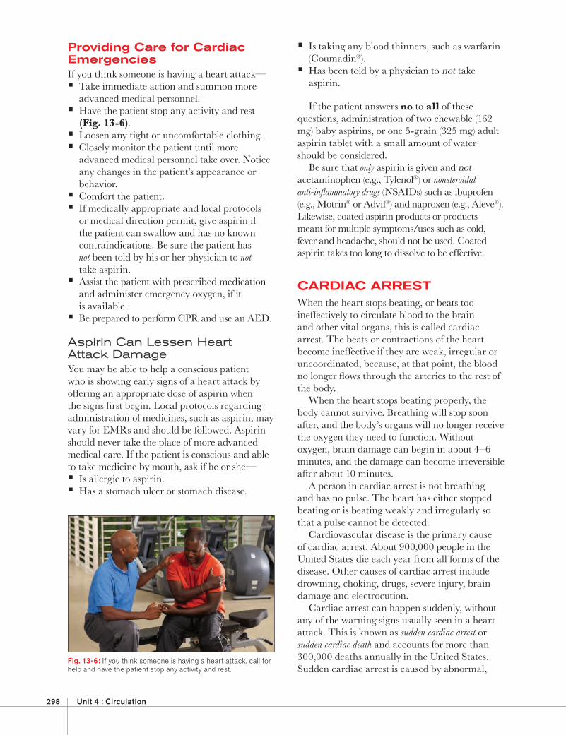
Fig. 13-6: If you think someone is having a heart attack, call for help and have the patient stop any activity and rest.
13 : Circulation and Cardiac Emergencies 299
In the Cardiac Chain of Survival, each link of the chain depends on and is connected to the other links. The layperson or bystander is the fi rst link in the cardiac chain of survival. But for this four-step sequence to work and ensure the greatest chance of survival, it is very important to quickly recognize the emergency and call for help, start CPR promptly and continue until an AED is ready to use or more advanced medical personnel take over.
Laypersons should be informed through community outreach programs and public awareness campaigns that by taking quick action, including calling 9-1-1 or the local emergency number, starting CPR immediately and using an AED if one is available, it is more likely a person in cardiac arrest will survive.
CPRA patient who is unconscious, not breathing and has no pulse is in cardiac arrest and needs CPR. CPR is a combination of chest compressions and ventilations which circulate blood containing oxygen to the brain and other vital organs for a person whose heart and breathing have stopped. Summoning more advanced medical personnel immediately is critical for the patient’s survival. If an AED is available, use it in combination with CPR and according to your local protocols until more advanced medical personnel take over.
Artifi cial Ventilation Artifi cial ventilation is a way of forcing air into the lungs of a patient who is not breathing. The oxygen in the air will be absorbed by blood fl owing through the lungs and carried to tissues and the body’s vital organs.
There are several different methods of artifi cial ventilation, including— Mouth-to-mask ventilations. Resuscitation using a bag-valve-mask resuscitator
(BVM).
chaotic electrical activity of the heart (known as arrhythmias). The most common life-threatening abnormal arrhythmia is ventricular fi brillation (V-fi b).
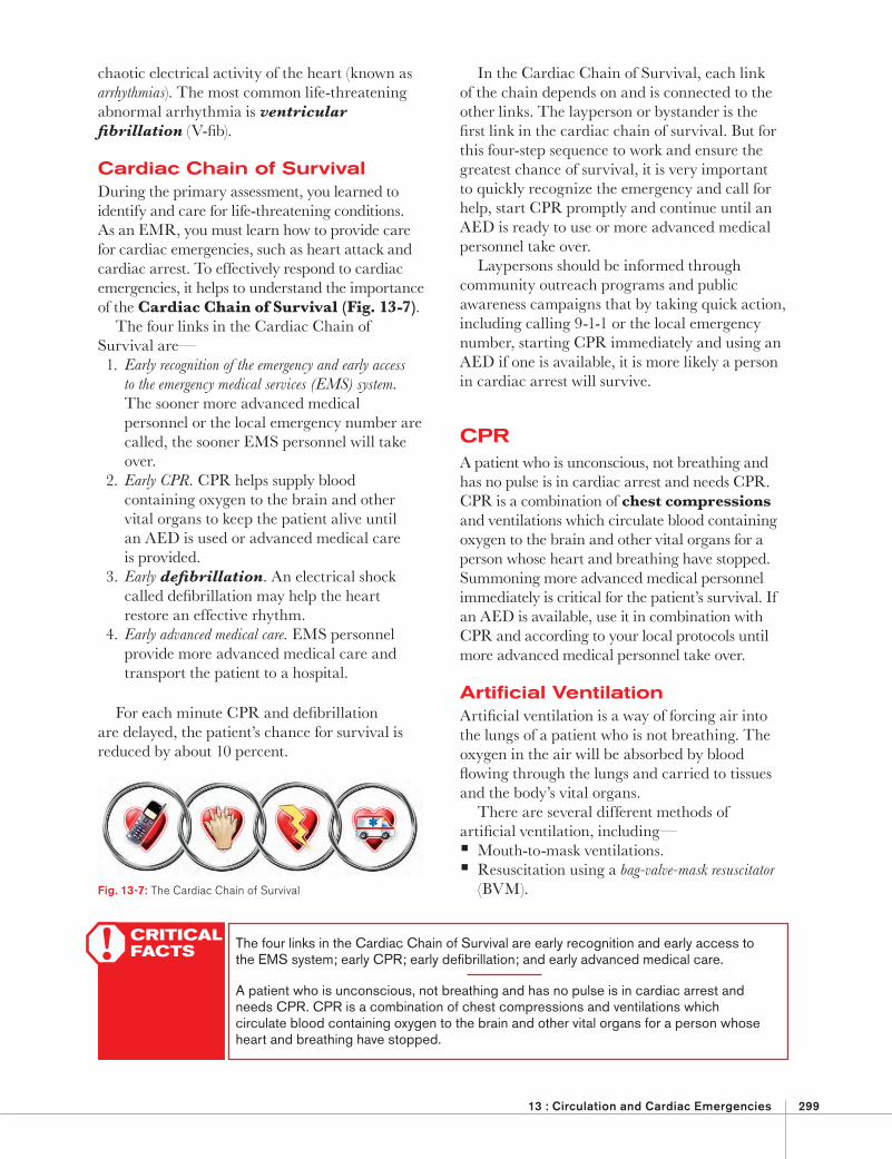
Cardiac Chain of SurvivalDuring the primary assessment, you learned to identify and care for life-threatening conditions. As an EMR, you must learn how to provide care for cardiac emergencies, such as heart attack and cardiac arrest. To effectively respond to cardiac emergencies, it helps to understand the importance of the Cardiac Chain of Survival (Fig. 13-7).
The four links in the Cardiac Chain of Survival are— 1. Early recognition of the emergency and early access
to the emergency medical services (EMS) system. The sooner more advanced medical personnel or the local emergency number are called, the sooner EMS personnel will take over.
2. Early CPR. CPR helps supply blood containing oxygen to the brain and other vital organs to keep the patient alive until an AED is used or advanced medical care is provided.
3. Early defi brillation. An electrical shock called defi brillation may help the heart restore an effective rhythm.
4. Early advanced medical care. EMS personnel provide more advanced medical care and transport the patient to a hospital.
For each minute CPR and defi brillation are delayed, the patient’s chance for survival is reduced by about 10 percent.
CRITICAL FACTS! The four links in the Cardiac Chain of Survival are early recognition and early access to
the EMS system; early CPR; early defi brillation; and early advanced medical care.
A patient who is unconscious, not breathing and has no pulse is in cardiac arrest and needs CPR. CPR is a combination of chest compressions and ventilations which circulate blood containing oxygen to the brain and other vital organs for a person whose heart and breathing have stopped.
Fig. 13-7: The Cardiac Chain of Survival
300 Unit 4 : Circulation
The effectiveness of compressions can be reduced if— Compressions are too shallow. Compression rate is too slow. There is sub-maximum recoil (not letting the
chest come all the way back up). There are frequent interruptions. The patient is not on a fi rm, fl at surface.
Correct Hand PositionKeeping your hands in the correct position allows you to give the most effective compressions. The correct position for your hands is over the lower half of the sternum (breastbone) in the middle of the chest (Fig. 13-9). At the lowest point of the sternum is an arrow-shaped piece of hard tissue called the xiphoid process. Avoid pressing directly on the xiphoid process, which can break off and puncture underlying organs and tissues causing potentially serious injury.
To fi nd the correct hand position, place the heel of one hand on the center of the chest, along the sternum, and then place the other hand on top. Use only the heel of your hand to apply pressure on the sternum when compressing the chest. Try to keep your fi ngers off the chest by interlacing
Fixed- and variable-fl ow oxygen when used in conjunction with delivery devices.
Artifi cial ventilation can save a patient’s life, but over-ventilation can be potentially harmful, especially for a patient in cardiac arrest. For example, if the ventilation is given too forcefully, or at too fast a rate, the pressure in the patient’s chest will remain too high even between breaths. This stops the blood from returning to the right side of the heart, and means that less blood is available to be pumped to other vital organs and tissues as CPR continues.
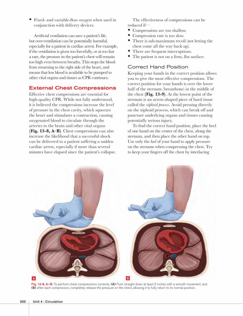
External Chest Compressions Effective chest compressions are essential for high-quality CPR. While not fully understood, it is believed the compressions increase the level of pressure in the chest cavity, which squeezes the heart and stimulates a contraction, causing oxygenated blood to circulate through the arteries to the brain and other vital organs (Fig. 13-8, A–B). Chest compressions can also increase the likelihood that a successful shock can be delivered to a patient suffering a sudden cardiac arrest, especially if more than several minutes have elapsed since the patient’s collapse.
Fig. 13-8, A–B: To perform chest compressions correctly: (A) Push straight down at least 2 inches with a smooth movement, and (B) after each compression, completely release the pressure on the chest, allowing it to fully return to its normal position.
A B
13 : Circulation and Cardiac Emergencies 301
Kneel at the patient’s side opposite the chest with your hands in the correct position. Keep your elbows as straight as possible, with your shoulders directly over your hands (Fig. 13-11).
them or holding them upward. Applying pressure with your fi ngers can cause ineffi cient chest compressions or unnecessary injury to the chest. Positioning the hands correctly allows for the most effective compressions and decreases the chance of causing injury.
If you have arthritis or a similar condition in your hands or wrists, you may use an alternative hand position. Find the correct hand position, as above, and then grasp the wrist of the hand on the chest with the other hand (Fig. 13-10).
The patient’s clothing will not necessarily interfere with your ability to position your hands correctly. If you can fi nd the correct position without removing thin clothing, such as a T-shirt, do so. Sometimes a layer of thin clothing will help keep your hands from slipping, since the patient’s chest may be moist with sweat. However, if you are not sure you can fi nd the correct hand position, bare the patient’s chest. Fat does not accumulate over the sternum; therefore, fi nding the correct hand position is the same regardless of patient size.
Position of the RescuerYour body position is important when giving chest compressions. Compressing the chest straight down provides the best blood fl ow. The correct body position is also less tiring for you.
Xyphoid Process
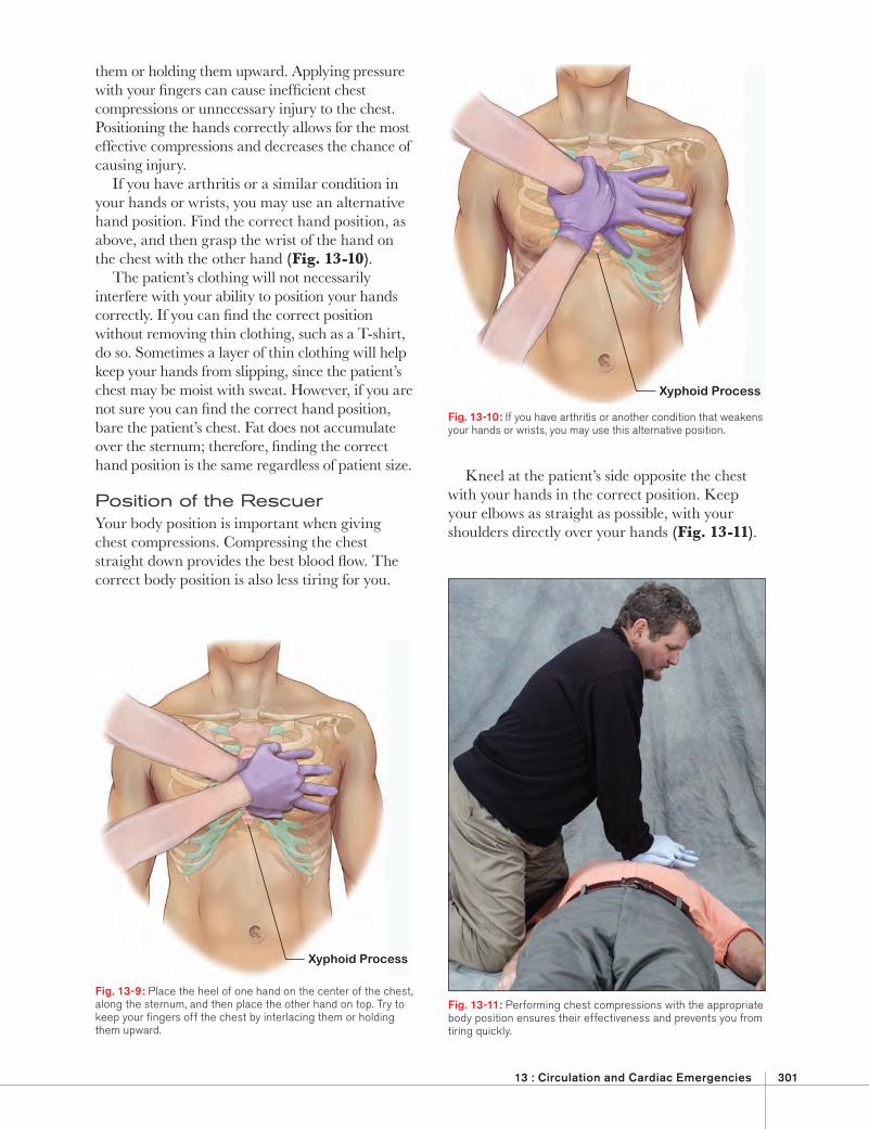
Fig. 13-9: Place the heel of one hand on the center of the chest, along the sternum, and then place the other hand on top. Try to keep your fi ngers off the chest by interlacing them or holding them upward.
Xyphoid Process
Fig. 13-10: If you have arthritis or another condition that weakens your hands or wrists, you may use this alternative position.
Fig. 13-11: Performing chest compressions with the appropriate body position ensures their effectiveness and prevents you from tiring quickly.
302 Unit 4 : Circulation
compressions, then give ventilations, before starting another cycle of compressions.
Depth of CompressionsEach time you push down, the breastbone of an adult should move at least 2 inches. The downward movement should be smooth, not jerky. Maintain a steady down-and-up rhythm and do not pause in between. If your hands slip out of position, follow the steps listed earlier to quickly reposition them.
RecoilAfter each compression, completely release the pressure on the chest. Do not break contact with the chest; simply allow the chest to fully return to its normal position (full recoil) before you start the next compression. It is during this phase of CPR that the chambers of the heart will refi ll with blood, ready to be circulated throughout the body with the next compression. Chest compressions are more effective when the patient is on a fi rm, fl at surface. If the patient is on a softer surface such as a bed, couch or pressure relieving mattress, carefully position the patient face up on the fl oor or a backboard.
When you press down in this position, you are pushing straight down onto the patient’s sternum. Keeping your arms as straight as possible prevents you from tiring quickly.
Compressing the chest requires less effort in this position. When you press down, the weight of your upper body creates the force needed to compress the chest. Push with the weight of your upper body, not with the muscles of your arms. Push straight down. Do not rock back and forth. Rocking results in less effective compressions and wastes energy. If your arms and shoulders tire quickly, you are not using the correct body position.
Compression TechniqueRate of CompressionGive compressions at a rate of at least 100 per minute. You can help yourself maintain the right pace by counting either aloud or in your head: one (as you press down) and (as you release the pressure) two (pressing down again) and (release again) and so on. When you get into the twenties, you can drop the “and” as it may be tiring and may alter the timing of compressions. Count the number of
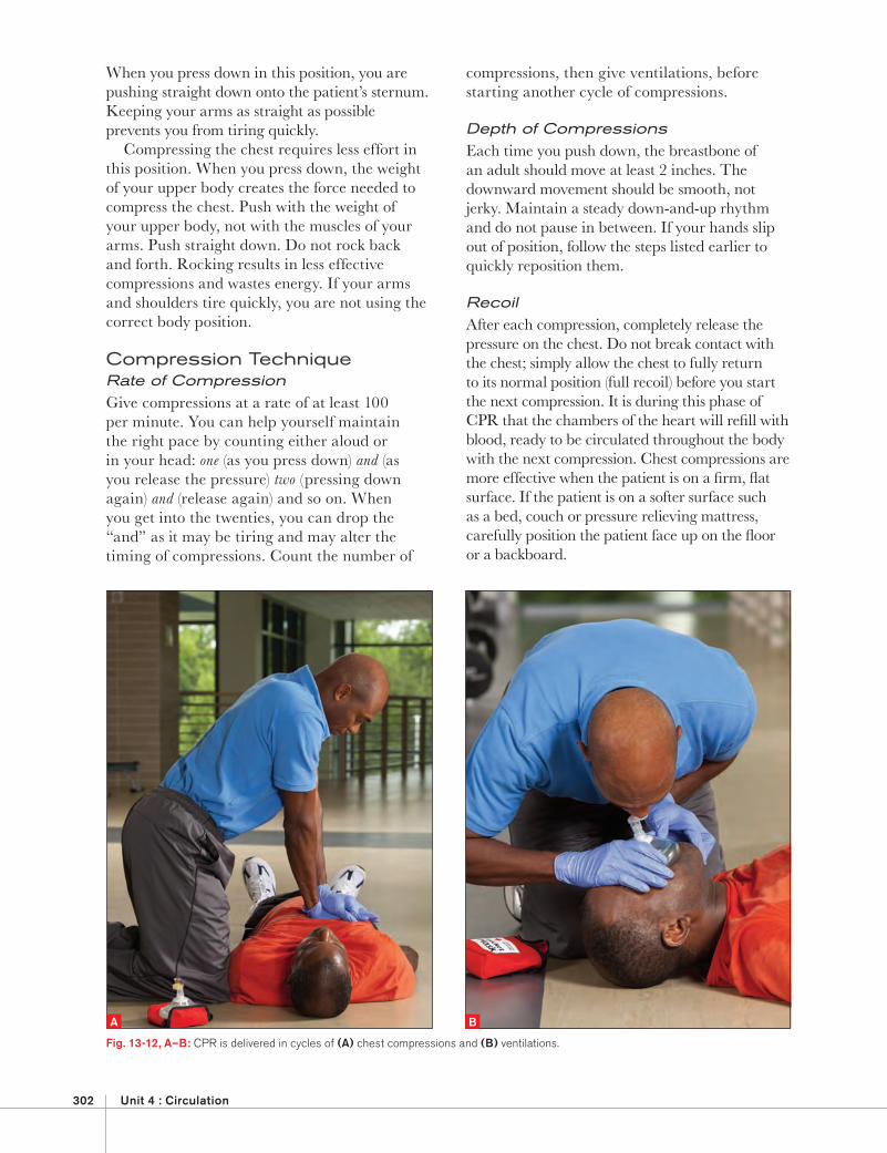
Fig. 13-12, A–B: CPR is delivered in cycles of (A) chest compressions and (B) ventilations.
A B
13 : Circulation and Cardiac Emergencies 303
Consensus on Science with Treatment Recommendations (CoSTR) by international experts in the fi eld of emergency medicine.
InterruptionsMinimize interruptions in giving chest compressions. If compressions must be interrupted, do so for no more than a few seconds. For example, you may need to move the patient to a location where CPR can be more effectively administered, such as if the patient is on a bed or couch, moving the patient to lie fl at on the fl oor. CPR may also be interrupted briefl y for defi brillation, insertion of an advanced airway or when two rescuers change positions between compressions and ventilations. Continue CPR while the patient
Compression and Breathing CyclesWhen performing CPR on an adult, child or infant, it is delivered in cycles of chest compressions followed by ventilations (Fig. 13-12, A–B). Complete the compressions, then re-establish an open airway by tilting the patient’s head and lifting the chin and then provide ventilations. When you are fi nished giving ventilations, quickly reposition your hands on the center of the chest and start another cycle of compressions and ventilations. See Table 13-1 regarding the compression-to-ventilation ratios for adult, child and infant one- and two-rescuer CPR. Note that these ratios can change every 5 years due to updates in scientifi c research and evidence that result from the
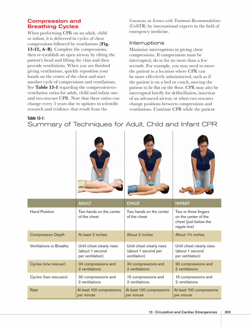
Table 13-1: Summary of Techniques for Adult, Child and Infant CPR
ADULT CHILD INFANT
Hand Position Two hands on the center of the chest
Two hands on the center of the chest
Two or three fi ngers on the center of the chest (just below the nipple line)
Compression Depth At least 2 inches About 2 inches About 1½ inches
Ventilations or Breaths Until chest clearly rises (about 1 second per ventilation)
Until chest clearly rises (about 1 second per ventilation)
Until chest clearly rises (about 1 second per ventilation)
Cycles (one rescuer) 30 compressions and 2 ventilations
30 compressions and 2 ventilations
30 compressions and 2 ventilations
Cycles (two rescuers) 30 compressions and 2 ventilations
15 compressions and 2 ventilations
15 compressions and 2 ventilations
Rate At least 100 compressions per minute
At least 100 compressions per minute
At least 100 compressions per minute
304 Unit 4 : Circulation
and the other rescuer gives chest compressions. Rescuers should change positions (alternate turns performing compressions and ventilations) about every 2 minutes to reduce the possibility of rescuer fatigue. Changing positions should take less than 5 seconds.
Perform two-rescuer CPR in the following situations: Two rescuers arrive on the scene at the same
time and begin CPR together. One rescuer is performing CPR and a second
rescuer becomes available.
When CPR is in progress by one rescuer and a second rescuer arrives, the second rescuer should confi rm whether more advanced medical personnel have been summoned. If they have not, the second rescuer should do so before getting the AED or assisting with care. If more advanced medical personnel have been summoned, the second rescuer should get the AED, or if an AED is not available, the second rescuer should help perform two-rescuer CPR.
Hands-Only CPRHands-only CPR or continuous chest compressions, is a simplifi ed form of CPR that eliminates ventilations or rescue breaths. It has its roots in dispatcher-assisted cardiac emergency situations where the caller is untrained, unwilling, unsure or otherwise unable to perform full CPR (chest compressions with ventilations or rescue breaths). Providing instruction on how to give chest compressions alone is less complex than trying to explain full CPR. The main focus of hands-only CPR is on the untrained layperson or a bystander who witnesses the sudden collapse of an adult. EMRs should be aware that if they come upon a bystander giving chest compressions only, that person is performing CPR correctly.
Chest compressions alone may provide effective circulation of blood containing oxygen in the fi rst few minutes of an out-of-hospital cardiac arrest. The same quality compression techniques of full CPR apply to compression-
is being moved into an ambulance or other transport vehicle or from the ambulance into a hospital emergency department.
When to Stop CPROnce you begin CPR, do not stop. Do not stop CPR except in one of these situations: You see an obvious sign of life, such as
breathing. An AED is ready to use. Another trained responder takes over. More advanced medical personnel take over. You are presented with a valid Do Not
Resuscitate (DNR) order. You are too exhausted to continue. The scene becomes unsafe.
Pediatric Considerations
CPR
The principles of CPR (compressing the chest and providing ventilations) are the same for children and infants as for adults, but the techniques are modifi ed because children’s and infants’ bodies are smaller and weaker. Cardiac arrest in children and infants is usually caused by a respiratory emergency. If you recognize that a child or an infant is in respiratory distress or arrest, provide care immediately. If cardiac arrest occurs, begin CPR.
To perform CPR on a child or an infant, also perform cycles of chest compressions and ventilations at the rate of at least 100 compressions per minute. As with an adult, let the chest fully recoil to its normal position after each compression. For a child, use two hands on the center of the chest and compress about 2 inches. For an infant, use two or three fi ngers on the center of the chest, just below the nipple line, and compress about 1½ inches.
Two-Rescuer CPRWhen an additional rescuer is available, perform two-rescuer CPR. One rescuer gives ventilations
CRITICAL FACTS! Once you begin CPR, do not stop. If you must, do so for no more than a few seconds.
Reasons to discontinue CPR include more advanced medical personnel taking over for you, seeing obvious signs of life, an AED being available and ready to use or being too exhausted to continue.
13 : Circulation and Cardiac Emergencies 305
person’s chest, analyzing the heart’s rhythm, delivering a shock if needed and reminders to perform CPR when appropriate. Some AEDs can be confi gured to deliver lower energy levels considered appropriate for children and infants.
When EMRs and other responders are trained to use AEDs, they can signifi cantly reduce the amount of time it takes to administer a fi rst shock in a sudden cardiac arrest, researchers say. In Eugene and Springfi eld, Oregon, AEDs were placed on every fi re truck, and all fi refi ghters were trained to use them. Researchers saw these communities’ survival rates for cardiac arrest increase by 18 percent in the fi rst year.
3
The vast majority of states recognize defi brillator training for EMTs, EMRs and other responders. All states and the District of Columbia have enacted AED Good Samaritan protection for lay responders.
4 Today, AEDs are
widely dispersed and can be found in areas where large groups of people gather, such as convention centers, airports, stadiums, shopping malls, large businesses, schools and industrial complexes.
The most common abnormal heart rhythm that causes sudden cardiac arrest occurs when the ventricles simply quiver, or fi brillate, without any organized rhythm. This condition is called ventricular fi brillation (V-fi b). In V-fi b, the electrical impulses fi re at random, creating chaos and preventing the heart from pumping and circulating blood.
Another less common life-threatening heart rhythm, called ventricular tachycardia (V-tach), occurs when the heart beats too fast. In V-tach, an abnormal electrical impulse controls the heart, originating in the ventricles instead of in the SA node. This abnormal impulse fi res so fast that the heart’s chambers do not have time to fi ll, and the heart is unable to pump blood effectively. With little or no blood circulating, there may be no pulse. As with V-fi b, there is no breathing or pulse.
3Graves JR, Austin D Jr, Cummins RO: Rapid zap: automated defi brillation, Englewood Cliffs, NJ, 1989, Prentice-Hall.4American Heart Association–AED Legislation/Good Samaritan Laws by State reviewed/updated 07/16/2008.
only CPR, including hand position, compression depth, speed, full recoil and minimal interruptions. Hands-only CPR does not affect the use of an AED.
AUTOMATED EXTERNAL DEFIBRILLATIONEach year, more than 300,000 Americans die suddenly of cardiac arrest. CPR can help by supplying blood containing oxygen to the brain and other vital organs. In many cases, however, an AED is needed to correct an abnormal electrical problem and allow the heart to restore an effective rhythm. Sudden cardiac arrest can happen to anyone at any time and, although rare, can occur in children and infants.
History of Defi brillationThe presence of cardiac arrhythmias or disturbances of the heart’s electrical system, and the ability to correct fi brillation with electrical shock, has been known since the mid-19th century.1 Electrical-shocking devices, or defi brillators, were fi rst developed during the 1920s. A portable version was introduced onto mobile coronary units in Belfast, Northern Ireland in 1966.
2
Defi brillation by emergency medical technicians (EMTs) without the presence of a physician was fi rst performed in Portland, Oregon in 1969.
As technology improved over the years, newer generations of more compact, simple to operate, semi-automatic defi brillators known as AEDs evolved allowing EMTs and EMRs, as well as trained citizen responders, to provide this life-saving technology. With these newer devices, a computer analyzes the heart’s rhythm and advises whether a shock is needed. Typically, the responder is guided through the steps of providing defi brillation by voice instructions and visual prompts from the AED. This includes placing the electrode (defi brillation) pads on the
1Bocka, Joseph J., MD: Automatic External Defi brillation, eMedicine, April 3, 2006.2Pantridge JF, Geddes JS: A mobile intensive care unit in the treatment of myocardial infarction, Lancet 2:271, 1967.
CRITICAL FACTS! V-fi b is the most common cause of sudden cardiac arrest. In V-fi b, heart ventricles quiver
instead of beating properly, due to erratic electrical impulses.
306 Unit 4 : Circulation
the body until defi brillation and increases the likelihood that the defi brillation shock will allow the heart to correct the abnormal rhythm.
Use an AED when the following conditions are present: The patient is unresponsive. There is no breathing. You do not detect a pulse.
Using an AEDWhen a cardiac arrest occurs, an AED should be used as soon as it is available and ready to use. If the AED advises that a shock is needed, follow protocols to give 1 shock followed by about 2 minutes of CPR. If CPR is in progress, chest compressions should not be interrupted until the AED is turned on, the defi brillation pads are applied and the AED is ready to analyze the heart rhythm.
Chest compressions can increase the likelihood that a defi brillation shock will be successful, especially if more than 4 minutes have elapsed since the patient’s collapse. Always follow local protocols and medical direction when using an AED and performing CPR. Be thoroughly familiar with the manufacturer’s operating instructions and maintenance guidelines for the device that you will be operating.
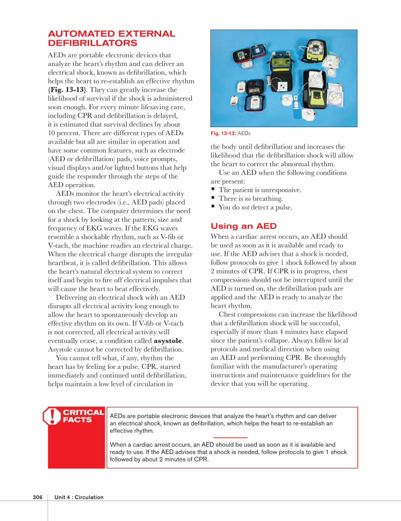
AUTOMATED EXTERNAL DEFIBRILLATORSAEDs are portable electronic devices that analyze the heart’s rhythm and can deliver an electrical shock, known as defi brillation, which helps the heart to re-establish an effective rhythm (Fig. 13-13). They can greatly increase the likelihood of survival if the shock is administered soon enough. For every minute lifesaving care, including CPR and defi brillation is delayed, it is estimated that survival declines by about 10 percent. There are different types of AEDs available but all are similar in operation and have some common features, such as electrode (AED or defi brillation) pads, voice prompts, visual displays and/or lighted buttons that help guide the responder through the steps of the AED operation.
AEDs monitor the heart’s electrical activity through two electrodes (i.e., AED pads) placed on the chest. The computer determines the need for a shock by looking at the pattern, size and frequency of EKG waves. If the EKG waves resemble a shockable rhythm, such as V-fi b or V-tach, the machine readies an electrical charge. When the electrical charge disrupts the irregular heartbeat, it is called defi brillation. This allows the heart’s natural electrical system to correct itself and begin to fi re off electrical impulses that will cause the heart to beat effectively.
Delivering an electrical shock with an AED disrupts all electrical activity long enough to allow the heart to spontaneously develop an effective rhythm on its own. If V-fi b or V-tach is not corrected, all electrical activity will eventually cease, a condition called asystole. Asystole cannot be corrected by defi brillation.
You cannot tell what, if any, rhythm the heart has by feeling for a pulse. CPR, started immediately and continued until defi brillation, helps maintain a low level of circulation in
AEDs are portable electronic devices that analyze the heart’s rhythm and can deliver an electrical shock, known as defi brillation, which helps the heart to re-establish an effective rhythm.
When a cardiac arrest occurs, an AED should be used as soon as it is available and ready to use. If the AED advises that a shock is needed, follow protocols to give 1 shock followed by about 2 minutes of CPR.
CRITICAL FACTS!
Fig. 13-13: AEDs
13 : Circulation and Cardiac Emergencies 307
defi brillation shock by absorbing some of the electrical energy. After a shock is delivered, or if no shock is advised, a period of time is programmed to allow for CPR until the next rhythm analysis begins. If the AED prompts to troubleshoot a problem such as “check electrodes” or “check pads,” check to see that the AED pads are connected properly to the device and placed on the patient’s chest with good adhesion, according to the manufacturer’s instructions and local protocols. Spare batteries should be available in case of a “low battery” warning, but shocks can still be delivered with a low battery warning on some models.
After a shock is delivered, or if no shock is indicated, perform about 2 minutes of CPR before the AED begins rhythm analysis again. If at any time you notice an obvious sign of life, such as breathing, stop CPR and monitor the patient's condition.
Pediatric Considerations
AED Use
While the incidence of cardiac arrest in children and infants is relatively low compared with that for adults, cardiac arrest resulting from V-fi b does happen in young children. Most cardiac arrests in children and infants are not sudden and may be caused by— • Airway and breathing problems. • Traumatic injuries or accidents (e.g., motor-vehicle
collision, drowning, electrocution or poisoning).• A hard blow to the chest.• Congenital heart disease.• Sudden infant death syndrome (SIDS).
AEDs equipped with pediatric defi brillation pads are capable of delivering lower levels of energy considered appropriate for children and infants up to 8 years old or weighing less than 55 pounds. Use pediatric AED pads and/or equipment, if available. If pediatric-specifi c equipment is not available, an AED designed for adults can be used on children and infants. In any event, always follow local protocols and medical direction and the manufacturer’s instructions. For a child or an infant in cardiac arrest, follow the same general steps and precautions that you would when using an AED on an adult. If the pads risk touching each other because of the
The general steps of operating an AED include— 1. Turning on the AED and preparing it for
use. Once the AED is turned on, it will guide the responder through all the steps of operation with voice and visual prompts. Some models have a power button that must be pressed, while others will activate upon opening the case or lid.
2. Exposing the patient’s chest and wiping the chest dry. The AED pads must be applied to the patient’s bare, dry chest. If the patient’s chest is moist or wet, it should be wiped with a small towel or gauze pads to ensure the best adhesion of the AED pads.
3. Attaching the AED pads to the patient’s bare, dry chest. Remove the AED pads from their sealed packaging. Peel the backing off from each pad, one at a time, to expose the adhesive, conductive surface of the pad before it is applied to the patient’s bare chest. Many AED pads have illustrations on them that show correct pad placement. Some AED pads are preconnected to the device, and some must be plugged into the device before rhythm analysis can begin. The pads should be appropriate to the patient. For example, pediatric AED pads must not be used on an adult patient because the lower energy levels may not be enough to defi brillate the patient.
4. Analyzing the heart rhythm. Some AEDs will automatically begin analysis when the pads are attached to the patient and connected to the device, while others have an “analyze” button that must be pushed. No one should touch or bump into the patient during the rhythm analysis as this could produce faulty readings.
5. Delivering a defi brillation shock. Once the analysis of the rhythm is complete, the AED will advise either to shock or not to shock the patient. If a shockable rhythm is detected, the AED will cycle up an electrical energy charge which will supply the shock to the patient. Some models can deliver the shock automatically while others have a “shock” button that must be manually pushed to deliver the shock. No one should be in contact with the patient when the shock is delivered, because they could also receive a shock and thereby reduce the effectiveness of the
308 Unit 4 : Circulation
automatically recognize and restore abnormal heart rhythms. Sometimes, a patient’s heart beats irregularly, even if the patient has a pacemaker or an ICD.
If the implanted device is visible, or you know that the patient has one, do not place the defi brillation pad directly over the device (Fig. 13-15). This may interfere with the delivery of the shock. Adjust pad placement if necessary and continue to follow established protocols. If you are not sure, use the AED as needed. It will not harm the patient or rescuer.
Rescuers should be aware that it is possible to receive a mild shock if an implantable ICD delivers a shock to the patient while CPR is performed. This risk of injury to rescuers is minimal and the amount of electrical energy involved is low. Much of the electrical energy is absorbed by the patient’s own body tissues. Some protocols may include temporarily deactivating the shock capability of an ICD with a donut magnet or other precautions. EMRs should be aware of and follow any special precautions associated with ICDs, but delays in delivering CPR and defi brillation shocks from an AED should not occur.
AEDs Around WaterIf the patient is in freestanding water, remove the patient before defi brillation. A shock delivered in water could conduct to rescuers or bystanders. Once you have removed the patient from the water, be sure there are no puddles of water around you, the patient or the AED. Remove wet clothing for proper pad placement, if necessary. Dry the patient’s chest and attach the AED pads.
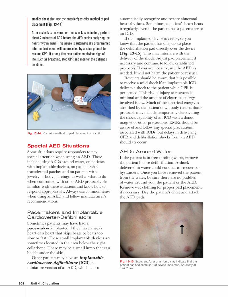
smaller chest size, use the anterior/posterior method of pad placement (Fig. 13-14).
After a shock is delivered or if no shock is indicated, perform about 2 minutes of CPR before the AED begins analyzing the heart rhythm again. This pause is automatically programmed into the device and will be preceded by a voice prompt to resume CPR. If at any time you notice an obvious sign of life, such as breathing, stop CPR and monitor the patient's condition.
Special AED SituationsSome situations require responders to pay special attention when using an AED. These include using AEDs around water, on patients with implantable devices, on patients with transdermal patches and on patients with jewelry or body piercings, as well as what to do when confronted with other AED protocols. Be familiar with these situations and know how to respond appropriately. Always use common sense when using an AED and follow manufacturer’s recommendations.
Pacemakers and Implantable Cardioverter-Defi brillatorsSometimes patients may have had a pacemaker implanted if they have a weak heart or a heart that skips beats or beats too slow or fast. These small implantable devices are sometimes located in the area below the right collarbone. There may be a small lump that can be felt under the skin.
Other patients may have an implantable cardioverter-defi brillator (ICD), a miniature version of an AED, which acts to
Fig. 13-14: Posterior method of pad placement on a child
Fig. 13-15: Scars and/or a small lump may indicate that the patient has had some sort of device implanted. Courtesy of Ted Crites.
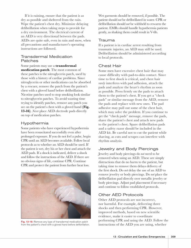
13 : Circulation and Cardiac Emergencies 309
Wet garments should be removed, if possible. The patient should not be defi brillated in water. CPR or defi brillation should not be withheld to rewarm the patient. EMRs should handle hypothermia patients gently, as shaking them could result in V-fi b.
TraumaIf a patient is in cardiac arrest resulting from traumatic injuries, an AED may still be used. Defi brillation should be administered according to local protocols.
Chest HairSome men have excessive chest hair that may cause diffi culty with pad-to-skin contact. Since time to fi rst shock is critical, and chest hair rarely interferes with pad adhesion, attach the pads and analyze the heart’s rhythm as soon as possible. Press fi rmly on the pads to attach them to the patient’s chest. If you get a “check pads” or similar message from the AED, remove the pads and replace with new ones. The pad adhesive may pull out some of the chest hair, which may solve the problem. If you continue to get the “check pads” message, remove the pads, shave the patient’s chest and attach new pads to the patient’s chest. Spare defi brillation pads and a safety razor should be included in the AED kit. Be careful not to cut the patient while shaving, as cuts and scrapes can interfere with rhythm analysis.
Jewelry and Body PiercingsJewelry and body piercings do not need to be removed when using an AED. These are simply distractions that do no harm to the patient, but taking time to remove them delays delivery of the fi rst shock. Do not delay the use of an AED to remove jewelry or body piercings. Do not place the defi brillation pad directly over metallic jewelry or body piercings. Adjust pad placement if necessary and continue to follow established protocols.
Other AED ProtocolsOther AED protocols are not incorrect, nor harmful. For example, delivering three shocks and then performing CPR. However, improved methods, based on new scientifi c evidence, make it easier to coordinate performing CPR and using the AED. Follow the instructions of the AED you are using, whether
If it is raining, ensure that the patient is as dry as possible and sheltered from the rain. Wipe the patient’s chest dry. Minimize delaying defi brillation when taking steps to provide for a dry environment. The electrical current of an AED is very directional between the pads. AEDs are quite safe, even in rain and snow, when all precautions and manufacturer’s operating instructions are followed.
Transdermal Medication PatchesSome patients may use a transdermal medication patch. The most common of these patches is the nitroglycerin patch, used by those with a history of cardiac problems. Since nitroglycerin or other medications can be absorbed by a rescuer, remove the patch from the patient’s chest with a gloved hand before defi brillation. Nicotine patches used to stop smoking look similar to nitroglycerin patches. To avoid wasting time trying to identify patches, remove any patch you see on the patient’s chest with a gloved hand (Fig. 13-16). Never place AED electrode pads directly on top of medication patches.
HypothermiaSome patients who have experienced hypothermia have been resuscitated successfully even after prolonged exposure. If you do not feel a pulse, begin CPR until an AED becomes available. Follow local protocols as to whether an AED should be used. If the patient is wet, dry his or her chest and attach the AED pads. If a shock is indicated, deliver a shock and follow the instructions of the AED. If there are no obvious signs of life, continue CPR. Continue CPR and protect the patient from further heat loss.
Fig. 13-16: Remove any type of transdermal medication patch from the patient’s chest with a gloved hand before defi brillation.
310 Unit 4 : Circulation
)
AED MAINTENANCEFor defi brillators to function optimally, they must be maintained like any other machine. AEDs require minimal maintenance. These devices have various self-testing features. However, it is important that operators be familiar with any visual or audible prompts the AED may have to warn of malfunction or a low battery. It is important that you read the operator’s manual thoroughly and check with the manufacturer to obtain all necessary information regarding maintenance.
In most instances, if the machine detects any malfunction, you should contact the manufacturer. The device may need to be returned to the manufacturer for service. While AEDs require minimal maintenance, it is important to remember the following: Follow the manufacturer’s specifi c
recommendations for periodic equipment checks. Make sure that the batteries have enough
energy for one complete rescue. (A fully charged backup battery should be readily available.) Make sure that the correct defi brillation pads
are in the package and are properly sealed. Check any expiration dates on defi brillation
pads and batteries and replace as necessary. After use, make sure that all accessories are
replaced and that the machine is in proper working order before placing it back in service. If at any time the machine fails to work
properly or warning indicators are recognized, discontinue use, place it out-of-service and contact the manufacturer immediately.
PUTTING IT ALL TOGETHERWhen the heart stops beating, or beats too ineffectively to circulate blood to the brain and other vital organs, this is called cardiac arrest. Irreversible brain damage is likely to occur after about 10 minutes from lack of oxygen. By starting CPR immediately, and using an AED, you can help keep the patient’s brain and other vital organs
it is to give one shock and then perform CPR or to give three shocks followed by CPR.
AED PRECAUTIONSWhen operating an AED, follow these general precautions: Do not use alcohol to wipe the patient’s chest
dry; alcohol is fl ammable. Do not use an AED and/or pads designed for
adults on a child or an infant under age 8 or weighing less than 55 pounds, unless pediatric pads specifi c to the device are not available. Local protocols may differ on this and should be followed. Do not use pediatric AED pads on an adult,
as they may not deliver enough energy for defi brillation. Do not touch the patient while the AED is
analyzing. Touching or moving the patient may affect the analysis. Before shocking a patient with an AED,
make sure that no one is touching or is in contact with the patient or the resuscitation equipment. Do not touch the patient while defi brillating.
You or someone else could be shocked. Do not defi brillate someone when around fl ammable or combustible materials such as gasoline or free-fl owing oxygen. Do not use an AED in a moving vehicle.
Movement may affect the analysis. Do not use an AED on a patient who is in
contact with water. Move the patient away from puddles of water or swimming pools, or out of the rain, before defi brillating. Do not use an AED on a patient wearing a nitroglycerin patch or other patch on the chest. With a gloved hand, remove any patches from the chest before attaching the device. Do not use a mobile phone or radio within 6 feet of the AED. Radio frequency interference (RFI) and electromagnetic interference (EMI), as well as infrared interference, generated by radio signals can disrupt analysis.
13 : Circulation and Cardiac Emergencies 311
the heart’s electrical system, resulting in an abnormal heart rhythm that can stop circulation. The two most common treatable abnormal rhythms initially present in patients suffering sudden cardiac arrest are V-fi b and V-tach.
An AED is a portable electronic device that analyzes the heart’s rhythm and delivers an electrical shock to the heart, called defi brillation. Defi brillation disrupts the electrical activity of V-fi b and V-tach long enough to allow the heart to develop an effective rhythm on its own. AEDs are used in conjunction with CPR.
Use an AED as soon as one becomes available. The sooner the shock is administered, the greater the likelihood of the patient’s survival. AEDs are appropriate for use on adults, children and infants in cardiac arrest. When using an AED, follow your local protocols and the manufacturer’s operating instructions and be aware of AED precautions and special situations.
supplied with oxygen and help the heart restore an effective, pumping rhythm. By summoning more advanced medical personnel, you can increase the cardiac arrest patient’s chances for survival.
A patient who is unconscious, not breathing and has no pulse is in cardiac arrest and needs immediate CPR. When performing CPR, always remember the following points regarding the quality and maximum effectiveness of CPR: Chest compressions should be given fast,
smooth and deep. Let the chest fully recoil or return to its normal
position after each compression before starting the downstroke of the next compression. Minimize any interruptions of
chest compressions.
If two rescuers are available, begin two-rescuer CPR as soon as possible. Change positions about every 2 minutes and continue CPR. Once you start CPR, do not stop unnecessarily.
The heart’s electrical system controls the pumping action of the heart. Damage to the heart from disease or injury can disrupt
))) YOU ARE THE EMERGENCY MEDICAL RESPONDER
The man who collapsed is unconscious, is not breathing and does not have a pulse.
You send another MERT member to summon more advanced medical personnel and
to bring the AED from inside the building. You begin CPR. Once the AED arrives,
the other MERT prepares the AED for use. How would you respond? When can you
stop performing CPR?
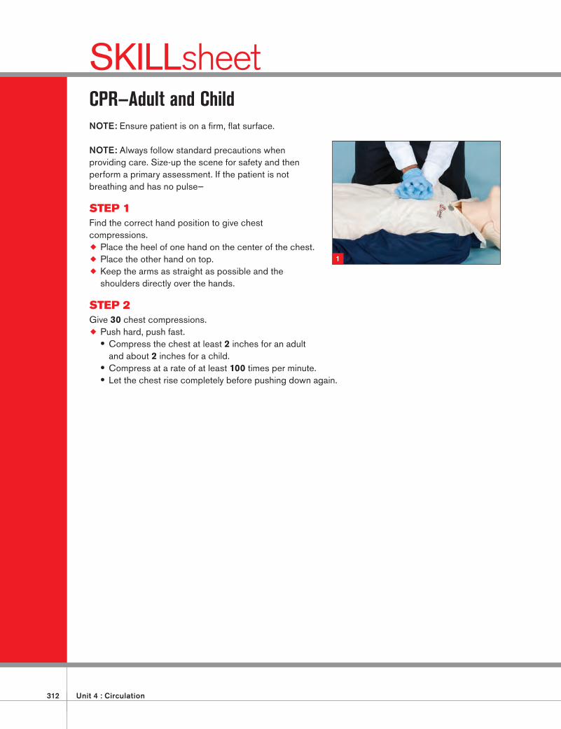
312 Unit 4 : Circulation
SKILLsheetCPR—Adult and ChildNOTE: Ensure patient is on a fi rm, fl at surface.
NOTE: Always follow standard precautions when providing care. Size-up the scene for safety and then perform a primary assessment. If the patient is not breathing and has no pulse—
STEP 1Find the correct hand position to give chest compressions.
Place the heel of one hand on the center of the chest.
Place the other hand on top.
Keep the arms as straight as possible and the shoulders directly over the hands.
STEP 2Give 30 chest compressions.
Push hard, push fast. • Compress the chest at least 2 inches for an adult and about 2 inches for a child.
• Compress at a rate of at least 100 times per minute. • Let the chest rise completely before pushing down again.
1
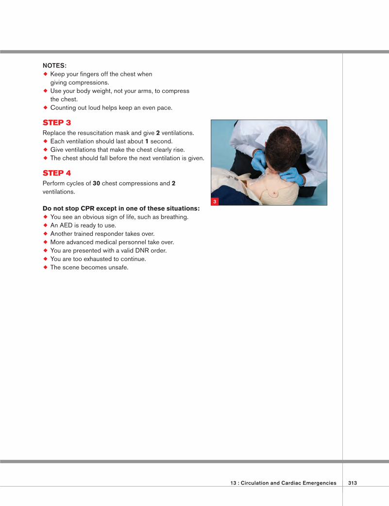
13 : Circulation and Cardiac Emergencies 313
NOTES: Keep your fi ngers off the chest when giving compressions.
Use your body weight, not your arms, to compress the chest.
Counting out loud helps keep an even pace.
STEP 3Replace the resuscitation mask and give 2 ventilations.
Each ventilation should last about 1 second.
Give ventilations that make the chest clearly rise.
The chest should fall before the next ventilation is given.
STEP 4Perform cycles of 30 chest compressions and 2 ventilations.
Do not stop CPR except in one of these situations:
You see an obvious sign of life, such as breathing.
An AED is ready to use.
Another trained responder takes over.
More advanced medical personnel take over.
You are presented with a valid DNR order.
You are too exhausted to continue.
The scene becomes unsafe.
3
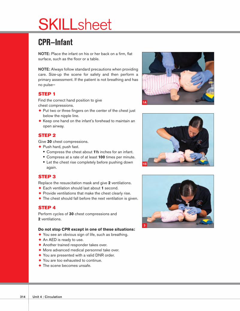
314 Unit 4 : Circulation
SKILLsheetCPR—InfantNOTE: Place the infant on his or her back on a fi rm, fl at surface, such as the fl oor or a table.
NOTE: Always follow standard precautions when providing care. Size-up the scene for safety and then perform a primary assessment. If the patient is not breathing and has no pulse—
STEP 1Find the correct hand position to give chest compressions.
Put two or three fi ngers on the center of the chest just below the nipple line.
Keep one hand on the infant’s forehead to maintain an open airway.
STEP 2Give 30 chest compressions.
Push hard, push fast. • Compress the chest about 1½ inches for an infant. • Compress at a rate of at least 100 times per minute. • Let the chest rise completely before pushing down again.
STEP 3Replace the resuscitation mask and give 2 ventilations.
Each ventilation should last about 1 second.
Provide ventilations that make the chest clearly rise.
The chest should fall before the next ventilation is given.
STEP 4Perform cycles of 30 chest compressions and 2 ventilations.
Do not stop CPR except in one of these situations:
You see an obvious sign of life, such as breathing.
An AED is ready to use.
Another trained responder takes over.
More advanced medical personnel take over.
You are presented with a valid DNR order.
You are too exhausted to continue.
The scene becomes unsafe.
1A
1B
3
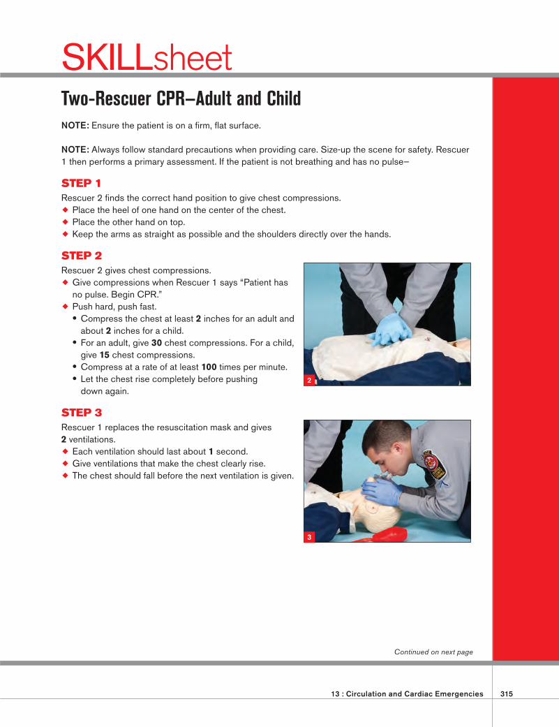
13 : Circulation and Cardiac Emergencies 315
SKILLsheetTwo-Rescuer CPR—Adult and ChildNOTE: Ensure the patient is on a fi rm, fl at surface.
NOTE: Always follow standard precautions when providing care. Size-up the scene for safety. Rescuer 1 then performs a primary assessment. If the patient is not breathing and has no pulse—
STEP 1Rescuer 2 fi nds the correct hand position to give chest compressions.
Place the heel of one hand on the center of the chest.
Place the other hand on top.
Keep the arms as straight as possible and the shoulders directly over the hands.
STEP 2Rescuer 2 gives chest compressions.
Give compressions when Rescuer 1 says “Patient has no pulse. Begin CPR.”
Push hard, push fast. • Compress the chest at least 2 inches for an adult and about 2 inches for a child.
• For an adult, give 30 chest compressions. For a child, give 15 chest compressions.
• Compress at a rate of at least 100 times per minute. • Let the chest rise completely before pushing down again.
STEP 3Rescuer 1 replaces the resuscitation mask and gives 2 ventilations.
Each ventilation should last about 1 second.
Give ventilations that make the chest clearly rise.
The chest should fall before the next ventilation is given.
2
3
Continued on next page
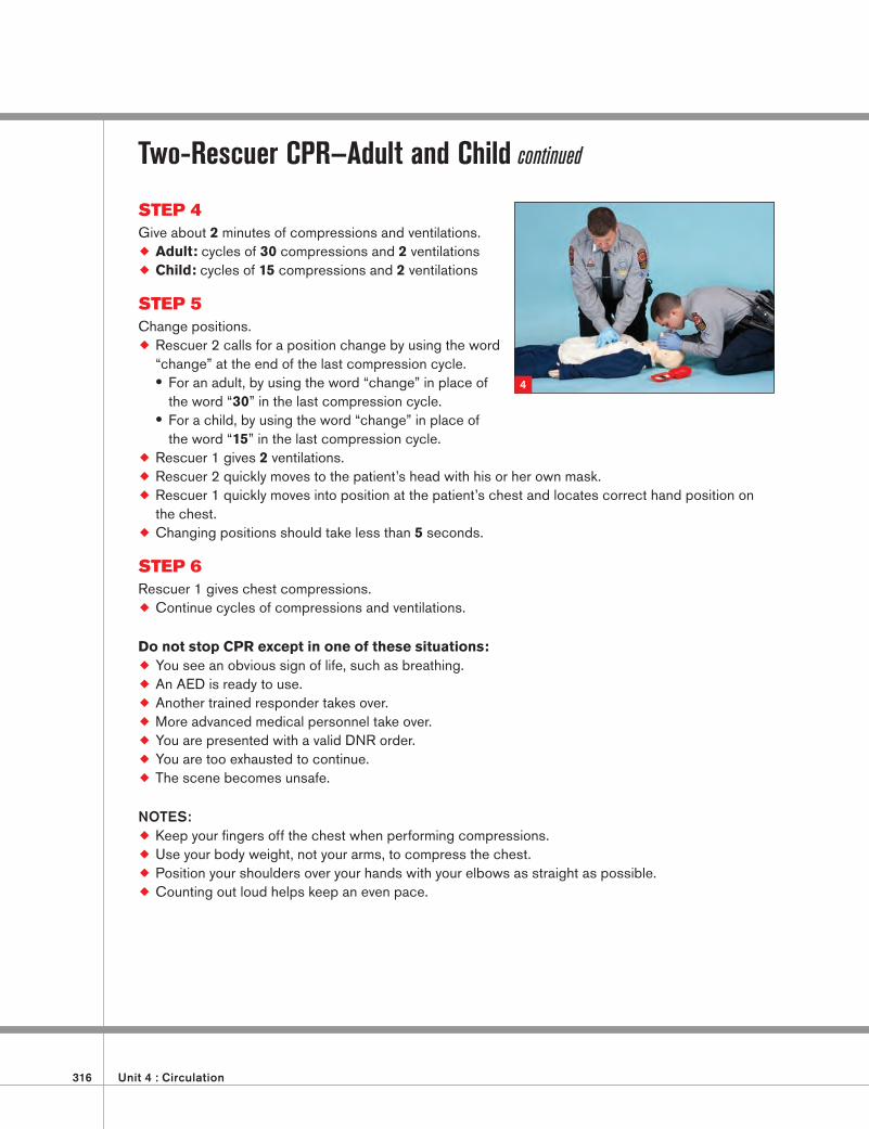
316 Unit 4 : Circulation
Two-Rescuer CPR—Adult and Child continued
STEP 4Give about 2 minutes of compressions and ventilations.
Adult: cycles of 30 compressions and 2 ventilations
Child: cycles of 15 compressions and 2 ventilations
STEP 5Change positions.
Rescuer 2 calls for a position change by using the word “change” at the end of the last compression cycle. • For an adult, by using the word “change” in place of the word “30” in the last compression cycle.
• For a child, by using the word “change” in place of the word “15” in the last compression cycle.
Rescuer 1 gives 2 ventilations.
Rescuer 2 quickly moves to the patient’s head with his or her own mask.
Rescuer 1 quickly moves into position at the patient’s chest and locates correct hand position on the chest.
Changing positions should take less than 5 seconds.
STEP 6Rescuer 1 gives chest compressions.
Continue cycles of compressions and ventilations.
Do not stop CPR except in one of these situations:
You see an obvious sign of life, such as breathing.
An AED is ready to use.
Another trained responder takes over.
More advanced medical personnel take over.
You are presented with a valid DNR order.
You are too exhausted to continue.
The scene becomes unsafe.
NOTES: Keep your fi ngers off the chest when performing compressions.
Use your body weight, not your arms, to compress the chest.
Position your shoulders over your hands with your elbows as straight as possible.
Counting out loud helps keep an even pace.
4
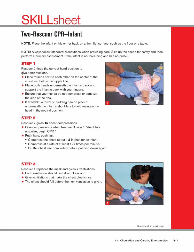
13 : Circulation and Cardiac Emergencies 317
SKILLsheetTwo-Rescuer CPR—InfantNOTE: Place the infant on his or her back on a fi rm, fl at surface, such as the fl oor or a table.
NOTE: Always follow standard precautions when providing care. Size-up the scene for safety and then perform a primary assessment. If the infant is not breathing and has no pulse—
STEP 1Rescuer 2 fi nds the correct hand position to give compressions.
Place thumbs next to each other on the center of the chest just below the nipple line.
Place both hands underneath the infant’s back and support the infant’s back with your fi ngers.
Ensure that your hands do not compress or squeeze the side of the ribs.
If available, a towel or padding can be placed underneath the infant’s shoulders to help maintain the head in the neutral position.
STEP 2Rescuer 2 gives 15 chest compressions.
Give compressions when Rescuer 1 says “Patient has no pulse, begin CPR.”
Push hard, push fast. • Compress the chest about 1½ inches for an infant. • Compress at a rate of at least 100 times per minute. • Let the chest rise completely before pushing down again.
STEP 3Rescuer 1 replaces the mask and gives 2 ventilations.
Each ventilation should last about 1 second.
Give ventilations that make the chest clearly rise.
The chest should fall before the next ventilation is given.
Continued on next page
1
3
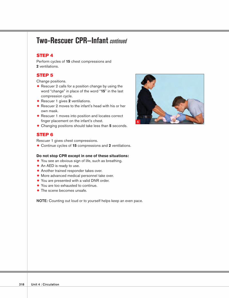
318 Unit 4 : Circulation
Two-Rescuer CPR—Infant continued
STEP 4Perform cycles of 15 chest compressions and 2 ventilations.
STEP 5Change positions.
Rescuer 2 calls for a position change by using the word “change” in place of the word “15” in the last compression cycle.
Rescuer 1 gives 2 ventilations.
Rescuer 2 moves to the infant’s head with his or her own mask.
Rescuer 1 moves into position and locates correct fi nger placement on the infant’s chest.
Changing positions should take less than 5 seconds.
STEP 6Rescuer 1 gives chest compressions.
Continue cycles of 15 compressions and 2 ventilations.
Do not stop CPR except in one of these situations:
You see an obvious sign of life, such as breathing.
An AED is ready to use.
Another trained responder takes over.
More advanced medical personnel take over.
You are presented with a valid DNR order.
You are too exhausted to continue.
The scene becomes unsafe.
NOTE: Counting out loud or to yourself helps keep an even pace.
5
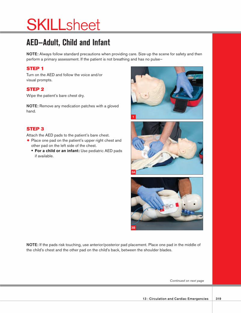
13 : Circulation and Cardiac Emergencies 319
SKILLsheetAED—Adult, Child and InfantNOTE: Always follow standard precautions when providing care. Size-up the scene for safety and then perform a primary assessment. If the patient is not breathing and has no pulse—
STEP 1Turn on the AED and follow the voice and/or visual prompts.
STEP 2Wipe the patient’s bare chest dry.
NOTE: Remove any medication patches with a gloved hand.
STEP 3Attach the AED pads to the patient’s bare chest.
Place one pad on the patient’s upper right chest and other pad on the left side of the chest. • For a child or an infant: Use pediatric AED pads if available.
NOTE: If the pads risk touching, use anterior/posterior pad placement. Place one pad in the middle of the child’s chest and the other pad on the child’s back, between the shoulder blades.
Continued on next page
1
3A
3B
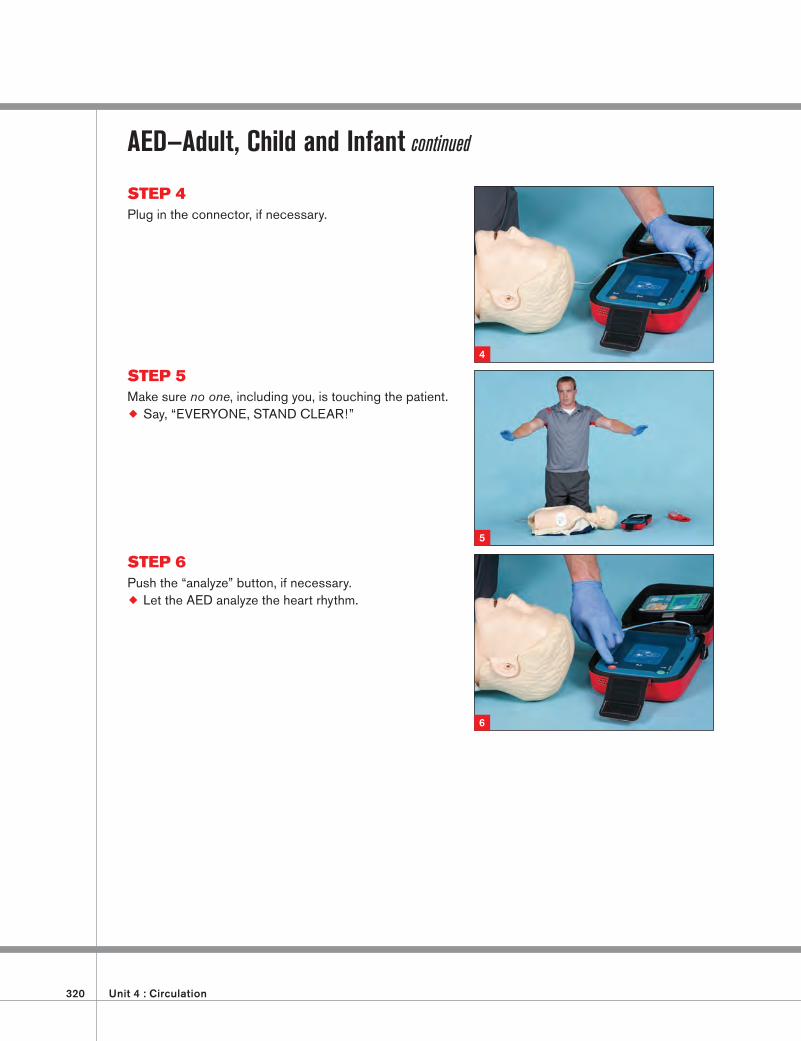
320 Unit 4 : Circulation
AED—Adult, Child and Infant continued
STEP 4Plug in the connector, if necessary.
STEP 5Make sure no one, including you, is touching the patient.
Say, “EVERYONE, STAND CLEAR!”
STEP 6Push the “analyze” button, if necessary.
Let the AED analyze the heart rhythm.
4
5
6
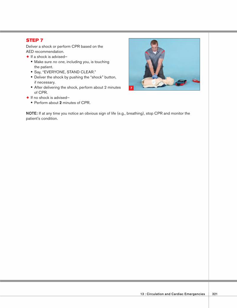
13 : Circulation and Cardiac Emergencies 321
STEP 7Deliver a shock or perform CPR based on the AED recommendation.
If a shock is advised— • Make sure no one, including you, is touching the patient.
• Say, “EVERYONE, STAND CLEAR.” • Deliver the shock by pushing the “shock” button, if necessary.
• After delivering the shock, perform about 2 minutes of CPR.
If no shock is advised— • Perform about 2 minutes of CPR.
NOTE: If at any time you notice an obvious sign of life (e.g., breathing), stop CPR and monitor the patient's condition.
7
322 Unit 4 : Circulation
Recognizing a heart attack and getting the necessary care at once may prevent a patient from going into cardiac arrest. However, preventing a heart attack in the fi rst place is even more effective—there is no substitute for prevention. Heart attacks are usually the result of disease of the heart and blood vessels. Coronary heart disease (CHD) is the leading cause of death for adults in the United States. It accounts for nearly 500,000 deaths each year.
CHD develops slowly. Deposits of cholesterol, a fatty substance made by the body and present in certain foods, build up on the inner walls of the arteries. As the arteries that carry blood to the heart get narrower, less oxygen-rich blood fl ows to the heart. This reduced oxygen supply to the heart can eventually cause a heart attack.
Although a heart attack may seem to strike suddenly, many people gradually put their hearts in danger from cardiovascular disease. Because cardiovascular disease develops slowly, people may not be aware of it for many years. Fortunately, it is possible to slow the progress of cardiovascular disease by making lifestyle changes.
Behavior that can harm the heart and blood vessels may begin in early childhood. Junk food, which is high in cholesterol and saturated fats but has little real nutritional value, can contribute to cardiovascular disease. Cigarette smoking also greatly contributes to cardiovascular disease and to other diseases.
There are many factors that increase a person’s chances of developing cardiovascular disease. These are called risk factors. Some of them you cannot change. For instance, although more women than men die each year from cardiovascular disease in the United States, heart disease generally affects men at younger ages than it does women.
Besides gender, ethnicity also plays an important role in determining the risk for heart disease. African Americans and Native Americans in the United States have higher rates of heart disease than do other populations. A history of heart disease in your family also increases your risk.
ALTERING RISK FACTORS
Many risk factors can be altered, however. Cigarette smoking; uncontrolled diabetes, high blood cholesterol or high blood pressure; obesity; and lack of regular exercise all increase the risk of heart disease. When you combine one risk factor, such as smoking, with others, such as high blood pressure and lack of regular exercise, the risk of heart attack is much greater.
It is never too late to take steps to control risk factors, thereby improving your chances for living a long and healthy life. It is important to know how to perform CPR and use an AED. However, since the chances of surviving cardiac arrest are poor, the best way to deal with cardiac arrest is to be aware of risk factors and take steps to help prevent it, including exercise and quitting smoking.
EnrichmentPreventing Coronary Heart Disease