Embed Size (px)
Citation preview
Emergency Medical Response INSTRUCTOR’S MANUAL
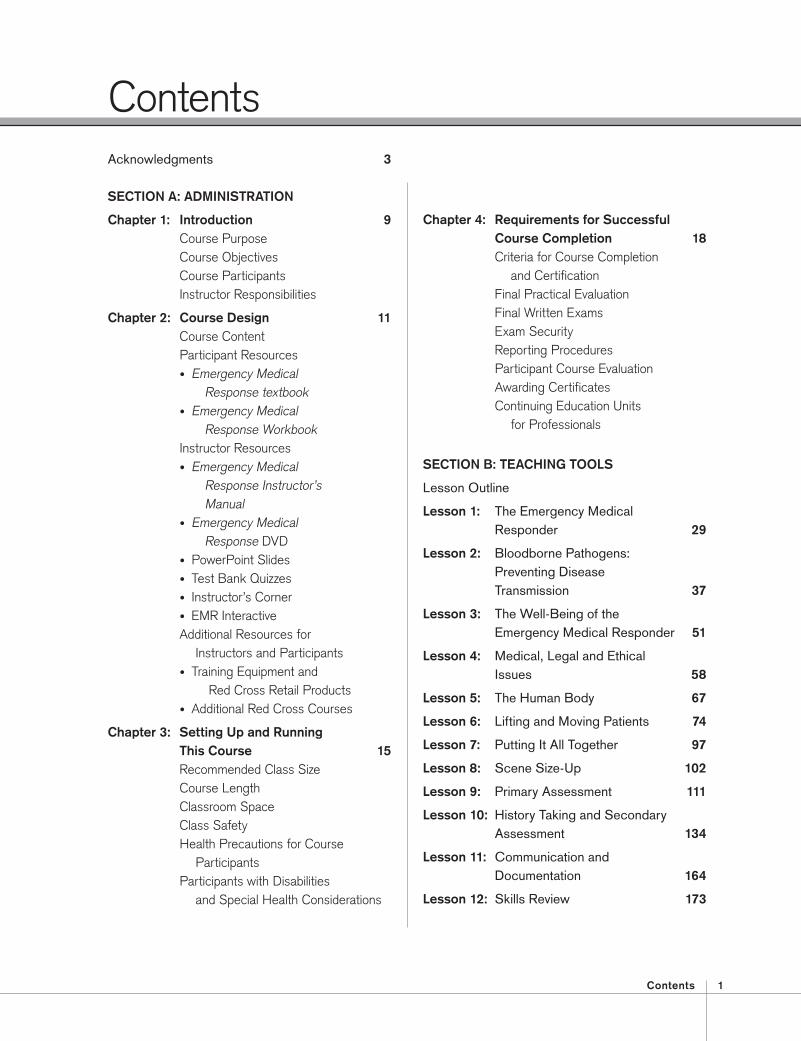
Contents 1
Acknowledgments 3
SECTION A: ADMINISTRATION
Chapter 1: Introduction 9 Course Purpose Course Objectives Course Participants Instructor Responsibilities
Chapter 2: Course Design 11 Course Content
Participant Resources • Emergency Medical
Response textbook • Emergency Medical
Response Workbook Instructor Resources • Emergency Medical
Response Instructor’s Manual
• Emergency Medical Response DVD
• PowerPoint Slides • Test Bank Quizzes • Instructor’s Corner • EMR Interactive
Additional Resources for Instructors and Participants
• Training Equipment and Red Cross Retail Products
• Additional Red Cross Courses
Chapter 3: Setting Up and Running This Course 15
Recommended Class Size Course Length Classroom Space Class Safety
Health Precautions for Course Participants
Participants with Disabilities and Special Health Considerations
Chapter 4: Requirements for Successful Course Completion 18
Criteria for Course Completion and Certification
Final Practical Evaluation Final Written Exams Exam Security Reporting Procedures Participant Course Evaluation Awarding Certificates Continuing Education Units
for Professionals
SECTION B: TEACHING TOOLS
Lesson Outline
Lesson 1: The Emergency Medical Responder 29
Lesson 2: Bloodborne Pathogens: Preventing Disease Transmission 37
Lesson 3: The Well-Being of the Emergency Medical Responder 51
Lesson 4: Medical, Legal and Ethical Issues 58
Lesson 5: The Human Body 67
Lesson 6: Lifting and Moving Patients 74
Lesson 7: Putting It All Together 97
Lesson 8: Scene Size-Up 102
Lesson 9: Primary Assessment 111
Lesson 10: History Taking and Secondary Assessment 134
Lesson 11: Communication and Documentation 164
Lesson 12: Skills Review 173
Contents
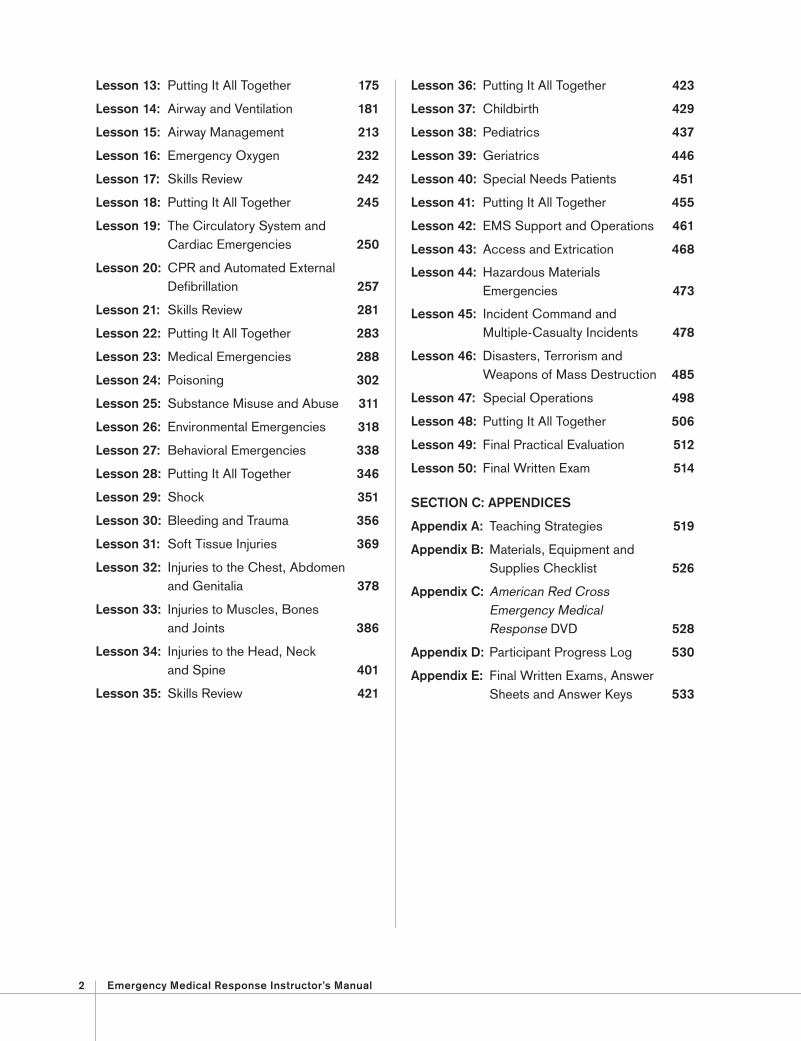
Lesson 13: Putting It All Together 175
Lesson 14: Airway and Ventilation 181
Lesson 15: Airway Management 213
Lesson 16: Emergency Oxygen 232
Lesson 17: Skills Review 242
Lesson 18: Putting It All Together 245
Lesson 19: The Circulatory System and Cardiac Emergencies 250
Lesson 20: CPR and Automated External Defibrillation 257
Lesson 21: Skills Review 281
Lesson 22: Putting It All Together 283
Lesson 23: Medical Emergencies 288
Lesson 24: Poisoning 302
Lesson 25: Substance Misuse and Abuse 311
Lesson 26: Environmental Emergencies 318
Lesson 27: Behavioral Emergencies 338
Lesson 28: Putting It All Together 346
Lesson 29: Shock 351
Lesson 30: Bleeding and Trauma 356
Lesson 31: Soft Tissue Injuries 369
Lesson 32: Injuries to the Chest, Abdomen and Genitalia 378
Lesson 33: Injuries to Muscles, Bones and Joints 386
Lesson 34: Injuries to the Head, Neck and Spine 401
Lesson 35: Skills Review 421
Lesson 36: Putting It All Together 423
Lesson 37: Childbirth 429
Lesson 38: Pediatrics 437
Lesson 39: Geriatrics 446
Lesson 40: Special Needs Patients 451
Lesson 41: Putting It All Together 455
Lesson 42: EMS Support and Operations 461
Lesson 43: Access and Extrication 468
Lesson 44: Hazardous Materials Emergencies 473
Lesson 45: Incident Command and Multiple-Casualty Incidents 478
Lesson 46: Disasters, Terrorism and Weapons of Mass Destruction 485
Lesson 47: Special Operations 498
Lesson 48: Putting It All Together 506
Lesson 49: Final Practical Evaluation 512
Lesson 50: Final Written Exam 514
SECTION C: APPENDICES
Appendix A: Teaching Strategies 519
Appendix B: Materials, Equipment and Supplies Checklist 526
Appendix C: American Red Cross Emergency Medical Response DVD 528
Appendix D: Participant Progress Log 530
Appendix E: Final Written Exams, Answer Sheets and Answer Keys 533
2 Emergency Medical Response Instructor’s Manual
Lesson 14 : Airway and Ventilation 181
L E S S O N
Airway and Ventilation 14 Lesson Length 115 minutes (185 minutes with Enrichments)
Materials, Equipment and Supplies Emergency Medical Response textbook PowerPoint slides 1–26 LCD projector, screen and computer Emergency Medical Response DVD DVD player and monitor Non-latex disposable gloves (multiple sizes)
and other personal protective equipment (PPE), as indicated
Adult and infant manikins (one for every two participants; child manikins optional)
Decontamination supplies Resuscitation masks (adult and pediatric;
one for each participant) Bag-valve-mask resuscitators (BVMs) (adult
and pediatric; one for every two participants) Examples of delivery systems for asthma
medications and peak flow meters, such as asthma training inhalers ( Enrichment: Assisting the Patient with Asthma [optional])
Lesson Objectives
Knowledge
After completing this lesson, participants will be able to— Describe the structure and function of the
respiratory system. List the signs of inadequate breathing. Describe how to care for a patient
experiencing respiratory distress. Explain why basic airway management and
ventilation skills take priority over many other basic life-support skills.
Describe how to perform mouth-to-mouth, mouth-to-nose and mouth-to-stoma ventilations.
Describe how to assess for breath sounds (Enrichment) .
Skill
After completing this lesson, participants will be able to—
Demonstrate how to give ventilations using a resuscitation mask.
Demonstrate how to give ventilations using a bag-valve-mask resuscitator (BVM).
Demonstrate how to give ventilations if a head, neck or spinal injury is suspected.
Demonstrate how to perform cricoid pressure (Sellick’s maneuver) ( Enrichment skill ).
Demonstrate how to assist a patient with an asthma inhaler ( Enrichment skill ).
182 Emergency Medical Response Instructor’s Manual
TOPIC: INTRODUCTION Time: 5 minutes PowerPoint slides 1–2
Activity:
Review the opening scenario:
Your medical emergency response team has been called to the fitness center by building security on a report that an employee complained of having difficulty breathing. You and your partner arrive and find the man conscious but in distress. The patient’s chief complaint is difficulty breathing. He says he just “overdid it” on the treadmill. He appears to be out of breath and is having trouble speaking in full sentences. You begin a primary assessment and determine that the patient is in respiratory distress.
Ask participants— “What should you do?” “What can you do to assist the patient with his breathing?”
Tell participants: “Ensuring an open airway is the most important step that you can take in caring for a patient because a person cannot breathe without an open airway. A patient who can speak or cry is conscious, has an open airway, is breathing and has a pulse.”
TOPIC: THE RESPIRATORY SYSTEM Time: 10 minutes PowerPoint slides 3–4
Key Points:
The respiratory system is divided into the upper and lower airway tracts. The upper airway tract begins at the mouth and includes the nose, pharynx and larynx. The lower airway tract begins below the level of the vocal cords and includes the trachea,
bronchi, bronchioles and alveoli. Breathing requires that the respiratory, circulatory, nervous and musculoskeletal systems work
together. Injuries or illnesses that affect any of these systems may cause breathing emergencies.
INSTRUCTOR’S NOTE Let participants provide responses, guiding them to important areas related to respiratory emergencies, such as assessing for an open airway and respiratory function, underlying causes and signs and symptoms of respiratory distress and respiratory arrest.
INSTRUCTOR’S NOTE Urge participants to review Chapter 4: The Human Body .
Lesson 14 : Airway and Ventilation 183
Pathophysiology
Key Points:
Breathing difficulties may occur for various reasons including — Inadequate amount of oxygen breathed in during respiration. Breathing in a low-oxygen environment or when poisonous gases are in the air. Infection of the lungs. Illness, such as asthma, excess fluid in the lungs, excess fluid between the lungs and
blood vessels or poor circulation. Upper airway problems due to swelling, obstruction or trauma. Ineffective circulation, such as from shock or cardiac arrest.
Choking, caused by airway obstruction, is one of the most common causes of breathing emergencies and can occur due to anatomical or mechanical obstruction.
An inadequate rate or depth of breathing leads to insufficient air volume moving in and out of the lungs.
Respirations may be ineffective due to unconsciousness, altered level of consciousness, chest injury, poisoning, overdose or diseases, such as chronic obstructive pulmonary disease (COPD) or emphysema.
Oxygenation refers to the amount of oxygen in the bloodstream. An insufficient amount of oxygen delivered to the cells is referred to as hypoxia, which may result from — Obstructed airway. Shock. Inadequate breathing. Drowning. Strangulation. Choking. Suffocation. Cardiac arrest. Head trauma. Carbon monoxide poisoning. Complications of general anesthesia.
TOPIC: RESPIRATORY EMERGENCIES Time: 20 minutes PowerPoint slides 5–9
Key Points:
A respiratory emergency occurs when air cannot travel freely and easily into the lungs; it can be life threatening because it greatly cuts down on the oxygen the body receives or because it cuts off oxygen entirely.
INSTRUCTOR’S NOTE Refer participants to Chapter 4: The Human Body for information about the physiology of respiration and Chapter 8: History Taking and Secondary Assessment for a review of information about respiratory rate and normal rate ranges.
184 Emergency Medical Response Instructor’s Manual
Unless the brain receives oxygen within 4 to 6 minutes, permanent brain damage or death can occur.
There are two types of respiratory emergencies: Respiratory distress, which involves difficulty breathing Respiratory arrest, which involves the cessation of breathing
Respiratory distress is a condition in which breathing becomes difficult; it can be caused by— A partially obstructed airway. Illness. Chronic conditions, such as asthma. Electrocution, including lightning strikes. Heart attack. Injury to the head, chest, lungs or abdomen . Allergic reactions . Drugs . Poisoning . Emotional distress .
Trouble breathing can be the first sign of a more serious emergency, such as anaphylaxis or a heart problem; therefore, recognizing the signs of breathing problems and providing care often are the keys to preventing these problems from becoming more serious emergencies.
Patients with breathing problems will most likely be conscious. Breathing problems can be identified by watching and listening to the patient’s breathing and
by asking how the patient feels. Signs and symptoms of respiratory emergencies include—
Slow or rapid breathing. Unusually deep or shallow breathing. Gasping for breath. Wheezing, gurgling or high-pitched noises. Unusually moist or cool skin. Flushed, pale, ashen or bluish skin color. Shortness of breath. Dizziness or light-headedness. Pain in the chest or tingling in the hands, feet or lips. Apprehensive or fearful feelings.
Activity Option A:
Time: 5 minutes Ask participants if they have ever experienced a respiratory infection. Ask those that responded
positively to share with the class what signs and symptoms they experienced.
INSTRUCTOR’S NOTE Possible responses may include— Signs and symptoms, such as coughing with or without mucus, difficulty breathing, fever, chills,
shortness of breath or chest pain.
Lesson 14 : Airway and Ventilation 185
Activity Option B:
Time: 5 minutes Using the following scenario, ask participants to identify possible causes for the patient’s condition
and to suggest additional questions that would be appropriate to ask when determining what the patient is experiencing.
You and your partner are summoned to a local conference center in response to an emergency call. A person who was scheduled to speak at a conference began complaining of difficulty breathing about 10 minutes before he was scheduled to speak. On arrival at the scene, you find the patient sitting on the floor, breathing rapidly. The patient states that all of sudden he began to feel dizzy and his lips started tingling.
Activity Option C:
Time: 10 minutes Divide the participants into small groups and ask them to compare and contrast the signs and
symptoms of COPD with an asthma attack. Then bring the groups back together to discuss their conclusions.
Activity Option D:
Time: 10 minutes Divide the participants into seven small groups. Assign each group one of the following conditions:
COPD, asthma, pneumonia, acute pulmonary edema, hyperventilation, pulmonary embolism and emphysema.
Have participants use their textbooks to describe the condition and explore its signs and symptoms and care, then have each group present its information to the rest of the class.
INSTRUCTOR’S NOTE Possible responses may include— Causes such as stress or anxiety related to speaking before a group of people or an underlying
infection, heart failure or lung disease. Additional questions, such as—
“Have you had an infection recently?” “Do you have a history of heart or lung problems?” “Do you have a history of asthma?” “Have you ever experienced this type of feeling before?”
INSTRUCTOR’S NOTE Responses should include the following: Comparison of similarities including elevated shoulders and pursed lip breathing, wheezing, shortness
of breath, coughing and confusion Contrasting differences include increased mucus, loss of appetite, tiring easily and age (middle aged
or older patient) for COPD, and rapid, shallow breathing, sweating, chest tightness and age (typically child or young adult) for an asthma attack
186 Emergency Medical Response Instructor’s Manual
INSTRUCTOR’S NOTE Responses should include the following: For COPD:
COPD is a progressive lung disease characterized by difficulty breathing due to partial obstruction of the airways and loss of the alveolis’ ability to fill with air.
Cigarette smoking is the most common cause. Other possible causes are inhalation of other lung irritants, pollution, dust or chemicals over a long
period of time. Signs and symptoms include coughing with copious mucus; tendency to tire easily; loss of appetite;
bent posture with elevated shoulders and pursed lips; fast pulse; round, barrel-shaped chest; and confusion (due to lack of oxygen to the brain).
Care includes helping the patient focus on his or her breathing, as deep breaths help to fill the lungs with air and maintain flexibility in the chest wall.
For asthma: Asthma is an ongoing illness involving airway swelling and inflammation that is more common in
children and young adults. Exposure to a trigger, such as exercise, cold air, allergens or other irritants, affects the airways,
causing them to swell and narrow. Signs and symptoms include coughing or wheezing noises, difficulty breathing, shortness of
breath, sweating, tightness in the chest, rapid and shallow breathing, inability to talk without stopping for breathing, bent posture with elevated shoulders, pursed lips and feelings of fear and confusion.
Acute care includes controlling environmental variables, medications (e.g., bronchodilators) and other forms of treatment.
For pneumonia: Pneumonia is an infection due to virus, bacteria, fungus or other organism that causes inflammation
of the lungs that leads to the air sacs filling with fluid, thus interfering with oxygen exchange. Signs and symptoms include high fever and chills, chest pain, shortness of breath, increased
respiratory rate, breathing difficulty, congestion (in older adults) and altered mental status (in older adults).
Care includes use of emergency oxygen administration. For acute pulmonary edema:
Acute pulmonary edema is an abnormal buildup of fluid in the lungs causing severe respiratory distress, altered mental status and coughing, with some bloody sputum.
Signs and symptoms develop gradually, such as difficulty breathing when lying flat, awakening at night with a feeling of breathlessness, unusual shortness of breath during activity and significant weight gain.
Acute signs and symptoms include shortness of breath; difficulty breathing, such as wheezing or gasping; cyanosis; frothy (foamy) pink sputum; pale skin; excessive sweating; restlessness; anxiety and a feeling of apprehension; feeling of suffocating or drowning; and chest pain (when condition is caused by coronary artery disease).
Care includes emergency oxygen administration as the first step. For hyperventilation:
Hyperventilation is the faster and shallower or deeper-than normal breathing leading to a decrease in blood carbon dioxide levels, reducing blood flow to the brain and leading to fear, anxiety, confusion, dizziness, and numbness and tingling in the fingers and toes.
Care focuses on calming the patient (if symptoms are not life threatening), listening to the patient’s concerns and trying to reassure and encourage the patient to breathe slower or breathe through pursed lips and administering emergency oxygen if patient does not respond.
For pulmonary embolism: Pulmonary embolism is a blockage in the arteries of the lungs that interferes with oxygen and carbon
dioxide exchange leading to respiratory distress (degree depends on size of clot). Signs and symptoms include sudden onset of dyspnea; chest pain that is localized and does not
radiate; coughing, including coughing up blood; and fainting. Larger clots can cause death very quickly. Rapid care and transport of the patient to a hospital is
crucial.
Lesson 14 : Airway and Ventilation 187
TOPIC: AIRWAY Time: 20 minutes PowerPoint slides 10–14
Signs of an Open Airway
Key Points:
An airway is open and clear when the following occur: The patient’s chest rises and falls. Air is heard and felt coming out of the patient’s mouth and nose as the patient exhales. The conscious patient is able to speak in full sentences without distress. The conscious patient is speaking in normal tones.
Signs of an Inadequate Airway
Key Points:
A patient with an inadequate airway needs close attention and monitoring. Any unusual sounds may be signs of an airway obstruction.
The patient may be visibly unable to catch his or her breath or he or she may gasp for air and make grunting sounds.
Stridor (a harsh, high-pitched sound on inhalation) may be due to a swollen larynx blocking the upper airway.
Snoring may indicate that the tongue or other tissues in the mouth may be relaxed and blocking the upper airway.
A patient who is awake and alert but unable to speak or can only speak a few words or has a hoarse-sounding voice may be having severe difficulty breathing.
Inadequate breathing also may be due to trauma, infection or an allergic reaction. No air movement indicates apnea (the complete absence of breathing); the patient needs
artificial ventilation. Sometimes, no detectable air movement may be due to an airway obstruction.
For emphysema: Emphysema is a chronic disease caused by damage to the air sacs in the lungs; it also is
degenerative, worsening over time with alveoli losing their elasticity, becoming distended with trapped air interfering with their proper function. Over time, the number of alveoli affected increases, leading to increasing difficulty with breathing.
Signs and symptoms include shortness of breath (most common), cyanosis, barrel-shaped chest, fatigue, loss of appetite, weight loss, mild cough , breathing through pursed lips, restlessness, confusion and weakness.
INSTRUCTOR’S NOTE Briefly review the two methods for opening the airway: the head-tilt/chin-lift technique and the jaw-thrust (without head extension) maneuver found in Lesson 9. Refer participants to Chapter 7: Primary Assessment. Have participants review the skill sheets for both techniques.
188 Emergency Medical Response Instructor’s Manual
If efforts to open the airway are unsuccessful and ventilations do not make the chest rise and fall, you must check for an airway obstruction, such as liquid, food, teeth, dentures, blood, vomit or other foreign objects.
Causes of Airway Obstruction
Key Points:
The two types of airway obstruction include mechanical and anatomical. Mechanical obstruction involves any foreign body lodged in the airway; it is an emergency
situation that needs immediate attention. The most common cause of foreign body airway obstruction (FBAO) in adults is a solid
object, such as food. In children younger than 4 years, large chunks of food and small objects, such as toy parts
or balloons, commonly cause airway obstruction. An anatomical obstruction is most commonly due to the tongue.
It occurs in an unresponsive patient, as muscle tone is lost allowing the tongue to fall back and block the airway.
Other conditions that may cause an anatomical obstruction include swelling due to trauma, infection, asthma, emphysema or anaphylaxis.
Trauma to the neck also may cause an obstruction.
Techniques to Clear an Airway Obstruction
Key Points:
Protocols vary related to the methods used to clear an airway obstruction. Abdominal thrusts, back blows and chest thrusts have been proven to effectively clear an
obstructed airway in conscious patients. Unconscious patients with FBAO should receive a modified form of CPR.
Techniques to Remove Foreign Matter from the Upper Airway
Key Points:
Two techniques can be used to remove visible foreign matter and fluids from the upper airway of an unconscious patient: finger sweeps and suctioning.
Finger sweeps involve removing an object or other foreign matter from a patient’s mouth with a finger. It is performed only on an unconscious patient and only when foreign matter is seen in the
patient’s mouth. Be sure to always wear gloves when performing a finger sweep. Be sure to use the index finger for an adult and little finger for a child or an infant.
Suctioning removes blood, fluids or food particles from the airway. Some suctioning devices cannot remove solid objects, such as teeth, foreign bodies
or food.
Lesson 14 : Airway and Ventilation 189
Recovery Positions
Key Points:
In some cases, the person may be unconscious but breathing. Generally that person should not be moved from a face-up position, especially if there is a suspected spinal injury.
However, if you are alone and have to leave the person (e.g., to call for help), or you cannot maintain an open and clear airway because of fluids or vomit, place the patient in a modified high arm in endangered spine (H.A.IN.E.S.) recovery position to help keep the airway open and clear .
TOPIC: ASSESSING BREATHING Time: 10 minutes PowerPoint slides 15–16
Inadequate Breathing
Key Points:
Inadequate breathing is indicated by the following signs: Muscles between the ribs pulling in on inhalation Pursed lip breathing Nasal flaring Fatigue Excessive use of abdominal muscles to breathe Sweating Sitting upright and leaning forward (tripod position) Deviated trachea
Abnormal breath sounds also are a sign of inadequate breathing; these may include— Stridor. Wheezing. Crackles/rales.
Inadequate Oxygenation
Key Points:
Problems with inadequate oxygenation may occur for a variety of reasons. A reduction in oxygen in the body causes headaches, increased breathing rate, nausea and
vomiting, altered mental status and, ultimately, death.
INSTRUCTOR’S NOTE Urge participants to review the information from previous lessons on opening the airway and on primary and secondary assessments.
190 Emergency Medical Response Instructor’s Manual
TOPIC: ARTIFICIAL VENTILATION Time: 45 minutes PowerPoint slide 17
Key Points:
Artificial ventilation refers to the various mechanical ways that can be used to help a patient breathe.
When delivering artificial ventilations, the force of air must be consistent and just strong enough to cause the chest to clearly rise in about 1 second; gastric distention can occur if too much force is used.
A resuscitation mask allows you to breathe expired air into a patient without making mouth-to-mouth contact, reducing the risk of disease transmission.
Approximately 16 percent oxygen is provided in an exhaled breath, enough to sustain life. However, this amount is considerably less than that which is delivered using a bag-valve-mask resuscitator (BVM) or emergency oxygen.
If the patient vomits when providing ventilations, quickly turn the patient onto the side, supporting the head and neck and turning the body as a unit; after vomiting has stopped, clear the patient’s airway using a finger sweep and suction if necessary and then turn the patient onto the back and continue with ventilations.
Dentures help to support the patient’s mouth and cheeks, making it easier to seal the resuscitation mask; they should be removed only if they become loose or block the airway.
Ventilations may need to be provided through the nose if the patient’s mouth is injured. The mask is placed over the patient’s mouth and nose.
On rare occasions, a patient may have an opening (stoma) in his or her neck for breathing.
SKILL Session
GIVING VENTILATIONS—ADULT AND CHILD
Activity:
INSTRUCTOR’S NOTES You may choose either method below depending on the experience of the participants
and whether your training facility can accommodate the Practice-While-You-Watch method, which works best when there is adequate practice space and a monitor large enough for everyone to see clearly.
Inform participants that they only need to demonstrate giving ventilations to an adult and can point out the differences for giving ventilations to a child, such as using a properly sized resuscitation mask for the patient, tilting the head slightly past neutral and only giving 1 ventilation about every 3 seconds.
Lesson 14 : Airway and Ventilation 191
SKILL Session
Practice-While-You-Watch
Ask participants to take their textbooks, disposable gloves and resuscitation masks with them to the practice area.
Explain to the participants that for this skill, they will follow along with and practice the steps for giving ventilations as they are guided by the DVD, using a manikin, disposable gloves and resuscitation mask.
Show the DVD segment, “Giving Ventilations—Adult, Child and Infant” (4:08). Do not interrupt this practice session to lecture or communicate anything other than
guidance related to skill practice. In general, answering questions should occur after the video segment (and skill practice) has ended.
Observe participants performing the technique and evaluate completion of the skill using the Skill Checklist.
Be sure to point out any common errors, such as tilting a child’s head too far back, failing to reassess for breathing and a pulse, not leaving the patient in a face-up position with return of breathing, not obtaining a seal with the resuscitation mask or using an improperly sized mask for the patient.
Check off participant’s progress on the Participant Progress Log.
Watch-Then-Practice
Tell participants to watch the DVD segment without practicing until you pause the DVD, even though the narration may say to follow along.
After the DVD segment, tell them to practice the skill using a manikin, disposable gloves and resuscitation mask.
If there is a 2-to-1 ratio of participants-to-manikins, partners should be instructed to follow along with the skill sheet and observe the other person practicing the skill.
Show the DVD segment, “Giving Ventilations—Adult, Child and Infant” (4:08). Initially guide participants through the steps listed on the skill chart, then encourage
them to practice independently, without assistance. Observe participants performing the technique and evaluate completion of the skill using
the Skill Checklist. Intervene and provide positive and corrective feedback as needed to each participant individually when possible.
Be sure to point out any common errors, such as tilting a child’s head too far back, failing to reassess for breathing and a pulse, not leaving the patient in a face-up position with return of breathing, not obtaining a seal with the resuscitation mask or using an improperly sized mask for the patient.
Check off participant’s progress on the Participant Progress Log.
192 Emergency Medical Response Instructor’s Manual
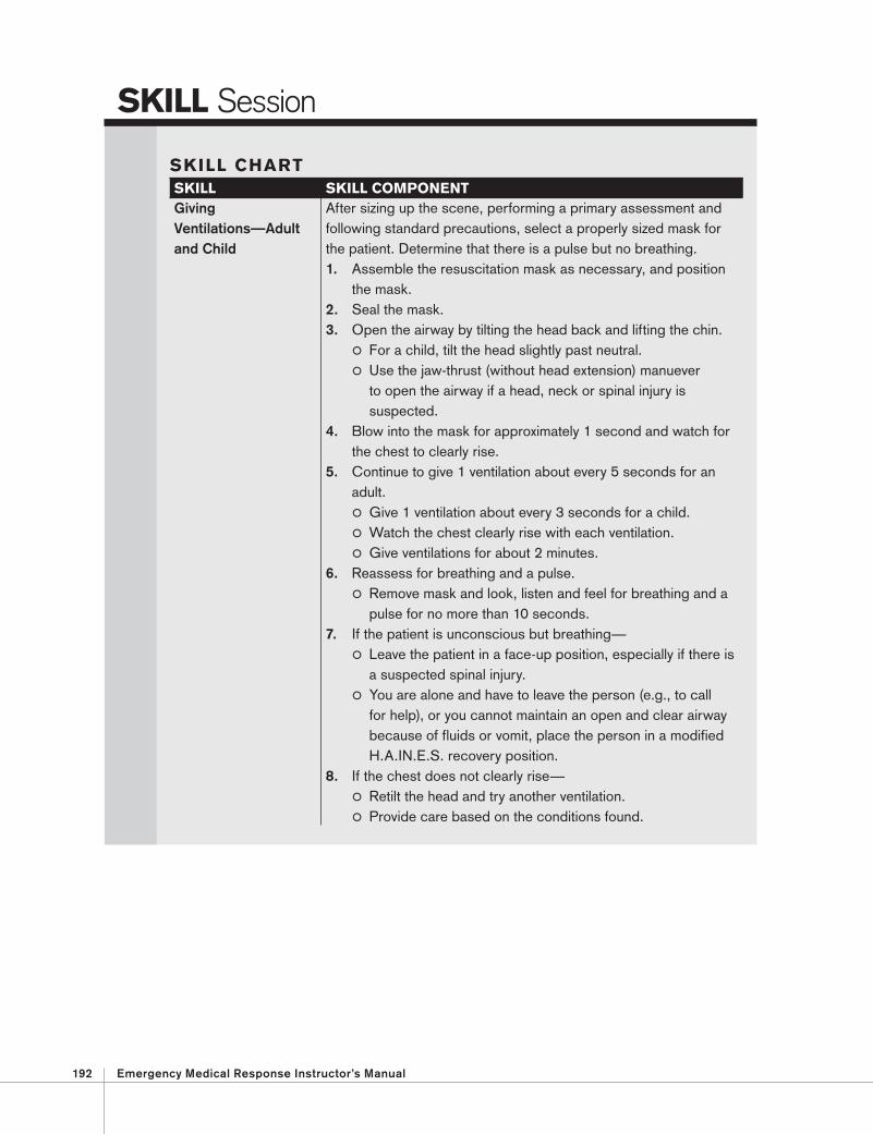
SKILL CHART SKILL SKILL COMPONENTGiving Ventilations—Adult and Child
After sizing up the scene, performing a primary assessment and following standard precautions, select a properly sized mask for the patient. Determine that there is a pulse but no breathing.1. Assemble the resuscitation mask as necessary, and position
the mask.2. Seal the mask.3. Open the airway by tilting the head back and lifting the chin.
For a child, tilt the head slightly past neutral. Use the jaw-thrust (without head extension) manuever to open the airway if a head, neck or spinal injury is suspected.
4. Blow into the mask for approximately 1 second and watch for the chest to clearly rise.
5. Continue to give 1 ventilation about every 5 seconds for an adult.
Give 1 ventilation about every 3 seconds for a child. Watch the chest clearly rise with each ventilation. Give ventilations for about 2 minutes.
6. Reassess for breathing and a pulse. Remove mask and look, listen and feel for breathing and a pulse for no more than 10 seconds.
7. If the patient is unconscious but breathing— Leave the patient in a face-up position, especially if there is a suspected spinal injury.
You are alone and have to leave the person (e.g., to call for help), or you cannot maintain an open and clear airway because of fluids or vomit, place the person in a modified H.A.IN.E.S. recovery position.
8. If the chest does not clearly rise— Retilt the head and try another ventilation. Provide care based on the conditions found.
SKILL Session
Lesson 14 : Airway and Ventilation 193
SKILL Session
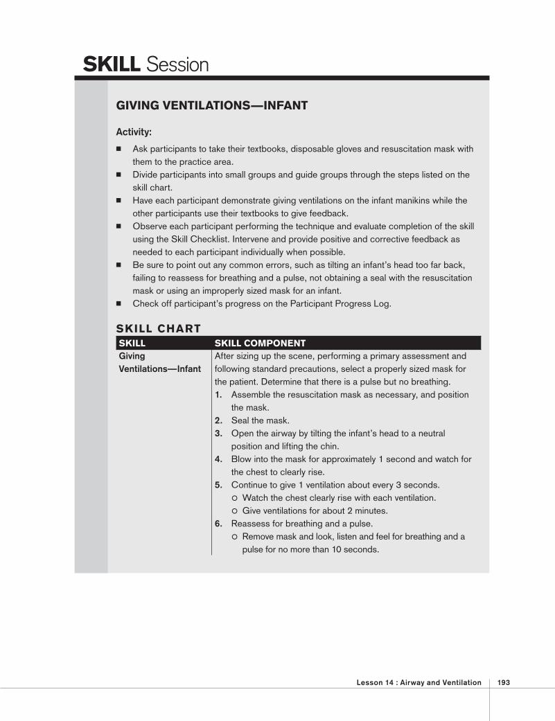
GIVING VENTILATIONS—INFANT
Activity:
Ask participants to take their textbooks, disposable gloves and resuscitation mask with them to the practice area.
Divide participants into small groups and guide groups through the steps listed on the skill chart.
Have each participant demonstrate giving ventilations on the infant manikins while the other participants use their textbooks to give feedback.
Observe each participant performing the technique and evaluate completion of the skill using the Skill Checklist. Intervene and provide positive and corrective feedback as needed to each participant individually when possible.
Be sure to point out any common errors, such as tilting an infant’s head too far back, failing to reassess for breathing and a pulse, not obtaining a seal with the resuscitation mask or using an improperly sized mask for an infant.
Check off participant’s progress on the Participant Progress Log.
SKILL CHART SKILL SKILL COMPONENTGiving Ventilations—Infant
After sizing up the scene, performing a primary assessment and following standard precautions, select a properly sized mask for the patient. Determine that there is a pulse but no breathing.1. Assemble the resuscitation mask as necessary, and position
the mask.2. Seal the mask.3. Open the airway by tilting the infant’s head to a neutral
position and lifting the chin.4. Blow into the mask for approximately 1 second and watch for
the chest to clearly rise.5. Continue to give 1 ventilation about every 3 seconds.
Watch the chest clearly rise with each ventilation. Give ventilations for about 2 minutes.
6. Reassess for breathing and a pulse. Remove mask and look, listen and feel for breathing and a pulse for no more than 10 seconds.
194 Emergency Medical Response Instructor’s Manual
SKILL Session
INSTRUCTOR’S NOTE If preferred, review giving ventilations (adult, child and infant) first and then divide the class into groups to practice the two techniques.
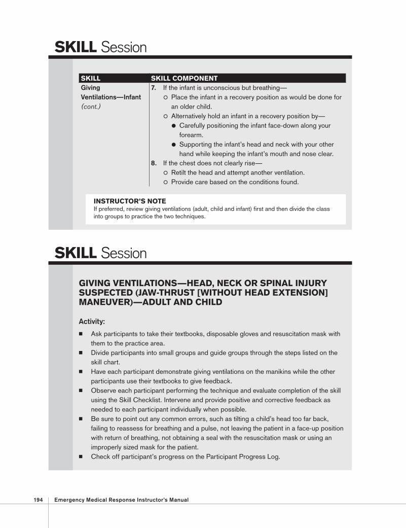
SKILL SKILL COMPONENTGiving Ventilations—Infant(cont.)
7. If the infant is unconscious but breathing— Place the infant in a recovery position as would be done for an older child.
Alternatively hold an infant in a recovery position by— Carefully positioning the infant face-down along your forearm.
Supporting the infant’s head and neck with your other hand while keeping the infant’s mouth and nose clear.
8. If the chest does not clearly rise— Retilt the head and attempt another ventilation. Provide care based on the conditions found.
SKILL Session
GIVING VENTILATIONS—HEAD, NECK OR SPINAL INJURY SUSPECTED (JAW-THRUST [WITHOUT HEAD EXTENSION]MANEUVER)—ADULT AND CHILD
Activity:
Ask participants to take their textbooks, disposable gloves and resuscitation mask with them to the practice area.
Divide participants into small groups and guide groups through the steps listed on the skill chart.
Have each participant demonstrate giving ventilations on the manikins while the other participants use their textbooks to give feedback.
Observe each participant performing the technique and evaluate completion of the skill using the Skill Checklist. Intervene and provide positive and corrective feedback as needed to each participant individually when possible.
Be sure to point out any common errors, such as tilting a child’s head too far back, failing to reassess for breathing and a pulse, not leaving the patient in a face-up position with return of breathing, not obtaining a seal with the resuscitation mask or using an improperly sized mask for the patient .
Check off participant’s progress on the Participant Progress Log.
Lesson 14 : Airway and Ventilation 195
SKILL Session
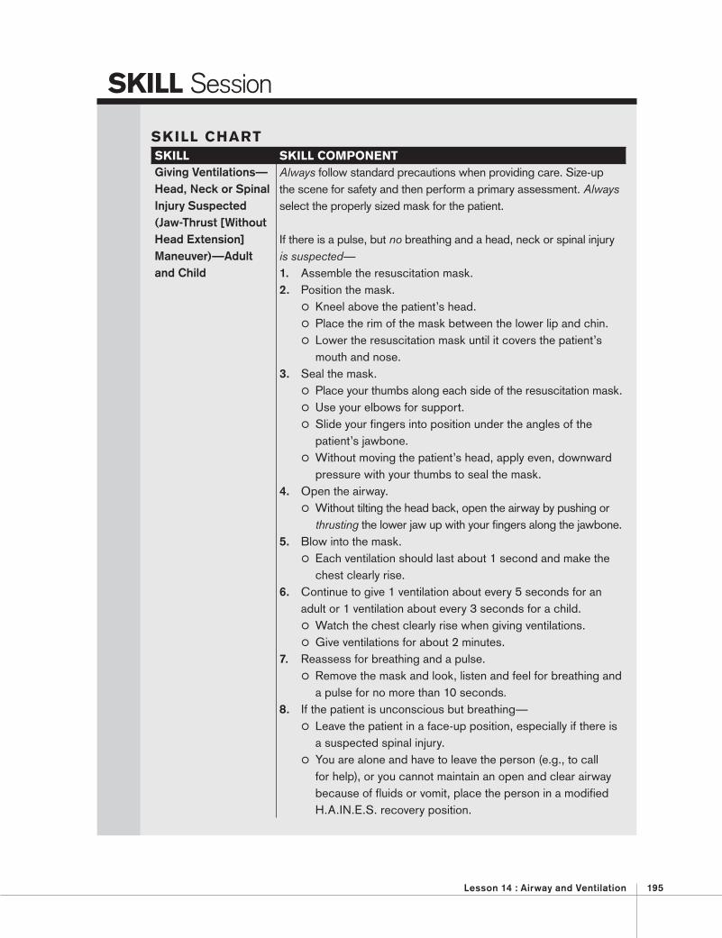
SKILL CHART SKILL SKILL COMPONENTGiving Ventilations—Head, Neck or Spinal Injury Suspected (Jaw-Thrust [Without Head Extension]Maneuver)—Adult and Child
Always follow standard precautions when providing care. Size-up the scene for safety and then perform a primary assessment. Always select the properly sized mask for the patient.
If there is a pulse, but no breathing and a head, neck or spinal injury is suspected — 1. Assemble the resuscitation mask. 2. Position the mask.
Kneel above the patient’s head. Place the rim of the mask between the lower lip and chin. Lower the resuscitation mask until it covers the patient’s
mouth and nose. 3. Seal the mask.
Place your thumbs along each side of the resuscitation mask. Use your elbows for support. Slide your fingers into position under the angles of the
patient’s jawbone. Without moving the patient’s head, apply even, downward
pressure with your thumbs to seal the mask. 4. Open the airway.
Without tilting the head back, open the airway by pushing or thrusting the lower jaw up with your fingers along the jawbone.
5. Blow into the mask. Each ventilation should last about 1 second and make the
chest clearly rise. 6. Continue to give 1 ventilation about every 5 seconds for an
adult or 1 ventilation about every 3 seconds for a child. Watch the chest clearly rise when giving ventilations. Give ventilations for about 2 minutes.
7. Reassess for breathing and a pulse. Remove the mask and look, listen and feel for breathing and
a pulse for no more than 10 seconds. 8. If the patient is unconscious but breathing—
Leave the patient in a face-up position, especially if there is a suspected spinal injury.
You are alone and have to leave the person (e.g., to call for help), or you cannot maintain an open and clear airway because of fluids or vomit, place the person in a modified H.A.IN.E.S. recovery position.
196 Emergency Medical Response Instructor’s Manual
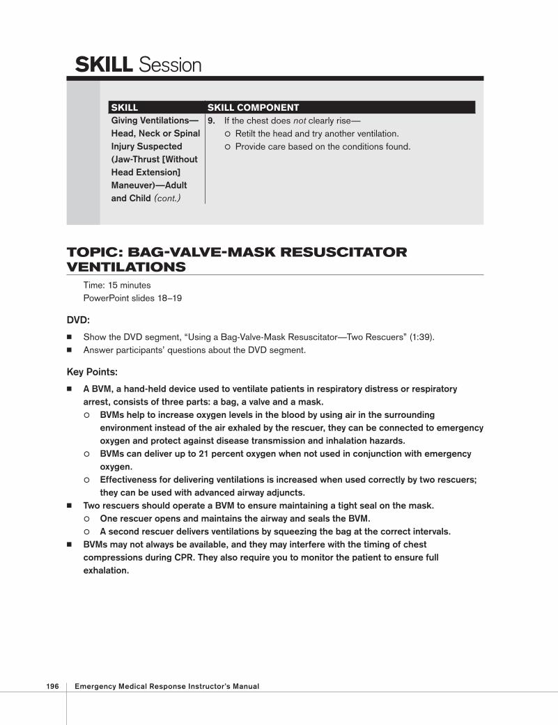
TOPIC: BAG-VALVE-MASK RESUSCITATOR VENTILATIONS
Time: 15 minutes PowerPoint slides 18–19
DVD:
Show the DVD segment, “Using a Bag-Valve-Mask Resuscitator—Two Rescuers” (1:39). Answer participants’ questions about the DVD segment.
Key Points:
A BVM, a hand-held device used to ventilate patients in respiratory distress or respiratory arrest, consists of three parts: a bag, a valve and a mask. BVMs help to increase oxygen levels in the blood by using air in the surrounding
environment instead of the air exhaled by the rescuer, they can be connected to emergency oxygen and protect against disease transmission and inhalation hazards.
BVMs can deliver up to 21 percent oxygen when not used in conjunction with emergency oxygen.
Effectiveness for delivering ventilations is increased when used correctly by two rescuers; they can be used with advanced airway adjuncts.
Two rescuers should operate a BVM to ensure maintaining a tight seal on the mask. One rescuer opens and maintains the airway and seals the BVM. A second rescuer delivers ventilations by squeezing the bag at the correct intervals.
BVMs may not always be available, and they may interfere with the timing of chest compressions during CPR. They also require you to monitor the patient to ensure full exhalation.
SKILL Session
SKILL SKILL COMPONENTGiving Ventilations—Head, Neck or Spinal Injury Suspected (Jaw-Thrust [Without Head Extension]Maneuver)—Adult and Child (cont.)
9. If the chest does not clearly rise— Retilt the head and try another ventilation. Provide care based on the conditions found.
Lesson 14 : Airway and Ventilation 197
SKILL Session
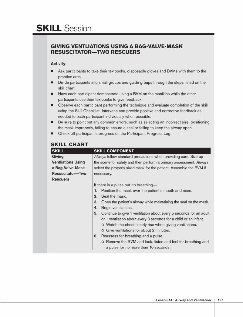
GIVING VENTLIATIONS USING A BAG-VALVE-MASK RESUSCITATOR—TWO RESCUERS
Activity:
Ask participants to take their textbooks, disposable gloves and BVMs with them to the practice area.
Divide participants into small groups and guide groups through the steps listed on the skill chart.
Have each participant demonstrate using a BVM on the manikins while the other participants use their textbooks to give feedback.
Observe each participant performing the technique and evaluate completion of the skill using the Skill Checklist. Intervene and provide positive and corrective feedback as needed to each participant individually when possible.
Be sure to point out any common errors, such as selecting an incorrect size, positioning the mask improperly, failing to ensure a seal or failing to keep the airway open.
Check off participant’s progress on the Participant Progress Log.
SKILL CHART SKILL SKILL COMPONENTGiving Ventliations Using a Bag-Valve-Mask Resuscitator—Two Rescuers
Always follow standard precautions when providing care. Size-up the scene for safety and then perform a primary assessment. Always select the properly sized mask for the patient. Assemble the BVM if necessary. If there is a pulse but no breathing— 1. Position the mask over the patient’s mouth and nose. 2. Seal the mask. 3. Open the patient’s airway while maintaining the seal on the mask. 4. Begin ventilations. 5. Continue to give 1 ventilation about every 5 seconds for an adult
or 1 ventilation about every 3 seconds for a child or an infant. Watch the chest clearly rise when giving ventilations. Give ventilations for about 2 minutes.
6. Reassess for breathing and a pulse. Remove the BVM and look, listen and feel for breathing and
a pulse for no more than 10 seconds.
198 Emergency Medical Response Instructor’s Manual
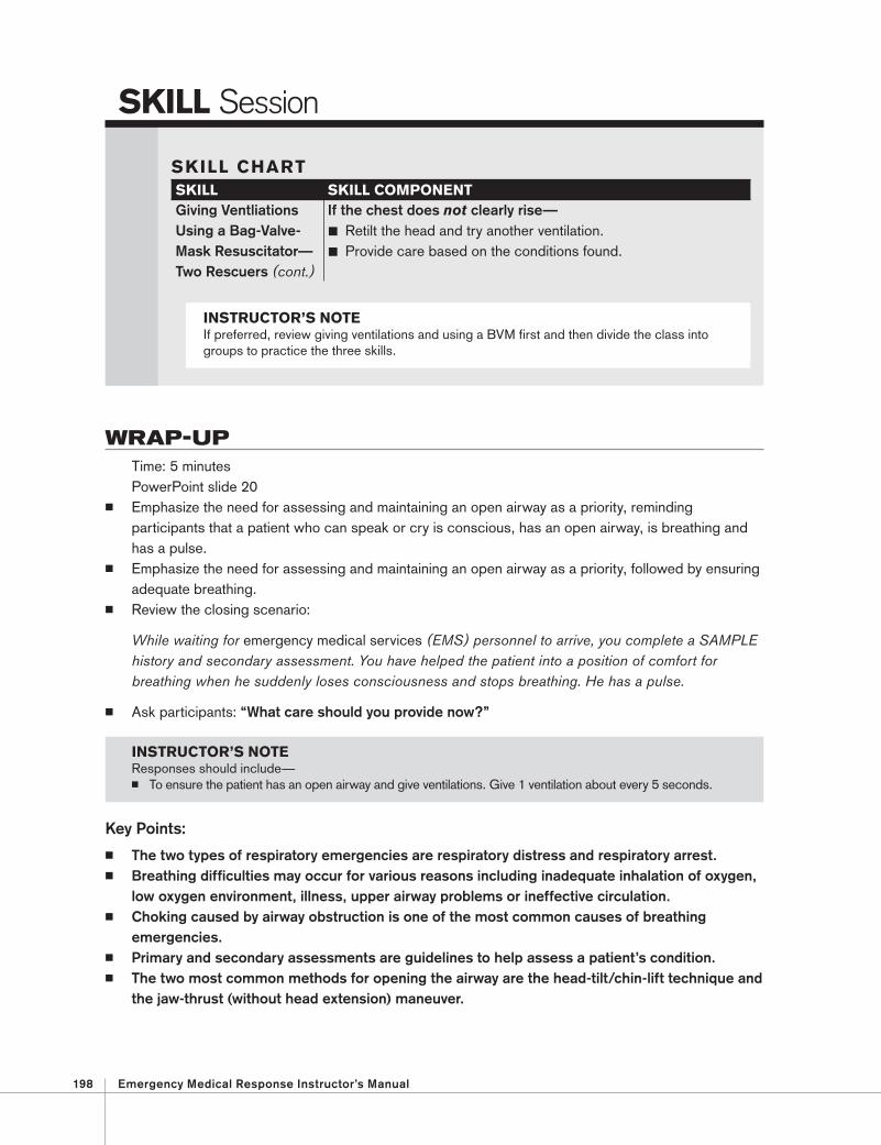
WRAP-UP Time: 5 minutes PowerPoint slide 20
Emphasize the need for assessing and maintaining an open airway as a priority, reminding participants that a patient who can speak or cry is conscious, has an open airway, is breathing and has a pulse.
Emphasize the need for assessing and maintaining an open airway as a priority, followed by ensuring adequate breathing.
Review the closing scenario:
While waiting for emergency medical services (EMS) personnel to arrive, you complete a SAMPLE history and secondary assessment. You have helped the patient into a position of comfort for breathing when he suddenly loses consciousness and stops breathing. He has a pulse.
Ask participants: “What care should you provide now?”
Key Points:
The two types of respiratory emergencies are respiratory distress and respiratory arrest. Breathing difficulties may occur for various reasons including inadequate inhalation of oxygen,
low oxygen environment, illness, upper airway problems or ineffective circulation. Choking caused by airway obstruction is one of the most common causes of breathing
emergencies. Primary and secondary assessments are guidelines to help assess a patient’s condition. The two most common methods for opening the airway are the head-tilt/chin-lift technique and
the jaw-thrust (without head extension) maneuver.
SKILL Session
SKILL CHART SKILL SKILL COMPONENTGiving Ventliations Using a Bag-Valve-Mask Resuscitator—Two Rescuers (cont.)
If the chest does not clearly rise— Retilt the head and try another ventilation. Provide care based on the conditions found.
INSTRUCTOR’S NOTE If preferred, review giving ventilations and using a BVM first and then divide the class into groups to practice the three skills.
INSTRUCTOR’S NOTE Responses should include— To ensure the patient has an open airway and give ventilations. Give 1 ventilation about every 5 seconds.
Lesson 14 : Airway and Ventilation 199
Abdominal thrusts, back blows and chest thrusts are effective methods to clear an obstructed airway in conscious patients.
Finger sweep and suctioning may be used to remove visible foreign matter and fluids from the upper airway of an unconscious patient.
Breathing abnormalities can be assessed by observing physical signs and breath sounds and by measuring the rate and depth of breathing.
For a patient who is not breathing, artificial ventilation is provided by using a resuscitation mask or BVM.
Assignment for the Next Lesson Read Chapter 11: Airway Management. Read Enrichment: Nasopharyngeal Airway (optional), pages 275–276 .
Instructor Preparation Review Chapter 11: Airway Management. Review Enrichment: Nasopharyngeal Airway (optional), pages 275–276. Review the DVD segments, “Using a Mechanical Suctioning Device” (1:59), “Using a Manual
Suctioning Device” (0:57), “Conscious Choking—Adult and Child” (2:34), “Conscious Choking—Infant,” (1:13), “Unconscious Choking—Adult, Child and Infant” (3:45).
Review the skills and obtain any necessary equipment and supplies for Lesson 15.
200 Emergency Medical Response Instructor’s Manual
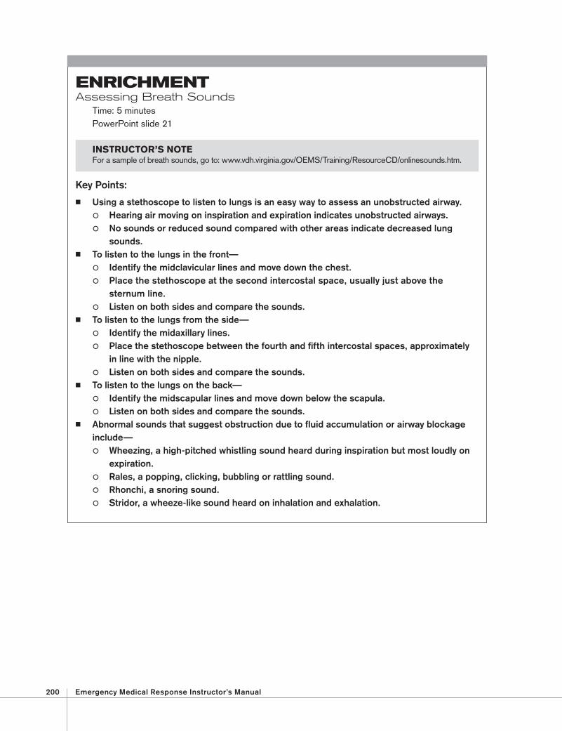
ENRICHMENTAssessing Breath Sounds
Time: 5 minutes PowerPoint slide 21
Key Points:
Using a stethoscope to listen to lungs is an easy way to assess an unobstructed airway. Hearing air moving on inspiration and expiration indicates unobstructed airways. No sounds or reduced sound compared with other areas indicate decreased lung
sounds. To listen to the lungs in the front—
Identify the midclavicular lines and move down the chest. Place the stethoscope at the second intercostal space, usually just above the
sternum line. Listen on both sides and compare the sounds.
To listen to the lungs from the side— Identify the midaxillary lines. Place the stethoscope between the fourth and fifth intercostal spaces, approximately
in line with the nipple. Listen on both sides and compare the sounds.
To listen to the lungs on the back— Identify the midscapular lines and move down below the scapula. Listen on both sides and compare the sounds.
Abnormal sounds that suggest obstruction due to fluid accumulation or airway blockage include— Wheezing, a high-pitched whistling sound heard during inspiration but most loudly on
expiration. Rales, a popping, clicking, bubbling or rattling sound. Rhonchi, a snoring sound. Stridor, a wheeze-like sound heard on inhalation and exhalation.
INSTRUCTOR’S NOTEFor a sample of breath sounds, go to: www.vdh.virginia.gov/OEMS/Training/ResourceCD/onlinesounds.htm.
Lesson 14 : Airway and Ventilation 201
ENRICHMENTSellick’s Maneuver (Cricoid Pressure)
Time: 25 minutes PowerPoint slide 22
DVD:
Show the DVD segment, “Sellick’s Maneuver (Cricoid Pressure)” (0:59). Answer participants’ questions about the DVD segment.
Key Points:
The Sellick’s maneuver should be used during positive pressure ventilation situations when the patient requires intubation (a skilled procedure performed by more advanced medical personnel). This maneuver reduces the chance of air entering the esophagus, particularly in
patients who are unresponsive and/or have no gag reflex. It enables the rescuer performing the intubation to have a better view of the vocal
cords, which allows for easier insertion of the endotracheal tube. One rescuer performs the maneuver while another performs the intubation.
The cricoid cartilage is located just below and behind the thyroid cartilage, encircling the esophagus.
After locating the cartilage, pressure is applied on both sides using the thumb and index finger, pressing firmly toward the back of the neck. Pressure is applied until the intubation is complete.
This maneuver is not used if— The patient is vomiting or begins to vomit. The patient is responsive. A breathing tube will be placed by advanced-level providers when they arrive .
INSTRUCTOR’S NOTE This topic should be taught only if local protocols allow and you meet the state and local requirements for teaching this skill.
202 Emergency Medical Response Instructor’s Manual
SKILL Session (Enrichment)
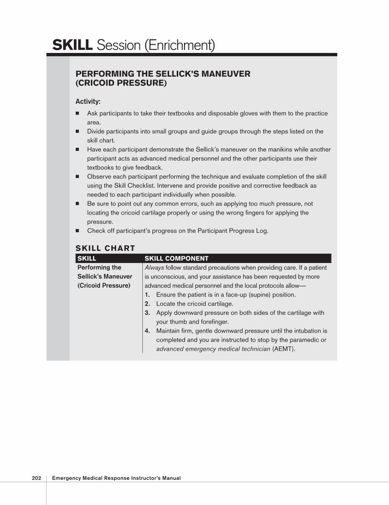
PERFORMING THE SELLICK’S MANEUVER (CRICOID PRESSURE)
Activity:
Ask participants to take their textbooks and disposable gloves with them to the practice area.
Divide participants into small groups and guide groups through the steps listed on the skill chart.
Have each participant demonstrate the Sellick’s maneuver on the manikins while another participant acts as advanced medical personnel and the other participants use their textbooks to give feedback.
Observe each participant performing the technique and evaluate completion of the skill using the Skill Checklist. Intervene and provide positive and corrective feedback as needed to each participant individually when possible.
Be sure to point out any common errors, such as applying too much pressure, not locating the cricoid cartilage properly or using the wrong fi ngers for applying the pressure.
Check off participant’s progress on the Participant Progress Log.
SKILL CHART SKILL SKILL COMPONENTPerforming the Sellick’s Maneuver (Cricoid Pressure)
Always follow standard precautions when providing care. If a patient is unconscious, and your assistance has been requested by more advanced medical personnel and the local protocols allow— 1. Ensure the patient is in a face-up (supine) position. 2. Locate the cricoid cartilage. 3. Apply downward pressure on both sides of the cartilage with
your thumb and forefinger. 4. Maintain firm, gentle downward pressure until the intubation is
completed and you are instructed to stop by the paramedic or advanced emergency medical technician (AEMT).
Lesson 14 : Airway and Ventilation 203
ENRICHMENTAssisting the Patient with Asthma
Time: 40 minutes PowerPoint slides 23–26
Key Points:
There are three types of medication used in the management of asthma: Long-term control medications to control chronic symptoms and prevent attacks Quick-relief medications (rescue medications) to relieve symptoms during an asthma attack Medications for allergy-induced asthma that decrease sensitivity to a particular
allergen and prevent the immune system from reacting to allergens Indications for asthma medication include recurrent wheezing, coughing, difficulty
breathing and chest tightness; contraindications include increased risk for thinning of the skin and bruising.
Delivery Systems for Asthma Medication
Key Points:
Asthma medications are supplied in different delivery systems. Metered-dose inhaler is a small, hand-held aerosol canister with a mouthpiece; some
are equipped with a spacer (a tube attached to the inhaler) that serves as a reservoir for the medication.
Dry powder inhaler (DPI), similar to a metered-dose inhaler, is a small, hand-held device that delivers a dry powder form of the medication within a small capsule, disc or compartment inside the inhaler.
Small-volume nebulizer delivers aerosolized medications (mist) over a few minutes. Pill or liquid form is available for some medications. Injection just under the skin is a recently developed method for giving asthma medications.
Peak Flow Meter
Key Points:
A peak flow meter measures a person’s ability to push air out of the lungs in one quick breath.
It is a hand-held asthma management tool that tracks a person’s breathing. It helps to warn the person if his or her asthma is worsening and shows his or her response
to treatment.
DVD:
Show the DVD segment, “Asthma” (2:52). Answer participants’ questions about the DVD segment.
INSTRUCTOR’S NOTE If available, show participants different types of delivery systems available for asthma medications.
204 Emergency Medical Response Instructor’s Manual
ENRICHMENT (cont.)
Assisting the Patient in the Use of an Inhaler
Key Points:
An EMR may be involved in assisting a patient in the use of an inhaler. It is necessary to obtain an order from medical direction via radio or phone contact
with the medical director or through protocols and standing orders. The order is always verified by restating the name of the medication. You are responsible for knowing and following local protocols for assisting a patient
with an asthma inhaler. After obtaining consent and ensuring that local protocols allow such action, follow these
general guidelines: If the patient has prescribed asthma medication, help the person take it. Ensure that the prescription is in the patients’ name and is prescribed for “quick relief”
or “acute” attacks. Ensure that the expiration date of the medication has not passed and read and follow
all instructions printed on the inhaler prior to administering the medication. Shake the inhaler and then remove the cover from the mouthpiece. Position the spacer
if you are using one. Have the patient breathe out fully through the mouth and then place the lips tightly
around the inhaler mouthpiece. Have the patient inhale deeply and slowly as you or the patient depresses the inhaler
canister to release the medication, which then is inhaled into the lungs. Ensure that the patient holds his or her breath for a count of 10. If using a spacer, the patient
takes 5 to 6 deep breaths with the spacer still in the mouth, without holding the breath. Once the inhalation is complete, have the patient rinse his or her mouth out with water
to reduce side effects. Reassess the patient’s breathing. Always wash your hands immediately after providing care.
After administration, be alert for possible common side effects including— Increased heart rate. Palpitations. Nausea. Vomiting. Nervousness. Sleeplessness. Dry mouth. Cough. Hoarseness. Headache. Throat irritation.
Lesson 14 : Airway and Ventilation 205
SKILL Session (Enrichment)
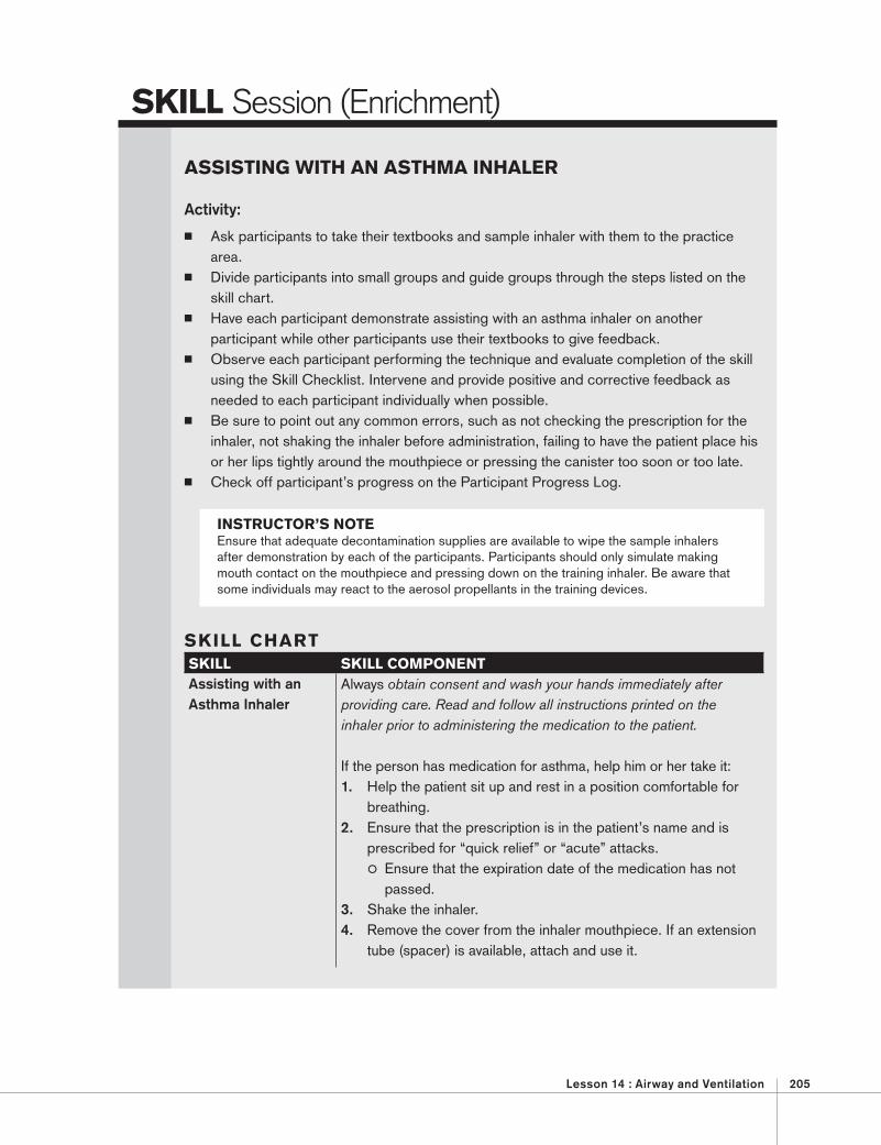
ASSISTING WITH AN ASTHMA INHALER
Activity:
Ask participants to take their textbooks and sample inhaler with them to the practice area.
Divide participants into small groups and guide groups through the steps listed on the skill chart.
Have each participant demonstrate assisting with an asthma inhaler on another participant while other participants use their textbooks to give feedback.
Observe each participant performing the technique and evaluate completion of the skill using the Skill Checklist. Intervene and provide positive and corrective feedback as needed to each participant individually when possible.
Be sure to point out any common errors, such as not checking the prescription for the inhaler, not shaking the inhaler before administration, failing to have the patient place his or her lips tightly around the mouthpiece or pressing the canister too soon or too late.
Check off participant’s progress on the Participant Progress Log.
INSTRUCTOR’S NOTE Ensure that adequate decontamination supplies are available to wipe the sample inhalers after demonstration by each of the participants. Participants should only simulate making mouth contact on the mouthpiece and pressing down on the training inhaler. Be aware that some individuals may react to the aerosol propellants in the training devices.
SKILL CHART SKILL SKILL COMPONENTAssisting with an Asthma Inhaler
Always obtain consent and wash your hands immediately after providing care. Read and follow all instructions printed on the inhaler prior to administering the medication to the patient.
If the person has medication for asthma, help him or her take it: 1. Help the patient sit up and rest in a position comfortable for
breathing. 2. Ensure that the prescription is in the patient’s name and is
prescribed for “quick relief” or “acute” attacks. Ensure that the expiration date of the medication has not
passed. 3. Shake the inhaler. 4. Remove the cover from the inhaler mouthpiece. If an extension
tube (spacer) is available, attach and use it.
206 Emergency Medical Response Instructor’s Manual
SKILL Session
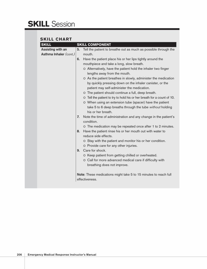
SKILL CHART SKILL SKILL COMPONENTAssisting with an Asthma Inhaler (cont.)
5. Tell the patient to breathe out as much as possible through the mouth.
6. Have the patient place his or her lips tightly around the mouthpiece and take a long, slow breath. Alternatively, have the patient hold the inhaler two finger
lengths away from the mouth. As the patient breathes in slowly, administer the medication
by quickly pressing down on the inhaler canister, or the patient may self-administer the medication.
The patient should continue a full, deep breath. Tell the patient to try to hold his or her breath for a count of 10. When using an extension tube (spacer) have the patient
take 5 to 6 deep breaths through the tube without holding his or her breath.
7. Note the time of administration and any change in the patient’s condition. The medication may be repeated once after 1 to 2 minutes.
8. Have the patient rinse his or her mouth out with water to reduce side effects. Stay with the patient and monitor his or her condition. Provide care for any other injuries.
9. Care for shock. Keep patient from getting chilled or overheated. Call for more advanced medical care if difficulty with
breathing does not improve.
Note: These medications might take 5 to 15 minutes to reach full effectiveness.
Lesson 14 : Airway and Ventilation 207
SKILL Checklist
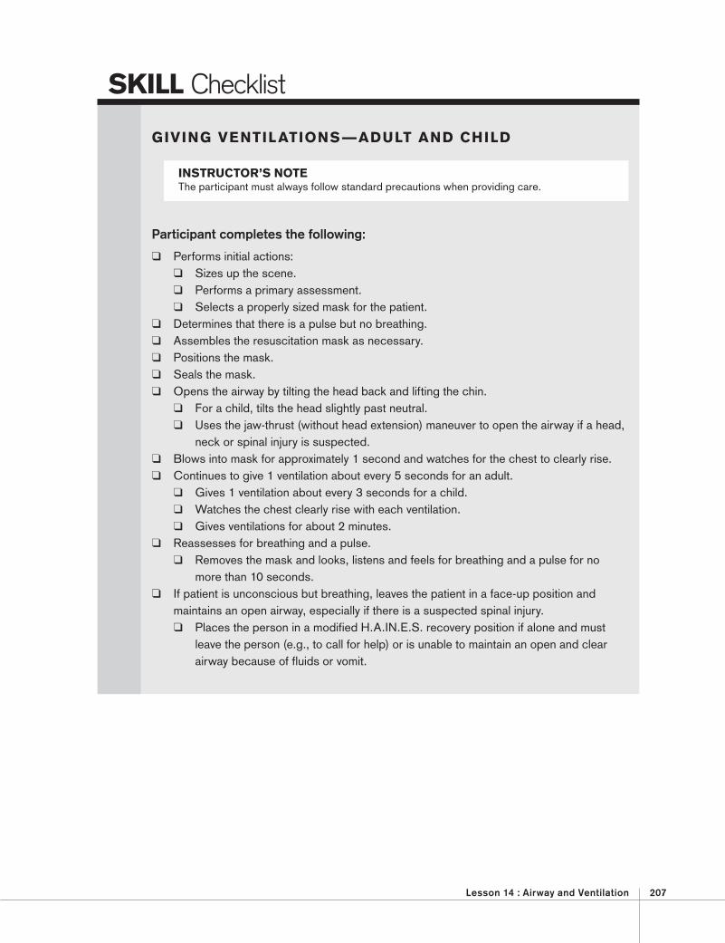
GIVING VENTILATIONS —ADULT AND CHILD
Participant completes the following:
Performs initial actions: Sizes up the scene. Performs a primary assessment . Selects a properly sized mask for the patient.
Determines that there is a pulse but no breathing. Assembles the resuscitation mask as necessary. Positions the mask. Seals the mask. Opens the airway by tilting the head back and lifting the chin.
For a child, tilts the head slightly past neutral. Uses the jaw-thrust (without head extension) maneuver to open the airway if a head,
neck or spinal injury is suspected. Blows into mask for approximately 1 second and watches for the chest to clearly rise. Continues to give 1 ventilation about every 5 seconds for an adult.
Gives 1 ventilation about every 3 seconds for a child. Watches the chest clearly rise with each ventilation. Gives ventilations for about 2 minutes.
Reassesses for breathing and a pulse. Removes the mask and looks, listens and feels for breathing and a pulse for no
more than 10 seconds. If patient is unconscious but breathing, leaves the patient in a face-up position and
maintains an open airway, especially if there is a suspected spinal injury. Places the person in a modified H.A.IN.E.S. recovery position if alone and must
leave the person (e.g., to call for help) or is unable to maintain an open and clear airway because of fluids or vomit.
INSTRUCTOR’S NOTEThe participant must always follow standard precautions when providing care.
208 Emergency Medical Response Instructor’s Manual
SKILL Checklist
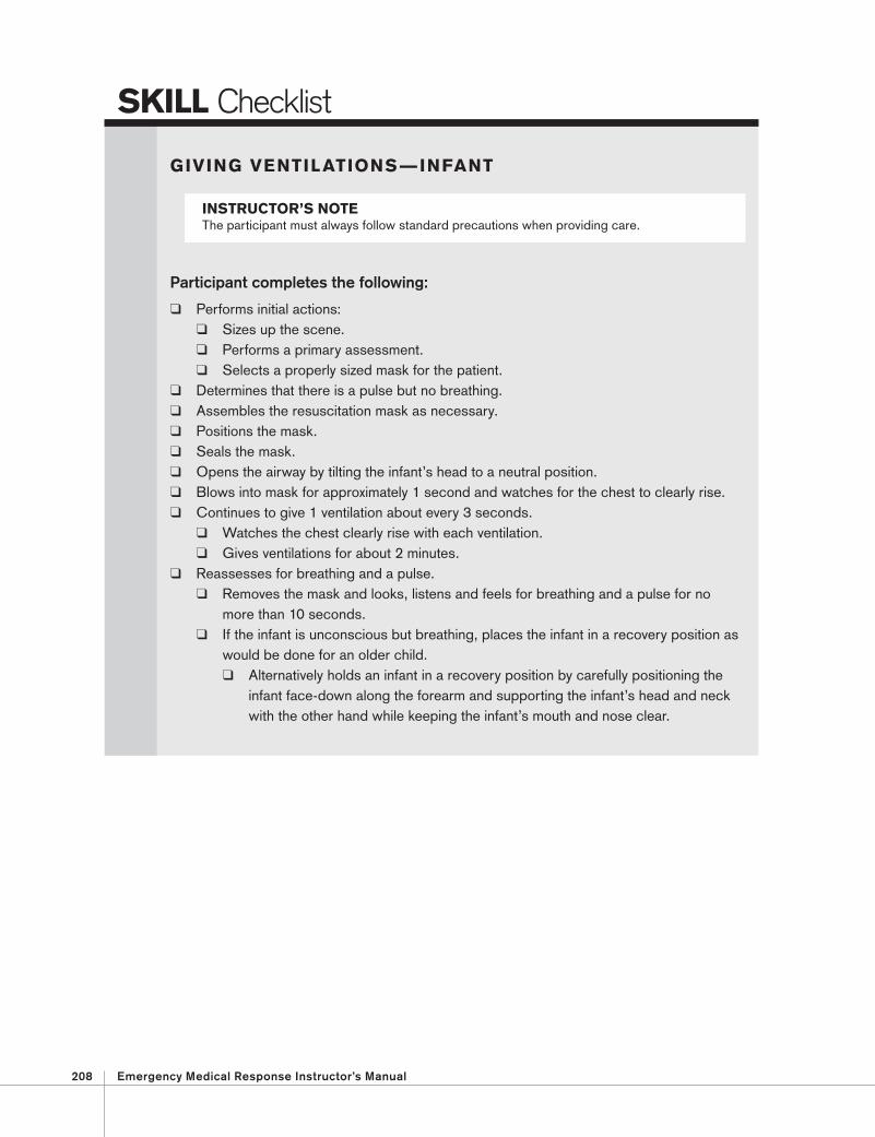
GIVING VENTILATIONS — INFANT
Participant completes the following:
Performs initial actions: Sizes up the scene. Performs a primary assessment. Selects a properly sized mask for the patient.
Determines that there is a pulse but no breathing. Assembles the resuscitation mask as necessary . Positions the mask . Seals the mask. Opens the airway by tilting the infant’s head to a neutral position. Blows into mask for approximately 1 second and watches for the chest to clearly rise. Continues to give 1 ventilation about every 3 seconds.
Watches the chest clearly rise with each ventilation. Gives ventilations for about 2 minutes.
Reassesses for breathing and a pulse. Removes the mask and looks, listens and feels for breathing and a pulse for no
more than 10 seconds. If the infant is unconscious but breathing, places the infant in a recovery position as
would be done for an older child. Alternatively holds an infant in a recovery position by carefully positioning the
infant face-down along the forearm and supporting the infant’s head and neck with the other hand while keeping the infant’s mouth and nose clear.
INSTRUCTOR’S NOTEThe participant must always follow standard precautions when providing care.
Lesson 14 : Airway and Ventilation 209
SKILL Checklist
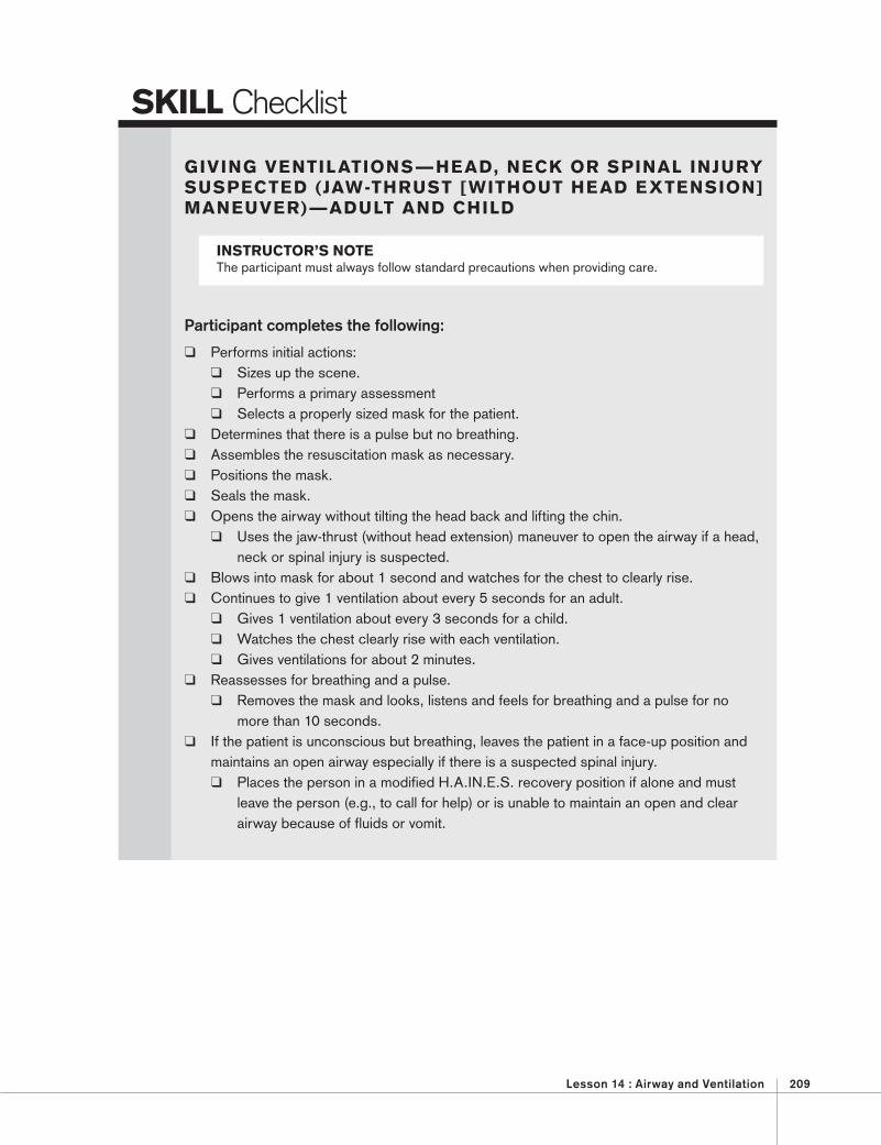
GIVING VENTILATIONS — HEAD, NECK OR SPINAL INJURY SUSPECTED (JAW-THRUST [WITHOUT HEAD EXTENSION] MANEUVER) —ADULT AND CHILD
Participant completes the following:
Performs initial actions: Sizes up the scene. Performs a primary assessment Selects a properly sized mask for the patient.
Determines that there is a pulse but no breathing . Assembles the resuscitation mask as necessary. Positions the mask. Seals the mask. Opens the airway without tilting the head back and lifting the chin.
Uses the jaw-thrust (without head extension) maneuver to open the airway if a head, neck or spinal injury is suspected.
Blows into mask for about 1 second and watches for the chest to clearly rise. Continues to give 1 ventilation about every 5 seconds for an adult.
Gives 1 ventilation about every 3 seconds for a child. Watches the chest clearly rise with each ventilation. Gives ventilations for about 2 minutes.
Reassesses for breathing and a pulse. Removes the mask and looks, listens and feels for breathing and a pulse for no
more than 10 seconds. If the patient is unconscious but breathing, leaves the patient in a face-up position and
maintains an open airway especially if there is a suspected spinal injury. Places the person in a modified H.A.IN.E.S. recovery position if alone and must
leave the person (e.g., to call for help) or is unable to maintain an open and clear airway because of fluids or vomit.
INSTRUCTOR’S NOTE The participant must always follow standard precautions when providing care.
210 Emergency Medical Response Instructor’s Manual
SKILL Checklist
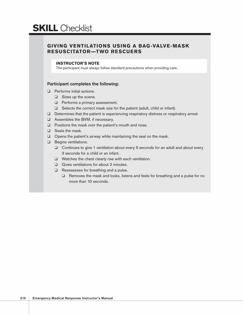
GIVING VENTILATIONS USING A BAG-VALVE-MASK RESUSCITATOR—TWO RESCUERS
Participant completes the following:
Performs initial actions: Sizes up the scene. Performs a primary assessment. Selects the correct mask size for the patient (adult, child or infant).
Determines that the patient is experiencing respiratory distress or respiratory arrest. Assembles the BVM, if necessary. Positions the mask over the patient’s mouth and nose. Seals the mask. Opens the patient’s airway while maintaining the seal on the mask. Begins ventilations.
Continues to give 1 ventilation about every 5 seconds for an adult and about every 3 seconds for a child or an infant.
Watches the chest clearly rise with each ventilation. Gives ventilations for about 2 minutes. Reassesses for breathing and a pulse.
Removes the mask and looks, listens and feels for breathing and a pulse for no more than 10 seconds.
INSTRUCTOR’S NOTE The participant must always follow standard precautions when providing care.
Lesson 14 : Airway and Ventilation 211
SKILL Checklist (Enrichment)
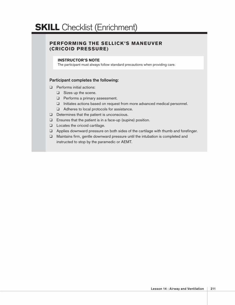
PERFORMING THE SELLICK’S MANEUVER (CRICOID PRESSURE)
Participant completes the following:
Performs initial actions: Sizes up the scene. Performs a primary assessment. Initiates actions based on request from more advanced medical personnel. Adheres to local protocols for assistance.
Determines that the patient is unconscious. Ensures that the patient is in a face-up (supine) position. Locates the cricoid cartilage. Applies downward pressure on both sides of the cartilage with thumb and forefinger. Maintains firm, gentle downward pressure until the intubation is completed and
instructed to stop by the paramedic or AEMT.
INSTRUCTOR’S NOTEThe participant must always follow standard precautions when providing care.
212 Emergency Medical Response Instructor’s Manual
SKILL Checklist (Enrichment)
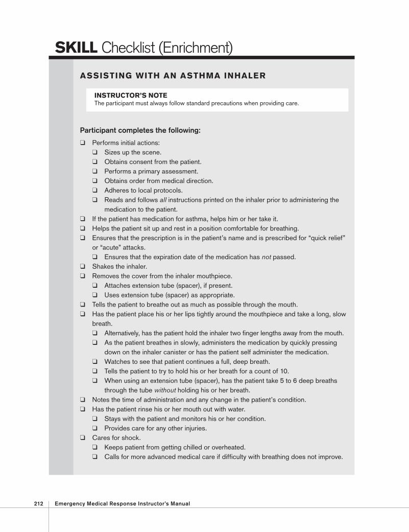
ASSISTING WITH AN ASTHMA INHALER
Participant completes the following:
Performs initial actions: Sizes up the scene. Obtains consent from the patient. Performs a primary assessment. Obtains order from medical direction. Adheres to local protocols. Reads and follows all instructions printed on the inhaler prior to administering the
medication to the patient. If the patient has medication for asthma, helps him or her take it. Helps the patient sit up and rest in a position comfortable for breathing. Ensures that the prescription is in the patient’s name and is prescribed for “quick relief”
or “acute” attacks. Ensures that the expiration date of the medication has not passed.
Shakes the inhaler. Removes the cover from the inhaler mouthpiece.
Attaches extension tube (spacer), if present. Uses extension tube (spacer) as appropriate.
Tells the patient to breathe out as much as possible through the mouth. Has the patient place his or her lips tightly around the mouthpiece and take a long, slow
breath. Alternatively, has the patient hold the inhaler two finger lengths away from the mouth. As the patient breathes in slowly, administers the medication by quickly pressing
down on the inhaler canister or has the patient self administer the medication. Watches to see that patient continues a full, deep breath. Tells the patient to try to hold his or her breath for a count of 10. When using an extension tube (spacer), has the patient take 5 to 6 deep breaths
through the tube without holding his or her breath. Notes the time of administration and any change in the patient’s condition. Has the patient rinse his or her mouth out with water.
Stays with the patient and monitors his or her condition. Provides care for any other injuries.
Cares for shock. Keeps patient from getting chilled or overheated. Calls for more advanced medical care if difficulty with breathing does not improve.
INSTRUCTOR’S NOTEThe participant must always follow standard precautions when providing care.