Embed Size (px)
Citation preview
1
EMBRYONIC STEM CELLS AS MODELS OF TROPHOBLAST DIFFERENTIATION: 1
PROGRESS, OPPORTUNITIES AND LIMITATIONS 2
3
4
Thaddeus G. Golos1,2,3, M. Giakoumopoulos1,2, and M. A. Garthwaite1,2 5
6
7
1Wisconsin National Primate Research Center, 2Department of Obstetrics and 8
Gynecology, 3Department of Comparative Biosciences, 9
University of Wisconsin, Madison, Madison, WI 53715-1299 USA 10
11
Running title: 12
Trophoblasts from human embryonic stem cells 13
14
Key words: 15
Trophoblast, embryonic stem cell, iPS cell, embryoid body, placenta 16
17
Corresponding author: Thaddeus G. Golos, Ph.D., Wisconsin National Primate 18
Research Center, University of Wisconsin, 1223 Capitol Court, Madison, WI 53715�-19
1299; Phone: (608) 263-3567, Fax: (608) 263-3524, email: [email protected] 20
21
22
Page 1 of 25 Reproduction Advance Publication first posted on 31 March 2010 as Manuscript REP-09-0544
Copyright © 2010 by the Society for Reproduction and Fertility.
2
Abstract 23
While the determination of the trophoblast lineage and the facilitation of placental 24
morphogenesis by trophoblast interactions with other cells of the placenta are crucial 25
components of the establishment of pregnancy, these processes are not tractable at the 26
time of human implantation. Embryonic stem cells provide an embryonic surrogate to 27
derive insights into these processes. In this review we will summarize current paradigms 28
which promote trophoblast differentiation from embryonic stem cells, and potential 29
opportunities for their use to further define signals directing morphogenesis of the 30
placenta following implantation of the embryo into the endometrium. 31
Page 2 of 25
3
Introduction 32
33
The earliest lineage commitment and differentiation event in mammalian preimplantation 34
embryo development is the formation of the trophectoderm, heralded by embryo 35
compaction at the morula stage of development 4-6 days following fertilization in human 36
and nonhuman primates, and manifested by blastocyst formation within the next 1-2 37
days. These trophectodermal cells function as progenitor cells of the trophoblast lineage, 38
giving rise in the primate placenta to cells of villous and extravillous phenotypes. 39
Trophoblasts of the villous phenotype include the relatively undifferentiated villous 40
cytotrophoblasts (CTB) and the terminally differentiated villous syncytiotrophoblasts 41
(STB), which arise from fusion of the mononuclear CTB. These villous trophoblasts are 42
found within the chorionic villi which form beginning during the 2nd week after 43
implantation of the human or nonhuman primate embryo into the receptive endometrium 44
(Kaufmann & Burton, 1994). A third trophoblast population is found at the tips of the 45
chorionic villi which anchor the placenta to the endometrium. These are cytotrophoblasts 46
of the cell column (CC). 47
48
While the CC anchors the placenta to the decidualized endometrium, an additional 49
population of trophoblasts which expands from the column, the extravillous trophoblasts 50
(EVT), invade into the maternal endometrium in the first weeks of gestation. These EVT 51
play a key role in the responses to implantation of the maternal vasculature within the 52
decidua. Histologically, two populations of EVT are recognizable within the decidua. 53
Endovascular EVT are found in the lumen of the spiral arterioles from 6 weeks of human 54
gestation onwards. These cells are likely to be present within maternal vessels earlier, 55
since in the rhesus monkey, endovascular EVT are seen having substantially invaded 56
spiral arterioles as early as 1 week after embryo attachment (Slukvin, et al. 2000; Enders 57
2007). However, such very early human implantation sites are not readily available for 58
Page 3 of 25
4
detailed study. In addition to this endovascular location, EVT are seen within the 59
decidualized stroma, outside of the blood vessels, and these cells are termed interstitial 60
EVT. 61
62
Because of the limitations inherent in the study of static histological specimens, the 63
pathway by which endovascular EVT arrive within the lumen of the spiral arteriole 64
remains somewhat uncertain. It is possible that EVT arrive at an endovascular location 65
primarily if not exclusively by breaching the vessel wall from an interstitial location. 66
Alternatively, EVT may access the spiral arteriole lumen directly via erosion of the 67
capillary microvasculature at the most internal region of the functional endometrium, and 68
migrate down the walls of those vessels. Regardless of the route of invasion, vessels 69
which are invaded by trophoblasts ultimately exhibit a loss of the endothelium, loss of 70
vascular smooth muscle and replacement of the tunica intima and tunica media with 71
endovascular EVT (Harris & Aplin 2007). These trophoblasts form vascular channels 72
which, in the absence of the vascular smooth muscle, now provide a non-vasoactive 73
blood conduit to the intervillous space. Regardless of the route of migration, the 74
remodeling of these vessels is a critical event in the first trimester of pregnancy. Poor 75
EVT invasion and failure to appropriately remodel the decidual and distal myometrial 76
vessels is widely associated with poor pregnancy outcomes, including uteroplacental 77
insufficiency, intrauterine growth restriction, and preeclampsia (Redman and Sargent, 78
2005; Pijenborg et al. 2007). Overall placental growth and villous morphogenesis are 79
widely recognized as crucial for pregnancy success. For these reasons, understanding 80
early trophoblast differentiation, villous morphogenesis and placenta formation, and EVT 81
differentiation and invasion are fundamental to considering potential therapeutic 82
approaches for preventing or treating suboptimal implantation and pregnancy 83
complications related to placental development. 84
Page 4 of 25
5
85
Trophoblast differentiation from embryonic stem cells: importance of human and 86
rhesus systems. 87
88
Very early (< 6 weeks of gestation) human implantation sites are not generally available 89
for research or experimental purposes. Ideally, blastocyst-stage embryos should be an 90
excellent starting point for defining the molecular events which underlie trophoblast 91
differentiation, and the interaction of early invasive EVT with stromal, immune and 92
vascular elements of the decidua. The additional limitations on our understanding of the 93
earliest events in human implantation are imposed by ethical restrictions on the use of 94
human embryos for in vitro experimental purposes. 95
96
The derivation of human embryonic stem cells (hESC) from in vitro fertilization-produced 97
blastocysts donated for research (Thomson et al. 1998) provided an unprecendented 98
opportunity for new explorations in early development, of embryonic and fetal cell 99
lineages, including the extraembryonic membranes. It has been known for many years 100
that in general, mouse ESC differentiate in vitro to the trophoblast lineage only poorly, if 101
differentiation is at all noted, although some modifications (e.g., PARP-1 deletion by 102
homologous recombination) have been shown to increase the rate of mouse ESC 103
differentiation into trophoblasts (Hemberger et al. 2003). When Thomson et al. (1998) 104
reported the first derivation of hESC, they noted that differentiation to trophoblasts was 105
indicated in colonies undergoing spontaneous differentiation, as judged by the secretion 106
of human chorionic gonadotropin (hCG), a generally trophoblast-specific marker 107
essential for the establishment of human pregnancy. This result had in fact been 108
foreshadowed by the previous observation from the Thomson lab that a subset of rhesus 109
monkey ESC, derived from in vivo produced blastocysts obtained by flushing of the 110
uterine cavity 24-48 hrs before the anticipated day of embryo implantation (Thomson et 111
Page 5 of 25
6
al. 1995), also spontaneously differentiated into trophoblasts, increasing the transcription 112
of the CG alpha and beta subunit mRNAs. While CG is typically thought of as a marker 113
of differentiated trophoblasts, it has long been known (but not widely appreciated) that 114
preimplantation human and nonhuman primate embryos already secrete detectable 115
levels of CG in vitro (Fishel & Edwards 1984; Hearn et al. 1988; Pope et al., 1982; 116
Seshagari et al. 1994), most likely as a component of the program of gene expression 117
activated during differentiation to the trophectoderm lineage. Thus, CG secretion in 118
spontaneously differentiating human and rhesus ESC was reasonably interpreted as 119
indicating that these ESCs could serve as a surrogate for blastocyst stage embryos, at 120
least from the perspective of trophectoderm/trophoblast formation. Expression of alpha-121
fetoprotein also indicated differentiation of yolk sac (endoderm) in rhesus (Thomson et 122
al. 1995) and later, human (Reubinoff et al., 2000) ESC. The formation of these 123
extraembryonic lineages was exciting in that it allowed for the first time study of placenta 124
formation, not from tissues which had already undergone morphogenesis and 125
differentiation to villous and extravillous lineages (i.e., first trimester placental tissues, or 126
term delivered placentas), but from the “blank slate” represented by ESC. 127
128
Modifiers of ESC culture conditions for trophoblast formation: Bone 129
Morphogenetic Protein (BMP), Embryoid bodies, and Extracellular Matrix. 130
131
Xu et al. (2002) reported that treatment of undifferentiated hESC lines developed at the 132
University of Wisconsin with BMP4 or related ligands resulted in their uniform 133
differentiation to cells of an epithelial morphology. Microarray analysis revealed that 134
when compared to undifferentiated cells, many of the most highly up-regulated genes 135
were associated with trophoblast function, particularly transcription factors (chiefly 136
defined in mouse development), and endocrine markers of human trophoblast 137
differentiation, including hCG, and enzymes important for steroid hormone synthesis 138
(Gerami et al. 2004). Interestingly, it has also been reported that the effect of BMP or 139
Page 6 of 25
7
related ligands on hESC is very cell line-dependent. For example, treatment of the HES 140
human embryonic stem cell lines also derived from IVF-derived preimplantation human 141
blastocysts (Reubinoff et al., 2000; Pera et al. 2004) with BMPs results in only sporadic 142
formation of small colonies of CG-expressing cells of epithelial morphology, and the 143
more widespread outcome in this study was the formation of endoderm and yolk sac-like 144
structures. It has not been determined what the specific differences may be that result in 145
diverse responses to these factors, but derivation and culture conditions, receptor 146
populations and signaling environments are all likely to contribute to the diversity of 147
differentiation potential among hESC lines derived in different laboratories. While there 148
seems little question that the cells derived from hESC under various experimental 149
conditions express many features of human placental trophoblasts, it seems prudent to 150
recognize that as they are not yet clearly shown to represent a specific differentiated 151
trophoblast cell type, and indeed, the periimplantation trophoblast phenotype in human 152
pregnancy remains a formidable challenge to define at a molecular level. Thus, it is 153
reasonable to consider these hESC derivatives as “trophoblast-like” cells, although for 154
the sake of simplicity, we will refer to them as trophoblasts in this review. 155
156
157
A second approach with H1 and H9 hESC cells for trophoblast differentiation from hESC 158
was identified with subsequent studies (Gerami et al. 2004). Reasoning that mimicking 159
the spherical configuration of the preimplantation embryo with pluripotent hESC may 160
initiate signals parallel to those acting during preimplantation embryo development, we 161
directed embryoid body (EB) formation by enzymatic release of undifferentiated ESC 162
colonies from culture plates, and maintained them in suspension culture in bacterial-163
grade culture dishes. Within 48 hours there is a siginifcant elevation in the level of hCG 164
secretion in suspension EB culture. Moreover, hCG secretion is accompanied by 165
Page 7 of 25
8
progesterone secretion, as well as secretion of estradiol-17β (Gerami et al. 2004). 166
Additional studies demonstrated that the endogenous androgens of the fetal calf serum 167
were the substrate for cytochrome P450aromatase, rather than androgen secretion by 168
the EB itself (Gerami et al., 2004). Thus, these hormones represent a footprint of 169
trophoblast differentiation, and recapitulated what was observed in BMP-treated 170
cultures. 171
172
However, hormone secretion was not dramatically sustained in the suspension culture 173
paradigm. Again taking into consideration the in vivo environment that the 174
preimplantation embryo is exposed to, we developed an approach, based on previous 175
work in the Aplin lab at the University of Manchester (Aplin et al. 1999), modeling 176
extravillous trophoblast invasion of the extracellular matrix of the decidua during 177
implantation. We developed an approach to embed hESC-derived EBs in pre-gelled 178
Matrigel droplets, and transfer those Matrigel “rafts” to suspension culture. In this 179
situation, within several weeks there were reliably discernable outgrowths from the EBs 180
into the Matrigel scaffold. In parallel with this invasion, significant maintenance of 181
differentiated function was observed, as manifested by sustained hormone secretion into 182
the culture media (Gerami et al. 2004). Thus, signals conveyed either by extracellular 183
matrix, or the 3-dimensional environment, provide support for sustained trophoblast 184
differentiation. 185
186
The BMP and EB-based approaches should be considered complementary, and each 187
has its strengths and limitations. A significant strength of the BMP-treatment paradigm 188
is that the relatively uniform response potentially allows the investigation of molecular 189
mechanisms which promote the initiation of the trophoblast lineage. This is somewhat 190
tempered by the recognition that the response appears to be divergent among hESC cell 191
Page 8 of 25
9
lines, and additionally, it remains unclear which in vivo trophoblast population is 192
represented by BMP4-driven trophoblasts. Finally, the in vivo role of BMP4 (or related 193
factor(s)) in placenta formation is not known. 194
195
It does not seem to have been widely explored if the EB-based approach is cell-line 196
dependent; current studies are largely restricted to the use of the original cell lines 197
derived in the Thomson lab (Thomson et al. 1998). One advantage of the EB-based 198
approach is that it could perhaps be reasoned that the spontaneous formation of 199
trophoblasts may be more likely to represent in vivo trophectoderm formation. However, 200
this model is complicated by the heterogeneous nature of the EB itself, since all 201
embryonic germ layers differentiate simultaneously during prolonged culture (Gerecht-202
Nir et al., 2004). Thus, the appropriate system should be chosen based on the question 203
to be addressed. 204
205
The dynamics of differentiation of hESC into trophoblast-like cells are influenced by 206
spatial considerations with in vitro monolayer culture as well as in the 3-dimensional 207
context of the embryoid body. In BMP-directed differentiation, Schulz et. al. (2008) have 208
demonstrated that expression of chorionic gonadotropin, as determined by 209
immunohistochemistry, is more advanced at the outer edges of the adherent hESC 210
colonies. We have also observed (Gerami et al, unpublished results) that when EBs are 211
cultured as adherant aggregates replated on Matrigel-coated culture dishes (Gerami et. 212
al., 2004), the morphology of the outermost cells of the outgrowing cells from the 213
adherent EBs tends to be similar to that shown by Schulz et. al. (2008). The influences 214
or factors that promote this spatial variation in morphology and function are not well-215
understood. It seems possible that variation in the deposition of extracellular matrix 216
throughout the colony also influences ESC differentiation. Collagen IV has been reported 217
Page 9 of 25
10
to influence mouse trophoblast differentiation (Schenke-Layland et al, 2007). Although 218
not well-understood, it also seems likely that there are gradients of regulatory factors, 219
both soluble and likely paracrine or juxtacrine, which influence the progress of 220
differentiation within the ESC colonies as they respond to BMP stimulation. This may 221
also influence the heterogeneity of morphology in BMP-directed differentiation (Schulz 222
et. al., 2008). 223
224
Important areas for further study: additional cell systems. A recent important 225
development in the drive to understand pluripotency is the demonstration first in the 226
mouse (Takahashi & Yamanaka 2006), and more recently with human cells (Takahashi 227
et al. 2007; Yu et al. 2007), that pluripotency is itself inducible and that differentiation is a 228
reversible state. It has been shown that forced expression of a discrete subset of genes 229
allows the reestablishment of a state of pluripotency in both human cells and mouse 230
cells. The therapeutic potential for these induced pluripotent stem (iPS) cells is 231
significant, with the possibility to overcome transplantation issues in regenerative 232
therapies. The question arises as to whether the pluripotency of iPS cells extends to the 233
ability to differentiate into trophoblasts. Mali et al (2008) have recently reported that 234
BMP4 treatment or EB formation lead to trophoblast differentiation, however it should be 235
noted that this conclusion was based on immunostaining with a TROMA-1 antibody, 236
which recognizes cytokeratin-7, which is not a specific trophoblast marker. Thus, further 237
study of this question is warranted. 238
239
Aggregation studies and morphogenesis models. One of the unique opportunities 240
that hESC (and primate ESC) provide is the opportunity to develop models for early 241
morphogenesis and development for areas where there are physiological or 242
morphological limitations to rodent models. One of these areas is in the formation of the 243
Page 10 of 25
11
placenta. During early development non-trophoblastic elements from the fetal embryonic 244
germ layers (fibroblasts, endothelia) will become invested in the developing trophoblast 245
during the formation of chorionic villi. With the hypothesis that these nontrophoblastic 246
elements will influence placental morphogenesis, approaches will need to be developed 247
in which they can become integrated into the embryoid body, and influence its 248
morphology and function in either suspension culture, or growth in a 3-dimensional 249
environment. A platform for this approach, we have recently found, is the Aggrewell 250
system (Stem Cell Technologies, Vancouver, BC, Canada), which utilizes a 251
micropatterned culture surface and centrifugal forced aggregation to direct the formation 252
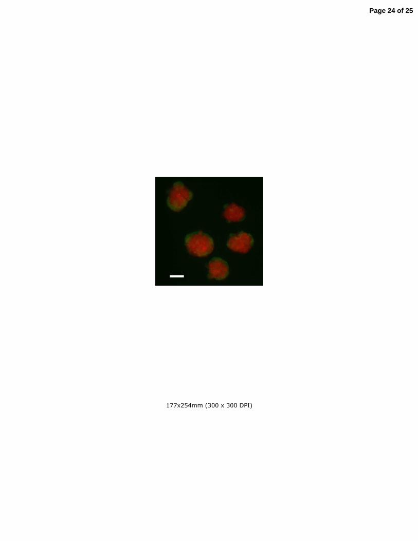
of EBS of defined cell numbers (Ungrin et al. 2008). The differentially stained ESC and 253
fibroblasts shown in Fig. 1 illustrate the ability to consistently prepare aggregated EBs of 254
experimentally determined heterogeneous composition (Fig. 1). Studies are underway to 255
define trophoblast differentiation in these composite EBs. 256
257
258
ESC-derived trophoblasts: modeling endovascular invasion and trophoblast-259
endothelial interaction. Early in human pregnancy, the invasion and remodeling of the 260
maternal spiral arterioles by extravillous trophoblasts provide the basis for the 261
establishment of a low resistance, high blood flow environment allowing for greater 262
nutrient and gas exchange to the developing fetus. Human umbilical vein endothelial 263
cells (HUVECs) provide an established endothelial cell model for angiogenesis and 264
endovascular remodeling. Studies have shown that the first trimester extravillous 265
trophoblast cell line HTR8 will undergo spontaneous migration toward, and align with 266
endothelial cells in contrast to term primary trophoblasts and immortalized third trimester 267
extravillous trophoblasts (TCL1) that actually inhibit endothelial cell tube formation 268
(Kalkunte et al. 2008). In addition vascular endothelial growth factor C (VEGF C) is 269
secreted at higher levels from the first trimester extravillous trophoblasts (HTR8) 270
Page 11 of 25
12
compared to the third trimester and term trophoblasts further establishing the differential 271
function of the first trimester cells. Localization of angiogenic growth factors in the 272
placental bed has further established the angiogenic potential of different trophoblast 273
subtypes (Schiessl et al. 2009). 274
275
Although the use of first trimester placental tissue and cell lines provide useful 276
information about pregnancy, most of the tissue and established cell lines originate from 277
weeks 8-12 of gestation. We therefore have extended the use of the hESC-derived EB 278
paradigm to study the HUVEC migratory potential in response to EB-derived trophoblast 279
outgrowths, and conversely, the EB-derived trophoblast outgrowth migratory potential in 280
response to HUVECs. The aims of these experiments are to better understand the bi-281
directional relationship between early pregnancy trophoblasts and endothelial cells in 282
vitro. Pilot studies shown in Fig. 2 (Giakoumopoulos et al., unpublished) illustrate the 283
time-dependent migration of trophoblastic outgrowths from EBs plated on Matrigel-284
coated invasion chambers. Demonstration that trophoblast-like cells derived from hESC 285
have biological characteristics of human placental cells will be important to define their 286
trophoblast identity. 287
288
Our previous studies demonstrating close association of macrophages with decidual 289
vessels n the first week of rhesus monkey gestation (Slukvin et al. 2004), and recent 290
studies of decidual vessels in human pregnancy at 8-12 weeks of gestation (Smith et al., 291
2009), are in concordance with endothelial-leukocyte dialog in early gestation as well. 292
Leukocyte-derived chemokines have been shown to influence trophoblast migration in a 293
immunodeficient mouse transplant model (Hanna et al. 2006). Further definition of 294
endothelial, leukocyte, and chemokine influences on the migration of a EB-derived 295
Page 12 of 25
13
trophoblast can provide an opportunity to interrogate the mechanisms that provide the 296
cues for development of the human implantation site. 297
298
Future directions: further studies with rhesus ESC. 299
The derivation of hESC was preceded by the isolation of cells from rhesus monkey 300
preimplantation blastocysts with ESC-like characteristics. These included the expression 301
of markers of pluripotency including alkaline phosphatase, Tra-1-60, Tra-1-81, SSEA-3, 302
and SSEA-4 (Thomson et al. 1995). In addition, there was morphological formation of 303
derivatives of all three embryonic germ layers upon spontaneous differentiaton, in the 304
context of teratomas when injected into immunocompromised mice (Thomson et al. 305
1995), and spontaneous formation of trophoblast as evidenced by transcription of CGα 306
and β subunit genes. Based on the efficient differentiation of hESC to trophoblasts with 307
the EB and BMP-regulated paradigms, we anticipated that these would as well be able 308
to induce trophoblast differentiation in rhesus ESC lines. However, in ongoing studies, 309
we have not found this to be the case. The differentiation of trophoblast populations from 310
rhesus ESC has proved to be very highly dependent on the cell line used (Garthwaite 311
and Golos, unpublished studies). A potentially important difference between the human 312
and rhesus ESC is the fact that the rhesus ESC derived in the Thomson lab were from in 313
vivo produced blastocysts, whereas all hESC have been derived from IVF-produced 314
embryos. Further studies are needed with rhesus ESC derived from IVF-produced 315
embryos (Mitalipov et al. 2006) or from rhesus nuclear transfer-generated blastocysts 316
(Byrne et al. 2007). 317
318
Epigenetic alterations with ESC differentiation. Recent studies with mouse ESC and 319
mouse trophoblast stem cells (TSC) have provided insight into the epigenetic 320
Page 13 of 25
14
mechanisms that influence trophoblast lineage specification (Ng et. al., 2008). TSC can 321
be derived from the trophectoderm of the mouse blastocyst under selective culture 322
conditions, and can give rise to all the differentiated trophoblasts of the mouse placenta 323
(Tanaka et al, 1998). In early embryonic development, cells of the inner cell mass 324
undergo global DNA methylation, whereas the cells of the trophectoderm remain in a 325
hypomethylated state (Santos et al, 2002). With mouse ESC in which the maintenance 326
DNA methyltransferase Dnmt1 has been deleted, culture conditions that support TSC 327
growth dramatically increased the formation of trophoblast giant cells from ESC in vitro 328
(Ng et. al., 2008). In these cells a progression of transcription factor expression similar to 329
that seen with TSC differentiation was also observed. These authors demonstrated that 330
a similar increase in trophoblast differentiation was induced by treatment with the DNA 331
methylation inhibitor 5-azacytidine (Ng et. al., 2008). In particular, the Ets family 332
transcription factor Elf5 was shown to be differentially methylated in TSC vs. ESC, and 333
formed a positive feedback loop with the transcription factors Cdx2 and Eomes, critical 334
for trophoblast lineage determination (Rossant, 2007). This study may have significance 335
for understanding the ability of human and nonhuman primate ESC to form of 336
trophoblast-like cells. It would seem to be imperative to define the methylation status of 337
key genes in trophoblast differentiation with the BMP-directed or EB-initiated formation 338
of trophoblast-like cells. Comparison with the methylation status of human placenta-339
derived trophoblasts will help clarify the status of derivatives of pluripotent ESC originally 340
derived from human and nonhuman primate embryos (as well as iPSC). Additionally, 341
the methylation status of transcription factors critical for trophoblast differentiation may 342
provide insight into the apparent differences in differentiation potential among hESC 343
cells derived from different labs (Xu et. al., 2002; Pera et. al., 2004) or between different 344
rhesus ESC lines derived under similar conditions (Garthwaite et. al., unpublished data). 345
346
Page 14 of 25
15
Summary and future prospects. 347
348
While the use of human ESC to study trophoblast differentiation is of significant merit, 349
progress needs to be made in several key areas in order to further refine the approaches 350
currently available. First, it is very important to determine what in vivo trophoblast 351
population(s) the hESC-derived trophoblasts represent. A better understanding of the 352
human placental trophoblasts during the first weeks of pregnancy would be ideal but is 353
not likely to be easily obtained, and nonhuman primates will likely be the surrogate for 354
this information. There is a pressing need for this information. As trophoblast 355
phenotypes are defined, new opportunities may arise to study trophoblast-leukocyte 356
interactions. The differentiation of all leukocyte lineages from hESC has been recently 357
advanced dramatically (Vodyanik et al. 2005), and the ability to derive autologous 358
leukocytes for hESC-derived trophoblast studies is highly attractive. Finally, while the in 359
vitro hESC-derived trophoblast models are important, of equal importance is the need to 360
develop in vivo opportunities to study ESC-derived trophoblast function. Again, this 361
arena will likely be restricted to nonhuman primate models, where it is realistic to be able 362
to conduct in vivo studies with ESC derivatives. As methods for the development of 363
rhesus monkey or other primate iPS cells are refined, autologous sources of ESC 364
differentiated derivatives will be essential for developing in vivo models. 365
366
Page 15 of 25
16
Declaration of Interest 367
368
The authors have no conflict of interest arising from these studies. 369
370
Funding 371
372
This research was supported by NIH grants HD046919 and HD038843 to T.G.G., and a 373
T32 HD041921 Predoctoral Fellowship to M.G. This research was conducted in part at 374
a facility constructed with support from Research Facilities Improvement Program grant 375
numbers RR15459-01 and RR020141-01. This publication's contents are solely the 376
responsibility of the authors and do not necessarily represent the official views of NCRR 377
or NIH. 378
379
Page 16 of 25

17
Acknowledgements 380
381
We would like to acknowledge the contributions of past and present members of the 382
Golos lab for contributions to the work summarized herein, including Behzad Gerami-383
Naini, Oksana Dovzhenko, Maureen Durning, and Leah Pollastrini Siegfried. We thank 384
James A. Thomson for ongoing interactions regarding human ES cells; and Judith 385
Peterson for help with manuscript preparation. 386
387
388
389
390
Page 17 of 25
18
REFERENCES 391
392
Aplin JD, Haigh T, Jones CJ, Church GJ & Vicovac L 1999 Development of 393
cytotrophoblast columns from explanted first-trimester human placental villi: role of 394
fibronectin and integrin alpha5beta1. Biol Reprod 60 828-838. 395
Byrne JA, Pedersen DA, Clepper LL, Nelson M, Sanger WG, Gokhale S, Wolf DP & 396
Mitalipov SM 2007 Producing primate embryonic stem cells by somatic cell nuclear 397
transfer. Nature 450 497-502. 398
Enders AC 2007 Implantation in the macaque: expansion of the implantation site during 399
the first week of implantation. Placenta 28 794-802. 400
Fishel SB & Edwards RG 1984 Human chorionic gonadotropin secreted by 401
preimplantation embryos cultured in vitro. Science 223 816-818. 402
Gerami-Naini B, Dovzhenko OV, Durning M, Wegner FH, Thomson JA & Golos TG 403
2004 Trophoblast differentiation in embryoid bodies derived from human embryonic stem 404
cells as a model for trophoblast differentiation. Endocrinology 145 1517-1524. 405
Gerecht-Nir S, Cohen S, and Itskovitz-Eldor J 2004 Bioreactor cultivation enhances 406
the efficiency of human embryoid body (hEB) formation and differentiation. Biotech 407
Bioeng. 86 493-502. 408
Hanna J, Goldman-Wohl D, Hamani Y, Avraham I, Greenfield C, Natanson-Yaron S, 409
Prus D, Cohen-Daniel L, Arnon TI, Manaster I, et al.. 2006 Decidual NK cells regulate 410
key developmental processes at the human fetal-maternal interface. Nat Med 12 1065-411
1074. 412
Harris LK & Aplin JD 2007 Vascular remodeling and extracellular matrix breakdown in 413
the uterine spiral arteries during pregnancy. Reprod Sci 14 28-34. 414
Hearn JP, Hodges JK & Gems S 1988 Early secretion of chorionic gonadotrophin by 415
marmoset embryos in vivo and in vitro. J Endrocinol 119:249-255 416
Page 18 of 25
19
Hemberger M, Nozaki T, Winterhager E, Yamamoto H, Nakagama H, Kamada N, 417
Suzuki H, Ohta T, Ohki M, Masutani M & Cross JC 2003 Parp1-deficiency induces 418
differentiation of ES cells into trophoblast derivatives. Dev Biol 257 371-81. 419
Kalkunte S, Lai Z, Tewari N, Chichester C, Romero R, Padbury J & Sharma S 2008 420
In vitro and in vivo evidence for lack of endovascular remodeling by third trimester 421
trophoblasts. Placenta 29 871-878. 422
Kaufmann P & Burton G. Anatomy and Genesis of the Placenta. 1994 In The 423
Physiology of Reproduction, edn 2, pp 441-484. Eds E Knobil and JD Neill. New York, 424
Raven Press. 425
Mali P, Ye Z, Hommond HH, Yu X, Lin J, Chen G, Zou J, and Cheng L. 2008 426
Improved efficiency and pace of generating induced pluripotent stem cells from human 427
adult and fetal fibroblasts. Stem Cells 26: 1998-2005. 428
Mitalipov S, Kuo H-C, Byrne J, Clepper L, Meisner L, Johnson J, Zeier R & Wolf D 429
2006 Isolation and Characterization of Novel Rhesus Monkey Embryonic Stem Cell 430
Lines. Stem Cells 24 2177-2186. 431
Ng RK, Dean W, Dawson C, Lucifero D, Madeja Z, Reik W & Hemberger M 2008 432
Epigenetic restriction of embryonic cell lineage fate by methylation of Elf5. Nature Cell 433
Biology 10 1280-1290. 434
Pera, MF, Andrade J, Houssami S, Reubinoff B, Trounson A, Stanley EG, Ward-van 435
Oostwaard D and Mummery C 2004 Regulation of human embryonic stem cell 436
differentiation by BMP-2 and its antagonist noggin. J. Cell Sci 117 1269-1280. 437
Pijnenborg R, Vercruysse L, Hanssens M and Van Asshe A 2007 Trophoblast 438
invasion in pre-eclampsia and other pregnancy disorders. In Pre-eclampsia: Etiology and 439
Clinical Practice, pp. 3-19. Eds F Lyall F and M Belfort. Cambridge, New York: 440
Cambridge University Press. 441
Page 19 of 25
20
Pope VZ, Pope CE & Beck LR 1982 Gonadotropin production by baboon embryos in 442
vitro. In Vitro Fertilization and embryo transfer. Eds: Hafey & Semm. Lancaster: MTP 443
Press, Ltd. 444
Redman CW and Sargent IL 2005. Latest advances in understanding preeclampsia. 445
Science 308 1592-1594. 446
Reubinoff BE, Pera MF, Fong CY, Trounson A & Bongso A 2000 Embryonic stem 447
cell lines from human blastocysts: somatic differentiation in vitro. Nat Biotechnol 18 399-448
404. 449
Rossant, J 2007. Stem cells from the mammalian blastocyst. Stem Cells 19 477-482. 450
Santos F, Hendrich B, Reik W & Dean W 2002 Dynamic reprogramming of DNA 451
methylation in the early mouse embryo. Dev Biol. 241 172-182. 452
Schenke-Layland K, Angelis E, Rhodes KE, Heydarkhan-Hagvall S, Mikkola HK & 453
& MacLellan WR 2007 Collagen IV induces trophectoderm differentiation of mouse 454
embryonic stem cells. Stem Cells 25 1529-1538. 455
Schiessl B, Innes BA, Bulmer JN, Otun HA, Chadwick TJ, Robson SC & Lash GE 456
2009 Localization of angiogenic growth factors and their receptors in the human 457
placental bed throughout normal human pregnancy. Placenta 30 79-87. 458
Schulz LC, Ezashi T, Das P, Westfall SD, Livingston KA, & Roberts RM 2008 459
Human embryonic stem cells as models for trophoblast differentiation. Placenta 22 S10-460
S16. 461
Seshagiri PB, Terasawa E & Hearn JP 1994 The secretion of gonadotrophin-releasing 462
hormone by peri-implantation embryos of the rhesus monkey: comparison with the 463
secretion of chorionic gonadotrophin. Hum Reprod 9 1300-1307. 464
Slukvin I, Breburda EE & Golos TG 2004. Dynamic changes in primate endometrial 465
leukocyte populations: differential distribution of macrophages and natural killer cells at 466
the rhesus monkey implantation site and in early pregnancy. Placenta 25 297-307. 467
Page 20 of 25
21
Slukvin, II, Lunn DP, Watkins DI & Golos TG 2000 Placental expression of the 468
nonclassical MHC class I molecule Mamu-AG at implantation in the rhesus monkey. 469
Proc Natl Acad Sci. USA 97 9104-9109. 470
Smith SD, Dunk CE, Aplin JD, Harris LK, and Jones RL 2009 Evidence for immune 471
cell involvement in decidual spiral arteriole remodeling in early human pregnancy. Am J 472
Pathol 174 1959-1971. 473
Takahashi K, Tanabe K, Ohnuki M, Narita M, Ichisaka T, Tomoda K & Yamanaka S 474
2007 Induction of pluripotent stem cells from adult human fibroblasts by defined factors. 475
Cell 131 861-872. 476
Takahashi K & Yamanaka S 2006 Induction of pluripotent stem cells from mouse 477
embryonic and adult fibroblast cultures by defined factors. Cell 126 663-676. 478
Tanaka S, Kunath T & Hadjantonakis AK 1998. A. Nagy and J. Rossant, Promotion of 479
trophoblast stem cell proliferation by FGF4. Science 282 2072-2075. 480
Thomson JA, Itskovitz-Eldor J, Shapiro SS, Waknitz MA, Swiergiel JJ, Marshall VS 481
& Jones JM 1998 Embryonic stem cell lines derived from human blastocysts. Science 482
282 1145-1147. 483
Thomson JA, Kalishman J, Golos TG, Durning M, Harris CP, Becker RA &Hearn JP 484
1995 Isolation of a primate embryonic stem cell line. Proc Natl Acad Sci U S A 92 7844-485
7848. 486
Ungrin MD, Joshi C, Nica A, Bauwens C & Zandstra PW 2008 Reproducible, ultra 487
high-throughput formation of multicellular organization from single cell suspension-488
derived human embryonic stem cell aggregates. PloS ONE 3 e1565. 489
Vodyanik MA Bork JA, Thomson JA & Slukvin II 2005 Human embryonic stem cell-490
derived CD34+ cells: efficient production in the coculture with OP9 stromal cells and 491
analysis of lymphohematopoietic potential. Blood 105 617-626. 492
Page 21 of 25
22
Xu RH, Chen X, Li DS, Li R, Addicks GC, Glennon C, Zwaka TP & Thomson JA 493
2002 BMP4 initiates human embryonic stem cell differentiation to trophoblast. Nat 494
Biotechnol 20 1261-1264. 495
Yu J, Vodyanik MA, Smuga-Otto K, Antosiewicz-Bourget J, Frane JL, Tian S, 496
Nie J, Jonsdottir GA, Ruotti V, Stewart R, Slukvin II & Thomson JA 2007 497
Induced pluripotent stem cell lines derived from human somatic cells. Science 318 498
1917-1920. 499
500
Page 22 of 25
23
Figure Legends 501
502
Figure 1. Aggrewell generated combination EBs. Aggrewell EBs were prepared 503
generate EBs comprised of 1000 cells (Ungrin et al.., 2008). Term placental fibroblasts 504
were stained for 1 hour with 25 µM Cell Tracker Red (Invitrogen, Carlsbad, CA) and ES 505
cells were stained at the same concentration with Cell Tracker Green. Fibroblasts were 506
then trypsinized, counted and seeded at 600,000 cells per Aggrewell. ES cells were 507
enzymatically dispersed with Accutase, counted and also seeded at 600,000 cells per 508
Aggrewell bringing the total number of cells per Aggrewell to 1,200,000 (as per 509
manufacturers recommendation for the production of 1000-cell EBs). The ROCK 510
inhibitor (Y-27632) was added to each well at a concentration of 10 µM, cells were 511
centrifuged into the Aggrewells and left overnight at 37 C with CO2 and 95% humidity to 512
form EBs. The following day, EBs were gently pipetted and passed through a 40 µm cell 513
strainer to remove excess cellular debris and were imaged. The photomicrograph 514
illustrates the presence of an internal core of red fibroblasts and an outer zone of green 515
ES cells. Scale bar = 0.1 mm. 516
517
518
Figure 2. Migration of embryoid body-derived trophoblasts in Transwell invasion 519
chambers. Left panel: schematic diagram of experimental setup, illustrating two 520
possible treatments to identify effects on trophoblast invasion/migration. Right panel: 521
time-dependent migration of EB-derived trophoblast outgrowths through the chamber 522
membrane. The means of 5 photomicrographic fields from each of 3 independent 523
chambers is shown. 524
525
Page 23 of 25
177x254mm (300 x 300 DPI)
Page 24 of 25
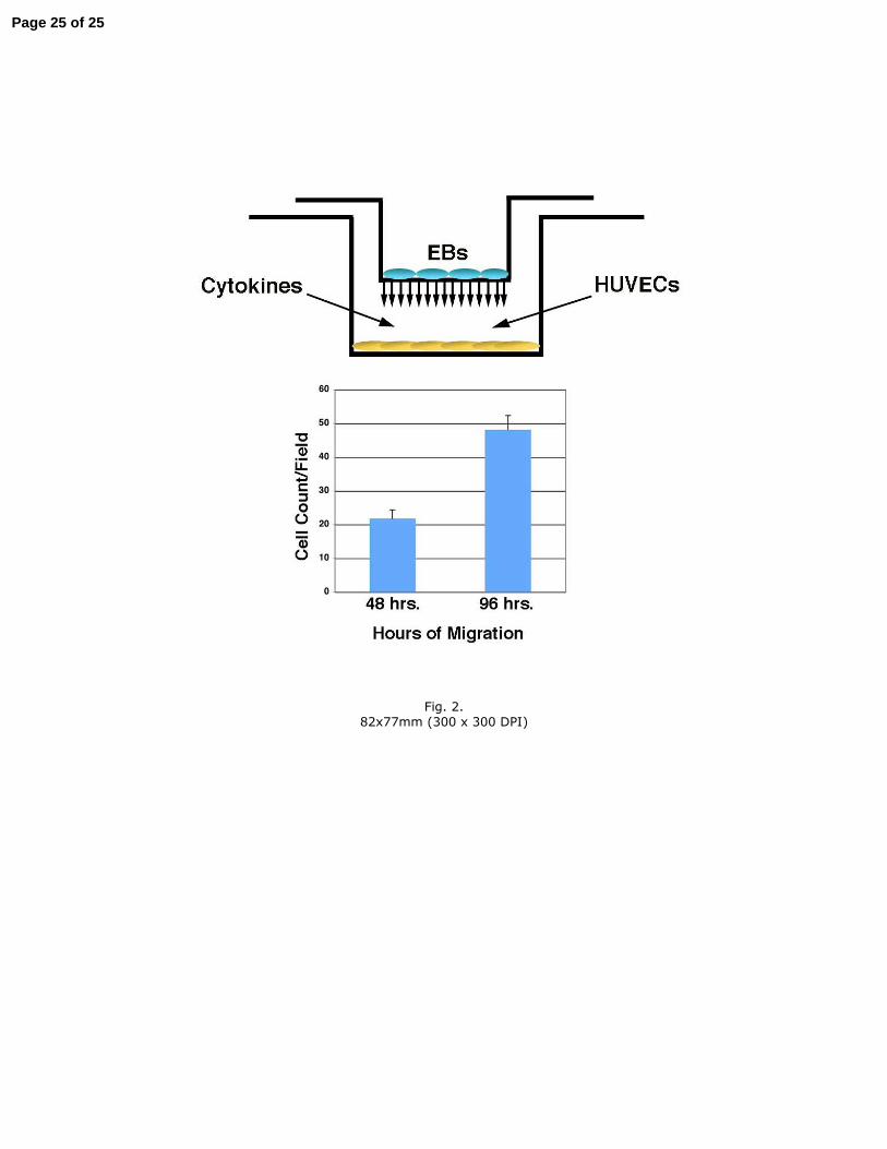
Fig. 2.
82x77mm (300 x 300 DPI)
Page 25 of 25


![STEM CELLS EMBRYONIC STEM CELLS/INDUCED PLURIPOTENT STEM CELLS Stem Cells.pdf · germ cell production [2]. Human embryonic stem cells (hESCs) offer the means to further understand](https://img.pdfslide.us/doc/110x75/6014b11f8ab8967916363675/stem-cells-embryonic-stem-cellsinduced-pluripotent-stem-cells-stem-cellspdf.jpg)