Embed Size (px)
Citation preview
Embryology of the Respiratory System I
Prof. Abdulameer Al-Nuaimi
E-mail: [email protected] E. mail: [email protected]
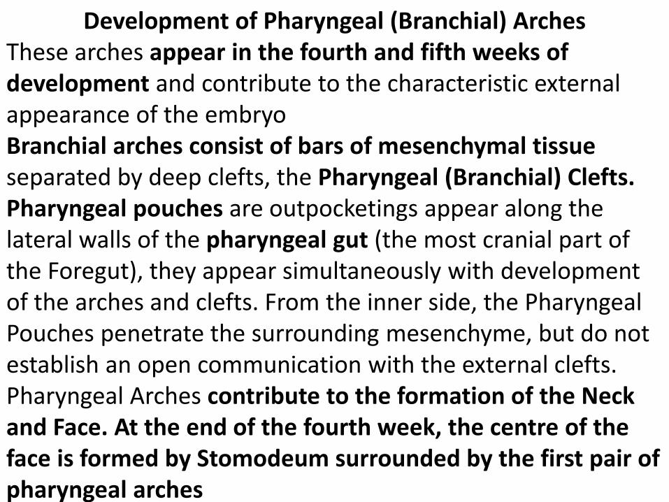
Development of Pharyngeal (Branchial) Arches These arches appear in the fourth and fifth weeks of development and contribute to the characteristic external appearance of the embryo Branchial arches consist of bars of mesenchymal tissue separated by deep clefts, the Pharyngeal (Branchial) Clefts. Pharyngeal pouches are outpocketings appear along the lateral walls of the pharyngeal gut (the most cranial part of the Foregut), they appear simultaneously with development of the arches and clefts. From the inner side, the Pharyngeal Pouches penetrate the surrounding mesenchyme, but do not establish an open communication with the external clefts. Pharyngeal Arches contribute to the formation of the Neck and Face. At the end of the fourth week, the centre of the face is formed by Stomodeum surrounded by the first pair of pharyngeal arches
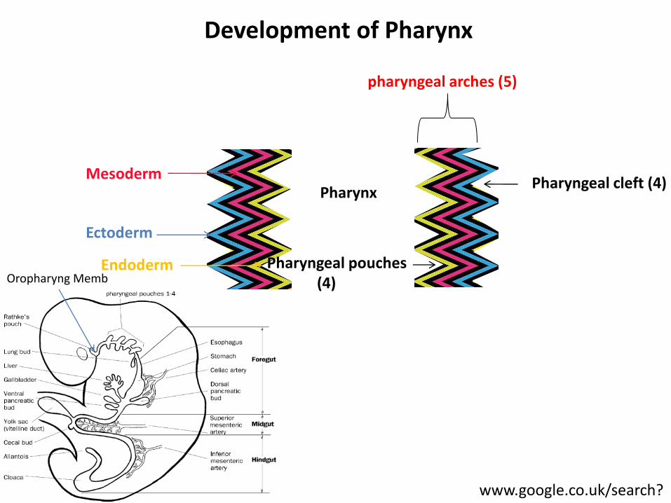
Mesoderm
Ectoderm
Endoderm
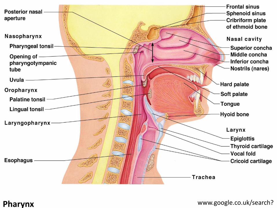
Pharynx
pharyngeal arches (5)
Pharyngeal cleft (4)
Pharyngeal pouches (4) Oropharyng Memb
www.google.co.uk/search?
Development of Pharynx
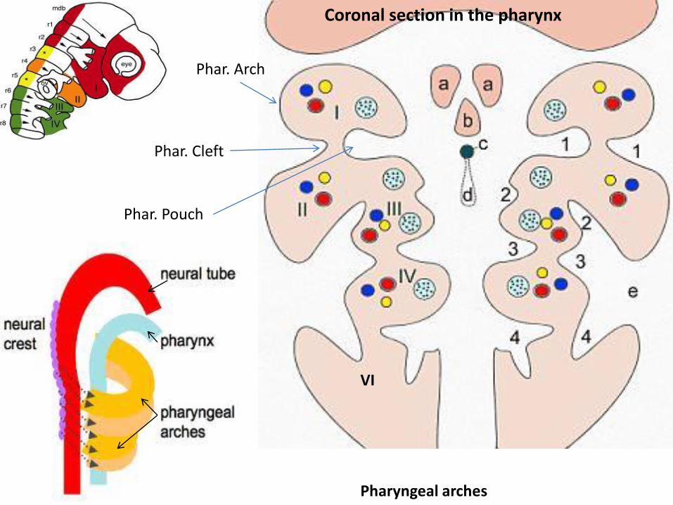
Phar. Arch
Phar. Cleft
Phar. Pouch
Pharyngeal arches
Coronal section in the pharynx
VI
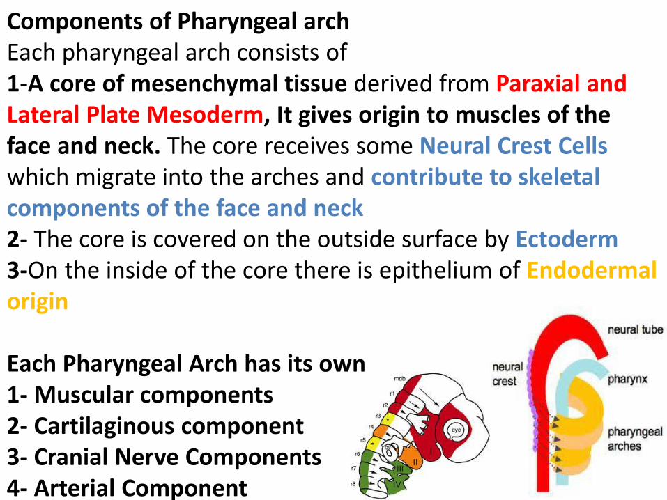
Components of Pharyngeal arch Each pharyngeal arch consists of 1-A core of mesenchymal tissue derived from Paraxial and Lateral Plate Mesoderm, It gives origin to muscles of the face and neck. The core receives some Neural Crest Cells which migrate into the arches and contribute to skeletal components of the face and neck 2- The core is covered on the outside surface by Ectoderm 3-On the inside of the core there is epithelium of Endodermal origin Each Pharyngeal Arch has its own 1- Muscular components 2- Cartilaginous component 3- Cranial Nerve Components 4- Arterial Component
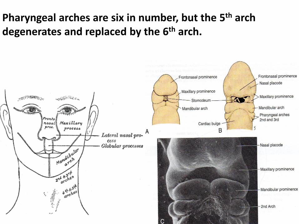
Pharyngeal arches are six in number, but the 5th arch degenerates and replaced by the 6th arch.
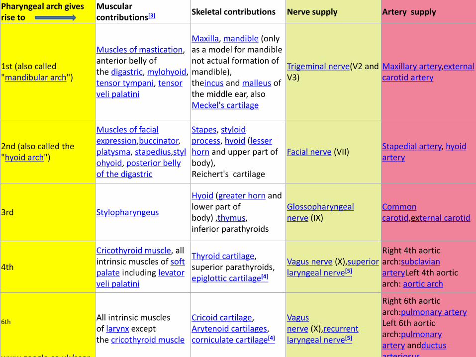
Pharyngeal arch gives rise to
Muscular contributions[3]
Skeletal contributions Nerve supply Artery supply
1st (also called "mandibular arch")
Muscles of mastication, anterior belly of the digastric, mylohyoid,tensor tympani, tensor veli palatini
Maxilla, mandible (only as a model for mandible not actual formation of mandible), theincus and malleus of the middle ear, also Meckel's cartilage
Trigeminal nerve(V2 and V3)
Maxillary artery,external carotid artery
2nd (also called the "hyoid arch")
Muscles of facial expression,buccinator, platysma, stapedius,stylohyoid, posterior belly of the digastric
Stapes, styloid process, hyoid (lesser horn and upper part of body), Reichert's cartilage
Facial nerve (VII) Stapedial artery, hyoid artery
3rd Stylopharyngeus
Hyoid (greater horn and lower part of body) ,thymus, inferior parathyroids
Glossopharyngeal nerve (IX)
Common carotid,external carotid
4th
Cricothyroid muscle, all intrinsic muscles of soft palate including levator veli palatini
Thyroid cartilage, superior parathyroids, epiglottic cartilage[4]
Vagus nerve (X),superior laryngeal nerve[5]
Right 4th aortic arch:subclavian arteryLeft 4th aortic arch: aortic arch
6th www.google.co.uk/sear
All intrinsic muscles of larynx except the cricothyroid muscle
Cricoid cartilage, Arytenoid cartilages, corniculate cartilage[4]
Vagus nerve (X),recurrent laryngeal nerve[5]
Right 6th aortic arch:pulmonary artery Left 6th aortic arch:pulmonary artery andductus arteriosus
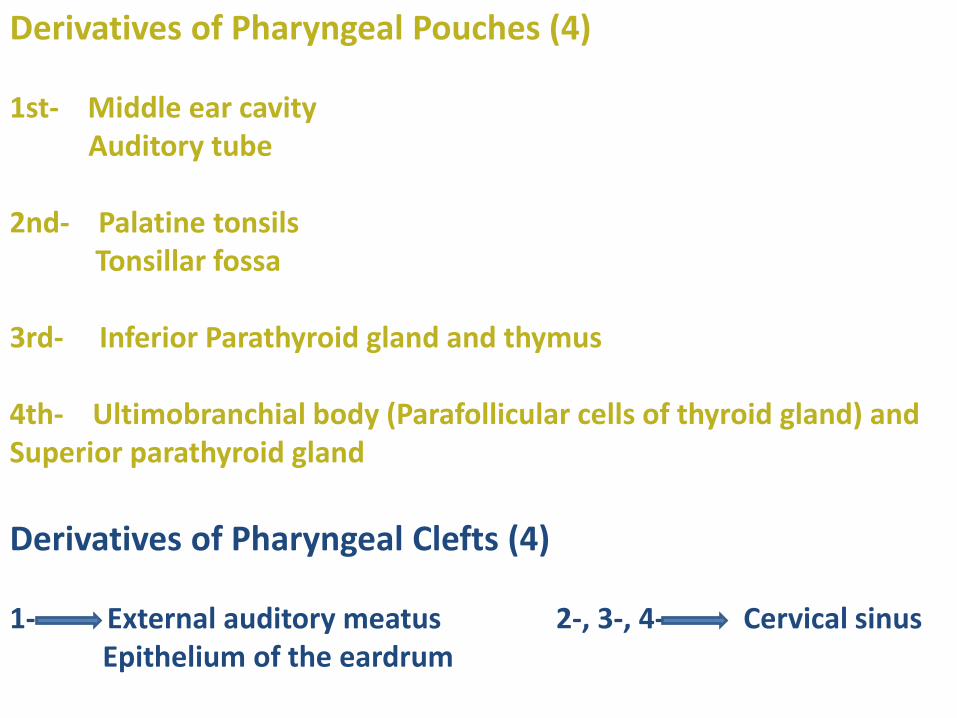
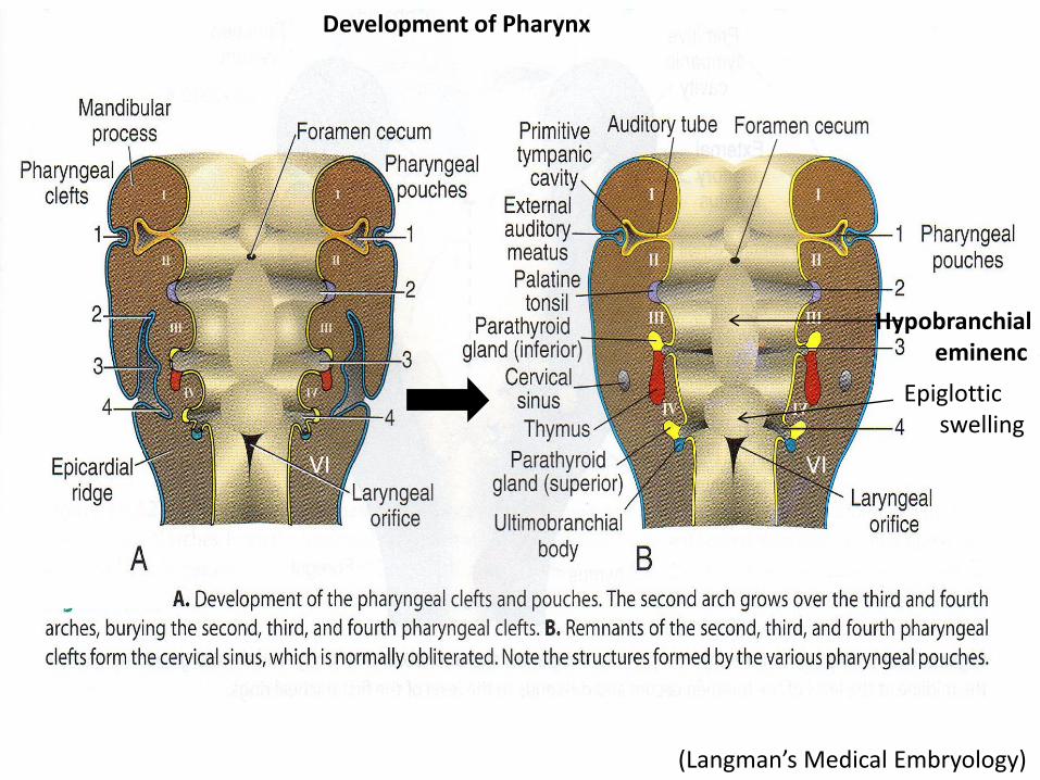
Derivatives of Pharyngeal Pouches (4) 1st- Middle ear cavity Auditory tube 2nd- Palatine tonsils Tonsillar fossa 3rd- Inferior Parathyroid gland and thymus 4th- Ultimobranchial body (Parafollicular cells of thyroid gland) and Superior parathyroid gland
Derivatives of Pharyngeal Clefts (4) 1- External auditory meatus 2-, 3-, 4- Cervical sinus Epithelium of the eardrum
(Langman’s Medical Embryology)
Development of Pharynx
Hypobranchial eminenc
Epiglottic swelling
VI VI
Pharynx www.google.co.uk/search?
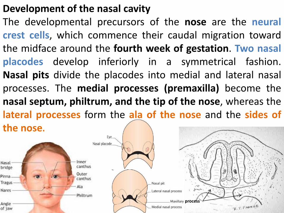
Development of the nasal cavity The developmental precursors of the nose are the neural crest cells, which commence their caudal migration toward the midface around the fourth week of gestation. Two nasal placodes develop inferiorly in a symmetrical fashion. Nasal pits divide the placodes into medial and lateral nasal processes. The medial processes (premaxilla) become the nasal septum, philtrum, and the tip of the nose, whereas the lateral processes form the ala of the nose and the sides of the nose.
process
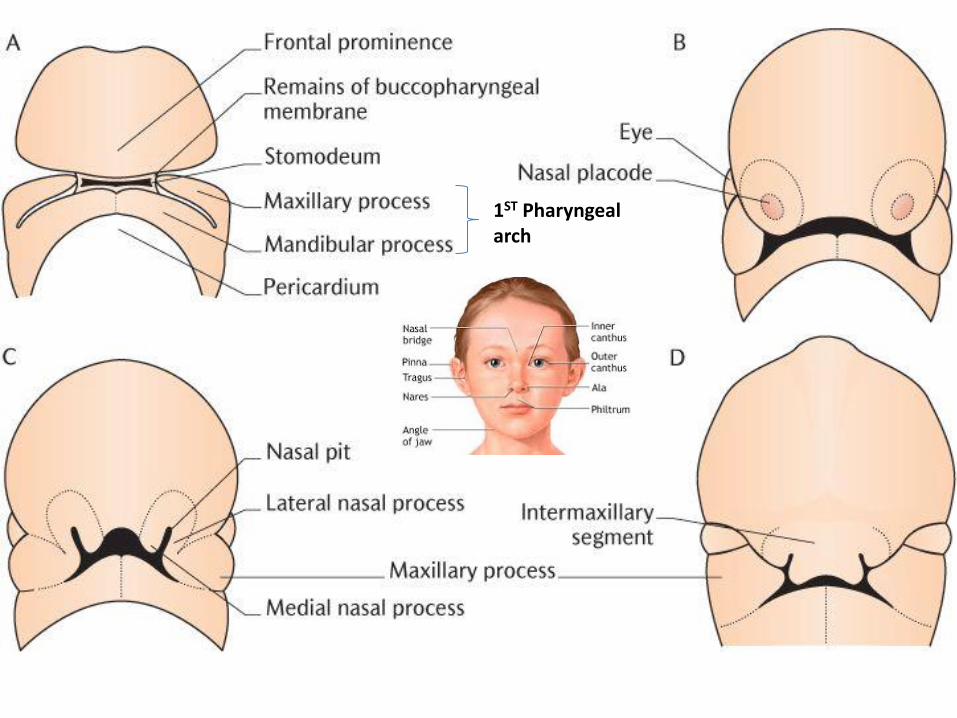
1ST Pharyngeal arch
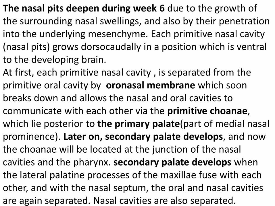
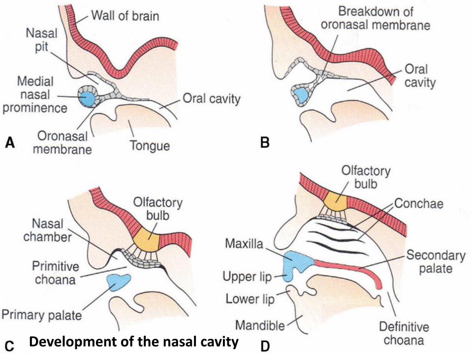
The nasal pits deepen during week 6 due to the growth of the surrounding nasal swellings, and also by their penetration into the underlying mesenchyme. Each primitive nasal cavity (nasal pits) grows dorsocaudally in a position which is ventral to the developing brain. At first, each primitive nasal cavity , is separated from the primitive oral cavity by oronasal membrane which soon breaks down and allows the nasal and oral cavities to communicate with each other via the primitive choanae, which lie posterior to the primary palate(part of medial nasal prominence). Later on, secondary palate develops, and now the choanae will be located at the junction of the nasal cavities and the pharynx. secondary palate develops when the lateral palatine processes of the maxillae fuse with each other, and with the nasal septum, the oral and nasal cavities are again separated. Nasal cavities are also separated.
Development of the nasal cavity
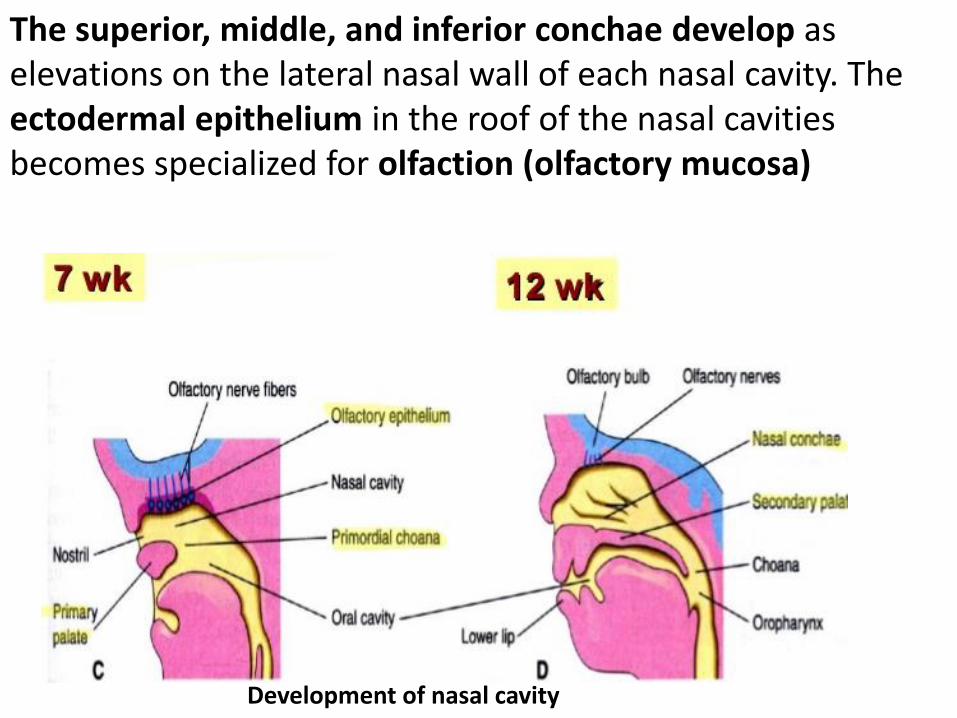
The superior, middle, and inferior conchae develop as elevations on the lateral nasal wall of each nasal cavity. The ectodermal epithelium in the roof of the nasal cavities becomes specialized for olfaction (olfactory mucosa)
Development of nasal cavity
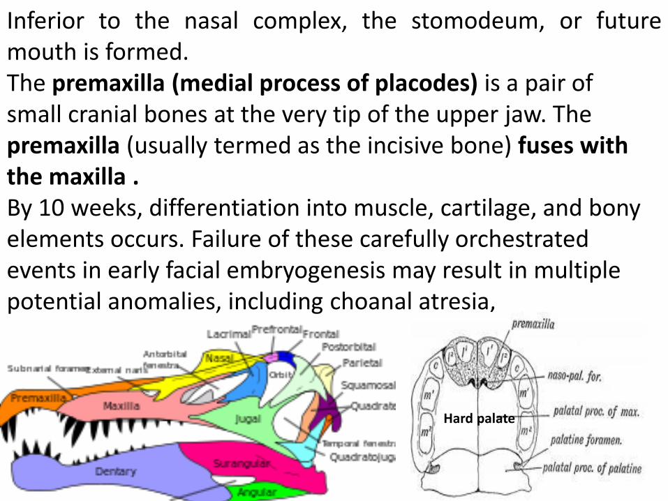
Inferior to the nasal complex, the stomodeum, or future mouth is formed. The premaxilla (medial process of placodes) is a pair of small cranial bones at the very tip of the upper jaw. The premaxilla (usually termed as the incisive bone) fuses with the maxilla . By 10 weeks, differentiation into muscle, cartilage, and bony elements occurs. Failure of these carefully orchestrated events in early facial embryogenesis may result in multiple potential anomalies, including choanal atresia,
Hard palate
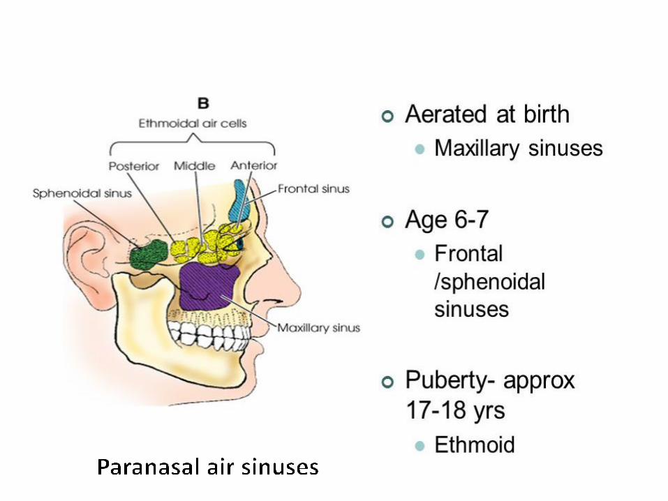
Development of paranasal air sinuses Paranasal air sinuses develop as a series of diverticula from each nasal cavity, they penetrate the maxillary, frontal, ethmoidal and sphenoid bones. They are lined by extensions of the nasal mucoperiosteum. This process begins prenatally, paranasal sinuses are small at birth, enlarge mainly during the eruption of the second dentation and reach their adult size soon after puberty. The natural ventilation-rate of a sinus with a single sinus opening, is extremely slow. Such limited ventilation may be protective for the sinus, as it would help prevent drying of its mucosal surface and maintain a nearly sterile environment with high carbon dioxide concentrations and minimal pathogen access. Composition of gas content in the sinuses is similar to the venous blood, with high carbon dioxide and lower oxygen levels compared to breathing air.

Thank You Thank You