Embed Size (px)
Citation preview
Embryology of the Head and Neck
Selected High-Yield Topics
Joseph L. Russell, MDFaculty Advisor: Harold S. Pine, MD, FAAP, FACS
Department of Otolaryngology-Head & Neck SurgeryThe University of Texas Medical Branch
Grand Rounds PresentationMarch 28, 2014
Series Editor: Francis B. Quinn, Jr., MD, FACS – Archivist: Melinda Stoner Quinn, MSICS
Overview
• Branchial arches, pouches, and clefts• Ear development• Nasal, lip, and facial development• Thyroid development• Laryngeal development
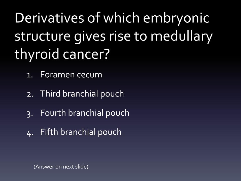
Derivatives of which embryonic structure gives rise to medullary thyroid cancer?
1. Foramen cecum
2. Third branchial pouch
3. Fourth branchial pouch
4. Fifth branchial pouch
(Answer on next slide)
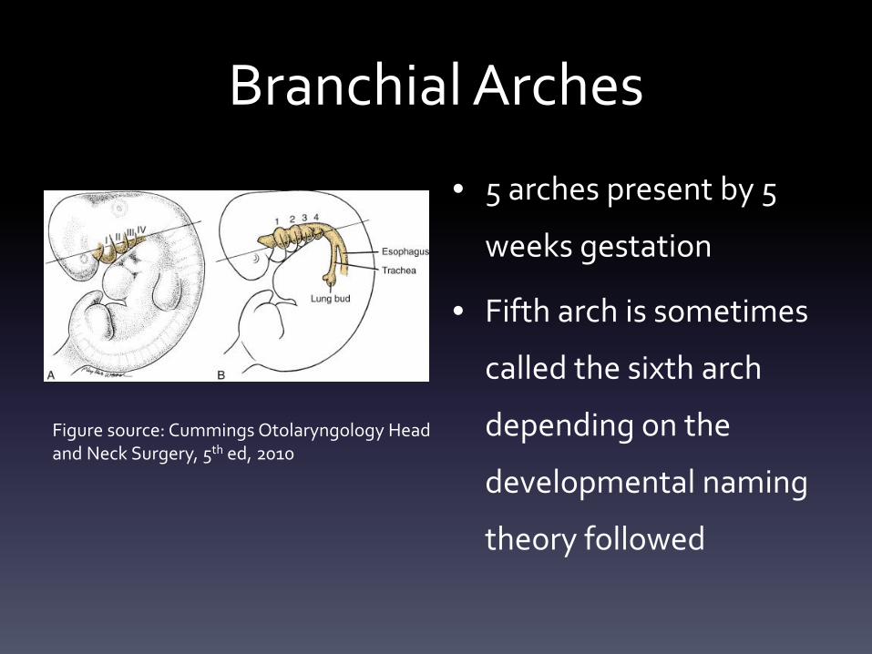
Branchial Arches
• 5 arches present by 5
weeks gestation
• Fifth arch is sometimes
called the sixth arch
depending on the
developmental naming
theory followed
Figure source: Cummings Otolaryngology Head and Neck Surgery, 5th ed, 2010
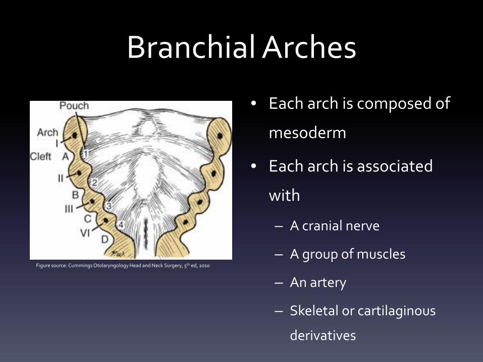
Branchial Arches
• Each arch is composed of
mesoderm
• Each arch is associated
with
– A cranial nerve
– A group of muscles
– An artery
– Skeletal or cartilaginous
derivatives
Figure source: Cummings Otolaryngology Head and Neck Surgery, 5th ed, 2010
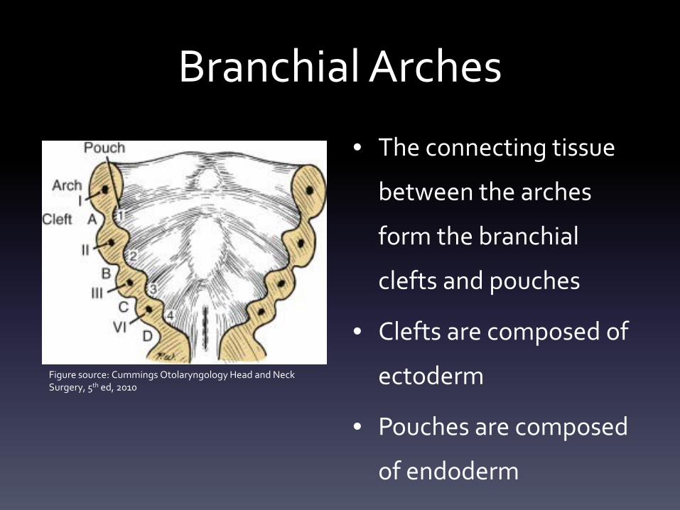
Branchial Arches
• The connecting tissue
between the arches
form the branchial
clefts and pouches
• Clefts are composed of
ectoderm
• Pouches are composed
of endoderm
Figure source: Cummings Otolaryngology Head and Neck Surgery, 5th ed, 2010
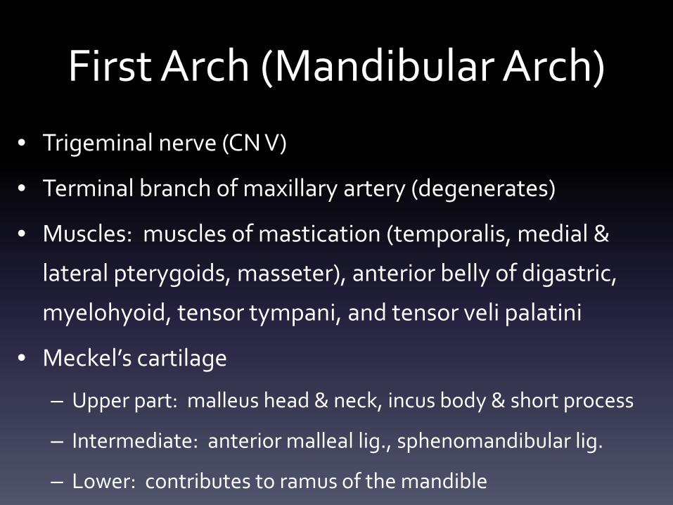
First Arch (Mandibular Arch)
• Trigeminal nerve (CN V)
• Terminal branch of maxillary artery (degenerates)
• Muscles: muscles of mastication (temporalis, medial &
lateral pterygoids, masseter), anterior belly of digastric,
myelohyoid, tensor tympani, and tensor veli palatini
• Meckel’s cartilage
– Upper part: malleus head & neck, incus body & short process
– Intermediate: anterior malleal lig., sphenomandibular lig.
– Lower: contributes to ramus of the mandible
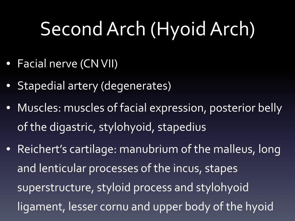
Second Arch (Hyoid Arch)
• Facial nerve (CN VII)
• Stapedial artery (degenerates)
• Muscles: muscles of facial expression, posterior belly
of the digastric, stylohyoid, stapedius
• Reichert’s cartilage: manubrium of the malleus, long
and lenticular processes of the incus, stapes
superstructure, styloid process and stylohyoid
ligament, lesser cornu and upper body of the hyoid
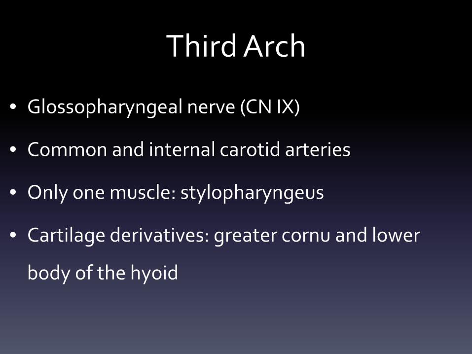
Third Arch
• Glossopharyngeal nerve (CN IX)
• Common and internal carotid arteries
• Only one muscle: stylopharyngeus
• Cartilage derivatives: greater cornu and lower
body of the hyoid
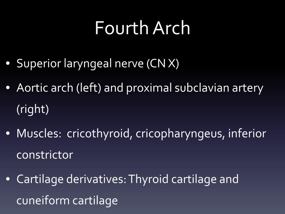
Fourth Arch
• Superior laryngeal nerve (CN X)
• Aortic arch (left) and proximal subclavian artery
(right)
• Muscles: cricothyroid, cricopharyngeus, inferior
constrictor
• Cartilage derivatives: Thyroid cartilage and
cuneiform cartilage
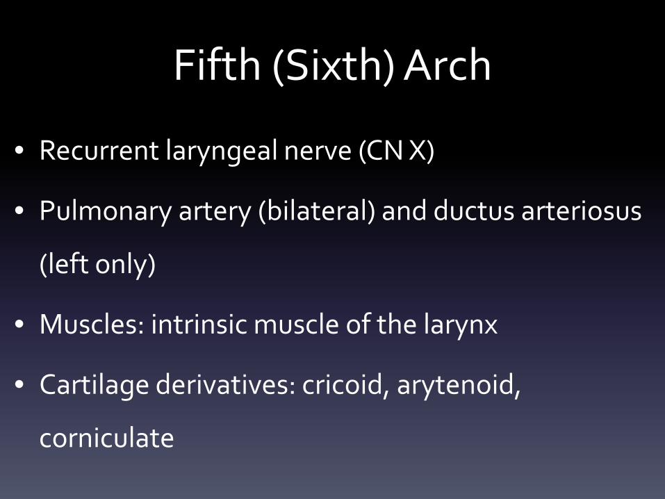
Fifth (Sixth) Arch
• Recurrent laryngeal nerve (CN X)
• Pulmonary artery (bilateral) and ductus arteriosus
(left only)
• Muscles: intrinsic muscle of the larynx
• Cartilage derivatives: cricoid, arytenoid,
corniculate
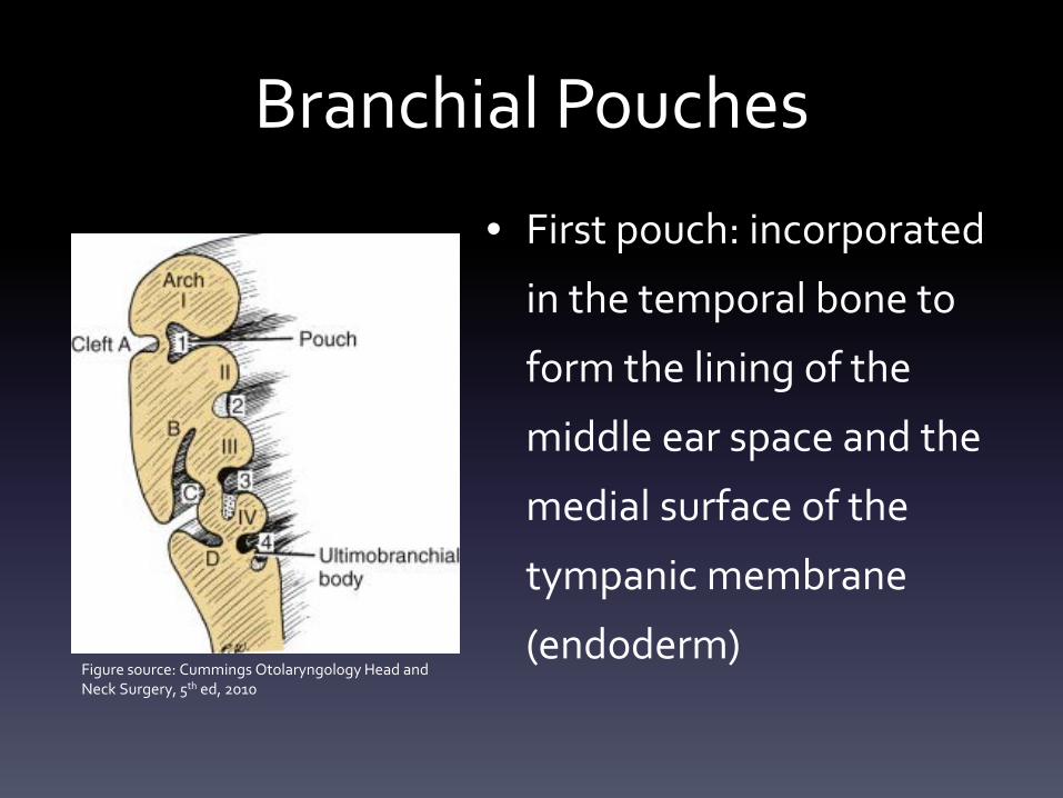
Branchial Pouches
• First pouch: incorporated
in the temporal bone to
form the lining of the
middle ear space and the
medial surface of the
tympanic membrane
(endoderm)Figure source: Cummings Otolaryngology Head and Neck Surgery, 5th ed, 2010
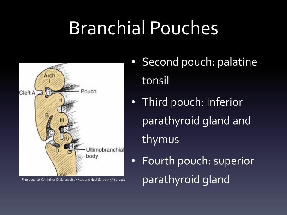
Branchial Pouches
• Second pouch: palatine
tonsil
• Third pouch: inferior
parathyroid gland and
thymus
• Fourth pouch: superior
parathyroid glandFigure source: Cummings Otolaryngology Head and Neck Surgery, 5th ed, 2010
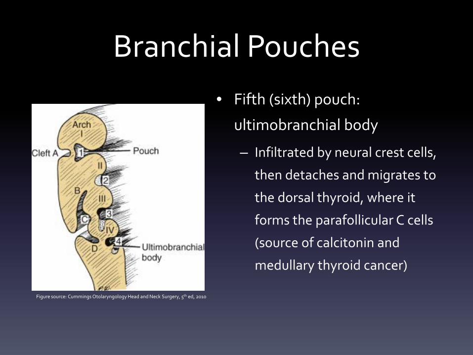
Branchial Pouches• Fifth (sixth) pouch:
ultimobranchial body
– Infiltrated by neural crest cells,
then detaches and migrates to
the dorsal thyroid, where it
forms the parafollicular C cells
(source of calcitonin and
medullary thyroid cancer)
Figure source: Cummings Otolaryngology Head and Neck Surgery, 5th ed, 2010
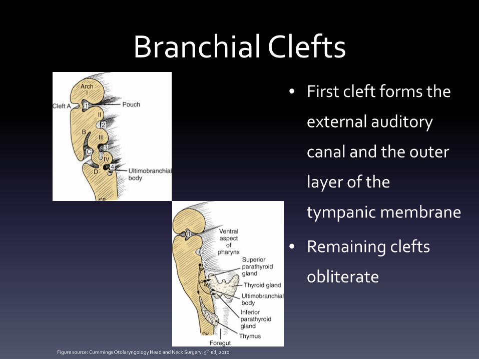
Branchial Clefts• First cleft forms the
external auditory
canal and the outer
layer of the
tympanic membrane
• Remaining clefts
obliterate
Figure source: Cummings Otolaryngology Head and Neck Surgery, 5th ed, 2010

Branchial Cleft Anomalies• Cyst: a mucosa or epithelium-lined structure with no external
opening
• Sinus: a tract, with or without a cyst, that communicates with the
pharynx or skin
• Fistula: a tract connecting the pharynx and skin
• For sinuses and fistulae, the tract runs deep to its arch and
superficial to more distal arches
• All tracts run anterior to the sternocleidomastoid muscle and
superficial to CN XII
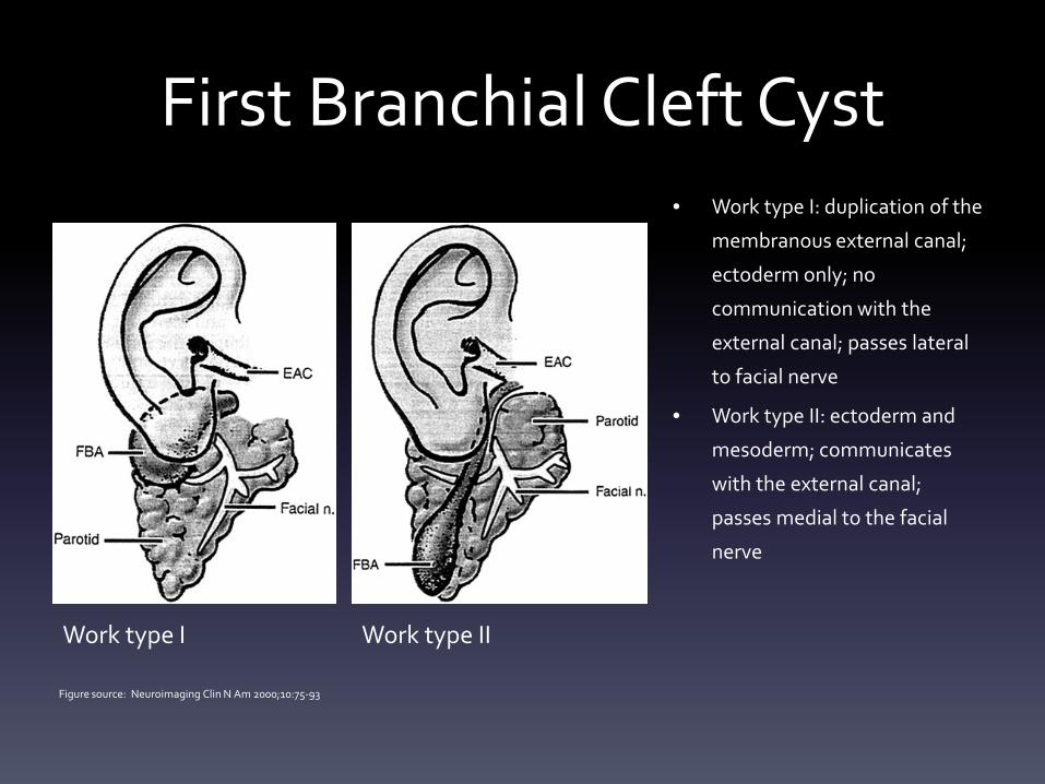
First Branchial Cleft Cyst• Work type I: duplication of the
membranous external canal;
ectoderm only; no
communication with the
external canal; passes lateral
to facial nerve
• Work type II: ectoderm and
mesoderm; communicates
with the external canal;
passes medial to the facial
nerve
Work type I Work type II
Figure source: Neuroimaging Clin N Am 2000;10:75-93
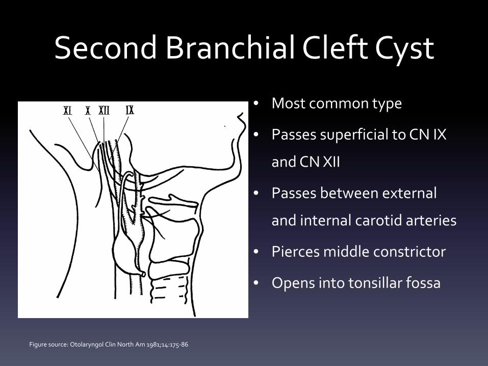
Second Branchial Cleft Cyst• Most common type
• Passes superficial to CN IX
and CN XII
• Passes between external
and internal carotid arteries
• Pierces middle constrictor
• Opens into tonsillar fossa
Figure source: Otolaryngol Clin North Am 1981;14:175-86
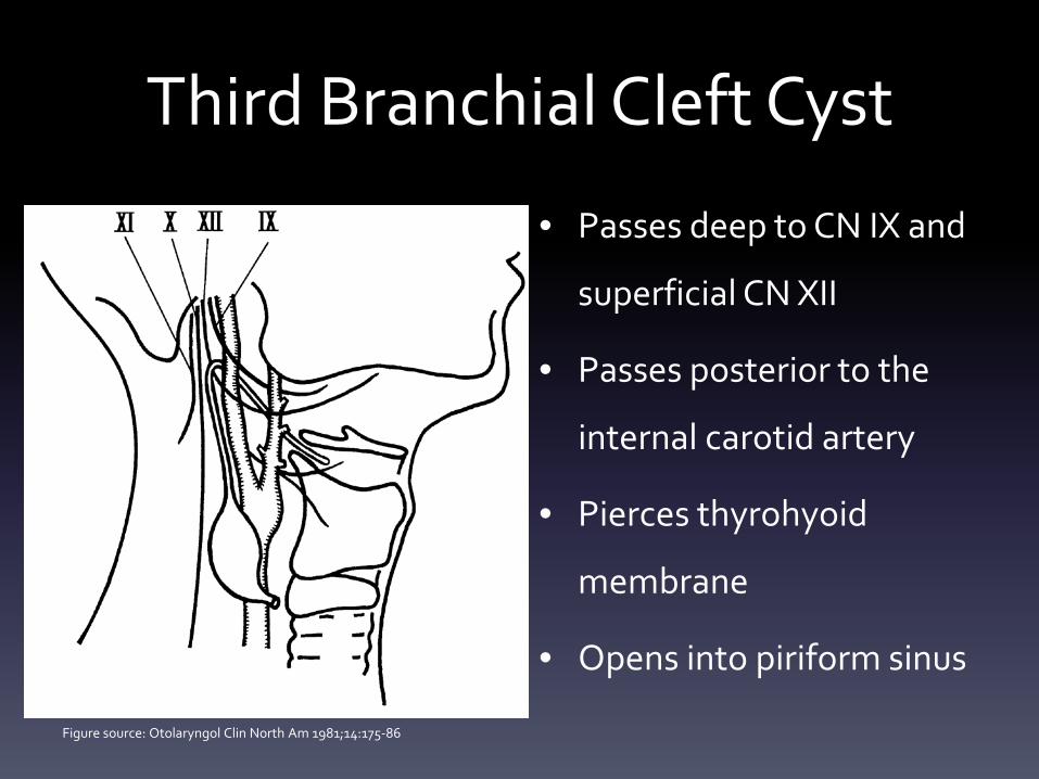
Third Branchial Cleft Cyst
• Passes deep to CN IX and
superficial CN XII
• Passes posterior to the
internal carotid artery
• Pierces thyrohyoid
membrane
• Opens into piriform sinus
Figure source: Otolaryngol Clin North Am 1981;14:175-86
Fourth Branchial Cleft Cyst
• Very rare
• Tract descends along the carotid sheath, passes
around the subclavian artery (right) or aortic arch
(left), and ascends back into the neck to open into the
piriform sinus
• Majority occur on the left
• Can present as suppurative thyroiditis or thyroid
abscess = Direct laryngoscopy with focus on the
piriform sinus for these patients

EAR DEVELOPMENT
What is the most common cochlear aplasia?
1. Alexander aplasia
2. Michel aplasia
3. Mondini aplasia
4. Scheibe aplasia
(Answer on next slide)
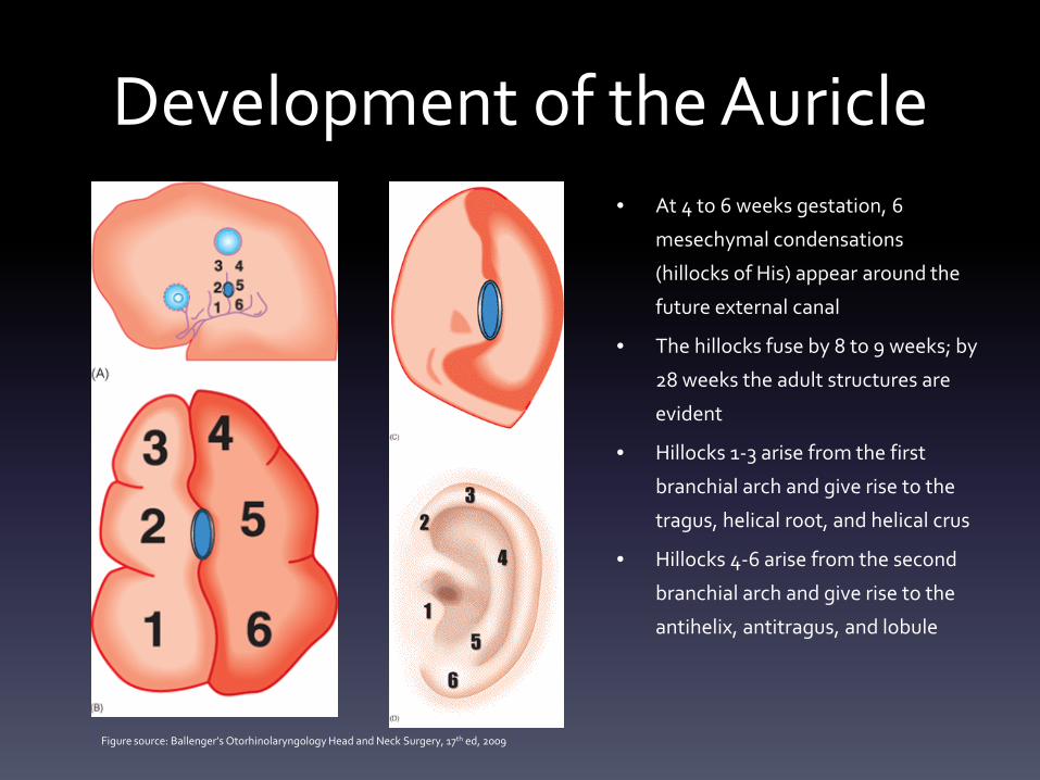
Development of the Auricle• At 4 to 6 weeks gestation, 6
mesechymal condensations
(hillocks of His) appear around the
future external canal
• The hillocks fuse by 8 to 9 weeks; by
28 weeks the adult structures are
evident
• Hillocks 1-3 arise from the first
branchial arch and give rise to the
tragus, helical root, and helical crus
• Hillocks 4-6 arise from the second
branchial arch and give rise to the
antihelix, antitragus, and lobule
Figure source: Ballenger’s Otorhinolaryngology Head and Neck Surgery, 17th ed, 2009
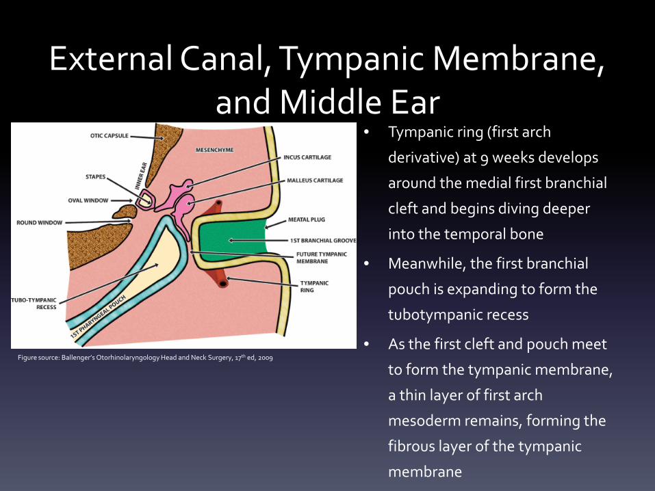
External Canal, Tympanic Membrane, and Middle Ear
• Tympanic ring (first arch
derivative) at 9 weeks develops
around the medial first branchial
cleft and begins diving deeper
into the temporal bone
• Meanwhile, the first branchial
pouch is expanding to form the
tubotympanic recess
• As the first cleft and pouch meet
to form the tympanic membrane,
a thin layer of first arch
mesoderm remains, forming the
fibrous layer of the tympanic
membrane
Figure source: Ballenger’s Otorhinolaryngology Head and Neck Surgery, 17th ed, 2009
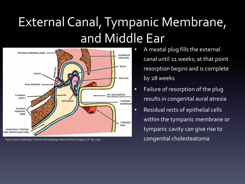
External Canal, Tympanic Membrane, and Middle Ear
• A meatal plug fills the external
canal until 21 weeks; at that point
resorption begins and is complete
by 28 weeks
• Failure of resorption of the plug
results in congenital aural atresia
• Residual rests of epithelial cells
within the tympanic membrane or
tympanic cavity can give rise to
congenital cholesteatomaFigure source: Ballenger’s Otorhinolaryngology Head and Neck Surgery, 17th ed, 2009
Notable Anomalies of the External and Middle Ear
• Fissures of Santorini (cartilaginous EAC) and foramina of Huschke (bony EAC)
– Connect the EAC to the parotid gland
– Can lead to spread of infection/tumor from one location to the other
• Hyrtl fissure
– Connects the middle ear space to the meninges; possible route for intracranial spread of
otitis media
• Dehiscent facial nerve
– Tympanic segment
– 30 to 50% of patients
– Increases the likelihood for facial paralysis from otitis media or middle ear surgery
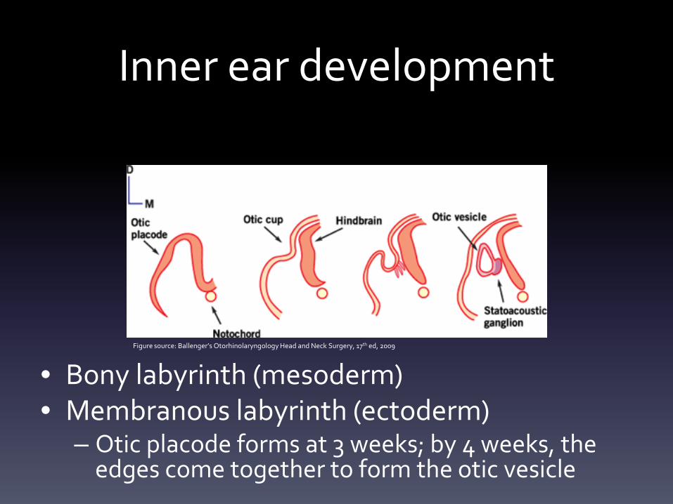
Inner ear development
• Bony labyrinth (mesoderm)• Membranous labyrinth (ectoderm)
– Otic placode forms at 3 weeks; by 4 weeks, the edges come together to form the otic vesicle
Figure source: Ballenger’s Otorhinolaryngology Head and Neck Surgery, 17th ed, 2009
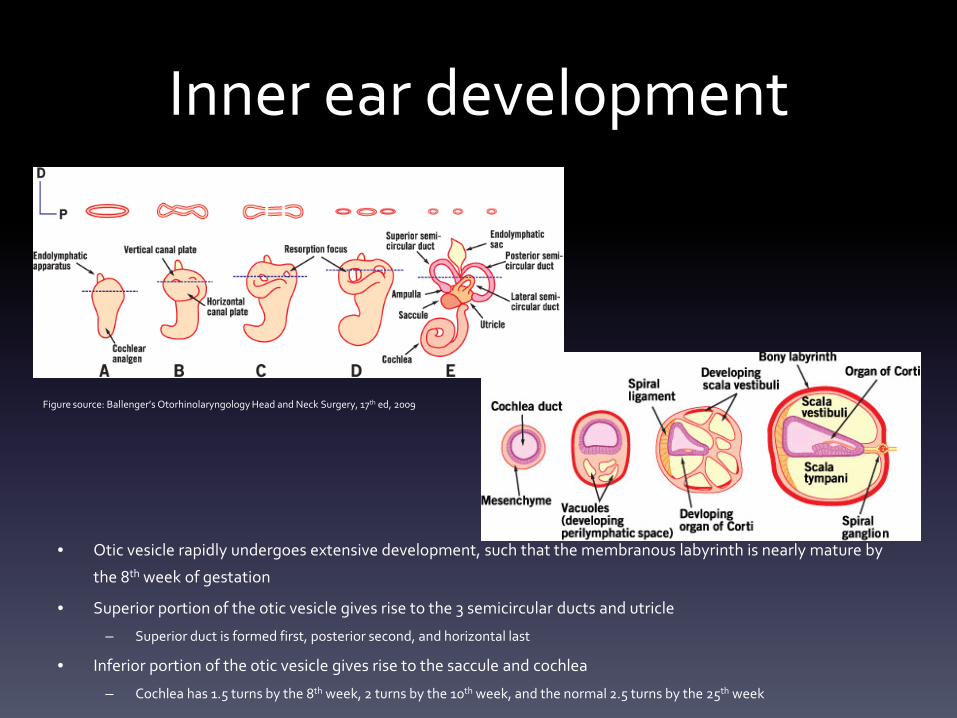
Inner ear development
• Otic vesicle rapidly undergoes extensive development, such that the membranous labyrinth is nearly mature by
the 8th week of gestation
• Superior portion of the otic vesicle gives rise to the 3 semicircular ducts and utricle
– Superior duct is formed first, posterior second, and horizontal last
• Inferior portion of the otic vesicle gives rise to the saccule and cochlea
– Cochlea has 1.5 turns by the 8th week, 2 turns by the 10th week, and the normal 2.5 turns by the 25th week
Figure source: Ballenger’s Otorhinolaryngology Head and Neck Surgery, 17th ed, 2009
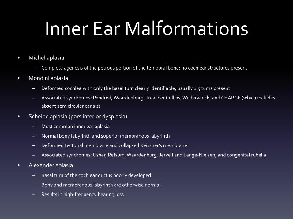
Inner Ear Malformations• Michel aplasia
– Complete agenesis of the petrous portion of the temporal bone; no cochlear structures present
• Mondini aplasia
– Deformed cochlea with only the basal turn clearly identifiable; usually 1.5 turns present
– Associated syndromes: Pendred, Waardenburg, Treacher Collins, Wildervanck, and CHARGE (which includes
absent semicircular canals)
• Scheibe aplasia (pars inferior dysplasia)
– Most common inner ear aplasia
– Normal bony labyrinth and superior membranous labyrinth
– Deformed tectorial membrane and collapsed Reissner’s membrane
– Associated syndromes: Usher, Refsum, Waardenburg, Jervell and Lange-Nielsen, and congenital rubella
• Alexander aplasia
– Basal turn of the cochlear duct is poorly developed
– Bony and membranous labyrinth are otherwise normal
– Results in high-frequency hearing loss
NASAL, LIP, AND FACIAL DEVELOPMENT
A newborn is discovered to have a unilateral cleft lip. This was caused by failure of fusion of which of the
following pairs of embryonic structures?
1. Medial nasal process and maxillary prominence
2. Lateral nasal process and maxillary prominence
3. Medial nasal process and lateral nasal process
4. Palatine shelf and medial nasal process
(Answer on next slide)
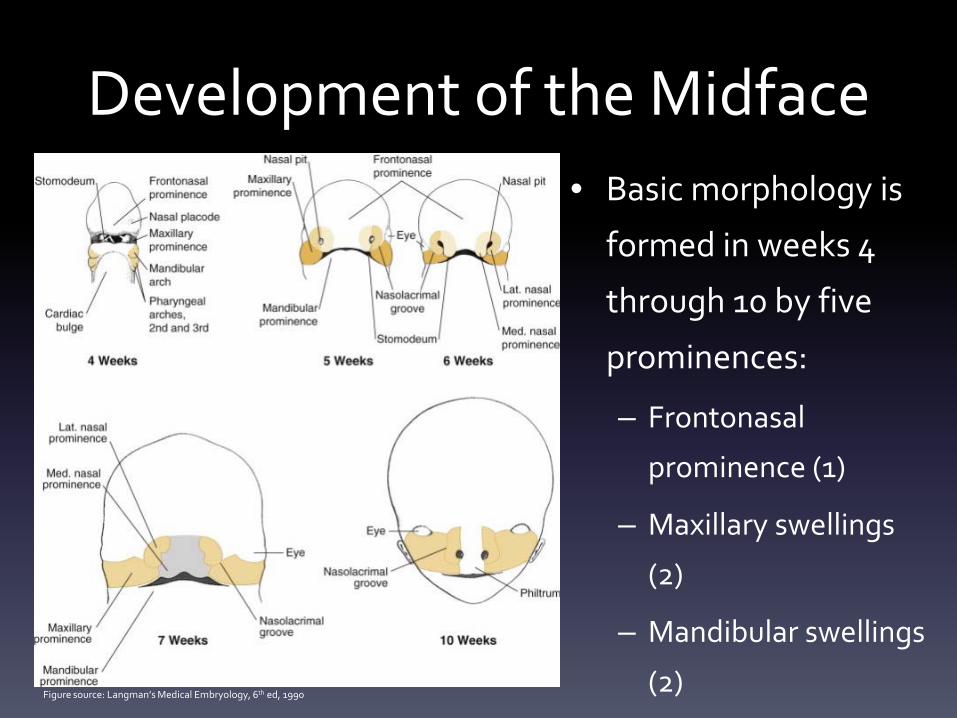
Development of the Midface• Basic morphology is
formed in weeks 4
through 10 by five
prominences:
– Frontonasal
prominence (1)
– Maxillary swellings
(2)
– Mandibular swellings
(2)Figure source: Langman’s Medical Embryology, 6th ed, 1990
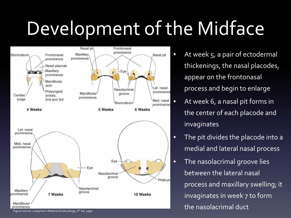
Development of the Midface• At week 5, a pair of ectodermal
thickenings, the nasal placodes,
appear on the frontonasal
process and begin to enlarge
• At week 6, a nasal pit forms in
the center of each placode and
invaginates
• The pit divides the placode into a
medial and lateral nasal process
• The nasolacrimal groove lies
between the lateral nasal
process and maxillary swelling; it
invaginates in week 7 to form
the nasolacrimal ductFigure source: Langman’s Medical Embryology, 6th ed, 1990
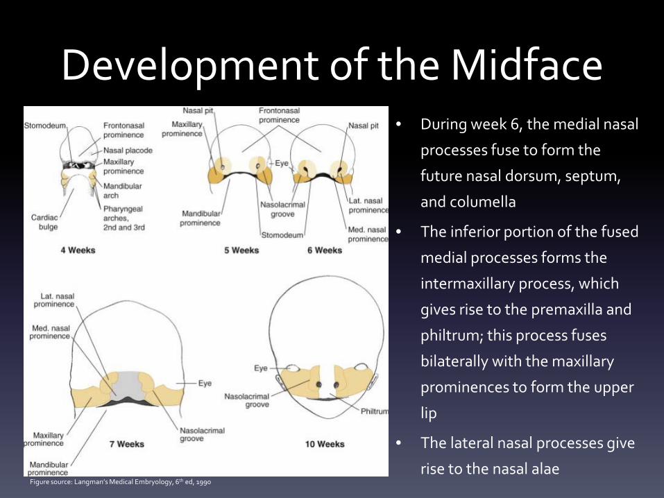
Development of the Midface• During week 6, the medial nasal
processes fuse to form the
future nasal dorsum, septum,
and columella
• The inferior portion of the fused
medial processes forms the
intermaxillary process, which
gives rise to the premaxilla and
philtrum; this process fuses
bilaterally with the maxillary
prominences to form the upper
lip
• The lateral nasal processes give
rise to the nasal alaeFigure source: Langman’s Medical Embryology, 6th ed, 1990
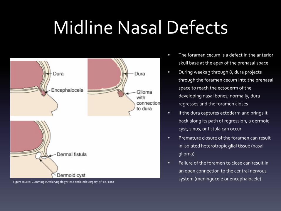
Midline Nasal Defects• The foramen cecum is a defect in the anterior
skull base at the apex of the prenasal space
• During weeks 3 through 8, dura projects
through the foramen cecum into the prenasal
space to reach the ectoderm of the
developing nasal bones; normally, dura
regresses and the foramen closes
• If the dura captures ectoderm and brings it
back along its path of regression, a dermoid
cyst, sinus, or fistula can occur
• Premature closure of the foramen can result
in isolated heterotropic glial tissue (nasal
glioma)
• Failure of the foramen to close can result in
an open connection to the central nervous
system (meningocele or encephalocele)Figure source: Cummings Otolaryngology Head and Neck Surgery, 5th ed, 2010
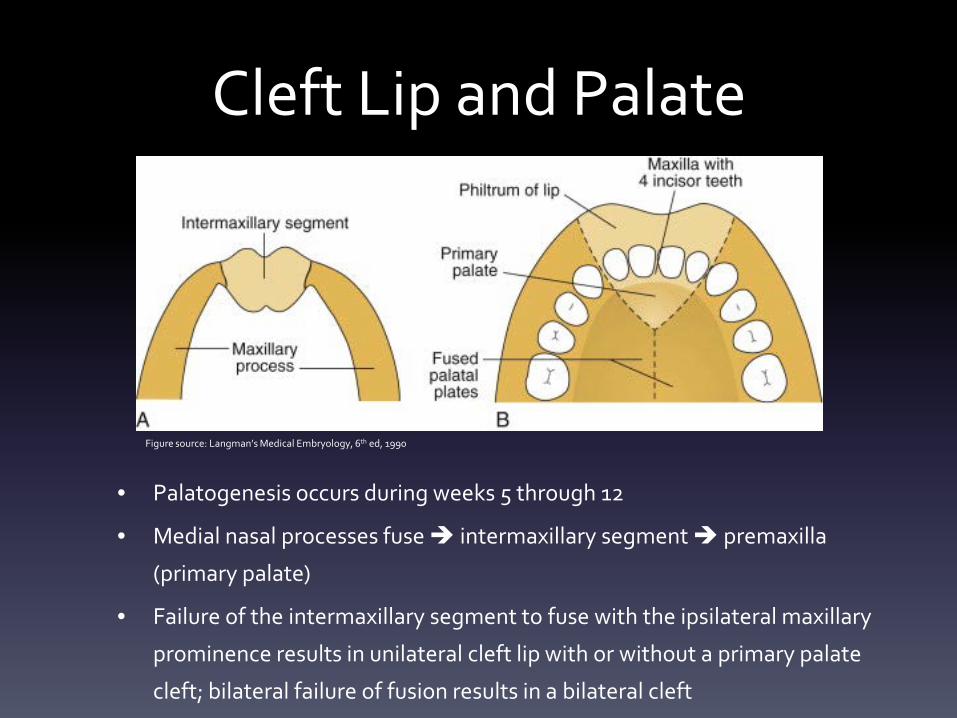
Cleft Lip and Palate
• Palatogenesis occurs during weeks 5 through 12
• Medial nasal processes fuse intermaxillary segment premaxilla
(primary palate)
• Failure of the intermaxillary segment to fuse with the ipsilateral maxillary
prominence results in unilateral cleft lip with or without a primary palate
cleft; bilateral failure of fusion results in a bilateral cleft
Figure source: Langman’s Medical Embryology, 6th ed, 1990
Cleft Lip and Palate
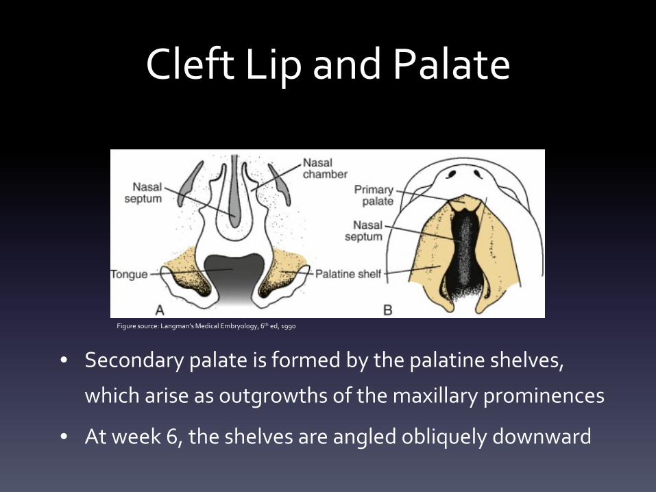
• Secondary palate is formed by the palatine shelves,
which arise as outgrowths of the maxillary prominences
• At week 6, the shelves are angled obliquely downward
Figure source: Langman’s Medical Embryology, 6th ed, 1990
Cleft Lip and Palate
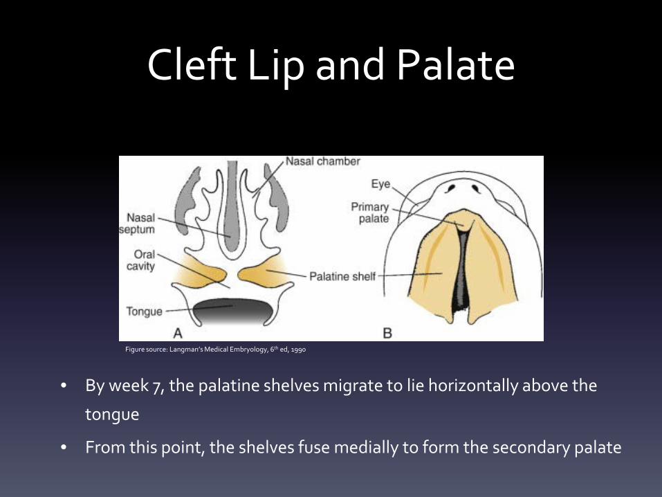
• By week 7, the palatine shelves migrate to lie horizontally above the
tongue
• From this point, the shelves fuse medially to form the secondary palate
Figure source: Langman’s Medical Embryology, 6th ed, 1990
Cleft Lip and Palate
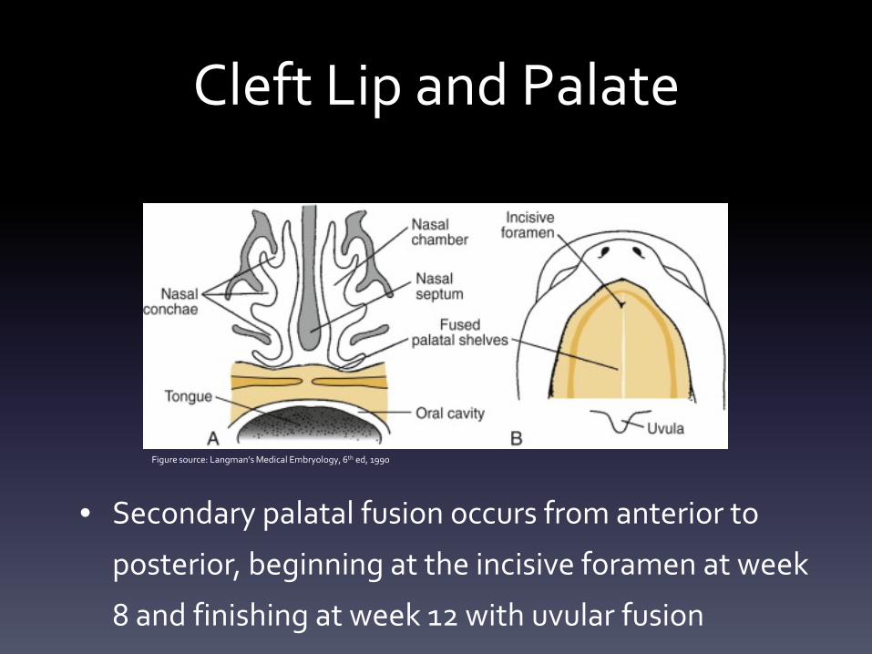
• Secondary palatal fusion occurs from anterior to
posterior, beginning at the incisive foramen at week
8 and finishing at week 12 with uvular fusion
Figure source: Langman’s Medical Embryology, 6th ed, 1990
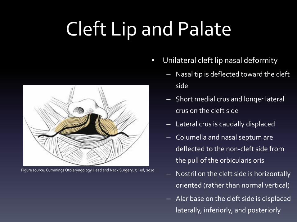
Cleft Lip and Palate• Unilateral cleft lip nasal deformity
– Nasal tip is deflected toward the cleft
side
– Short medial crus and longer lateral
crus on the cleft side
– Lateral crus is caudally displaced
– Columella and nasal septum are
deflected to the non-cleft side from
the pull of the orbicularis oris
– Nostril on the cleft side is horizontally
oriented (rather than normal vertical)
– Alar base on the cleft side is displaced
laterally, inferiorly, and posteriorly
Figure source: Cummings Otolaryngology Head and Neck Surgery, 5th ed, 2010
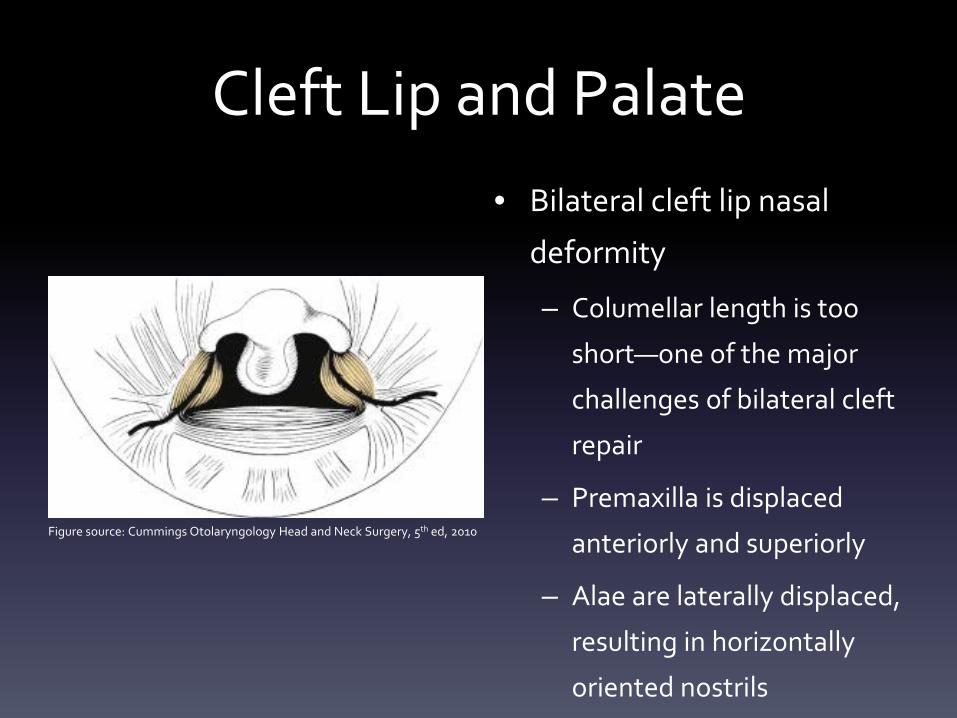
Cleft Lip and Palate• Bilateral cleft lip nasal
deformity
– Columellar length is too
short—one of the major
challenges of bilateral cleft
repair
– Premaxilla is displaced
anteriorly and superiorly
– Alae are laterally displaced,
resulting in horizontally
oriented nostrils
Figure source: Cummings Otolaryngology Head and Neck Surgery, 5th ed, 2010
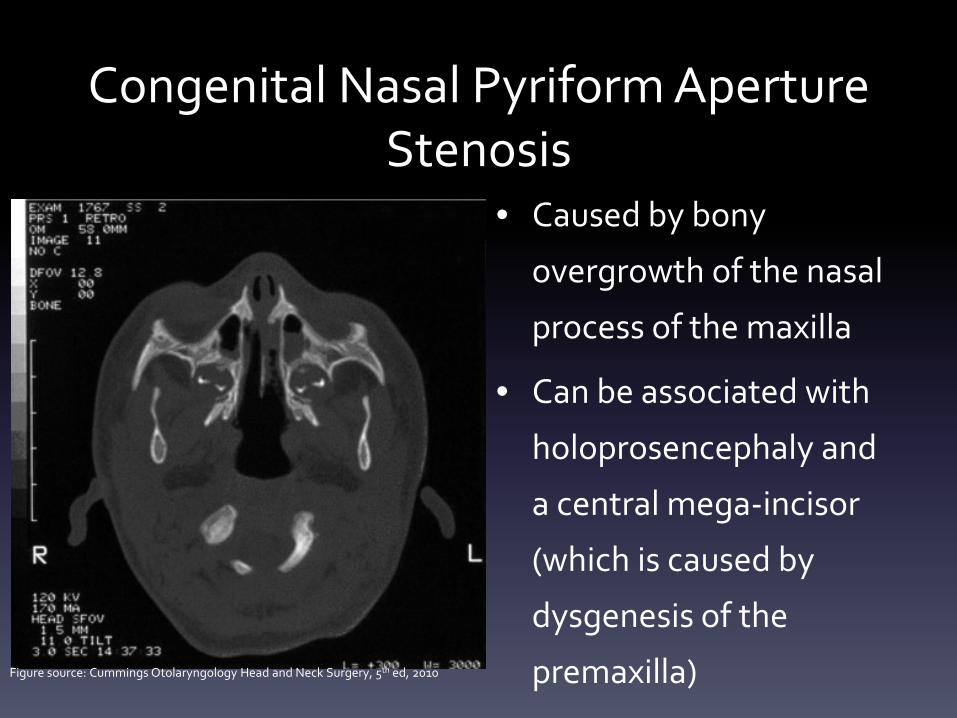
Congenital Nasal Pyriform Aperture Stenosis
• Caused by bony
overgrowth of the nasal
process of the maxilla
• Can be associated with
holoprosencephaly and
a central mega-incisor
(which is caused by
dysgenesis of the
premaxilla)Figure source: Cummings Otolaryngology Head and Neck Surgery, 5th ed, 2010
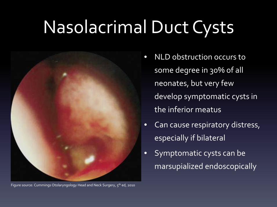
Nasolacrimal Duct Cysts• NLD obstruction occurs to
some degree in 30% of all
neonates, but very few
develop symptomatic cysts in
the inferior meatus
• Can cause respiratory distress,
especially if bilateral
• Symptomatic cysts can be
marsupialized endoscopically
Figure source: Cummings Otolaryngology Head and Neck Surgery, 5th ed, 2010
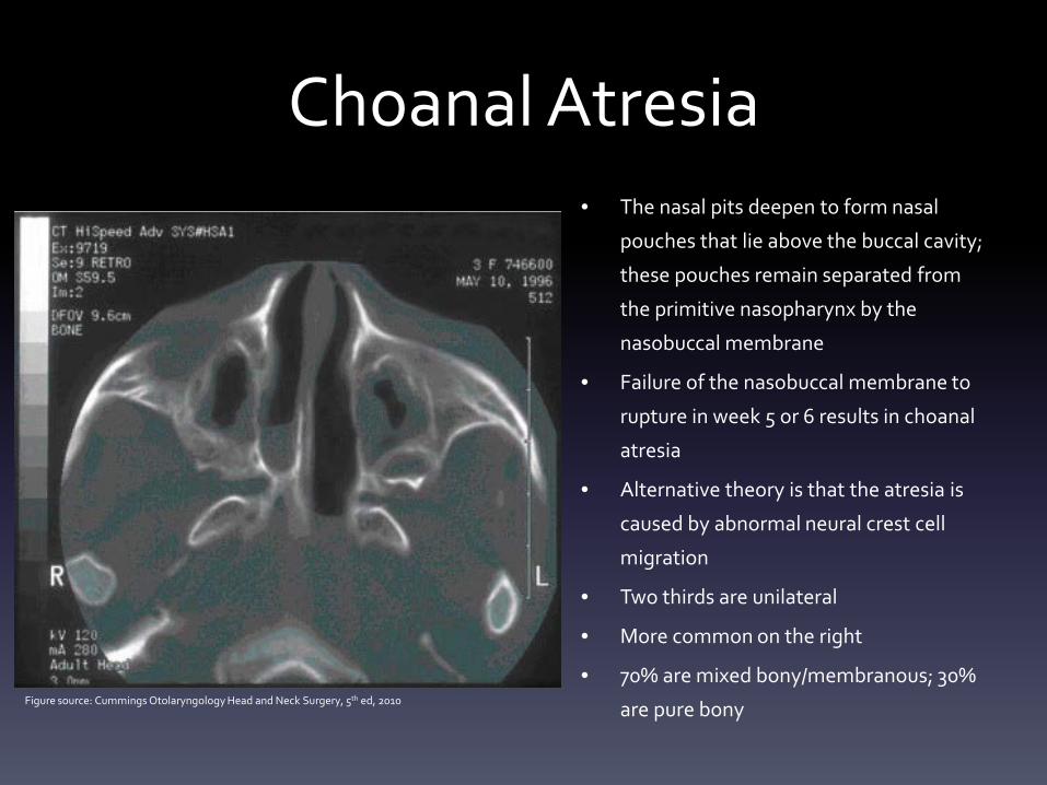
Choanal Atresia• The nasal pits deepen to form nasal
pouches that lie above the buccal cavity;
these pouches remain separated from
the primitive nasopharynx by the
nasobuccal membrane
• Failure of the nasobuccal membrane to
rupture in week 5 or 6 results in choanal
atresia
• Alternative theory is that the atresia is
caused by abnormal neural crest cell
migration
• Two thirds are unilateral
• More common on the right
• 70% are mixed bony/membranous; 30%
are pure bonyFigure source: Cummings Otolaryngology Head and Neck Surgery, 5th ed, 2010
THYROID DEVELOPMENT
The most common site of congenital ectopic thyroid tissue
is:1. Anterior neck
2. Mediastinum
3. Tongue
4. Thyroglossal duct cyst
(Answer on next slide)
Thyroid Development
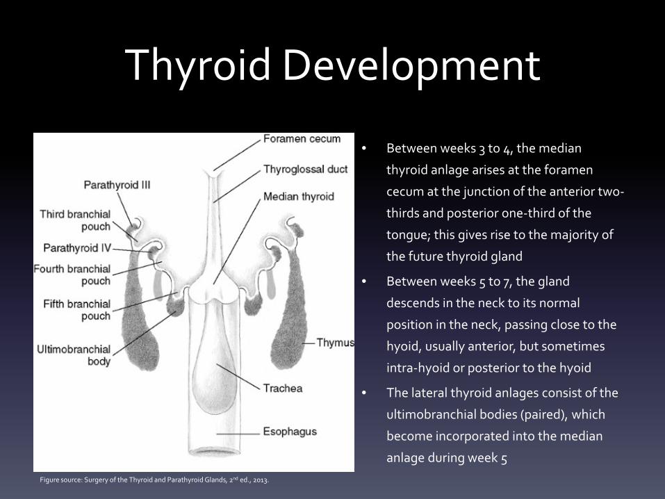
• Between weeks 3 to 4, the median
thyroid anlage arises at the foramen
cecum at the junction of the anterior two-
thirds and posterior one-third of the
tongue; this gives rise to the majority of
the future thyroid gland
• Between weeks 5 to 7, the gland
descends in the neck to its normal
position in the neck, passing close to the
hyoid, usually anterior, but sometimes
intra-hyoid or posterior to the hyoid
• The lateral thyroid anlages consist of the
ultimobranchial bodies (paired), which
become incorporated into the median
anlage during week 5Figure source: Surgery of the Thyroid and Parathyroid Glands, 2nd ed., 2013.
Ectopic Thyroid Tissue
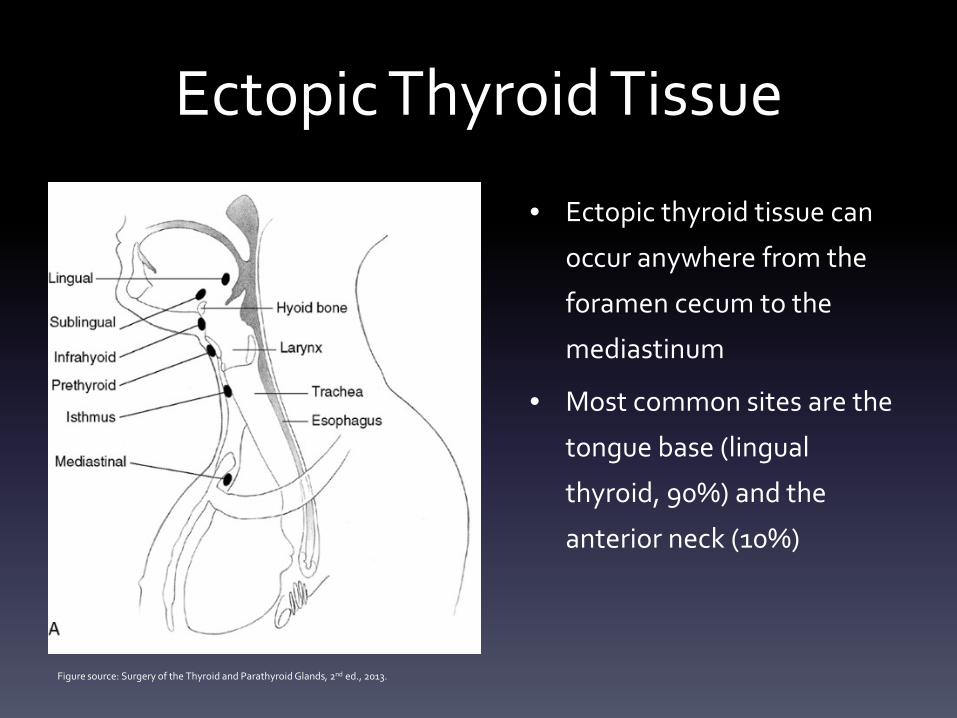
• Ectopic thyroid tissue can
occur anywhere from the
foramen cecum to the
mediastinum
• Most common sites are the
tongue base (lingual
thyroid, 90%) and the
anterior neck (10%)
Figure source: Surgery of the Thyroid and Parathyroid Glands, 2nd ed., 2013.
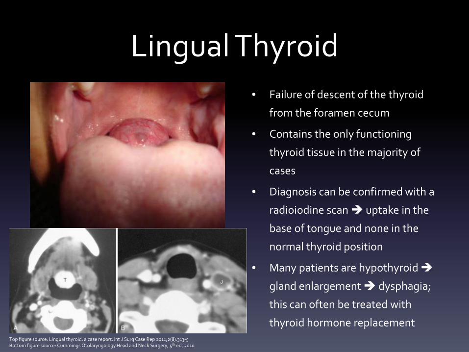
Lingual Thyroid• Failure of descent of the thyroid
from the foramen cecum
• Contains the only functioning
thyroid tissue in the majority of
cases
• Diagnosis can be confirmed with a
radioiodine scan uptake in the
base of tongue and none in the
normal thyroid position
• Many patients are hypothyroid
gland enlargement dysphagia;
this can often be treated with
thyroid hormone replacement Top figure source: Lingual thyroid: a case report. Int J Surg Case Rep 2011;2(8):313-5Bottom figure source: Cummings Otolaryngology Head and Neck Surgery, 5th ed, 2010
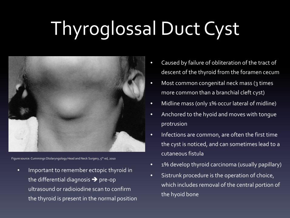
Thyroglossal Duct Cyst
• Important to remember ectopic thyroid in
the differential diagnosis pre-op
ultrasound or radioiodine scan to confirm
the thyroid is present in the normal position
• Caused by failure of obliteration of the tract of
descent of the thyroid from the foramen cecum
• Most common congenital neck mass (3 times
more common than a branchial cleft cyst)
• Midline mass (only 1% occur lateral of midline)
• Anchored to the hyoid and moves with tongue
protrusion
• Infections are common, are often the first time
the cyst is noticed, and can sometimes lead to a
cutaneous fistula
• 1% develop thyroid carcinoma (usually papillary)
• Sistrunk procedure is the operation of choice,
which includes removal of the central portion of
the hyoid bone
Figure source: Cummings Otolaryngology Head and Neck Surgery, 5th ed, 2010
LARYNGEAL DEVELOPMENT
What is the correct order, from most frequent to least frequent, for causes of stridor in an infant?
1. Laryngomalacia, subglottic stenosis, vocal fold paralysis, laryngeal web
2. Vocal fold paralysis, laryngomalacia, sublgottic stenosis, laryngeal web
3. Subglottic stenosis, laryngomalacia, laryngeal web, vocal fold paralysis
4. Laryngomalacia, vocal fold paralysis, subglottic stenosis, laryngeal web
5. Laryngomalacia, laryngeal web, subglottic stenosis, vocal fold paralysis
(Answer on next slide)
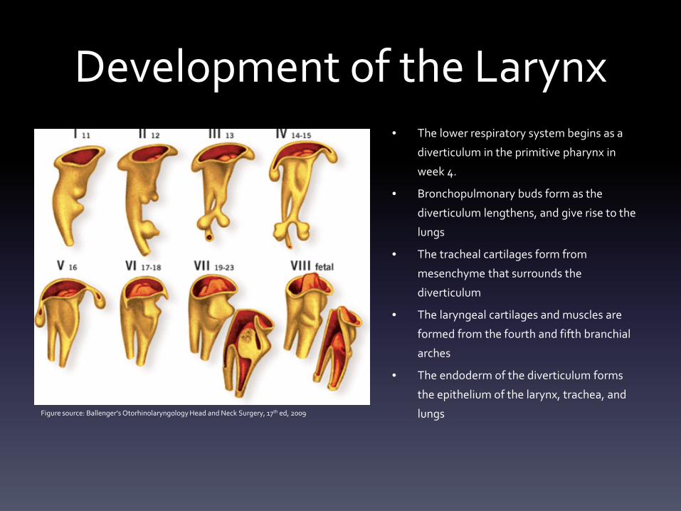
Development of the Larynx• The lower respiratory system begins as a
diverticulum in the primitive pharynx in
week 4.
• Bronchopulmonary buds form as the
diverticulum lengthens, and give rise to the
lungs
• The tracheal cartilages form from
mesenchyme that surrounds the
diverticulum
• The laryngeal cartilages and muscles are
formed from the fourth and fifth branchial
arches
• The endoderm of the diverticulum forms
the epithelium of the larynx, trachea, and
lungsFigure source: Ballenger’s Otorhinolaryngology Head and Neck Surgery, 17th ed, 2009
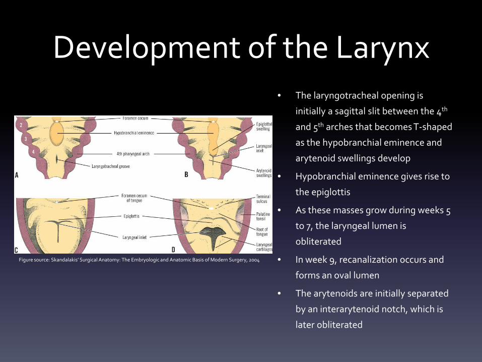
Development of the Larynx• The laryngotracheal opening is
initially a sagittal slit between the 4th
and 5th arches that becomes T-shaped
as the hypobranchial eminence and
arytenoid swellings develop
• Hypobranchial eminence gives rise to
the epiglottis
• As these masses grow during weeks 5
to 7, the laryngeal lumen is
obliterated
• In week 9, recanalization occurs and
forms an oval lumen
• The arytenoids are initially separated
by an interarytenoid notch, which is
later obliterated
Figure source: Skandalakis’ Surgical Anatomy: The Embryologic and Anatomic Basis of Modern Surgery, 2004
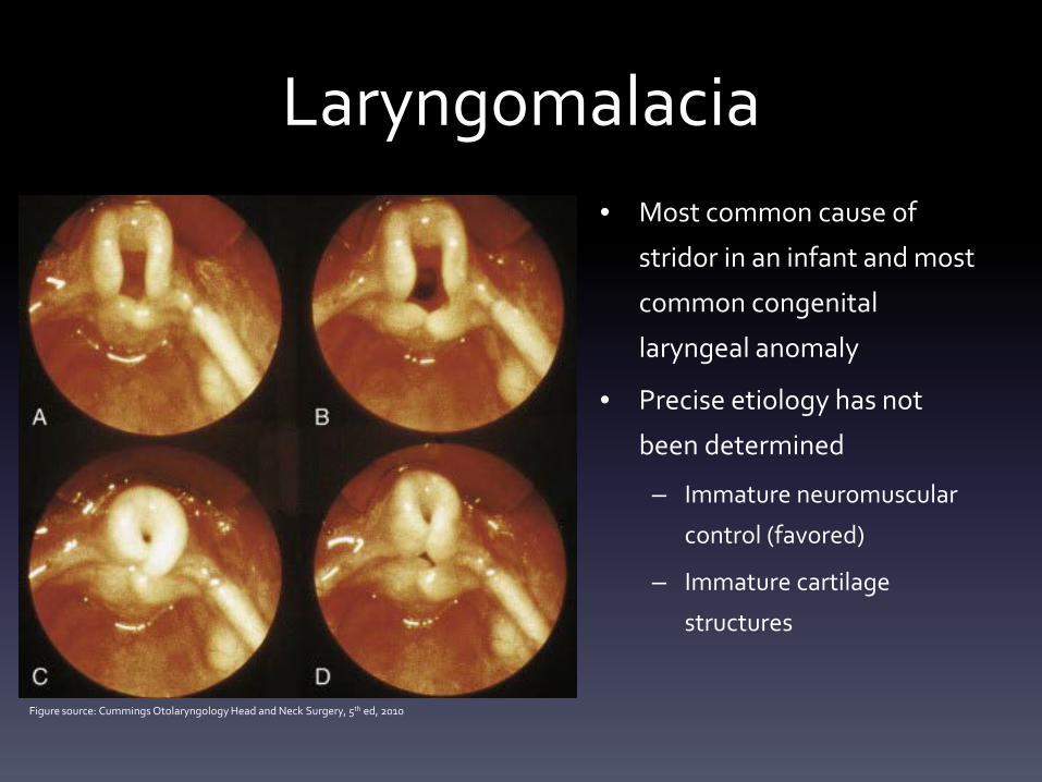
Laryngomalacia• Most common cause of
stridor in an infant and most
common congenital
laryngeal anomaly
• Precise etiology has not
been determined
– Immature neuromuscular
control (favored)
– Immature cartilage
structures
Figure source: Cummings Otolaryngology Head and Neck Surgery, 5th ed, 2010
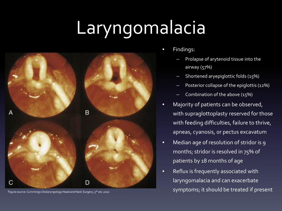
Laryngomalacia• Findings:
– Prolapse of arytenoid tissue into the
airway (57%)
– Shortened aryepiglottic folds (15%)
– Posterior collapse of the epiglottis (12%)
– Combination of the above (15%)
• Majority of patients can be observed,
with supraglottoplasty reserved for those
with feeding difficulties, failure to thrive,
apneas, cyanosis, or pectus excavatum
• Median age of resolution of stridor is 9
months; stridor is resolved in 75% of
patients by 18 months of age
• Reflux is frequently associated with
laryngomalacia and can exacerbate
symptoms; it should be treated if presentFigure source: Cummings Otolaryngology Head and Neck Surgery, 5th ed, 2010
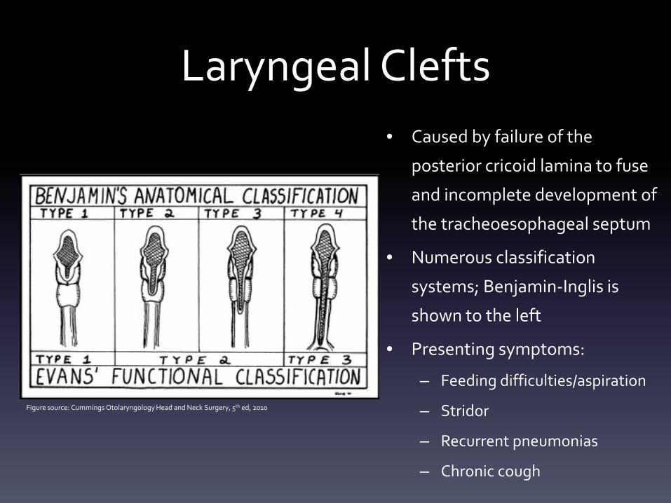
Laryngeal Clefts• Caused by failure of the
posterior cricoid lamina to fuse
and incomplete development of
the tracheoesophageal septum
• Numerous classification
systems; Benjamin-Inglis is
shown to the left
• Presenting symptoms:
– Feeding difficulties/aspiration
– Stridor
– Recurrent pneumonias
– Chronic cough
Figure source: Cummings Otolaryngology Head and Neck Surgery, 5th ed, 2010
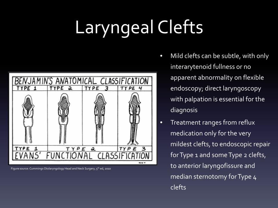
Laryngeal Clefts• Mild clefts can be subtle, with only
interarytenoid fullness or no
apparent abnormality on flexible
endoscopy; direct laryngoscopy
with palpation is essential for the
diagnosis
• Treatment ranges from reflux
medication only for the very
mildest clefts, to endoscopic repair
for Type 1 and some Type 2 clefts,
to anterior laryngofissure and
median sternotomy for Type 4
clefts
Figure source: Cummings Otolaryngology Head and Neck Surgery, 5th ed, 2010
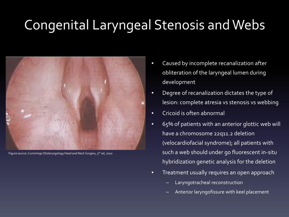
Congenital Laryngeal Stenosis and Webs
• Caused by incomplete recanalization after
obliteration of the laryngeal lumen during
development
• Degree of recanalization dictates the type of
lesion: complete atresia vs stenosis vs webbing
• Cricoid is often abnormal
• 65% of patients with an anterior glottic web will
have a chromosome 22q11.2 deletion
(velocardiofacial syndrome); all patients with
such a web should under go fluorescent in-situ
hybridization genetic analysis for the deletion
• Treatment usually requires an open approach
– Laryngotracheal reconstruction
– Anterior laryngofissure with keel placement
Figure source: Cummings Otolaryngology Head and Neck Surgery, 5th ed, 2010
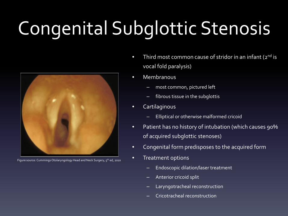
Congenital Subglottic Stenosis• Third most common cause of stridor in an infant (2nd is
vocal fold paralysis)
• Membranous
– most common, pictured left
– fibrous tissue in the subglottis
• Cartilaginous
– Elliptical or otherwise malformed cricoid
• Patient has no history of intubation (which causes 90%
of acquired subglottic stenoses)
• Congenital form predisposes to the acquired form
• Treatment options
– Endoscopic dilation/laser treatment
– Anterior cricoid split
– Laryngotracheal reconstruction
– Cricotracheal reconstruction
Figure source: Cummings Otolaryngology Head and Neck Surgery, 5th ed, 2010
Conclusions• This is just the tip of the iceberg—head and neck
development from embryo to birth is intricately complex
• Anomalies during development—including lack of fusion of
two structures, persistence of a structure that should
regress, trapping of one tissue within another, or failure of
a vital structure to form—underlie the majority of the
congenital lesions in the head and neck
• Knowledge of the normal developmental pathways and
the manner in which anomalies arise is essential for any
surgical treatment of congenital lesions
References1. Graney DO, Sie KCY. Anatomy and developmental embryology of the neck. In: Flint PW, Haughey BH, Lund VJ, et al, editors. Cummings Otolaryngology—Head and Neck Surgery.
5th edition. Philadelphia: Mosby Elsevier; 2010. p. 2577–86.
2. Larsen WJ. Development of the Head and Neck. Human Embryology. 3rd edition. Philadelphia: Churchill Livingstone; 2001. p. 351-78.
3. Farrior J, Lee KJ. Embryology of clefts and pouches. In: Lee KJ, editor. Essential Otolaryngology Head and Neck Surgery. 10th edition. New York: McGraw-Hill; 2012. p. 269-84.
4. Mukherji SK, Fatterpekar G, Castillo M, et al. Imaging of congenital anomalies of the branchial apparatus. Neuroimaging Clin N Am 2000;10:75-93.
5. Chandler JR, Mitchell B. Branchial cleft cysts, sinuses, and fistulas. Otolaryngol Clin North Am 1981;14:175-86.
6. Rowe LD. Congenital anomalies of the head and neck. In: Snow JB, Wackym PA, editors. Ballenger’s Otorhinolaryngology Head and Neck Surgery. 17th edition. Shelton, Connecticut:
People’s Medical Publishing House/BC Decker; 2009. p. 829-38.
7. Choo DI, Richter GT. Development of the ear. In: Snow JB, Wackym PA, editors. Ballenger’s Otorhinolaryngology Head and Neck Surgery. 17th edition. Shelton, Connecticut: People’s
Medical Publishing House/BC Decker; 2009. p. 17-27.
8. Sadler TW. Head and neck embryology. In: Sadler TW, editor. Langman’s Medical Embryology. 6th edition. Baltimore: Williams & Wilkins; 1990:315-8.
9. Elluru RG, Wootten CT. Congenital malformations of the nose. In: Flint PW, Haughey BH, Lund VJ, et al, editors. Cummings Otolaryngology—Head and Neck Surgery. 5th edition.
Philadelphia: Mosby Elsevier; 2010.
10. Friedman O, Wang TD, Milczuk HA. Cleft lip and palate. In: Flint PW, Haughey BH, Lund VJ, et al, editors. Cummings Otolaryngology—Head and Neck Surgery. 5th edition.
Philadelphia: Mosby Elsevier; 2010. p. 2659-75.
11. Agarwal A, Mishra AK, Lombardi CP, Raffaelli M. Applied embryology of the thyroid and parathyroid glands. In: Randolph GW, ed. Surgery of the Thyroid and Parathyroid Glands. 2nd
edition. Philadelphia: Elsevier Saunders; 2013.
12. Amr B, Monib S. Lingual thyroid: a case report. Int J Surg Case Rep 2011;2(8):313-5
13. Aygun N, Zinreich SJ. Overview of diagnostic imaging of the head and neck. In: Flint PW, Haughey BH, Lund VJ, et al, editors. Cummings Otolaryngology—Head and Neck Surgery.
5th edition. Philadelphia: Mosby Elsevier; 2010.
14. Wetmore RF, Potsic WP. Differential diagnosis of neck masses. In: Flint PW, Haughey BH, Lund VJ, et al, editors. Cummings Otolaryngology—Head and Neck Surgery. 5th edition.
Philadelphia: Mosby Elsevier; 2010.
15. Sasaki CT, Kim YH, LeVay AJ. Development, anatomy, and physiology of the larynx. .In: Snow JB, Wackym PA, editors. Ballenger’s Otorhinolaryngology Head and Neck Surgery. 17th
edition. Shelton, Connecticut: People’s Medical Publishing House/BC Decker; 2009.
16. Messner AH. Congenital disorders of the larynx. In: Flint PW, Haughey BH, Lund VJ, et al, editors. Cummings Otolaryngology—Head and Neck Surgery. 5th edition. Philadelphia:
Mosby Elsevier; 2010.
17. Zalzal GH, Cotton RT. Glottic and subglottic stenosis. In: Flint PW, Haughey BH, Lund VJ, et al, editors. Cummings Otolaryngology—Head and Neck Surgery. 5th edition. Philadelphia:
Mosby Elsevier; 2010.
18. Jacobs IN, DelGaudio J, Colborn GL, et al. Larynx. In: Skandalakis’ Surgical Anatomy: The Embryologic and Anatomic Basis of Modern Surgery. Greece: PMP/Mcgraw-Hill; 2004.


![Embryology Head and Neck[1]](https://img.pdfslide.us/doc/110x75/55cf8541550346484b8c049f/embryology-head-and-neck1.jpg)