Embed Size (px)
Citation preview
The Plant Cell, Vol. 9, 989-1 000, July 1997 O 1997 American Society of Plant Physiologists
Embryogenesis: A New Start in Life
Thomas Laux and Gerd Jürgens' Lehrstuhl für Entwicklungsgenetik, Universitat Tübingen, Spemannstrasse 37-39, D-72076 Tübingen, Germany
INTRODUCTION
Embryogenesis effects the transition from the fertilized egg to the new multicellular generation, the seedling, which dis- plays the basic body plan and organization of the plant. An apical-basal pattern along the main body axis of the embryo consists of a linear array of distinct elements, including the shoot meristem, cotyledons, hypocotyl, root, and root meri- stem. A radial pattern around the apical-basal axis is repre- sented by the concentric arrangement of the primary tissues: epidermis at the periphery, ground tissue underneath, and conductive tissue in the center. During postembryonic devel- opment, the two primary meristems give rise to the elaborate structures of the adult plant (see Clark, 1997; Kerstetter and Hake, 1997; Schiefelbein et al., 1997, in this issue).
The basic body plan is established within the first one- third of embryogenesis and becomes fully apparent by the time dicot embryos reach the heart stage. Subsequent events include further growth of the embryo, morphogenesis, activity of the primary meristems, cell differentiation, and preparation of both embryo and seed for dormancy. Previous reviews have covered various aspects of embryogenesis, including the formation of embryo initials (Mordhorst et al., 1997), fer- tilization (Russell, 1993), endosperm development (Lopes and Larkins, 1993), somatic embryogenesis (Zimmerman, 1993; Emons, 1994), axis formation (Jürgens, 1995), gene expres- sion (Thomas, 1993), and related topics (Goldberg et al., 1994; Laux and Jürgens, 1994; Yadegari and Goldberg, 1997).
In this review, we discuss how pattern formation gener- ates different cell fates during embryogenesis. By drawing mainly on recent genetic and molecular studies in Arabidop- sis, we first summarize what is known about the successive generation of cell fates and then discuss mechanisms that may underlie the establishment of diverse cell identities.
SUCCESSIVE GENERATION OF CELL FATES IN THE EMBRYO
The developing embryo consists of a growing population of cells, the fate of each of which must be determined in a po- sition-dependent manner to form a functional organism.
'To whom correspondence should be addressed. E-mail geju@mailer. mpib-tuebingen.mpg.de; fax 49-7071 -601 862.
Here, we discuss the events that establish the basic body plan of the Arabidopsis embryo. Where informative, we in- clude relevant data from other species. Arabidopsis embryo- genesis has been described in detail previously (Mansfield and Briarty, 1991; Jürgens and Mayer, 1994).
Setting the Stage: Formation of the Apical-Basal Axis of the Embryo
A common feature of higher plant embryos is that their api- cal-basal axes are aligned according to the chalaza-micro- pyle axis of the ovule, suggesting an orienting influence of the surrounding maternal tissue. The embryo sac, egg cell, and zygote appear polarized in many higher plant species, including Arabidopsis (Esau, 1977; Willemse and Van Went, 1984; Mansfield and Briarty, 1991; Mansfield et al., 1991). In maize, for example, the cytoplasm and nucleus are shifted toward the apical end upon fertilization of the egg cell (Mbl et al., 1994). Although somatic embryos demonstrate that apical-basal polarity can be established without maternal in- formation (Backs-Hüsemann and Reinert, 1970; Nomura and Komamine, 1985), the strict correlation between the ori- entation of the apical-basal a i s of the embryo and the struc- ture of the ovule suggests that such information could play an important role in zygotic embryogenesis.
The nature of such maternal information is unknown, al- though diffusable factors andlor physical constraints are valid possibilities. Moreover, maternal mutations affecting polarity and axis formation similar to those described in Drosophila (Johnston and Nüsslein-Volhard, 1992) have not been iden- tified in plants. By contrast to higher plants, the apical-basal axis of the embryo is not oriented relative to maternal struc- tures in the brown alga fucus, in which the free-living zygote becomes polarized in response to externa1 cues such as light (see Kropf, 1997, in this issue).
Another extra-embryonic tissue that may influence forma- tion of the apical-basal axis of the higher plant embryo is the triploid endosperm, which is initiated after the fusion of the second sperm cell with the central cell of the female gameto- phyte (Mansfield and Briarty, 1991). The roles of the en- dosperm in embryogenesis appear diverse; they include nutrition of the embryo (Lopes and Larkins, 1993) and reg- ulation of both embryo size (Hong et al., 1996) and fruit
990 The Plant Cell
development (Chad et al., 1996). However, there is no evi-dence that the endosperm plays an instructive role in embryopattern formation.
The zygote generates the embryo and the extra-embry-onic suspensor, which provides nutrients to the young em-bryo (Yeung and Sussex, 1979) and pushes it into the lumenof the embryo sac (Yeung and Meinke, 1993). Suspensorcells can initiate embryogenesis if the embryo is aborted orarrested (Gerlach-Cruse, 1969; Schwartz et al., 1994; Yadegariand Goldberg, 1997), suggesting that the embryo normallyrepresses the developmental potential of the suspensor(Marsden and Meinke, 1985; Yeung and Meinke, 1993). Intwin seeds, a suspensor cell can give rise to an additional em-bryo, although the primary embryo develops normally. Thesecondary embryo has normal or reversed apical-basal po-larity (Vernon and Meinke, 1994), raising the possibility thatthe juxtaposition of embryonic and extra-embryonic cells mayhelp to orient the apical-basal axis of the embryo. In thisview, the juxtaposition of the wild-type embryo proper andthe suspensor is instrumental in establishing the basal em-
bryo pole. By contrast, twin embryos that initiate within thesuspensor are flanked by suspensor cells on both sides, andthus, their basal pole may be established at random. Alterna-tively, the differences between the primary and secondaryembryos may simply reflect that the former originates from apolarized zygote, whereas the latter arises from a suspensorcell that lacks information directing embryo polarity.
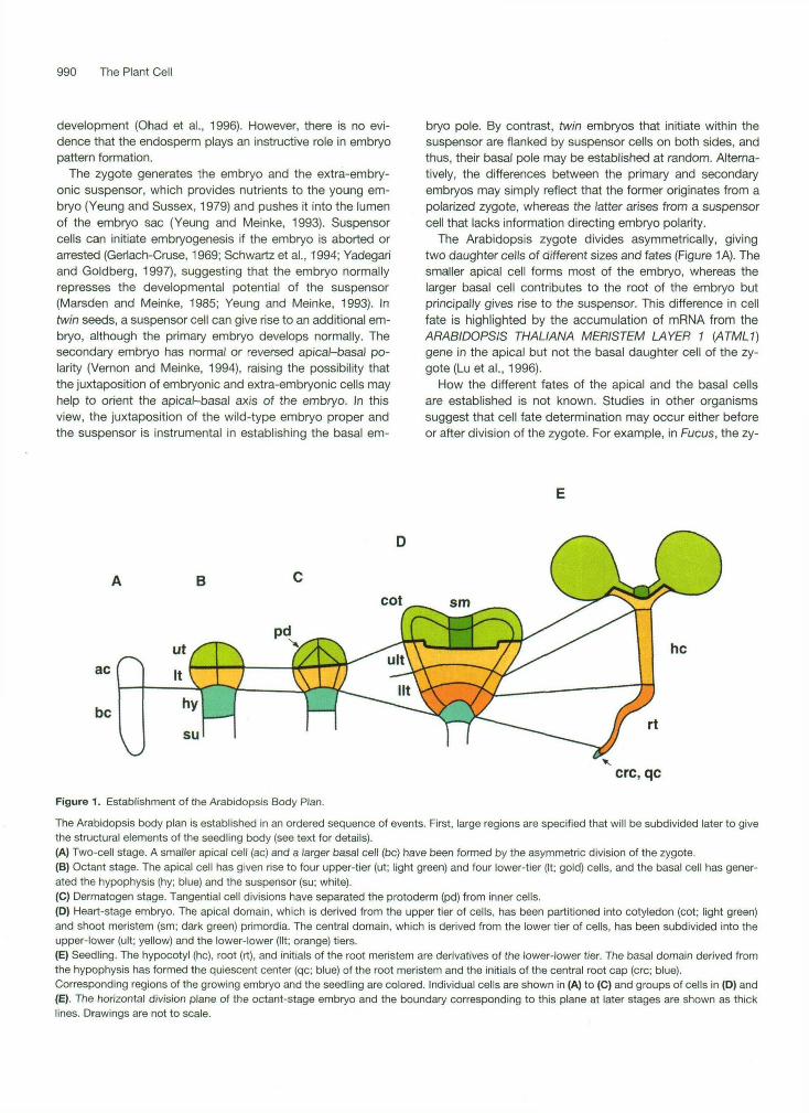
The Arabidopsis zygote divides asymmetrically, givingtwo daughter cells of different sizes and fates (Figure 1 A). Thesmaller apical cell forms most of the embryo, whereas thelarger basal cell contributes to the root of the embryo butprincipally gives rise to the suspensor. This difference in cellfate is highlighted by the accumulation of mRNA from theARABIDOPSIS THALIANA MERISTEM LAYER 1 (ATML1)gene in the apical but not the basal daughter cell of the zy-gote (Lu et al., 1996).
How the different fates of the apical and the basal cellsare established is not known. Studies in other organismssuggest that cell fate determination may occur either beforeor after division of the zygote. For example, in Fucus, the zy-
B
ac
be
\
It ^ J_hyl
su rt
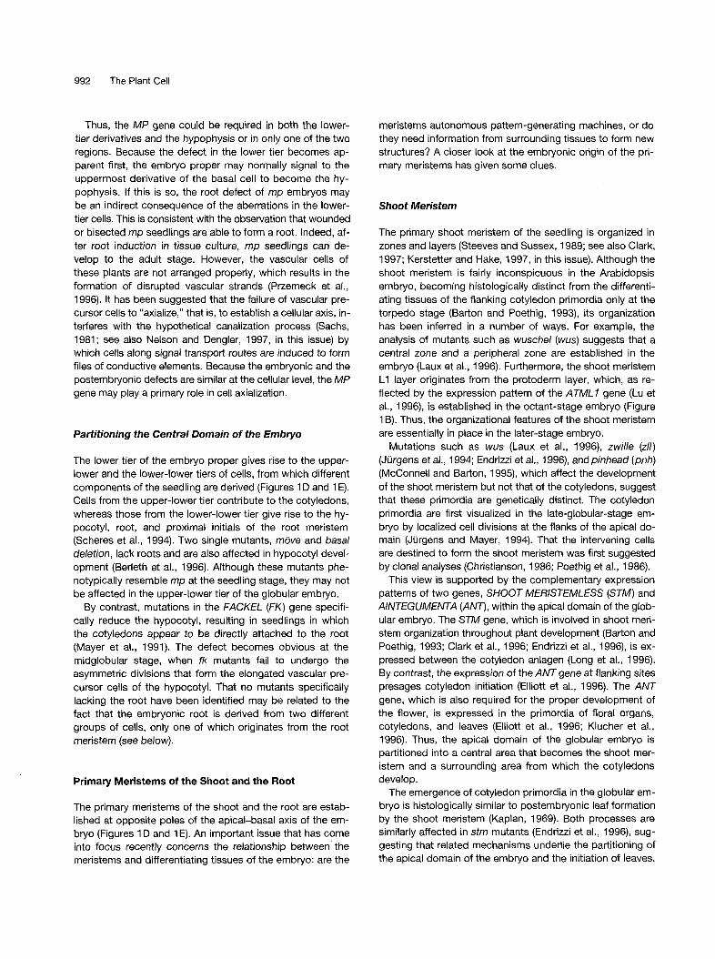
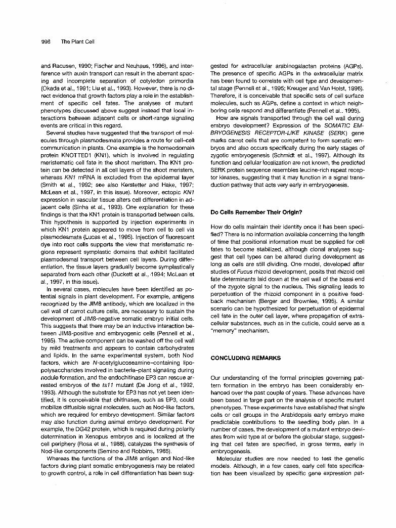
crc, qcFigure 1. Establishment of the Arabidopsis Body Plan.
The Arabidopsis body plan is established in an ordered sequence of events. First, large regions are specified that will be subdivided later to givethe structural elements of the seedling body (see text for details).(A) Two-cell stage. A smaller apical cell (ac) and a larger basal cell (be) have been formed by the asymmetric division of the zygote.(B) Octant stage. The apical cell has given rise to four upper-tier (ut; light green) and four lower-tier (It; gold) cells, and the basal cell has gener-ated the hypophysis (hy; blue) and the suspensor (su; white).(C) Dermatogen stage. Tangential cell divisions have separated the protoderm (pd) from inner cells.(D) Heart-stage embryo. The apical domain, which is derived from the upper tier of cells, has been partitioned into cotyledon (cot; light green)and shoot meristem (sm; dark green) primordia. The central domain, which is derived from the lower tier of cells, has been subdivided into theupper-lower (ult; yellow) and the lower-lower (lit; orange) tiers.(E) Seedling. The hypocotyl (he), root (rt), and initials of the root meristem are derivatives of the lower-lower tier. The basal domain derived fromthe hypophysis has formed the quiescent center (qc; blue) of the root meristem and the initials of the central root cap (crc; blue).Corresponding regions of the growing embryo and the seedling are colored. Individual cells are shown in (A) to (C) and groups of cells in (D) and(E). The horizontal division plane of the octant-stage embryo and the boundary corresponding to this plane at later stages are shown as thicklines. Drawings are not to scale.
Plant Embryogenesis 991
gote is polarized before cell division such that rhizoid cell fate determinants become associated with the cell wall at the basal pole and are inherited by the basal daughter cell (Berger et al., 1994; see also Kropf, 1997, in this issue). Asymmetric divisions of embryonic cells in the green alga Volvox give large generative and small vegetative daughter cells, and this cell fate segregation has been attributed to the postmitotic difference in cell size rather than to unequal distribution of molecules (Kirk et al., 1993). Finally, one rea- son the two daughter cells of the Arabidopsis zygote may acquire different fates in response to positional information could be because the basal but not the apical cell is attached to the surrounding maternal tissue. Whatever the underlying mechanism may be, the initial decision generates the distinc- tion between embryonic and nonembryonic cell fates.
Of the Arabidopsis embryonic pattern mutants analyzed so far, only mutations in the GNOMfEMB30 (GN) gene affect the apical-basal polarity of the embryo. The gn zygote does not elongate to the same extent as the wild-type zygote and tends to divide symmetrically (Mayer et al., 1993). Neverthe- less, the reduced basal daughter cell does give rise to a shortened suspensor, and the apical cell forms an embryo proper, suggesting that an asymmetric division of the zygote is not required to establish the fate of the two daughter cells. Cell divisions are irregular in the developing gn embryo, and the expression of the LIPlD TRANSFER PROTflN (LTP) gene, which is normally restricted to the apical end of the later stage embryo, is variable along the apical-basal axis (vroemen et al., 1996). This observation suggests that the po- larity of the gn zygote, as expressed in the different fates of its daughter cells, may not be sufficient to establish the apical- basal axis of the embryo.
The GN gene appears to be expressed throughout devel- opment (Shevell et al., 1994), and the GN protein shows se- quence similarity to two yeast proteins, Gea2p (Yec2p; Busch et al., 1996) and Gealp, which are guanine-nucleotide ex- change factors involved in vesicle transport between the en- doplasmic reticulum and the Golgi complex (Peyroche et al., 1996). These findings raise the possibility that the GN protein participates in directional vesicle transport, which may func- tion to stabilize the apical-basal axis of the embryo. Targeted vesicle fusion is also thought to play a role in axis stabilization in the Fucus embtyo (see Kropf, 1997, in this issue).
Partitioning the Apical-Basal Axis of the Embryo
The apical-basal axis of the seedling is subdivided into five major components: shoot meristem, cotyledons, hypocotyl, root, and root meristem (Figure 1 E). These components do not originate simultaneously by partitioning of the axis of the embryo but are established in steps. First, transverse cell di- visions in the four-cell embryo result in upper and lower tiers, each with four cells (Figure 1 B). The boundary between the two tiers can be followed throughout embryo develop- ment (Tykarska, 1976, 1979) and passes through the cotyle-
dons (Figure 1; Scheres et al., 1994). Whereas the upper tier gives rise to the apical domain, which comprises the shoot meristem and most of the cotyledons, the lower tier gener- ates the central domain, which contributes the “shoulder” to the cotyledons and also produces hypocotyl, root, and the proximal initials of the root meristem (Figure 1). The remain- ing parts of the root meristem, the quiescent center and the initials of the central root cap, are derived from the hypophy- sis, the uppermost derivative of the basal daughter cell of the zygote (Figure 1). Thus, the three domains established in the early embryo do not correspond to primordia of the seedling components. Nevertheless, analyses of mutant phenotypes argue that the early establishment of these do- mains plays a role in apical-basal patterning.
Proper Development of the Apical Domain Requires GURKE Activity
Mutations in the GURKE (GK) gene specifically affect the apical domain (Torres-Ruiz et al., 1996). Although cotyledon development appears to be more sensitive to the leve1 of GK activity than does that of the shoot meristem, strong gk al- leles abolish apical structures altogether and lead to the for- mation of a disorganized green mass of cells at the apical end of gk seedlings. Defects are first recognized in the api- cal domain of the heart-stage gk embryo, but defects in the central domain become obvious during later stages of em- bryogenesis. In the most extreme manifestation of the gk phenotype, the complete elimination of the cotyledons, the shoulders of which are derived from the central domain, and the reduction of the hypocotyl raise the possibility that GK is required not only in the apical but also in the central domain of the embryo. Alternatively, the reduction of the hypocotyl may be an indirect consequence of a primary defect in the apical domain, which would imply that cells from the central domain may be entrained to become incorporated into the incipient cotyledon primordia.
The MONOPTEROS Gene 1s Required in a Complementary Domain to GURKE
monopteros (mp) seedlings lack roots and hypocotyls and also display defective vascularization of the cotyledons. The earliest deviation from wild-type development is observed at the eight-cell stage, when the mp embryo proper consists of four rather than two tiers of cells (Berleth and Jürgens, 1993). Subsequently, the cells of the central domain divide abnormally and fail to produce the elongated cell files that represent the hypocotyl and root primordia of wild-type glob- ular embryos. In addition, the uppermost derivative of the basal cell, which normally would become the hypophysis and contribute to the root meristem, divides horizontally, similar to a suspensor cell, to generate a “central pile” of cells.
992 The Plant Cell
Thus, the MP gene could be required in both the lower- tier derivatives and the hypophysis or in only one of the two regions. Because the defect in the lower tier becomes ap- parent first, the embryo proper may normally signal to the uppermost derivative of the basal cell to become the hy- pophysis. If this is so, the root defect of mp embryos may be an indirect consequence of the aberrations in the lower- tier cells. This is consistent with the observation that wounded or bisected mp seedlings are able to form a root. Indeed, af- ter root induction in tissue culture, mp seedlings can de- velop to the adult stage. However, the vascular cells of these plants are not arranged properly, which results in the formation of disrupted vascular strands (Przemeck et al., 1996). It has been suggested that the failure of vascular pre- cursor cells to “axialize,” that is, to establish a cellular axis, in- terferes with the hypothetical canalization process (Sachs, 1981; see also Nelson and Dengler, 1997, in this issue) by which cells along signal transport routes are induced to form files of conductive elements. Because the embryonic and the postembryonic defects are similar at the cellular level, the MP gene may play a primary role in cell axialization.
Partitioning the Central Domain of the Embryo
The lower tier of the embryo proper gives rise to the upper- lower and the lower-lower tiers of cells, from which different components of the seedling are derived (Figures 1 D and 1 E). Cells from the upper-lower tier contribute to the cotyledons, whereas those from the lower-lower tier give rise to the hy- pocotyl, root, and proximal initials of the root meristem (Scheres et al., 1994). Two single mutants, move and basal deletion, lack roots and are also affected in hypocotyl devel- opment (Berleth et al., 1996). Although these mutants phe- notypically resemble mp at the seedling stage, they may not be affected in the upper-lower tier of the globular embryo.
By contrast, mutations in the FACKEL (FK) gene specifi- cally reduce the hypocotyl, resulting in seedlings in which the cotyledons appear to be directly attached to the root (Mayer et al., 1991). The defect becomes obvious at the midglobular stage, when fk mutants fail to undergo the asymmetric divisions that form the elongated vascular pre- cursor cells of the hypocotyl. That no mutants specifically lacking the root have been identified may be related to the fact that the embryonic root is derived from two different groups of cells, only one of which originates from the root meristem (see below).
Primary Meristems of the Shoot and the Root
The primary meristems of the shoot and the root are estab- lished at opposite poles of the apical-basal axis of the em- bryo (Figures 1 D and 1 E). An important issue that has come into focus recently concerns the relationship between the meristems and differentiating tissues of the embryo: are the
meristems autonomous pattern-generating machines, or do they need information from surrounding tissues to form new structures? A closer look at the embryonic origin of the pri- mary meristems has given some clues.
Shoot Meristem
The primary shoot meristem of the seedling is organized in zones and layers (Steeves and Sussex, 1989; see also Clark, 1997; Kerstetter and Hake, 1997, in this issue). Although the shoot meristem is fairly inconspicuous in the Arabidopsis embryo, becoming histologically distinct from the differenti- ating tissues of the flanking cotyledon primordia only at the torpedo stage (Barton and Poethig, 1993), its organization has been inferred in a number of ways. For example, the analysis of mutants such as wuschel (wus) suggests that a central zone and a peripheral zone are established in the embryo (Laux et al., 1996). Furthermore, the shoot meristem L1 layer originates from the protoderm layer, which, as re- flected by the expression pattern of the ATML1 gene (Lu et al., 1996), is established in the octant-stage embryo (Figure 1 B). Thus, the organizational features of the shoot meristem are essentially in place in the later-stage embryo.
Mutations such as wus (Laux et al., 1996), zwille (zll) (Jürgens et al., 1994; Endrizzi et al., 1996), and pinhead (pnh) (McConnell and Barton, 1995), which affect the development of the shoot meristem but not that of the cotyledons, suggest that these primordia are genetically distinct. The cotyledon primordia are first visualized in the late-globular-stage em- bryo by localized cell divisions at the flanks of the apical do- main (Jürgens and Mayer, 1994). That the intervening cells are destined to form the shoot meristem was first suggested by clonal analyses (Christianson, 1986; Poethig et al., 1986).
This view is supported by the complementary expression patterns of two genes, SHOOT MERlSTEMLESS (STM) and AINTEGUMENTA (ANT), within the apical domain of the glob- ular embryo. The STM gene, which is involved in shoot meri- stem organization throughout plant development (Barton and Poethig, 1993; Clark et al., 1996; Endrizzi et al., 1996), is ex- pressed between the cotyledon anlagen (Long et al., 1996). By contrast, the expression of the ANT gene at flanking sites presages cotyledon initiation (Elliott et al., 1996). The ANT gene, which is also required for the proper development of the flower, is expressed in the primordia of floral organs, cotyledons, and leaves (Elliott et al., 1996; Klucher et al., 1996). Thus, the apical domain of the globular embryo is partitioned into a central area that becomes the shoot mer- istem and a surrounding area from which the cotyledons develop.
The emergence of cotyledon primordia in the globular em- bryo is histologically similar to postembryonic leaf formation by the shoot meristem (Kaplan, 1969). 60th processes are similarly affected in stm mutants (Endrizzi et al., 1996), sug- gesting that related mechanisms underlie the partitioning of the apical domain of the embryo and the initiation of leaves.
Plant Embryogenesis 993
Moreover, organ primordia emerge during the initiation of adventitious shoot meristems before the cellular organiza- tion of the shoot meristem can be discerned, which is similar to the situation in the embryo during cotyledon initiation (Sussex and Rosenthal, 1973; Davis and Steeves, 1977; Tian and Marcotrigiano, 1993; see also Kerstetter and Hake, 1997, in this issue).
Before the onset of dormancy, the shoot meristem of the Arabidopsis embryo produces the first two leaf primordia perpendicular to the cotyledons, suggesting that the cotyle- dons serve as a reference point from which to establish the subsequent phyllotactic pattern. This view is supported by the correlation between defects in phyllotaxis and cotyledon number in altered meristem program (ampl; Chaudhury et al., 1993), hauptling (hpt; Jürgens et al., 1991), and fass (Torres-Ruiz and Jürgens, 1994) mutants. In these mutants, leaves are usually initiated between two adjacent cotyle- dons, irrespective of their number. The shoot meristem may also derive patterning information from the hypocotyl, be- cause tissues added to the shoot during postembryonic de- velopment are contiguous with the tissue layers formed in the embryo. In conclusion, the shoot meristem may best be viewed as a population of dividing cells that are established in the early embryo and receive patterning information from differentiated tissues.
Root Meristem
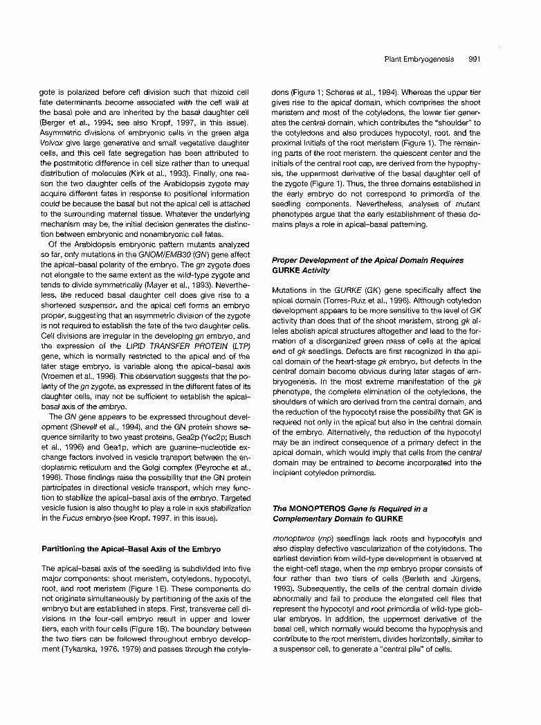
The primary root meristem consists of two tiers of initials that surround a group of mostly mitotically inactive cells, the quiescent center (Figure 2; Dolan et al., 1993; see also Schiefelbein et al., 1997, in this issue). The proximal initials above the quiescent center add new cell tiers to the concen- tric layers of root tissues in a regular fashion: fixed numbers of initials each give rise to lateral root cap and epidermis, cortex and endodermis, and pericycle and vascular tissue. The distal initials below the quiescent center add cell tiers to the central root cap. The root meristem becomes active in the heart-shaped embryo, at which time the embryonic root begins to extend.
The embryonic origin of the root meristem has been ana- lyzed in some detail, both histologically and by clonal analy- sis (Dolan et al., 1993; Scheres et al., 1994). The quiescent center and the initials of the central root cap derive from the basal domain of the embryo, which is established by the hy- pophysis. The hypophysis, in turn, originates from the basal daughter cell of the zygote (Figure 1). By contrast, the initials for the remaining root tissues derive from the lowest cell tier of the central domain and ultimately from the apical daugh- ter cell of the zygote. Thus, a clonal boundary runs across the root meristem (Figure 2; Dolan et al., 1994; Scheres et al., 1994), suggesting that inductive events play a role in es- tablishing the initials for the root tissues. Indeed, no root meristem is formed in the “hypophyseal group” of mutants (e.g., hobbit), in which the first recognizable defect is the ab-
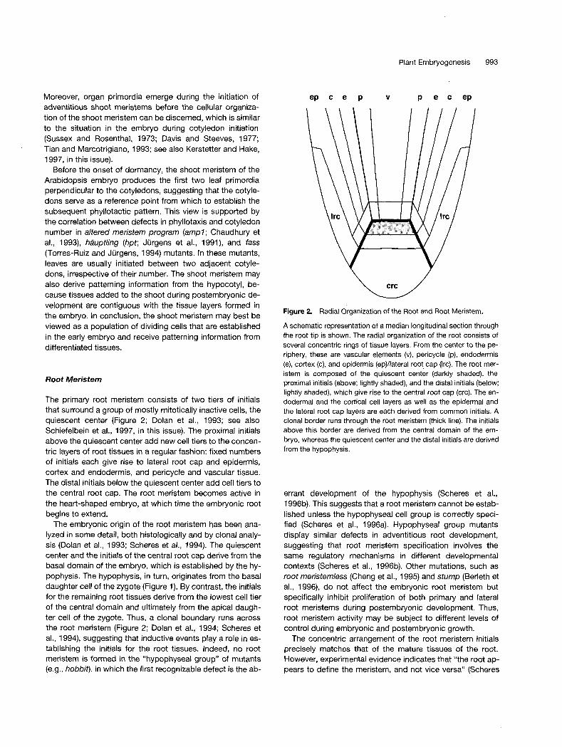
Figure 2. Radial Organization of the Root and Root Meristem.
A schematic representation of a median longitudinal section through the root tip is shown. The radial organization of the root consists of severa1 concentric rings of tissue layers. From the center to the pe- riphery, these are vascular elements (v), pericycle (p), endodermis (e), cortex (c), and epidermis (ep)/lateral root cap (Irc). The root mer- istem is composed of the quiescent center (darkly shaded), the proximal initials (above; lightly shaded), and the distal initials (below; lightly shaded), which give rise to the central root cap (crc). The en- dodermal and the cortical cell layers as well as the epidermal and the lateral root cap layers are each derived from common initials. A clonal border runs through the root meristem (thick line). The initials above this border are derived from the central domain of the em- bryo, whereas the quiescent center and the distal initials are derived from the hypophysis.
errant development of the hypophysis (Scheres et al., 199613). This suggests that a root meristem cannot be estab- lished unless the hypophyseal cell group is correctly speci- fied (Scheres et al., 1996a). Hypophyseal group mutants display similar defects in adventitious root development, suggesting that root meristem specification involves the same regulatory mechanisms in different developmental contexts (Scheres et al., 199613). Other mutations, such as root meristemless (Cheng et al., 1995) and stump (Berleth et al., 1996), do not affect the embryonic root meristem but specifically inhibit proliferation of both primary and lateral root meristems during postembryonic development. Thus, root meristem activity may be subject to different levels of control during ernbryonic and postembryonic growth.
The concentric arrangement of the root meristem initials precisely matches that of the mature tissues of the root. However, experimental evidence indicates that “the root ap- pears to define the meristem, and not vice versa” (Scheres
994 The Plant Cell
et al., 1996a). Indeed, after laser ablation of cortedendoder- mis initials in the Arabidopsis seedling root, the underlying pericycle initials take over and function properly according to their new position. Furthermore, ablation of three adjacent daughter cells of cortex/endodermis initials resulted in abnor- mal cell files, suggesting that root tissue initials receive pat- terning information from mature root tissues (Van den Berg et al., 1995). In carrot, the upper part of transected somatic embryos regenerated root tissues before a new root mer- istem was formed (Schiavone and Racusen, 1991). This se- quence of events is also seen during lateral root formation in Arabidopsis-that is, the lateral root primordium acquires a radial organization of tissue layers before the root meristem becomes active (Malamy and Benfey, 1997). Thus, the root meristem initials can be viewed as dividing cells that lack in- trinsic patterning information.
comes restricted to the protoderm layer and is no longer detectable in the inner cells.
In an alternative model based on the action of morpho- gens in animals (Green and Smith, 1990), protoderm and in- ner cells could acquire different fates in response to positional information passing along the radial axis. Hypothetical signal- ing molecules may enter the inner cells from the suspensor and/or the protoderm cells from the endosperm. Although de- livery of molecules to the embryo from both the suspensor and the endosperm has been discussed, there is no evi- dente that such substances affect cell fate determination in plant embryos. Whatever the mechanism of protoderm. formation, the epidermal fate of the outer cell layer, once established, is stably maintained.
Stable Expression of Cell Fates Requires Physical Separation
Establishing the Radial Axis: Protoderm Formation
The radial axis of the embryo, which is defined as the con- centric arrangement of tissue layers from the center to the periphery, becomes apparent after tangential cell divisions in the octant-stage embryo that partition the cell mass into an outer cell layer, the protoderm, and inner cells (Figures 1 B and 1 C). The protoderm gives rise to the epidermis by strictly anticlinal cell divisions, whereas the inner cells originate ground tissue and vascular elements. Subsequently, these primary tissues undergo further specialization.
Determination of Cell Fates along the Radial Axis
How are protoderm and inner cell fates segregated? By analogy with rhizoid cell fate segregation in the brown alga Fucus (Berger et al., 1994), cell fate information may be laid down in the cell wall of the zygote and passed on to all progeny with cell walls derived from the zygote wall. This idea was originally proposed for the determination of epider- mal cell fate in Cifrus jambhiri on the basis of the observa- tion that the zygote is coated with a cuticle layer, which is a morphological marker for epidermal identity (Bruck and Walker, 1985). Nonepidermal cell fate would thus represent a developmental “ground state” corresponding to the ab- sence of epidermal determinants derived from the zygote. In this model, cell fate segregation does not require strictly ori- ented cell divisions: any cell division that disconnects cells from the zygote-derived cell wall would suffice.
In support of this hypothesis, protoderm formation is not affected in embryos of the Arabidopsis fass mutant, which display an irregular cell division pattern (Torres-Ruiz and Jürgens, 1994). Moreover, the expression pattern of the ATML7 gene is consistent with this model. ATML7 is ex- pressed in the apical daughter cell of the zygote and in all cells of the eight-cell embryo proper (Lu et al., 1996). How- ever, after the tangential divisions, ATML7 expression be-
Mutations in the KNOLLE (KN) gene perturb the segregation of protoderm and inner cell fate, a defect that has been cor- related with incomplete cytokinesis (Lukowitz et al., 1996). Epidermal and subepidermal markers appear to be abnor- mally distributed in early kn embryos (Lukowitz et al., 1996; Vroemen et al., 1996), but at later stages, some inner cells stop expressing the epidermal marker and form vascular tis- sue. The attenuation of the mutant phenotype during later stages of embryogenesis may reflect either increasingly complete cytokineses, perhaps a result of the activation of redundant function(s), or the increasing distance of the inner cells from the embryo surface.
The KN gene encodes a syntaxin-like protein that is re- quired for cell plate formation (Lukowitz et al., 1996). In kn embryos, with their incomplete cell walls, the hypothetical protoderm and inner cell fate determinants may not be fully segregated to adjacent cells but remain present within the adjoined cytoplasm. lnjection experiments on root cells sup- port the view that physical separation is necessary for proper cell differentiation (Duckett et al., 1994). For example, in the differentiated part of the root, fluorescent dye did not spread from the epidermis to the subepidermal layer but en- tered adjacent epidermal cells. In more general terms, be- fore groups of cells can acquire different developmental fates, the uncoupling of their symplastic domains may be re- quired to restrict the passage of cell type-specific mole- cules. Conversely, cells within any one tissue may use their symplastic continuity to disseminate cell-specific informa- tion for the specification of newly formed cells (see McLean et al., 1997, in this issue).
Elaboration of the Radial Pattern in the Central Domain of the Embryo
Although the protoderm forms around both the apical and central domains of the embryo, subsequent steps of radial
Plant Embryogenesis 995
patterning are confined to the central domain (Figures 1 and 2). Periclinal divisions of the inner cells at the protoderm stage produce a layer of ground tissue that surrounds a cen- tral vascular primordium. After further periclinal cell divi- sions, this primordium gives rise to a layer of pericycle cells that encircle the conductive tissues (Scheres et al., 1995). It is only in the torpedo-stage embryo that the ground tissue splits into an outer cortex and an inner endodermis layer. This radial pattern is modified at two levels along the apical- basal axis: the hypocotyl primordium has two layers of corti- cal cells, and the lowest tier of the root primordium forms the outermost layer of lateral root cap cells after periclinal divisions in the epidermis layer.
Severa1 mutations affect the radial pattern of the embryo. The asymmetric cell division of the ground tissue that gener- ates the cortical and endodermal cell layers is absent in three mutants, resulting in a single cell layer instead of two (Scheres et al., 1995). The mutant cell layer appears to cor- respond to cortex in short root (shr) and to endodermis in pi- nocchio (pic) but displays both endodermal and cortical traits in scarecrow (scr). The SCR gene encodes a putative transcription factor and is expressed in both the cortex/ endodermis initial of the root meristem and the endodermal cell layer (Di Laurenzio et al., 1996). Mutations in another gene, WOODEN LEG (WOL), result in a reduced number of vascular cells, all of which differentiate into xylem vessels (Scheres et al., 1995). All of these mutations affect the radial organization of the hypocotyl, root, and proximal initials of the root meristem, suggesting an intimate relation between root and hypocotyl development.
1s the absence of a specific cell layer the result of defec- tive specification of cell fate or of a shortage of cells? This question has been addressed by double mutant analyses with fass, which causes an increased number of radial cell layers (Torres-Ruiz and Jürgens, 1994). The fass mutation was able to rescue the defects of scr and wol, suggesting that neither SCR nor WOL specifies cell fate (Scheres et al., 1995). Thus, the lack of phloem in wol mutants implies that the xylem must be formed before the phloem. A similar first- come-first-served mechanism has been suggested for the allocation of cells to floral organ primordia (Laux et al., 1996). By contrast, the shr defect was not suppressed by fass, suggesting that SHR specifies endodermal cell fate (Scheres et al., 1995). It should be noted that the mutations affecting the radial pattern of the embryo also display the same defects in lateral roots, implying that the same pat- terning mechanism operates during postembryonic devel- opment. This notion is also supported by corresponding patterns of marker gene expression (Malamy and Benfey, 1997).
In conclusion, the radial pattern of the embryo evolves se- quentially. The initial separation of protoderm and nonproto- derm cell fates establishes polarity along the radial axis, which subsequently may be used for the induction of addi- tional cell fates within the central domain of the embryo. That the radial pattern is elaborated differently along the axis
suggests that apical-basal positional information modulates the response of cells to radial patterning signals.
MECHANISMS THAT ESTABLISH CELL FATE IN THE EMBRYO
Cell fate diversity can be generated by two different mecha- nisms: unequal division of a polarized cell to generate two daughter cells that assume different fates and intercellular communication to provide information for position-depen- dent cell fate determination. The former cell-intrinsic mecha- nism may apply ih specific cases, such as the asymmetric divisions of the zygote and the hypophysis and possibly the tangential divisions of the octant-stage cells. But this can- not be a general mechanism (see below). Why then is the cell division pattern in Arabidopsis early embryogenesis so invariant?
Considering the pattern defects in scr and wol mutants, which result from a shortage of cells, it is tempting to specu- late that pattern elements originate from a small number of founder cells so that the stereotyped cell division pattern of the Arabidopsis wild-type early embryo ensures that the complete body plan is established. Conversely, because all cell types are formed at the correct position in fass and ton- neau (Traas et al., 1995), mutants that display highly irregu- lar cell divisions, the orientation of cell division per se seems not to be instrumental in establishing the basic body plan. Thus, the stereotyped cell division pattern in the Arabidopsis wild-type embryo may reflect, rather than establish, cell fate specification.
Position-dependent cell fate specification was inferred from observations indicating that there are no cell lineages of fixed fate in plant development and that cells can adopt alternate fates if exposed to different developmental cues (Stewart and Dermen, 1975; Irish, 1991; Scheres et al., 1994). Although this flexibility may seem very different from the situation in animals, it should be noted that, for example, the cells in developing imagina1 discs of Drosophila also continuously reassess their fate according to their position until they become irreversibly committed at the end of the proliferative period (Lawrence and Struhl, 1996). Thus, there may be some fundamental similarities in mechanisms that specify cell fate in animals and plants.
J
Positional Information: How Do Embryo Cells Sense What Makes Sense?
Plant development is integrated by long-range signals, such as growth factors, which are transported along the shoot- root axis (Lyndon, 1990; see also Creelman and Mullet, 1997; Kende and Zeevaart, 1997, in this issue). Auxin, for example, promotes the elongation of the embryo and coty- ledon outgrowth (Schiavone and Cooke, 1987; Schiavone
996 The Plant Cell
and Racusen, 1990; Fischer and Neuhaus, 1996), and inter- ference with auxin transport can result in the aberrant spac- ing and incomplete separation of cotyledon primordia (Okada et al., 1991; Liu et al., 1993). However, there is no di- rect evidence that growth factors play a role in the establish- ment of specific cell fates. The analyses of mutant phenotypes discussed above suggest instead that local in- teractions between adjacent cells or short-range signaling events are critical in this regard.
Severa1 studies have suggested that the transport of mol- ecules through plasmodesmata provides a route for cell-cell communication in plants. One example is the homeodomain protein KNOTTEDl (KNl), which is involved in regulating meristematic cell fate in the shoot meristem. The KN1 pro- tein can be detected in all cell layers of the shoot meristem, whereas KN7 mRNA is excluded from the epidermal layer (Smith et al., 1992; see also Kerstetter and Hake, 1997; McLean et al., 1997, in this issue). Moreover, ectopic KN7 expression in vascular tissue alters cell differentiation in ad- jacent cells (Sinha et al., 1993). One explanation for these findings is that the KN1 protein is transported between cells. This hypothesis is supported by injection experiments in which KN1 protein appeared to move from cell to cell via plasmodesmata (Lucas et al., 1995). lnjection of fluorescent dye into root cells supports the view that meristematic re- gions represent symplastic domains that exhibit facilitated plasmodesmal transport between cell layers. During differ- entiation, the tissue layers gradually become symplastically separated from each other (Duckett et al., ,1994; McLean et al., 1997, in this issue).
In severa1 cases, molecules have been identified as po- tential signals in plant development. For example, antigens recognized by the JIM8 antibody, which are localized in the cell wall of carrot culture cells, are necessary to sustain the development of JIM8-negative somatic embryo initial cells. This suggests that there may be an inductive interaction be- tween JIM8-positive and embryogenic cells (Pennell et al., 1995). The active component can be washed off the cell wall by mild treatments and appears to contain carbohydrates and lipids. In the same experimental system, both Nod factors, which are N-acetylglucoseamine-containing lipo- polysaccharides involved in bacteria-plant signaling during nodule formation, and the endochitinase EP3 can rescue ar- rested embryos of the ts77 mutant (De Jong et al., 1992, 1993). Although the substrate for EP3 has not yet been iden- tified, it is conceivable that chitinases, such as EP3, could mobilize diffusible signal molecules, such as Nod-like factors, which are required for embryo development. Similar factors may also function during animal embryo development. For example, the DG42 protein, which is required during polarity determination in Xenopus embryos and is localized at the cell periphery (Rosa et al., 1988), catalyzes the synthesis of Nod-like components (Semino and Robbins, 1985).
Whereas the functions of the JIM8 antigen and Nod-like factors during plant somatic embryogenesis may be related to growth control, a role in cell differentiation has been sug-
gested for extracellular arabinogalactan proteins (AGPs). The presence of specific AGPs in the extracellular matrix has been found to correlate with cell type and developmen- tal stage (Pennell et al., 1995; Kreuger and Van Holst, 1996). Therefore, it is conceivable that specific sets of cell surface molecules, such as AGPs, define a context in which neigh- boring cells respond and differentiate (Pennell et al., 1995).
How are signals transported through the cell wall during embryo development? Expression of the SOMATlC EM- BRYOGENESIS RECEPTOR-LIKE KlNASE (SERK) gene marks carrot cells that are competent to form somatic em- bryos and also occurs specifically during the early stages of zygotic embryogenesis (Schmidt et al., 1997). Although its function and cellular localization are not known, the predicted SERK protein sequence resembles leucine-rich repeat recep- tor kinases, suggesting that it may function in a signal trans- duction pathway that acts very early in embryogenesis.
Do Cells Remember Their Origin?
How do cells maintain their identity once it has been speci- fied? There is no information available concerning the length of time that positional information must be supplied for cell fates to become stabilized, although clonal analyses sug- gest that cell types can be altered during development as long as cells are still dividing. One model, developed after studies of Fucus rhizoid development, posits that rhizoid cell fate determinants laid down at the cell wall of the basal end of the zygote signal to the nucleus. This signaling leads to perpetuation of the rhizoid component in a positive feed- back mechanism (Berger and Brownlee, 1995). A similar scenario can be hypothesized for perpetuation of epidermal cell fate in the outer cell layer, where propagation of extra- cellular substances, such as in the cuticle, could serve as a “memory” mechanism.
CONCLUDING REMARKS
Our understanding of the formal principles governing pat- tern formation in the embryo has been considerably en- hanced over the past couple of years. These advances have been based in large part on the analysis of specific mutant phenotypes. These experiments have established that single cells or cell groups in the Arabidopsis early embryo make predictable contributions to the seedling body plan. In a number of cases, the development of a mutant embryo devi- ates from wild type at or before the globular stage, suggest- ing that cell fates are specified, in gross terms, early in embryogenesis.
Molecular studies are now needed to test the genetic models. Although, in a few cases, early cell fate specifica- tion has been visualized by specific gene expression pat-
Plant Embryogenesis 997
terns, it is hoped that molecular analyses of early patterning genes will not only give clues to their functions but also facil- itate the testing of genetic models of cell interactions. A dif- ferent kind of molecular approach may also circumvent one of the problems inherent in genetic analyses of develop- ment: some developmentally important genes may not readily mutate to cause specific phenotypes, due, for exam- ple, to functional redundancy, and may thus have been missed in the extensive screens for pattern mutants. With this in mind, it may be rewarding to isolate genes with spe- cific expression patterns by using the enhancer or gene trap approach, or to isolate genes known to play regulatoj, roles in other systems and subsequently to search for insertions in those genes to determine their biological functions. By combining various approaches, eventually we will learn how a plant embryo gets organized.
ACKNOWLEDGMENTS
We are grateful to Fréderic Berger, Thomas Berleth, Markus Grebe, Christoph Maulbetsch, Klaus F.X. Mayer, Ulrike Mayer, Bernard Moussian, Ben Scheres, and Heiko Schoof for helpful comments. We thank Thomas Berleth, Robert Goldberg, Andreas Mordhorst, Sharman O'Neill, Ben Scheres, Sacco de Vries, and Ramin Yadegari for sending manuscripts before publication. Work in our laboratories is supported by the Deutsche Forschungsgemeinschaft (Grant No. La606/2 to T.L. and Grant No. Jul79/3-3 to G.J.).
REFERENCES
Backs-Hüsemann, D., and Reinert, J. (1970). Embryobildung durch isolierte Einzelzellen aus Gewebekulturen von Daucus carota. Protoplasma 70,4940.
Barton, M.K., and Poethig, R.S. (1993). Formation of the shoot api- cal meristem in Arabidopsis thaliana: An analysis of development in the wild type and in the shoot meristemless mutant. Develop- ment 119,823-831.
Berger, F., and Brownlee, C. (1995). Establishment of the apical- basal axis in multicellular plant embryos. Biol. Cell84, 7-1 1.
Berger, F., Taylor, A., and Brownlee, C. (1994). Cell fate determi- nation by the cell wall in early fucus development. Science 263, 1421-1423.
Berleth, T., and Jiirgens, G. (1993). The role of the Monopteros gene in organising the basal body region of the Arabidopsis embryo. Development 118,575-587.
Berleth, T., Hardtke, C.S., Przemeck, G.K.H., and Müller, J. (1996). Mutational analysis of root initiation in the Arabidopsis embryo. Plant Soil 187, 1-9.
Bruck, D.K., and Walker, D.B. (1985). Cell determination during embryogenesis in Citrus jambhiri. I . Ontogeny of the epidermis. Bot. GZ. 146,188-195.
Busch, M., Mayer, U., and Jlirgens, G. (1996). Molecular analysis of the Arabidopsis pattern formation gene GNOM: Gene structure and intragenic complementation. MOI. Gen. Genet. 250, 681-691.
Chaudhuty, A.M., Letham, S., Craig, S., and Dennis, E.S. (1 993). amp7-A mutant with high cytokinin levels and altered embryonic pattern, faster vegetative growth, constitutive photomorphogene- sis and precocious flowering. Plant J. 4, 907-916.
Cheng, J.-C., Seeley, K.A., and Sung, Z.R. (1995). RML7 and RML2, Arabidopsis genes required for cell proliferation at the root tip. Plant Physiol. 107,365-376.
Christianson, M.L. (1986). Fate map of the organizing shoot apex in Gossypium. Am. J. Bot. 73,947-958.
Clark, S.E. (1997). Organ formation at the vegetative shoot meri- stem. Plant Cell 9, 1067-1 076.
Clark, S.E., Jacobsen, S.E., Levin, J.Z., and Meyerowitz, E.M. (1 996). The CLAVATA and SHOOT Ad€R/ST€ML€SS loci competi- tively regulate meristem activity in Arabidopsis. Development 122,
Creelman, R.A., and Mullet, J.E. (1 997). Oligosaccharins, brassino- lides, and jasmonates: Nontraditional regulators of plant growth, development, and gene expression. Plant Cell 9, 121 1-1223.
Davis, E.L., and Steeves, T.A. (1977). Experimental studies on the shoot apex of Helianthus annuus: The effect of surgical bisection on quiescent cells in the apex. Can. J. Bot. 55, 606-614.
De Jong, A.J., Cordewener, J., Lo Schiavo, F., Terzi, M., Vandekerckhove, J., Van Kammen, A., and De Vries, S.C. (1992). A carrot somatic embryo mutant is rescued by chitinase. Plant Cell4,425-433.
De Jong, A.J., Heidstra, R., Spaink, H.P., Hartog, M.V., Meijer, E.A., Hendriks, T., Lo Schiavo, F., Terzi, M., Bisseling, T., Van Kammen, A., and De Vries, S.C. (1993). Rhizobium lipooligosac- charides rescue a carrot somatic embryo mutant. Plant Cell 5, 61 5-620.
Di Laurenzio, L., Wysocka-Diller, J., Malamy, J.E., Pysh, L., Helariutta, Y., Freshour, G., Hahn, M.G., Feldmann, K.A., and Benfey, P.N. (1996). The SCARECROW gene regulates an asym- metric cell division that is essential for generating the radial orga- nization of the Arabidopsis root. Cell86, 423-433.
Dolan, L., Janmaat, K., Willernsen, V., Linstead, P., Poethig, S., Roberts, K., and Scheres, 8. (1993). Cellular organisation of the Arabidopsis thaliana root. Development 119, 71-84.
Dolan, L., Duckett, C.M., Grierson, C., Linstead, P., Schneider, K., Lawson, E., Dean, C., Poethig, S., and Roberts, K. (1994). Clonal relationships and cell patterning in the root epidermis of Arabidopsis. Development 120, 2465-2474.
Duckett, C.M., Oparka, K.J., Prior, D.A.M., Dolan, L., and Roberts, K. (1994). Dye-coupling in the root epidermis of Arabi- dopsis is progressively reduced during development. Develop- ment 120,3247-3255.
Elliott, R.C., Betzner, A.S., Huttner, E., Oakes, M.P., Tucker, W.Q.J., Gerentes, D., Perez, P., and Smyth, D.R. (1996). AIN- T€GUM€NTA, an APETALAP-like gene of Arabidopsis with pleio- tropic roles in ovule development and floral organ growth. Plant Cell8, 155-1 68.
Emons, A.M.C. (1 994). Somatic embryogenesis: Cell biological aspects. Acta Bot. Neerl. 43, 1-14.
1565-1 575.
998 The Plant Cell
Endriui, K., Moussian, E., Haecker, A., Levin, J., and Laux, T. (1 996). The SHOOT MERISTEMLESS gene is required for mainte- nance of undifferentiated cells in Arabidopsis shoot and floral meristems and acts at a different regulatory leve1 than the meri- stem genes WUSCHEL and ZWILLE. Plant J. 10,967-979.
Esau, K. (1977). Anatomy of Seed Plants. (New York: John Wiley and Sons).
Fischer, C., and Neuhaus, G. (1996). lnfluence of auxin on the establishment of bilateral symmetry in monocots. Plant J. 9,
Gerlach-Cruse, D. (1969). Embryo- und Endospermentwicklung nach einer Rontgenbestrahlung der Fruchtknoten von Arabidopsis thaliana. Rad. Bot. 9, 433-442.
Goldberg, R.B., De Paiva, G., and Yadegari, R. (1994). Plant embryogenesis: Zygote to seed. Science 266,605-61 4.
Green, J.B., and Smith, J.C. (1990). Graded changes in dose of a Xenopus activin A homologue elicit stepwise transitions in embry- onic cell fate. Nature 347, 391-394.
Hong, S.K., Kitano, H., Satoh, H., and Nagato, Y. (1996). How is embryo size genetically regulated in rice? Development 122,
Irish, V.F. (1991). Cell lineage in plant development. Curr. Opin. Genet. Dev. 1, 169-1 73.
Johnston, D.S., and Nüsslein-Volhard, C. (1992). The origin of pat- tern and polarity in the Drosophila embryo. Cell68, 201-219.
Jürgens, G. (1995). Axis formation in plant embryogenesis: Cues and clues. Cell81,467-470.
Jürgens, G., and Mayer, U. (1994). Arabidopsis. In A Colour Atlas of Developing Embryos, J. Bard, ed (London: Wolfe Publishing), pp.
Jürgens, G., Mayer, U., Torres-Ruiz, R.A., Berleth, T., and Miséra, S. (1991). Genetic analysis of pattern formation in the Arabidopsis embryo. Development (suppl.) I , 27-38.
Jürgens, G., Torres-Ruiz, R.A., Laux, T., Mayer, U., and Berleth, T. (1994). Early events in apical-basal pattern formation in Ara- bidopsis. In Plant Molecular Biology: Molecular-Genetic Analy- sis of Plant Development and Metabolism, G. Coruzzi and P. Puigdomenech, eds (Berlin: Springer-Verlag), pp. 95-1 03.
Kaplan, D. (1 969). Seed development in Downingia. Phytomorphol-
Kende, H., and Zeevaart, J.A.D. (1 997). The five “classical” plant hormones. Plant Cell9, 11 97-1 21 O.
Kerstetter, R.A., and Hake, S. (1997). Shoot meristem formation in vegetative development. Plant Cell9, 1001-101 O.
Kirk, M.M., Ransick, A., McRae, SE, and Kirk, D.L. (1993). The relationship between cell size and cell fate in Volvox carteri. J. Cell Biol. 123, 191-208.
Klucher, K.M., Chow, H., Reiser, L., and Fischer, R.L. (1996). The AINTEGUMENTA gene of Arabidopsis required for ovule and female gametophyte development is related to the floral homeotic gene APETALAZ. Plant Cell8, 137-1 53.
Kreuger, M., and Van Holst, G.-J. (1 996). Arabinogalactan proteins and plant differentiation. Plant MOI. Biol. 30, 1077-1086.
Kropf, D.L. (1997). lnduction of polarity in fucoid zygotes. Plant Cell
659-669.
2051-2058.
7-21.
Ogy 19,253-278.
9,l o1 1-1 020.
Laux, T., and Jürgens, G. (1994). Establishing the body of the Ara- bidopsis embryo. Acta Bot. Neerl. 43, 247-260.
Laux, T., Mayer, K.F.X., Berger, J., and Jürgens, G. (1996). The WUSCHH gene is required for shoot and floral meristem integrity in Arabidopsis. Development 122, 87-96.
Lawrence, P.A., and Struhl, G. (1 996). Morphogens, compart- ments, and pattern: Lessons from Drosophila? Cell 85, 951-961.
Liu, C.-m., Xu, 2.-h., and Chua, N.-H. (1993). Auxin polar transport is essential for the establishment of bilateral symmetry during early plant embryogenesis. Plant Cell 5, 621-630.
Long, J.A., Moan, E.I., Medford, J.I., and Barton, M.K. (1996). A member of the KNOTTED class of homeodomain proteins encoded by the STM gene of Arabidopsis. Nature 379, 66-69.
Lopes, M.A., and Larkins, B.A. (1 993). Endosperm origin, develop- ment, and function. Plant Cell 5, 1383-1399.
Lu, P., Porat, R., Nadeau, J.A., and O’Neill, S.D. (1996). Identifica- tion of a meristem LI layer-specific gene in Arabidopsis that is expressed during embryonic pattern formation and defines a new class of homeobox genes. Plant Cell8, 2155-2168.
Lucas, W.J., Bouché-Pillon, S., Jackson, D.P., Nguyen, L., Baker, L., Ding, B., and Hake, S. (1995). Selective trafficking of KNOTTEDl homeodomain protein and its mRNA through plas- modesmata. Science 270,1980-1 983.
Lukowitz, W., Mayer, U., and Jürgens, G. (1996). Cytokinesis in the Arabidopsis embryo involves the syntaxin-related KNOLLE gene product. Cell84,61-71.
Lyndon, R.F. (1990). Plant Development: The Cellular Basis. (win- chester, MA: Unwin Hyman Inc.).
Malamy, J.E., and Benfey, P.N. (1 997). Organization and cell differ- entiation in lateral roots of Arabidopsis thaliana. Development 124, 33-44.
Mansfield, S.G., and Briarty, L.G. (1991). Early embryogenesis in Arabidopsis thaliana. II. The developing embryo. Can. J. Bot. 69,
Mansfield, S.G., Briarty, L.G., and Erni, S. (1991). Early embryo- genesis in Arabidopsis thaliana. I. The mature embryo sac. Can. J. Bot. 69, 447-460.
Marsden, M.P.F., and Meinke, D.W. (1 985). Abnormal develop- ment of the suspensor in an embryo-lethal mutant of Arabidopsis fhaliana. Am. J. Bot. 72, 1801-1812.
Mayer, U., Torres-Ruiz, R.A., Berleth, T., Miséra, S., and Jürgens, G. (1991). Mutations affecting body organisation in the Arabidop- sis embryo. Nature 353,402-407.
Mayer, U., Biittner, G., and Jiirgens, G. (1993). Apical-basal pat- tern formation in the Arabidopsis embryo: Studies on the role of the gnom gene. Development 11 7,149-1 62.
McConnell, J.R., and Barton, M.K. (1995). Effect of mutations in the PINHEAD gene of Arabidopsis on the formation of shoot api- cal meristems. Dev. Genet. 16, 358-366.
McLean, B.G., Hempel, F.D., and Zambryski, P.C. (1997). Plant intercellular communication via plasmodesmata. Plant Cell 9,
Mbl, R., Matthys-Rochon, E., and Dumas, C. (1994). The kinetics of cytological events during double fertilization in Zea mays L. Plant J. 5, 197-206.
461-476.
1043-1 054.
Plant Embryogenesis 999
Mordhorst, A.P., Toonen, M.A.J., and De Vries, S.C. (1997). Plant embryogenesis. Crit. Rev. Plant Sci., in press.
Nelson, T., and Dengler, N. (1997). Leaf vascular pattern formation. Plant Cell 9, 1121-1135.
Nomura, K., and Komamine, A. (1985). ldentification and isolation of single cells that produce somatic embryos at a high frequency in a carrot suspension culture. Plant Physiol. 79, 988-991.
Ohad, N., Margossian, L., Hsu, Y., Williams, C., Repetti, P., and Fischer, R.L. (1 996). A mutation that allows endosperm develop- ment without fertilization. Proc. Natl. Acad. Sci. USA93,5319-5324.
Okada, K., Ueda, J., Komaki, M.K., Bell, C.J., and Shimura, Y. (1991). Requirement of the auxin polar transport system in early stages of Arabidopsis floral bud formation. Plant Cell3, 677-684.
Pennell, R.I., Cronk, Q.C.B., Forsberg, L.S., Stohr, C., Snogerup, S., Kjellbom, P., and McCabe, P.F. (1995). Cell-contex signal- ling. Philos. Trans. R. SOC. Lond. Biol. Sci. 350, 87-93.
Peyroche, A., Paris, S., and Jackson, C.L. (1996). Nucleotide exchange on ARF mediated by yeast Geal protein. Nature 384,
Poethig, R.S., Coe, E.H., and Johri, M.M. (1986). Cell lineage pat- terns in maize embryogenesis: A clonal analysis. Dev. Biol. 117, 392404.
Przemeck, G.K.H., Mattsson, J., Hardtke, C.S., Sung, Z.R., and Berleth, T. (1996). Studies on the role of the Arabidopsis gene MONOPTEROS in vascular development and plant cell axializa- tion. Planta 200, 229-237.
Rosa, F., Sargent, T.D., Rebbert, M.L., Michaels, G.S., Jamrich, M., Grunz, H., Jonas, E., Winkles, J.A., and Dawid, I.B. (1988). Accumulation and decay.of DG42 gene products follow a gradient pattern during Xenopus embryogenesis. Dev. Biol. 129, 114-123.
Russell, S.D. (1 993). The egg cell: Development and role in fertiliza- tion and early embryogenesis. Plant Cell 5, 1349-1 359.
Sachs, T. (1981). The control of the patterned differentiation of vas- cular tissue. Adv. Bot. Res. 9, 151-162.
Scheres, B., Wolkenfelt, H., Willemsen, V., Terlouw, M., Lawson, E., Dean, C., and Weisbeek, P. (1994). Embryonic origin of the Arabidopsis primary root and root meristem initials. Development
Scheres, B., Di Laurenzio, L., Willemsen, V., Hauser, M., Janmaat, K., Weisbeek, P., and Benfey, P.N. (1995). Mutations affecting the radial organisation of the Arabidopsis root display specific defects throughout the embryonic axis. Development 121, 53-62.
Scheres, B., McKhann, H.I., and Van den Berg, C. (1996a). Roots redefined: Anatomical and genetical analysis of root development. Plant Physiol. 111, 959-964.
Scheres, B., McKhann, H., Van den Berg, C., Willemsen, V., Wolkenfelt, H., de Vrieze, G., and Weisbeek, P. (1996b). Experi- mental and genetic analyses of root development in Arabidopsis thaliana. Plant Soil 187, 97-105.
Schiavone, F.M., and Cooke, T.J. (1987). Unusual patterns of somatic embryogenesis in the domesticated carrot: Developmen- tal effects of exogenous auxins and auxin transport inhibitors. Cell Differ. 21, 53-62.
Schiavone, F.M., and Racusen, R.H. (1 990). Microsurgery reveals regional capabilities for pattern reestablishment in somatic carrot embryos. Dev. Biol. 141, 211-219.
479-484.
120,2475-2487.
Schiavone, F.M., and Racusen, R.H. (1 991). Regeneration of surgi- cally transected carrot embryos occurs by position-dependent, proximodistal replacement of missing tissues. Development 113,
Schiefelbein, J.W., Masucci, J.D., and Wang, H. (1997). Building a root: The control of patterning and morphogenesis during root development. Plant Cell9, 1089-1 098.
Schmidt, E.D.L., Guuo, F., Toonen, M.A.J., and De Vries, S.C. (1 997). A leucine-rich repeat containing receptor-like kinase marks somatic plant cells competent to form embryos. Develop- ment 124,2049-2062.
Schwartz, B.W., Yeung, E.C., and Meinke, D.W. (1994). Disrup- tion of morphogenesis and transformation of the suspensor in abnormal suspensor mutants of Arabidopsis. Development 120,
Semino, C.E., and Robbins, P.W. (1 985). Synthesis of “Nod”-like chitin oligosaccharides by the Xenopus developmental protein DG42. Proc. Natl. Acad. Sci. USA 92,3498-3501.
Shevell, D.E., Leu, W.-M., Gilimor, C.S., Xia, G., Feldmann, K.A., and Chua, N.-H. (1994). EMB3O is essential for normal cell divi- sion, cell expansion, and cell adhesion in Arabidopsis and encodes a protein that has similarity to Sec7. Cell 77, 1051-1062.
Sinha, N.R., Williams, R.E., and Hake, S. (1993). Overexpression of the maize homeobox gene, KNOTTED-7, causes a switch from determinate to indeterminate cell fates. Genes Dev. 7 , 787-795.
Smith, L.G., Breene, B., Veit, B., and Hake, S. (1992). A dominant mutation in the maize homeobox gene, Knotted-7, causes its ectopic expression in leaf cells with altered fates. Development
Steeves, T.A., and Sussex, I.M. (1989). Patterns in Plant Develop- ment. (Cambridge, UK: Cambridge University Press).
Stewart, R.N., and Dermen, H. (1975). Flexibility in ontogeny as shown by the contribution of the shoot apical layers to leaves of periclinal chimeras. Am. J. Bot. 62, 935-947.
Sussex, I.M., and Rosenthal, D. (1 973). Differential 3H-thymidine labelling of nuclei in the shoot apical meristem of Nicotiana. Bot. Gaz. 134,295-301.
Thomas, T.L. (1 993). Gene expression during plant embryogenesis and germination: An overview. Plant Cell5, 1401-1410.
Tian, H.-C., and Marcotrigiano, M. (1 993). Origin and development of adventitious shoot meristems on plant chimeras. Dev. Biol.
Torres-Ruiz, R.A., and Jürgens, G. (1994). Mutations in the FASS gene uncouple pattern formation and morphogenesis in Arabi- dopsis development. Development 120, 2967-2978.
Torres-Ruiz, R.A., Lohner, A., and Jürgens, G. (1 996). The GURKE gene is required for normal organisation of the apical region in the Arabidopsis embryo. Plant J. 10, 1005-1 O1 6.
Traas, J., Bellini, C., Nacry, P., Kronenberger, J., Bouchez, D., and Caboche, M. (1995). Normal differentiation patterns in plants lacking microtubular preprophase bands. Nature 375, 676-677.
Tykarska, T. (1976). Rape embryogenesis. I. The proembryo devel- opment. Acta SOC. Bot. Pol. 45, 3-15.
Tykarska, T. (1 979). Rape embryogenesis. II. Development of
1305-1 31 3.
32353245,
116,21-30.
155,259-269.
embryo proper. Acta SOC. Bot. Pol. 48, 391421.
1 O00 The Plant Cell
Van den Berg, C., Willemsen, V., Hage, W., Weisbeek, P., and Scheres, 8. (1995). Cell fate in the Arabidopsis root meristem determined by directional signalling. Nature 378, 62-65.
Vernon, D.M., and Meinke, D.W. (1 994). Embryogenic transforma- tion of the suspensor in twin, a polyembryonic mutant of Arabi- dopsis. Dev. Biol. 165, 566-573.
Vroemen, C.W., Langeveld, S., Mayer, U., Ripper, G., Jürgens, G., Van Kammen, A., and De Vries, S.C. (1 996). Pattern formation in the Arabidopsis embryo revealed by position-specific lipid trans- fer protein gene expression. Plant Cell8, 783-791.
Willemse, M.T.M., and Van Went, J.L. (1984). The female gameto- phyte. In Embryology of Angiosperms, B.M. Johri, ed (Berlin: Springer-Verlag), pp. 159-1 96.
Yadegari, R., and Goldberg, R.B. (1997). Cellular and molecular biology of plant seed development. In Advances in Molecular Biology of Plants, B. Larkins and I.K. Vas, eds (Dordrecht, The Netherlands: Kluwer Academic Publishers), in press.
Yeung, E.C., and Meinke, D.W. (1993). Embryogenesis in
angiosperms: Development of the suspensor. Plant Cell 5, 1371- 1381.
Yeung, E.C., and Sussex, I.M. (1979). Embryogeny of fhaseolus coccineus: The suspensor and the growth of the embryo-proper in vitro. Z. Pflanzenphysiol. 91, 423-433.
Zimmerman, J.L. (1993). Somatic embryogenesis: A model for development in higher plants. Plant Cell5, 1411-1423.
NOTE ADDED IN PROOF
In this article we have discussed zygotic mutations that affect em- bryo development. Recently, Ray et al. described variable maternal effects of the short integument mutation on embryo development. This mutation can affect cotyledon number and/or the embryonic shoot meristem (Ray, S., Golden, T., and Ray, A. [1996]. Maternal effects of the short integument mutation on embryo development in Arabidopsis. Dev. Biol. 180, 365-369).