Embed Size (px)
Citation preview

Learning and Teaching Investment Fund final report
Title of project Embedding Virtual RadiographyTM within the curriculum to enhance student learning: Authentic learning in a safe environment using a multichannel delivery approach
Project leader Dr Madeleine Shanahan, formerly Senior Lecturer / Discipline Leader, Medical Radiations; now Adjunct Associate Professor of RMIT University School/service unit name/college School of Health and Biomedical Sciences, SEH
Project conducted 2016, Report written February 2017.
Strategic objectives addressed: This project addresses the following priorities for 2016 LTIF identified priority areas.
• Improving student or cohort experience, student retention or actions arising from student feedback
• Designing curricula for new learning spaces, multichannel delivery or future industry/professional/international requirements
Internal order number: 360543 Project leader contact details: Email: [email protected]
Phone: (02) 6206 8521
Project team members:
• Internal (2016) – Dr Madeleine Shanahan, Project Leader
• Internal – Associate Professor Moshi Geso
• External Clinical Radiography tutors Mr Keith Jansz, Mr Ben Grinsted, Mr Adam Steward
On the original application (September, 2015) but not involved in the project in 2016
• Mr Luke Barclay (internal), Left RMIT University, Dec 2015
• Mr Greg Trypis (external) Was not involved in project 2016 due to clinical work commitments
• Ms Zoe Brady, (external) Was not involved in project 2016 due to parental leave

Title of Report Unclassified
Information Technology Services
Document: Embedding Virtual RadiographyTM within the
curriculum to enhance student learning- Authentic learning in a safe environment using a multichannel
delivery approach.doc Page 2 of 12
Funding scheme (please type an ‘X’ in the correct box)
LTIF contestable x
RMIT Vietnam Program Development Fund

Title of Report Unclassified
Document: Embedding Virtual RadiographyTM within the curriculum to enhance student learning- Authentic
learning in a safe environment using a multichannel delivery approach.doc
Page 3 of 12
1 Executive summary Diagnostic radiography involves the use of ionising radiation and, consequently, student use of radiography equipment for methods-based and physics-based courses is tightly controlled at RMIT University and in clinical placements. This limits student ability to develop their understanding and practice their skills in preparation for their clinical placements. This project aimed to overcome this limitation to student learning by introducing the software simulation Virtual RadiographyTM into methods-based and physics-based courses. The Virtual Radiography simulation generates radiographic images and x-ray tube output data without the use of ionising radiation. This means this software provides students with enhanced opportunities to develop their understanding and practice their skills in a safe learning environment, without having to use ionising radiation in preparation for their clinical placements.
In particular, this project aimed to:
1) Improve student or cohort experience by introducing a new simulation, Virtual RadiographyTM, cohesively across multiple courses within the BAppSc (Medical Radiations) program.
Virtual Radiography was successfully introduced into the following core courses, within the Bachelor of Applied Science (Medical Radiations) program
Year level Courses Student number
1st year Introduction to Medical Radiations (RADI1125) Medical Radiations Technology 1 (ONPS2343) Introduction to Medical Imaging (RADI1184)
167 153 76
2nd year Medical Imaging Methods 1 (RADI1130) Medical Imaging Methods 2 (RADI1131)
58 62
New curriculum resources were developed as outputs of workshops held with industry partners. This approach was adopted to ensure the developed resources and learning activities were industry-relevant. Student surveys were developed and implemented (Ethics Approval granted). Key findings from the surveys demonstrate that the simulation software, Virtual RadiographyTM, is easy to use (88%), that students like using the simulation (89%) and that developed curriculum resources were easy to follow (83%).
2) Designing curricula for multichannel delivery and to meet professional requirements The simulation was introduced using a multichannel delivery through lectures, laboratories and student self-directed learning. Key findings from the surveys: The vast majority of students agreed that Virtual RadiographyTM should continue to be used in laboratories (90%) and in lectures (87%). Sixteen percent of students reported using the simulation outside of scheduled class times.
The introduction of Virtual RadiographyTM sought to support students in meeting professional requirements by increasing their radiographic knowledge and skills. Students identified that use of the simulation had a positive effect on their ability to: set up radiographic examinations (87%), evaluate radiographic images (93%), correct errors that they see on radiographic images (94%) and select exposure factors that lower patient dose (85%). Students also reported that use of Virtual RadiographyTM encouraged problem solving (87%) and that having used the simulation they felt better prepared for clinical placement (78%).

Title of Report Unclassified
Document: Embedding Virtual RadiographyTM within the curriculum to enhance student learning- Authentic
learning in a safe environment using a multichannel delivery approach.doc
Page 4 of 12
2 Outcomes Provide a brief overview of the project's outcomes and impacts including:
• Number of courses/programs/students benefitting from the project
This project sought to improve the student experience by introducing a new simulation, Virtual RadiographyTM, cohesively across multiple courses within the BAppSc (Medical Radiations) program. Virtual Radiography was successfully introduced into the following core courses, within the Bachelor of Applied Science (Medical Radiations) program
Year level Courses Student number
1st year Introduction to Medical Radical Radiations (RADI1125) Medical Radiations Technology 1 (ONPS2343) Introduction to Medical Imaging (RADI1184)
167 153
76 2nd year Medical Imaging Methods 1 (RADI1130)
Medical Imaging Methods 2 (RADI1131) 58 62
In RADI1130/RADI1131 and RADI1184, learning activities using Virtual RadiographyTM were conducted throughout the semester in the laboratory component of these courses. RADI1125 and ONPS2343 utilised the simulation in lectures and in a single laboratory session.
• Student feedback on project deliverables/ GTS/OSI
New curriculum resources were developed as outputs of workshops held with industry partners. This approach was adopted to ensure the developed resources and learning activities were industry relevant. Student surveys were developed and implemented (Ethics Approval granted). Key findings from the surveys demonstrate that the simulation software, Virtual RadiographyTM, is easy to use (88%, n=316), students like using the simulation (89%, n=318) and that developed curriculum resources were easy to follow (83%, n=297). See Section 3.1.
The simulation was introduced using a multichannel delivery through lectures, laboratories and student self-directed learning. Key findings from student surveys: 90% (n=322) of students agreed that Virtual RadiographyTM should continue to be used in laboratories and 87% (n=152) and in lectures (n= 152). Whilst 70% (n=250) of students agreed that they could access Virtual RadiographyTM outside their scheduled laboratory time, 16% (n=29) of students reported using the simulation outside of scheduled class times. See Section 3.2.1.
The introduction of Virtual RadiographyTM sought to provide students with enhanced opportunities to meet professional requirements by increasing their radiographic knowledge and skills. Students identified that use of the simulation had a positive effect on their ability to: set up radiographic examinations (87%, n=290), evaluate radiographic images (93%, n=310), correct errors that they see on radiographic images (94%, n=171) and select exposure factors that lower patient dose (85%, n=154). Students also reported that use of Virtual RadiographyTM encouraged problem solving (87%, n=271) and that having used the simulation they felt better prepared for clinical placement (78%, n=142). See Section 3.2.1.
Multiple factors impact on GTS/OSI within courses. This project team makes no association between GTS/OSI of individual courses and this project.

Title of Report Unclassified
Document: Embedding Virtual RadiographyTM within the curriculum to enhance student learning- Authentic
learning in a safe environment using a multichannel delivery approach.doc
Page 5 of 12
2016 GTS /OSI (Average as reported by SHBS data)
RADI1125 ONPS2343 RADI1130 RADI1131 RADI1184
GTS 58.5 81.9 84.8 87.8 91.8
OSI 60.4 91.5 82.6 91.7 95.5
At this stage there have been no scholarly outputs from this project. Full data analysis is being undertaken. It is anticipated that scholarly outputs will occur during 2017.
3 Project outcomes and impacts Discuss the outcomes the project was designed to achieve and the outcomes the project has achieved. Include evidence of the project's impact on courses/programs/students benefitting from the project; and student feedback on project deliverables/changes in GTS or OSI.
Describe any disciplinary and interdisciplinary linkages that have emerged as a result of the project.
Describe briefly any issues that may have prevented you achieving all of the outcomes in your application.
3.1 Improve student experience This project aimed to improve the student experience by introducing a new simulation, Virtual RadiographyTM, cohesively across multiple courses within the BAppSc (Medical Radiations) program. Virtual Radiography was successfully introduced into the following core courses, within the Bachelor of Applied Science (Medical Radiations) program
Year level Courses Student number
1st year Introduction to Medical Radiations (RADI1125) Medical Radiations Technology 1 (ONPS2343) Introduction to Medical Imaging (RADI1184)
167 153
76 2nd year Medical Imaging Methods 1 (RADI1130)
Medical Imaging Methods 2 (RADI1131) 58 62
New curriculum resources were developed as outputs of workshops held with industry partners. This approach was adopted to ensure the developed resources and learning activities were industry relevant. Student surveys were developed and implemented (Ethics Approval granted). A total of 356 surveys were returned.
The number of surveys returned from students for each course
RADI1125 ONPS2343 RADI1184 RADI1130/RADI1131
Total
Survey response N=153 N=21 N=64 N=118 N=356
The project sought to improve the student experience by introducing a user-friendly simulation software that would provide students with enhanced learning opportunities. Key findings from the surveys demonstrate that the simulation software, Virtual RadiographyTM, is easy to use (88%, n=316). Student like using the simulation (89%, n=318) and that developed curriculum resources were easy to follow (83%, n=297).

Title of Report Unclassified
Document: Embedding Virtual RadiographyTM within the curriculum to enhance student learning- Authentic
learning in a safe environment using a multichannel delivery approach.doc
Page 6 of 12
The percent (%) and number of students (n) who agreed or strongly agreed with each statement
RADI1125 ONPS2343 RADI1184 RADI1130/RADI1131
Total
% and n % and n % and n % and n % and n
Virtual RadiographyTM is easy to use
92% (141) 90% (19) 83% (53) 87% (103) 88% (316)
I could control the equipment as I needed when using Virtual RadiographyTM
96% (147) 95% (20) 81% (52) 87%(103) 90% (322)
I liked using Virtual RadiographyTM
94% (144) 90% (19) 88% (56) 84% (99) 89% (318)
The Virtual RadiographyTM lab activity sheet(s) were easy to follow
76% (117) 86% (18) 92% (59) 87% (103) 83% (297)
Not all students answered all questions on the survey. Percentages reflect percentage of students that responded to a given question
3.2 Designing curricula for multichannel delivery and to meet professional requirements
3.2.1 Designing curricula for multichannel delivery The simulation was introduced using a multichannel delivery through lectures, laboratories and student self-directed learning. Key findings from the surveys: The vast majority of students agreed that Virtual RadiographyTM should continue to be used in laboratories (90%, n=322)) and in lectures (87%, n=152).
The percent (%) and number of students (n) who agreed or strongly agreed with each statement
RADI1125 ONPS2343 RADI1184 RADI1130/RADI1131
Total
% (n) % (n) % (n) % (n) % (N)
Virtual RadiographyTM should continue to be used in laboratories
94% (145) 95% (18) 94% (60) 84% (99) 90% (322)
Virtual RadiographyTM should continue to be used lectures
90% (138) 74% (14) - - 87% (152)
- Indicates this question not part of the survey for this course
Whilst 70% students agreed that they could access Virtual RadiographyTM outside their scheduled laboratory time, 16% (n=29) of students reported using the simulation outside of scheduled class times.
Use of Virtual RadiographyTM RADI1130/1131 RADI1184 Total (N)
% (n) % (n) % (N)
Laboratory time only 86% (102) 80% (51) 84% (153)
In laboratory and outside scheduled lab 14% (16) 20% (13) 16% (29)

Title of Report Unclassified
Document: Embedding Virtual RadiographyTM within the curriculum to enhance student learning- Authentic
learning in a safe environment using a multichannel delivery approach.doc
Page 7 of 12
Total 100% (118) 100%(64) 100% (182)
3.2.2 Designing curricula to meet professional requirements The introduction of Virtual RadiographyTM sought to provide students with enhanced opportunities to meet professional requirements by increasing their radiographic knowledge and skills. Students identified that use of the simulation had a positive effect on their ability to: set up radiographic examinations (87%, n=290), evaluate radiographic images (93%, n=310), correct errors that they see on radiographic images (94%, n=171) and select exposure factors that lower patient dose (85%, n=154). Students also reported that use of Virtual RadiographyTM encouraged problem solving (87%, n=271) and that having used the simulation they felt better prepared for clinical placement (78%, n=142).
The percent (%) and number of students (n) who agreed or strongly agreed with each statement
RADI1125 ONPS2343 RADI1184 RADI1130/RADI1131
Total
Using Virtual RadiographyTM… % (n) % (n) % (n) % (n) % (N)
Allowed me to quickly see images and understand if changes need to be made
97% (150) 89% (17) 97% (62) 92% (108) 95% (337)
Helped me to learn as I was able to repeat activities until I was satisfied
97% (150) 84% (16) 95% (61) 93% (107) 94% (334)
Encouraged me to solve problems 90% (138) -‐ 88% (56) 82% (97) 87% (291)
Had a positive effect on my ability to set-‐up a radiographic examination
93% (142) -‐ 86% (55) 80% (93) 87% (290)
Had a positive effect on my ability to evaluate radiographic images
97% (148) -‐ 97% (62) 85% (100) 93% (310)
Encouraged me to think more about radiographic procedures
95% (145) -‐ 89% (57) 85% (100) 91% (302)
Encouraged me to think more about radiographic image evaluation
97% (148) -‐ 95% (61) 86% (101) 93% (310)
Encouraged me to think more about medical radiation technology
-‐ 63% (12) -‐ -‐ 63% (12)
Had a positive effect on my ability to correct errors that I see of a radiographic image
-‐ -‐ 95% (61) 94% (110) 94% (171)
Had a positive effect on my ability to select exposure factors that lower patient dose
-‐ -‐ 80% (51) 88% (103) 85% (154)
I feel better prepared for clinical practice having used VR
-‐ -‐ 83% (53) 76% (89) 78% (142)
- Indicates this question not part of the survey for this course

Title of Report Unclassified
Document: Embedding Virtual RadiographyTM within the curriculum to enhance student learning- Authentic
learning in a safe environment using a multichannel delivery approach.doc
Page 8 of 12
Prospectively the linkages between the courses will become apparent as skills are reinforced and extended.
3.3 Interdisciplinary linkages
Students within the chiropractic program were provided with an opportunity to use the software in a voluntary tutorial, specifically designed for them (an amendment to the original ethics application was approved). Forty-eight students enrolled in Chiropractic 6 attended the tutorial and provided feedback via survey. Responses were received from 44 out of the 48 students (response rate 92%). Overall students were positive about this simulation identifying that it was easy to use (95%) and that they could control the equipment as needed (95%). The main reported benefits included students being enabled to repeat tasks until they were satisfied with the results (98%) and being able to quickly assess images and understand if changes needed to be made (98%). Participants reported improvement in their understanding of the effect of exposure factors on patient radiation dose (93%) as well as their technical (73%), image evaluation (84%) and problem solving (80%) skills. The results from this small scale study indicate there is potential for this simulation to be embedded within the Chiropractic program.
3.4 Issues preventing achievement of all outcomes in your application
Due to project team staff changes (project team member leaving RMIT University), project team member having changed responsibilities in SHBS, and project team member on extended leave in 2016 there are some curriculum resources that were not developed e.g. short videos on using the simulation. All other curriculum resources have been developed and made available on BlackBoard for semester 1 and semester 2 courses. In addition, the noted changes within the project team resulted in the simulation not being used within lectures in some courses.
Furthermore, due to project team changes during 2016, the Curriculum Design pattern has not been developed. However, the model used in this project of using a pilot study (STeLR, 2015) to identify and resolve any IT issues associated with the implementation of the simulation is strongly recommended for any potential large scale computer-based simulation projects. In addition, the approach adopted in this study of having industry partners directly involved in curriculum resource development through workshops ensures the developed resources and learning activities are industry-relevant. There was good overall survey response from students, except in ONPS2343. As such the implementation of simulation software into ONPS2343 has not yet been rigorously evaluated.
4 Dissemination strategies and outputs
Dissemination strategy:
• LTIF presentation at SHBS / SEH / University. The Project Leader is now an Adjunct Associate Professor of RMIT University and is very happy to share the knowledge gained from this project with the RMIT community
• Development and provision of key project resources on BlackBoard – This has been completed

Title of Report Unclassified
Document: Embedding Virtual RadiographyTM within the curriculum to enhance student learning- Authentic
learning in a safe environment using a multichannel delivery approach.doc
Page 9 of 12
• Publication of the project in peer reviewed journals – Full data analysis is currently being undertaken. Once this has been undertaken manuscripts will be developed for submission to peer-reviewed journals.
• Presentation of the project at conferences -– Full data analysis is currently being undertaken. Once this has been undertaken abstracts will be developed for submission for peer-reviewed conferences.
In addition, staff members who were not part of the project team have been involved through workshops and implementation of the simulation into courses. These staff are: Dr Pradip Deb (ONPS2343), Ms Angela Drew (RADI1184) and Ms Cara Kerr (RADI1125, RADI1184).
This approach has extended the number of staff within the Discipline of Medical Radiations Science who have knowledge and experience of working with the simulation software. This is a very valuable approach as it ensures that when staff members leave the university, other staff are able to fully utilise the educational software, Virtual RadiographyTM.
5 Evaluation of project outcomes As the introduction of Virtual RadiographyTM simulation is innovatory, this project adopted a developmental evaluation framework to support continuous quality improvement of adoption of Virtual RadiographyTM simulation within RMIT courses. A concurrent nested Mixed Methods design was adopted. In this design quantitative survey data was the primary data collected and analysed to evaluate the effectiveness of Virtual RadiographyTM as:
• A tool to support student learning in medical imaging methods and introductory medical radiation physics
Please see Sections 2 and 3 for key findings. Students reported that engaging in purposefully designed Virtual RadiographyTM activities has enhanced their technical and image evaluation skills, which arefundamental aspects of radiographic practice of the Australian accredited medical radiation practitioner. With these capabilities enhanced in the pre-clinical environment, this should allow better utilisation of student time in the clinical setting to concentrate on those skills and experiences that can only be obtained in that setting. Seventy-eight percent of students reported that having used the simulation they feel better prepared for clinical placement.
• A tool to support student learning as teacher-led and as self-directed learning activity
Students have used the Virtual RadiographyTM simulation as both a tutor-led and as self-directed learning activities.
The percent (%) and number of students (n) who expressed a preference for who led the learning activity
RADI1130/1131 RADI1184
Tutor-‐directed learning activity 44% (52) 56% (36)
Self-‐directed learning activity 8% (9) 3% (2)
Either tutor or self-‐directed learning activity 48% (56) 41% (26)
Title of Report Unclassified
Document: Embedding Virtual RadiographyTM within the curriculum to enhance student learning- Authentic
learning in a safe environment using a multichannel delivery approach.doc
Page 10 of 12
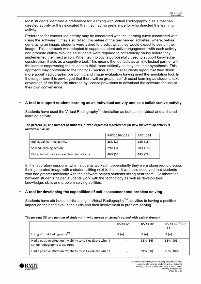
Most students identified a preference for learning with Virtual RadiographyTM as a teacher-directed activity or they indicated that they had no preference for who directed the learning activity.
Preference for teacher-led activity may be associated with the learning curve associated with using the software. It may also reflect the nature of the teacher-led activities, where, before generating an image, students were asked to predict what they would expect to see on their image. This approach was adopted to support student active engagement with each activity and promote critical thinking as students were required to consciously pause before they implemented their next action. When technology is purposefully used to support knowledge construction, it acts as a cognitive tool. This means the tool acts as an intellectual partner with the learner empowering the student to think more critically as they test their hypothesis. This approach may contribute to the findings (Section 3.2.2) that students report that they “think more about” radiographic positioning and image evaluation having used the simulation tool. In the longer term it is envisaged that there will be greater self-directed learning as students take advantage of the flexibility afforded by licence provisions to download the software for use at their own convenience.
• A tool to support student learning as an individual activity and as a collaborative activity
Students have used the Virtual RadiographyTM simulation as both an individual and a sharedlearning activity.
The percent (%) and number of students (n) who expressed a preference for how the learning activity isundertaken as an
RADI1130/1131 RADI1184
Individual learning activity 25% (29) 28% (18)
Shared learning activity 29% (34) 28% (18)
Either individual or shared learning activity 46% (54) 44% (28)
In the laboratory sessions, when students worked independently they were observed to discuss their generated image with a student sitting next to them. It was also observed that students who had greater familiarity with the software helped students sitting near them. Collaboration between students helped students work with the technology as well as develop their knowledge, skills and problem solving abilities.
• A tool for developing the capabilities of self-assessment and problem solving
Students have attributed participating in Virtual RadiographyTM activities to having a positiveimpact on their self-evaluation skills and their involvement in problem solving.
The percent (%) and number of students (n) who agreed or strongly agreed with each statement
RADI1125 RADI1184 RADI1130/RADI1131
Using Virtual RadiographyTM… % (n) % (n) % (n)
Had a positive effect on my ability to self-‐evaluate when I set up radiographic procedures
-‐ 88% (56) 85% (99)
Had a positive effect on my ability to self-‐evaluate when I -‐ 94% (60) 85% (100)

Title of Report Unclassified
Document: Embedding Virtual RadiographyTM within the curriculum to enhance student learning- Authentic
learning in a safe environment using a multichannel delivery approach.doc
Page 11 of 12
evaluate radiographic images
Encouraged me to solve problems 90% (138) 88% (56) 82% (97)
- Indicates this question not part of the survey for this course
Application of critical and reflective thinking to solve clinical challenges is a key attribute of the Australian accredited medical radiation practitioner. This suggests that purposeful use of Virtual radiography may also develop capabilities necessary for autonomous professional practice.

Title of Report Unclassified
Document: Embedding Virtual RadiographyTM within the curriculum to enhance student learning- Authentic
learning in a safe environment using a multichannel delivery approach.doc
Page 13 of 13
Appendix A Include any material that may support your claims of outcomes and impact.
Attach pictures, presentation material, web links and so on that may be important. In particular, please provide an image that can be used for publications, such as a poster.
Example images from Virtual RadiographyTM
Top row: Ideal patient position and resultant generated image
Bottom row: Non-ideal position (patient is rotated) and the resultant generated image