Embed Size (px)
Citation preview

The University of Southern Mississippi The University of Southern Mississippi
The Aquila Digital Community The Aquila Digital Community
Dissertations
Spring 5-2017
Elucidating the Role of Interleukin-17A in West Nile Virus Infection Elucidating the Role of Interleukin-17A in West Nile Virus Infection
Dhiraj Acharya University of Southern Mississippi
Follow this and additional works at: https://aquila.usm.edu/dissertations
Part of the Immunology of Infectious Disease Commons, Immunoprophylaxis and Therapy Commons,
and the Virology Commons
Recommended Citation Recommended Citation Acharya, Dhiraj, "Elucidating the Role of Interleukin-17A in West Nile Virus Infection" (2017). Dissertations. 1376. https://aquila.usm.edu/dissertations/1376
This Dissertation is brought to you for free and open access by The Aquila Digital Community. It has been accepted for inclusion in Dissertations by an authorized administrator of The Aquila Digital Community. For more information, please contact [email protected].

ELUCIDATING THE ROLE OF INTERLEUKIN-17A IN
WEST NILE VIRUS INFECTION
by
Dhiraj Acharya
A Dissertation Submitted to the Graduate School
and the Department of Biological Sciences at The University of Southern Mississippi in Partial Fulfillment of the Requirements for the Degree of Doctor of Philosophy
Approved:
_________________________________________ Dr. Fengwei Bai, Committee Chair Assistant Professor, Biological Sciences
_________________________________________ Dr. Mohamed O. Elasri, Committee Member Professor, Biological Sciences
_________________________________________ Dr. Faqing Huang, Committee Member Professor, Chemistry and Biochemistry
_________________________________________ Dr. Yanlin Guo, Committee Member Professor, Biological Sciences
_________________________________________ Dr. Shahid Karim, Committee Member Associate Professor, Biological Sciences
_________________________________________ Dr. Janet Donaldson Department Chair, Biological Sciences
_________________________________________ Dr. Karen S. Coats Dean of the Graduate School
May 2017

COPYRIGHT BY
Dhiraj Acharya
2017
Published by the Graduate School

ii
ABSTRACT
ELUCIDATING THE ROLE OF INTERLEUKIN-17A IN
WEST NILE VIRUS INFECTION
by Dhiraj Acharya
May 2017
West Nile virus (WNV) is a neurotropic flavivirus of significant public health
importance for which no therapeutics and vaccine are currently available.
Interleukin-17A (IL-17A) is an inflammatory cytokine that regulates diverse
immune functions, while its role is unclear in host’s immune response to WNV.
Furthermore, CD8+ T cells are crucial components of immunity and play a vital
role in recovery from WNV infection. Here, we report a previously unrecognized
function of IL-17A in regulating CD8+ T cell cytotoxicity. We show that WNV
induces the expression of IL-17A in both mouse splenocytes and human
peripheral blood mononuclear cells cultured in vitro, and in plasma of WNV-
infected mice and humans. In a mouse model of WNV infection, we demonstrate
that IL-17A deficient mice (Il17a-/-) are more susceptible to WNV and develop a
higher viral burden compared to wild-type (WT) mice. Interestingly, the CD8+ T
cells isolated from WNV-infected Il17a-/- mice are less cytotoxic and express
lower levels of cytotoxic mediator genes, which can be restored by supplying
recombinant IL-17A in vitro and in vivo. Moreover, treatment of WNV-infected
mice with recombinant IL-17A, as late as day 6 post-infection, significantly
reduces viral burden and increases survival, suggesting a therapeutic potential of
IL-17A. In conclusion, we demonstrate a novel function of IL-17A in promoting

iii
CD8+ T cell cytotoxicity against WNV infection, which may have broad
implications in other microbial infections and cancers.

iv
ACKNOWLEDGMENTS
I am grateful to my Ph.D. advisor Dr. Fengwei Bai for his excellent
supervision and providing a wonderful opportunity to accomplish my research in
his lab. I appreciate his tremendous help, scientific expertise and valuable time.
His encouragement and support considerably helped me to understand science,
learn scientific techniques, and conduct research. I am also grateful to my
research committee members Dr. Faqing Huang, Dr. Mohamed Elasri, Dr. Yanlin
Guo, and Dr. Shahid Karim and like to greatly appreciate their scientific insight,
expertise, and valuable time. I like to thank Dr. Penghua Wang (Yale University)
for his assistance in preliminary studies.
I like to thank my lab mate Amber M. Paul for her assistance in performing
experiments. I am also thankful to Biswas Neupane and all other lab members
for their help and creating an excellent working environment. I like to thank Nick
and Emily for their help in animal care.
I like to thank Dr. Dobrivoje S. Stokic and Dr. Arturo Leis from University of
Mississippi Medical Center for their encouragement in conducting WNV research.
I like to extend my sincere thank to Dr. Richard A. Flavell (Yale University) for
providing IL-17A knockout mice, Dr. John F. Anderson (Connecticut Agricultural
Experiment Station) for providing WNV (CT2471), and Dr. Michael S. Diamond
(Washington University in St. Louis) for providing MC57GLWNV-E and
MC57GLvector cells. Finally, I like to thank Mississippi IDeA Network of Biomedical
Research Excellence (MS-INBRE) for providing excellent research facility.

v
DEDICATION
I dedicate this dissertation to my father Durga Datta Sharma Acharya and
my mother Sharada Sharma Acharya, who are endless source of inspiration and
encouragement throughout my life. I like to extend my gratitude to my beloved
wife Ranjana Baral, and my sisters Rasmi and Roshani for their love and care.
Finally, I like to thank all my friends for their motivation and encouragement.

vi
TABLE OF CONTENTS
ABSTRACT ...........................................................................................................ii
ACKNOWLEDGMENTS .......................................................................................iv
DEDICATION ....................................................................................................... v
LIST OF ILLUSTRATIONS ................................................................................... x
LIST OF ABBREVIATIONS ..................................................................................xi
CHAPTER I - INTRODUCTION ............................................................................ 1
1.1 West Nile Virus ........................................................................................... 1
1.1.1 WNV Is a Major Public Health Problem ...................................................... 2
1.1.2 Pathogenesis of WNV Infection Is Not Well Understood ......................... 3
1.1.3 Host’s Immunity Is Crucial in WNV Recovery ............................................ 4
1.2 Interleukin-17A and Its Signaling ................................................................ 5
1.2.1 IL-17A Regulates Diverse Immune Functions ........................................... 6
1.2.2 Role of IL-17A in Viral Infections Is Unclear .............................................. 7
1.3 Possible Implication of IL-17A in WNV Infection ......................................... 8
1.3.1 IL-17A Is Crucial in the CNS Inflammation ................................................ 8
1.3.2 IL-17A May Control Host’s Immunity to WNV Infection ........................... 9
CHAPTER II – SIGNIFICANCE, HYPOTHESIS, AND INNOVATION ................ 11
2.1 Significance ............................................................................................... 11
2.2 Hypothesis ................................................................................................ 11

vii
2.3 Innovation ................................................................................................. 13
CHAPTER III – EXPERIMENTAL APPROACHES ............................................. 15
3.1 Ethics Statement and Biosafety ................................................................ 15
3.2 Virus Stock and Animal Studies ................................................................ 15
3.3 Cell Culture and In Vitro Infection ............................................................. 16
3.4 Quantitative Polymerase Chain Reaction (qPCR) ..................................... 17
3.5 Enzyme Linked Immunosorbent Assay (ELISA) ....................................... 18
3.6 Confocal Microscopy ................................................................................. 18
3.7 Flow Cytometry ......................................................................................... 19
3.8 CD8+ T cell Isolation and Cytotoxicity Assay ............................................. 19
3.9 Ex Vivo and In Vivo IL-17A Treatment Assays .......................................... 20
3.10 Statistical Analyses ................................................................................. 21
CHAPTER IV – RESULTS.................................................................................. 22
4.1 WNV Induces Expression of Il17a and Il17ra in Humans .......................... 22
4.2 WNV Induces Expression of Il17a and Il17ra in Mice ............................... 23
4.3 WNV-mediated IL-17A Production Depends on IL-23 Signaling ............... 25
4.4 IL-17A Protects Mice from Lethal WNV Infection ...................................... 26
4.5 Mice Deficient in IL-17A Develop Higher Viral Burden .............................. 27
4.6 CNS Leukocyte Slightly Elevates in WNV-infected Il17a-/- mice ............... 28
4.7 WNV-infected Il17a-/- Mice Upregulate CCL5 ........................................... 31
viii
4.8 IL-17A Does Not Affect Interferon Response and Inflammation ................ 32
4.9 IL-17A Does Not Have a Direct Anti-WNV Effect ...................................... 35
4.10 IL-17A Does Not Control Humoral Immune Response ........................... 35
4.11 IL-17A Deficiency Causes the Reduced CD8+ T Cell Cytotoxicity ........... 36
4.12 Il17a-/- CD8+ T Cells Express Less Cytotoxicity Mediators ...................... 39
4.13 IL-17A Induces Cytotoxic Mediators Independent of CD4+ T Cells ......... 41
4.14 IL-17A-induced Cytotoxicity Mediators May Involve ACT-1 Signaling ..... 43
4.15 IL-17A Directly Induces Cytotoxic mediators in CD8+ T Cells.................. 44
4.16 IL-17A Treatment Increases Survival of WNV-infected Mice .................. 47
4.17 IL-17A Induces Cytotoxicity Mediators in WNV-infected Mice ................. 48
CHAPTER V – DISCUSSION ............................................................................. 50
5.1 IL-17A Has a Protective Function in WNV Infection .................................. 50
5.2 Role of IL-17A in IFN, Cytokines and Antibody Response ........................ 51
5.3 Role of IL-17A in Leukocyte Homing to CNS ............................................ 52
5.4 IL-17A Regulates CD8+ T Cell Cytotoxicity ............................................... 53
5.5 IL-17A Has a Potential Therapeutic Role Against WNV ............................ 56
CHAPTER VI – CONCLUSION AND FUTURE DIRECTIONS ........................... 59
6.1 Conclusion ................................................................................................ 59
6.2 Future Directions ....................................................................................... 59
APPENDIX A IACUC and IRB Approval Letter ................................................... 61

ix
REFERENCES ................................................................................................... 64

x
LIST OF ILLUSTRATIONS
Figure 4.1 WNV induces expression of Il17a and Il17ra in humans ................... 23
Figure 4.2 WNV induces expression of Il17a and Il17ra in mice......................... 24
Figure 4.3 WNV-mediate IL-17A response in a IL-23 dependent manner .......... 25
Figure 4.4 Il17a-/- mice are more susceptible to WNV infection .......................... 26
Figure 4.5 IL-17A deficiency results in deficient WNV clearance in mice ........... 28
Figure 4.6 CNS leukocyte profile in WNV-infected Il17a-/- mice .......................... 30
Figure 4.7 WNV infected Il17a-/- mice upregulate CCL5 ..................................... 32
Figure 4.8 Antiviral and inflammatory responses of WNV-infected Il17a-/- mice . 34
Figure 4.9 IL-17A does not directly control WNV replication .............................. 35
Figure 4.10 WNV-mediated IL-17A does not affect antibody production ............ 36
Figure 4.11 CD8+ T cells from Il17a-/- mice have reduced cytotoxicity ................ 38
Figure 4.12 Il17a-/- CD8+ T cells express reduced cytotoxicity mediators ........... 40
Figure 4.13 IL-17A promotes cytotoxic mediators independently of CD4+ cells . 42
Figure 4.14 IL-17A induces the expression of ACT-1 ......................................... 44
Figure 4.15 IL-17A directly induces cytotoxicity mediators in CD8+ T cells ........ 46
Figure 4.16 Recombinant IL-17A increases survival of WNV-infected mice ....... 47
Figure 4.17 Recombinant IL-17A induces cytotoxicity in WNV-infected mice. .... 48

xi
LIST OF ABBREVIATIONS
ACT-1 NF-κB activator 1
ANOVA Analysis of variance
ATCC American type culture collection
BBB Blood brain barrier
BSL3 Biosafety level 3
cDNA Complementary DNA
CHIKV Chikungnya virus
CNS Central nervous system
d.p.i. Days post infection
DMEM Dulbecco’s modified Eagle medium
DNA Deoxyribonucleic acid
ELISA Enzyme linked immunosorbent assay
ERK Extracellular signal-regulated kinase
FBS Fetal bovine serum
Fig Figure
FCB Flow cytometry buffer
hPBMC Human peripheral blood mononuclear cells
i.p. Intraperitoneal
IFN Interferon
IL Interleukin
IL-17A Interleukin-17A
IL-17R Interleukin-17 receptor

xii
IRB Institutional review board
IACUC Institutional animal care and use committee
LCMV Lymphocytic choriomeningitis virus
MOI Multiplicity of infection
mRNA Messenger RNA
NK Natural killer
NKT Natural killer T
NS Nonstructural
PBS Phosphate buffer saline
PFA Paraformaldehyde
PFU Plaque-forming unit
qPCR Quantitative polymerase chain reaction
RANTES Regulated on activation, normal T cell expressed and secreted
RFC Relative fold change
rIL-17A Recombinant IL-17A
RNA Ribonucleic acid
SEFIR Similar expression to fibroblast growth factor and IL-17R
SEM Standard error of mean
TLR Toll-like receptor
TNF Tumor necrosis factor
USM The University of Southern Mississippi
WNV West Nile virus

xiii
WNV-E WNV envelop
WT Wild-type

1
CHAPTER I - INTRODUCTION
1.1 West Nile Virus
West Nile virus (WNV), a neurotropic single-stranded RNA virus belonging
to flaviviridae family, is generally transmitted to human by an infected mosquito
bite, primarily of Culex species (Colpitts et al., 2012; Kuno and Chang, 2005).
However, WNV can also be transmitted through other less frequent transmission
routes, including blood transfusion (Pealer et al., 2003; Stramer et al., 2005),
organ transplantation (Centers for Disease and Prevention, 2009), breastfeeding
(Blazquez and Saiz, 2010), and congenital infections (Alpert et al., 2003). WNV
infection in human can cause fever and may result in injury and death of
neurons, the latter can lead to various neurological manifestations, such as
encephalitis, flaccid paralysis, meningitis, chronic neurologic sequelae and
possibly death, particularly in the elderly and immunocompromised individuals
(Colpitts et al., 2012; Hayes et al., 2005; Samuel and Diamond, 2006).
Structurally, WNV is an enveloped virus with 50 nm diameter, which
comprises an icosahedral nucleocapsid surrounded by a host-derived lipid
envelope (Mukhopadhyay et al., 2003). The WNV genome contains a capped,
single-stranded, and plus-sensed RNA genome of approximately 11 Kb in size.
The WNV genome translates to a polyprotein precursor, which undergoes
posttranslational processing by viral and cellular proteases to generate three
structural (capsid [C], premembrane [PrM], and envelope [E]), and seven non-
structural proteins (NS1, NS2A, NS2B, NS3, NS4A, NS4B, and NS5). Among
these proteins, the structural proteins form the structure of virion, whereas the

2
non-structural proteins are essential in genome replication, virion assembly, and
viral pathogenesis (Chung et al., 2006; Liu et al., 2006; Mukherjee et al., 2011a).
1.1.1 WNV Is a Major Public Health Problem
WNV was first discovered in Uganda about 80 year ago and primarily
maintained in a mosquito-bird-mosquito transmission cycle (Colpitts et al., 2012).
Until 1990’s it caused several sporadic outbreaks of minor public health
importance in Africa, Asia, Europe and the Middle East with a limited capacity to
cause neuroinvasive diseases (Draganescu, 1979; Kramer et al., 2008). The first
report of neuroinvasive cases of WNV in Algeria in 1994 (Le Guenno et al.,
1996), and a huge report of neurological impairments during subsequent WNV
outbreaks in South-East of Romania (Cernescu et al., 1997), and other parts of
southern Europe (Hubalek and Halouzka, 1999) brought this virus into a real
public health concern. In northern hemisphere, WNV was first identified in New
York in 1999 (Hayes, 2001; Mostashari et al., 2001), which subsequently spread
throughout the USA within a few years (Benjelloun et al., 2015).
Currently, WNV is causing endemic diseases throughout the North
America and has a wide geographical distribution in all continents of globe
except Antarctica (Benjelloun et al., 2015). In the US alone, there have been over
40,000 cases of WNV reported between 1999 and 2014, out of which about 45%
were classified as neuroinvasive cases (CDC, 2015). However, the actual burden
of WNV is likely much higher than previously thought because only a proportion
(about 20%) of infected individual develop a clinical disease and a majority of
infected individuals remain asymptomatic (Mostashari et al., 2001). It has been

3
recently estimated that over 3 million individuals have been infected with WNV in
the USA alone, out of which about 780,000 had a symptomatic illness (Petersen
et al., 2013). Considering the worldwide circulation of WNV and supportive
evidence of its capacity to change pathogenicity and transmission potential
(Davis et al., 2005; Ebel et al., 2004; Moudy et al., 2007; Prow et al., 2014; van
den Hurk et al., 2014), there is an urgent need to develop a safe and effective
antiviral drug or vaccine against WNV infection (Martina et al., 2010). However,
no licensed antiviral drug or vaccine is currently available for WNV infection in
humans.
1.1.2 Pathogenesis of WNV Infection Is Not Well Understood
After a bite of WNV-infected mosquito inoculates infectious viruses into
human skin, WNV first infects skin-resident cells such as dendritic cells
(Langerhans cells) and keratinocytes. Infected dendritic cells can carry the
viruses to draining lymph nodes, after which WNV circulates through lymphatic
systems and enters the blood stream (Lim et al., 2011; Ye et al., 2011). WNV can
infect various cells including macrophages, neutrophils, and other leukocytes
leading to development of a transient viremia. Subsequently, WNV can
disseminate to peripheral organs, such as liver and spleen, and then to the brain
and spinal cord. In most of the immune-competent individuals, WNV symptoms
range from unapparent infection to mild febrile illness (Colpitts et al., 2012). In a
few symptomatic cases, particularly in the elderly and immunocompromised
individuals, WNV can cause injury and apoptosis of neuron, which can potentially
cause various neurological impairments and death (Colpitts et al., 2012). Viral

4
entry into the brain is considered a hallmark of WNV pathogenesis and disease
severity. Although the mechanism of neurotropism and how WNV enters the
central nervous system (CNS) is not clearly understood, hematogenous entry via
disrupted blood-brain barrier (BBB) and transneural entry via retrograde axonal
transport have been suggested to play a role (Suen et al., 2014; Suthar et al.,
2013).
1.1.3 Host’s Immunity Is Crucial in WNV Recovery
WNV can infects various type of immune cells and strongly activates host
immune responses, which play important roles in controlling viremia, reducing
viral dissemination to the CNS, and recovery from the disease (Samuel and
Diamond, 2006). Clinical reports of WNV-infected human cases along with
research using mouse models or in vitro approaches have identified critical roles
of several components of immune system in WNV infections (Arjona et al., 2011;
Suthar et al., 2013). In brief, type-I interferon (IFNs) (Lazear et al., 2011; Samuel
and Diamond, 2005), the complement system (Mehlhop and Diamond, 2006),
and humoral immunity (Diamond et al., 2003a; Diamond et al., 2003b) limit
viremia and control WNV dissemination to CNS. Similarly, components of cell-
mediated immunity, including CD4+ (Sitati and Diamond, 2006) and CD8+
(Shrestha and Diamond, 2004) T cells, have been shown to clear WNV from the
CNS, limit viral persistence, and facilitate recovery. In contrast, the roles of
neutrophils, NK cells, and γδ-T cells are still unclear (Bai et al., 2010; Shrestha et
al., 2006a; Welte et al., 2008). Cytokine signaling of interleukin (IL)-23 (Town et
al., 2009), interferon-γ (IFN-γ)(Shrestha et al., 2006b), and IL-1β (Ramos et al.,

5
2012) have been shown to protect against WNV infection, while cytokine such as
IL-10 (Bai et al., 2009) and IL-22 (Wang et al., 2012) have been shown to favor
WNV pathogenicity. The role of tumor necrosis factor-α (TNF-α) still remains
elusive (Shrestha et al., 2008b; Wang et al., 2004a). However, the role of several
other components of immune system yet remains unknown and the mechanism
of WNV pathogenesis, including its tropism to neurons and CNS invasion,
remains unclear. In addition, we have limited knowledge on viral or host factors
that contribute to imbalance between viral pathology and host to WNV infection.
1.2 Interleukin-17A and Its Signaling
IL-17A, a major cytokine of IL-17 family (IL-17A, B, C, D, E, and F), was
discovered in 1993 as the cytotoxic T-lymphocyte antigen-8 (Rouvier et al.,
1993). CD4+ Th17 (Th-17) cells are one of the most common sources of IL-17A.
Besides Th-17 cells, a wide variety of immune cells, including CD8+ T cells, γδ-T
cells, and natural killer T (NKT) cells can also produce IL-17A under various
pathological conditions (Gu et al., 2013). IL-17A signals through IL-17 receptor
(IL-17R) complex composed of IL-17RA and IL-17RC subunits (Ho and Gaffen,
2010). Both subunit of IL-17R complex encode a SEFIR (Similar Expression to
Fibroblast growth factor genes and IL-17 Receptor) domain that mediates various
downstream signaling events through the adaptor protein NF-κB activator-1
(ACT1) (Gaffen, 2009; Ho and Gaffen, 2010). Specifically, the ACT1 recruits to
IL-17R through interaction with SEFIR domain and activates TNF receptor-
associated factor-6 (TRAF6) and TRAF3, which are essential upstream
activators of the nuclear factor- B (NF- B) signaling pathway. In addition, ACT1

6
is also required for IL-17A mediated stabilization of mRNAs that encode several
chemokines and cytokines. Moreover, IL-17A also signals through ACT1
independent pathway by activation of extracellular signal-regulated kinase (ERK)
(Ho and Gaffen, 2010).
1.2.1 IL-17A Regulates Diverse Immune Functions
The finding that IL-23 drives expression of IL-17 in CD4+ T cells led to the
realization of a new paradigm that many functions formerly attributed to CD4+ Th-
1 cells were in fact mediated by a distinct, IL-17-producing T cell subset known
as Th-17 cells (Aggarwal et al., 2003; Langrish et al., 2005). It is now well
established that IL-1β, IL-6, TGF-β, and IL-21 drive development, while IL-23
stabilizes Th-17 cells (Korn et al., 2007; Nurieva et al., 2007; Sutton et al., 2006;
Zhou et al., 2007).
IL-17A signaling regulates diverse immune functions including the
expression of various inflammatory cytokines, chemokines and antimicrobial
peptide, activation and recruitment of leukocytes, and production of antibodies
(Gu et al., 2013; Song and Qian, 2013) during infection and immunity. For
example, IL-17A has often been described as a mediator of inflammation
(Witowski et al., 2004) with prominent roles in allergic and autoimmune diseases
including multiple sclerosis (McFarland and Martin, 2007; Zepp et al., 2011),
rheumatoid arthritis (van den Berg and Miossec, 2009), psoriasis (Raychaudhuri,
2013), asthma (Newcomb and Peebles, 2013), and Crohn’s disease (Siakavellas
and Bamias, 2012). Similarly, IL-17A also plays protective roles against some
bacterial and fungal infections. For instance, IL-17A may enhance neutrophil

7
recruitment and protect against bacterial and fungal pathogens such as
Klebsiella pneumonia and Escherichia coli (Happel et al., 2005; Shibata et al.,
2007; Ye et al., 2001), Listeria monocytogenes (Hamada et al., 2008),
Mycobacterium tuberculosis (Witowski et al., 2004), Francisella tularensis (Lin et
al., 2009), Chlamydia muridarum (Zhang et al., 2009), Candida albicans (Conti et
al., 2009; Huang et al., 2004) and Pneumocystis carinii (Rudner et al., 2007).
Conversely, IL-17A signaling has been suggested to facilitate toxoplasmosis
(Guiton et al., 2010) and certain fungal infections (Zelante et al., 2007).
1.2.2 Role of IL-17A in Viral Infections Is Unclear
The IL-17A has been originally identified as a homolog of virally encoded
ORF13 gene of herpesvirus saimiri (Yao et al., 1995). This discovery generated
interest to study the role of this cytokine in viral infection. It has been
demonstrated that genetically constructed vaccinia virus (VV) expressing IL-17A
(VVIL-17A) caused more severe disease in mice (Patera et al., 2002), but VVIL-17A
was also reported less virulent and IL-17 deficient (Il17a-/-) mice were more
susceptible to VV infection (Kohyama et al., 2007). Thus, the role of IL-17A
during VV infection remains inconclusive.
Interestingly, it has been reported that type I IFNs potently suppress IL-
17A expression (Curtis et al., 2009; Tilg et al., 2009), which further makes the
functional role of IL-17A during viral infections more complex to understand. In
most of the viral infections, type I IFN-mediated antiviral response primarily
occurs during early phase of infection (Lazear et al., 2011; Pinto et al., 2011). It
can be speculated that the IL-17A functions may be more dominant in the later

8
phase of infection when type I IFN expression levels become low. It has been
shown that viral infections can induce expression of IL-17A (Mukherjee et al.,
2011b; Town et al., 2009; Wang et al., 2013), which has been implicated in
priming T cell responses during lymphocytic choriomeningitis virus (LCMV)
hepatitis (Jie et al., 2014) and mediating immunopathogenicity of viral infections
such as influenza virus (Crowe et al., 2009), respiratory syncytial virus (de
Almeida Nagata et al., 2014; Mukherjee et al., 2011b), murine encephalomyelitis
virus (Hou et al., 2009) and hepatitis B virus (Wang et al., 2013). However, the
role of IL-17A in WNV infection in not clearly understood.
1.3 Possible Implication of IL-17A in WNV Infection
It has been previously reported that WNV infection induces Toll-like
receptor-7 (TLR7) dependent production of IL-23 in mice (Town et al., 2009). IL-
23 is known as a prime regulator for stabilization and maintenance of CD4+ Th-17
cells, which are the major cell type secreting IL-17A (Aggarwal et al., 2003;
McGeachy et al., 2009; Stritesky et al., 2008). Thus, it is likely that WNV infection
induces IL-17A expression, which may play a role in WNV immunity. The
following literature provides strong evidence for possible involvement of IL-17A
signaling during WNV infection.
1.3.1 IL-17A Is Crucial in the CNS Inflammation
WNV primarily targets neurons and causes inflammation in the CNS,
which play important role in its pathogenesis. Astrocytes and microglia up-
regulate the expression of functional IL-17A receptor and respond to this cytokine
during brain inflammatory conditions (Das Sarma et al., 2009). In addition, IL-17A

9
is also described as a mediator of CNS inflammation (McFarland and Martin,
2007; Zepp et al., 2011) and suggested as a blood-brain-barrier permeability
factor (Kebir et al., 2007). Therefore, the WNV pathogenesis and host’s immune
response in the CNS are likely impacted by IL-17A.
1.3.2 IL-17A May Control Host’s Immunity to WNV Infection
Considering the role of IL-17A in regulating diverse immune function, it
can be speculated that this cytokine may play an important role in WNV
immunity. For instance, IL-17A has been shown to regulate the expression of
other cytokines including IL-1β, IFN-γ and TNF-α (Maione et al., 2009; Song and
Qian, 2013), infiltration of leukocytes, and production of antibodies (Yao et al.,
1995; Yuan et al., 2010). As these cytokines are previously described to
modulate immunity to WNV (Ramos et al., 2012; Shrestha et al., 2006b;
Shrestha et al., 2008b; Wang et al., 2004a), one can expect possible involvement
of IL-17A in WNV immunity. Moreover, brain-infiltrating leukocytes are crucial in
clearance of WNV (Bai et al., 2010; Shrestha and Diamond, 2004), which can be
potentially controlled by IL-17A signaling. Furthermore, the humoral immune
response, which plays an important role in controlling viremia and WNV
dissemination to CNS (Diamond et al., 2003a), can be potentially regulated by IL-
17A (Yuan et al., 2010).
Among the CNS-infiltrating leukocytes, CD8+ T cells play crucial role in
WNV immunity (Shrestha and Diamond, 2004, 2007; Shrestha et al., 2012;
Shrestha et al., 2006a). Although the relation between IL-17A signaling and CD8+
T cell cytotoxicity is not yet studied, several literatures support this novel axis

10
may play a role in WNV and other viral infections. First, both Th-17 cells and γδ-T
cells, the major cells that produce IL-17A, have been previously shown to
promote CD8+ T cell cytotoxicity during intracellular infections (Hamada et al.,
2009; Xu et al., 2010). Second, the Th-17 cells and IL-17 have also been shown
to control CD8+ T cell function in autoimmune diseases (Ankathatti Munegowda
et al., 2011a). Third, the CD8+ T cells have a crucial role in clearance of tumor
cells (Benchetrit et al., 2002; Martin-Orozco and Dong, 2009). Several studies
also demonstrated the expression of IL-17A in tumor cells (Benchetrit et al.,
2002; Martin-Orozco and Dong, 2009). Interestingly, it has been shown that Th-
17 cell and IL-17A induce cytotoxic function of CD8+ T cells and control tumor
progression (Benchetrit et al., 2002; Martin-Orozco and Dong, 2009), however,
the mechanism for this IL-17A-CD8+ T cell axis remains yet unknown. Therefore,
considering the crucial role of CD8+ T cells in WNV immunity, WNV is a suitable
model to study IL-17A-CD8+ T cell axis.

11
CHAPTER II – SIGNIFICANCE, HYPOTHESIS, AND INNOVATION
2.1 Significance
WNV infection is a significant public health problem, for which no
therapeutics or vaccine are currently available. In addition, IL-17A and CD8+ T
cells regulate diverse immune functions during microbial infections,
malignancies, and autoimmune diseases. In this study, we studied the function of
IL-17A in WNV immunity and identified a potential therapeutic role of IL-17A in
facilitating WNV clearance by promoting CD8+ T cells cytotoxicity. Further
understanding of the interplay between CD8+ T cells and IL-17A axis may lead to
identification of novel therapeutic strategies. Moreover, this novel function of IL-
17A may have broad implications in deciphering the immunopathology of other
viral infections and malignancies, where CD8+ T cells functions are crucial.
2.2 Hypothesis
Despite intensive research over the past 15 years, WNV pathogenesis
and host’s immune response still remains unclear. IL-17A, a major cytokine in the
IL-17 family regulates diverse immune functions including the expression of
various inflammatory cytokines and chemokines, activation and recruitment of
leukocytes, and production of antibodies (Gu et al., 2013; Song and Qian, 2013).
Moreover, IL-17A has often been described as a mediator of inflammation
(Witowski et al., 2004) with an important role in allergic and autoimmune
diseases (McFarland and Martin, 2007; Newcomb and Peebles, 2013;
Raychaudhuri, 2013; van den Berg and Miossec, 2009; Zepp et al., 2011),
malignancies (Martin-Orozco and Dong, 2009; Murugaiyan and Saha, 2009), and

12
infections (Conti et al., 2009; Hamada et al., 2008; Happel et al., 2005; Huang et
al., 2004; Lin et al., 2009; Rudner et al., 2007; Shibata et al., 2007; Ye et al.,
2001; Zhang et al., 2009). However, the role of IL-17A signaling is not clear
during viral infections and has been previously studied in WNV infection. As we
described in the introduction (section 1.7), there are several evidences
suggesting a possible role of IL-17A in controlling host’s immune response to
WNV. Therefore, we hypothesized that IL-17A may play a crucial role in WNV
immunity. In this study, we tested this hypothesis under following four specific
aims.
Specific Aim 1: Study the expression of IL-17A during WNV infection in
vitro and in vivo. It has been previously reported that TLR7 mediates IL-23-
dependent protective immune responses against WNV infection in mice (Town et
al., 2009). IL-23 is known as a prime regulator for stabilization and maintenance
of CD4+ Th-17 cells, which are the major cell type secreting IL-17A (Aggarwal et
al., 2003; McGeachy et al., 2009; Stritesky et al., 2008). Since IL-17A is also
described as a mediator of CNS inflammation (McFarland and Martin, 2007;
Zepp et al., 2011), and a factor contributing to blood-brain-barrier permeability
(Kebir et al., 2007), we asked if WNV induces IL-17A production and whether it
involves IL-23 signaling.
Specific Aim 2: Study the role IL-17A in WNV immunity in a mouse model.
Based on the literature, it is likely that IL-17A may play either protective or
pathogenic role during WNV infection. To dissect this question, we used a mouse

13
model of WNV infection and compared survival and viral burden of WNV infected
IL-17A deficient (Il17a-/-) and WT control mice.
Specific Aim 3: Study the immune functions of IL-17A during WNV
infection. IL-17A has been shown to recruit leukocytes (Happel et al., 2005; Ye et
al., 2001), control cytokine and chemokine expression (Maione et al., 2009; Song
and Qian, 2013), and regulate humoral immune response (Grund et al., 2012;
Tarlinton, 2008; Yuan et al., 2010). Under this specific aim, we asked how
various immune functions were controlled by IL-17A during WNV infection.
Specific Aim 4: Study the mechanism of IL-17A mediated regulation of
WNV immunity. From Aim 3, we identified a novel function of IL-17A in regulation
of CD8+ T cell cytotoxicity. Under Aim 4, we studied the mechanism of IL-17A
mediated control of CD8+ T cells cytotoxicity and its potential therapeutic role
against WNV infection.
2.3 Innovation
Cytotoxic CD8+ T cells are important component of immunity, particularly
in clearance of intracellular infections and tumors. One of the effector
mechanisms used by these cells is cytotoxic killing of target cells. However,
regulators of CD8+ T cell cytotoxicity, in particular, the expression of cytotoxicity
mediators in these cells is poorly understood. In addition, functions of several
molecules expressed in CD8+ T cells remain uncharacterized. One such
molecule is the IL-17A receptor (IL-17R), which is expressed on the surface of
CD8+ cells (Lindemann et al., 2008; Yao et al., 1995). In additions, subsets of
CD8+ cells can produce IL-17A (Hamada et al., 2009). Considering the role of IL-

14
17A in regulation of diverse immune functions during microbial infections,
malignancies, and autoimmune diseases, it is likely that IL-17A signaling may
play a role in biology of CD8+ T cells. CD8+ T cells are particularly important in
controlling viral infections and have been shown to play a crucial role in
clearance of WNV infection (Shrestha and Diamond, 2004). In this study, we
studied the role of IL-17A in a mouse model of WNV infections and discovered a
novel, previously unrecognized, function of IL-17A in promoting CD8+ T cells
cytotoxicity.

15
CHAPTER III – EXPERIMENTAL APPROACHES
3.1 Ethics Statement and Biosafety
Written informed consent was obtained from all human volunteers and
human WNV cases prior to their enrollment in this study. The protocol for human
subject has been reviewed and approved by the University of Southern
Mississippi (USM) Institutional Review Board (IRB, protocol # CH-R11120601,
Appendix A). All animal experimental procedures were reviewed and approved
by the Institutional Animal Care and Use Committee at USM (protocol
#12041201, Appendix A). All the in vitro experiments and animal studies
involving infectious WNV were performed by the certified personnel in biosafety
level 3 (BSL3) or animal biosafety level 3 (ABSL3) laboratories following
standard biosafety protocols approved by USM Institutional Biosafety Committee.
3.2 Virus Stock and Animal Studies
Low-passaged WNV isolate CT2741 (Anderson et al., 1999) was provided
by Dr. John F. Anderson at the Connecticut Agricultural Experiment Station. Low-
passaged CHIKV LR OPY1 2006 strain was provided by Dr. Robert B. Tesh at
the University of Texas Medical Branch. WNV and CHIKV stocks used in this
study were prepared by propagating the viruses in Vero cells through a single
passage and titrated in Vero cells by a plaque-forming assay as previously
described (Bai et al., 2005). Vero cells (ATCC CCL-81) were cultured in a 37°C
incubator with 5% CO2 in Dulbecco’s modified Eagle medium (DMEM, Life
Technologies) supplemented with 10% fetal bovine serum (FBS). Interleukin-17A
deficient (Il17a-/-) mouse breeding pairs (C57BL/6J background) were provided

16
by Dr. Richard A. Flavell at the Yale University School of Medicine and wild-type
(WT) control mice (C57BL/6J) were purchased from The Jackson Laboratory
(Bar Harbor, ME). IL-23 deficient (Il23p19-/-) breeding pairs on a mixed C57BL/6
× 129 background were obtained from the Mutant Mouse Regional Resource
Center (MMRRC). Mice were housed under standard conditions in the animal
facility at USM. Gender-matched and 7 to 9 weeks old Il17a-/- and WT control
mice were infected with 1,000 plaque forming units (PFUs) of WNV by i.p.
injection in 100 µl of phosphate buffer saline (PBS) containing 5% gelatin (Town
et al., 2009). For footpad inoculation, 100 PFU of WNV in 50 µl PBS containing
1% FBS was injected into the mouse footpad after isofluorane anesthesia (Bai et
al., 2009). Infected animals were observed twice daily for up to 21 days for
morbidity and mortality.
3.3 Cell Culture and In Vitro Infection
Human peripheral blood mononuclear cells (hPBMCs) were isolated from
blood of healthy human volunteers using Ficoll-paqueTM PLUS (GE healthcare).
To isolate murine splenocytes, healthy C57BL/6J mice (7-week old) were
euthanized and spleens were collected to make single cell suspension. After red
blood cell lysis, both mouse splenocytes and hPBMCs were purified and cultured
in the Dulbecco’s modified Eagle medium (DMEM, Life Technologies)
supplemented with 10% FBS, 2 mM L-glutamine, and 1% non-essential amino
acids. Cells were infected with WNV (MOI = 0.1, 1 or 5) and collected in
TRIreagent (Molecular Research Center) at 24 h and 48 h for total RNA
extraction.

17
3.4 Quantitative Polymerase Chain Reaction (qPCR)
Total RNA was extracted from cells or animal tissues (i.e. blood, spleen,
and brain) using the TRIreagent or RNeasy kit with on-column DNA digestion
(Qiagen). First-stranded complementary DNA (cDNA) was synthesized using the
iSCRIPTTM cDNA synthesis kit (Bio-Rad). WNV-envelope (WNVE) RNA copy
numbers were quantified using probe-based qPCR and normalized to cellular β-
actin gene, as previously described (Bai et al., 2005). qPCR assays for cytokine,
chemokine, and other immunological marker genes were performed using SYBR
Green supermix (Bio-Rad) and data are presented either as the relative fold
change (RFC) by ΔΔCT method using β-actin as a housekeeping gene or as a
copy number ratio of target gene to cellular β-actin. Primer sequences for mice
(Bai et al., 2005) and human (Kozaci et al., 2007) β-actin are previously
described. Primers sequences for human Il17a (F, 5’-
TGTGATCTGGGAGGCAAAGT-3’; R, 5’-GATCTCTTGCTGGATGGGGA-3’), and
mouse Il17a (F, 5’-TCTCCACCGCAATGAAGACC-3’, R, 5’-
TTTCCCTCCGCATTGACACA-3’); perforin-1 (F, 5'-
TGTTCCTCCTGGGCCTTTTC-3'; R, 5'-CCATACACCTGGCACGAACT-3'),
granzyme-A (F, 5'-CACGTGAGGGGGATCTACAAC-3'; R, 5'-
TCTCCCCCATCCTGCTACTC-3'), granzyme-B (F, 5'-
TGCTACTGCTGACCTTGTCTC-3'; R, 5'-CCATGTAGGGTCGAGAGTGG-3'),
fasL (F, 5'-GAACTGGCAGAACTCCGTGA-3'; R, 5'-
TGAGTGGGGGTTCCCTGTTA-3'), Ifn-α (F, 5'-TTCCCCTGACCCAGGAAGAT-
3'; R, 5'-CTTCTGCTCTGACCACCTCC-3'), Ifn-β (F, 5'-

18
TGTCCTCAACTGCTCTCCAC-3'; R, 5'-ATCTCTGCTCGGACCACCAT-3'), Act-1
(F, 5’-GAGGACGAGCATGGCTTACA-3’; R, 5’-TGGCATTTGGGAAGAGCACA -
3’) were designed using NCBI’s primer designing tool and synthesized by
Integrated DNA Technologies.
3.5 Enzyme Linked Immunosorbent Assay (ELISA)
IL-17A, IFN-β, IL-1β, IFNγ, TNFα, IL-6, IL-10, IL-12p40, and anti-WNV-E
IgM antibody in plasma of WNV-infected mice (1,000 PFU, i.p.) were measured
using ELISA kit (R&D Systems) following manufacturer’s instructions. Level of IL-
17A in culture media, and sera of human WNV cases and healthy controls was
measured by ELISA kit from Enzo Life Sciences.
3.6 Confocal Microscopy
Brains were collected from WNV-infected mice (1,000 PFU, i.p.) after
intracardial PBS perfusion, fixed overnight in 4% PFA at 4°C, and cryoprotected
in sucrose. Para-median sagittal sections (25 μm) of brain were pre-blocked for
30 min at room temperature and then probed overnight at 4°C with a combination
of primary antibodies against CD11b, CD45 and WNV antigen (anti-WNV
antibody provided by John F. Anderson, other antibodies purchased from BD
Biosciences). After a PBS wash, sections were probed with appropriate
fluorescent labeled secondary antibodies for 1 h at room temperature,
counterstained with DAPI (Invitrogen), and mounted in fluorescence mounting
medium (ProLong Gold). Images were acquired in independent channels using a
Nikon A1R confocal microscope.

19
3.7 Flow Cytometry
Brains were collected from WNV-infected (1,000 PFU, i.p.) mice after
intracardial PBS perfusion and processed into a single cell suspension. Brain
leukocytes were isolated using a discontinuous Percoll gradient (GE Healthcare)
and probed with CD45, CD4, CD8, and CD11b (BD Biosciences or eBioscience).
After staining, cells were washed two times in flow cytometry buffer (FCB, PBS
with 2% FBS) and fixed in 4% paraformaldehyde (PFA) for 15 minutes. For
intracellular staining, cells were permeabilized and probed with antibodies
against perforin, and granzyme A (eBioscience). Cells were then washed and re-
suspended in FCB. Data were acquired on a flow cytometer (BD LSRFortessa)
and analyzed with FlowJo or FACSDivaTM software (BD Biosciences).
3.8 CD8+ T cell Isolation and Cytotoxicity Assay
Spleens were collected from WNV-infected (100 PFU, i.p.) mice at 10
d.p.i.. Splenic CD8+ T cells were isolated by negative antibody selection with
magnetic beads using the mouse CD8+ T Lymphocyte Enrichment Set-DM (BD
Biosciences). Purity of CD8+ T cells was examined by flow cytometry after
staining with fluorescent labeled anti-CD3 and anti-CD8 antibodies
(eBioscience). Cytotoxicity of CD8+ T cells was measured as described
previously (Shrestha and Diamond, 2004), with some modifications. Briefly,
purified CD8+ T cells (~ 80-90% purity) were co-cultured for 4 h in 96-well plates
with target cells expressing the ectodomain of WNV-E (MC57GLWNV-E) or control
cells containing the expression vector (MC57GLvector) (gifted by Dr. Michael S.
Diamond) with effector to target cell ratios of 50:1. CD8+ T cell cytotoxicity was

20
measured using a LDH Cytotoxicity Detection Kit (Thermo Scientific). Transcripts
of perforin-1, granzyme A, granzyme B and Fas-ligand (FasL) genes in purified
CD8+ T and CD8- cells were measured by qPCR, as described above.
3.9 Ex Vivo and In Vivo IL-17A Treatment Assays
For ex vivo studies, splenocytes were isolated from WNV-infected (100
PFU, i.p.) mice at 8 d.p.i. and splenic CD8+ T cells were purified as described
above. In some experiments, CD4+ T cells were depleted from splenocytes using
CD4 Magnetic Particles - DM (BD Biosciences). Splenocytes, purified CD8+ T
cells, or CD4- splenocytes were cultured for 24 h in the presence of mouse
recombinant IL-17A (50 ng/ml, eBioscience). In some experiments, splenocytes
were cultured for 24 h in the presence of mouse recombinant IL-17A (50 ng/ml)
and subjected to CD8+ T cell purification. Expression of perforin-1, granzyme A,
granzyme B and FasL in splenocytes, CD8+ T cells, and splenic CD8- cells were
measured by qPCR, as described above.
For in vivo IL-17A treatment and survival studies, WT female mice (8-
week old) were inoculated with WNV (100 PFU) via i.p. route. At day 6 p.i, mice
were treated (2.5 μg/mouse) with i.p. administration of carrier-free mouse
recombinant IL-17A (eBioscience) or PBS, and monitored daily for mortality and
morbidity for up to 21 days. Randomly selected mice were euthanized at day 8
p.i. and WNV burden in brain and the expression of the cytotoxic mediators in
splenic CD8+ and CD8- T cells were measured by qPCR as described above.

21
3.10 Statistical Analyses
Data were analyzed using two-tailed student’s t-test or analysis of
variance (ANOVA) in GraphPad Prism (GraphPad Software, version 6), with p <
0.05 considered statistically significant.

22
CHAPTER IV – RESULTS
4.1 WNV Induces Expression of Il17a and Il17ra in Humans
We previously reported that WNV induces IL-23 production in mice in a
TLR7-dependent manner (Town et al., 2009). Considering the role of IL-23 in
Th17 cell stabilization and IL-17A production (Aggarwal et al., 2003), we
hypothesized that IL-17A may play a role in WNV infection. To test this, we
measured the expression of Il17a in human cells infected with WNV in vitro.
Human peripheral blood mononuclear cells (hPBMCs) isolated from healthy
human volunteers without a history of WNV infection were infected with WNV
(MOI = 0.1 and 5) for 24 h and 48 h in vitro. WNV-infected hPBMCs were
collected, total RNA isolated, and cDNA synthesis and quantitative real-time PCR
(qPCR) was performed to measure transcripts of Il17a and cellular β-actin as a
housekeeping gene. The qPCR results showed that Il17a gene expression was
up-regulated in WNV-infected hPBMCs (Fig. 4.1A), which was further confirmed
by measuring IL-17A production in hPBMC culture supernatants (Fig. 4.1B) by an
enzyme-linked immunosorbent assay (ELISA).
To relate these in vitro results to WNV infection in humans, we used
ELISA to measure the production of IL-17A in the sera of human cases with
active WNV infection (fever or neuroinvasive disease), with a history of recovered
neuroinvasive WNV diseases, and healthy controls who have no history of WNV
infection. The cases with active and long-standing history of neuroinvasive WNV
diseases showed a trend of higher levels of IL-17A in sera compared to WNV
fever cases and healthy controls (Fig. 4.1C), with no difference between the latter

23
two. These results demonstrate that WNV infection induces a production of IL-
17A in humans and suggest that this cytokine may play a role in WNV infection.
Figure 4.1 WNV induces expression of Il17a and Il17ra in humans
(A) Il17a transcripts were measured by qPCR and expressed as relative fold change (RFC) after normalization to cellular
β -actin in human PBMCs infected with WNV for 24 h or 48 h. (B) IL-17A production in culture supernatant of WNV-
infected hPBMC was measured by ELISA. (C) Levels of IL-17A in sera of human WNV patients and healthy controls were
measured by ELISA. Error bars represent standard error of mean (mean ± SEM). Data in A and B represent two
independent experiments performed in triplicates and analyzed by a one-way ANOVA. “ns” denotes no significant
difference (p > 0.05).
4.2 WNV Induces Expression of Il17a and Il17ra in Mice
To expand upon the findings from human samples, we used a mouse
model of WNV infection because it reflects various aspects of human WNV
disease (Bai et al., 2009; Bai et al., 2005; Town et al., 2009). Splenocytes
isolated from C57BL/6J mice were infected with WNV (MOI = 0.1) in vitro for 24 h
Mock 0.1 MOI 5 MOI 0.1 MOI 5 MOI
0
10
20
30
40
50
IL17A
RF
C
WNV (24h) WNV (48h)
p < 0.05
0
10
20
30
40
IL-1
7A
(p
g/m
l)
Healthy
control
WN
fever
Neuroinvasive
(acute)
Neuroinvasive
(recovered)
n=7 n=8
n=10
n=14
Mock 0.1 MOI 5 MOI0.0
0.5
1.0
1.5
2.0
IL-1
7A
(p
g/m
l)
Human
PBMCs
WNV (48h)
p < 0.005

24
and 48 h, and the expression of Il17a gene was measured by qPCR. Similar to
hPBMC, Il17a transcript levels were up-regulated at both 24 and 48 h.p.i. in
mouse splenocytes infected with WNV in vitro (Fig. 4.2A).
Figure 4.2 WNV induces expression of Il17a and Il17ra in mice
(A) RFC of Il17a transcripts after normalization to cellular β -actin in mouse splenocytes infected with WNV (MOI = 0.1) in
vitro. (B to C) WT (C57BL/6) mice were infected with WNV (1,000 PFU, i.p.) and expression of Il17a (B) and Il17ra (C)
transcripts were measured in brain tissue by qPCR. Error bars represent standard error of mean (mean ± SEM). Data in A
represent two independent experiments performed in triplicates and analyzed by a one-way ANOVA. Data in B and C
represent two independent experiments (n = 5 mice/group) and analyzed by two-tailed student's t-test; “ns” denotes no
significant difference (p > 0.05).
Since astrocytes, microglia, and brain infiltrating immune cells express
functional interleukin-17 receptor A (IL-17RA) in brain inflammatory conditions
(Das Sarma et al., 2009), we measured the expression of Il17a and Il17ra genes
in brain of WNV-infected mice. For this, we infected a group of WT mice with
WNV (1,000 PFU, i.p.), sacrificed them at various time points to collect brains,
and measured levels of Il17a and Il17ra transcripts by qPCR. Indeed, there was
a significantly up-regulated expression of both Il17a (Fig. 4.2B) and Il17ra (Fig.
4.2C) genes in brain of WNV-infected mice compared to uninfected controls.
Mock 24h 48h0
2
4
6
Il17a R
FC
p < 0.05
WNV (0.1 MOI)
Brain
Control 1 2 40
1
2
3
4
Il17a R
FC
d.p.i.
p < 0.05
Mice per group (n=5)
Control 2 4 60
1
2
3
Il17ra
R
FC
p < 0.05
d.p.i
Mice per group (n=5)
Brain

25
Collectively, these results indicate that WNV infection elevates the expression of
both Il17a and Il17ra in mice, suggesting a possible role of IL-17A in WNV
infection.
4.3 WNV-mediated IL-17A Production Depends on IL-23 Signaling
To further measure Il17a expression in mice and to test whether its
production was IL-23 dependent, we intraperitoneally (i.p.) infected a group of
wild-type (WT) littermates and IL-23 deficient (Il23p19-/-) mice (both are in mixed
C57BL/6×129 background) with 1,000 plaque-forming units (PFUs) of WNV and
measured IL-17A protein in plasma by ELISA. The results showed that WNV
infection in mice induced IL-17A production (Fig. 4.3), but the level of this
cytokine was undetectable in serum samples from mock-infected control mice
(data not shown).
Figure 4.3 WNV-mediate IL-17A response in a IL-23 dependent manner
IL-17A production was measured in plasma of Il23p19-/- and their littermate WT control mice (7-9 week old)
infected with WNV (1,000 PFU, i.p.) by ELISA. Error bars represent standard error of mean (mean ± SEM).
Data represent two independent experiments (n = 3 to 5 mice/group) and analyzed by two-tailed student's t-
test; “ns” denotes no significant difference (p > 0.05).

26
Moreover, there was approximately 80% reduction in Il17a expression in
Il23p19-/- mice at 3 days post infection (d.p.i), suggesting that IL-17A production
during WNV infection in mice largely depends on IL-23 signaling (Fig. 4.3).
4.4 IL-17A Protects Mice from Lethal WNV Infection
To further investigate the role of IL-17A in WNV immunity, we used a
mouse model of WNV encephalitis (Town et al., 2009). IL-17A deficient (Il17a-/-)
and WT control mice (7-8 weeks old, sex-matched, strain C57BL/6J) were
challenged via i.p. injection with 1,000 PFUs of WNV (Town et al., 2009), a dose
that kills approximately 40 ~ 50% of WT animals. Morbidity and mortality was
monitored twice daily for 21 days. We found strikingly greater susceptibility of
Il17a-/- mice (20% survival) vs. WT control mice (60% survival) to lethal WNV
infection (Fig. 4.4A).
Figure 4.4 Il17a-/- mice are more susceptible to WNV infection
Seven to nine weeks old WT (C57BL/6J) and Il17a-/- mice were infected with WNV via i.p. (1,000 PFU) or
footpad (100 PFU) routes and monitored for mortality twice daily for 21 days; percentage (%) survival was
compared using the Kaplan-Meier survival and log-rank test. (A) Survival curves after i.p inoculation, or (B)
footpad inoculation.
0 5 10 15 200
20
40
60
80
100
d.p.i
Su
rviv
al (%
)
WT (n=28)
Il17a-/- (n=26)
i.p.p < 0.01
0 5 10 15 200
20
40
60
80
100
d.p.i
Su
rviv
al (%
)
WT (n=10)
Il17a-/-(n=8)
p < 0.01footpad

27
To exclude the possibility of an inoculation route specific response, we
also challenged Il17a-/- and WT mice with 100 PFUs of WNV via footpad (Bai et
al., 2009), and performed survival analysis. Similar to the i.p. route, footpad
inoculation showed that Il17a-/- mice were more susceptible to WNV infection
(Fig. 4.4B). Together, these data suggest that IL-17A protects mice from lethal
WNV infection.
4.5 Mice Deficient in IL-17A Develop Higher Viral Burden
To further expand the protective function of IL-17A in controlling WNV
infection, we compared the virological profiles of WNV-infected Il17a-/- vs. WT
mice in various tissues such as blood, spleen, liver and brain. Measurement of
WNV viremia by qPCR revealed no difference at 2 d.p.i, however, a 3-fold
increase in the transcript level of WNV envelope gene (WNVE) was observed at
4 d.p.i. in WNV-infected Il17a-/- mice compared to WT controls (Fig. 4.5A).
To assess the viral burden in peripheral organs, we sacrificed WNV-
infected mice, collected liver, spleen and brain samples at the selected time
points, and performed a qPCR analysis. When compared to WT controls, we
found approximately 2-fold increase in WNV-E transcripts in the liver of Il17a-/-
mice (Fig. 4.5B). Consistent with the survival results, there were about 6-fold (at
7 d.p.i.) and 30-fold (at 8 d.p.i.) increases in WNVE transcripts in the brain of
WNV-infected Il17a-/- mice compared to WT controls (Fig. 4.5C). Although there
was no difference in viral burden in spleens of WT vs. Il17a-/- mice at 4 d.p.i,
Il17a-/- mice had significantly higher level (about 3-fold) of WNVE transcripts on 8
d.p.i (Fig. 4.5D).
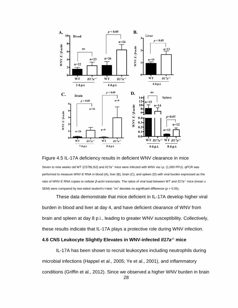
28
Figure 4.5 IL-17A deficiency results in deficient WNV clearance in mice
Seven to nine weeks old WT (C57BL/6J) and Il17a-/- mice were infected with WNV via i.p. (1,000 PFU). qPCR was
performed to measure WNV-E RNA in blood (A), liver (B), brain (C), and spleen (D) with viral burden expressed as the
ratio of WNV-E RNA copies to cellular β-actin transcripts. The ratios of viral load between WT and Il17a-/- mice (mean ±
SEM) were compared by two-tailed student's t-test; “ns” denotes no significant difference (p > 0.05).
These data demonstrate that mice deficient in IL-17A develop higher viral
burden in blood and liver at day 4, and have deficient clearance of WNV from
brain and spleen at day 8 p.i., leading to greater WNV susceptibility. Collectively,
these results indicate that IL-17A plays a protective role during WNV infection.
4.6 CNS Leukocyte Slightly Elevates in WNV-infected Il17a-/- mice
IL-17A has been shown to recruit leukocytes including neutrophils during
microbial infections (Happel et al., 2005; Ye et al., 2001), and inflammatory
conditions (Griffin et al., 2012). Since we observed a higher WNV burden in brain
WT Il17a-/- WT Il17a-/-
0
2
4
6
8
10
WN
V E
/ β
-act
in
2 d.p.i. 4 d.p.i.
ns
p < 0.05
n=22
n=23 n=26
n=24
WT Il17a-/-
0
1
2
3
WN
V E
/ β
-act
in
p < 0.05
4 d.p.i.
n=15
n=13
WT Il17a-/- WT Il17a-/-
0
1
2
3
4
5
WN
V E
/ β
-act
in
p < 0.05
7 d.p.i.
n=24
n=21
p < 0.05
n=9
n=9
8 d.p.i.
WT Il17a-/- WT Il17a-/-
0.0
0.1
0.2
0.3
0.4
68
101214
WN
V E
/ β
-act
in
ns
4 d.p.i.
n=15n=14
8 d.p.i.
p<0.05
n=12
n=12

29
of Il17a-/- mice, we asked if this was related to IL-17A-mediated control of
leukocyte infiltration into brain during WNV infection. For this, we performed
confocal microscopy to detect WNV-E antigen, CD45 (pan-leukocyte marker),
and CD11b (microglial and macrophage marker) in brain sections of WNV-
infected Il17a-/- and WT mice sacrificed at 6 d.p.i. We focused on the olfactory
bulb because we have shown that this brain region is most sensitive to WNV
infection (Bai et al., 2009; Town et al., 2009; Wang et al., 2004a). Consistent with
the qPCR measurement of WNVE RNA in brain tissue (Fig. 4.5C), the confocal
imaging revealed more WNVE antigens in the brains of WNV-infected Il17a-/-
mice compared to WT controls (Fig. 4.6A and B). Similar results were also
obtained in other brain regions, including the cerebral cortex, brainstem,
cerebellum, and striatum (data not shown). Unexpectedly, confocal imaging
results showed more CD45+ (Fig. 4.6A) and CD11b+ leukocytes (Fig. 4.6B) in the
brain of WNV-infected Il17a-/- mice than WT controls.
To confirm these data and further quantify brain infiltrating immune cells,
we performed flow cytometric analysis of brain leukocytes isolated from WT and
Il17a-/- mice infected with 1,000 PFU (i.p.) of WNV for 6 days. We characterized
CD45+, CD11b+, CD3+CD4+ and CD3+CD8+ cell populations, as previously
described (Town et al., 2009). Consistent with the confocal imaging results, we
observed a trend towards elevation of all leukocyte population in the brain of
WNV-infected Il17a-/- mice compared to WT controls (Fig. 4.6C).

30
Figure 4.6 CNS leukocyte profile in WNV-infected Il17a-/- mice
Seven to nine weeks old WT (C57BL/6J) and Il17a-/- mice were challenged with WNV (1,000 PFU) through
i.p.. (A to B) PBS perfused brains were isolated on 6 d.p.i, and WNV antigen (green signal) and CD45
(leukocyte common antigen, red, panel A) or CD11b (macrophage and microglia marker, red, panel B) were

31
detected by Nikon A1R confocal microscope (original magnification = 20X). DAPI (blue signal) was used as
a nuclear counterstain; representative images are shown. (C) Brain leukocytes isolated on day 6 d.p.i. were
characterized by flow cytometry after probing with antibodies against WNVE, CD45, CD4, CD8, and CD11b.
Signal color used in dot plots and numbers representing the percentages of positive cells within gated
populations are shown (right). Data represent two independent experiments (n = 5 mice per group for each
experiment).
To test whether the higher leukocyte infiltration into WNV-infected Il17a-/-
mouse brains was affected by leukocyte expansion or differentiation in the
periphery, we compared leukocyte populations in spleen of Il17a-/- and WT mice
infected with WNV. There was no difference in leukocyte populations in spleens
of WNV-infected Il17a-/- vs. WT mice (data not shown), suggesting that more
leukocytes in brain may not be due to the possible effects of IL-17A on leukocyte
expansion in the periphery.
4.7 WNV-infected Il17a-/- Mice Upregulate CCL5
To further dissect the mechanism by which more leukocytes migrate into
the brains of WNV infected Il17a-/- mice, we performed qPCR to measure the
expression of selected chemokines genes (Cxcl1, Cxcl10, Ccl5) known to
mediate recruitment of leukocytes. There was significantly elevated expression of
Ccl5 (also known as RANTES) (Fig. 4.7A) and its receptor Ccr5 (Fig. 4.7B) in
blood of WNV infected Il17a-/- mice at 4 d.p.i., but no difference in expression of
other chemokines, such as Cxcl1 and Cxcl10 (data not shown). In addition, there
was a significant increase of Ccl5 expression (Fig. 4.7C) in the brains of WNV-
infected Il17a-/- mice at 8 d.p.i., but no difference in expression of other leukocyte
recruiting chemokines or chemokine receptors, such as Cxcl10 (Fig. 4.7D) and

32
Cxcr3 (Fig. 4.7E). These results may imply a link between deficient IL-17A and
higher Ccl5 expression that may contribute to more leukocyte homing to the brain
of Il17a-/- mice during the course of WNV infection.
Figure 4.7 WNV infected Il17a-/- mice upregulate CCL5
Expressions of Ccl5 (A) and Ccr5 (B) gene at the mRNA levels were measured by qPCR in the blood of
WNV-infected mice at 2 and 4 d.p.i.. mRNA levels of Ccl5 (C), Cxcl10 (D) and Cxcr3 (E) genes were
measured by qPCR in brain of WNV-infected mice at 8 d.p.i. Gene expression data were normalized to
cellular β-actin mRNA and compared by two-tailed student's t-test; “ns” denotes no significant difference (p >
0.05).
4.8 IL-17A Does Not Affect Interferon Response and Inflammation
WNV infection induces potent type I IFN responses in mice, which plays a
critical role in controlling both viremia and encephalitis (Samuel and Diamond,
2005). We tested whether deficient IL-17A alters type I IFN expression during
WT Il17a-/- WT Il17a-/-
0
5
10
15
20
Ccl
5/ β-a
ctin
2 d.p.i. 4 d.p.i.
ns
p < 0.005
n=25 n=24
n=10
n=10
WT Il17a-/- WT Il17a-/-
0.00
0.02
0.04
0.06
0.08
Ccr
5/ β-a
ctin
ns
p < 0.005
2 d.p.i. 4 d.p.i.
n=25 n=23
n=25
n=23
WT Il17a-/-
0
5
10
15
20
25
Ccl
5/ β-a
ctin
8 d.p.i.
p < 0.05
n=9
n=9
WT Il17a-/-
0.0
0.1
0.2
0.3
0.4
0.5
Cxcl
10/ β-a
ctin
8 d.p.i.
ns
n=6 n=6
WT Il17a-/-
0.00
0.02
0.04
0.06
Cxcr
3/ β-a
ctin
8 d.p.i.
ns
n=6 n=6

33
WNV infection by qPCR and ELISA. The qPCR results showed no difference in
Ifn- expression in blood of Il17a-/- vs. WT control mice at 4 d.p.i. (Fig. 4.8A).
Similarly, no difference in expression of Ifn- gene was observed in blood (Fig.
4.8B), spleen (Fig. 4.8C), liver (Fig. 4.8D), and brain (Fig. 4.8E) samples from
WNV-infected Il17a-/- vs. WT control mice measured at various time points. To
further confirm these results, we also measured IFN- protein in plasma of WNV-
infected WT and Il17a-/- mice at 3 d.p.i. by ELISA and found no difference in IFN-
expression (Fig. 4.8F). These results suggest that the type I IFNs response
remains unaltered in Il17a-/- mice during WNV infection.
IL-17A cause potent inflammation and regulate expression of several
cytokines including IL-1β, IFNγ and TNFα (Maione et al., 2009; Song and Qian,
2013). We assessed the possible role of IL-17A in inflammatory responses
during WNV infection by measuring inflammatory cytokine expression in plasma
by ELISA. Again, there was no difference in levels of IL-1β, IL-6, IL-10, IFN-γ, IL-
12 p40, and TNF-α in plasma from WNV-infected Il17a-/- vs. WT control mice at
both 1 and 3 d.p.i. (Fig. 4.8G-L). In addition, no significant difference in
expression of these cytokines was detected by qPCR in brain of WNV-infected
Il17a-/- vs. WT control mice at 8 d.p.i. (data not shown). Collectively, these results
demonstrate that higher viral load in Il17a-/- mice is likely not due to altered
production of interferon and other inflammatory cytokines.

34
Figure 4.8 Antiviral and inflammatory responses of WNV-infected Il17a-/- mice
Seven to nine weeks old WT (C57BL/6J) and Il17a-/- were infected with 1,000 PFU (i.p.) of WNV. Blood, plasma, and
tissue samples were collected at the selected time points (d.p.i.) for cytokine and anti-WNV-E IgM measurement.
Expression of interferon- (Ifn- ) gene transcripts in blood (A), and expression of interferon- β (Ifn- β) gene transcripts in
blood (B), spleen (C), liver (D), and brain (E) were measured by qPCR (normalized to cellular β -actin mRNA). (F to L)
WT Il17a-/-
0
20
40
60
80
100
IFN
-a/ β-a
ctin
4 d.p.i.
ns
n=10
n=9
WT Il17a-/-
0
5
10
15
IFN
-b/ β-a
ctin
4 d.p.i.
ns
n=18
n=16
WT Il17a-/- WT Il17a-/-
0
10
20
30
40
IFN
-γ (
pg/m
l) ns
ns
WT (n=5), Il17a-/- (n=5)
1 d.p.i. 3 d.p.i.
WT Il17a-/- WT Il17a-/-
0
20
40
60
80
100
IL-1
2p
40 (
pg/m
l)
ns
ns
WT (n=5), Il17a-/- (n=5)
1 d.p.i. 3 d.p.i.
WT Il17a-/-
0
1
2
3
4
IFN
-b/ β-a
ctin
7 d.p.i.
ns
n=10
n=9
WT Il17a-/-
0
1
2
3
4
IFN
-b/ β-a
ctin
4 d.p.i.
ns
n=16
n=20
WT Il17a-/-
0.00
0.05
0.10
0.15
IFN
-b/ β-a
ctin
4 d.p.i.
ns
n=10
n=9
WT Il17a-/- WT Il17a-/-
0
20
40
60
80
TN
F-α
(p
g/m
l)
WT (n=5), Il17a-/- (n=5)
ns
ns
1 d.p.i. 3 d.p.i.
WT Il17a-/-
0
50
100
150
IFN
-β (
pg / m
L)
ns
WT (n=10),
Il17a-/- (n=10)
3 d.p.i.
WT Il17a-/- WT Il17a-/-
0.0
0.5
1.0
1.5
2.0
IL-6
(p
g/m
l)
ns
nsWT (n=5), Il17a-/- (n=5)
1 d.p.i. 3 d.p.i.
WT Il17a-/- WT Il17a-/-
0
10
20
30
40
IL-1
0 (
pg/m
l)
ns
ns
WT (n=5), Il17a-/- (n=5)
1 d.p.i. 3 d.p.i.
WT Il17a-/- WT Il17a-/-
0
10
20
30
40
IL-β
(p
g/m
l)
WT (n=5), Il17a-/- (n=5)
ns
ns
Day 1 p.i. Day 3 p.i.

35
Protein level of IFN-β IL-1β IL-6, IL-10, IFN- γ, IL-12p40, and TNF-α in plasma were measured by ELISA. Data
(mean ± SEM) represent at least two independent experiments performed in triplicates and analyzed by two-tailed
student's t-test; “ns” denotes no significant difference (p > 0.05).
4.9 IL-17A Does Not Have a Direct Anti-WNV Effect
We next asked if IL-17A has a direct antiviral activity against WNV
infection, which has been shown for some other cytokines, such as TNF-α
(Ruggiero et al., 1989; Shrestha et al., 2008b) and IL-6 (Moore et al., 2012).
However, no effect in replication of WNV was observed in Raw 264.7 (mouse
macrophage, Fig. 4.9A) and Neuro 2a (mouse neuroblast, Fig. 4.9B) cells that
were pretreated with mouse recombinant IL-17A (1 to 100 ng/ml), suggesting that
IL-17A may not have a direct antiviral effect against WNV replication.
Figure 4.9 IL-17A does not directly control WNV replication
Replication of WNV was analyzed by qPCR in Raw 264.7 (A) and Neuro 2a (B) cells that were pretreated with mouse
recombinant IL-17A (1 to 100 ng/ml) for 6 h followed by infection with WNV (1 MOI) for 24 h. Data (mean ± SEM)
represent at least two independent experiments performed in triplicates and analyzed by one-way ANOVA; “ns” denotes
no significant difference (p > 0.05).
4.10 IL-17A Does Not Control Humoral Immune Response
Besides type I IFN, humoral immune response also plays an important
role in clearance of WNV from the blood and peripheral organs and limits viral
Control 1 10 50 100
0
5
10
15
WN
V E
/ 1
000 β
-act
in
nsNeuro 2a cells
rIL17A
(ng/ml)Control 1 10 50 100
0
5
10
ns
Raw 264.7 cells
rIL17A
(ng/ml)
WN
V E
/ 1
000 β
-acti
n

36
dissemination to the CNS (Diamond et al., 2003a). Although the role of IL-17A in
humoral immunity is not well understood, it has been shown that B cells express
IL-17RA (Yao et al., 1995), whereas Th17 cells (major IL-17A producers)
promote B cells to produce antibodies (Yuan et al., 2010). To test the possible
effect of IL-17A in humoral immune responses during WNV infection, we
compared WNV-E specific IgM antibody production in WNV-infected Il17a-/- and
WT mice by ELISA. Both Il17a-/- and WT mice produced similar levels of anti-
WNV-E IgM when measured at 2, 3, and 5 d.p.i. (Fig. 4.10). These results
demonstrate that higher viral load in Il17a-/- mice is likely, not due to altered
antibody responses.
Figure 4.10 WNV-mediated IL-17A does not affect antibody production
Seven to nine weeks old WT (C57BL/6J) and Il17a-/- were infected with 1,000 PFU (i.p.) of WNV. Anti-WNV-E IgM
antibody levels in plasma were measured by ELISA. Data (mean ± SEM) represent at least two independent experiments
performed in triplicates and analyzed by two-tailed student's t-test; “ns” denotes no significant difference (p > 0.05).
4.11 IL-17A Deficiency Causes the Reduced CD8+ T Cell Cytotoxicity
Brain-infiltrating leukocytes play a vital role in clearing WNV from the CNS
during WNV infection (Shrestha and Diamond, 2004; Sitati and Diamond, 2006;
Town et al., 2009). In particular, CD8+ T cells are crucial for clearance of WNV
WT Il17a-/- WT Il17a-/- WT Il17a-/-
0.0
0.2
0.4
0.6
0.8
An
ti-
WN
V E
IgM
(O
D 4
50n
m)
3 d.p.i. 5 d.p.i.
ns
ns
7 d.p.i.
ns
WT (n=10), Il17a-/- (n=10)

37
from the CNS and spleens (Shrestha and Diamond, 2004, 2007; Shrestha et al.,
2006a; Wang et al., 2004b). Despite a modest elevation trend of brain infiltrating
CD8+ T cells in Il17a-/- mice, viral burden in the brains of these mice was higher
than in WT controls (Fig. 4.5C). In addition, Il17a-/- mice were also deficient in
clearing WNV from spleen (Fig. 4.5D). Therefore, we hypothesized that CD8+ T
cells in Il17a-/- mice may be functionally defective in their ability to clear WNV-
infected target cells. To test this, we infected WT and Il17a-/- mice through i.p.
with a sub-lethal dose of WNV (100 PFU) to prolong the course of WNV infection.
This is important because CD8+ T cells play a major role in clearing WNV-
infected cells during the later phase (day 8 to 12) of infection (Shrestha and
Diamond, 2004; Wang et al., 2004b), and most of Il17a-/- mice infected with a
higher dose (e.g. 1,000 PFU or more) develop severe diseases and die during
this time period. At 10 d.p.i., mice were sacrificed and splenic CD8+ T cells were
purified using a negative antibody selection method. The purified effector CD8+ T
cells were then co-cultured with the target cells (MC57GLWNV-E) or control cells
(MC57GLvector) for 4 h. The target cells express the ectodomain of the WNVE in a
pcDNA3.1 vector, while the control cells only express the parent vector (Shrestha
and Diamond, 2004). The cytotoxicity of effector CD8+ T cells to WNV specific
target cells was assessed by measuring the quantity of intracellular lactate
dehydrogenase released into culture supernatants from the lysed target cells.
Strikingly, the cytotoxicity assay showed about 2-fold reduction in cytotoxicity of
CD8+ T cells isolated from WNV-infected Il17a-/- mice in comparison to WNV-
infected WT mice (Fig. 4.11A). These results demonstrate that CD8+ T cells from

38
Il17a-/- mice failed to mount an effective target cell-specific cytotoxic response
during WNV infection.
Figure 4.11 CD8+ T cells from Il17a-/- mice have reduced cytotoxicity
Seven to nine weeks old WT (C57BL/6J) and Il17a-/- mice were infected with WNV at 100 PFU (i.p.) for 10 days. (A)
Purified splenic CD8+ T cells were co-cultured with the target (MC57GLWNV-E) or control (MC57GLvector) cells at a 50:1
effector/target ratio for 4 h and cytotoxicity was assayed by measuring the release of intracellular lactate dehydrogenase
in culture supernatants. (B to E) RFC in transcripts of perforin-1 (B), granzyme A (C), granzyme B (D), and FasL (E) in
indicated tissues of WT or Il17a-/ - mice were measured by qPCR (normalized to cellular β -actin mRNA). Data (mean ±
SEM) represent three independent experiments (n = 3 mice/group). “ns” denotes no significant difference (p > 0.05).
Cytotoxicity of CD8+ T cells employ granule (e.g., perforin and granzyme)
exocytosis and Fas-Fas ligand (FasL) dependent mechanisms to kill target cells
Control Target Control Target0
5
10
15
20
25
CD
8+ T
cel
l cy
toto
xic
ity (
%)
p < 0.0001
WT Il17a-/-
WT (n=9) Il17a-/- (n=9)
0.0
0.5
1.0
1.5
Per
fori
n-1
RF
C
Blood Spleen Liver Brain
p < 0.01ns p < 0.05
p < 0.05
0.0
0.5
1.0
1.5
Gra
nzy
me
A R
FC
Blood Spleen Liver Brain
p < 0.005
ns
p < 0.05p < 0.05
0.0
0.5
1.0
1.5
2.0
Gra
nzy
me
B R
FC
Blood Spleen Liver Brain
p < 0.05
p < 0.005p < 0.05
ns
0.0
0.5
1.0
1.5
2.0
2.5
FasL
RF
C
Blood Spleen Liver Brain
p < 0.05
ns
p < 0.01ns

39
(Shresta et al., 1998). To test whether IL-17A regulates the expression of
cytotoxicity mediator genes, we performed qPCR assay in blood, spleen, liver,
and brain of WT and Il17a-/- mice that were infected with WNV (100 PFU) for 10
days. Interestingly, the expression levels of perforin-1 (Fig. 4.11B), granzyme A
(Fig. 4.11C), granzyme B (Fig. 4.11D) and FasL (Fig. 4.11E) were significantly
lower in most of these tissues collected from Il17a-/- mice compared to WT
controls.
4.12 Il17a-/- CD8+ T Cells Express Less Cytotoxicity Mediators
Although CD8+ cells are the major cells that express these cytotoxic
mediators, other cells, such as NK cells, may also express these genes and
contribute to cytotoxic effector function by the mechanism similar to CD8+ T cells.
To specifically test if the attenuated cytotoxicity of CD8+ T cells in Il17a-/- mice
was due to the lower expression of these cytotoxic mediator genes, we
performed qPCR to measure the expression of perforin-1, granzyme A,
granzyme B and FasL in CD8+ T cells purified from spleens of WNV-infected WT
and Il17a-/- mice at 10 d.p.i.. Consistent with the cytotoxicity assay results, the
qPCR showed that CD8+ T cells isolated from the Il17a-/- mice have significantly
reduced expression of perforin-1 (Fig. 4.12A), granzyme A (Fig. 4.12B),
granzyme B (Fig. 4.12C) and FasL (Fig. 4.12D) compared to WT controls.

40
Figure 4.12 Il17a-/- CD8+ T cells express reduced cytotoxicity mediators
Seven to nine weeks old WT (C57BL/6J) and Il17a-/- mice were infected with WNV at 100 PFU (i.p.) for 10 days and
euthanized to obtain splenic CD8+ T cells. (A to D) RFC in expression of transcripts of perforin-1 (A), granzyme A (B),
granzyme B (C), and FasL (D) in splenic CD8+ T cells or CD8- cells from WT or Il17a-/ - mice were measured by qPCR
(normalized to cellular β -actin mRNA). (E to F) RFC in the expressions of perforin-1 (E), and granzyme A (F) in the
spleens of CHIKV-infected (105 PFU, i.p.) WT and Il17a-/- mice were measured on 12 d.p.i. by qPCR. Data (mean ± SEM)
shown in A to D represent three independent experiments (n = 3 mice/group); and data in E to F represent two
independent experiments (n = 3 mice/group). “ns” denotes no significant difference (p > 0.05).
To test if these cytotoxicity mediators were also less expressed in other
immune cells in Il17a-/- mice, we performed qPCR assay on CD8- splenocytes
(mixed immune cells including NK cells) isolated from WNV-infected WT and
Il17a-/- mice at 10 d.p.i. In contrast to the CD8+ cells, we found no difference in
the expression of perforin-1, granzyme A, granzyme B and FasL genes in CD8-
splenocytes from Il17a-/- mice (Fig. 4.12A-D). Whereas in WNV-infected WT
mice, the expression of these cytotoxicity marker genes was significantly higher
in CD8+ than CD8- cells, no such differences were detected between CD8+ and
0.0
0.5
1.0
1.5
Per
fori
n-1
RF
C
p<0.01
CD8+ CD8-
ns
0.0
0.5
1.0
1.5
2.0
Gra
nzy
me
A R
FC
p<0.01
ns
CD8+ CD8-
0.0
0.5
1.0
1.5
Gra
nzy
me
B R
FC
p<0.001
ns
CD8+ CD8-
0.0
0.5
1.0
1.5
FasL
RF
C
p<0.001
ns
CD8+ CD8-
WT (n=9) Il17a-/- (n=9)
WT Il17a-/-0.0
0.5
1.0
1.5
2.0
Per
fori
n 1
RF
C
p < 0.05
WT Il17a-/-0.0
0.5
1.0
1.5
Gra
nzy
me
A R
FC
p < 0.05

41
CD8- cells isolated from Il17a-/- mice (Fig. 4.12A-D). In addition, there was no
significant difference in the expression of these cytotoxicity marker genes
between uninfected Il17a-/- and WT mice (data not shown).
To further test whether the IL-17A-mediated cytotoxicity mediator
expression is specific to WNV, or common across viral infections, we measured
the expression of cytotoxicity mediator genes in WT and Il17a-/- mice infected
with chikungunya virus (CHIKV), a single-stranded RNA virus from genus
Alphavirus of the family Togaviridae. Similar to WNV, we found significantly
reduced expressions of perforin-1 (Fig. 4.12E) and granzyme A (Fig. 4.12F) in
the splenocytes isolated from CHIKV-infected Il17a-/- mice compared to WT
controls. Collectively, these results suggest that IL-17A promotes the expression
of the cytotoxicity mediators and facilitate CD8+ T cells cytotoxicity during WNV
and other viral infections, such as CHIKV.
4.13 IL-17A Induces Cytotoxic Mediators Independent of CD4+ T Cells
The role of IL-17A in regulating the expression of cytotoxic mediators in
CD8+ T cells has not been previously reported. To further investigate and confirm
this, we isolated splenocytes from WNV-infected WT mice at 8 d.p.i., cultured
them with recombinant IL-17A (50 ng/ml) ex vivo for 24 h, and measured the
expression of perforin-1, granzyme A, granzyme B and FasL genes by qPCR
assay. The treatment with recombinant IL-17A significantly up-regulated the
expression of perforin-1, granzyme A, and granzyme B, but not FasL (Fig. 4.13A-
D), in the splenocytes isolated from WNV-infected WT mice. Similar results were
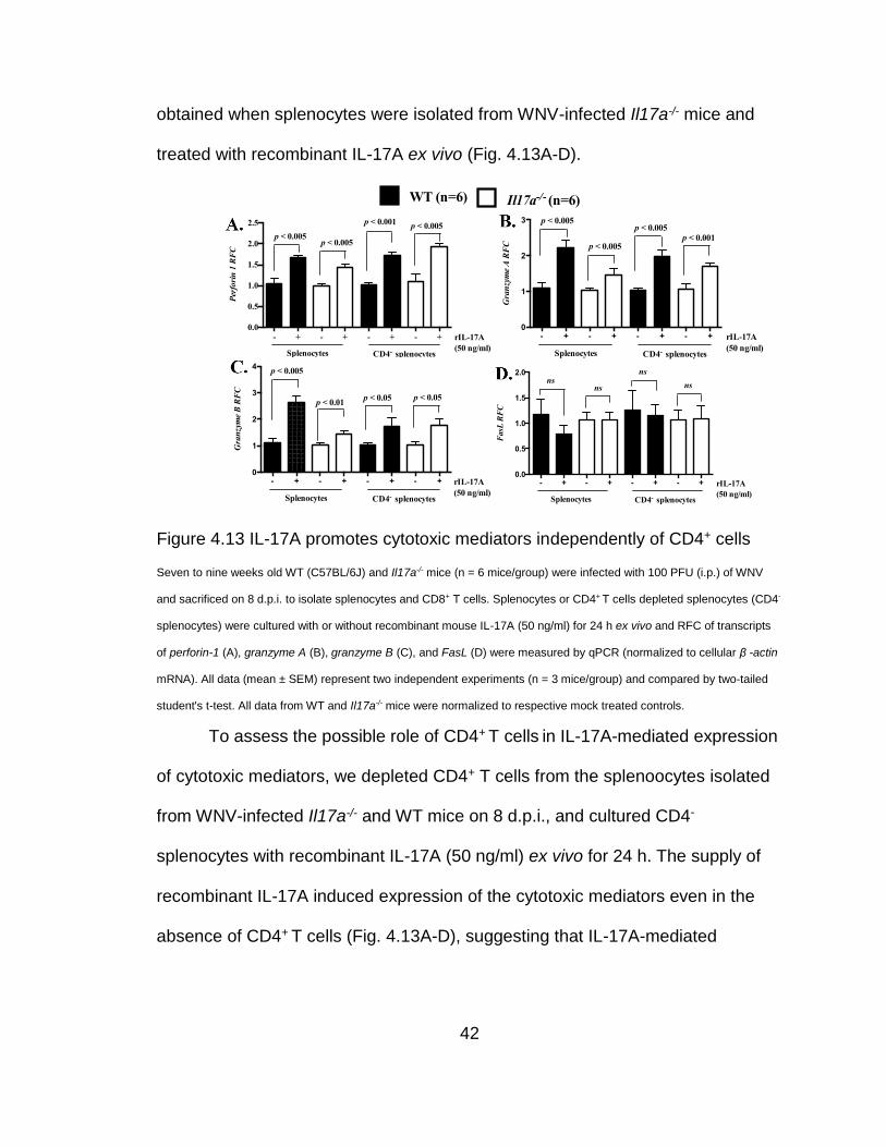
42
obtained when splenocytes were isolated from WNV-infected Il17a-/- mice and
treated with recombinant IL-17A ex vivo (Fig. 4.13A-D).
Figure 4.13 IL-17A promotes cytotoxic mediators independently of CD4+ cells
Seven to nine weeks old WT (C57BL/6J) and Il17a-/- mice (n = 6 mice/group) were infected with 100 PFU (i.p.) of WNV
and sacrificed on 8 d.p.i. to isolate splenocytes and CD8+ T cells. Splenocytes or CD4+ T cells depleted splenocytes (CD4-
splenocytes) were cultured with or without recombinant mouse IL-17A (50 ng/ml) for 24 h ex vivo and RFC of transcripts
of perforin-1 (A), granzyme A (B), granzyme B (C), and FasL (D) were measured by qPCR (normalized to cellular β -actin
mRNA). All data (mean ± SEM) represent two independent experiments (n = 3 mice/group) and compared by two-tailed
student's t-test. All data from WT and Il17a-/- mice were normalized to respective mock treated controls.
To assess the possible role of CD4+ T cells in IL-17A-mediated expression
of cytotoxic mediators, we depleted CD4+ T cells from the splenoocytes isolated
from WNV-infected Il17a-/- and WT mice on 8 d.p.i., and cultured CD4-
splenocytes with recombinant IL-17A (50 ng/ml) ex vivo for 24 h. The supply of
recombinant IL-17A induced expression of the cytotoxic mediators even in the
absence of CD4+ T cells (Fig. 4.13A-D), suggesting that IL-17A-mediated
Figure 6.
- + - + - + - +0
1
2
3
4
5
Per
fori
n 1
RF
C
CD8+ CD8-
IL-17A
(50 ng/ml)
ns ns
p < 0.05
p < 0.05
E.
- + - + - + - +0
1
2
3
4
5
Gra
nzy
me A
RF
C
CD8+ CD8-
IL-17A
(50 ng/ml)
ns ns
p < 0.05
p < 0.05F.
- + - + - + - +0
1
2
3
4
Gra
nzy
me B
RFC
CD8+ CD8-
IL-17A
(50 ng/ml)
nsns
p < 0.005
p < 0.005G.
- + - + - + - +0.0
0.5
1.0
1.5
2.0
FasL
RF
C
CD8+ CD8-
IL-17A
(50 ng/ml)
ns
ns
ns
p <0.05
H.
- + - + - + - +0.0
0.5
1.0
1.5
2.0
2.5
Per
forin 1
RFC
Splenocytes CD4- splenocytes
IL-17A
(50 ng/ml)
p < 0.005p < 0.005
p < 0.005p < 0.001
- + - + - + - +0
1
2
3
Gra
nzy
me A
RF
C
IL-17A
(50 ng/ml)
p < 0.005
p < 0.005
Splenocytes CD4- splenocytes
p < 0.01
p < 0.005A. B.
- + - + - + - +0
1
2
3
4
Gra
nzy
me
B R
FC
IL-17A
(50 ng/ml)
p < 0.01
p < 0.005
Splenocytes CD4- splenocytes
p < 0.05 p < 0.05
C.
- + - + - + - +0.0
0.5
1.0
1.5
2.0
FasL
RF
C
IL-17A
(50 ng/ml)
ns
nsns
ns
Splenocytes CD4- splenocytes
D.
WT (n=6)
Il17a-/- (n=6)
Figure 6.
- + - + - + - +0
1
2
3
4
5
Per
fori
n 1
RF
C
CD8+ CD8-
IL-17A
(50 ng/ml)
ns ns
p < 0.05
p < 0.05
E.
- + - + - + - +0
1
2
3
4
5
Gra
nzy
me A
RF
C
CD8+ CD8-
IL-17A
(50 ng/ml)
ns ns
p < 0.05
p < 0.05F.
- + - + - + - +0
1
2
3
4
Gra
nzy
me B R
FC
CD8+ CD8-
IL-17A
(50 ng/ml)
nsns
p < 0.005
p < 0.005G.
- + - + - + - +0.0
0.5
1.0
1.5
2.0
FasL
RF
C
CD8+ CD8-
IL-17A
(50 ng/ml)
ns
ns
ns
p <0.05
H.
- + - + - + - +0.0
0.5
1.0
1.5
2.0
2.5
Per
forin 1
RFC
Splenocytes CD4- splenocytes
IL-17A
(50 ng/ml)
p < 0.005p < 0.005
p < 0.005p < 0.001
- + - + - + - +0
1
2
3
Gra
nzy
me A
RF
C
IL-17A
(50 ng/ml)
p < 0.005
p < 0.005
Splenocytes CD4- splenocytes
p < 0.01
p < 0.005A. B.
- + - + - + - +0
1
2
3
4
Gra
nzy
me
B R
FC
IL-17A
(50 ng/ml)
p < 0.01
p < 0.005
Splenocytes CD4- splenocytes
p < 0.05 p < 0.05
C.
- + - + - + - +0.0
0.5
1.0
1.5
2.0
FasL
RF
C
IL-17A
(50 ng/ml)
ns
nsns
ns
Splenocytes CD4- splenocytes
D.
WT (n=6)
Il17a-/- (n=6)
- + - + - + - +0
1
2
3
Gra
nzy
me
A R
FC
rIL-17A
(50 ng/ml)
p < 0.005
p < 0.005
Splenocytes CD4- splenocytes
p < 0.001
p < 0.005
- + - + - + - +0.0
0.5
1.0
1.5
2.0
2.5P
erfo
rin
1 R
FC
Splenocytes CD4- splenocytes
rIL-17A
(50 ng/ml)
p < 0.005p < 0.005
p < 0.005p < 0.001
- + - + - + - +0
1
2
3
4
Gra
nzy
me
B R
FC
rIL-17A
(50 ng/ml)
p < 0.01
p < 0.005
Splenocytes CD4- splenocytes
p < 0.05p < 0.05
- + - + - + - +0.0
0.5
1.0
1.5
2.0
FasL
RF
C
rIL-17A
(50 ng/ml)
ns
nsns
ns
Splenocytes CD4- splenocytes

43
induction of cytotoxic mediators expression in CD8+ T cell is independent of CD4+
T cells.
4.14 IL-17A-induced Cytotoxicity Mediators May Involve ACT-1 Signaling
IL-17A exerts its function through IL-17A receptor (IL-17R), which is a
heterodimeric receptor complex of IL-17RA and IL-17RC. Although the signaling
pathway downstream of IL-17R has not been completely understood, one
common pathway that has been characterized is the activation of the classical
NF-κB pathway (Gaffen, 2009; Gu et al., 2013). Upon binding to IL-17A, SEFIR
domain of IL-17R recruits the signaling adaptor ACT-1, which further recruits an
essential upstream activator of the classical NF-κB pathway called TRAF6
(Gaffen, 2009). To test if IL-17A signaling in WNV-infected cells occurs via ACT-
1, we measured the expression of Act-1 in splenocytes and CD8+ T cells isolated
from WNV-infected mice and treated ex vivo with recombinant IL-17A, as
described above. The qPCR results showed that IL-17A treatment up-regulated
the expression of Act-1 gene in splenocytes (Fig. 4.14A) and CD8+ T cells (Fig.
4.14B) isolated from WNV-infected mice.
To further test if IL-17A-mediated expression of cytotoxicity mediators
involves NF-κB activation, we treated splenocytes isolated from WNV-infected
WT mice with recombinant IL-17A in the presence of Bay-11-7082 (an inhibitor of
NF-κB) and measured the expression of the cytotoxicity mediators, as described
above. Consistent with previous reports (Huang et al., 2007; Ribaux et al., 2007),
Bay-11-7082 inhibited the IL-17A mediated Cxcl1 expression that involves NF-κB
pathway. However, inhibition of NF-κB did not inhibit, but interestingly further up-

44
regulated the IL-17A-mediated expression of perforin-1, granzyme A, and
granzyme B (Fig. 4.14C). These data indicate that IL-17A-mediated expression
of cytotoxicity mediators may involve ACT-1, but may occur independently of NF-
κB pathway, which requires further investigation.
Figure 4.14 IL-17A induces the expression of ACT-1
Seven to nine weeks old WT (C57BL/6J) and Il17a-/- mice (n = 6 mice/group) were infected with 100 PFU (i.p.) of WNV
and sacrificed on 8 d.p.i. to isolate splenocytes and CD8+ T cells. Splenocytes (A) or purified CD8+ T cells (B) were
cultured ex vivo with or without recombinant IL-17A (rIL17A, 50 ng/ml) for 24 h, and expression (RFC) of Act-1 was by
measured qPCR. (C) RFC of Act-1 in splenocytes cultured ex vivo for 24 h with or without rIL17A (50 ng/ml), in the
presence of Bay-11-7082 (4 μM) or DMSO as vehicle control (< 0.05%). All data (mean ± SEM) represent two
independent experiments (n = 3 mice/group) and compared by two-tailed student's t-test. All data from WT and Il17a-/-
mice were normalized to respective mock treated controls. * denotes p < 0.05; ** denotes p < 0.005; and “ns” denotes not
significant (p > 0.05).
4.15 IL-17A Directly Induces Cytotoxic mediators in CD8+ T Cells
IL-17R is expressed by virtually all cell types and tissues examined
(Moseley et al., 2003; Yao et al., 1995; Yao et al., 1997). CD8+ T cells also
express IL-17R (Lindemann et al., 2008; Yao et al., 1995), however, the

45
functional role of this receptor in CD8+ T cell biology has not yet been
recognized. To test if IL-17A may directly promote cytotoxic mediators
expression in CD8+ T cells, we treated CD8+ T cells purified (~ 80 to 90% pure)
from spleens of WNV-infected WT mice with the recombinant IL-17A (50 ng/ml)
ex vivo for 24 h, then measured expression of cytotoxicity mediators by qPCR.
IL-17A treatment significantly induced the expression of perforin-1 (Fig. 4.15A),
granzyme A (Fig. 4.15B), and granzyme B (Fig. 4.15C), but not FasL (Fig. 4.15D)
in CD8+ T cells purified from WNV-infected WT mice. Similar results were also
obtained in CD8+ T cell purified from WNV-infected Il17a-/- mice (Fig. 4.15A-D). In
a separate experiment, we cultured splenocytes from WNV-infected Il17a-/- and
WT mice with recombinant IL-17A (50 ng/ml) ex vivo for 24 h, separated CD8+ T
cells from CD8- cells, and then measured expression of cytotoxic mediators by
qPCR. Consistently, IL-17A treatment induced the expression of cytotoxic
mediators in CD8+ T cells (data not shown), but not in CD8- cells (Fig. 4.15A-D).
To further confirm the role of IL-17A in promoting the expression of the
cytotoxic mediators in brain CD8+ T cells in vivo, we infected Il17a-/- mice with
WNV (100 PFU), treated them with recombinant IL-17A (2.5 μg per mouse at day
6 p.i.), and performed flow cytometric analysis of brain leukocytes. The results
showed that treatment of WNV-infected Il17a-/- mice with recombinant IL-17A
significantly induced the production of perforin and granzyme A in brain
infiltrating CD8+ T cells (Fig. 4.15E). Taken together, these results suggest that
IL-17A promotes the expression of cytotoxic mediator genes in CD8+ T cells
during WNV infection in mice.

46
Figure 4.15 IL-17A directly induces cytotoxicity mediators in CD8+ T cells
(A-D) Seven to nine weeks old WT (C57BL/6J) and Il17a-/- mice (n = 6) were infected with 100 PFU (i.p.) of
WNV and sacrificed at 8 d.p.i. to collect spleens, followed by magnetic separation of CD8+ T cells. Relative
fold change (RFC) of the transcripts of perforin-1 (A), granzyme A (B), granzyme B (C), and FasL (D) were
measured in CD8+ or CD8- T cells cultured ex vivo for 24 h with or without recombinant mouse IL-17A (50
pg/ml) by qPCR. (E) Eight week old Il17a-/- mice (n = 4 per group) infected with WNV (100 PFU, i.p.) were
Figure 6.
- + - + - + - +0
1
2
3
4
5
Per
fori
n 1
RF
C
CD8+ CD8-
IL-17A
(50 ng/ml)
ns ns
p < 0.05
p < 0.05
E.
- + - + - + - +0
1
2
3
4
5
Gra
nzy
me A
RF
C
CD8+ CD8-
IL-17A
(50 ng/ml)
ns ns
p < 0.05
p < 0.05F.
- + - + - + - +0
1
2
3
4
Gra
nzy
me B
RFC
CD8+ CD8-
IL-17A
(50 ng/ml)
nsns
p < 0.005
p < 0.005G.
- + - + - + - +0.0
0.5
1.0
1.5
2.0
FasL
RF
C
CD8+ CD8-
IL-17A
(50 ng/ml)
ns
ns
ns
p <0.05
H.
- + - + - + - +0.0
0.5
1.0
1.5
2.0
2.5
Per
forin 1
RFC
Splenocytes CD4- splenocytes
IL-17A
(50 ng/ml)
p < 0.005p < 0.005
p < 0.005p < 0.001
- + - + - + - +0
1
2
3
Gra
nzy
me A
RF
C
IL-17A
(50 ng/ml)
p < 0.005
p < 0.005
Splenocytes CD4- splenocytes
p < 0.01
p < 0.005A. B.
- + - + - + - +0
1
2
3
4
Gra
nzy
me
B R
FC
IL-17A
(50 ng/ml)
p < 0.01
p < 0.005
Splenocytes CD4- splenocytes
p < 0.05 p < 0.05
C.
- + - + - + - +0.0
0.5
1.0
1.5
2.0
FasL
RF
C
IL-17A
(50 ng/ml)
ns
nsns
ns
Splenocytes CD4- splenocytes
D.
WT (n=6)
Il17a-/- (n=6)
Figure 6.
- + - + - + - +0
1
2
3
4
5
Per
fori
n 1
RF
C
CD8+ CD8-
IL-17A
(50 ng/ml)
ns ns
p < 0.05
p < 0.05
E.
- + - + - + - +0
1
2
3
4
5
Gra
nzy
me A
RF
C
CD8+ CD8-
IL-17A
(50 ng/ml)
ns ns
p < 0.05
p < 0.05F.
- + - + - + - +0
1
2
3
4
Gra
nzy
me B R
FC
CD8+ CD8-
IL-17A
(50 ng/ml)
nsns
p < 0.005
p < 0.005G.
- + - + - + - +0.0
0.5
1.0
1.5
2.0
FasL
RF
C
CD8+ CD8-
IL-17A
(50 ng/ml)
ns
ns
ns
p <0.05
H.
- + - + - + - +0.0
0.5
1.0
1.5
2.0
2.5
Per
forin 1
RFC
Splenocytes CD4- splenocytes
IL-17A
(50 ng/ml)
p < 0.005p < 0.005
p < 0.005p < 0.001
- + - + - + - +0
1
2
3
Gra
nzy
me A
RF
C
IL-17A
(50 ng/ml)
p < 0.005
p < 0.005
Splenocytes CD4- splenocytes
p < 0.01
p < 0.005A. B.
- + - + - + - +0
1
2
3
4
Gra
nzy
me
B R
FC
IL-17A
(50 ng/ml)
p < 0.01
p < 0.005
Splenocytes CD4- splenocytes
p < 0.05 p < 0.05
C.
- + - + - + - +0.0
0.5
1.0
1.5
2.0F
asL
RF
C
IL-17A
(50 ng/ml)
ns
nsns
ns
Splenocytes CD4- splenocytes
D.
WT (n=6)
Il17a-/- (n=6)
rIL17A
CD45
CD
8
Control
CD45
CD
8
Control rIL-17A200
300
400
500
600
Per
fori
n
(Mea
n flu
ore
scen
t in
tensi
ty)
*
Control rIL-17A0
50
100
150
200
250
Gra
nzy
me
A
(Mea
n flu
ore
scen
t in
tensi
ty) *
Brain day 8 p.i.
- + - +0
1
2
3
4
Per
fori
n 1
RF
C
CD8+ CD8-
ns
p < 0.05
- + - +0
1
2
3
4
5
CD8+ CD8-
rIL-17A
(50 ng/ml)
ns
p < 0.05
- + - +0
1
2
3
4
5
Gra
nzy
me
A R
FC
ns
p < 0.05
CD8+ CD8-
- + - +0
1
2
3
4
CD8+ CD8-
rIL-17A
(50 ng/ml)
ns
p < 0.05
- + - +0
1
2
3
4
Gra
nzy
me
B R
FC
ns
p < 0.005
CD8+ CD8-
- + - +0.0
0.5
1.0
1.5
2.0
CD8+ CD8-
rIL-17A
(50 ng/ml)
ns
p < 0.005
- + - +0.0
0.5
1.0
1.5
2.0
FasL
RF
C
nsns
CD8+ CD8-
- + - +0.0
0.5
1.0
1.5
CD8+ CD8-
rIL-17A
(50 ng/ml)
ns
p < 0.05

47
treated with recombinant IL-17A (rIL17A, 2.5 μg/mouse) or PBS (control) via i.p. at day 6 p.i and sacrificed at
8 p.i. to characterize brain leukocytes by flow cytometry. Mean fluorescence intensities of perforin and
granzyme A within the gated CD45hi CD8+ cells (green) are shown in bar (right). All data (mean ± SEM)
represent two independent experiments and compared by two-tailed student's t-test. All data from WT and
Il17a-/- mice (shown in figures A to D) were normalized to respective mock treated controls. “ns” denotes no
significant difference (p > 0.05).
4.16 IL-17A Treatment Increases Survival of WNV-infected Mice
Since CD8+ T cells are essential to clear WNV from the CNS at a later
phase of infection, we hypothesized that recombinant IL-17A treatment in vivo
may offer protection from WNV infection by promoting cytotoxicity of CD8+ T
cells. To test if IL-17A may serve as a therapeutic reagent to treat WNV infection
in mice, we infected WT female mice with WNV (100 PFU) via i.p. route. At day 6
p.i, mice were injected with the carrier-free mouse recombinant IL-17A
(eBioscience) or PBS as control via i.p. route for survival analysis.
Figure 4.16 Recombinant IL-17A increases survival of WNV-infected mice
WT (C57BL/6J) mice (8-week old, female) were infected with 100 PFU of WNV via i.p. route. At day 6 p.i,
mice were injected with i.p. administration of carrier-free mouse recombinant IL-17A (rIL17A, 2.5 μg/mouse)
or PBS. (A) Percent (%) of survival was compared using the Kaplan-Meier survival and log-rank test. Viral
burden in brain (B) was measured by qPCR and compared by two-tailed student's t-test; “ns” denotes no
significant difference (p > 0.05).
0 5 10 15 200
20
40
60
80
100
d.p.i
Per
cen
t su
rviv
al
PBS (n=25)rIL17A (n=22)
p < 0.05
0
10
20
30
WN
V E
/ 1
000 β
-act
in
p < 0.01
Brain (day 8 p.i.)
rIL-17A (n=7)
PBS (n=9)

48
Mice that received recombinant IL-17A showed significantly increased
survival rate compared to the PBS-treated control mice (Fig. 4.16A). Consistent
with the survival results, mice treated with recombinant IL-17A also showed lower
viral burden in the brain compared to the PBS-treated control mice at day 8 p.i.
(Fig. 4.16B).
4.17 IL-17A Induces Cytotoxicity Mediators in WNV-infected Mice
To test if the effect of the recombinant IL-17A treatment involves a
promotion of cytotoxicity of CD8+ T cells, we also measured the expression of the
cytotoxicity mediators in splenic CD8+ and CD8- T cells
Figure 4.17 Recombinant IL-17A induces cytotoxicity in WNV-infected mice.
WT (C57BL/6J) mice (8-week old, female) were infected with 100 PFU of WNV via i.p. route. At day 6 p.i, mice were
injected with i.p. administration of carrier-free mouse recombinant IL-17A (rIL17A, 2.5 μg/mouse) or PBS. (A to E) The
transcripts of perforin-1, granzyme A, granzyme B, and FasL in splenic CD8+ and CD8- T cells (A-D), and brain (E) were
measured by qPCR and compared by two-tailed student's t-test; “ns” denotes no significant difference (p > 0.05).
0.0
0.5
1.0
1.5
Per
fori
n-1
RF
C
CD8+ CD8-
p <0 .05
ns
0.0
0.5
1.0
1.5
2.0
Gra
nzy
me
A R
FC
CD8+ CD8-
p <0 .05
ns
0
1
2
3
Gra
nzy
me
B R
FC
CD8+ CD8-
p <0 .05
ns
0.0
0.5
1.0
1.5
2.0
FasL
RF
C
CD8+ CD8-
p <0 .05
ns
rIL-17A (n=7)
PBS (n=9)
0
1
2
10
20
30
40
50
60
mR
NA
RF
C
ns
p < 0.005
p < 0.005
p < 0.005
Perforin 1 Granzyme A Granzyme B FasL

49
. Consistent with reduced viral burden and increased survival,
administration of recombinant IL-17A in WNV-infected mice induced the
expression of the cytotoxic mediators in CD8+ T cells, but not in CD8- T cells (Fig.
4.17A-D), which agreed with our in vitro results. In addition, we also detected
increased expression of granzyme A, granzyme B, and FasL in brain tissues of
WNV-infected mice after IL-17A treatment (Fig. 17E). Collectively, these results
suggest a novel and promising therapeutic role of IL-17A in facilitating WNV
clearance by promoting CD8+ T cell cytotoxicity.
50
CHAPTER V – DISCUSSION
This study reveals a novel role of IL-17A in facilitating WNV clearance by
inducing the expression of cytotoxic mediator genes and promoting CD8+ T cell
cytotoxicity. Specifically, we report here that 1) WNV induces IL-17A expression
in both mice and humans; 2) Il17a-/- mice, generate a higher viral burden and are
more susceptible to WNV infection; 3) CD8+ T cells purified from Il17a-/- mice are
less cytotoxic and express lower levels of cytotoxic mediators, i.e., perforin-1,
granzyme A, granzyme B and FasL; and most importantly; and 4) ex vivo supply
of recombinant IL-17A as late as day 6 post-infection significantly reduces viral
burden in the brain and increases survival rate of WNV-infected mice, suggesting
a therapeutic potential of IL-17A.
5.1 IL-17A Has a Protective Function in WNV Infection
It has been previously reported that WNV mediates production of IL-23, a
cytokine that regulate IL-17A expression (Town et al., 2009). As expected, we
found that WNV induced IL-17A production in human and mouse in an IL-23
dependent manner. We further found a protective function of IL-17A against
WNV and its crucial role in clearance of WNV from brain, which was also
consistent with function of IL-23 (Town et al., 2009). In support of our finding, it
has been previously reported that mice depleted of IL-17A function by
administration of anti-IL-17A antibody showed about 20% reduced survival rate
compared to control mice after a lethal WNV challenge (Welte et al., 2011).
However, no difference in WNV viral burden was observed between anti-IL-17A
antibody treated and control mice (Welte et al., 2011). In contrast, our results

51
clearly demonstrate that Il17a-/- mice are more susceptible to WNV infection. The
discrepancy between these two studies may be due to the transient effects of
anti-IL-17A treatment when compared to complete genetic deficiency of IL-17A in
Il17a-/- mice. More to this point, γδ-T cell produce IL-17A during early phage of
WNV infection, whereas CD4+ Th-17 cells become the major IL-17A producer
during the late phase of infection (Hamada et al., 2008). Thus, the administration
of anti-IL-17A antibody at an early phase of WNV infection (0 and 5 d.p.i.) (Welte
et al., 2011), may not have sufficiently blocked IL-17A production and its function
during the later phase of infection. Indeed, we found that IL-17A was not
essential for controlling the viremia on 2 d.p.i, but became critical for reducing
viremia on 4 d.p.i and clearing the viruses from spleen and brain on 8 d.p.i..
Thus, it appears that IL-17A-mediated protective immunity against WNV infection
predominantly occurs during the late phase of infection, which requires further
investigation.
5.2 Role of IL-17A in IFN, Cytokines and Antibody Response
IL-17A is a pleiotropic cytokine that plays key roles during infection
(Iwakura et al., 2008), inflammation (Park et al., 2005) and autoimmune diseases
(Onishi and Gaffen, 2010). Further, it regulates the expression of a number of
cytokines, including IL-1β, IFN-γ, and TNF-α (Maione et al., 2009; Song and
Qian, 2013). Considering their critical roles in WNV pathogenesis (Ramos et al.,
2012; Shrestha et al., 2006b; Shrestha et al., 2008b; Wang et al., 2004a), it is
plausible that IL-17A may regulate the expression of these cytokines during WNV
infection. However, we did not detect significant differences in the expression of
52
IL-1β, IFN-γ, and TNF-α in WNV-infected Il17a-/- mice when compared to WT
controls. These results suggest the likelihood that IL-17A-mediated protective
immunity against WNV infection may not be associated with functions controlled
by these cytokines. Previous studies have suggested that type I IFNs (antiviral
cytokines), which can potently suppress IL-17A expression (Curtis et al., 2009;
Tilg et al., 2009), play a prominent role in controlling WNV infection (Lazear et al.,
2011; Samuel and Diamond, 2005). Again, we did not detect any change in the
expression of type I IFNs in Il17a-/- vs. WT control mice during WNV infection.
Moreover, IL-17A has been suggested to regulate the humoral immune response
(Grund et al., 2012; Tarlinton, 2008; Yuan et al., 2010), however, we did not see
such effect during WNV infection in mice. Despite unaltered inflammatory
response, type I IFN expression and humoral immune response, Il17a-/- mice
generated higher viral burden, suggesting that IL-17A mediated protective
immunity during WNV infection is independent of these immune responses.
5.3 Role of IL-17A in Leukocyte Homing to CNS
WNV invades the CNS and infects neurons and CNS-resident cells, such
as microglia and astrocytes, which leads to production of cytokines and
chemokines that recruit peripheral leukocytes into the brain (Glass et al., 2005;
Hussmann and Fredericksen, 2014; Shrestha and Diamond, 2004; Town et al.,
2009). We previously reported that IL-23 has an important role in recruiting
CD11b+ monocytes and macrophages into the CNS to control WNV infection
(Town et al., 2009). Considering the role of IL-23 in producing IL-17A, and the
role of both of these cytokines in inducing and sustaining leukocyte recruitment to

53
infected sites (Roussel et al., 2010; Shahrara et al., 2009), we hypothesized that
leukocyte migration into the CNS would be reduced in Il17a-/- mice. Surprisingly,
we detected a modestly elevated leukocyte influx into WNV-infected Il17a-/-
mouse brains by both flow cytometry and confocal microscopy.
In dissecting the mechanism, we found that the expression of CCL5 was
elevated in both the CNS and the peripheral tissues of WNV-infected Il17a-/- mice
compared to WT controls. CCL5 plays a protective role in WNV, influenza and
parainfluenza infections by promoting leukocyte trafficking to infected tissues
(Glass et al., 2005; Kohlmeier et al., 2008). Also, Ccl5 expression sustains CD8+
T cell responses during influenza (Kohlmeier et al., 2008), parainfluenza
(Kohlmeier et al., 2008) and chronic lymphocytic choriomeningitis virus (LCMV)
(Crawford et al., 2011) infections. The up-regulation of Ccl5 in Il17a-/- mice may
be due to several reasons. First, the higher viral load in Il17a-/- mice could trigger
a stronger Ccl5 expression (Matsukura et al., 1998; Saito et al., 1997). Secondly,
the presence of IL-17A in WT mice may suppress Ccl5 expression, since IL-17A
can down-regulate Ccl5 expression (Andoh et al., 2002; Shen et al., 2005).
Although increased Ccl5 expression may account for modest elevation in brain
infiltrating leukocytes in Il17a-/- mice, this did not result in protection of these mice
from WNV infection, implying a possible functional defect in brain infiltrating
effector leukocytes in Il17a-/- mice.
5.4 IL-17A Regulates CD8+ T Cell Cytotoxicity
CD8+ T cells play a critical role in clearance of viruses from the CNS
(Binder and Griffin, 2001; Griffin, 2010; Shrestha and Diamond, 2004). CD8+ T

54
cells can control viral infection directly by inducing apoptosis of virus-infected
cells via perforin, granzyme, or Fas-FasL interactions (Harty and Badovinac,
2002; Shresta et al., 1998), or indirectly by immune-mediated non-cytolytic
clearance of viruses from neurons by producing cytokines, such as TNF-α and
IFN-γ (Binder and Griffin, 2001; Griffin, 2010). Mice deficient in the expression of
cytotoxic mediators such as perforin, FasL, or cytokines, such as TNF-α and IFN-
γ, exhibit increased mortality and viral burden in the CNS and peripheral organs
following WNV infection (Shrestha and Diamond, 2007; Shrestha et al., 2012;
Shrestha et al., 2006a; Shrestha et al., 2006b; Shrestha et al., 2008b; Wang et
al., 2004b). However, we did not detect any significant difference in the
expression of the cytokines including TNF-α and IFN-γ between WNV-infected
WT and Il17a-/- mice, suggesting that IL-17A-mediated control of WNV by CD8+ T
cells may occur independently of the effects of these cytokines. We found that
CD8+ T cells isolated from Il17a-/- mice have significantly reduced cytotoxicity
compared to WT controls, which may explain higher viral burden and lower
survival rates of Il17a-/- mice, despite elevated leukocyte infiltration (including
CD8+ T cells) into the CNS. While we are not certain whether CD8+ T cells
predominantly contribute to WNV clearance by causing cytolysis of WNV-infected
neurons due to lower MHC I expression in these cells (Kimura and Griffin, 2000;
Mucke and Oldstone, 1992), it is likely that CD8+ T cells can cause cytolysis of
WNV-infected non-neural cells, such as infiltrating macrophages and neutrophils,
and microglia in the CNS, thus facilitating the virus clearance (Bai et al., 2010;
Shrestha and Diamond, 2004, 2007; Shrestha et al., 2006a). In line with reduced

55
cytotoxicity, CD8+ T cells from Il17a-/- mice had significantly reduced expression
of perforin-1, granzyme A, granzyme B and FasL in most of the tissue examined
during WNV infection. Although NK cells (type of CD8- cells) also express these
genes and mediate apoptosis of target cell, we did not detect difference in
expression of cytotoxic mediators in CD8 negative (CD8-) cell populations
isolated from WNV-infected WT and Il17a-/- mouse spleens. This indicates that
NK cells (type of CD8- cells) may not play a prominent role in IL-17A-mediated
cytotoxicity during WNV infection. This notion is consistent with the previous
reports that NK cells have a little or no role in controlling WNV infection (Shrestha
et al., 2006a).
It is worthy to note that CD8+ T cells have also been shown an
immunopathological role when mice were infected intravenously with a high dose
(108 PFU) of WNV (Sarafend strain), resulting in 100% mortality with a 6-day
mean survival time (Wang et al., 2003). However, such an immunopathology role
of CD8+ T cells was likely not due to their cytotoxic functions because these cells
are usually activated after one week post-infection. In support of this, that same
study and many other reports also showed a recovery role of CD8+ T cells when
mice were infected with low doses (102-103 PFU) of WNV, which is similar to the
current study (Brien et al., 2011; Brien et al., 2007; Shrestha et al., 2008a;
Shrestha et al., 2012; Shrestha et al., 2006a; Wang et al., 2003). In addition,
Th17 cells and IL-17A have been implicated in inflammation and
immunopathology associated with autoimmune diseases (McFarland and Martin,
2007; van den Berg and Miossec, 2009; Zepp et al., 2011) and some virus-

56
induced chronic CNS diseases (Hou et al., 2014; Hou et al., 2009; Savarin et al.,
2012), suggesting that Th-17/IL-17A axis may have different implications in such
immunopathological and chronic inflammatory conditions.
5.5 IL-17A Has a Potential Therapeutic Role Against WNV
Our data demonstrates that IL-17A deficiency leads to an attenuated CD8+
T cell cytotoxicity and deficient WNV clearance. Both CD4+ Th-17 cells and γδ T
cells, the major cells that produce IL-17A, have been previously shown to
promote CD8+ T cell cytotoxicity during infection (Hamada et al., 2009; Xu et al.,
2010), autoimmune disease (Ankathatti Munegowda et al., 2011a), and cancers
(Ankathatti Munegowda et al., 2011b; Martin-Orozco et al., 2009). Also, NK cells
treated with IL-17A overexpress perforin and granzymes, and have better
cytotoxic functions (Al Omar et al., 2013), which provides additional evidence that
IL-17A signaling can induce the expression of cytotoxicity mediators. These
reports support our finding and suggest that IL-17A may facilitate cytotoxicity of
CD8+ T cells in diverse disease conditions.
Various types of immune cells, such as CD4+ Th-17 cells, γδ-T cells, NKT
cells, and CD8+ T cells can produce IL-17A (Gu et al., 2013; Welte et al., 2011).
In addition, the IL-17A receptor (IL-17R) is expressed ubiquitously in virtually all
cell types and tissues examined (Andoh et al., 2002; Moseley et al., 2003; Yao et
al., 1997). Although CD8+ T cells can produce IL-17A and also express its
cognate receptor (Lindemann et al., 2008; Yao et al., 1995), the link between IL-
17A and CD8+ T cell cytotoxic function has not been previously recognized. In
theory, it is possible that IL-17A may affect CD8+ T cell development and/or

57
function either by directly acting on these cells or indirectly through other cell
types. Since we found similar levels of CD8+ T cells and CD4+/CD8+ ratios in
splenocytes isolated from Il17a-/- and WT mice infected with WNV, it appears that
CD8+ T cell development remains unaltered in Il17a-/- mice and IL-17A may
largely control CD8+ T cell function during WNV infection. This hypothesis is
confirmed by ex vivo treatment of IL-17A to splenocytes isolated from WNV-
infected Il17a-/- and WT mice, which showed an up-regulation of perforin-1,
granzyme A, and granzyme B expression in CD8+ T cells by IL-17A, in a CD4+ T
cell-independent manner. These results suggest that IL-17A can induce the
expression of cytotoxic mediators by directly acting on CD8+ T cells. However,
the induction of FasL by recombinant IL-17A was differentially affected in our
experiments. For instance, in vitro IL-17A treatment did not induce the
expression of FasL in CD8+ T cells, while it was induced after in vivo
administration of IL-17A. This discrepancy is likely due to the experimental
conditions or may reflect differential role of IL-17A in regulating the expression of
these cytotoxic mediators, which requires further investigation. We further
demonstrated that the treatment of WNV-infected Il17a-/- mice with recombinant
IL-17A significantly induced the production of perforin and granzyme A in brain
infiltrating CD8+ T cells, which confirmed the role of IL-17A in promoting the
expression of the cytotoxic mediators in brain infiltrating CD8+ T cells in vivo.
To extend upon our finding, we tested therapeutic potential of IL-17A in a
mouse model of WNV infection. Importantly, administration of a single dose of
recombinant IL-17A to WNV-infected mice, even as late as day 6 p.i., induced

58
the expression of cytotoxic mediators in CD8+ T cells, dramatically reduced viral
burden in the brain, and significantly increased survival rate, suggesting a
promising therapeutic role of IL-17A against WNV infection.

59
CHAPTER VI – CONCLUSION AND FUTURE DIRECTIONS
6.1 Conclusion
IL-17A and CD8+ T cells regulate diverse immune functions during
microbial infections, malignancies, and autoimmune diseases. IL-17A is a
proinflammatory cytokine produced by diverse cell types, while the CD8+ T cells
(known as cytotoxic T cells) are major cells that provide immunity against
intracellular pathogens. Previous studies have demonstrated a crucial role of
CD8+ T cells in recovery from WNV infection. Here, we demonstrate that IL-17A
protects mice from lethal WNV infection by promoting CD8+ T-cell-mediated
clearance of WNV. In addition, treatment of WNV-infected mice with recombinant
IL-17A reduces viral burden and increases survival of mice, suggesting a
potential therapeutic. In conclusion, this study uncovers a novel function of IL-
17A in promoting CD8+ T cell cytotoxicity during WNV infection in mice. This
novel IL-17A-CD8+ T cell axis may also have broad implications in immunity to
other microbial infections and cancers, where CD8+ T cell functions are crucial.
6.2 Future Directions
In this study, we found the production of IL-17A in WNV-infected human
and mice, which we also confirmed in a mixed culture of human and mouse
immune cells infected in vitro with WNV. We further demonstrated a novel
function of IL-17A in promoting the cytotoxicity of CD8+ T cell against WNV
infection. In addition, a supply of recombinant IL-17A to a culture of CD8+ T cells
and WNV-infected mice clearly induced the expression of cytotoxicity mediator
genes. The role of IL-17A in promoting the expression of cytotoxic mediators in
60
CD8+ T cells has not been previously reported and the signaling mechanism by
which IL-17A regulates the expression of cytotoxicity mediators is not currently
understood. In addition, diverse cell types including immune cells, astrocytes,
and microglia can produce IL-17A and express IL-17A receptor. Future studies
are required for characterization of IL-17A producing and responding cells in both
CNS and periphery during various stage of WNV infection. In addition, a better
understanding of the regulation of IL-17A production and dissection of the
signaling mechanism by which IL-17A induces expression of cytotoxicity
mediators and promotes CD8+ T cell cytotoxicity in the context of WNV and other
diseases may help to exploit novel IL-17A-based therapeutic strategies.

61
APPENDIX A IACUC and IRB Approval Letter
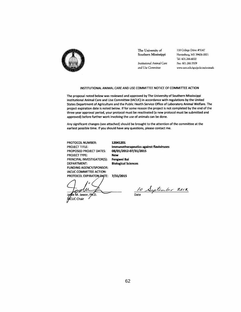
62

63
64
REFERENCES
Aggarwal, S., Ghilardi, N., Xie, M.H., de Sauvage, F.J., and Gurney, A.L.
(2003). Interleukin-23 promotes a distinct CD4 T cell activation state
characterized by the production of interleukin-17. The Journal of biological
chemistry 278, 1910-1914.
Al Omar, S., Flanagan, B.F., Almehmadi, M., and Christmas, S.E. (2013). The
effects of IL-17 upon human natural killer cells. Cytokine 62, 123-130.
Alpert, S.G., Fergerson, J., and Noel, L.P. (2003). Intrauterine West Nile
virus: ocular and systemic findings. Am J Ophthalmol 136, 733-735.
Anderson, J.F., Andreadis, T.G., Vossbrinck, C.R., Tirrell, S., Wakem, E.M.,
French, R.A., Garmendia, A.E., and Van Kruiningen, H.J. (1999). Isolation of
West Nile virus from mosquitoes, crows, and a Cooper's hawk in Connecticut.
Science 286, 2331-2333.
Andoh, A., Fujino, S., Bamba, S., Araki, Y., Okuno, T., Bamba, T., and
Fujiyama, Y. (2002). IL-17 selectively down-regulates TNF-alpha-induced
RANTES gene expression in human colonic subepithelial myofibroblasts.
Journal of immunology 169, 1683-1687.
Ankathatti Munegowda, M., Deng, Y., Chibbar, R., Xu, Q., Freywald, A.,
Mulligan, S.J., van Drunen Littel-van den Hurk, S., Sun, D., Xiong, S., and
Xiang, J. (2011a). A distinct role of CD4+ Th17- and Th17-stimulated CD8+
CTL in the pathogenesis of type 1 diabetes and experimental autoimmune
encephalomyelitis. J Clin Immunol 31, 811-826.

65
Ankathatti Munegowda, M., Deng, Y., Mulligan, S.J., and Xiang, J. (2011b).
Th17 and Th17-stimulated CD8(+) T cells play a distinct role in Th17-induced
preventive and therapeutic antitumor immunity. Cancer immunology,
immunotherapy: CII 60, 1473-1484.
Arjona, A., Wang, P., Montgomery, R.R., and Fikrig, E. (2011). Innate
immune control of West Nile virus infection. Cell Microbiol 13, 1648-1658.
Bai, F., Kong, K.F., Dai, J., Qian, F., Zhang, L., Brown, C.R., Fikrig, E., and
Montgomery, R.R. (2010). A paradoxical role for neutrophils in the
pathogenesis of West Nile virus. J Infect Dis 202, 1804-1812.
Bai, F., Town, T., Qian, F., Wang, P., Kamanaka, M., Connolly, T.M., Gate,
D., Montgomery, R.R., Flavell, R.A., and Fikrig, E. (2009). IL-10 signaling
blockade controls murine West Nile virus infection. PLoS Pathog 5,
e1000610.
Bai, F., Wang, T., Pal, U., Bao, F., Gould, L.H., and Fikrig, E. (2005). Use of
RNA interference to prevent lethal murine west nile virus infection. J Infect Dis
191, 1148-1154.
Benchetrit, F., Ciree, A., Vives, V., Warnier, G., Gey, A., Sautes-Fridman, C.,
Fossiez, F., Haicheur, N., Fridman, W.H., and Tartour, E. (2002). Interleukin-
17 inhibits tumor cell growth by means of a T-cell-dependent mechanism.
Blood 99, 2114-2121.

66
Benjelloun, A., El Harrak, M., and Belkadi, B. (2015). West Nile Disease
Epidemiology in North-West Africa: Bibliographical Review. Transboundary
and emerging diseases.
Binder, G.K., and Griffin, D.E. (2001). Interferon-gamma-mediated site-
specific clearance of alphavirus from CNS neurons. Science 293, 303-306.
Blazquez, A.B., and Saiz, J.C. (2010). West Nile virus (WNV) transmission
routes in the murine model: intrauterine, by breastfeeding and after cannibal
ingestion. Virus Res 151, 240-243.
Brien, J.D., Daffis, S., Lazear, H.M., Cho, H., Suthar, M.S., Gale, M., Jr., and
Diamond, M.S. (2011). Interferon regulatory factor-1 (IRF-1) shapes both
innate and CD8(+) T-cell immune responses against West Nile virus infection.
PLoS pathogens 7, e1002230.
Brien, J.D., Uhrlaub, J.L., and Nikolich-Zugich, J. (2007). Protective capacity
and epitope specificity of CD8(+) T cells responding to lethal West Nile virus
infection. European journal of immunology 37, 1855-1863.
CDC (2015). West Nile virus.
Centers for Disease, C., and Prevention (2009). West Nile virus transmission
via organ transplantation and blood transfusion - Louisiana, 2008. MMWR
Morb Mortal Wkly Rep 58, 1263-1267.
Cernescu, C., Ruta, S.M., Tardei, G., Grancea, C., Moldoveanu, L., Spulbar,
E., and Tsai, T. (1997). A high number of severe neurologic clinical forms

67
during an epidemic of West Nile virus infection. Romanian journal of virology
48, 13-25.
Chung, K.M., Liszewski, M.K., Nybakken, G., Davis, A.E., Townsend, R.R.,
Fremont, D.H., Atkinson, J.P., and Diamond, M.S. (2006). West Nile virus
nonstructural protein NS1 inhibits complement activation by binding the
regulatory protein factor H. Proc Natl Acad Sci U S A 103, 19111-19116.
Colpitts, T.M., Conway, M.J., Montgomery, R.R., and Fikrig, E. (2012). West
Nile Virus: biology, transmission, and human infection. Clin Microbiol Rev 25,
635-648.
Conti, H.R., Shen, F., Nayyar, N., Stocum, E., Sun, J.N., Lindemann, M.J.,
Ho, A.W., Hai, J.H., Yu, J.J., Jung, J.W., et al. (2009). Th17 cells and IL-17
receptor signaling are essential for mucosal host defense against oral
candidiasis. The Journal of experimental medicine 206, 299-311.
Crawford, A., Angelosanto, J.M., Nadwodny, K.L., Blackburn, S.D., and
Wherry, E.J. (2011). A role for the chemokine RANTES in regulating CD8 T
cell responses during chronic viral infection. PLoS Pathog 7, e1002098.
Crowe, C.R., Chen, K., Pociask, D.A., Alcorn, J.F., Krivich, C., Enelow, R.I.,
Ross, T.M., Witztum, J.L., and Kolls, J.K. (2009). Critical role of IL-17RA in
immunopathology of influenza infection. Journal of immunology 183, 5301-
5310.

68
Curtis, M.M., Way, S.S., and Wilson, C.B. (2009). IL-23 promotes the
production of IL-17 by antigen-specific CD8 T cells in the absence of IL-12
and type-I interferons. Journal of immunology 183, 381-387.
Das Sarma, J., Ciric, B., Marek, R., Sadhukhan, S., Caruso, M.L., Shafagh,
J., Fitzgerald, D.C., Shindler, K.S., and Rostami, A. (2009). Functional
interleukin-17 receptor A is expressed in central nervous system glia and
upregulated in experimental autoimmune encephalomyelitis. Journal of
neuroinflammation 6, 14.
Davis, C.T., Ebel, G.D., Lanciotti, R.S., Brault, A.C., Guzman, H., Siirin, M.,
Lambert, A., Parsons, R.E., Beasley, D.W., Novak, R.J., et al. (2005).
Phylogenetic analysis of North American West Nile virus isolates 2001-2004:
evidence for the emergence of a dominant genotype. Virology 342, 252-265.
de Almeida Nagata, D.E., Demoor, T., Ptaschinski, C., Ting, H.A., Jang, S.,
Reed, M., Mukherjee, S., and Lukacs, N.W. (2014). IL-27R-mediated
regulation of IL-17 controls the development of respiratory syncytial virus-
associated pathogenesis. The American journal of pathology 184, 1807-1818.
Diamond, M.S., Shrestha, B., Marri, A., Mahan, D., and Engle, M. (2003a). B
cells and antibody play critical roles in the immediate defense of disseminated
infection by West Nile encephalitis virus. Journal of virology 77, 2578-2586.
Diamond, M.S., Sitati, E.M., Friend, L.D., Higgs, S., Shrestha, B., and Engle,
M. (2003b). A critical role for induced IgM in the protection against West Nile
virus infection. The Journal of experimental medicine 198, 1853-1862.

69
Draganescu, N. (1979). Study of arboviruses in Romania (1958--1978).
Results obtained in the "Stefan S. Nicolau" Institute of Virology. Virologie 30,
283-293.
Ebel, G.D., Carricaburu, J., Young, D., Bernard, K.A., and Kramer, L.D.
(2004). Genetic and phenotypic variation of West Nile virus in New York,
2000-2003. Am J Trop Med Hyg 71, 493-500.
Gaffen, S.L. (2009). Structure and signalling in the IL-17 receptor family.
Nature reviews Immunology 9, 556-567.
Glass, W.G., Lim, J.K., Cholera, R., Pletnev, A.G., Gao, J.L., and Murphy,
P.M. (2005). Chemokine receptor CCR5 promotes leukocyte trafficking to the
brain and survival in West Nile virus infection. The Journal of experimental
medicine 202, 1087-1098.
Griffin, D.E. (2010). Recovery from viral encephalomyelitis: immune-mediated
noncytolytic virus clearance from neurons. Immunologic research 47, 123-
133.
Griffin, G.K., Newton, G., Tarrio, M.L., Bu, D.X., Maganto-Garcia, E., Azcutia,
V., Alcaide, P., Grabie, N., Luscinskas, F.W., Croce, K.J., et al. (2012). IL-17
and TNF-alpha sustain neutrophil recruitment during inflammation through
synergistic effects on endothelial activation. Journal of immunology 188,
6287-6299.

70
Grund, L.Z., Komegae, E.N., Lopes-Ferreira, M., and Lima, C. (2012). IL-5
and IL-17A are critical for the chronic IgE response and differentiation of long-
lived antibody-secreting cells in inflamed tissues. Cytokine 59, 335-351.
Gu, C., Wu, L., and Li, X. (2013). IL-17 family: cytokines, receptors and
signaling. Cytokine 64, 477-485.
Guiton, R., Vasseur, V., Charron, S., Arias, M.T., Van Langendonck, N.,
Buzoni-Gatel, D., Ryffel, B., and Dimier-Poisson, I. (2010). Interleukin 17
receptor signaling is deleterious during Toxoplasma gondii infection in
susceptible BL6 mice. J Infect Dis 202, 427-435.
Hamada, H., Garcia-Hernandez Mde, L., Reome, J.B., Misra, S.K., Strutt,
T.M., McKinstry, K.K., Cooper, A.M., Swain, S.L., and Dutton, R.W. (2009).
Tc17, a unique subset of CD8 T cells that can protect against lethal influenza
challenge. Journal of immunology 182, 3469-3481.
Hamada, S., Umemura, M., Shiono, T., Tanaka, K., Yahagi, A., Begum, M.D.,
Oshiro, K., Okamoto, Y., Watanabe, H., Kawakami, K., et al. (2008). IL-17A
produced by gammadelta T cells plays a critical role in innate immunity
against listeria monocytogenes infection in the liver. Journal of immunology
181, 3456-3463.
Happel, K.I., Dubin, P.J., Zheng, M., Ghilardi, N., Lockhart, C., Quinton, L.J.,
Odden, A.R., Shellito, J.E., Bagby, G.J., Nelson, S., et al. (2005). Divergent
roles of IL-23 and IL-12 in host defense against Klebsiella pneumoniae. The
Journal of experimental medicine 202, 761-769.

71
Harty, J.T., and Badovinac, V.P. (2002). Influence of effector molecules on
the CD8(+) T cell response to infection. Curr Opin Immunol 14, 360-365.
Hayes, C.G. (2001). West Nile virus: Uganda, 1937, to New York City, 1999.
Annals of the New York Academy of Sciences 951, 25-37.
Hayes, E.B., Sejvar, J.J., Zaki, S.R., Lanciotti, R.S., Bode, A.V., and
Campbell, G.L. (2005). Virology, pathology, and clinical manifestations of
West Nile virus disease. Emerg Infect Dis 11, 1174-1179.
Ho, A.W., and Gaffen, S.L. (2010). IL-17RC: a partner in IL-17 signaling and
beyond. Seminars in immunopathology 32, 33-42.
Hou, W., Jin, Y.H., Kang, H.S., and Kim, B.S. (2014). Interleukin-6 (IL-6) and
IL-17 synergistically promote viral persistence by inhibiting cellular apoptosis
and cytotoxic T cell function. Journal of virology 88, 8479-8489.
Hou, W., Kang, H.S., and Kim, B.S. (2009). Th17 cells enhance viral
persistence and inhibit T cell cytotoxicity in a model of chronic virus infection.
The Journal of experimental medicine 206, 313-328.
Huang, F., Kao, C.Y., Wachi, S., Thai, P., Ryu, J., and Wu, R. (2007).
Requirement for both JAK-mediated PI3K signaling and ACT1/TRAF6/TAK1-
dependent NF-kappaB activation by IL-17A in enhancing cytokine expression
in human airway epithelial cells. Journal of immunology 179, 6504-6513.

72
Huang, W., Na, L., Fidel, P.L., and Schwarzenberger, P. (2004). Requirement
of interleukin-17A for systemic anti-Candida albicans host defense in mice. J
Infect Dis 190, 624-631.
Hubalek, Z., and Halouzka, J. (1999). West Nile fever--a reemerging
mosquito-borne viral disease in Europe. Emerging infectious diseases 5, 643-
650.
Hussmann, K.L., and Fredericksen, B.L. (2014). Differential induction of CCL5
by pathogenic and non-pathogenic strains of West Nile virus in brain
endothelial cells and astrocytes. The Journal of general virology 95, 862-867.
Iwakura, Y., Nakae, S., Saijo, S., and Ishigame, H. (2008). The roles of IL-
17A in inflammatory immune responses and host defense against pathogens.
Immunol Rev 226, 57-79.
Jie, Z., Liang, Y., Hou, L., Dong, C., Iwakura, Y., Soong, L., Cong, Y., and
Sun, J. (2014). Intrahepatic innate lymphoid cells secrete IL-17A and IL-17F
that are crucial for T cell priming in viral infection. Journal of immunology 192,
3289-3300.
Kebir, H., Kreymborg, K., Ifergan, I., Dodelet-Devillers, A., Cayrol, R.,
Bernard, M., Giuliani, F., Arbour, N., Becher, B., and Prat, A. (2007). Human
TH17 lymphocytes promote blood-brain barrier disruption and central nervous
system inflammation. Nat Med 13, 1173-1175.
Kimura, T., and Griffin, D.E. (2000). The role of CD8(+) T cells and major
histocompatibility complex class I expression in the central nervous system of

73
mice infected with neurovirulent Sindbis virus. Journal of virology 74, 6117-
6125.
Kohlmeier, J.E., Miller, S.C., Smith, J., Lu, B., Gerard, C., Cookenham, T.,
Roberts, A.D., and Woodland, D.L. (2008). The chemokine receptor CCR5
plays a key role in the early memory CD8+ T cell response to respiratory virus
infections. Immunity 29, 101-113.
Kohyama, S., Ohno, S., Isoda, A., Moriya, O., Belladonna, M.L., Hayashi, H.,
Iwakura, Y., Yoshimoto, T., Akatsuka, T., and Matsui, M. (2007). IL-23
enhances host defense against vaccinia virus infection via a mechanism
partly involving IL-17. Journal of immunology 179, 3917-3925.
Korn, T., Bettelli, E., Gao, W., Awasthi, A., Jager, A., Strom, T.B., Oukka, M.,
and Kuchroo, V.K. (2007). IL-21 initiates an alternative pathway to induce
proinflammatory T(H)17 cells. Nature 448, 484-487.
Kozaci, D.L., Chernajovsky, Y., and Chikanza, I.C. (2007). The differential
expression of corticosteroid receptor isoforms in corticosteroid-resistant and -
sensitive patients with rheumatoid arthritis. Rheumatology 46, 579-585.
Kramer, L.D., Styer, L.M., and Ebel, G.D. (2008). A global perspective on the
epidemiology of West Nile virus. Annual review of entomology 53, 61-81.
Kuno, G., and Chang, G.J. (2005). Biological transmission of arboviruses:
reexamination of and new insights into components, mechanisms, and unique
traits as well as their evolutionary trends. Clin Microbiol Rev 18, 608-637.

74
Langrish, C.L., Chen, Y., Blumenschein, W.M., Mattson, J., Basham, B.,
Sedgwick, J.D., McClanahan, T., Kastelein, R.A., and Cua, D.J. (2005). IL-23
drives a pathogenic T cell population that induces autoimmune inflammation.
The Journal of experimental medicine 201, 233-240.
Lazear, H.M., Pinto, A.K., Vogt, M.R., Gale, M., Jr., and Diamond, M.S.
(2011). Beta interferon controls West Nile virus infection and pathogenesis in
mice. Journal of virology 85, 7186-7194.
Le Guenno, B., Bougermouh, A., Azzam, T., and Bouakaz, R. (1996). West
Nile: a deadly virus? Lancet 348, 1315.
Lim, P.Y., Behr, M.J., Chadwick, C.M., Shi, P.Y., and Bernard, K.A. (2011).
Keratinocytes are cell targets of West Nile virus in vivo. Journal of virology 85,
5197-5201.
Lin, Y., Ritchea, S., Logar, A., Slight, S., Messmer, M., Rangel-Moreno, J.,
Guglani, L., Alcorn, J.F., Strawbridge, H., Park, S.M., et al. (2009).
Interleukin-17 is required for T helper 1 cell immunity and host resistance to
the intracellular pathogen Francisella tularensis. Immunity 31, 799-810.
Lindemann, M.J., Hu, Z., Benczik, M., Liu, K.D., and Gaffen, S.L. (2008).
Differential regulation of the IL-17 receptor by gammac cytokines: inhibitory
signaling by the phosphatidylinositol 3-kinase pathway. The Journal of
biological chemistry 283, 14100-14108.
Liu, W.J., Wang, X.J., Clark, D.C., Lobigs, M., Hall, R.A., and Khromykh, A.A.
(2006). A single amino acid substitution in the West Nile virus nonstructural

75
protein NS2A disables its ability to inhibit alpha/beta interferon induction and
attenuates virus virulence in mice. J Virol 80, 2396-2404.
Maione, F., Paschalidis, N., Mascolo, N., Dufton, N., Perretti, M., and
D'Acquisto, F. (2009). Interleukin 17 sustains rather than induces
inflammation. Biochem Pharmacol 77, 878-887.
Martin-Orozco, N., and Dong, C. (2009). The IL-17/IL-23 axis of inflammation
in cancer: friend or foe? Current opinion in investigational drugs 10, 543-549.
Martin-Orozco, N., Muranski, P., Chung, Y., Yang, X.O., Yamazaki, T., Lu, S.,
Hwu, P., Restifo, N.P., Overwijk, W.W., and Dong, C. (2009). T helper 17
cells promote cytotoxic T cell activation in tumor immunity. Immunity 31, 787-
798.
Martina, B.E., Koraka, P., and Osterhaus, A.D. (2010). West Nile Virus: is a
vaccine needed? Current opinion in investigational drugs 11, 139-146.
Matsukura, S., Kokubu, F., Kubo, H., Tomita, T., Tokunaga, H., Kadokura, M.,
Yamamoto, T., Kuroiwa, Y., Ohno, T., Suzaki, H., et al. (1998). Expression of
RANTES by normal airway epithelial cells after influenza virus A infection. Am
J Respir Cell Mol Biol 18, 255-264.
McFarland, H.F., and Martin, R. (2007). Multiple sclerosis: a complicated
picture of autoimmunity. Nature immunology 8, 913-919.
McGeachy, M.J., Chen, Y., Tato, C.M., Laurence, A., Joyce-Shaikh, B.,
Blumenschein, W.M., McClanahan, T.K., O'Shea, J.J., and Cua, D.J. (2009).

76
The interleukin 23 receptor is essential for the terminal differentiation of
interleukin 17-producing effector T helper cells in vivo. Nature immunology
10, 314-324.
Mehlhop, E., and Diamond, M.S. (2006). Protective immune responses
against West Nile virus are primed by distinct complement activation
pathways. The Journal of experimental medicine 203, 1371-1381.
Moore, T.C., Bush, K.L., Cody, L., Brown, D.M., and Petro, T.M. (2012).
Control of early Theiler's murine encephalomyelitis virus replication in
macrophages by interleukin-6 occurs in conjunction with STAT1 activation
and nitric oxide production. Journal of virology 86, 10841-10851.
Moseley, T.A., Haudenschild, D.R., Rose, L., and Reddi, A.H. (2003).
Interleukin-17 family and IL-17 receptors. Cytokine & growth factor reviews
14, 155-174.
Mostashari, F., Bunning, M.L., Kitsutani, P.T., Singer, D.A., Nash, D., Cooper,
M.J., Katz, N., Liljebjelke, K.A., Biggerstaff, B.J., Fine, A.D., et al. (2001).
Epidemic West Nile encephalitis, New York, 1999: results of a household-
based seroepidemiological survey. Lancet 358, 261-264.
Moudy, R.M., Meola, M.A., Morin, L.L., Ebel, G.D., and Kramer, L.D. (2007).
A newly emergent genotype of West Nile virus is transmitted earlier and more
efficiently by Culex mosquitoes. Am J Trop Med Hyg 77, 365-370.
Mucke, L., and Oldstone, M.B. (1992). The expression of major
histocompatibility complex (MHC) class I antigens in the brain differs

77
markedly in acute and persistent infections with lymphocytic choriomeningitis
virus (LCMV). Journal of neuroimmunology 36, 193-198.
Mukherjee, S., Lin, T.Y., Dowd, K.A., Manhart, C.J., and Pierson, T.C.
(2011a). The infectivity of prM-containing partially mature West Nile virus
does not require the activity of cellular furin-like proteases. J Virol 85, 12067-
12072.
Mukherjee, S., Lindell, D.M., Berlin, A.A., Morris, S.B., Shanley, T.P.,
Hershenson, M.B., and Lukacs, N.W. (2011b). IL-17-induced pulmonary
pathogenesis during respiratory viral infection and exacerbation of allergic
disease. The American journal of pathology 179, 248-258.
Mukhopadhyay, S., Kim, B.S., Chipman, P.R., Rossmann, M.G., and Kuhn,
R.J. (2003). Structure of West Nile virus. Science 302, 248.
Murugaiyan, G., and Saha, B. (2009). Protumor vs antitumor functions of IL-
17. Journal of immunology 183, 4169-4175.
Newcomb, D.C., and Peebles, R.S., Jr. (2013). Th17-mediated inflammation
in asthma. Curr Opin Immunol 25, 755-760.
Nurieva, R., Yang, X.O., Martinez, G., Zhang, Y., Panopoulos, A.D., Ma, L.,
Schluns, K., Tian, Q., Watowich, S.S., Jetten, A.M., et al. (2007). Essential
autocrine regulation by IL-21 in the generation of inflammatory T cells. Nature
448, 480-483.

78
Onishi, R.M., and Gaffen, S.L. (2010). Interleukin-17 and its target genes:
mechanisms of interleukin-17 function in disease. Immunology 129, 311-321.
Park, H., Li, Z., Yang, X.O., Chang, S.H., Nurieva, R., Wang, Y.H., Wang, Y.,
Hood, L., Zhu, Z., Tian, Q., et al. (2005). A distinct lineage of CD4 T cells
regulates tissue inflammation by producing interleukin 17. Nature immunology
6, 1133-1141.
Patera, A.C., Pesnicak, L., Bertin, J., and Cohen, J.I. (2002). Interleukin 17
modulates the immune response to vaccinia virus infection. Virology 299, 56-
63.
Pealer, L.N., Marfin, A.A., Petersen, L.R., Lanciotti, R.S., Page, P.L., Stramer,
S.L., Stobierski, M.G., Signs, K., Newman, B., Kapoor, H., et al. (2003).
Transmission of West Nile virus through blood transfusion in the United
States in 2002. N Engl J Med 349, 1236-1245.
Petersen, L.R., Carson, P.J., Biggerstaff, B.J., Custer, B., Borchardt, S.M.,
and Busch, M.P. (2013). Estimated cumulative incidence of West Nile virus
infection in US adults, 1999-2010. Epidemiol Infect 141, 591-595.
Pinto, A.K., Daffis, S., Brien, J.D., Gainey, M.D., Yokoyama, W.M., Sheehan,
K.C., Murphy, K.M., Schreiber, R.D., and Diamond, M.S. (2011). A temporal
role of type I interferon signaling in CD8+ T cell maturation during acute West
Nile virus infection. PLoS Pathog 7, e1002407.
Prow, N.A., Setoh, Y.X., Biron, R.M., Sester, D.P., Kim, K.S., Hobson-Peters,
J., Hall, R.A., and Bielefeldt-Ohmann, H. (2014). The West Nile virus-like

79
flavivirus Koutango is highly virulent in mice due to delayed viral clearance
and the induction of a poor neutralizing antibody response. J Virol 88, 9947-
9962.
Ramos, H.J., Lanteri, M.C., Blahnik, G., Negash, A., Suthar, M.S., Brassil,
M.M., Sodhi, K., Treuting, P.M., Busch, M.P., Norris, P.J., et al. (2012). IL-
1beta signaling promotes CNS-intrinsic immune control of West Nile virus
infection. PLoS Pathog 8, e1003039.
Raychaudhuri, S.P. (2013). Role of IL-17 in psoriasis and psoriatic arthritis.
Clin Rev Allergy Immunol 44, 183-193.
Ribaux, P., Ehses, J.A., Lin-Marq, N., Carrozzino, F., Boni-Schnetzler, M.,
Hammar, E., Irminger, J.C., Donath, M.Y., and Halban, P.A. (2007). Induction
of CXCL1 by extracellular matrix and autocrine enhancement by interleukin-1
in rat pancreatic beta-cells. Endocrinology 148, 5582-5590.
Roussel, L., Houle, F., Chan, C., Yao, Y., Berube, J., Olivenstein, R., Martin,
J.G., Huot, J., Hamid, Q., Ferri, L., et al. (2010). IL-17 promotes p38 MAPK-
dependent endothelial activation enhancing neutrophil recruitment to sites of
inflammation. Journal of immunology 184, 4531-4537.
Rouvier, E., Luciani, M.F., Mattei, M.G., Denizot, F., and Golstein, P. (1993).
CTLA-8, cloned from an activated T cell, bearing AU-rich messenger RNA
instability sequences, and homologous to a herpesvirus saimiri gene. Journal
of immunology 150, 5445-5456.

80
Rudner, X.L., Happel, K.I., Young, E.A., and Shellito, J.E. (2007). Interleukin-
23 (IL-23)-IL-17 cytokine axis in murine Pneumocystis carinii infection. Infect
Immun 75, 3055-3061.
Ruggiero, V., Antonelli, G., Gentile, M., Conciatori, G., and Dianzani, F.
(1989). Comparative study on the antiviral activity of tumor necrosis factor
(TNF)-alpha, lymphotoxin/TNF-beta, and IL-1 in WISH cells. Immunology
letters 21, 165-169.
Saito, T., Deskin, R.W., Casola, A., Haeberle, H., Olszewska, B., Ernst, P.B.,
Alam, R., Ogra, P.L., and Garofalo, R. (1997). Respiratory syncytial virus
induces selective production of the chemokine RANTES by upper airway
epithelial cells. J Infect Dis 175, 497-504.
Samuel, M.A., and Diamond, M.S. (2005). Alpha/beta interferon protects
against lethal West Nile virus infection by restricting cellular tropism and
enhancing neuronal survival. Journal of virology 79, 13350-13361.
Samuel, M.A., and Diamond, M.S. (2006). Pathogenesis of West Nile Virus
infection: a balance between virulence, innate and adaptive immunity, and
viral evasion. J Virol 80, 9349-9360.
Savarin, C., Stohlman, S.A., Hinton, D.R., Ransohoff, R.M., Cua, D.J., and
Bergmann, C.C. (2012). IFN-gamma protects from lethal IL-17 mediated viral
encephalomyelitis independent of neutrophils. Journal of neuroinflammation
9, 104.

81
Shahrara, S., Pickens, S.R., Dorfleutner, A., and Pope, R.M. (2009). IL-17
induces monocyte migration in rheumatoid arthritis. Journal of immunology
182, 3884-3891.
Shen, F., Ruddy, M.J., Plamondon, P., and Gaffen, S.L. (2005). Cytokines
link osteoblasts and inflammation: microarray analysis of interleukin-17- and
TNF-alpha-induced genes in bone cells. J Leukoc Biol 77, 388-399.
Shibata, K., Yamada, H., Hara, H., Kishihara, K., and Yoshikai, Y. (2007).
Resident Vdelta1+ gammadelta T cells control early infiltration of neutrophils
after Escherichia coli infection via IL-17 production. Journal of immunology
178, 4466-4472.
Shresta, S., Pham, C.T., Thomas, D.A., Graubert, T.A., and Ley, T.J. (1998).
How do cytotoxic lymphocytes kill their targets? Curr Opin Immunol 10, 581-
587.
Shrestha, B., and Diamond, M.S. (2004). Role of CD8+ T cells in control of
West Nile virus infection. Journal of virology 78, 8312-8321.
Shrestha, B., and Diamond, M.S. (2007). Fas ligand interactions contribute to
CD8+ T-cell-mediated control of West Nile virus infection in the central
nervous system. Journal of virology 81, 11749-11757.
Shrestha, B., Ng, T., Chu, H.J., Noll, M., and Diamond, M.S. (2008a). The
relative contribution of antibody and CD8+ T cells to vaccine immunity against
West Nile encephalitis virus. Vaccine 26, 2020-2033.

82
Shrestha, B., Pinto, A.K., Green, S., Bosch, I., and Diamond, M.S. (2012).
CD8+ T cells use TRAIL to restrict West Nile virus pathogenesis by controlling
infection in neurons. Journal of virology 86, 8937-8948.
Shrestha, B., Samuel, M.A., and Diamond, M.S. (2006a). CD8+ T cells
require perforin to clear West Nile virus from infected neurons. Journal of
virology 80, 119-129.
Shrestha, B., Wang, T., Samuel, M.A., Whitby, K., Craft, J., Fikrig, E., and
Diamond, M.S. (2006b). Gamma interferon plays a crucial early antiviral role
in protection against West Nile virus infection. Journal of virology 80, 5338-
5348.
Shrestha, B., Zhang, B., Purtha, W.E., Klein, R.S., and Diamond, M.S.
(2008b). Tumor necrosis factor alpha protects against lethal West Nile virus
infection by promoting trafficking of mononuclear leukocytes into the central
nervous system. Journal of virology 82, 8956-8964.
Siakavellas, S.I., and Bamias, G. (2012). Role of the IL-23/IL-17 axis in
Crohn's disease. Discov Med 14, 253-262.
Sitati, E.M., and Diamond, M.S. (2006). CD4+ T-cell responses are required
for clearance of West Nile virus from the central nervous system. Journal of
virology 80, 12060-12069.
Song, X., and Qian, Y. (2013). The activation and regulation of IL-17 receptor
mediated signaling. Cytokine 62, 175-182.

83
Stramer, S.L., Fang, C.T., Foster, G.A., Wagner, A.G., Brodsky, J.P., and
Dodd, R.Y. (2005). West Nile virus among blood donors in the United States,
2003 and 2004. N Engl J Med 353, 451-459.
Stritesky, G.L., Yeh, N., and Kaplan, M.H. (2008). IL-23 promotes
maintenance but not commitment to the Th17 lineage. Journal of immunology
181, 5948-5955.
Suen, W.W., Prow, N.A., Hall, R.A., and Bielefeldt-Ohmann, H. (2014).
Mechanism of West Nile virus neuroinvasion: a critical appraisal. Viruses 6,
2796-2825.
Suthar, M.S., Diamond, M.S., and Gale, M., Jr. (2013). West Nile virus
infection and immunity. Nat Rev Microbiol 11, 115-128.
Sutton, C., Brereton, C., Keogh, B., Mills, K.H., and Lavelle, E.C. (2006). A
crucial role for interleukin (IL)-1 in the induction of IL-17-producing T cells that
mediate autoimmune encephalomyelitis. The Journal of experimental
medicine 203, 1685-1691.
Tarlinton, D. (2008). IL-17 drives germinal center B cells? Nature immunology
9, 124-126.
Tilg, H., Moschen, A.R., and Kaser, A. (2009). Suppression of interleukin-17
by type I interferons: a contributing factor in virus-induced
immunosuppression? Eur Cytokine Netw 20, 1-6.

84
Town, T., Bai, F., Wang, T., Kaplan, A.T., Qian, F., Montgomery, R.R.,
Anderson, J.F., Flavell, R.A., and Fikrig, E. (2009). Toll-like receptor 7
mitigates lethal West Nile encephalitis via interleukin 23-dependent immune
cell infiltration and homing. Immunity 30, 242-253.
van den Berg, W.B., and Miossec, P. (2009). IL-17 as a future therapeutic
target for rheumatoid arthritis. Nat Rev Rheumatol 5, 549-553.
van den Hurk, A.F., Hall-Mendelin, S., Webb, C.E., Tan, C.S., Frentiu, F.D.,
Prow, N.A., and Hall, R.A. (2014). Role of enhanced vector transmission of a
new West Nile virus strain in an outbreak of equine disease in Australia in
2011. Parasites & vectors 7, 586.
Wang, P., Bai, F., Zenewicz, L.A., Dai, J., Gate, D., Cheng, G., Yang, L.,
Qian, F., Yuan, X., Montgomery, R.R., et al. (2012). IL-22 signaling
contributes to West Nile encephalitis pathogenesis. PLoS One 7, e44153.
Wang, Q., Zhou, J., Zhang, B., Tian, Z., Tang, J., Zheng, Y., Huang, Z., Tian,
Y., Jia, Z., Tang, Y., et al. (2013). Hepatitis B virus induces IL-23 production
in antigen presenting cells and causes liver damage via the IL-23/IL-17 axis.
PLoS Pathog 9, e1003410.
Wang, T., Town, T., Alexopoulou, L., Anderson, J.F., Fikrig, E., and Flavell,
R.A. (2004a). Toll-like receptor 3 mediates West Nile virus entry into the brain
causing lethal encephalitis. Nat Med 10, 1366-1373.

85
Wang, Y., Lobigs, M., Lee, E., and Mullbacher, A. (2003). CD8+ T cells
mediate recovery and immunopathology in West Nile virus encephalitis.
Journal of virology 77, 13323-13334.
Wang, Y., Lobigs, M., Lee, E., and Mullbacher, A. (2004b). Exocytosis and
Fas mediated cytolytic mechanisms exert protection from West Nile virus
induced encephalitis in mice. Immunol Cell Biol 82, 170-173.
Welte, T., Aronson, J., Gong, B., Rachamallu, A., Mendell, N., Tesh, R.,
Paessler, S., Born, W.K., O'Brien, R.L., and Wang, T. (2011). Vgamma4+ T
cells regulate host immune response to West Nile virus infection. FEMS
Immunol Med Microbiol 63, 183-192.
Welte, T., Lamb, J., Anderson, J.F., Born, W.K., O'Brien, R.L., and Wang, T.
(2008). Role of two distinct gammadelta T cell subsets during West Nile virus
infection. FEMS Immunol Med Microbiol 53, 275-283.
Witowski, J., Ksiazek, K., and Jorres, A. (2004). Interleukin-17: a mediator of
inflammatory responses. Cell Mol Life Sci 61, 567-579.
Xu, S., Han, Y., Xu, X., Bao, Y., Zhang, M., and Cao, X. (2010). IL-17A-
producing gammadeltaT cells promote CTL responses against Listeria
monocytogenes infection by enhancing dendritic cell cross-presentation.
Journal of immunology 185, 5879-5887.
Yao, Z., Fanslow, W.C., Seldin, M.F., Rousseau, A.M., Painter, S.L.,
Comeau, M.R., Cohen, J.I., and Spriggs, M.K. (1995). Herpesvirus Saimiri

86
encodes a new cytokine, IL-17, which binds to a novel cytokine receptor.
Immunity 3, 811-821.
Yao, Z., Spriggs, M.K., Derry, J.M., Strockbine, L., Park, L.S., VandenBos, T.,
Zappone, J.D., Painter, S.L., and Armitage, R.J. (1997). Molecular
characterization of the human interleukin (IL)-17 receptor. Cytokine 9, 794-
800.
Ye, C., Abraham, S., Wu, H., Shankar, P., and Manjunath, N. (2011).
Silencing early viral replication in macrophages and dendritic cells effectively
suppresses flavivirus encephalitis. PLoS One 6, e17889.
Ye, P., Rodriguez, F.H., Kanaly, S., Stocking, K.L., Schurr, J.,
Schwarzenberger, P., Oliver, P., Huang, W., Zhang, P., Zhang, J., et al.
(2001). Requirement of interleukin 17 receptor signaling for lung CXC
chemokine and granulocyte colony-stimulating factor expression, neutrophil
recruitment, and host defense. The Journal of experimental medicine 194,
519-527.
Yuan, J., Cao, A.L., Yu, M., Lin, Q.W., Yu, X., Zhang, J.H., Wang, M., Guo,
H.P., and Liao, Y.H. (2010). Th17 cells facilitate the humoral immune
response in patients with acute viral myocarditis. J Clin Immunol 30, 226-234.
Zelante, T., De Luca, A., Bonifazi, P., Montagnoli, C., Bozza, S., Moretti, S.,
Belladonna, M.L., Vacca, C., Conte, C., Mosci, P., et al. (2007). IL-23 and the
Th17 pathway promote inflammation and impair antifungal immune
resistance. Eur J Immunol 37, 2695-2706.

87
Zepp, J., Wu, L., and Li, X. (2011). IL-17 receptor signaling and T helper 17-
mediated autoimmune demyelinating disease. Trends Immunol 32, 232-239.
Zhang, X., Gao, L., Lei, L., Zhong, Y., Dube, P., Berton, M.T., Arulanandam,
B., Zhang, J., and Zhong, G. (2009). A MyD88-dependent early IL-17
production protects mice against airway infection with the obligate intracellular
pathogen Chlamydia muridarum. Journal of immunology 183, 1291-1300.
Zhou, L., Ivanov, II, Spolski, R., Min, R., Shenderov, K., Egawa, T., Levy,
D.E., Leonard, W.J., and Littman, D.R. (2007). IL-6 programs T(H)-17 cell
differentiation by promoting sequential engagement of the IL-21 and IL-23
pathways. Nature immunology 8, 967-974.