Embed Size (px)
Citation preview

Elemental Impurities in Drug Products: Challenges for the pharmaceutical industry in establishing limits
based on ICH Q3D for alternative routes of administration
Wissenschaftliche Prüfungsarbeit
zur Erlangung des Titels
„Master of Drug Regulatory Affairs“
der Mathematisch-Naturwissenschaftlichen Fakultät
der Rheinischen Friedrich-Wilhelms-Universität Bonn
vorgelegt von
Dr. Stefanie Rodler
aus Valley
Bonn 2019

Betreuer und 1. Referent: Dr. Cornelia Nopitsch-Mai Zweiter Referent: Dr. Susanne Ding

Master Thesis Table of Contents
Dr. Stefanie Rodler
I
Table of Contents
List of Tables ............................................................................................................................. III List of Figures ............................................................................................................................ III List of Abbreviations .................................................................................................................. IV Elemental Impurities according to ICH Q3D .............................................................................. VI 1 Introduction ............................................................................................................. 1 2 Background ............................................................................................................. 3
2.1 General ................................................................................................................... 3 2.2 Principles of ICH Q3D ............................................................................................. 3
2.2.1 Classification of elemental impurities and PDEs ................................................. 3 2.2.2 Risk Assessment ................................................................................................ 4
2.3 Revision of ICH Q3D ............................................................................................... 5 2.3.1 ICH Q3D(R1) - Revision 1 .................................................................................. 5 2.3.2 ICH Q3D(R2) - Revision 2 .................................................................................. 5
3 Applying ICH Q3D to other Routes of Administration ............................................... 6 3.1 Routes of administration.......................................................................................... 7
3.1.1 Local / Topical administration .............................................................................. 7 3.1.2 Systemic administration ...................................................................................... 7
3.2 Challenges for the pharmaceutical industry in applying ICH Q3D ............................ 7 3.2.1 PDEs for products to be administered to the skin ............................................... 8
3.2.1.1 Skin structure and function ............................................................................. 8 3.2.1.2 Pathways across the skin ............................................................................. 10 3.2.1.3 Factors altering the structure of the skin ....................................................... 11 3.2.1.4 Topical vs. transdermal application ............................................................... 12
3.2.1.4.1 Topical applications for local action ...................................................... 12 3.2.1.4.2 Transdermal drug delivery systems ..................................................... 13
3.2.1.5 Establishing Limits for Dermal Absorption of Elemental Impurities ............... 17 3.2.1.5.1 Percutaneous penetration of metals through the skin .......................... 17 3.2.1.5.2 Skin Sensitisation ................................................................................ 23
3.2.1.6 Relevant elemental impurities in dermally applied drug products ................. 28 3.2.1.7 Case Study – Transdermal Delivery System ................................................ 33 3.2.1.8 Conclusion ................................................................................................... 38
3.2.2 PDEs for products to be administered to mucous membranes .......................... 40 3.2.2.1 Mucosa structure and function ..................................................................... 40 3.2.2.2 Pathways across mucous membranes ......................................................... 40 3.2.2.3 Products administered to mucous membranes ............................................. 41
3.2.2.3.1 Products administered nasally ............................................................. 41 3.2.2.3.2 Products administered rectally ............................................................. 42

Master Thesis Table of Contents
Dr. Stefanie Rodler
II
3.2.2.4 Elemental impurity limits for the mucosal route of administration .................. 43 3.2.2.5 Conclusion .................................................................................................... 43
4 Discussion - Plausibility of ICH Q3D for other routes of administration .................. 44 5 Summary ............................................................................................................... 48 References ............................................................................................................................... 51

Master Thesis Lists of Tables and Figures
Dr. Stefanie Rodler
III
List of Tables
Table 1: Transdermal Delivery Systems - Overview ................................................................. 15 Table 2: Dermal absorption data for metals and inorganic metal compounds [42, 45] .............. 21 Table 3: Regulatory interventions on contact allergy to metals within the EU [61] .................... 26 Table 4: Oral permissible daily exposures, dermal PDEs and dermal concentration limits [35] 30 Table 5: Dermal absorption of elemental impurities ................................................................. 31 Table 6: Product information Phantasin TDS 70 µg/h .............................................................. 33 Table 7: Composition of Phantasin TDS .................................................................................. 33 Table 8: Manufacturing equipment for Phantasin TDS ............................................................. 35 Table 9: Potentially contained EIs in Phantasin TDS 70 µg/h ................................................... 36 Table 10: Calculation of route-specific PDEs ........................................................................... 37
List of Figures
Figure 1: Skin structure, cross-section (illustration kindly provided by Eucerin® [16]) ................. 9 Figure 2: Structure of the epidermis in layers (illustration kindly provided by Eucerin® [16]) ....... 9 Figure 3: Routes across the epidermis: (a) through the appendages, (b) through the corneocytes (transcellular), (c) through the matrix layers (intercellular); [19] ............................ 11 Figure 4: Schematic representation of a matrix patch (a) and a reservoir patch (b) ................. 14 Figure 5: Potential sources of elemental impurities in Phantasin TDS ..................................... 34

Master Thesis List of Abbreviations
Dr. Stefanie Rodler
IV
List of Abbreviations
ACD Allergic contact dermatitis AL Acceptance Level API Active Pharmaceutical Ingredient BfR Bundesinstitut für Risikobewertung
CCS Container Closure System
CF Correction Factor CHMP Committee for Medicinal Products for Human Use
CSF Cerebrospinal Fluid
DMSO Dimethylsulfoxide
DP Drug Product dPDE dermal Permitted Daily Exposure
EASE Estimation and Assessment of Substance Exposure
EC European Commission
ECHA European Chemicals Agency
EDQM European Directorate for the Quality of Medicines
EI Elemental Impurity
EMA European Medicines Agency
ESR Existing Substances Regulation
EU European Union
EWG Expert Working Group
FDA (US) Food and Drug Administration GHS Globally Harmonized System of Classification and Labelling of Chemicals
GI gastrointestinal GMP Good Manufacturing Practice
HERAG Health Risk Assessment Guidance For Metals
ICH International Council for Harmonisation of Technical Requirements for Pharmaceuticals for Human Use
ICP-MS Inductively coupled plasma mass spectrometry
ICP-OES Inductively coupled plasma optical emission spectrometry
IWG Implementation Working Group
MA Marketing Authorisation
MAA Marketing Authorisation Application
MDD Maximum Daily Dose

Master Thesis List of Abbreviations
Dr. Stefanie Rodler
V
MW Molecular Weight n.a. not applicable
nmt not more than
NOAEL No-Observed-Adverse-Effect Level NOEL No-Observed-Effect Level NSAID Nonsteroidal Anti-Inflammatory Drug
OECD Organisation for Economic Co-operation and Development PDE Permitted Daily Exposure
PE Polyethylene
PETP Polyethylene terephthalate
PGM Platinum group metals
Ph. Eur. European Pharmacopoeia
Pow Partition coefficient between octanol (o) and water (w) ppm parts per million
PSA Pressure Sensitive Adhesive
PVP Polyvinylpyrrolidone
QTPP Quality Target Product Profile
RA Risk Assessment RAPEX Rapid Exchange of Information System
RAR Risk Assessment Report REACH Registration, Evaluation, Authorisation and Restriction of Chemicals
ResAP Resolution on requirements and criteria for the safety of tattoos and permanent make-up
SC Stratum Corneum
SED Systemic Exposure Dose
SWP Safety Working Party
TDS Transdermal Delivery System
TGD Technical Guidance Document UN United Nations
US United States
USP United States Pharmacopoeia
UVB Ultraviolet B (Radiation) VRA Voluntary Risk Assessment WHO World Health Organization

Master Thesis Elemental Impurities according to ICH Q3D
Dr. Stefanie Rodler
VI
Elemental Impurities according to ICH Q3D
Element Class Oral PDE [µg/day]
Parenteral PDE [µg/day]
Inhalation PDE [µg/day]
Cd Cadmium 1 5 2 2 Pb Lead 1 5 5 5 As Arsenic 1 15 15 2
Hg Mercury 1 30 3 1 Co Cobalt 2A 50 5 3 V Vanadium 2A 100 10 1
Ni Nickel 2A 200 20 5 Tl Thallium 2B 8 8 8 Au Gold 2B 100 100 1
Pd Palladium 2B 100 10 1 Ir Iridium 2B 100 10 1 Os Osmium 2B 100 10 1
Rh Rhodium 2B 100 10 1
Ru Ruthenium 2B 100 10 1 Se Selenium 2B 150 80 130 Ag Silver 2B 150 10 7
Pt Platinum 2B 100 10 1 Li Lithium 3 550 250 25 Sb Antimony 3 1200 90 20
Ba Barium 3 1400 700 300 Mo Molybdenum 3 3000 1500 10 Cu Copper 3 3000 300 30
Sn Tin 3 6000 600 60 Cr Chromium 3 11000 1100 3

Master Thesis
Introduction Dr. Stefanie Rodler
1
1 Introduction
The potential presence of unwanted trace amounts of metals in drug products has been a real cause for concerns with regulators worldwide for years.
In Europe no guide for safety limits on metals was available until – in September 2008 – the „Guideline on the specification limits for residues of metal catalysts or metal reagents“ (EMEA/CHMP/SWP/4446/2000) came into effect covering 14 metals. This guideline was replaced with the implementation of the more extensive ICH guideline Q3D on elemental impurities which goes beyond catalysts and reagents and requires the holistic consideration of all potential sources of contamination in a product [1]. ICH Q3D is valid in the European Union since June 2016 for new marketing authorisation applications and since December 2017 for authorised medicinal products, respectively.
Although several metal compounds are known for their important role as pharmaceuticals (e.g. platinum-based anticancer drugs or gold-containing antirheumatics) or in cosmetic care products (e.g. aluminium salts in antiperspirants), essential elements are undesirable as impurities in medicinal products. The intention of ICH Q3D is to control the exposure to these undesirable elements in drug products which do not have any therapeutic benefit for the patient, thus, to limit the potential toxicological risks.
In total ICH Q3D covers 24 elements among them the alkali and alkaline earth metals lithium and barium, respectively, as well as several transition metals like chromium or palladium but also metalloids like arsenic and antimony.
There are numerous potential sources of contamination with elemental impurities in the manufacturing process of drug products. While the most significant risk comes from intentionally added metal catalysts during synthesis, other sources such as the manufacturing equipment itself, solvents, water and reagents should also be considered [2]. Particularly challenging is assessing the potential contribution of elemental impurities from excipients.
ICH Q3D applies a risk-based approach for drug products to control the exposure to elemental impurities which may pose a risk to patient health due to toxicological effects. Thereby, toxicity limits which are specified and defined as maximum PDE (Permitted Daily Exposure) levels are established for each of the 24 listed elements. Currently ICH Q3D only establishes PDEs for the oral, parenteral and inhalation route of administration which comprise the majority of available pharmaceuticals.
But what about products which are applied via an alternative route like the skin (e.g. transdermal systems) or mucous membranes (e.g. suppositories)? Do these products have to comply with the same ICH Q3D limits although the respective elemental impurities may not be relevant as they cannot be absorbed at all or to a less extent via the intended route of administration?
Master Thesis
Introduction Dr. Stefanie Rodler
2
Indeed, ICH Q3D provides some rough guidance on how to establish PDEs also for these kinds of products. Nevertheless, the general request of ICH Q3D to establish PDEs also for products administered via other routes of administration comprises a big challenge for the pharmaceutical industry as only limited information is available if, how and to what extent (i.e. bioavailability) the respective elemental impurities are absorbed via the respective biological barrier. Hence, different levels of elemental impurities contained in drug products could be considered acceptable by regulators in the different regions worldwide if no harmonised limits are established.
In general, a serious evaluation on the toxicological risk regarding elemental impurities in alternatively applied drugs requires the consideration of the overall physiological relevance and the establishment of generally accepted and harmonised limits worldwide, if necessary.

Master Thesis
Background Dr. Stefanie Rodler
3
2 Background
2.1 General Beside the intention to increase the safety of drug products by assessing the risk of availability of elemental impurities (EIs) in general, another reason for the establishment of ICH guideline Q3D was the replacement of the unselective test on heavy metals on raw materials with low and variable recovery rates which has been contained in pharmacopoeias for decades and required the use of highly toxic reagents. The limits for the respective metals obtained with this test which is based on a subjective colorimetric principle did not correlate well with their toxicities.
ICH guideline Q3D provides a real paradigm shift by stipulating limits for elemental impurities in the finished drug product instead of setting limits for the single raw materials. As a consequence, the European Pharmacopoeia (Ph. Eur.) Commission decided to delete the cross-reference to wet chemical testing for heavy metals from all Ph. Eur. monographs on substances for human use only and for human and veterinary use, respectively [3].
ICH guideline Q3D mainly addresses three big groups of themes:
• Determination of toxicity values for potential elemental impurities, • Definition of appropriate limits for elemental impurities, • Applying a holistic risk-based approach to adequately control the exposure to
toxicologically critical elements in drug products.
2.2 Principles of ICH Q3D
2.2.1 Classification of elemental impurities and PDEs The elements listed in ICH guideline Q3D are classified as category 1, 2A, 2B and 3, respectively, according to their general toxicity, their toxicity considering the respective route of administration and their likelihood of being contained in drug products. Thereby, ICH Q3D does not only consider EIs arising from reagents and catalysts in drug substances and excipients, respectively, but also from manufacture, in particular from manufacturing equipment, as well as from added water or emerging from the used container closure system.
For the determination of health based exposure limits, permitted daily exposure (PDE) values in drug products are given in ICH guideline Q3D for the 24 listed elements for each the oral, inhalation and parenteral route of administration. They are scientifically based on the No-Observed-Adverse-Effect Level (NOAEL) and the No-Observed-Effect Level (NOEL) which are considered to be maximum permissible doses without any adverse effect and without any effect on the organism at all, respectively [3].
These established PDE levels are considered to be protective for public health and are commonly accepted as maximum tolerated limits.

Master Thesis
Background Dr. Stefanie Rodler
4
2.2.2 Risk Assessment Q3D emphasises to evaluate the potential presence of elemental impurities in a drug product via a risk-based control strategy i.e. risk identification, risk assessment and risk control thereby considering all potential sources of contamination (holistic concept) [4]:
• Drug Substance • Excipients • Manufacturing Equipment • Utilities (e.g. water) • Container Closure System
The requirement to include an EI in the risk assessment depends on its class membership (1, 2A, 2B or 3), whereas the class 1 and 2A elements Cd, Pb, As, Hg, Co, V and Ni always have to be considered in the risk assessment due to their high probability of occurrence.
Class 2B elements (Tl, Au, Pd, Ir, Os, Rh, Ru, Se, Ag, Pt) – with a low likeliness of being present in drug products – only need to be considered in the RA if they are intentionally added during manufacturing (e.g. as catalyst). Intentionally added elements intended to contribute to the mode of action of the product or to increase its stability are not considered as impurities and, hence, are not part of the RA.
Risk assessment for class 3 elements is only needed in case that a drug product is applied via the more sensitive parenteral and inhalation route, respectively [4].
The holistic quality risk management required according to ICH Q3D offers two alternative approaches to evaluate the EI extent in a drug product:
Drug product assessment approach: In case of only limited knowledge about the presence of elemental impurities in the single components of a drug product or in case of high risk of EI contribution by interaction of the equipment or the container closure system, the “product approach“ control strategy should be applied, i.e. the level of impurities is assessed in the final drug product (usually by analytical evaluation) [3, 5].
Component assessment approach: Alternative to the above described “product approach“ an assessment of the single components is possible (“component approach“). Therefore, the evaluation of all single aspects (raw material, equipment, container closure system, etc.) is followed by a subsequent summation of the potentially contained elemental impurities. This approach is comfortable for products containing well characterised, reproducible ingredients for which sufficient supplier information is available [5].
In general, the risk assessment is summarised by reviewing relevant product- and/or component-specific data in combination with information from the manufacturing process in order to identify relevant elemental impurities that may be contained in the final product.

Master Thesis
Background Dr. Stefanie Rodler
5
The significance of the observed or predicted level relative to the established PDE of the respective EI should be verified by applying a control threshold which is defined as a level that is 30% of the established PDE in the drug product. The control threshold can been seen as a “warning limit” which is used to determine if additional controls may be required to ensure that the elemental impurity level does not exceed the PDE in the drug product. Periodic verification via analytical testing may be applied using suitable methods (e.g. ICP-MS or ICP-OES) specific for the respective elemental impurities to confirm that the expected levels are consistent and predictive across the life-cycle of the product.
The information on the control of elemental impurities should be provided in the drug product dossier of a regulatory submission and includes, but is not limited to, a summary of the risk assessment, appropriate data (if necessary), and a description of the established measures to limit and control elemental impurities.
2.3 Revision of ICH Q3D Although only finalised in December 2014 the existing ICH guidance for industry “Q3D Elemental Impurities” is currently already under revision:
2.3.1 ICH Q3D(R1) - Revision 1 Revision 1 of ICH Q3D is focused on an error correction of the PDE for cadmium by the inhalation route. Revision of the guideline resulted in version Q3D(R1) which was adopted in March 2019.
2.3.2 ICH Q3D(R2) - Revision 2 Much more extensive than Revision 1 is the currently ongoing second revision of ICH Q3D, which will result in the future Q3D(R2) version comprising the incorporation of PDEs for new elemental impurities and routes of administration, respectively, as well as reevaluation of EI limits already listed in Q3D as new toxicological data for EI may be available [6].
Products administered to the skin remain the largest area where PDEs for EIs have not yet been established. As interest was expressed by industry in developing harmonised limits, an Expert Working Group (EWG) was established to develop PDE levels for elemental impurities for products administered by the cutaneous and transdermal route of administration [7].
Currently [July 2019] Step 1 of the formal ICH procedure of guideline revision, i.e. consensus building [8], is still ongoing. Hence, the implementation of PDE levels for cutaneous and transdermal products in ICH Q3D is clearly behind the scheduled timeline as according to the initial EWG work plan from February 2018, Q3D(R2) Step 1 and the subsequent release for public were anticipated to already be completed by December 2018 [9]. According to new work plan from February 2019 finalisation of Step 2 is scheduled for July 2019 and the final adoption of Revision 2, i.e. Step 4, for May 2020 [10].

Master Thesis
Applying ICH Q3D to other Routes of Administration
Dr. Stefanie Rodler
6
3 Applying ICH Q3D to other Routes of Administration
ICH guideline Q3D establishes PDEs for 24 elemental impurities for drug products administered either orally, parenterally or via inhalation. Admittedly, pharmaceuticals of these three routes of administration comprise the majority of available drug products. Nevertheless, according to ICH Q3D PDE derivation is also mandatory for products intended for alternative routes of administration (e.g. transdermal, rectal or nasal) and acceptable levels for EIs should be derived.
Details for developing PDEs for products for routes other than oral, parenteral and inhalation are discussed in section 3.2 of the guideline [11]. Furthermore training material in the form of different modules is provided by the ICH IWG whereas Module 1 provides assistance in “Developing an Acceptable Level for Other Routes of Administration“. This document provides further insights on the approach to define PDE limits for products intended to be administered by other routes of administration including some examples, but still does not provide a real guidance for the pharmaceutical industry how to face this problem.
For derivation of PDEs for other routes of administration the following approach is recommended according to the current guideline [4, 12, 13]:
• Consider the oral PDE as a starting point in developing a route-specific PDE. If applicable, the parenteral and inhalation PDEs may be a more appropriate starting point based on a scientific evaluation.
• Assess if the respective EI is expected to have local effects when administered by the intended route of administration: • If local effects are expected, assess whether a modification to an established PDE is
necessary. • Consider the doses/exposures at which these effects can be expected relative to the
adverse effect that was used to set an established PDE. • If local effects are not expected, no adjustment to an established PDE is necessary.
• If available, evaluate the bioavailability of the element via the intended route of administration and compare this to the bioavailability of the element by the route with an established PDE: • When a difference is observed, a correction factor may be applied to an established
PDE. • If a PDE proposed for the new route is increased relative to an established PDE, quality
attributes may need to be considered.
In sum, a scientific evaluation should be conducted for PDE derivation taking into account the characteristics of the product as well as potential local effects, duration of exposure and bioavailability of the element via the intended route. Thereby, Q3D recommends to use the PDE developed for the oral route as the starting point for a risk assessment unless other PDEs (e.g. parenteral) are scientifically more justified. Guidance from other areas may be used to obtain

Master Thesis
Applying ICH Q3D to other Routes of Administration
Dr. Stefanie Rodler
7
estimates on systemic intake, e.g. where a superficial application is made and retention of the drug has an influence on exposure. An assessment may either increase or decrease an established PDE.
3.1 Routes of administration Different routes of administration may be used to achieve either systemic or local drug delivery:
3.1.1 Local / Topical administration The drug is applied to/via:
• Skin (either for local action or as transdermal system for systemic action) • Mucous membranes (e.g. vaginal, nasal) • Inhalation
3.1.2 Systemic administration Enteral Enteral administration involves absorption of the drug via the gastrointestinal (GI) tract:
• Oral (e.g. tablets, capsules, solutions or suspensions) • Rectal (e.g. suppositories or enema) • Sublingual / buccal (e.g. tablets, films)
Parenteral Parenteral administration is related to drugs which are usually administered via invasive methods like injection or infusion, thereby circumventing the GI tract:
• intramuscular • intravenous • subcutaneous • intra-arterial • intra-articular • intrathecal • intradermal
3.2 Challenges for the pharmaceutical industry in applying ICH Q3D Although ICH Q3D provides detailed guidance and information on the establishment of acceptable PDEs for 24 EIs for products administered via the oral, parenteral and inhalation route only limited information is provided in the guideline on exposure to EIs for products administered via alternate routes.
The main alternative routes comprise the administration via the skin and mucous membranes, respectively. Therefore the following elaboration will mainly focus on the exposure to EIs resulting
Master Thesis
Applying ICH Q3D to other Routes of Administration
Dr. Stefanie Rodler
8
from products administered to the skin as well as to mucous membranes like the nasal or the rectal mucosa.
Only limited information may be available if or how elemental impurities are absorbed via one of the above mentioned alternative administration routes as the exact mechanism of uptake in the concerned compartment over the respective barrier may not be known. Therefore it is essential to understand the structure and composition of the relevant body compartment and its biological barriers.
3.2.1 PDEs for products to be administered to the skin ICH Q3D does not specifically address dermal PDE limits of elemental impurities for products administered on skin and its appendages (e.g. hair, nails) as they have not been developed at the time of implementation of the guideline.
Thus, manufacturers of dermally delivered drugs are challenged to establish and define their own limits, thereby considering that available data relating to dermal exposure to specific elements may be rare or not available at all and, hence, different levels of EIs could be deemed acceptable by regulators in the different ICH and non-ICH regions for similar products leading to a lack of harmonisation [7].
Although ICH Q3D requests to consider specific toxic endpoints of transposed limits based on other routes of administration (i.e. to apply oral or even parenteral limits), it is questionable if established limits will adequately address actual risk (if any).
In addition, it is likely that not all EIs listed in Q3D are relevant for the risk assessment and the establishment of cutaneous and transdermal PDEs. Generally, since the intact skin serves as a significant, rate limiting barrier to absorption, it is worth to discuss if EI update via the skin provides a real concern to human health at all considering the maximum possible total exposure via the dermal route. A differentiated consideration may be necessary for EIs in dermally applied products in terms of total systemic exposure on the one hand and the risk to develop allergic sensitisation for which only very low amounts are necessary on the other hand.
In general, for the determination of a safe limit after exposure to elemental impurities from dermal drug products, it is important that the toxic endpoint is considered in lieu of information on absorption and bioavailability from the derived route.
3.2.1.1 Skin structure and function
With ca. 2 m² and roughly 10% of the body weight the skin is the largest and heaviest organ of the human body [14]. The skin is a waterproof, airtight and flexible barrier between the environment and the internal organs, thereby protecting the body from heat, UV radiation, injuries and micro-organisms, allergens or chemicals. It keeps the internal environment stable by regulating the body temperature via perspiration and is capable to synthesise vitamin D induced

Master Thesis
Applying ICH Q3D to other Routes of Administration
Dr. Stefanie Rodler
9
by sun exposure (UVB) [15]. The skin can broadly be divided into 3 layers (from outside to inside): Epidermis, dermis and subcutis (Figure 1).
Figure 1: Skin structure, cross-section (illustration kindly provided by Eucerin® [16])
Epidermis The epidermis is the outmost layer of the skin and, hence, comprises the primary barrier for xenobiotics. It is composed of keratinised, stratified squamous epithelium and does not bear any blood vessels. As displayed in Figure 2 it can be further subdivided into four layers of epithelial cells (from outside to inside): Stratum corneum, stratum granulosum, stratum spinosum and stratum basale [17].
Figure 2: Structure of the epidermis in layers (illustration kindly provided by Eucerin® [16])

Master Thesis
Applying ICH Q3D to other Routes of Administration
Dr. Stefanie Rodler
10
Depending on the body site, the epidermis has a thickness of ca. 75-600 µm [18]. Thereby, the stratum corneum as the outermost layer (“horny layer”) of the epidermis comprises the primary barrier to overcome the skin with a thickness of about 10-20 µm. It is composed of ca. 10-15 high density, low hydration cell layers (corneocytes). These corneocytes are compact cells arranged like a brick wall with intercellular lipids as “cement-like” matrix (for details see Figure 3). Like the epidermis in general, also the horny layer shows variations in thickness and structure when comparing different parts of the body [14, 19, 20].
Dermis The dermis is a thick layer (≥1 mm) of connective tissue underneath the epidermis. It is responsible for the skin’s elasticity and stability and is mainly composed of collagen and elastin. The dermis contains nerve endings, hair follicles, sweat glands, oil (sebaceous) glands and blood vessels [14].
Subcutis (subcutaneous layer) The subcutaneous tissue layer is the innermost layer of the skin which is mainly composed of fat with larger lymphatic and blood vessels embedded. It serves as energy storage and protection against cold [14].
3.2.1.2 Pathways across the skin
Compounds applied to the skin are in general poorly absorbed, if at all, due to the protective barrier function of the skin. Whereas products like cosmetics, sunscreens or repellents are intended to remain on the skin, other formulations and drugs, respectively, are meant to penetrate the skin to a deeper target layer for local action or even to permeate through the skin to reach the systemic circulation (transdermal) [21].
Thereby, passive absorption of drugs across the outer layer of the epidermis – the stratum corneum (SC) with its unique composition and structural arrangement in multiple layers within a continuous lipid matrix – is considered as the major hurdle of skin penetration and permeation, respectively.
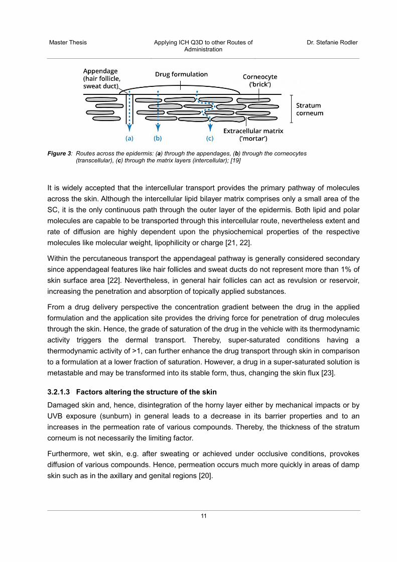
In general, there are three possible routes across the stratum corneum (Figure 3):
• Through the appendages • Transcellular (through the corneocytes) • Intercellular (through the lipid matrix layers)
These pathways are not mutually exclusive and most compounds permeate through the skin via a combination of pathways based on their physiochemical properties [19].
Master Thesis
Applying ICH Q3D to other Routes of Administration
Dr. Stefanie Rodler
11
Figure 3: Routes across the epidermis: (a) through the appendages, (b) through the corneocytes (transcellular), (c) through the matrix layers (intercellular); [19]
It is widely accepted that the intercellular transport provides the primary pathway of molecules across the skin. Although the intercellular lipid bilayer matrix comprises only a small area of the SC, it is the only continuous path through the outer layer of the epidermis. Both lipid and polar molecules are capable to be transported through this intercellular route, nevertheless extent and rate of diffusion are highly dependent upon the physiochemical properties of the respective molecules like molecular weight, lipophilicity or charge [21, 22].
Within the percutaneous transport the appendageal pathway is generally considered secondary since appendageal features like hair follicles and sweat ducts do not represent more than 1% of skin surface area [22]. Nevertheless, in general hair follicles can act as revulsion or reservoir, increasing the penetration and absorption of topically applied substances.
From a drug delivery perspective the concentration gradient between the drug in the applied formulation and the application site provides the driving force for penetration of drug molecules through the skin. Hence, the grade of saturation of the drug in the vehicle with its thermodynamic activity triggers the dermal transport. Thereby, super-saturated conditions having a thermodynamic activity of >1, can further enhance the drug transport through skin in comparison to a formulation at a lower fraction of saturation. However, a drug in a super-saturated solution is metastable and may be transformed into its stable form, thus, changing the skin flux [23].
3.2.1.3 Factors altering the structure of the skin
Damaged skin and, hence, disintegration of the horny layer either by mechanical impacts or by UVB exposure (sunburn) in general leads to a decrease in its barrier properties and to an increases in the permeation rate of various compounds. Thereby, the thickness of the stratum corneum is not necessarily the limiting factor.
Furthermore, wet skin, e.g. after sweating or achieved under occlusive conditions, provokes diffusion of various compounds. Hence, permeation occurs much more quickly in areas of damp skin such as in the axillary and genital regions [20].

Master Thesis
Applying ICH Q3D to other Routes of Administration
Dr. Stefanie Rodler
12
For all possible routes across the skin, pathological processes and skin diseases, such as neurodermitis or psoriasis, have an effect on the barrier properties of skin and can increase the extent of penetration of xenobiotics.
In addition, skin exposure to irritant compounds can enhance penetration due to disruption of the stratum corneum, e.g. by means of protein denaturating agents like detergents and soaps or through lipid extraction, leading to a potentially increased passage of substances through the skin, enabling them to easily reach the viable layers of the epidermis as well as, from there, the dermis and even the general circulation, inducing the potential for systemic intoxication [24].
3.2.1.4 Topical vs. transdermal application
The terms “topical” and “transdermal” are often used interchangeably. Nevertheless it is important to understand the difference to evaluate the effect of drug uptake and to assess the extent of exposure to elemental impurities possibly contained in the respective drug products.
In general, all preparations applied to the skin are topical by definition (applied to the top of the skin). Drug products topically administered via the skin fall into two categories, those applied for local action and those for systemic effects. Hence, the term “topical action” generally refers to formulations administered to the skin creating a local effect at the application site allowing the active to penetrate into deeper regions of the skin (e.g. corticosteroid creams). In contrast to that transdermal formulations cross the skin barrier aiming to deliver the active ingredient into the systemic circulation, thereby to circumvent the first-pass effect [19].
It is, however, difficult to strictly differentiate between 100% local and 100% transdermal action because once a compound has overcome the limiting SC barrier and reached to the living epidermis it may also penetrate into deeper skin layers which might bear blood vessels enabling the active to enter the systemic circulation [21].
3.2.1.4.1 Topical applications for local action
Topically administered drugs for local action are mainly applied in the form of creams, lotions, ointments, gels (semisolid dosage forms) or patches comprising drugs like corticoids (e.g. hydrocortisone), localanaesthetics (e.g. lidocaine), NSAIDs (e.g. diclofenac) or alcaloids (e.g. capsaicin) intended to act in tissues mainly through receptors and/or ion channels [25]. Topically acting drugs are an important part of therapy especially for common and chronic dermatologic diseases like cold sores (Herpes simplex), psoriasis or acne.
Depending on the physicochemical properties of the active substance, the drug formulation design and the site of action, semisolids can show their activity on the surface of the skin without stratum corneum penetration (e.g. repellents) as well as by exerting their action into the stratum corneum or by modulation of the function of the epidermis and the dermis. The barrier nature of the stratum corneum with its interstitial lipid pathway and proteinaceous cellular compartment greatly limits the uptake of drugs. Nonetheless, if a drug is to act locally, it must penetrate the

Master Thesis
Applying ICH Q3D to other Routes of Administration
Dr. Stefanie Rodler
13
stratum corneum to a certain extent. Usually, molecules penetrate the skin primarily via the tortuous and continuous intercellular pathway although transport of topically administered drugs may also occur through the transcellular route, particularly when solvents or enhancers are contained in the formulation. Furthermore occlusive effects, e.g. by means of ointment application on the skin, may lead to the retention of significant amounts of transepidermal water, thus facilitating drug transport through the hydrated skin [23].
3.2.1.4.2 Transdermal drug delivery systems
Systemic delivery of drugs from semisolid preparations has several drawbacks, including inconvenience of administration, inaccuracy of administered dose or difficulties in removing the residual formulation from the skin. Owing to these disadvantages, transdermal drug delivery systems (TDS), commonly referred to as “patches”, have to a large extent replaced semisolid preparations for systemic action.
TDS are flexible, multi-layered, pharmaceutical single dose preparations of varying size containing one or more active substance(s) intended to be applied to the intact skin in order to deliver the active(s) through the skin to the systemic circulation bypassing the destructive hepatic first-pass metabolism.
Although the intact skin provides a protective barrier for the body from the external environment, certain active substances are capable (depending on their physicochemical properties like molecular weight or melting point) to passively diffuse through the skin in order to achieve a therapeutic effect. Most active substances suitable to be delivered transdermally are hydrophobic with scopolamine, nicotine, fentanyl or estradiol and testosterone as the most prominent candidates used in TDS formulations.
Transdermal patches are designed to release the active ingredient(s) in a zero-order kinetics in vivo over a period of 1-7 days. Thereby, the active substance is absorbed through the intact skin (rate limiting step), resulting in a prolonged and controlled drug delivery rate involving the following steps:
• Release of the active from the formulation • Penetration / Diffusion through the SC • Partitioning from the SC into the viable epidermis before reaching the capillaries in the
dermis
Transdermal delivery systems usually offer significant advantages over oral administration due to circumvention of the first-pass metabolism and avoidance of the adverse gastrointestinal environment thereby enhancing patient compliance due to reduced side effects caused from temporary over dose. Due to the constant release rate uniform plasma levels are achieved. Furthermore TDS offer the advantage of a reduced dosing frequency due to the prolonged action up to one week [26].

Master Thesis
Applying ICH Q3D to other Routes of Administration
Dr. Stefanie Rodler
14
Currently two main TDS designs are available: A reservoir (membrane-controlled) and a matrix system. For details please refer to Figure 4 and Table 1, respectively.
Figure 4: Schematic representation of a matrix patch (a) and a reservoir patch (b)
In the reservoir TDS the active ingredient is contained in a gel or solution chamber from which it is controlled released by a semi-permeable membrane, whereas in the matrix patch the drug is homogenously embedded in an adhesive polymer matrix from which it is continuously released directly to the skin [27].

Master Thesis
Applying ICH Q3D to other Routes of Administration
Dr. Stefanie Rodler
15
Table 1: Transdermal Delivery Systems - Overview
Component Matrix system “Drug-in-adhesive system“
Reservoir system
Backing Layer − Usually impermeable − Protects the formulation while the patch is worn − Considerations: Occlusivity, patient comfort, cosmetic appearance.
Reservoir n.a. − Contains the drug(s) − Can be in the form of a solution,
suspension or gel or dispersed in a solid polymer matrix
Membrane n.a. − Usually semi-permeable − Possible incorporation of penetration
enhancers/solvents − Controls release of the drug from the
reservoir
Adhesive Matrix Layer(s)
− Backbone of TDS − Contains the drug(s) − Multilayers possible − Incorporation of additional excipients
e.g. stabilisers, thickeners, penetration enhancers
− Regulates drug release − Ensures adhesion on the skin for the
intended application period
− Ensures adhesion on the skin for the intended application period
Release Liner − Protects the adhesive layer and the drug formulation − Removed prior to patch application − Usually siliconised
In order to expand the range of possible candidates suitable for transdermal application and to promote the systemic availability, skin permeation enhancement is more and more used in TDS development and may be reached either via active or passive methods.
PASSIVE permeation enhancement:
Chemical approaches Several excipients are able to promote the transport of an active substance across the skin by a variety of mechanisms all of them temporarily altering the skin barrier function. The most important are [28, 29]:
• Disruption of the highly ordered structure of the stratum corneum via interaction with intercellular lipids
• Interaction with intercellular protein and keratin denaturation • Increasing solubility and improving partitioning of the drug into the SC.

Master Thesis
Applying ICH Q3D to other Routes of Administration
Dr. Stefanie Rodler
16
Different chemical classes of enhancers are known: Alcohols (e.g. ethanol) or glycols (e.g. propylene glycol) increase the solubility and improve the partitioning coefficient. Long-chain fatty acids like oleic acid or esters like isopropyl myristate interact with and modify the lipid domains of the SC whereas sulfoxides like DMSO interact with the keratin structure in the corneocytes. One challenge with the use of chemical enhancers is their correlation with increased skin irritation.
Formulation approaches Penetration enhancement with special formulation approaches is mainly based on the usage of colloidal carriers. Submicron-sized particles are intended to transport entrapped active molecules into the skin. Such carriers include micro- and nanoparticles or liposomes [28].
Other approaches Supersaturation may increase skin penetration without altering the skin barrier. The mechanism of enhancement is based on the increased thermodynamic activity of the active substance in the formulation by increasing the concentration gradient and, thus forcing the active out of the formulation and into and across the stratum corneum thereby carrying the risk of drug crystallisation due to thermodynamic instability [29].
ACTIVE permeation enhancement:
Physical approaches In order to enhance and expand transdermal drug delivery, permeation enhancement may also be achieved in an active manner by physical technologies such as iontophoresis or microporation.
Whereas iontophoresis uses an externally applied potential difference and a small electric current to enhance the transdermal delivery of charged and neutral compounds through the skin [30], microporation involves the creation of micropores or microchannels in the stratum corneum which allows water soluble molecules and macromolecules to overcome the skin. Technologies which can create these microchannels include mechanical microneedles, ultrasound, electroporation, radiofrequency and laser. These technologies are promising methods especially for the transdermal delivery of biopharmaceuticals, as these macromolecules usually are not able to permeate passively through the skin [31].
Microporation as an invasive technique leads to microscopic small skin injuries thereby disrupting the physiological barrier of the skin. Hence, micropores and microchannels do not only lead to a skin permeation enhancement for drugs but may also open the way for elemental impurities enabling them to enter the systemic circulation through the skin to a higher extent in contrast to the passive way. Therefore, the parenteral PDE set out in ICH Q3D may be relevant as starting point for PDE establishment for a dermal product as open and damaged skin has a reduced barrier function.
Master Thesis
Applying ICH Q3D to other Routes of Administration
Dr. Stefanie Rodler
17
3.2.1.5 Establishing Limits for Dermal Absorption of Elemental Impurities
The intention of ICH Q3D is to establish limits for unwanted elemental impurities in drug products. Acceptable harmonised limits for cutaneous/transdermal drug products are still lacking presumably also due to the fact that data availability in terms of toxic and/or carcinogenic effects of metals and their derivatives by direct contact or systemic absorption through the skin is largely heterogenous. In particular, data relating to the ability of metals to penetrate the skin, are widely disseminated in literature. Furthermore, the fact that a majority of reliable experimental data was obtained more than 40 years ago with methods no longer up-to-date and the fact that methods on percutaneous penetration of metals are obtained under different non-standardised experimental conditions make a comparison of published results very difficult. Only in 2004 the OECD published guidelines for an in vivo (No. 427, [32]) and an in vitro (No. 428, [33]) test method to assess dermal absorption. The “OECD Guidelines for the Testing of Chemicals” is a collection of about 150 internationally agreed test methods used by government, industry and laboratories to identify and characterise potential hazards of chemicals but was only rarely used to characterise the dermal absorption of metals until now.
Most available published data on dermal absorption of elemental impurities concentrate on frequently used, omnipresent metals such as nickel, chromium or cobalt as their presence (including their derivatives) in the workplace and their accumulation in the environment causes concerns in terms of potential health hazards in general [34].
For topically applied drug product (either intended for local or for systemic action) other EIs as those mentioned in ICH Q3D may also be of relevance but data availability in terms of skin permeation and sensitisation potential may even be worse for these elements.
3.2.1.5.1 Percutaneous penetration of metals through the skin
The presence of certain metals or metal-based compounds as unwanted impurities in drug products administered by the cutaneous and transdermal route raises appropriate questions concerning human exposure related to their toxicity. Uptake of these materials through the skin may represent a route of exposure, which is not well characterised. Currently, an ICH Expert Working Group is trying to quantify this exposure to elemental impurities via the dermal route by developing PDEs in analogy to oral, parenteral and inhalation products. Nevertheless, not all elemental impurities currently listed in ICH Q3D may be relevant in terms of skin penetration. To continue the process of harmonisation, it would be beneficial to develop generally valid PDEs for products administered to the skin, where relevant. Thereby, the issues to be resolved include the evaluation if and to which extent EIs can penetrate the skin as well as the determination for which of the EIs a safety-based PDE will need to be established.
Anyhow, it is important to consider the structure of the skin, thereby taking into account that the outermost layer, the stratum corneum, is highly lipophilic and contains only very little water. As a result, hydrophilic or charged molecules are mostly hindered from penetrating into the lipid layer

Master Thesis
Applying ICH Q3D to other Routes of Administration
Dr. Stefanie Rodler
18
and, hence, from passing through the skin in significant levels. On the other hand, as the skin is the organ directly in contact with the drug product in frequent intervals or continuously over a longer period, specific toxic endpoints may be of relevance for some EIs and require a specific (lower) PDE limit. In this context, allergic sensitisation may have to be considered causing allergic contact dermatitis (ACD) through a hypersensitivity reaction after dermal contact. Such exposure represents an additional safety issue to face in the evaluation of EIs in dermal products [35].
Percutaneous penetration of metals is influenced by many factors such as oxidation state, molecular weight, lipophilicity, reactivity and the nature of the metal compounds (e.g. salts) as well as by the product properties itself (applied dose, duration of contact, vehicles used etc.) and by user-specific characteristics like thickness and integrity of the skin layers at the application site, sweating, gender or age [24]. This makes it difficult to create predictive models as most metals penetrate the skin in no particular order. The most studied compounds are probably Ni and Cr due to their high potential to cause allergy and ACD, respectively, and their widely use in consumer articles like jewellery, clothes, electronic devices as well as in leather tanning [34, 36].
In general, percutaneous penetration of unwanted impurities is of importance when the absorption through the skin contributes significantly to the body burden. Usually, from a scientific point of view, the characteristic that is relevant to patient safety is the total daily mass of an elemental impurity delivered to the patient as the toxicological risk depends mainly on the total exposure [11]. This may not be fully true for dermally applied products, as sensitization – e.g. caused by metals like nickel – can already happen with extremely low doses, which are not of relevance in terms of systemic intoxication for which usually higher concentrations are necessary [24].
Therefore, relevant data in terms of qualitative and (if possible) quantitative evaluation of EI permeation through the skin, the role of each element in metabolism, particularly with respect to the skin, and the potentially toxic effects that may result from dermal contact as well as the immunological characteristics (including allergenicity) should be considered when establishing PDE limits for cutaneous and transdermal products [37].
Dermal absorption
The degree of dermal absorption, i.e. the transport of a substance across the skin and its update into the body and, hence, its ultimate therapeutic or toxic effect, is a complex process and influenced by a variety of factors. Although for most metals, uptake through the skin is limited, experimental human data have demonstrated that metals can penetrate and permeate the skin – even though to a limited extent – and are able to reach the viable layers of the epidermis or even the dermis and from there the systemic circulation. Skin can also act as a reservoir for metals like it was shown for nickel in the stratum corneum when single doses of nickel were applied on the skin in various concentrations [24, 38].

Master Thesis
Applying ICH Q3D to other Routes of Administration
Dr. Stefanie Rodler
19
Published information, recommendations, guidance and risk assessments covering exposure to metals and their possible uptake in the body in the past were mainly intended for managing potential health risks arising for professions which are exposed to metals to a high extent like miners or employees in metal companies or refineries. These publications primarily focused on environmental exposure from sources such as soils. A guidance provided by the US Environmental Protection Agency (EPA) for example gives advice how to perform a health-based risk assessment for humans for the evaluation of oral bioavailability of metals in soils [39].
A key factor in determination of toxic effects associated with topical application of drug products containing elemental impurities is the ability of the respective impurity to be absorbed through the skin into the systemic circulation and its contribution to systemic body burden. A number of studies and reviews of metal absorption via topical exposure are published demonstrating that dermal absorption is in general significantly less than oral absorption, thereby further limiting systemic exposure:
Investigations in terms of dermal absorption of metals have already been conducted in the 1960s systematically studying the absorption of radioactive metal compounds through the skin of living guinea pigs [40, 41]. Aqueous solutions of CoCl2, ZnCl2, CdCl2, HgCl2, AgNO3, Na2CrO4 and methyl mercury dicyandiamide at various concentrations were applied revealing the highest relative absorption for methyl mercury dicyandiamide with a value of 4.5% over an application of 5 hours. All other compounds showed lower relative absorption rates at any concentration. Although these publications do not comply with current modern standards and the exact values are presumably no longer valid, they nevertheless indicate that inorganic metal compounds exhibit a rather low potential for dermal absorption [42].
Another review dealing with metal exposure and a possible uptake by the skin is from 1980 which also confirms the above mentioned assumption: Moore et al. determined that the dermal absorption of lead acetate from cosmetic preparations is in the range of 0-0.3% [43] while (according to more recent data) oral absorption of lead from food and water is estimated at 50% and from soil at 30% [39, 44].
In 1993 Hostynek et al. [37] aimed to collect data relevant for the qualitative and quantitative evaluation of metal permeation through skin. In total, they summarised 31 metals, but an assessment of ICH Q3D class 1 elements like mercury or lead is lacking. In general, they concluded that dermal absorption of metals is a complex process affected by multiple factors including size, charge and oxidation. They did not, however, draw any definitive overall conclusions regarding default estimates of absorption nor did they make any comparisons to other routes of administration [45].
One of the most significant publication is the fact sheet “HERAG (Health Risk Assessment Guidance For Metals) - Assessment Of Occupational Dermal Exposure And Dermal Absorption For Metals And Inorganic Metal Compounds” [42] critically evaluating existing data and models

Master Thesis
Applying ICH Q3D to other Routes of Administration
Dr. Stefanie Rodler
20
which are used to examine levels of dermal exposure and rating their value in assessing the absorption of inorganic metals.
Current EU guidance and available models for the prediction of dermal absorption
In the case of lack of any data on dermal absorption, the current Technical Guidance Document (TGD) on Risk Assessment (Part I) published by the European Commission in 2003 [46] consults the EASE (Estimation and Assessment of Substance Exposure) model, developed by the United Kingdom’s Health and Safety Executive, for prediction of dermal absorption by assigning two different default-values depending on substance-specific properties:
• 10% dermal absorption: for compounds with a molecular weight of >500 and a log Pow smaller than -1 or higher than 4 as a limited extent of skin permeation is assumed for substances above these values
• 100% dermal absorption: for all other compounds
The major point of criticism with such a general approach is that the model was developed for organic chemical compounds which is lacking in terms of metals: The common understanding of a compound to be able to penetrate the skin by diffusive mechanism is to dissolve first. For metals or an inorganic metal compound, this requires dissociation to the respective free metal ion, for which partition coefficients like the Log Pow value are not of relevance in the prediction of skin-related properties as a metal or an inorganic salt thereof may not permeate the skin via passive diffusion. The second criterion for assigning a dermal absorption rate, the molecular weight, is irrelevant for metals as the cut-off value of 500 is not exceeded by any metal cation.
In general, such puristical approaches like the EASE model are scientifically questionable and expected to significantly over-predict the actual levels of dermal exposure. And indeed, the TGD does in fact suggest to use alternative dermal absorption values where scientifically justified data are available [42, 45].
A large EU founded project on the Evaluation and Prediction of Dermal Absorption of Toxic Chemicals (EDETOX), i.e. a research program conducted between 2001 and 2004, aimed to generate new knowledge for the standardisation of in vitro systems to better predict percutaneous penetration [47]. Although mainly focused on organic substances, parts of this practical guidance on conduct of such studies are also applicable for metals and metal compounds, of which three were considered (sodium chromate, cobalt powder and nickel chloride) [42].
The above mentioned HERAG guidance document [42] provides a summary on conducted studies that further jeopardise the EASE model which assumes 10% as lowest possible rate for dermal absorption. Excerpts of the results of these studies are summarised in Table 2 for ICH Q3D-relevant elements. In addition, the HERAG guidance documents also summarises more recent data on dermal absorption for metals and inorganic metal compounds like Zn, Ni, Cd, Sb,

Master Thesis
Applying ICH Q3D to other Routes of Administration
Dr. Stefanie Rodler
21
Cu and Pb as results of various EU Risk Assessment Reports (EU RAR) published by ECHA (European Chemicals Agency) as well as of Voluntary Risk Assessments (VRA).
The EU RARs were prepared by different member states at the instance of the Commission within the frame of the “Existing Substances Regulation (ESR)” – one of various EU legislations for the regulation of chemicals before in June 2007 the “REACH regulation” (EC Regulation 1907/2006) came into effect. The ESR was intended to regularly provide updates of priority substances which require immediate attention because of their potential effects to human health or the environment. The complete overview on the risk assessments performed by the EU member states for each of the 141 ESR substances can be found on the ECHA website under “Information from the Existing Substances Regulation (ESR)” [48].
Table 2: Dermal absorption data for metals and inorganic metal compounds [42, 45]
Metal/compound Test system Results*
Data as extracted and concluded upon in the various existing EU RA reports:
Cadmium metal, Cadmium oxide
(analogy) <1% (EU RAR assessment, Rapporteur: Belgium)
Nickel metal, Nickel sulphate, Nickel chloride, Nickel nitrate, Nickel acetate
in vitro, human skin, tape stripping
0.2% (EU RAR assessment, Rapporteur: Denmark)
Nickel sulphate, Nickel chloride, Nickel nitrate, Nickel acetate
in vitro, human skin
2% (EU RAR assessment, Rapporteur: Denmark) 1% when material bound to stratum corneum is discounted
Diantimony trioxide
in vitro, human skin
0-0.1%
Copper compounds (not specified)
in vitro (unspecified)
0.3% soluble/insoluble Cu compounds (VRA Copper)
Lead oxide in vitro, human skin
0-0.1% (VRA Lead)
Additional (non-exhaustive compilation) data made available from metal industries participating in HERAG:
Cobalt metal in vitro, (Franz diffusion cell, human skin)
Absorption not given as a percentage of the applied dose but as a steady-state flow of (0.0123 ± 0.0054) μgcm-2h-1 with a lag time of (1.55 ± 0.71) h. Significant absorption only took place, when the metal was oxidised to Co2+ by stirring in artificial sweat for 30 minutes
* for detailed reference information see HERAG [42]

Master Thesis
Applying ICH Q3D to other Routes of Administration
Dr. Stefanie Rodler
22
Considerations in assessing dermal absorption
Various in silico, in vitro and in vivo models exist to estimate or measure dermal absorption of metals through skin. Published data from these studies have to be reviewed carefully in order to understand systemic exposure of various metals for risk assessment purposes and to assure an adequate safety margin for exposure to elemental impurities [44]:
Heterogeneity of published data and used techniques complicate the comparison of results leading to conflicting interpretation (e.g. species-specific differences in skin properties, sensitivity of used analytical methods, sampling techniques etc.)
Quantitative percutaneous absorption assessments are often based on the use of a skin permeability constant (Kp) However, the permeability coefficient (expressed in [cm/h]) is experimentally determined and characterised according to Fick's first law of diffusion by the ratio of flux and the applied concentration of the test compound. The Kp is usually derived in the laboratory from in vitro studies and the rate of penetration is ideally determined assuming steady state conditions. As infinite dose levels are not representative in vivo for percutaneous penetration of metals due to their affinity to the stratum corneum and their ability to build reservoirs their usefulness for dermal risk assessment is questionable [34, 49].
Some models may imply that the amount which is transported through the stratum corneum is a function of metal concentration and a metal specific permeability coefficient. Although most published data were generated with the intention to investigate dermal absorption in high-risk professions, i.e. considering worst-case circumstances with a high overall occupational exposure to metals, skin loading levels and saturation phenomena, i.e. reservoir forming in the epidermis, for the metal in questions may not be recognised. For example nickel is bound in a reversible manner in the epidermis forming a reservoir. Its affinity for keratin influences percutaneous absorption so that from some salts the breakthrough time is considerable long (from 24 to 48 hours) [34].
In order to establish rational limits for exposure to elemental impurities in topical products, a reliable prediction of the quantitative percutaneous absorption of the drug product itself is necessary first but lacks of the following general uncertainties and variabilities [20]:
• Dosing regimen: Time of exposure and frequency of exposure (e.g. leave-on or rinse-off products)
• Drug exposure and quantity of a topical preparation: Inaccuracies in dosing as e.g. in terms of creams or ointments the amount to be applied is not clearly defined and, thus, up to the user’s discretion.
• Influence of formulation: Vehicle (e.g. microparticles), type of formulation (e.g. w/o or o/w), use of permeation enhancers etc.
• Physicochemical properties of compound in question: size, lipophilicity, charge, oxidation stage

Master Thesis
Applying ICH Q3D to other Routes of Administration
Dr. Stefanie Rodler
23
• Skin diseases and permeability: Dermatoses and other pathological processes may impede the barrier function.
• Environmental factors and occlusion: Contact of the stratum corneum with water or defatting agents, grade of hydration, mechanical stress, UV irradiation, seasonal skin variations etc.
• Application site: Considerable differences with respect to skin thickness or grade of hydration
• Age: Different skin structure including the barrier function (e.g. in children and adults) • Accumulation in the epidermis
Based on these insecurities it becomes obvious that an exact quantitative evaluation of dermal absorption for drug products in general and single elemental impurities in particular is challenging. Model calculations or default dermal absorption factors may need to be considered instead.
In general, existing data do indicate that dermal exposure to metals is limited – usually far below the corresponding observed extent when the same material is administered orally. This conclusion is logical considering the nature of the epidermis and the fundamental barrier properties.
Available data as summarised in Table 2 reveal that dermal absorption levels for most metals are below 5%. This correlates well with the levels provided by the US EPA estimating e.g. a default dermal absorption of 3% for arsenic and 1% for other metals (e.g. for cadmium compounds from soil) [50, 51].
3.2.1.5.2 Skin Sensitisation
Sensitisation has been identified for few EIs after dermal contact from occupational exposure or from cosmetic and house-hold products. Such exposure represents an additional safety issue to consider in the evaluation of EIs in dermal drug products.
Skin sensitisation (also known as allergic contact dermatitis or contact hypersensitivity) is a skin rash or eczema caused by an allergy to specific substances. It is an immune reaction resulting from immunological priming induced by a first contact with an allergen activating the immune system. Another subsequent contact with this allergenic substance then leads to a local effect including early signs like dryness, redness, swelling or itchiness of the skin. This local effect is not limited to the skin area which was in direct contact with the substance but can also spread to other parts of the body.
Potentially allergenic substances penetrate the skin until they reach a viable dermis, after which they interact with skin proteins and immune cells inducing a complex immune response that involves the interaction with T-lymphocytes leading to a specialised immunological memory (“sensitisation phase”). During the “elicitation phase”, i.e. the next contact with the respective compound, a wide-spread elicitation of the immune system (i.e. allergic response) occurs in the sensitised individual due to a reaction between the allergen-specific T-cells and the allergen [52,
Master Thesis
Applying ICH Q3D to other Routes of Administration
Dr. Stefanie Rodler
24
53]. In general, trace amounts of skin allergens are sufficient to induce ACD, thereby, usually lower amounts are necessary for elicitation than for inducing hypersensitivity.
According to the UN Globally Harmonized System of Classification and Labelling of Chemicals (UN GHS) skin sensitisers are defined as substances that will lead to an allergic response following skin contact. They can either be assessed and classified based on human data showing a sensitisation response in a substantial number of individuals or positive results from appropriate animal tests [54].
Available data in terms of skin sensitisation potential of substances in questions are mainly based on investigations conducted due to occupational risk in professions with a high exposure to sensitising agents like certain metals and metal compounds. In general, contact dermatitis may be provoked by elements like Al, Au, Be, Co, Cr, Cu, Hg, Ir, Ni, Pd, Pt, Rh or Ti [55] but divergent data a published in terms of potentially irritating/sensitising metals presumably due to different tested salt forms. Consequently, the varying bioavailability of different metal compounds due to their different forms and salts should also be considered as it is essential for inducing and elicitating ACD. This is also the reason why some metals are harmless for the skin, whereas their salts may be potent skin sensitisers, e.g. water soluble chromates are known to be a very common cause of ACD while elementary chromium is not [34].
For some EIs covered by ICH Q3D a summary on their potential to cause skin sensitisation and its prevalence is given below:
Nickel Nickel sensitisation is a general global socio-demographic problem of vast proportions which usually occurs after exposure derived from releasing consumer products such as jewellery, keys, watches or piercings. Skin sensitisation to nickel is the most frequent cause of allergy in industrialised countries worldwide. With a prevalence of ca. 10-20% in the European population nickel represents the most important cause of ACD [56]. Despite the high prevalence, only limited is known about the exact skin penetration pathway of Ni compounds which could explain the rapid contact eczema elicitation in sensitised individuals after repeated simple contacts [34].
Chromium Leather products have been described as important causes of chromium contact allergy as Cr salts like chromium(III)hydroxide sulphate (Cr(OH)SO4) are usually used for leather tanning. Chromium (III) can be oxidised to chromium (VI) which is a suspected carcinogen and a well-known skin sensitiser.
Furthermore, sensitisation reactions from contact with cement is a classical occupational disease which is associated with chromium, especially chromium (VI). Wet cement has a high pH of >12 altering the stratum corneum and, hence, facilitating the penetration of water-soluble substances. Thus, skin contact with the alkaline cement-water suspension results in irritation, thereby enhancing absorption of soluble chromate compounds and the elicitation of allergic reactions.

Master Thesis
Applying ICH Q3D to other Routes of Administration
Dr. Stefanie Rodler
25
The prevalence of chromium allergy in Europe in a recently published study was found to be in the range of 1% [56].
Cobalt Potential sources of cobalt exposure are jewellery or other metal consumer objects as well as prosthetics, paints, and pigments. The prevalence of cobalt allergy in Europe was found to be in the range of 2%. [56]. Cobalt sensitisation is usually co-associated with chromium and/or nickel as Ni is often contaminated by Co and cement contains both Cr and Co. As a result, contact dermatitis to cobalt may often occur due to the combined exposure to these metals [34].
Mercury Induction of contact dermatitis by mercury was often associated with the use of antiseptics, disinfecting agents or dental amalgam. Thereby mercury salts are irritants on the skin causing dermatitis (especially under occlusive conditions), discoloration of the nails and corrosion of the mucous membranes. Generally, skin sensitivity to mercurial compounds has little clinical significance in developed countries nowadays as mercury-containing candidates like thimerosal which has been used as preserving agent in vaccines as well as dental amalgams fillings nearly disappeared in the recent past [34, 57].
Platinum Group Metals Platinum group metals (PGMs) or “platinoids” comprise 6 elements of groups 8, 9, and 10 in the periodic table constituting the transition metals platinum, palladium, rhodium, ruthenium, iridium, and osmium with platinum as the most prominent and most studied representative [58].
In general, occupational exposure to the PGM may cause contact dermatitis. Due to the limited information on toxicity, toxicological data for the PGM are often based on and derived from platinum. Palladium may be contained in jewellery and dental fillings. Sensitisation to Pd salts is not uncommon but may often occur due to the combined exposure and concurrent presence with nickel [59]. This is even confirmed by the in vivo study on skin sensitisation of palladium published by ECHA revealing a non-sensitising outcome [60].
In animals, rhodium has proven to be a powerful skin sensitiser. Furthermore individual cases of ACD from rhodium salts in jewellery manufacture and dental material are reported in literature [34].
Master Thesis
Applying ICH Q3D to other Routes of Administration
Dr. Stefanie Rodler
26
Legislations to limit the metal exposure in the EU
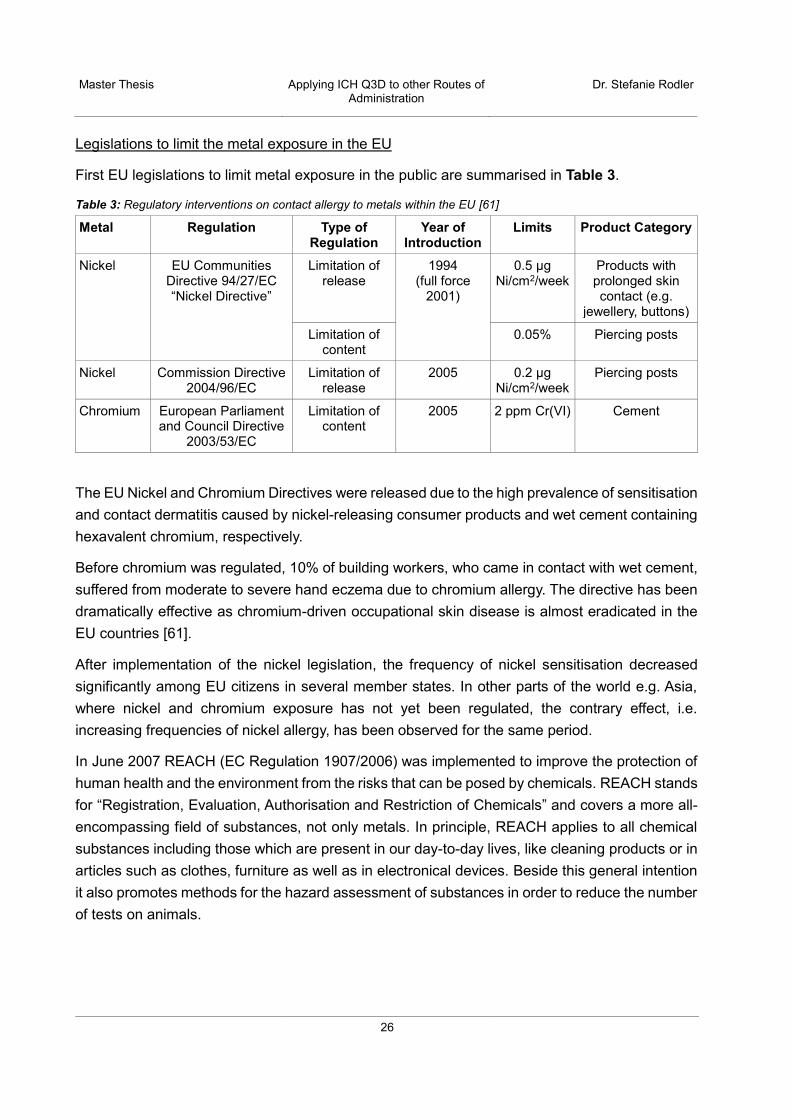
First EU legislations to limit metal exposure in the public are summarised in Table 3.
Table 3: Regulatory interventions on contact allergy to metals within the EU [61]
Metal Regulation Type of Regulation
Year of Introduction
Limits Product Category
Nickel EU Communities Directive 94/27/EC “Nickel Directive”
Limitation of release
1994 (full force
2001)
0.5 μg Ni/cm2/week
Products with prolonged skin contact (e.g.
jewellery, buttons)
Limitation of content
0.05% Piercing posts
Nickel Commission Directive 2004/96/EC
Limitation of release
2005 0.2 μg Ni/cm2/week
Piercing posts
Chromium European Parliament and Council Directive
2003/53/EC
Limitation of content
2005 2 ppm Cr(VI) Cement
The EU Nickel and Chromium Directives were released due to the high prevalence of sensitisation and contact dermatitis caused by nickel-releasing consumer products and wet cement containing hexavalent chromium, respectively.
Before chromium was regulated, 10% of building workers, who came in contact with wet cement, suffered from moderate to severe hand eczema due to chromium allergy. The directive has been dramatically effective as chromium-driven occupational skin disease is almost eradicated in the EU countries [61].
After implementation of the nickel legislation, the frequency of nickel sensitisation decreased significantly among EU citizens in several member states. In other parts of the world e.g. Asia, where nickel and chromium exposure has not yet been regulated, the contrary effect, i.e. increasing frequencies of nickel allergy, has been observed for the same period.
In June 2007 REACH (EC Regulation 1907/2006) was implemented to improve the protection of human health and the environment from the risks that can be posed by chemicals. REACH stands for “Registration, Evaluation, Authorisation and Restriction of Chemicals” and covers a more all-encompassing field of substances, not only metals. In principle, REACH applies to all chemical substances including those which are present in our day-to-day lives, like cleaning products or in articles such as clothes, furniture as well as in electronical devices. Beside this general intention it also promotes methods for the hazard assessment of substances in order to reduce the number of tests on animals.

Master Thesis
Applying ICH Q3D to other Routes of Administration
Dr. Stefanie Rodler
27
The restrictions for nickel were first established by Directive 94/27/EC and subsequently incorporated into REACH:
“Nickel shall not be used:
• in any post assemblies which are inserted into pierced ears and other pierced parts of the human body unless the nickel release […] is less than 0.2 μg/cm2/week.
• in articles intended to come into direct and prolonged contact with the skin such as: earrings, necklaces, bracelets and chains, anklets, finger rings, wrist-watch cases, watch straps and tighteners, rivet buttons, tighteners, rivets, zippers and metal marks, when these are used in garments, if the nickel release rate from the parts of these articles coming into direct and prolonged contact with the skin is greater than 0.5 μg/cm2/week.”
The ECHA is responsible for the implementation and supervision of the REACH requirements. In general, the EU legislation in terms of nickel and chromium restrictions was widely successful in most of the EU countries and occupational metal exposure could effectively be reduced although it still remains prevalent.
Potentially allergenic EIs in drug products
Allergic contact dermatitis reactions to metals generally occur if the metal salts are in solution, as occurs with perspiration or exposure to body fluids [62]. Potential trace amounts of typical skin allergens with a high immunogenic potential like nickel, chromium or cobalt and their associated salts, respectively, eventually contained in drug products do not noteworthy contribute to the overall exposure in terms of skin sensitisation as they are anyway ubiquitious in our environment. The same is applicable for aluminium salts which are frequently used is antiperspirants.
Nevertheless some other “rare” EIs may be relevant in the context of ICH Q3D in terms of skin sensitisation and ACD as they are exclusively contained in drug products but were not considered in the past because of their negligible presence or general lack of exposure in workplaces and the general environment, respectively, e.g. catalysts used for API synthesis like molybdenum, vanadium or ruthenium compounds.
Furthermore occlusive conditions with the administration of dermal drug products may alter the constitution of the skin, i.e. the stratum corneum, and thereby facilitating the entry of otherwise impermeable, harmless elemental impurities.
When applying ICH Q3D to dermal drug products, skin sensitisation is considered as a cause of potential safety concern for a few EIs, justifying a concentration limit in addition to the PDE which may improve the overall dermal safety evaluation [35]. When a dermal concentration limit is defined for an EI, this value should be compared to 30% of the PDE (i.e. the control threshold, see chapter 2.2.2) considering the daily posology of the dermal drug product to evaluate which of the two limits is the lowest.

Master Thesis
Applying ICH Q3D to other Routes of Administration
Dr. Stefanie Rodler
28
For example: The dermal PDE for cobalt is 50 µg/day; thus, the daily exposure from a dermatological DP should not exceed 30% (i.e. 15 µg/day). The dermal concentration limit is defined to 10 µg/g. Thus, up to a posology in the drug product of 1.5 g/day (= 15 µg/day divided by the concentration limit of 10 µg/g) the dermal concentration limit for Co (10 µg/g) must be applied as this is the lowest limit whereas for a drug product applied above 1.5 g/day the 30% PDE (15 µg/day) will be the applicable limit.
Transdermal delivery systems
For obtaining marketing authorisation for transdermal drug delivery systems it is required to investigate the irritation and sensitisation potential of the final formulation in vivo. Respective guidelines are in place both in the EU [63] and the USA [64]. Thereby, the whole drug product is examined as the ingredients and the general composition of a TDS formulation, including the nature of the drug substance as well as the occlusive effect and the transmission of water vapor from the skin, in conjunction with other factors such as the environmental humidity or the condition of the skin, may have the potential to irritate the skin or lead to a sensitisation reaction.
TDS as a whole have to comply with the requirements in terms of a possible irritation and sensitisation potential. Therefore, potentially contained allergenic EIs are assessed automatically in the in vivo investigation by definition. Therefore, in case that the requested risk assessment for a TDS according to ICH Q3D reveals the presence of potential relevant allergenic EIs, the respective hazard potential arising from these elements and its manifestation can be evaluated within the sensitisation studies. Theoretically identified EIs in the view of a possible skin interaction/sensitisation potential do not necessarily have to cause an allergic reaction in vivo as the incidence is not only a matter of potentially contained sensitising metal elemental impurities like nickel or chromium but rather attributed to the overall formulation of the drug product and the presence of skin-modifying agents among others.
3.2.1.6 Relevant elemental impurities in dermally applied drug products
ICH Q3D provides the option to re-evaluate PDEs for alternative routes of administration – when supported by data – taking into consideration differences in bioavailability and specific toxic endpoints by those alternative routes. Differences in absorption between the dermal and oral routes are known for several compounds [65]; thus, data in terms of dermal absorption may allow re-evaluation of given oral PDEs expected to result in equal or higher dermal PDEs (dPDE) due to the barrier function of the skin which is the primary organ in contact with dermal drug products. Among skin-specific toxic endpoints for dermal products, sensitisation and local effects have to be considered as additional safety issues.
Following the work published by Teasdale et al. in 2015 [45], Bouvier et al. [35] performed an evaluation of elemental impurities for which an oral PDE was assigned in ICH Q3D with the

Master Thesis
Applying ICH Q3D to other Routes of Administration
Dr. Stefanie Rodler
29
intention to identify differences between oral and dermal bioavailabilities and to establish specific dermal PDEs for single elements.
Oral PDEs for the single elemental impurities were used as starting point for the establishment of dPDEs for topically applied drug products: Where robust data were available from literature in terms of dermal penetration, the oral PDE limit was corrected by a factor considering the ratio of the oral versus the dermal absorptions (if dermal and oral absorptions are more than two-fold different). Thereby, in vitro data penetration studies on human skin and at last animal data were considered if no human data were available.
In addition, among skin-specific toxic endpoints, skin sensitisation was considered in the evaluation to offer recommendations for minimal dermal concentration limits [µg/g] for few EIs in the final drug product.
The results are presented in Table 4.

Master Thesis
Applying ICH Q3D to other Routes of Administration
Dr. Stefanie Rodler
30
Table 4: Oral permissible daily exposures, dermal PDEs and dermal concentration limits [35]
Element Class Oral PDE [µg/day]
Dermal PDE [µg/day]
Concentration limits [µg/g]
Cd Cadmium 1 5 5 n.a. Pb Lead 1 5 5 n.a. As Arsenic 1 15 15 n.a. Hg Mercury 1 30 30 10 Co Cobalt 2A 50 50 10
V Vanadium 2A 100 100* n.a. Ni Nickel 2A 200 110 5
Tl Thallium 2B 8 8* n.a. Au Gold 2B 100 100* n.a. Pd Palladium 2B 100 100* n.a. Ir Iridium 2B 100 100* n.a. Os Osmium 2B 100 100* n.a. Rh Rhodium 2B 100 100* n.a. Ru Ruthenium 2B 100 100* n.a. Se Selenium 2B 150 150* n.a. Ag Silver 2B 150 150 n.a. Pt Platinum 2B 100 50 n.a. Li Lithium 3 550 47600 n.a. Sb Antimony 3 1200 1200* n.a. Ba Barium 3 1400 1400* n.a. Mo Molybdenum 3 3000 3000* n.a. Cu Copper 3 3000 3000 n.a. Sn Tin 3 6000 6000* n.a. Cr** Chromium 3 11000 11000 100
* no dermal PDEs available due to missing data for oral or dermal absorption. ** As Cr(VI) is unstable and reactive and unlikely to be present in a DP, only Cr(III) was considered. n.a. not applicable (no specific limit related to skin toxicity was identified)
Based on the published literature presented before and additional screenings in terms of dermal absorption of certain metals the following data of elemental impurities covered by ICH Q3D can be summarised, including a proposed default value for dermal absorption which can be used for establishing PDE limits for topically applied products (Table 5).
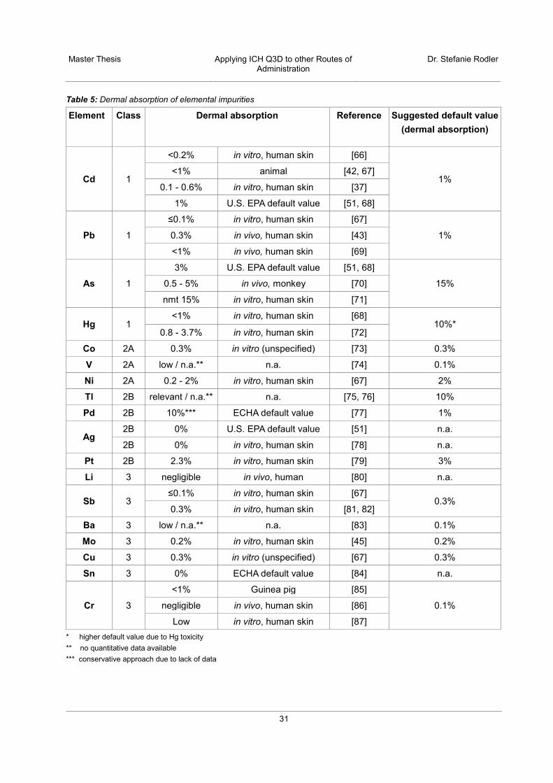
Master Thesis
Applying ICH Q3D to other Routes of Administration
Dr. Stefanie Rodler
31
Table 5: Dermal absorption of elemental impurities
Element Class Dermal absorption Reference Suggested default value (dermal absorption)
Cd 1
<0.2% in vitro, human skin [66]
1% <1% animal [42, 67]
0.1 - 0.6% in vitro, human skin [37]
1% U.S. EPA default value [51, 68]
Pb 1
≤0.1% in vitro, human skin [67]
1% 0.3% in vivo, human skin [43]
<1% in vivo, human skin [69]
As 1
3% U.S. EPA default value [51, 68]
15% 0.5 - 5% in vivo, monkey [70]
nmt 15% in vitro, human skin [71]
Hg 1 <1% in vitro, human skin [68]
10%* 0.8 - 3.7% in vitro, human skin [72]
Co 2A 0.3% in vitro (unspecified) [73] 0.3%
V 2A low / n.a.** n.a. [74] 0.1%
Ni 2A 0.2 - 2% in vitro, human skin [67] 2%
Tl 2B relevant / n.a.** n.a. [75, 76] 10%
Pd 2B 10%*** ECHA default value [77] 1%
Ag 2B 0% U.S. EPA default value [51] n.a.
2B 0% in vitro, human skin [78] n.a.
Pt 2B 2.3% in vitro, human skin [79] 3%
Li 3 negligible in vivo, human [80] n.a.
Sb 3 ≤0.1% in vitro, human skin [67]
0.3% 0.3% in vitro, human skin [81, 82]
Ba 3 low / n.a.** n.a. [83] 0.1%
Mo 3 0.2% in vitro, human skin [45] 0.2%
Cu 3 0.3% in vitro (unspecified) [67] 0.3%
Sn 3 0% ECHA default value [84] n.a.
Cr 3
<1% Guinea pig [85]
0.1% negligible in vivo, human skin [86]
Low in vitro, human skin [87] * higher default value due to Hg toxicity ** no quantitative data available *** conservative approach due to lack of data
Master Thesis
Applying ICH Q3D to other Routes of Administration
Dr. Stefanie Rodler
32
The PGMs rhodium, ruthenium, iridium, and osmium were not part of the screening as data on toxicokinetics, metabolism and distribution are mostly available for platinum and palladium, the most prominent PGM representatives. Gold was also excluded from analysis as misinterpretations are possible due to its wide use in the medicinal field e.g. gold nanoparticles. No results could be found for selenium.
It should be considered that the summary presented in Table 5 is based on collected data by screening literature without considering the particular oxidative state, the time of exposure, the physical characteristics (i.e. organic or inorganic), the integrity of the skin or the form (e.g. as solution, dry powder) and the vehicle in which the metal in questions was applied. Neither a possible retention in the skin and the potential for binding to skin proteins, respectively, was considered.
Some element may be toxic for the skin, regardless of route of administration. Specific dermal toxicity is e.g. reported for arsenic [88]. In addition, mercury may be of particular interest because of the differing toxicities of its inorganic and complex organic forms. Therefore, for some EIs like mercury – although their degree of dermal penetration may be low – a higher default value for dermal absorption is applied, hence, adequately addressing the increased toxicity.
For some EIs like chromium or mercury divergent results are reported. Furthermore, the terms “permeation” (diffusion through skin) and “penetration” (diffusion through/into various layers of the skin) were often used interchangeably. Additionally, the two pharmacokinetic terms, absorption and bioavailability, were considered synonymously, as a possible skin-related metabolism for metals was expected to be not relevant.
Due to the heterogeneity of data and the possible variabilities presented before, the establishment of a default value in terms of dermal absorption for the single EIs is difficult. In case of varying results were found, the most conservative value was taken except for palladium as the ECHA assumes that Pd does not undergo appreciable uptake by the dermal route as well as for chromium as all published data indicate a low to negligible amount of dermal uptake.
In sum, all available data indicate that bioavailability for most EIs after dermal administration is in general low (usually below 3%). Based on this summary, it is recommended to limit the volume of ICH Q3D for cutaneous and transdermal products to the relevant elemental impurities which may potentially comprise a risk to human health via this route of administration i.e. class 1 elements and – in terms of a safe-side approach – elements which may show a bioavailability of at least 2% i.e. nickel, thallium and platinum.

Master Thesis
Applying ICH Q3D to other Routes of Administration
Dr. Stefanie Rodler
33
3.2.1.7 Case Study – Transdermal Delivery System
The example provided in the following for a TDS may give advice how to approach specific reasonable limits for elemental impurities in topical products with systemic action.
Product: Phantasin TDS 70 µg/h (Matrix patch)
Table 6: Product information Phantasin TDS 70 µg/h Product Phantasin TDS 70 µg/h Dosage form Transdermal system Route of administration Transdermal use (to be applied on dry, intact skin) Mode of action systemic Strength 70 µg/h Drug load 40 mg
Patch area 30 cm2 Indication Moderate to severe pain Dosing regimen One TDS every 4 days
API quality reference Ph. Eur. Manufacturing Site GMP
Table 7: Composition of Phantasin TDS
Ingredient Amount / TDS Function Phantasin 40 mg API PVP 60 mg Cohesion Enhancer Levulinic Acid 40 mg Penetration Enhancer Acrylic polymer 450 mg PSA Ethanol n.a.* Solvent PETP Backing Film 30 cm² Backing Layer PETP Film, siliconised 35 cm² Release Liner
* not contained in the finished product

Master Thesis
Applying ICH Q3D to other Routes of Administration
Dr. Stefanie Rodler
34
Method of application:
• To be applied to non-irritated, dry, clean skin on a non-hairy flat surface without large scars. Preferable application sites: upper chest, upper back or upper arm. Any remaining hairs should be cut off with a pair of scissors (not shaved).
• Skin preparations that might affect adhesion of the transdermal patch to the area selected for application, e.g. body lotions, should be avoided.
Risk Assessment and Evaluation
According to ICH Q3D drug substance, excipients, manufacturing equipment, water and container closure system may contribute to the overall amount of elemental impurities in the final product and are to be considered in the risk assessment.
Figure 5: Potential sources of elemental impurities in Phantasin TDS
Each category and the potential to contribute elemental impurities to the drug product will be discussed in the following.

Master Thesis
Applying ICH Q3D to other Routes of Administration
Dr. Stefanie Rodler
35
Manufacturing equipment
Manufacturing equipment typically has the potential to contribute a limited number of elements to the drug product. Stainless steel equipment may contribute vanadium, chromium and nickel and therefore these would be the elements of concern in the risk assessment.
Table 8: Manufacturing equipment for Phantasin TDS
Manufacturing step
Equipment Equipment material
Conditions Evaluation
Coating solution Mixing drum Stainless steel Wet (including solvents)
- Low kinetic energy (low shear forces)
- No thermic energy - Non-corrosive solvent
Mixer / stirrer Stainless steel
Lamination Rotary piston pump
Tubes: PE Wet (including solvents)
- Low kinetic energy (low shear forces)
- Non-corrosive solvent Coating knife / barrel
Stainless steel Wet (including solvents)
Rollers Stainless steel Hot (solvent evaporation)
No contact of drug containing matrix with rollers
Cutting into daughter rolls
Cutting machine (knife)
Chrome-alloyed blade
Dry Marginal contact of cutting knives with backing layer and release liner of the solid laminate
Die-cutting of TDS
Cutting die Stainless steel Dry Marginal contact of cutting die with backing layer and release liner of the solid laminate
Packaging into pouches
Sealing plate Aluminium Dry No contact of sealing plates with the drug product
The overall risk of a significant elemental impurity contribution in the manufacturing of TDS (e.g. via abrasion) remains negligible as during the use of stainless steel or metal-containing equipment no high kinetic energy, no thermal stress or corrosive liquids are used.
Excipients
Statements about elemental impurities in the excipients were obtained from suppliers.
• PVP: Lead nmt 0.001% • Acrylate PSA: Cadmium nmt 1 mg/kg; Lead nmt 10 mg/kg • Levulinic acid: Cadmium nmt 1 mg/kg • PETP Release Liner/ Backing Layer: No EIs contained • Ethanol: No EIs contained

Master Thesis
Applying ICH Q3D to other Routes of Administration
Dr. Stefanie Rodler
36
Drug substance
Palladium and lithium are intentionally added during synthesis as catalysts resulting in a maximum allowed concentration of nmt 1 mg/kg (each).
Container Closure
The product is a solid drug in the form of a transdermal patch packed in a sachet made of a PE/aluminium/ polyamide composite laminate film (from inside to outside). As the drug containing matrix area is covered by an impermeable backing layer and an impermeable release liner during storage in the container closure system, the risk of metals leaching out of the CCS is negligible.
The components with the greatest potential for transfer of EIs into the drug product are the drug substance and the excipients. Considering the above mentioned scenario the potentially contained metals Cd and Pb in the excipients as well as Pd and Li in the API require further consideration. Assuming the worst-case scenario that the maximum allowed amounts of EIs are contained in the respective ingredient, the following amounts may be present in the drug product:
Table 9: Potentially contained EIs in Phantasin TDS 70 µg/h
El Specification in raw material Max. amount in
TDS*
Oral PDE
[µg/day]**
Parenteral PDE
[µg/day]**
Dermal PDE***
[µg/day]
Cd PSA nmt 1 mg/kg 0.45 µg 5 2 5
Levulinic acid nmt 1 mg/kg 0.04 µg
Pb PSA nmt 10 mg/kg 4.50 µg 5 5 5
PVP nmt 10 mg/kg 0.06 µg
Pd API nmt 1 mg/kg 0.04 µg 100 10 100
Li API nmt 1 mg/kg 0.04 µg 550 250 47600 * For composition of the DP see Table 7 ** according to ICH Q3D *** Reference: [35]
The composition of the DP presented in Table 7 reflects a typical and realistic composition of a marketed TDS. Furthermore the specifications of the API and excipients in terms of metals were taken from real suppliers.
Even if it is assumed that the potentially contained amounts of Cd, Pb, Pd and Li are released immediately after DP application within one day (although the patch is continuously worn for

Master Thesis
Applying ICH Q3D to other Routes of Administration
Dr. Stefanie Rodler
37
four days), the amount of EIs released per day from the TDS formulation is still below the parenteral PDEs [µg/day] given in ICH Q3D.
The same product is used for the development of a route specific Acceptance Level (according to ICH Q3D training module 1):
As a worst-case scenario the parenteral PDEs are used as starting point as
• the intended effect of the topically applied product is to be systemic, • local effects may be possible, • occlusive effects may alter the skin barrier properties and therefore alter bioavailability, • a penetration enhancer (levulinic acid) is used,
although it is assumed that the parenteral and even the oral bioavailability overestimates dermal absorption.
No sensitisation potential is reported for Cd, Pb, Pd and Li [60, 67, 89, 90]. Hence, no local toxicity concerns need to be considered.
As the parenteral PDEs are used as basis (assuming a bioavailability of 100%) a correction factor (CF) of 100 needs to be applied for each Cd, Pb and Pd as for all three EIs a dermal bioavailability of 1% is described. Lithium is not considered at all as no bioavailability is expected.
Consideration of a retention factor (RF) as introduced by the Scientific Committee on Cosmetic Products and Non-Food Products for cosmetics to take into account rinsing off and dilution of drug products is not applicable in this case as the product Phantasin TDS is continuously worn for several days (leave-on product).
The derived PDE values for each element is calculated by applying the following equation:
PDE (dermal) = PDE (parenteral) * CF * 1/RF.
Although the product is worn for 4 day the product exposure is conservatively assumed with 590 mg/day based on the composition given in Table 7.
Table 10: Calculation of route-specific PDEs
EI Dermal Absorption /
Bioavailability*
Parenteral PDE
[µg/day]**
CF RF Derived PDE: AL [µg/day]
Product Exposure [mg/day]
Concentration [ppm]
Cd 1% 2 100 1 200 590 339
Pb 1% 5 100 1 500 590 847 Pd 1% 10 100 1 1000 590 1995
* refer to Table 5 ** according to ICH Q3D

Master Thesis
Applying ICH Q3D to other Routes of Administration
Dr. Stefanie Rodler
38
The acceptable levels for the transdermal route are increased relative to the established parenteral PDEs for the EIs under assessment as systemic exposure from dermal absorption is lower than parenteral injection (and oral administration). Based on the above provided calculation, high amounts of 339, 847 and 1995 ppm for cadmium, lead and palladium, respectively, contained in Phantasin TDS would be acceptable to equal the parenteral PDE limit.
3.2.1.8 Conclusion
Reliable published data to correct the oral PDE and to evaluate for dermal absorption may be rare and available for a few elemental impurities only. Furthermore conflicting results and the heterogeneity of used and potentially outdated methods may complicate a reliable estimation and calculation for single elemental impurities based on literature data. Nevertheless, establishing a dPDE may increase the safety of dermatological products, especially in case of highly toxic elements.
Based on the heterogeneity of available data for metals in terms of dermal bioavailability and the insecurities associated with the application of topical drug products, the establishment of dermal exposure limits for elemental impurities with regard to ICH Q3D and the corresponding risk assessment may require a case-by-case approach taking into consideration product-specific factors e.g. formulation, application site, metal form and other relevant data necessary to establish scientifically reasonable limits. Nevertheless, the use of default dermal bioavailability values, which are expected to already overestimate dermal absorption, shows that an exposure to most elemental impurities and the risk of a possible intoxication through dermally applied drug products is extremely low to negligible.
Even for transdermal systems which are worn continuously for up to several days and which are intended to act systemically, the amount of metals and elemental impurities, respectively, capable to reach the systemic circulation is expected to be low. Although possible ingredients like penetration enhancers or skin-barrier modifying aspects like occlusive effects may in general increase the exposure to EIs, the oral PDEs given in ICH Q3D as starting point to develop a route-specific endpoint are still considered as a very conservative approach resulting in an overestimation of dermal EI absorption after TDS application.
For topically applied products intended to remain on the skin (e.g. sunscreens or repellents) or for semisolid preparations like creams and ointments for local action with only low penetration into the upper skin layers as well as for rinse-off products, however, acceptable limits for EIs are expected to be even higher in comparison to TDS.
Only for highly toxic elemental impurities or when carcinogenicity is suspected (e.g. arsenic or mercury compounds), a conservative approach with tighter dermal limits may be justified as worst-case consideration.
Route-specific endpoints for topically applied products like skin sensitisation may require tighter limits for single EIs as well, although it should be considered that the sensitisation potential of a

Master Thesis
Applying ICH Q3D to other Routes of Administration
Dr. Stefanie Rodler
39
drug product is not only a matter of potentially contained allergenic elemental impurities but rather a result from the overall product formulation.
In sum, all available evidence and data indicate that oral PDE limits – not to mention parenteral limits – are not appropriate to be used as starting point for the implementation of PDE levels for cutaneous and transdermal products as they are expected to overestimate dermal absorption. Furthermore only few elemental impurities like class 1 elements potentially comprise a possible health hazard after dermal absorption and should therefore be considered in the risk assessment for cutaneous and transdermal drug products.
Only products which are intended to be administered to injured skin or which use physical approaches to increase skin permeation (like technologies such as iontophoresis or microporation) may require a case-by-case evaluation for the establishment of PDE limits. In these cases all 24 elements mentioned in ICH Q3D should be part of the risk assessment and the parenteral PDEs may be an appropriate starting point for derivation of dermal limits as the barrier function of the skin is annulled.

Master Thesis
Applying ICH Q3D to other Routes of Administration
Dr. Stefanie Rodler
40
3.2.2 PDEs for products to be administered to mucous membranes Mucous membranes – also known as mucosa or mucosal tissue – are contiguous with the skin and line either the inner surface of hollow organs or body orifices that are exposed to the environment, thereby protecting the body and organs from external influences or harmful invades in a similar way how the skin protects the external body surface.
Examples of mucous membranes are among others found in the digestive system (e.g. mouth, stomach), the respiratory system (e.g. nose, lung) or in the urogenital tract (e.g. bladder, vagina) as well as inside the eyelids [91].
3.2.2.1 Mucosa structure and function
The majority of mucous membranes are of endodermal origin. They are non-keratinised and composed of one or more layers of epithelial cells and an underlying lamina propria of loose connective tissue.
Mucous membranes vary in structure between the body compartments but in general a three-layer mucosa shows the following composition:
• The surface layer (Lamina epithelialis mucosae) is a layer of epithelial tissue, composed of cells that are set closely against one another. The shape and arrangement of the epithelial cells vary depending on their location.
• The epithelial tissue is followed by a deeper layer of connective tissue and (elastic) fibres (Lamina propria mucosae).
• The deepest layer is a thin layer of smooth muscle cells (Lamina muscularis mucosae).
Mucous membranes are rich in mucous glands secreting mucus in order to keep the membranes and the underlying tissue moist. The secreted mucus primarily serves in protection and lubrication but the type of cells and the type of mucus may vary from organ to organ [17, 92].
3.2.2.2 Pathways across mucous membranes
Like the skin, mucosa offer a potential site for drug administration.
Mucous membranes as biological barriers are relatively permeable as the keratinised stratum corneum, the major barrier to absorption across the skin, is missing; they are rich in blood supply and, hence, allow the rapid uptake of a drug into systemic circulation [93].
The environment of the respective mucosa represents significant challenges for systemic drug delivery. The drug needs to be released from the formulation and pass through the mucosal layers to enter the systemic circulation. Certain physiological aspects of the respective body compartment play significant roles in this process, including pH, fluid volume, enzyme activity and the permeability of the mucosa.

Master Thesis
Applying ICH Q3D to other Routes of Administration
Dr. Stefanie Rodler
41
3.2.2.3 Products administered to mucous membranes
Transmucosal routes for drug delivery include e.g. the nasal, rectal, vaginal or ocular mucosal linings [94].
As the most commonly used medicines administered via mucous membranes comprise drug products administered either nasally or rectally, the following elaboration will focus on these two ways of application.
3.2.2.3.1 Products administered nasally
The nasal cavity – with a surface area of about 160 cm2 – is composed of three regions: The non-olfactory area of the nasal vestibule, the olfactory region and the respiratory region. The nasal vestibule is lined by skin-like stratified squamous epithelium, composed of basal cells along the basal lamina and several layers of squamous cells protecting the underlying tissues from potentially harmful environmental agents [95].
The mucosa of the olfactory region, (i.e. the smelling area) is composed of olfactory sensory neurons detecting odorants in the inhaled air which are embedded in a layer of supportive cells (sustentacular cells) in the epithelial layer and Bowman’s glands producing and secreting mucus. The olfactory mucosa is linked with the brain and the CSF via the olfactory bulb and, thus, comprises a possible direct route for compounds to enter the brain [96].
The respiratory region is covered with the nasal mucosa (respiratory mucosa) which is mainly composed of ciliated pseudostratified columnar epithelium attached to a basal membrane. With a thickness from about 300 µm up to several mm the respiratory mucosa is covered with a dynamic layer of mucus which plays an important role in the immune responses to allergens and infectious particles, hence, comprising an effective physical barrier against pathogens [97, 98]. The ciliated cells or the epithelium are covered with numerous microvilli enlarging the intranasal surface area – and, hence, the area for drug absorption – up to several square meters. Furthermore, the nasal mucosa is underlined by an extensive, highly vascular network of blood vessels. Vasoconstriction and vasodilatation and the extent of blood flow in the nasal area, respectively, may therefore influence the rate of absorption of different compounds.
The interest in intranasally delivered drugs has gradually increased over the past decades. Drugs administered through the nasal cavity and the nasal mucosa, respectively, are intended for either local or systemic action. Medications applied in the form of nasal sprays or aerosols are mainly acting locally with minimal systemic effects and contain drugs like α-adrenergic receptor agonist (e.g. oxymetazoline) or corticoids (e.g. budesonide) indicated for nasal symptoms of a common cold and allergic rhinitis, respectively. Examples of systemically active drugs available as nasal sprays are triptans (e.g. sumatriptane) for migraine attacks or opioids like fentanyl for the treatment of breakthrough cancer pain. Furthermore, an increased interest in intranasally administered vaccines and investigations to transport drugs directly to the brain via the nasal route, circumventing the blood-brain barrier, occurred during the last years [98].
Master Thesis
Applying ICH Q3D to other Routes of Administration
Dr. Stefanie Rodler
42
Nasal absorption of a drug occurs either by the transcellular or the paracellular route through the epithelial cell membrane after passing the mucus layer. Within nasal permeation lipophilic drugs may in general pass the epithelium via the transcellular way, whereas polar drugs may mainly use the aqueous route of transport and pass across the membrane paracellularly and through cell tight junctions, respectively [99].
In general, low membrane permeability, rapid elimination of administered drugs due to mucous-driven clearance mechanism and enzymatic degradation are limiting factors for nasal absorption of drugs. Methods like enzyme inhibition, the use of permeation enhancers, pharmaceutical formulation technologies or pro-drug approaches may be possibilities to enhance nasal drug delivery.
3.2.2.3.2 Products administered rectally
The rectum is about 12-19 cm long and constitutes the terminal part of the colon, i.e. the most distal portion of the large intestine. Over a total surface area of about 200-400 cm2, it is lined by a simple rectal epithelium – a highly vascular mucous membrane – which is mainly formed by columnar enterocytes and goblets cells but lacks of villi. The rectal mucosa is covered by a layer of mucus secreted by the goblet cells which provides a stable pH environment, acts as diffusion barrier and supports the movement of feces [100, 101].
The rectal cavity and its mucosa provide a formidable route of drug administration. Medications applied per rectum are either intended for local effects (e.g. laxatives, management of haemorrhoids) or for systemic action (e.g. analgesics). Rectally administered drugs are usually applied in the form of suppositories, semi-solid ointments and creams comprising drugs like paracetamol, glyceryl trinitrate or hydrocortisone.
The rectal mucosa allows a quick drug absorption and the rich rectal vasculature enables an easy uptake to the systemic circulation which may lead to drug plasma levels similar to those achieved via the oral and even parenteral routes. However, rectal drug absorption may to a certain extent be volatile due to potential expulsion of the dosage form. Furthermore, the rectal bioavailability of a drug depends on the site of drug administration and the concrete positioning of the dosage form within the rectum: As the superior rectal veins go directly to the hepatic system passing through the liver, but the inferior rectal veins bypass liver metabolism, drug absorption in the upper part of the rectum results in transportation to the portal system undergoing first-pass metabolism, whereas products absorbed in the lower part of the rectum are delivered directly into the systemic circulation avoiding any first-pass effect. However, a prediction on the extent of metabolism is hardly possible as there is no precise anatomical separation between the area going to the portal system and that draining to the systemic circulation [102, 103].

Master Thesis
Applying ICH Q3D to other Routes of Administration
Dr. Stefanie Rodler
43
3.2.2.4 Elemental impurity limits for the mucosal route of administration
Drug absorption and, hence, absorption of contained elemental impurities from medicinal products administered via mucous membranes is general expected to be more efficient in comparison to dermal absorption due to the lack of the major rate limiting barrier, i.e. the stratum corneum with its keratinised and corneocytic cell structure. Mucosal surfaces are usually rich in blood supply and therefore suitable for a rapid drug transport to the systemic circulation and avoiding, in most cases, degradation by first-pass metabolism [93].
Nevertheless a possible EI absorption via mucous membranes including an estimated quantification strongly depends on the formulation and vehicle of the drug (e.g. liquid, semi-solid preparation, nano-fomulation) as well as on environmental circumstances at the application site, e.g.:
• Composition of mucus and nature of the mucous membrane • Liquid volume at the application site • Venous drainage • pH (potential for ionisation, oxidation)
As the nasal and the rectal mucous membranes do also comprise a rate limiting barrier similar to the intestinal mucous membrane after oral administration – for which most information on metal absorption is available – it is assumed that a nasal or rectal application does not lead to higher amounts of elemental impurities than by oral application. Therefore the oral PDE may be a good starting point for the establishment of limits for products administered to mucous membranes. Also the revised USP chapter <232> on “Elemental Impurities - Limits” considers the mucosal route of administration to be comparable to the oral route for PDE establishment.
In analogy to the skin, route-specific endpoints like local mucosal irritation also need to be considered.
3.2.2.5 Conclusion
Although the oral PDEs established in ICH Q3D are considered as appropriate approach for mucosal drug administration, case-by-case situations due to certain application site or formulation specialties may require a differentiated consideration leading to increased or decreased limits.
A separate consideration may be necessary for paedriatic drug products as formulations like suppositories or nose drops for rectal and nasal transmucosal administration, respectively, are more often used in children and neonates than e.g. oral drugs and factors like the volume distribution, permeability for EIs and, hence, their potential toxicity are different for children in comparison to adults.
Due to the limited availability of data in terms of transmucosal absorption of EIs, it is recommended to include all 24 elements of ICH Q3D in the risk assessment.

Master Thesis
Discussion Dr. Stefanie Rodler
44
4 Discussion - Plausibility of ICH Q3D for other routes of administration
In general, the overall risk of intoxication via elemental impurities potentially contained in drug products is expected to be low in most cases due to controlled manufacturing conditions of drug products (GMP) and the use of qualified, well-controlled APIs and excipients. This is even confirmed by a recently published report of Martín and Alonso which summarises the impacts of ICH Q3D one year after its implementation [104]: A study on more than 1200 products revealed that a majority of 96.47% of the investigated products did not contain any elemental impurities which comprise a risk to patients’ health. In 2.79% of the products, impurities were contained but did not exceed the toxicological threshold. In 0.74% of the products, EI levels exceeded the acceptable thresholds which led to changes in either the manufacturing process, raw materials, specifications or even in the use of the drug product.
These results confirm the assumption that most of the available drug products administered via the oral, parenteral and inhalation route are safe in terms of metal impurities and the risk of possible health hazards is low, but they also revealed that the implementation of ICH Q3D is justified as rare cases of Q3D-non-compliant drugs could be identified which is all the more important especially for parenterally applied drug products directly entering the bloodstream.
Elemental impurities are omnipresent throughout life and the skin as the outer barrier of the body is in direct contact with these metals several times a day via many short but frequent contact events e.g. in the form of electronic devices like mobile phones or even permanently over a longer period (e.g. watches, jewellery). Also metal-containing particles from the surrounding environment might end up on the skin. Unexceptional high occupational exposure to metals is furthermore recognised for some professions such as metalworkers or locksmiths. Therefore, skin exposure to metals from normal daily activities can often be described as a continuous, low-dose exposure [105].
The focus of ICH Q3D is to limit the uptake of elemental impurities in the body through medicinal products by establishing PDE limits to control the overall exposure to certain elements. Currently the establishment of limits for elemental impurities in cutaneous and transdermal products is under examination.
The extent of dermal EI uptake from a drug product like a TDS in general depends on specific contact-related conditions including duration and frequency of contact, pH, the presence of sweat or the skin condition at the application site (thickness, integrity) as well as on product-related properties such as type of matrix or the use of permeation enhancers. But in sum, the dermal uptake of EIs in the human body via the skin from topically applied drug product (which are intended to be applied to intact skin and which do not use invasive methods) is expected to be low or even negligible due to the excellent barrier properties of the skin by itself.
Master Thesis
Discussion Dr. Stefanie Rodler
45
Furthermore drug products manufactured under GMP conditions are well controlled in terms of manufacturing and ingredients, respectively, expecting only trace amounts of elemental impurities (if any) in the final product.
But do these trace amounts of elemental impurities potentially contained in cutaneous and transdermal drug products seriously comprise a risk to human health and are they really of relevance in terms of total exposure and potential toxic effects, respectively, if we also think about all the other chemicals, metallic objects, consumer products or environmental factors our skin is exposed to every day during the whole life?
Of course, skin sensitisation is a route-specific toxic endpoint which needs to be considered in the overall evaluation of a drug product as here low amounts may indeed be sufficient to cause allergenic reactions. Nevertheless the sensitisation potential of a drug product is much more a matter of the complete formulation rather than of trace amounts of single metals.
It is undoubtable that levels of elemental impurities need to be controlled in medicines, especially for products which easily enter the body and which are intended for a long-term application. But in this course, the author seriously wonders if it is necessary to strictly apply ICH Q3D to all dermal drug products by default, keeping in mind the formidable barrier properties of the skin’s stratum corneum, the well-established product controls and the (usually) temporary use, whereas other, more frequently used and easily accessible consumer products like inks and pigments for the popular and invasive “lifestyle trend” of tattoos are almost completely unregulated.
Permanent tattoos are created by penetrating the epidermis with invasive electrical tattoo machines bringing the coloring pigments and inks in the area of the dermis, a layer of connective tissue underneath the protective epidermis. Although it is well known that tattoo inks may cause serious health problems, the ingredients allowed to be used for the manufacture of tattoo colors are still insufficiently labeled and even worse controlled: Pigments and tattoo inks are poorly regulated both in the US and Europe although their prevalence has been increasing in the last decades. In the United States, tattoo inks are classified as cosmetics but are not approved for injection into the dermis. In the EU, no generally valid regulation is available and only non-binding recommendations or few national regulations exist, among them the “Tattoo Ink Regulation” being effective in Germany since 2009 and just containing a short negative list of compounds which must not be present in tattoo inks. The EU resolution ResAP (2008)1 on requirements and criteria for the safety of tattoos and permanent make-up indeed provides a list of permitted metals, namely As, Ba, Cd, Co, Cr(VI), Hg, Ni, Pb, Sb, Se, Sn and Zn with maximum permitted concentrations – but the resolution is only a proposal.
Hence, the manufacturers are free in the choice of ingredients and neither preventive controls in terms of compliance or a comprehensive assessment of single substances regarding their use in tattoo products nor a formal approval exist. Hence, it is often not known which substances enter the body through tattoo inks and to what extent. Beside the general risk of microbiological

Master Thesis
Discussion Dr. Stefanie Rodler
46
contamination via tattooing, the presence of toxic substances in tattoo inks comprises a serious health risks for consumers [106, 107].
According to the European rapid alert system RAPEX for consumer safety regarding dangerous non-food products [108], more than 200 tattoo inks or permanent make-up products were withdrawn from the market or banned – mostly due to their cancerogenic potential. In some cases this cancer risk was also attributed to high amounts of toxic metals: In 2018 a dark brown tattoo ink (origin: USA) was identified by RAPEX to contain nickel, arsenic and lead in high amounts of 130, 6.9 and 20.5 ppm, respectively [109]. For comparison: The permitted concentrations according to ICH Q3D for the same elemental impurities Ni, As and Pb in orally administered drug products (with daily doses of not more than 10 g per day) are 20, 1.5 and 0.5 ppm.
Assuming an average applied amount of 0.4 mg/cm2 of ink after tattooing [110], the above mentioned amounts would for a small tattoo in the size of 100 cm2 already result in a chronic exposure to 5.2, 0.3 and 0.8 µg of Ni, As and Pb which would be permanently present underneath the skin and in the body, respectively. Although metal-based pigments are usually insoluble and deposited in the skin, it was reported that up to one third of the tattoo pigments do not stay at its application site but spread into the lymphatic system, hence, migrating in deeper regions of the body with an unknown risk for systemic intoxication if the substances enter the bloodstream [111]. According to ICH Q3D, PDEs for Ni, As and Pb in parenterally administered drug products are 5, 2 and 5 µg/day, respectively.
Heavy metals are readily present in tattoo inks due to the use of metal-based pigments: A study from 2017 provided by Tighe et al. in the US investigated more than 200 tattoo inks and identified 15 toxic metals commonly found in tattoo colors among them chromium, manganese, nickel, copper, bromine or barium [112].
An ICP-MS analysis of 56 tattoo inks and 10 different colors presented by the Italian Ministry of Health on the “First International Conference on Tattoo Safety” of the German Federal Institute for Risk Assessment (BfR) in 2013 revealed the highest concentrations for titanium, aluminium, copper, barium and chromium in tattoo inks. But also other harmful metals like antimony, cadmium and mercury were found. Contents of single metals strongly depend on the colorants. Among them red tattoo pigments are identified to be most toxic as some of them may contain mercury, while others may contain cadmium or iron oxide [113].
The clinical effects and reactions from heavy metals contained in tattoo inks are manifold and cannot be assigned to one single metal but skin irritation, induction of sensitisation and allergic responses are common adverse reactions resulting from nickel. Photosensitivity, e.g. in yellow colorants, may be attributed to cadmium sulfide in inks while granulomatous reactions and pseudolymphomas are mainly attributed to mercury, chromium or cobalt [113].
Considering the extensive requirements of ICH Q3D in terms 24 elemental impurities potentially contained in drug products it is incomprehensible why popular consumer products like tattoo inks

Master Thesis
Discussion Dr. Stefanie Rodler
47
which are known to contain toxic metals in relevant amounts and which are applied underneath the epidermis are almost completely unregulated. The need for a harmonised regulation in analogy to ICH Q3D or, at least, a set of minimal requirements is highly endorsed to improve the safety of tattoo inks whereas for cutaneous and transdermal drug products a strict obligation for an elemental impurity assessment according to ICH Q3D should be reconsidered due to the low bioavailability and penetration properties of most metals through the protective epidermis on the one hand and the highly regulated field for drug products in general on the other hand.
In terms of the general plausibility to apply ICH Q3D to products intended for the dermal route of administration, regulators should generally take into consideration the commensurability between the unavoidable omnipresence of metals throughout life, the unregulated field of tattoo inks – which may indeed comprise a serious risk to human health – and the objective benefit resulting from controls for cutaneous and transdermal drug products within the frame of ICH Q3D.
Furthermore, the general commercial impact for the pharmaceutical industry in conjunction with the restricted data availability e.g. in terms of dermal absorption of elemental impurities should be carefully traded off against the expected insignificant toxicological risk on the one hand and the expected benefit for the user on the other hand.

Master Thesis
Summary Dr. Stefanie Rodler
48
5 Summary
ICH guideline Q3D aims to control elemental impurity levels in drug products within acceptable limits using the principles of risk management. ICH Q3D is intended for all drug delivery forms in general but at present only contains scientifically evaluated elemental impurity limits for drug products administered either orally, parenterally or via inhalation.
The obligation to apply ICH Q3D also to drug products which are intended for other routes, like transdermal or transmucosal administration, challenges the pharmaceutical industry as currently only a rough guidance is provided on how to derive route-specific exposure limits from the specified oral or parenteral limits. It is up to the MAH to derive his own product-specific limits on the basis of published data.
Products administered via the cutaneous and transdermal route remain the largest area where PDEs for EIs have not yet been fixed although their establishment is currently in the focus of an ICH expert group. The establishment of PDE limits for elemental impurities in products administered to the skin is also challenging for regulators as dermal absorption of metals depends on numerous interconnected mechanisms and is influenced by various exogenous and endogenous factors such as application site, skin constitution, age, pathological influences, oxidation stage, drug formulation, vehicle, etc.
Only limited information is available if or how the respective elemental impurities are absorbed via alternative routes like the skin. Further challenges in gathering information on route-specific absorption and bioavailability of specific elemental impurities are the comparability of published data or the use of non-standardised and outdated analytical methods resulting in an heterogeneous landscape of available information as basis for the ICH Q3D risk assessment. This may lead to different acceptable EI levels worldwide and a lack of global harmonisation, respectively.
Although the skin is not a complete resistant barrier for xenobiotics, as penetration of substances through intracellular, intercellular and/or follicular routes is possible, published data show that dermal bioavailability and penetration of metals through the skin is generally low and usually overestimated for most elements due to the protective properties of the outermost layer of the skin, the stratum corneum. Hence, the risk of a possible systemic intoxication through dermally applied drug products in terms of elemental impurities is regarded neglectable, especially for products with a limited exposure, rinse-off products or products with non-systemic action. Only for highly toxic elemental impurities with carcinogenic potential or in terms of route-specific endpoints like skin sensitisation of topically applied products, a safe-side approach with tighter dermal limits may be considered for single EIs.
Elemental impurities and metals, respectively, are omnipresent throughout life and the skin as the outer barrier of the body is in direct contact with these elements every day. Therefore, skin exposure to metals from normal daily activities may be regarded as a continuous, low-dose

Master Thesis
Summary Dr. Stefanie Rodler
49
exposure. Also, metal-containing particles from the surrounding environment might end up in the skin which is most relevant for professions with a high occupational exposure to metals. Considering this relatively high exposure to metals in our daily life, the trace amounts of elemental impurities potentially contained in cutaneous and transdermal drug products applied to the skin are not expected to significantly contribute to the overall EI burden.
Ultimately, all available evidence and data to date support the view that dermal exposure to most elemental impurities is unlikely to represent a substantive toxicological concern. Therefore, for cutaneous and transdermal drug products ICH Q3D should be limited to only those elements which may potentially comprise a risk to human health via the dermal route of administration.
In contrast to drug products applied to the keratinised stratum corneum of the skin, medicines administered to mucous membranes require a more conservative evaluation in terms of elemental impurities as the biological barriers of mucosa are relatively permeable and rich in blood supply. Hence, an uptake of a drug into the bloodstream my also lead to relevant levels of elemental impurities in the systemic circulation. Nevertheless a possible EI absorption via mucous membranes including an estimated quantification strongly depends on the environmental circumstances in the respective compartment among others. Information on the transmucosal transport of elemental impurities is rare. In general the oral PDEs given in ICH Q3D are considered as suitable point of reference as the intestinal mucosa after oral administration may be regarded as comparable rate limiting barrier
A separate consideration may be necessary for paedriatic drug products as the permeability of children’s skin under normal conditions in comparison to adults’ skin may be higher. The relatively rich blood supply in the skin combined with thinner skin may have significant effects on the pharmacokinetics of dermally applied drugs and, hence, the toxicity of potentially contained elemental impurities for children. This may even be more relevant for products administered to mucous membranes, as drug products like suppositories or nose drops for rectal and nasal mucosal administration, respectively, are frequently used medicines for children and neonates
In sum, the implementation of guideline ICH Q3D in the regulatory framework of the ICH region revealed an extensive impact on the pharmaceutical industry which had to learn how to deal with the new requirements and the new risk management approach for their products. Collaboration with suppliers had to be intensified, internal and external resources needed to be created for the conduction of risk assessments or analytical evaluations but an overall better knowledge on the used ingredients was acquired.
It is undoubtable that the requirements given in ICH Q3D in general have a positive impact on the quality of medicines as now the drug product as a whole – depending on its route of administration – is assessed. Nevertheless, data evaluation more than one year after implementation of ICH Q3D revealed only few products for which new controls became necessary.
Master Thesis
Summary Dr. Stefanie Rodler
50
The given PDE limits in ICH Q3D for oral, parenteral or inhalation products facilitate the performance of risk assessments for the pharmaceutical industry leading to a harmonised approach and an outcome which can also be easily assessed by regulators. Due to the lack of available data or generally agreed limits in terms of elemental impurities for products administered by alternative routes, the efforts in evaluating the risk of potentially contained unwanted elements for both the pharmaceutical industry and regulators are much higher, even leading to divergent outcomes and accepted limits in various countries.
In order to harmonise the requirements especially for the large field of products administered via the cutaneous and transdermal route, either guidance should be provided for the pharmaceutical industry on a scientific basis including multi‐functional input from an analytical, regulatory and quality perspective instead of direct application of oral or parenteral limits for all 24 listed elements by default or the strict obligation for an elemental impurity assessment according to ICH Q3D should be reconsidered for cutaneous and transdermal drug products.

Master Thesis
References Dr. Stefanie Rodler
51
References [1] Spieldenner B. EDQM on Implementing ICH Q3D in Ph. Eur. and CEP Impact [Webinar]. 2017.
Available from: https://www.ipqpubs.com/wp-content/uploads/2017/08/EDQM-ON-IMPLEMENTING-ICH-Q3D-IN-PH.pdf; [Accessed: March 16, 2019].
[2] U.S. Food and Drug Administration, Elemental Impurities. 2018; Available from:
https://www.fda.gov/Drugs/DevelopmentApprovalProcess/Manufacturing/ucm590075.htm; [Accessed: June 23, 2019].
[3] Wollersen H. et al., Leitlinie ICH Q3D. Pharm. Ind., 2017. 79(4): 510-517. [4] European Medicines Agency, ICH guideline Q3D(R1) on elemental impurities - Step 5. Committee
for Human Medicinal Product, EMA/CHMP/ICH/353369/2013, 2019. [5] Reichert U., Elemental Impurities: ICH Q3D finalized. Pharm. Ind., 2016. 78(5): 670-676. [6] ICH, Revision of Q3D(R1) for cutaneous and transdermal products. 2019; Available from:
https://www.ich.org/products/guidelines/quality/article/quality-guidelines.html; [Accessed: July 12, 2019].
[7] ICH Management Committee, Final Concept Paper - Q3D: Elemental Impurities EWG for the
Cutaneous and Transdermal Route. International Council of Harmonisation of Technical Requirements for Pharmaceuticals for Human Use, 2016.
[8] ICH, Formal ICH Procedure. Available from: https://www.ich.org/products/process-of-
harmonisation/formalproc.html; [Accessed: March 03, 2019]. [9] ICH, ICH Q3D(R2) EWG Work Plan. 2018. [10] ICH, ICH Q3D(R1/R2) EWG Work Plan. 2019, Available from:
https://www.ich.org/fileadmin/Public_Web_Site/ICH_Products/Guidelines/Quality/Q3D/Q3D-R1-2_EWG_WorkPlan_2019_0201.pdf; [Accessed: July 04, 2019].
[11] ICH Q3D Expert Working Group, ICH Q3D: Elemental Impurities - Frequently Asked Questions.
Q3D Training Package, 2016. [12] Alonso P. and Martìn D., Implementation of ICH Q3D in products intended for other routes of
administration. azierta, 2018. [13] Herchenberg M., Q3D Step 4: Background , Impact and Consequences of an ICH Guideline upon
Elemental Impurities in Drug Products. 2015, Rheinische Friedrich-Wilhelms-Universität Bonn. [14] Ng K.W. and Lau W.M., Skin Deep: The Basics of Human Skin Structure and Drug Penetration, in
Percutaneous Penetration Enhancers Chemical Methods in Penetration Enhancement, Springer, 2015.
[15] Mostafa W.Z. and Hegazy R.A., Vitamin D and the skin: Focus on a complex relationship: A
review. Journal of Advanced Research, 2015. 6: 793-804. [16] Eucerin, Die Haut - Aufbau und Funktion der Haut. Available from:
https://www.eucerin.de/beratung/die-haut-grundlagen/aufbau-und-funktion-der-haut; [Accessed: March 04, 2019].
[17] OpenStax, Anatomy and Physiology, Rice University, 2013, OpenStax.

Master Thesis
References Dr. Stefanie Rodler
52
[18] Junqueira L.C. and Carneiro J., Haut, in Histologie, Springer, 2005. [19] Wilbur R.L., The Difference Between Topical and Transdermal Medications. Available from:
https://genscopharma.com/difference-topical-transdermal-medications/; [Accessed: March 07, 2019].
[20] Brandau R. and Lippold B.H., Dermal and Transdermal Absorption. 1982: Wissenschaftliche
Verlagsgesellschaft mbH Stuttgart. [21] Dayan N., Pathways for Skin Penetration. Cosmetics & Toiletries® magazine, 2005. 120(6). [22] Benson H.A.E. and Watkinson A.C., Transdermal and Topical Drug Delivery - Principles and
Practice. 2012, New Jersey: John Wiley & Sons, Inc. [23] Chang R.K. et al., Generic development of topical dermatologic products: formulation
development, process development, and testing of topical dermatologic products. AAPS J, 2013. 15(1): 41-52.
[24] Filon F.L., Penetration of Metals Through the Skin Barrier, in Metal Allergy: From Dermatitis to
Implant and Device Failure, Jennifer K. Chen and Jacob P. Thyssen, 2018, Springer International Publishing.
[25] Leppert W. et al., Transdermal and Topical Drug Administration in the Treatment of Pain.
Molecules, 2018. 23(3): 681. [26] Tanwar H. and Sachdeva R., Transdermal Drug Delivery System: A Review. International Journal
of Pharmaceutical Sciences and Research, 2016. 7(6): 2274-2290. [27] European Medicines Agency, Guideline on quality of transdermal patches. Committee for
Medicinal Products for Human Use (CHMP), EMA/CHMP/QWP/608924/2014, 2014. [28] Bharkatiya M. and Nema R.K., Skin Penetration Enhancement Techniques. Journal of Young
Pharmacists, 2009. 1(2): 110-115. [29] Pathan I.B. and Setty M., Chemical Penetration Enhancers for Transdermal Drug Delivery
Systems. Tropical Journal of Pharmaceutical Research, 2009. 8(2): 173-179. [30] Batheja P. et al., Transdermal iontophoresis. Expert Opinion on Drug Delivery, 2006. 3(1): 127-
138. [31] Banga A.K., Microporation applications for enhancing drug delivery. Expert Opinion on Drug
Delivery, 2009. 6(4): 343-354. [32] OECD, Test No. 427: Skin Absorption: In Vivo Method. 2004. [33] OECD, Test No. 428: Skin Absorption: In Vitro Method. 2004. [34] Sartorelli P. et al., Percutaneous penetration of metals and their effects on skin. Prevention and
Research in Medicine, 2012. 2(2): 158-164. [35] Bouvier G. et al., Determination of Dermal PDE for Pharmaceutical Products. Pharmaceutical
Technology, 2018. 42(1): 38-40. [36] Heim K. and Basketter D., Metal Exposure Regulations and Their Effect on Allergy Prevention, in
Metal Allergy: From Dermatitis to Implant and Device Failure, Jennifer K. Chen and Jacob P. Thyssen, 2018, Springer International Publishing.

Master Thesis
References Dr. Stefanie Rodler
53
[37] Hostynek J.J. et al., Metals and the Skin. Critical Reviews in Toxicology, 1993. 23(2): 171-235. [38] Hostynek J.J., Factors determining percutaneous metal absorption. Food and Chemical
Toxicology, 2003. 41(3): 327-345. [39] United States Environmental Protection Agency (US EPA), Guidance for Evaluating the Oral
Bioavailability of Metals in Soils for Use in Human Health Risk Assessment. OSWER 9285.7-80, 2007.
[40] Skog E. and Wahlberg J.E., A Comparative Investigation of the Percutaneous Absorption of Metal
Compounds in the Guinea Pig by Means of the Radioactive Isotopes: 51cr, 58co, 65zn, 110mag, 115mcd, 203hg. J Invest Dermatol, 1964. 43: 187-192.
[41] Wahlberg J.E., Percutaneous toxicity of metal compounds. A comparative investigation in guinea
pigs. Arch Environ Health, 1965. 11(2): 201-204. [42] Health Risk Assessment Guidance for Metals (HERAG), Fact sheet: Assessment of Occupational
Dermal Exposure and Dermal Absorption for Metals and Inorganic Metal Compounds. 2007, Available from: https://www.icmm.com/herag; [Accessed: April 14, 2019].
[43] Moore M.R. et al., The percutaneous absorption of lead-203 in humans from cosmetic
preparations containing lead acetate, as assessed by whole-body counting and other techniques. Food and Cosmetics Toxicology, 1980. 18(4): 399-405.
[44] International Pharmaceutical Excipients Council (IPEC) Japan, Newsletter: Dermal Absorption of
Elemental Impurities. Available from: www.jpec.gr.jp/newsletter/vol21/news3.pdf; [Accessed: April 19, 2019].
[45] Teasdale A. et al., Establishing Limits for Dermal Absorption of Elemental Impurities.
Pharmaceutical Technology, 2015. 39(9): 44-51. [46] European Commission, Technical Guidance Document on Risk Assessment - Part I. Institute for
Health and Consumer Protection, EUR 20418 EN/1, 2003. [47] Williams F.M., EDETOX. Evaluations and predictions of derm absorption of toxic chemicals.
International Archives of Occupational and Environmental Health, 2004. 77(2): 150-151. [48] European Chemicals Agency, Information from the Existing Substances Regulation (ESR).
Available from: https://echa.europa.eu/en/information-on-chemicals/information-from-existing-substances-regulation?p_p_id=viewsubstances_WAR_echarevsubstanceportlet&p_p_lifecycle=0&p_p_state=normal&p_p_mode=view&p_p_col_id=column-1&p_p_col_pos=1&p_p_col_count=2&_viewsubstances_WAR_echarevsubstanceportlet_keywords=&_viewsubstances_WAR_echarevsubstanceportlet_advancedSearch=false&_viewsubstances_WAR_echarevsubstanceportlet_andOperator=true&_viewsubstances_WAR_echarevsubstanceportlet_orderByCol=staticField_-105&_viewsubstances_WAR_echarevsubstanceportlet_orderByType=asc&_viewsubstances_WAR_echarevsubstanceportlet_resetCur=false&_viewsubstances_WAR_echarevsubstanceportlet_delta=200; [Accessed: May 26, 2019].
[49] Roberts M.S. and Walters K.A., Dermal Absorption and Toxicity Assessment. 2007: Taylor &
Francis Group. [50] Naval Facilities Engineering Service Center, Guide for Incorporating Bioavailability Adjustments
into Human Health and Ecological Risk Assessments at U. S. Navy and Marine Corps Facilities - Part 1: Overview of Metals Bioavailability. NFESC User’s Guide UG-2041-ENV, 2000.

Master Thesis
References Dr. Stefanie Rodler
54
[51] United States Environmental Protection Agency (US EPA), Risk Assessment Guidance for Superfund Volume I: Human Health Evaluation Manual (Part E, Supplemental Guidance for Dermal Risk Assessment). EPA/540/R/99/005, 2004.
[52] Marsh P., Skin Sensitisation. Available from: https://www.cobaltinstitute.org/cobalt-and-skin-
sensitisation.html; [Accessed: 24 April 2019]. [53] Saito M. et al., Molecular Mechanisms of Nickel Allergy. Int J Mol Sci, 2016. 17(2). [54] United Nations, Globally Harmonized System of Classification and Labelling of Chemicals (GHS)
- Fifth revised edition. 2013. [55] Forte G. et al., Metal allergens of growing significance: epidemiology, immunotoxicology,
strategies for testing and prevention. Inflamm Allergy Drug Targets, 2008. 7(3): 145-162. [56] Schuttelaar M.L.A. et al., Prevalence of contact allergy to metals in the European general
population with a focus on nickel and piercings: The EDEN Fragrance Study. Contact Dermatitis, 2018. 79(1): 1-9.
[57] Park J.-D. and Zheng W., Human Exposure and Health Effects of Inorganic and Elemental
Mercury. Journal of Preventive Medicine & Public Health, 2012. 45(6): 344-352. [58] International Platinum Group Metals Association, Safe Use of Platinum Group Metals in the
Workplace. 2017. [59] Goossens A. et al., Occupational allergic contact dermatitis caused by rhodium solutions. Contact
Dermatitis, 2011. 64(3): 158-161. [60] European Chemicals Agency, Palladium - Skin sensitisation. Available from:
https://echa.europa.eu/de/registration-dossier/-/registered-dossier/18747/7/5/2; [Accessed: July 04, 2019].
[61] European Commission, The Critical Review of Methodologies and Approaches to Assess the
Inherent Skin Sensitization Potential (skin allergies) of Chemicals. Executive Agency for Health and Consumers, 2009 61 04.
[62] Turcic P. et al., Patch test reactions to metal salts in patients with different types of dermatitis.
Acta Dermatovenerol Croat, 2013. 21(3): 180-184. [63] European Medicines Agency, Guideline on the pharmacokinetic and clinical evaluation of
modified release dosage forms Committee for Medicinal Products for Human Use (CHMP), EMA/CPMP/EWP/280/96 Corr1, 2014.
[64] U.S. Food and Drug Administration, Assessing the Irritation and Sensitization Potential of
Transdermal and Topical Delivery Systems for ANDAs. Center for Drug Evaluation and Research (CDER), 2018.
[65] Williams F.M. et al., Assessing the safety of cosmetic chemicals: Consideration of a flux decision
tree to predict dermally delivered systemic dose for comparison with oral TTC (Threshold of Toxicological Concern). Regul Toxicol Pharmacol, 2016. 76: 174-186.
[66] Wester R.C. et al., In vitro percutaneous absorption of cadmium from water and soil into human
skin. Fundamental and applied toxicology : official journal of the Society of Toxicology, 1992. 19(1): 1-5.

Master Thesis
References Dr. Stefanie Rodler
55
[67] European Commission, Cadmium Metal and Cadmium Oxide - Summary Risk Assessment Report. Institute for Health and Consumer Protection Toxicology and Chemical Substance (TCS), EUR 23424 EN, 2008.
[68] Schoof R.A., Guide for Incorporating Bioavailability Adjustments into Human Health and
Ecological Risk Assessments at US Department of Defense Facilities - Part 1: Overview of Metals Bioavailability. Tri-Service Ecological Risk Assessment Workgroup, 2003.
[69] U.S. Department of Health and Human Services, Toxicological Profile for Lead. Agency for Toxic
Substances and Disease Registry, 2019. [70] Lowney Y.W. et al., Dermal absorption of arsenic from soils as measured in the rhesus monkey.
Toxicological sciences : an official journal of the Society of Toxicology, 2007. 100(2): 381-392. [71] Ouypornkochagorn S. and Feldmann J., Dermal uptake of arsenic through human skin depends
strongly on its speciation. Environ Sci Technol, 2010. 44(10): 3972-3978. [72] Palmer R.B. et al., Transdermal kinetics of a mercurous chloride beauty cream: an in vitro human
skin analysis. J Toxicol Clin Toxicol, 2000. 38(7): 701-707. [73] European Communities, EU Risk Assessment - COPPER, COPPER II SULPHATE
PENTAHYDRATE, COPPER(I)OXIDE, COPPER(II)OXIDE, DICOPPER CHLORIDE TRIHYDROXIDE. Italy, 2007.
[74] World Health Organization, Vanadium, in Air Quality Guidelines. 2001. [75] Kemnic T.R. and Coleman M., Thallium Toxicity, in StatPearls2019. [76] Saha A., Thallium toxicity: A growing concern. Indian Journal of Occupational and Environmental
Medicine, 2005. 9(2): 53-56. [77] European Chemicals Agency, Palladium - Toxicokinetics, metabolism and distribution. Available
from: https://echa.europa.eu/de/registration-dossier/-/registered-dossier/18747/7/2/1; [Accessed: June 12, 2019].
[78] European Chemicals Agency, Silver - Dermal absorption. Available from:
https://echa.europa.eu/de/registration-dossier/-/registered-dossier/16155/7/2/3; [Accessed: June 11, 2019].
[79] Franken A. et al., In vitro permeation of platinum through African and Caucasian skin. Toxicology
letters, 2015. 232(3): 566-572. [80] McCarty J.D. et al., Study of lithium absorption by users of spas treated with lithium ion. Human &
experimental toxicology, 1994. 13(5): 315-319. [81] European Communities, EU Risk Assessment - Diantimony Trioxide. Sweden, 2008. [82] Bredsdorff L. and Nielsen E., Antimony - Evaluation of health hazards and proposal of a health
based quality criterion for soil. The Danish Environmental Protection Agency, 1727, 2015. [83] Nielsen E. and Ladefoged O., Barium, inorganic water-soluble compounds. The Danish
Environmental Protection Agency, 1516, 2013. [84] European Chemicals Agency, Tin - Toxicokinetics, metabolism and distribution. Available from:
https://echa.europa.eu/de/registration-dossier/-/registered-dossier/15457/7/2/1; [Accessed: June 12, 2019].

Master Thesis
References Dr. Stefanie Rodler
56
[85] Wahlberg J.E. and Skog E., The percutaneous absorption of sodium chromate (51Cr) in the guinea pig. Acta dermato-venereologica, 1963. 43: 102-108.
[86] Corbett G.E. et al., Systemic uptake of chromium in human volunteers following dermal contact
with hexavalent chromium (22 mg/L). Journal of exposure analysis and environmental epidemiology, 1997. 7(2): 179-189.
[87] Filon F.L. et al., In vitro absorption of metal powders through intact and damaged human skin.
Toxicol In Vitro, 2009. 23(4): 574-579. [88] Ratnaike R.N., Acute and chronic arsenic toxicity. Postgrad Med J, 2003. 79(933): 391-396. [89] European Chemicals Agency, Lithium - Skin sensitisation. Available from:
https://echa.europa.eu/registration-dossier/-/registered-dossier/14178/7/5/2; [Accessed: June 10, 2019].
[90] European Chemicals Agency, Lead - Skin sensitisation. Available from:
https://echa.europa.eu/de/registration-dossier/-/registered-dossier/16063/7/5/2; [Accessed: June 13, 2019].
[91] Hayes K., What Are Mucous Membranes? 2019; Available from:
https://www.verywellhealth.com/what-are-mucous-membranes-1191862; [Accessed: 20 March 2019].
[92] The Editors of Encyclopaedia Britannica, Mucous membrane - Anatomy. 2019; Available from:
https://www.britannica.com/science/mucous-membrane; [Accessed: 21 March 2019]. [93] American Academy of Pediatrics, Alternative Routes of Drug Administration—Advantages and
Disadvantages (Subject Review). Pediatrics, 1997. 100(1): 143-152. [94] Sangeetha S. et al., Mucosa as a route for systemic drug delivery. Research Journal of
Pharmaceutical, Biological and Chemical Sciences, 2010. 1(3): 178. [95] Kian Ang K. et al., Chapter 9 - The Nasal Cavity and Paranasal Sinuses, in Radiation Oncology,
James D. Cox and K. Kian Ang, 2010. [96] Ganger S. and Schindowski K., Tailoring Formulations for Intranasal Nose-to-Brain Delivery: A
Review on Architecture, Physico-Chemical Characteristics and Mucociliary Clearance of the Nasal Olfactory Mucosa. Pharmaceutics, 2018. 10(3).
[97] Beule A.G., Physiology and pathophysiology of respiratory mucosa of the nose and the paranasal
sinuses. GMS Curr Top Otorhinolaryngol Head Neck Surg, 2010. 9: Doc07. [98] Gizurarson S., Anatomical and histological factors affecting intranasal drug and vaccine delivery.
Curr Drug Deliv, 2012. 9(6): 566-582. [99] Ritthidej G.C., Chapter 3 - Nasal Delivery of Peptides and Proteins with Chitosan and Related
Mucoadhesive Polymers, in Peptide and Protein Delivery, C. Van Der Walle, 2011. [100] Baviskar P. et al., Drug Delivery on Rectal Absorption: Suppositories. International Journal of
Pharmaceutical Sciences Review and Research, 2013. 21(1): 70-76. [101] Strobel S. et al., Histological and histochemical analysis of the gastrointestinal tract of the
common pipistrelle bat (Pipistrellus pipistrellus). Eur J Histochem, 2015. 59(2): 2477. [102] Dodou K., Exploring the unconventional routes — rectal and vaginal dosage formulations. The
Pharmaceutical Journal, 2012(289): 238-239.

Master Thesis
References Dr. Stefanie Rodler
57
[103] De Boer A.G. et al., Drug absorption by sublingual and rectal routes. Br J Anaesth, 1984. 56(1):
69-82. [104] Martín D. and Alonso P., ICH Q3D: Vorteile und Auswirkungen. 2019, Available from:
https://www.gmp-verlag.de/content/de/gmp-news-uebersicht/gmp-newsletter/gmp-logfile-leitartikel/d/1428/gmp-logfile-15-2019-ichq3d-vorteile-auswirkungen; [Accessed: 23 June, 2019].
[105] Midander K., Deposition of Metals on the Skin and Quantification of Skin Exposure, in Metal
Allergy: From Dermatitis to Implant and Device Failure, Jennifer K. Chen and Jacob P. Thyssen, 2018, Springer International Publishing.
[106] Sträter B., Tattoo-Farben. Pharm. Ind., 2017. 79(11): 1478-1479. [107] Minghetti P. et al., The safety of tattoo inks: Possible options for a common regulatory framework.
The Science of the total environment, 2019. 651(Pt 1): 634-637. [108] European Commission, Safety Gate: the rapid alert system for dangerous non-food products.
Available from: https://ec.europa.eu/consumers/consumers_safety/safety_products/rapex/alerts/?event=main.search&lng=en; [Accessed: 25 June 2019].
[109] Produktwarnung, Rapex-Meldung: Nickel, Arsen und Blei in Tätowierfarbe der Marke INTENZE.
2018; Available from: https://www.produktwarnung.eu/2018/03/23/rapex-meldung-nickel-arsen-und-blei-in-taetowierfarbe-der-marke-intenze/8835; [Accessed: 25 June 2019].
[110] Prior G., Tattoo inks: legislation, pigments, metals and chemical analysis. Curr Probl Dermatol,
2015. 48: 152-157. [111] Engel E. et al., Tattooing of skin results in transportation and light-induced decomposition of
tattoo pigments--a first quantification in vivo using a mouse model. Exp Dermatol, 2010. 19(1): 54-60.
[112] Tighe M.E. et al., A Survey of Metals Found in Tattoo Inks. Journal of Environmental Protection,
2017. 8: 1243-1253. [113] Bocca B. Heavy metals in tattoo inks. in BfR-Symposium “First International Conference on
Tattoo Safety”. 2013. Berlin.
Master Thesis Dr. Stefanie Rodler
Eidesstattliche Versicherung
Hiermit erkläre ich an Eides statt, die Arbeit selbstständig verfasst und keine anderen als die angegebenen Hilfsmittel verwendet zu haben. _________________________________ ________________________________ Ort, Datum Unterschrift