Embed Size (px)
Citation preview

Elegant Solutions forComplex ParamedianForehead FlapReconstruction
Nathan Todd Nelson Schreiber, MD,Steven Ross Mobley, MD*KEYWORDS
� Paramedian forehead flap � Nasal reconstruction � Alar rim� Cartilage graft � Surgical delay � Topical nitroglycerin� Steroid injection � Nasal scars
m
Elegant solutions are frequently sought by bothartists and engineers. In dance, for example, ele-gance is defined by the minimum amount ofmotion that results in the maximum visual effect.Similarly, engineers strive to provide simple andpractical solutions to their challenges while effi-ciently balancing the demands of time, materials,and other constraints. The confluence of art andengineering is never more intertwined than it is incomplex multistage nasal reconstruction. Thesurgeon must draw on both the practical andscientific qualities of an engineer and the creativityof an artist. Experienced surgeons can quicklyidentify challenges, craft efficient solutions, andoptimize reconstructive benefits for their patientswith each surgery. In short, experienced surgeonsreconstruct complex nasal defects with the mostelegant of solutions.
The basic principles and techniques of facialreconstruction have been in use and relativelyunchanged for a surprising number of years. Asearly as the fourth century, a Byzantine physiciannamed Oribasius described advancement flaps,recognized the importance of tension-free closure,and warned of complications in poor wound heal-ers, the elderly, and individuals in generally poorhealth.1 Because the human eye can perceiveasymmetries of only millimeters, the modern facial
The authors have nothing to disclose.Division of Otolaryngology–Head and Neck Surgery, UMedical Drive, School of Medicine 3C120, Salt Lake City,* Corresponding author.E-mail address: [email protected]
Facial Plast Surg Clin N Am 19 (2011) 465–479doi:10.1016/j.fsc.2011.06.0031064-7406/11/$ – see front matter � 2011 Elsevier Inc. All
plastic surgeon must be creative and precise torecreate facial symmetry as much as is humanlypossible.
In evaluating a patient for facial reconstructivesurgery, the reconstructive ladder of increasingcomplexity and surgical involvement must alwaysbe discussed and patients must be guided to thesurgical option that best suits their needs andgoals. A skin defect can be closed primarily, al-lowed to heal by secondary intention, repairedwith a split or full-thickness skin graft, or recon-structed with a local, regional, or free flap. Thisarticle describes refinements in the technique ofparamedian forehead flap (PMFF) nasal recon-struction by the senior author (SRM) over his yearsof practice in a university setting.
PREOPERATIVE PLANNING
There are several factors to consider before initi-ating any discussion of reconstructive options fora specific patient. In patients undergoing Mohssurgery, themargins should be pathologically clearbefore reconstruction. If there is a significant risk ofrecurrence, methods of reconstruction may besuggested that allow for easy monitoring, such asskin grafting. In such a case, a more cosmetically
niversity of Utah Health Sciences Center, 50 NorthUT 84132, USA
rights reserved. facialplastic.theclinics.co

Schreiber & Mobley466
acceptable definitive reconstruction can bedeferred to a later date.Certain patient populations have poor peri-
pheral circulation, putting them at risk for flapnecrosis. Risk factors that cause endothelialdysfunction and impaired neoangiogenesis in-clude tobacco use, poorly controlled diabetes,and irradiation.2,3 Tobacco use, in particular, in-creases the risk of flap necrosis and skin slough,and this has been well documented in patientswho have undergone rhytidectomy.4,5 A studyin patients undergoing breast reconstructionwith a transverse rectus abdominis muscle flapsuggests the results are best when a patient ab-stains from smoking for at least 4 weeks bothpreoperatively and postoperatively.6 We alsoadvise our patients to abstain from smoking fora minimum of 4 weeks both preoperatively andpostoperatively. However, because many Mohsreconstructions present with little forewarning,the smoking status of the patient must be fac-tored into a safe reconstructive plan, with theperformance of a delayed PMFF often themethod of choice in a patient who smokes.
THE DELAY PHENOMENON
The practice of surgical delay improving a flap’sviability has long been noted. Surgical delayseems to cause several mostly transient effects,including division of sympathetic nerves causinginitial release and then depletion of adrenergicfactors from nerve endings, vasodilation occurringparallel to the long axis of the flap, ischemic condi-tioning, blunted release of vasoconstrictive metab-olites, and, later, neoangiogenesis. Animal studieshave reported maximally increased blood flow atthe distal ends of random pattern flaps in as fewas 4 days and lasting as long as 14 days.7–11
Most relevant human studies have investigatedbreast reconstruction and generally endorse bestresults with a delay of 7 to 14 days.12,13 Wecurrently recommend a delay of 7 to 10 days andhave a low threshold to perform a delay stagebefore reconstruction for at-risk patients. It is oftendifficult for both the surgeon and the patient tocommit to surgical delay because of the additionalstage of reconstruction that is required. However,we strongly believe that the addition of a delaystage before a given reconstruction can signifi-cantly decrease the chances of a flap-relatedcomplication. As otolaryngologists, many of uswere taught, “If you think of a tracheotomy,perform a tracheotomy”; as facial plastic sur-geons, we offer the perspective, “If you think ofa delay stage, perform a delay stage.” The
additional cost of a delay stage is worth theprevention of distal flap necrosis and the multiplesurgeries often required for its repair.
SURGICAL TECHNIQUEGeneral Principles
It is important to think of the face in terms ofaesthetic subunits and to approach each recon-struction with these in mind. As much as possible,incisions should be placed at the borders ofaesthetic subunits where they are least noticeable.Generally, a scar within a subunit is more obviousthan a scar located at a border between subunits.As a result, the best reconstruction may involveremoving additional tissue and rebuilding an entiresubunit if the defect involves 50% or more of thatsubunit.14 However, there are some exceptions,and a blind adherence to the 50% rule should beno substitute for an artistic reconstructive eye.For example, with lighter skin color, such as Fitz-patrick type I or II, a scar may be placed acrossa subunit without being noticed as much as withdarker skin color. In addition, in contrast to thinnernasal skin, thicker sebaceous nasal skin can bea poor match with the forehead. In this situation,a PMFF might not be the best choice for recon-struction, and one may wish to avoid excisingthe remaining portion of a subunit if it wouldmake the difference in requiring a pedicled flapfor coverage.15
PMFF
The PMFF is extremely useful for nasal defects witha diameter larger than 1.5 cm because it canprovide a significant amount of nasal coveragewith minimal donor defect and is usually an idealcolor match for the nose. The flap is centered onthe supratrochlear vessels at the medial canthusand should be about 1.2 cm in width at its base.A foil template should be created to match thedefect and outlined at the forehead with adequatelength to reach the defect in a tension-freemanner.The flap can then be elevated and inset. Thepedicle can be safely divided and inset 3 weekslater. For full-thickness defects that involve theintranasal lining, the PMFF can be folded over toclose the intranasal defect as an alternative tofull-thickness skin grafting or a mucosal flap.16–18
In this situation, a 3-stage operation can be per-formed at 3-week intervals with delayed placementof a large auricular cartilage batten graft in stage 2.A large cartilage graft, preferably from the septum,abutting the nasal sidewall is often used to provideextra support and prevent collapse.
Paramedian Forehead Flap Reconstruction 467
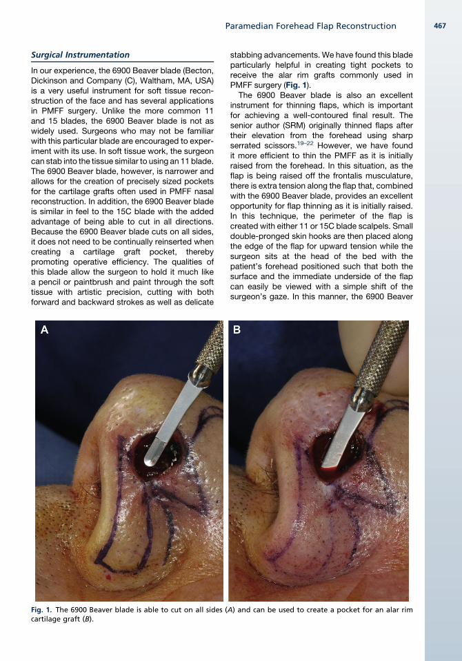
Surgical Instrumentation
In our experience, the 6900 Beaver blade (Becton,Dickinson and Company (C), Waltham, MA, USA)is a very useful instrument for soft tissue recon-struction of the face and has several applicationsin PMFF surgery. Unlike the more common 11and 15 blades, the 6900 Beaver blade is not aswidely used. Surgeons who may not be familiarwith this particular blade are encouraged to exper-iment with its use. In soft tissue work, the surgeoncan stab into the tissue similar to using an 11 blade.The 6900 Beaver blade, however, is narrower andallows for the creation of precisely sized pocketsfor the cartilage grafts often used in PMFF nasalreconstruction. In addition, the 6900 Beaver bladeis similar in feel to the 15C blade with the addedadvantage of being able to cut in all directions.Because the 6900 Beaver blade cuts on all sides,it does not need to be continually reinserted whencreating a cartilage graft pocket, therebypromoting operative efficiency. The qualities ofthis blade allow the surgeon to hold it much likea pencil or paintbrush and paint through the softtissue with artistic precision, cutting with bothforward and backward strokes as well as delicate
Fig. 1. The 6900 Beaver blade is able to cut on all sides (Acartilage graft (B).
stabbing advancements. We have found this bladeparticularly helpful in creating tight pockets toreceive the alar rim grafts commonly used inPMFF surgery (Fig. 1).
The 6900 Beaver blade is also an excellentinstrument for thinning flaps, which is importantfor achieving a well-contoured final result. Thesenior author (SRM) originally thinned flaps aftertheir elevation from the forehead using sharpserrated scissors.19–22 However, we have foundit more efficient to thin the PMFF as it is initiallyraised from the forehead. In this situation, as theflap is being raised off the frontalis musculature,there is extra tension along the flap that, combinedwith the 6900 Beaver blade, provides an excellentopportunity for flap thinning as it is initially raised.In this technique, the perimeter of the flap iscreated with either 11 or 15C blade scalpels. Smalldouble-pronged skin hooks are then placed alongthe edge of the flap for upward tension while thesurgeon sits at the head of the bed with thepatient’s forehead positioned such that both thesurface and the immediate underside of the flapcan easily be viewed with a simple shift of thesurgeon’s gaze. In this manner, the 6900 Beaver
) and can be used to create a pocket for an alar rim

Fig. 2. Asymmetry from distal necrosis of PMFF reconstruction of the left alar rim is shown in frontal (A), rightthree-quarter (B) and left three-quarter (C) profile views.
Schreiber & Mobley468
blade is gently stabbed into the tissue, leaving 1 to3 mm of subcutaneous fat on the underside of theforehead flap. The surgeon can watch from belowas the blade precisely enters the subcutaneous fatlayer and then watch the skin surface of the flap asthe blade causes the skin to gently rise, similar towhat is seen when the tips of a face-lift scissorpress on the underside of the cheek skin duringwide undermining. The ability to paint this bladeback and forth while gently advancing has allowedus to safely and more quickly raise nicely thinnedflaps. These thinner flaps eventually result ina superior contour match. Another advantage of
Fig. 3. Retraction after PMFF reconstruction of the righcompared to the native left alar rim (B).
this technique is that subcutaneous fat and fronta-lis muscle are left in place at the patient’s fore-head, which can more quickly provide a healthybase of living tissue on which granulation tissuecan more quickly form when the forehead defectcannot be closed primarily and must heal bysecondary intention.
Achieving Alar Rim Symmetry, Skin Layer
In PMFF reconstruction of the alar subunit, alarretraction is a significant complication that thefacial plastic surgeon must work diligently to
t alar rim resulting in a notched right alar rim (A)
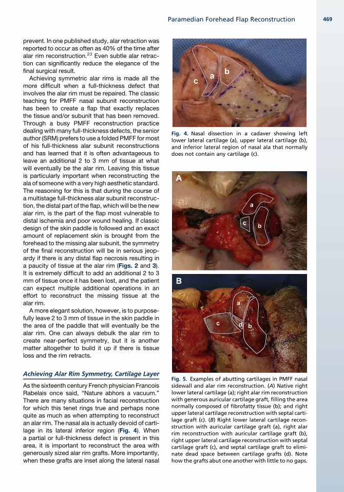
Fig. 4. Nasal dissection in a cadaver showing leftlower lateral cartilage (a), upper lateral cartilage (b),and inferior lateral region of nasal ala that normallydoes not contain any cartilage (c).
Paramedian Forehead Flap Reconstruction 469
prevent. In one published study, alar retraction wasreported to occur as often as 40% of the time afteralar rim reconstruction.23 Even subtle alar retrac-tion can significantly reduce the elegance of thefinal surgical result.
Achieving symmetric alar rims is made all themore difficult when a full-thickness defect thatinvolves the alar rim must be repaired. The classicteaching for PMFF nasal subunit reconstructionhas been to create a flap that exactly replacesthe tissue and/or subunit that has been removed.Through a busy PMFF reconstruction practicedealingwithmany full-thickness defects, the seniorauthor (SRM) prefers to use a foldedPMFF formostof his full-thickness alar subunit reconstructionsand has learned that it is often advantageous toleave an additional 2 to 3 mm of tissue at whatwill eventually be the alar rim. Leaving this tissueis particularly important when reconstructing theala of someonewith a very high aesthetic standard.The reasoning for this is that during the course ofa multistage full-thickness alar subunit reconstruc-tion, the distal part of the flap, which will be the newalar rim, is the part of the flap most vulnerable todistal ischemia and poor wound healing. If classicdesign of the skin paddle is followed and an exactamount of replacement skin is brought from theforehead to the missing alar subunit, the symmetryof the final reconstruction will be in serious jeop-ardy if there is any distal flap necrosis resulting ina paucity of tissue at the alar rim (Figs. 2 and 3).It is extremely difficult to add an additional 2 to 3mm of tissue once it has been lost, and the patientcan expect multiple additional operations in aneffort to reconstruct the missing tissue at thealar rim.
A more elegant solution, however, is to purpose-fully leave 2 to 3 mm of tissue in the skin paddle inthe area of the paddle that will eventually be thealar rim. One can always debulk the alar rim tocreate near-perfect symmetry, but it is anothermatter altogether to build it up if there is tissueloss and the rim retracts.
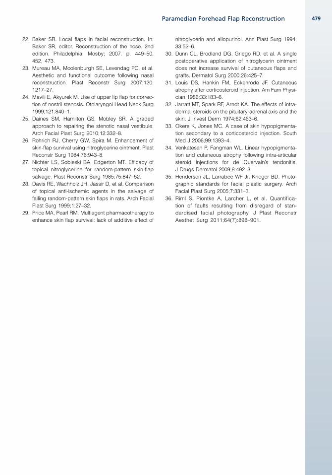
Fig. 5. Examples of abutting cartilages in PMFF nasalsidewall and alar rim reconstruction. (A) Native rightlower lateral cartilage (a); right alar rim reconstructionwith generous auricular cartilage graft, filling the areanormally composed of fibrofatty tissue (b); and rightupper lateral cartilage reconstructionwith septal carti-lage graft (c). (B) Right lower lateral cartilage recon-struction with auricular cartilage graft (a), right alarrim reconstruction with auricular cartilage graft (b),right upper lateral cartilage reconstruction with septalcartilage graft (c), and septal cartilage graft to elimi-nate dead space between cartilage grafts (d). Notehow the grafts abut one another with little to no gaps.
Achieving Alar Rim Symmetry, Cartilage Layer
As the sixteenth century French physician FrancoisRabelais once said, “Nature abhors a vacuum.”There are many situations in facial reconstructionfor which this tenet rings true and perhaps nonequite as much as when attempting to reconstructan alar rim. The nasal ala is actually devoid of carti-lage in its lateral inferior region (Fig. 4). Whena partial or full-thickness defect is present in thisarea, it is important to reconstruct the area withgenerously sized alar rim grafts. More importantly,when these grafts are inset along the lateral nasal

Schreiber & Mobley470
sidewall, it is imperative that there are no soft tissuegaps between abutting edges of cartilage grafts(Fig. 5). If gaps are left between adjacent cartilagegrafts, the sidewall can contract, obliteratingthe dead space and leaving behind a retractednasal ala.In patients with high aesthetic standards and
full-thickness defects involving the alar rim, repairis planned as follows (Fig. 6):
1. Design folded PMFF using a template to recon-struct missing internal lining as well as alarsubunit with 2 to 3 mm of additional tissue atwhat will be the alar rim.
2. If the patient is a smoker or otherwise perceivedto be at risk for flap failure, there should bea delay stage, and the PMFF should be suturedin its original position after being raised. Wait7 to 10 days for the next stage.
3. Raise the foldedPMFF to the nose to reconstructthe internal lining and alar subunit. Reconstruct
Fig. 6. Folded PMFF nasal reconstruction stage 1. Full-thifollowed by completed reconstruction (C) after placemenwith quilting sutures (before use of polytetrafluoroethyle
such that 2 to 3mmof additional tissue is locatedat the alar rim. Wait 3 weeks for the next stage.
4. Divide the internal lining from the external skinpaddle, and place delayed primary cartilagegrafts to support the alar rim, nasal sidewall,tip, and so forth. Wait an additional 3 weeks forthe next stage.
5. Divide the pedicle, and inset the flap. Wait 2months for the final stage.
6. Design a symmetric alar rim and remove anyexcessive tissue tomatch thenative side (Fig.7).
Supra-Alar Crease
Through-and-through quilting sutures are an im-portant technique for the control of contour thatis necessary for complex nasal reconstruction.These through-and-through sutures can have animportant and significant effect on the final nasalcontour in key areas such as the supra-alar crease.Although the quilting sutures are able to apply
ckness nasal defect (A) and planned folded PMFF (B)t of cartilage grafts and creation of supra-alar creasene pledgets) are shown.

Fig. 7. Result of right alar rim reconstruction with PMFF before final debulking. Markings show planned locationof final alar rim in the frontal (A) and right three-quarter (B) profile views. The left three-quarter profile view isshown for comparison (C).
Paramedian Forehead Flap Reconstruction 471
pressure in a precise location, these sutures canhave a cheese wire cutting effect on the skin. Alter-natively, cotton bolsters have been advocated forthis use to help apply broader pressure in the post-operative period without the cheese wire effect.22
The senior author’s (SRM) long-time surgical assis-tant, Brent Klev, RN, helped suggest an even moreelegant solution to control nasal contour. This sim-ple modification combines the benefits of applyingpressure in a precise location while at the sametime avoiding the cheese wire effect.
Fig. 8. Creation of supra-alar crease. Bulky supra-alar tissPTFE pledgets are placed to maintain pressure at the pPTFE pledget are shown (D).
In thismodification, a 5-0 black nylon is threadedover a polytetrafluoroethylene (PTFE) pledget thatis used to precisely place pressure. This not onlyprovides equal, if not superior, soft tissue com-pression compared with simple quilting but alsoprevents cheese wiring that occurs when stitchesalone are used to create this crease (Fig. 8). Inaddition, the PTFE pledget has 2 ready-madeperforations through which the needle can easilybe passed such that equal pressure can be placedover the entire area of the pledget. PTFE pledgets
ue (A) is debulked through planned incisions (B), andlanned supra-alar crease (C). The dimensions of the

Fig. 9. Preoperative (A), intraoperative (B), and postoperative (C) images of nasal vestibular stenosis repair. Notethat in the middle image the senior author (SRM) had not yet adopted the use of PTFE pledget, and, instead,a bulky sponge was used. (Modified from Daines SM, Hamilton GS, Mobley SR. A graded approach to repairingthe stenotic nasal vestibule. Arch Facial Plastic Surg 2010;12:336. Copyright � 2010 American Medical Association.All rights reserved; with permission.)
Schreiber & Mobley472
are specifically designed to prevent the suture fromtearing through the tissue and are used most oftenin cardiothoracic and vascular surgeries. Asa result, otolaryngologists and facial plasticsurgeons may not have been widely exposed tothe utility of PTFE pledgets in the operating room.PTFE pledgets are best used for this purpose bytying them such that they are just held in placewith just the smallest amount of initial tissuecompression. That way, in the days after surgery,the compression becomes more appropriate asnormal postoperative edema enters the tissues inthe days after the flap surgery. A short learningcurve can be anticipated by surgeons learning touse PTFE pledgets for soft tissue contourcompression. We routinely leave these pledgetsin for 5 to 8 days postoperatively and have notseen postoperative complications from excessivecompression or cheese wire cutting of tissue. Ina minority of cases, the sutures may tighten,causing the pledgets to excessively depress theskin with postoperative edema. If this situationoccurs, the pledgets may be removed soonerthan the 5 to 8 days they are normally left in place.
Fig. 10. Topical nitroglycerin usedon thedistal portionof a PMFF.
Nasal Vestibular Stenosis
Nasal vestibular stenosis can occur to varyingdegrees after full-thickness repair of nasal ala de-fects with folded PMFFs and is best prevented byusing large autologous grafts in the area of thenasal sidewall and ala. However, despite a sur-geon’s best efforts, this complication may stilloccur. Nasal vestibular stenosis in the setting oftrauma can be difficult to repair, has a high rate ofrecurrence, and can often result in significant func-tional impairment.24 The senior author (SRM) hashad success repairing nasal vestibular stenosisusing a batten graft to support the inherentlyweak nasal ala and a thermoplastic splint for 2
weeks (Fig. 9).25 Alar vestibular stenosis causedby burns and other trauma are less common butcan be corrected using the same technique.
POSTOPERATIVE CARETopical Nitroglycerin
Flap necrosis is a difficult complication that cansignificantly delay final repair and lead to

Fig. 11. Injection of triamcinolone to treat pincushionedema of a nasal flap.
Paramedian Forehead Flap Reconstruction 473
a prolonged course caring for an open wound. Onetechnique that has been useful for the seniorauthor (SRM) is the use of nitroglycerin paste,which is absorbed topically, creating nitric oxide
Fig. 12. Opposing three-quarter views allow comparisonseen at the reconstructed right alar rim (A) compared to
radicals that cause both arterial and venous vaso-dilation (Fig. 10). Topical nitroglycerin has shownefficacy in improved skin flap survival in severalanimal models.26–29 Importantly, a single postop-erative treatment with topical nitroglycerin showedno benefit over placebo in flap necrosis for Mohsreconstruction in the only published clinical studyto date.30 Our patients are instructed to use topicalnitroglycerin every 4 to 6 hours if their flaps showsigns of inadequate perfusion postoperatively.Patients are asked to continue this applicationfor 4 to 6 days or until a more stable flap color isobserved. The senior author (SRM) has used thistechnique for several years in a variety of patientswith a spectrum of comorbidities without anysignificant cardiovascular complications. Patientsare instructed to apply a pea-sized amount ofpaste, about 1 mm thick, over the pale or con-gested flap and must be warned that most peopleexperience an annoying but tolerable headache.Anecdotally, this method seems to work slightlybetter for arterial insufficiency as opposed tovenous congestion.
Triamcinolone
Pincushioning is often the result of edema andinadequate flap thinning and can reduce recon-struction camouflage. Triamcinolone injectionsthat are intradermal or immediately subdermalcan help reduce pincushioning through the use-ful side effects of lipolysis and inhibition of lipogen-esis. Before these injections, however, the patientmust be informed of possible complications, suchas telangiectasias, hypopigmentation, and skinbreakdown.31 These complications tend to occur
of nasal ala. A small amount of alar retraction can bethe left (B).

Fig. 13. Comparison of preoperative (A–D) and 4-year postoperative (E–H) PMFF reconstruction results for a large full-thickness right alar rim defect. This patient devel-oped alar retraction with a notched appearance similar to that in Fig. 3, requiring additional reconstructive procedures.
Schreiber&
Mobley
474

Paramedian Forehead Flap Reconstruction 475
with higher concentrations of triamcinolone, suchas 40mg/mL. Telangiectasiasmay appear amonthor more after injection and can even enlarge for upto 6 months.32 Hypopigmentation related tosteroid injection appears in multiple case reports,mainly in the orthopedic literature in which steroiddoses are significantly higher than those used bythe senior author (SRM).33 This effect is causedby decreased melanocyte function and is usuallytemporary and rarely permanent.34 Nevertheless,hypopigmentation can be particularly discon-certing and especially noticeable in darker-skinned individuals. The risk of skin breakdown,which is especially high in patients with thin skin,is reduced by starting with low concentrations oftriamcinolone. With the concentrations we use,we have seen a few cases of telangiectasias anda little hypopigmentation but no skin breakdown.
We usually begin with triamcinolone at a finalconcentration of 2.5 mg/mL (mixed with lidocaineand bicarbonate for patient comfort) in 0.1-mLaliquot injections from a 1-mL tuberculin-typesyringe for each approximate 5 mm2 of edema.The first injection is administered at 3 to 6 weeks
Fig. 14. Comparison of preoperative (A–D) and 5-year posright alar defect repaired with a melolabial flap and aa PMFF. This patient’s reconstruction was complicated byadditional reconstructive procedures.
postoperatively to the deep dermis or top layerof subcutaneous fat. When injected intradermally,the surgeon should see skin blanching (Fig. 11).The patient is followed up for 4 weeks, and, if noappreciable effect is noticed, an increased triam-cinolone concentration of 5 mg/mL can be in-jected, continuing with 0.1-mL aliquots per eachapproximate 5 mm2 of persistent tissue edema.There is a definite art to this technique, and thelearning surgeon is advised to proceed slowly,spreading treatments over longer intervals, per-haps 6 weeks initially, until comfort is gained withhow much edema resolves with a given injection.The possibility of injecting more triamcinolone ata later time is a particularly important conceptwith this technique.
REPORTING RESULTSPhotography
It would be difficult to overstate the importance ofphotography in facial plastic surgery. Standardizedand consistent photographs allow each surgeon tomore accurately assess and report their forehead
toperative (E–H) reconstruction results for a superficiallarge full-thickness left alar rim defect repaired withdistal necrosis of the PMFF, shown in Fig. 2, requiring

Schreiber & Mobley476
flap reconstruction results to the professionalcommunity at large. Ideally, photographs are takenof a patient without makeup or expression; afterremoving jewelry, hats, and other accessories;and at well-defined angles.35 In addition, nonad-herence to standard practices in photographycan lead to the publication of possibly misleadingresults. For example, variations of only 10� fromstandard camera angles can shorten or lengthenvarious facial features to a significant degree, re-sulting in distorted assessment.36 Nasal skincancer reconstruction photos, and especiallythose involving the alar rim, can fall victim to publi-cation of nonstandardized photography results. To
Fig. 15. Comparison of preresection (A–D), postresection/preconstruction results for a full-thickness right alar rim dedebulking is shown in Fig. 7.
ensure the most accurate assessment of a givenpostoperative result, the contralateral ala shouldalways be shown juxtaposed to the reconstructedala. Without showing the unaffected side, subtleamounts of alar rim retraction, which are quitenoticeable on direct observation, can be blindedto the observer of the published photo. We en-courage all nasal reconstruction results involvingrepair on or near the ala to be published or pre-sented with the contralateral ala opposite to the re-constructed result (Fig. 12). The adoption of thisstandard will ensure that the results of given re-constructive techniques are more objectivelypresented.
reoperative (E–H) and 1-year postoperative (I–L) PMFFfect. Appearance after reconstruction but before final

Fig. 17. Comparison of preoperative (A–D) and 10-month postoperative (E–H) PMFF reconstruction results fora long-standing, bilateral, full-thickness alar rim and nasal tip defect.
Fig. 16. Comparison of preoperative (A–D) and 7-month postoperative (E–H) PMFF reconstruction results fora large full-thickness right alar rim defect. Note that only preoperative images with surgical markings and post-operative images taken by the patient at home due to a long travel distance and inability to followup in clinicwere available. Intraoperative reconstruction images are shown in Fig. 6.
Paramedian Forehead Flap Reconstruction 477

Schreiber & Mobley478
SUMMARY
Reconstruction of nasal defects is a particularlychallenging task, requiring the reconstructivesurgeon to recreate facial symmetry to within milli-meters to reduce detection. The techniquesmentioned in this article have been refined overseveral years and have helped the senior author(SRM) to achieve increasingly symmetric recon-structive results while preventing complicationsand the need for unplanned surgeries along thereconstructive journey (Figs. 13–17). A summaryof key principles is as follows:
1. Always provide choices, and help patientschoose the reconstructive option that best fitstheir situation.
2. Consider comorbidities and have a lowthreshold to add a staged surgical delay.
3. Aim to reconstruct entire subunits, but usean artistic eye and do not adhere to this ruleblindly.
4. Add an additional 2 to 3 mm of skin paddlewhen reconstructing through-and-through de-fects of the ala.
5. Experiment with the surgical instruments andtechniques that are discussed in this article(the 6900 Beaver blade, PTFE pledgets, nitro-glycerin paste, and triamcinolone injection) toenhance results.
By following these principles, we have been ableto navigate our patients through a series of recon-structive stages that maximizes final aestheticresults while minimizing complications. Weencourage the use of these techniques amongstfacial plastic surgeons to improve reconstructiveresults in complex multistage nasal reconstructionand create more elegant solutions for patients.
REFERENCES
1. Lascaratos J, Cohen M, Voros D. Plastic surgery of
the face in Byzantium in the fourth century. Plast
Reconstr Surg 1998;102:1274–80.
2. Argacha JF, Adamopoulos D, Ghjic M, et al. Acute
effects of passive smoking on peripheral vascular
function. Hypertension 2008;51:1506–11.
3. Goessler UR, Bugert P, Kassner S, et al. In vitro anal-
ysis of radiation-induced dermal wounds. Otolar-
yngol Head Neck Surg 2010;142:845–50.
4. Rees TD, Liverett DM, Guy CL. The effect of ciga-
rette smoking on skin-flap survival in the face lift
patient. Plast Reconstr Surg 1984;73:911–5.
5. Riefkohl R, Wolfe JA, Cox EB, et al. Association
between cutaneous occlusive vascular disease,
cigarette smoking, and skin slough after rhytidec-
tomy. Plast Reconstr Surg 1986;77:592–5.
6. Chang DW, Reece GP, Wang B, et al. Effect of
smoking on complications in patients undergoing
free TRAM flap breast reconstruction. Plast Reconstr
Surg 2000;105:2374–80.
7. Milton S. The effects of delay on the survival of
experimental studies on pedicled skin flaps. Br J
Plast Surg 1965;22:244–52.
8. Myers MB, Cherry G. Differences in the delay
phenomenon in the rabbit, rat, and pig. Plast Re-
constr Surg 1971;47:73–8.
9. Finseth F, Cutting C. An experimental neurovascular
island skin flap for the study of the delay phenom-
enon. Plast Reconstr Surg 1978;61:412–20.
10. Pang CY, Forrest CR, Neligan PC, et al. Augmenta-
tion of blood flow in delayed random skin flaps in
the pig: effect of length of delay period and angio-
genesis. Plast Reconstr Surg 1986;78:68–74.
11. Holzbach T, Neshkova I, Vlaskou D, et al. Searching
for the right timing of surgical delay: angiogenesis,
vascular endothelial growth factor and perfusion
changes in a skin-flap model. J Plast Reconstr Aes-
thet Surg 2009;62:1534–42.
12. Erdmann D, Sundin BM, Moquin KJ, et al. Delay in
unipedicled TRAM flap reconstruction of the breast:
a review of 76 consecutive cases. Plast Reconstr
Surg 2002;110:762–7.
13. Christiano JG, Rosson GD. Clinical experience with
the delay phenomenon in autologous breast recon-
struction with the deep inferior epigastric artery
perforator flap. Microsurgery 2010;30:526–31.
14. Burget GC, Menick FJ. The subunit principle in
nasal reconstruction. Plast Reconstr Surg 1985;76:
239–47.
15. Singh DJ, Bartlett SP. Aesthetic considerations in
nasal reconstruction and the role of modified nasal
subunits. Plast Reconstr Surg 2003;111:639–48.
16. Burget GC, Menick FJ. Nasal support and lining: the
marriage of beauty and blood supply. Plast Reconstr
Surg 1989;84:189–202.
17. Menick FJ. A 10-year experience in nasal recon-
struction with the three-stage forehead flap. Plast
Reconstr Surg 2002;109:1839–55.
18. Menick FJ. A new modified method for nasal lining:
the Menick technique for folded lining. J Surg Oncol
2006;94:509–14.
19. BurgetGC,Menick FJ.Aesthetic reconstruction of the
nose. In: Burget GC, Menick FJ, editors. Aesthetics,
visual perception, and surgical judgement. St Louis
(MO): Mosby; 1993. p. 48.
20. Burget GC, Menick FJ. Aesthetic reconstruction of
the nose. In: Burget GC, Menick FJ, editors. The par-
amedian forehead flap. St Louis (MO): Mosby; 1993.
p. 77–9.
21. Baker SR. Local flaps in facial reconstruction. In:
Baker SR, editor. Interpolated paramedian fore-
head flaps. 2nd edition. Philadelphia: Mosby; 2007.
p. 284–5, 293–5.
Paramedian Forehead Flap Reconstruction 479
22. Baker SR. Local flaps in facial reconstruction. In:
Baker SR, editor. Reconstruction of the nose. 2nd
edition. Philadelphia: Mosby; 2007. p. 449–50,
452, 473.
23. Mureau MA, Moolenburgh SE, Levendag PC, et al.
Aesthetic and functional outcome following nasal
reconstruction. Plast Reconstr Surg 2007;120:
1217–27.
24. Mavili E, Akyurek M. Use of upper lip flap for correc-
tion of nostril stenosis. Otolaryngol Head Neck Surg
1999;121:840–1.
25. Daines SM, Hamilton GS, Mobley SR. A graded
approach to repairing the stenotic nasal vestibule.
Arch Facial Plast Surg 2010;12:332–8.
26. Rohrich RJ, Cherry GW, Spira M. Enhancement of
skin-flap survival using nitroglycerine ointment. Plast
Reconstr Surg 1984;76:943–8.
27. Nichter LS, Sobieski BA, Edgerton MT. Efficacy of
topical nitroglycerine for random-pattern skin-flap
salvage. Plast Reconstr Surg 1985;75:847–52.
28. Davis RE, Wachholz JH, Jassir D, et al. Comparison
of topical anti-ischemic agents in the salvage of
failing random-pattern skin flaps in rats. Arch Facial
Plast Surg 1999;1:27–32.
29. Price MA, Pearl RM. Multiagent pharmacotherapy to
enhance skin flap survival: lack of additive effect of
nitroglycerin and allopurinol. Ann Plast Surg 1994;
33:52–6.
30. Dunn CL, Brodland DG, Griego RD, et al. A single
postoperative application of nitroglycerin ointment
does not increase survival of cutaneous flaps and
grafts. Dermatol Surg 2000;26:425–7.
31. Louis DS, Hankin FM, Eckenrode JF. Cutaneous
atrophy after corticosteroid injection. Am Fam Physi-
cian 1986;33:183–6.
32. Jarratt MT, Spark RF, Arndt KA. The effects of intra-
dermal steroids on the pituitary-adrenal axis and the
skin. J Invest Derm 1974;62:463–6.
33. Okere K, Jones MC. A case of skin hypopigmenta-
tion secondary to a corticosteroid injection. South
Med J 2006;99:1393–4.
34. Venkatesan P, Fangman WL. Linear hypopigmenta-
tion and cutaneous atrophy following intra-articular
steroid injections for de Quervain’s tendonitis.
J Drugs Dermatol 2009;8:492–3.
35. Henderson JL, Larrabee WF Jr, Krieger BD. Photo-
graphic standards for facial plastic surgery. Arch
Facial Plast Surg 2005;7:331–3.
36. Riml S, Piontke A, Larcher L, et al. Quantifica-
tion of faults resulting from disregard of stan-
dardised facial photography. J Plast Reconstr
Aesthet Surg 2011;64(7):898–901.
![Dr Sharan Hiremath’s Preauricular flag flap for temple and ...€¦ · the technique of choice for the reconstruction of small-sized defects on the lateral forehead [1]. These flaps](https://img.pdfslide.us/doc/110x75/5fbe5be53c273e5ced393610/dr-sharan-hiremathas-preauricular-flag-flap-for-temple-and-the-technique-of.jpg)