Embed Size (px)
Citation preview
ANNALS O F C LIN IC A L AND LABORATORY SCIEN CE, Vol. 5, No 6 Copyright © 1975, Institute for Clinical Science
Electrotherm al Atomic Absorption Spectrom etry of Trace M etals in B iological F lu id s * !
F. WILLIAM SUNDERMAN, JR., M.D.
Department of Laboratory Medicine, University of Connecticut School of Medicine,
Farmington, CT 06032
ABSTRACT
Electrothermal atomic absorption spectrometry has five major advantages for measurements of trace metals in biological materials, in comparison to measurements by conventional flame atomic absorption analysis: (1) Trace metal contamination is minimized by avoidance of preliminary chemical extractions and additions of reagents; (2) Sample volumes are small (1 to 50 /*1). The entire sample is vaporized in electrothermal atomization, whereas in most flame nebulization systems only a small fraction of the sample enters the flame; (3) Atoms are released in higher concentrations. In electrothermal atomization, the atom cloud is released into a relatively small volume of gas. In contrast, in flame atomization, the atom cloud is diluted by the high flow rate of gases and by expansion of gases during combustion; (4) Molecular recombination of atoms is retarded by the atmosphere of inert gas which restricts chemical reactions (e.g., oxidation). In contrast, in flame atomization, oxidation occurs very rapidly; and (5) By programmed increments in. temperature, electrothermal atomization permits organic constituents to be pyrolyzed prior to vaporization and atomization of metals. Despite these advantages, electrothermal atomic absorption is particularly subject to interferences and sources of imprecision which necessitate critical evaluations in the prospective analyst’s laboratory before an electrothermal method can be confidently employed for diagnostic measurements of a specific trace metal in body fluids.
Introduction
As a consequence of the recent increase of scientific knowledge regarding the metabolism, biochemical pathology and toxi-
"Supported by Grant #AT(11-1 )-3140 from the U.S. Energy Research and Development Agency and by Public Health Service Contract #HSM 99-72-24 from the National Institute of Occupational Safety and Health.
tPrepared for the 1975 Lecture Series on “Current Topics in Clinical Chemistry,” under the joint sponsorship of the Chicago Sections of the American Chemical Society and the American Association of Clinical Chemists.
cology of trace metals, there has arisen widespread clinical interest in measurements of trace metals in body fluids, tissues and excreta.33'41,65,77 Laboratories in many university medical centers and governmental institutions have become involved in determinations of copper, zinc, lithium, gold, lead, cadmium, mercury, manganese, nickel and other metals in biological materials. Increasing attention has been focused upon clinical applications of electrothermal atomic absorption spectrometry, since this relatively new technique
4 2 2 SUNDERMAN
has appeared to provide the requisite analytical sensitivity, specificity and convenience.33,41,65,77 Regrettably, this technique is subject to pitfalls, interferences and sources of imprecision that have hindered its clinical use. As will be shown in this review, progressive refinements of electrothermal atomizers and of optical systems for background compensation have largely overcome these obstacles. It now appears probable that electrothermal atomic absorption spectrometry will be adopted by clinical chemistry laboratories as a routine method for measurements of trace metals in body fluids.
The scope of this review is limited to electrothermal atomic absorption spectrometry, with emphasis upon its clinical applications. This article is intended to supplement the recent treatises on the basic technique which have been written by Ag- gett and Sprott,1 Ingle,29 Kirkbright,34 Price63 and Woodriff.83 This resumé does not consider various related topics, such as(1 ) atomic fluorescence or emission spectrometry; (2) non-flame atomization devices which employ direct current arcs, radio-fre- quency plasma jets, microwave excitation fields, or demountable hollow-cathode sputtering cells; (3) relatively low-temperature atomic absorption spectrometry of Hg vapor; or (4) atomic absorption spectrometry of As and Se following volatization by chemical reduction to their respective gaseous hydrides.
Electrothermal Atomizers
In 1961, L ’vov44 described the first electrothermal atomizer for atomic absorption analysis of trace metals. L ’vov’s atomizer consisted of a graphite plug which fitted into a socket in an electrically heated graphite tube. A liquid sample was dried on the plug, which was then inserted into the tube. An alternating current arc was used to discharge the sample into an argon atmosphere within the graphite tube. Atomic
recombination was retarded by the high temperature and the inert gas, and atomic absorption spectrometry of the atom cloud was performed by an optical beam along the axis of the tube.44 Numerous ingenious variations of the electrothermal atomizer have been developed, including use of graphite tubes, rods and braided filaments,2 3'812,22,42,50,51,52,54,55,56,81,83,84 w e JJ a s m e t a l l i c
ribbons, cups and filaments constructed of tungsten, tantalum, molybdenum or platinum.12'26'27,28,48,82 In this paper, attention is focused primarily upon the three designs of electrothermal atomizers that have been most widely used for analyses of trace metals in biological materials.
Massmann51,52 devised the graphite tube furnace which has served as the prototype of commercial graphite furnaces that have been manufactured by Perkin-Elmer Co.,50 Beckman Instruments Gmbh. (Germany) and Jarrell-Ash Division of Fisher Scientific Co. A liquid sample with a volume from 5 to 50 ftl is introduced into a hollow graphite tube by means of a micropipet. The graphite tube is heated electrically by electrode connections at both ends of the tube. The graphite tube assembly is encased within a water-cooled jacket, and the cavity of the tube is slowly flushed with an inert gas such as argon or nitrogen. During each analysis, the temperature of the graphite tube is increased in a stepwise fashion so that (1) the sample is dried at 100 to 125° C; (2) the sample is pyrolyzed at 500 to 1200° C so that organic constituents are destroyed; and (3) the metals in the sample are vaporized and atomized at 1500 to 3000° C.
During this programmed sequence, the slow stream of inert gas flows through the cavity of the graphite tube, successively sweeping away (1 ) the water or solvent vapors that are released during drying, (2) the smoke and fumes that are generated during pyrolysis; and (3) the atom cloud that is liberated during atomization. The light path of the atomic absorption spectrometer traverses the length of the graphite tube.
ELECTRO TH ERM AL ATOMIC ABSORPTION 4 2 3
During the atomization phase, atomic absorption of the analyte in the atom cloud is measured by the spectrometer and is recorded with a strip-chart recorder. The peak atomic concentration, “ N” , (atoms per ml) in the cavity of the graphite furnace is related to the analyte concentration, “ C” , (mol per liter of solution injected into the graphite tube by a micropipet) by the following equation: N = 6 x 10²⁰ VCeB/Ve, where V = volume of analyte (ml); e = vaporization efficiency; B = atomization efficiency, and Vc = volume of the cavity of the graphite tube (ml), provided that the analyte diffuses slowly out of the cavity.28
West and Williams81 and Amos et al2 have employed an alternative design for an electrothermal atomizer, which has served as the prototype of the graphite rod atomizers manufactured by Varian-Techtron Pty, Ltd .53 The sample (0.5 to 5 ¿¿1) is dried within a small cavity located at the midlength of a thin graphite rod, which is supported between two electrodes. The graphite rod is unenclosed but is protected by a laminar stream of an inert gas such as argon or nitrogen. Optionally, the upward current of inert gas may be encircled by a concentric laminar stream of hydrogen. The elevated temperature of the heated graphite rod ignites the hydrogen, which forms a barrier that (1 ) entrains atmospheric oxygen, hindering oxidation of the atom cloud, and (2) assists in the reduction of metallic oxides to free metal atoms.84
During a brief atomization phase, the sample is suddenly vaporized, and the atom cloud is measured by its attenuation of a transverse optical beam that passes through an aperture at right angles to the length of the graphite rod. This simple, economical design has the apparent disadvantages of relatively limited sample volume and relatively short path-length of light in the atom cloud. However, Amos et al2 have found that this atomizer provides analytical sensitivity that rivals that obtained with Massmann’s graphite tube furnace.
A third design concept for an electrothermal atomizer uses a metallic or graphite boat, as described by Donega and Burgess.12 This configuration has been adapted by Hwang et al26,27,28 in the form of the tantalum ribbon atomizers which are manufactured by Instrumentation Laboratories, Inc. and by Jarrell-Ash Division of Fisher Scientific Co. The apparatus28 consists of a thin tantalum strip that is shaped to serve as a combination sample boat and heating element. The ends of the tantalum strip are clamped onto the tops of two electrodes, and the assembly is enclosed within an argon-purged chamber. The optical beam traverses quartz windows at the sides of the argon-purged chamber, and passes immediately over the sample cavity in the tantalum strip. A hinged door at the front of the chamber permits access for introduction of the sample. By use of different tantalum strips with sample cavities of various sizes, the atomizer can accommodate sample volumes from 1 to 100 ¿tl. When it is necessary to use atomization temperatures in the range from 2500 to 3000° C, a graphite cuvet can be substituted for the tantalum strip.
Torsi and Tessari78 studied the influence of heating rate upon analytical sensitivities in electrothermal atomic absorption. They observed that in the graphite tube furnace the residence time of the atom cloud in the optical path is relatively long (5 to' 20 sec), compared to the transitory burst of atoms (ca 0.5 sec) which occurs during the sudden heating of the graphite rod atomizer. The residence time of the atom cloud in the light-path of the tantalum ribbon atomizer is presumably intermediate between that achieved with the other two basic designs.
Inert Atmospheres for Electrothermal Atomization
According to Reeves et al,84 the inert atmosphere of the electrothermal atomizer serves the dual purposes of (1) protecting the
424 SUNDERMAN
graphite tube or rod against oxidation and(2) retarding the degradation of analyte atoms in the atom cloud by recombination {e.g. with 0 2). From theoretical viewpoints, argon is the most suitable inert gas for these purposes. Thus, Maessen and Posma46 showed that the diffusion coefficients of gold atoms in nitrogen and argon atmospheres are, respectively, one-fourth and one-fifth of that observed in a helium atmosphere. Hwang et al26 reported that the specific heat of argon is lower than that of nitrogen and helium. Moreover, with use of nitrogen as the atmosphere, there is a possibility of forming a toxic gas, cyanogen, which has an absorption band in the ultra-violet spectrum.
Despite these theoretical advantages of argon, many analysts prefer to employ nitrogen, which is less expensive than argon. Cruz and van Loon10 discounted the possibility of generating cyanogen from nitrogen at 3000° C and stated that cyanogen formation requires a higher temperature (ca 5000° C). From a practical viewpoint, there appears to be little difference between the use of argon and nitrogen atmospheres for most biological applications of electrothermal atomic absorption spectrometry. For greatest analytical sensitivity, the flow- rate of the inert gas should be adjusted to the minimum that is necessary to sweep away smoke and other interfering vapors, in order that the atom cloud be subjected to the least possible dilution.3 With some graphite tube atomizers, the flow of inert gas can be interrupted during the atomization cycle of the programmed sequence of changes in furnace temperature.
Characteristics of Graphite Tubes
After a brief period of use, the graphite tubes and rods that are commonly employed in electrothermal atomizers begin to deteriorate, and their electrical characteristics become subject to drift.7’9,47 This is one of the most troublesome sources of analytical
variability. Maessen et al47 demonstrated that the properties of graphite (e.g. porosity and conductivity) change appreciably during heating. Alteration of the electrical conductivity brings about changes in the rate of heating as well as in the ultimate temperature. Changes of porosity of graphite affect the deposition of liquid samples and also influence the diffusion rate of sample vapors into the graphite.
Substitution of pyrolytic graphite for standard graphite has been an important innovation in electrothermal atomic absorption spectrometry. According to Aspila et al,3 pyrolytic graphite has the following advantages over standard graphite: (1 ) lower permeability to liquids and gases; (2) higher thermal conductivity; (3) higher resistance to oxidation; and (4) higher sublimation point (ca 3700° C). Clyburn et al9 have introduced a simple technique for depositing a layer of pyrolytic graphite upon the surfaces of graphite heating elements. A mixture of methane and inert gas (argon or nitrogen) is introduced into the graphite atomizer system during operation at a temperature in excess of 2000° C. The layer of pyrolytic graphite which is thus generated is extremely dense, non-porous and resistant to oxidation. According to Clyburn et al,9 this treatment extends the effective life-time of the graphite elements and improves analytical precision.
Temperature Calibration and Regulation
Electrothermal apparatuses for atomic absorption spectrometry are currently sold by most instrument manufacturers with “ factory calibrations” of temperature scales. These temperature calibrations may be grossly inaccurate under the conditions which actually exist in analytical laboratories, owing to (1) local variations in electrical current, (2) fluctuations in pressure and temperature of coolant water, (3) variations in electrical resistance of batches of graphite tubes and rods or metallic ribbons or boats and (4) changes in thermal response which
ELECTRO TH ERM A L ATOMIC ABSORPTION 425
occur during repeated heating of atomization devices. Findlay et al18 observed that temperatures within a graphite furnace were substantially lower than were indicated by the manufacturer’s calibration, and that large temperature variations occurred within the furnace, both axially and radially. Lund- gren et al43 reported similar findings and concluded that factory calibrations cannot be relied upon for estimation of atomization temperatures. Based upon the findings of Findlay et al18 and Lundgren et al,43 temperature calibrations should be verified in each laboratory by use of a thermocouple, an optical pyrometer and/or by observations of the melting and boiling points of reference materials.
Inadequate regulation of atomizer temperature is a major source of imprecision in electrothermal atomic absorption spectrometry. The programmed heating of electrothermal atomizers can be achieved by five different methods, depending upon the electrical or physical parameters which are monitored during the heating steps.55 The first and most common method is (1) to utilize a two- or three-stage electrical current program for heating the atomizer. Montaser and Crouch55 noted that programmed heating can also be accomplished by monitoring and controlling (2) the voltage across the atomizer, (3) the power dissipated by the atomizer, (4) the infra-red radiation emitted by the atomizer, and (5) the actual atomizer temperature, as measured by a thermocouple. Montaser and Crouch55 found that thermal control by the radiation monitoring method is the most satisfactory means of achieving stable plateaus of temperature. They showed that the infra-red radiation which is emitted by the atomizer is directly related to the atomizer temperature, and that a radiation feed-back control system can effectively compensate for changes in atomizer properties and other fluctuations which influence the temperature produced by electrothermal systems. Similarly, Lundgren and Johansson42 described a graphite
tube furnace in which a photodiode detector for infra-red radiation is used as feed-back input for the temperature controller. They demonstrated that it is feasible to keep the atomizer temperature constant within ± 10° C .42,43 Such improved methods for temperature regulation should soon be adopted by instrument manufacturers and will undoubtedly improve the accuracy of trace metal analyses.
During analyses of protein-containing biological materials, steadily progressive increases of temperature may be essential during the drying and ashing steps in order to avoid foaming of the sample or sudden discharging of particles of ash, with attendant loss of analyte. Such gradual increments in temperature (i.e., a “ ramp-mode ”) have been recommended by Fuchs et al20 for analysis of serum aluminum; by Grafflage et al23 for analysis of serum chromium and manganese; and by Pekarek et al81 for analysis of serum chromium. The “ rampmode” for drying and ashing has also proven to be necessary for measurements of serum nickel, using a direct electrothermal technique which has recently been developed in the author’ s laboratory. The “ ramp-mode” can be achieved by careful manual adjustments of the atomizer controls, but this procedure is tedious and imprecise. Therefore, instruments which provide a “ ramp-mode” for programmed increase of temperature during the drying and ashing cycles are advantageous for biological applications. The Perkin-Elmer Co. has recently introduced a “ ramp-mode” accessory for their Model HGA 2100 graphite tube furnace. It is desirable that a sudden increase in temperature occur at the end of the ashing cycle, in order to reach the high- temperature plateau of the atomization cycle. This abrupt increase in temperature is necessary in order to achieve optimal analytical sensitivity.78 A brief (5 to 10 sec) “ after-burn” cycle at the upper thermal limit of the atomizer is oftimes necessary during analysis of biological materials, in
4 2 6 SUNDERMAN
order (1 ) to minimize the accumulation of ash, and (2) to prevent the “ memory” effects which are discussed later in this review. Unfortunately, the “ after-burn” cycle shortens the life-time of the heating elements.
Sampling and Contamination Problems
Micro-pipetting instruments, such as the “ Eppendorf” or “ Oxford” pipettors with disposable plastic cone tips, are customarily employed to dispense the liquid samples into electrothermal atomizers. Sampling problems which are associated with the use of these pipettors are among the troublesome aspects of electrothermal atomic absorption spectrometry.97,75 The plastic cone-tips are frequently contaminated with metals, and they should invariably be cleaned before use by soaking in dilute “ ultra pure” nitric acid, followed by multiple rinses with demineralized water which has been distilled in a quartz still. Each cone-tip should be then prepared by repeatedly pipetting and discarding the specimen which is to be analyzed. Particular care must be exercised to avoid contamination of the cone-tip by allowing it to touch any portion of the graphite tube or rod or of the metallic boat. The delivery of the sample into the atomizer must be done meticulously in order to avoid irregular splattering of droplets within the sample cavity.67 After repeated use, the porosity of graphite tubes becomes variable at different locations within the sample cavity. Hence, failure to deposit successive samples at the identical site can be a source of variability in analytical results.
Measurements of trace metals in body fluids should invariably be performed in duplicate or triplicate, in order to minimize imprecision and to detect sporadic contamination. The tedious attention that must be directed to manual sampling makes electrothermal analysis unacceptable to some technologists in clinical chemistry laboratories. For this reason, it would be desirable to employ automatic sampling devices such as are used in automated systems for gas
chromatography. An automatic sampler for electrothermal atomic absorption spectrometry has recently been designed by Maessen et al,47 and may be a harbinger of commercial developments in this field.
Techniques for sampling of solid materials for electrothermal atomic absorption spectrometry have been described by Barnett and Kahn4 and by Lundgren and Johansson,42 and a method for continuous sample introduction into an electrothermal atomizer has been introduced by Kantor et al.38 Perhaps the most promising device for automation of electrothermal atomic absorption is the “ graphite yarn thermal atomizer” that has been developed by Applied Research Laboratories Division of Bausch and Lomb Co. Graphite yarn is dispensed from a spool and is automatically unreeled through (1 ) a sampling chamber where the sample is pipetted onto the yarn, and (2) an electric furnace for desolvation and vaporization.
Sweat from the fingers and palms of the hands is rich in trace metals25 and is a common source of contamination in trace metal analysis. The graphite tubes, tantalum ribbons, plastic cone-tips and other components of electrothermal atomization systems should not be handled without use of plastic forceps or disposable plastic gloves. In electrothermal analyses of biological materials, it is necessary to remove the carbon ash from the sample cavity after almost every analysis. The lens paper which is customarily used for this purpose should be handled, only with forceps or gloves in order to minimize contamination from sweat. In the design of electrothermal atomizers, it would be desirable to include an automatic system for ash removal, (such as vigorous flushing of the sample cavity with a jet of inert gas).
Methods for Standardization and Computation
Aqueous standard solutions are a source of certain difficulties in electrothermal atomic
ELECTRO TH ERM A L ATOMIC ABSORPTION 427
absorption spectrometry of trace metals in biological fluids. The viscosities and surface tensions of aqueous standard solutions are substantially less than the viscosities and surface tensions of serum, blood and other pro- tein-containing fluids. These factors introduce volumetric disparities in pipetting of standard solutions and body fluids, and also cause differences in penetration of these liquids into porous graphite tubes or rods. Preliminary treatment of porous graphite with xylene may help to minimize the differences of liquid penetration.53,67
A more satisfactory solution of this problem is preparation of standards in aqueous solutions of metal-free dextran (50- 60 g per liter), as first proposed by Pekarek et al61 for the standardization of serum chromium analyses. This practice has been used successfully by the present author for standardization of analyses of serum nickel. The standard solutions which are prepared in aqueous dextran resemble serum in regard to viscosity and surface tension. Introduction of dextran-containing standard solutions is an important contribution to electrothermal atomic absorption analysis of trace metals in body fluids.
The “ method of standard additions” has been employed as a technique for standardization of atomic absorption analyses of metals in biological fluids.13,21 In this procedure, several concentrations of standard analyte are added to samples of the biological fluid to be analyzed. The calibration curve which is obtained after additions of the standard analyte to the biological fluid should parallel that obtained when aqueous standards are analyzed. Extrapolation of the standard additions curve back to a negative intercept on the abscissa furnishes an estimate of the concentration of the analyte in the original sample.21 This technique is helpful in assessing the validity of methods of trace metal analysis.11,13,58 However, in the author’s opinion, the “ method of standard additions” is neither practical nor reliable as a routine method for standardization of trace metal analyses in body fluids.
Cruz and van Loon10 and Fuller21 noted that non-linearity of the calibration curves obtained by electrothermal atomic absorption spectrometry limits the reliability of the “ method of standard additions.” Moreover, the dilution of the sample which this technique entails may mask matrix interferences.
Atomic absorption spectrometers with dual monochromators are commercially available from Jarrell-Ash Division of Fisher Scientific Co. and from Instrumentation Laboratories, Inc. These instruments with dual wave-length monitoring capability permit the use of “ internal standards.” In this technique, the atomic absorption of the analyte is measured as a proportion of the atomic absorption at a reference wavelength of a known amount of another metal that has been added to the sample. This arrangement can be very useful in compensating (1) for volumetric differences in sampling; (2) for variations in atomizer temperature, and (3) to some extent for background compensation, as discussed subsequently. However, care must be exercised in selecting as the internal standard a metal with melting point, vapor pressure, free energy of oxide dissociation and resonance wave-length which closely resemble those of the analyte.1
Computations of the results of electrothermal atomic absorption analyses are usually based upon measurements of the peak heights of the recorder tracings. This technique has the advantages of simplicity and convenience, but it may be responsible for analytical errors. Maessen and Posma46 showed that the time-constant for response for the measurement system has a critical influence upon the analytical signals. When they employed a measurement and recording system with a time-constant of 0.01 sec, undistorted signals were obtained by the strip-chart recorder. In contrast, when the time-constant was 0.3 sec (such as provided by common strip-chart recorders), highly distorted signals were obtained, and the peak heights were reduced to one-half or less of the undistorted peaks.46
4 2 8 SUNDERM AN
Schramel73 concluded from a careful study of matrix effects upon the curve-form of recorded absorption signals that the peak- height method of computation is inaccurate and inadequate. Integration of the output signal during the atomization phase furnishes a measure of peak area, which leads to more accurate and more precise analytical results. Schramel73 emphasized that peak area (not peak height) is the correct index of analyte atoms released into the atomic cloud during electrothermal atomization. Sarbeck and Landgraf70 employed a mini-computer system for automated integration of absorption signals which are detected during electrothermal atomization of trace metals. Such a system is marketed by Jarrell-Ash Division of Fisher Scientific Co.
Sources of Interference
In order to understand matrix interferences in electrothermal atomization, it is necessary to examine the mechanisms of thermal vaporization of metals. Aggett and Sprott1 showed that some metals, such as silver, gold and copper, vaporize at temperatures that are considerably below their boiling points, while other metals, such as calcium and magnesium, require temperatures in excess of their boiling points for vaporization. From a penetrating study of the mechanisms of atomization, Aggett and Sprott1 inferred that the higher the temperature of vaporization of a metal relative to its boiling point, the greater is the influence of the metallic oxide upon the atomization process. The free energy levels that are required for the dissociation of oxides of silver, gold and copper are negligible or very low, and these oxides do not play a significant role in the atomization process. On the other hand, the free energy levels that are required for dissociation of calcium and magnesium are relatively very high. Hence, the thermal dissociation of salts of calcium and magnesium (and various other metals) involves intermediate for
mation of metallic oxides prior to vaporization of the metal atoms.
When graphite is used as the electrothermal atomizer, the graphite may reduce metallic oxides to the metals, with attendant liberation of carbon monoxide. Aggett and Sprott1 found that the minimum temperatures for vaporization of Co, Fe, Ni and Zn from a graphite rod were significantly lower than from a tantalum filament, suggesting that these free metal atoms can be liberated by chemical reduction of their respective oxides, rather than by direct thermal dissociation. Findlay et al19 emphasized the hazards of preatomization losses of trace metals in electrothermal atomic absorption spectrometry, when the ashing temperature is permitted to exceed the minimum temperature for vaporization of the analyte.
Interferences by matrix components in electrothermal atomic absorption spectrometry have been categorized by Aggett and Sprott1 as either (1) “ vapor phase” or (2) “ surface phase.” Vapor-phase interference is usually attributed to condensation of gaseous atoms in a manner so that analyte atoms are occluded in clusters of condensing interferant atoms. Interfering metal atoms tend to combine with traces of oxygen and to condense as the oxides. Vapor phase interference can be identified by the fact that the suppressive effect of the interferant upon the atomic absorbance of the analyte increases as a function of distance from the graphite or tantalum surface.1 Machata and Binder45 have shown that addition of lanthanum salts to biological fluids effectively suppresses certain vapor phase interferences in trace metal analysis.
Surface-phase interference is independent of the distance from the graphite or tantalum surface and is attributed to changes in the thermal conductivity of the graphite or tantalum which are induced by the matrix.1 Surface phase interference results in broadening of the absorbance signal and can be reduced by measurements of peak areas rather than by peak heights.73 Moreover,
ELECTRO TH ERM A L ATOMIC ABSORPTION 429
surface-phase interference can be minimized by use of the infra-red radiation-monitoring technique of temperature regulation, as previously discussed.
Spectral overlap of emission and absorption wavelengths is a potential cause of interference in atomic absorption spectrometry.57 Thus, (1) the emission line of Fe at 352.424 nm is close to the resonance line of Ni at 352.454; (2) the emission line of Sb at 217.023 nm is close to the resonance line of Pb at 216.999 nm; and (3) the emission line of As at 228.812 nm is close to the resonance line of Cd at 228.802.57 To date, these practically coincident spectral lines have not been reported to be of practical importance as sources of analytical interference in atomic absorption analyses of biological materials.
Interference owing to sample “ carryover” or “ memory effect” can be a serious problem in electrothermal atomic absorption spectrometry.77 The memory effect may be caused by condensation of analyte in a cooler region of the atomizer and subsequent revaporization of analyte during the next atomization cycle.3 Another cause of memory effect is occlusion of the analyte within porous graphite, owing to the presence of other metals and carbon ash. Memory effect can be minimized by (1) increasing the flow-rate of inert gas; (2) increasing the temperature and duration of the atomization cycle; (3) using an “ after-burn” cycle, as previously mentioned; and (4) substituting pyrolytic graphite for common graphite.
Interference from molecular absorption bands may be troublesome in analyses of relatively volatile metals such as cadmium and bismuth, which vaporize within the temperature ranges that are necessary for destruction of organic constituents of biological materials.80 Another cause of interference in electrothermal atomic absorption spectrometry of body fluids is nonspecific light-scattering owing to smoke and combustion vapors. Interferences from
molecular absorption bands and light scattering can usually be ameliorated by the use of optical systems for background compensation, as will be described subsequently. In order to understand the various background correction systems, it is necessary to appreciate the optical maneuvers which are used to prevent interference by light that is emitted by electrothermal atomizers during the atomization phase.
In electrothermal atomic absorption spectrometry, the vaporized metal atoms within the atom cloud are predominently in the ground state and are capable of absorbing discreet wavelengths of incident light. These specific resonance wavelengths are provided as monochromatic emission lines by a lamp with a hollow cathode containing the metal to be analyzed. The beam of light is passed through the atom cloud and is focused upon the entrance slit of a monochromator. Light at the selected wavelength is detected by a photomultiplier tube. In order to avoid interference by the intense light which is emitted from the electrothermal atomizer, it is customary to modulate the incident light beam with a mechanical or electronic chopper and to tune the photomultiplier detector to the same frequency of modulation. Under these conditions, the photomultiplier detector circuit should respond only to the pulsed signal from the light beam and should not respond to the continuous signal produced by light emission from the electrothermal atomizer. The alternating current from the photomultiplier detector circuit is amplified and recorded.
Owing to idiosyncrasies in the design of optical systems, light from the electrothermal atomizer can sometimes be reflected from the surfaces of a mechanical chopper, resulting in a pulsed wide-band signal which is registered by the photodetector and recorded as a shift of base-line absorption during the atomization cycle. A system of optical baffles and apertures can be installed in recent models of the atomic absorption spectrometers which are manufactured by
4 3 0 SUNDERMAN
the Perkin-Elmer Co. The baffle and aperture system minimizes interference by pulsed reflection of light which is emitted from the graphite tube furnace.31 Accurate installation of this baffle and aperture system is necessary in order to achieve reliable measurements of trace metals at concentrations near the detection limits.
Background Correction Systems
When biological materials are vaporized in an electrothermal atomizer, there is atomic absorption at the specific resonance wave-length of the analyte, and there is also nonspecific continuum absorption owing to molecular absorption bands and light-scat- tering by smoke and combustion vapors. In order to measure the specific absorption of the analyte, it is essential to correct or compensate for the nonspecific background absorption. Several ingenious optical systems have been developed for this purpose.8’27,49,85 For example, Hwang et al27 described a hydrogen background correction system for use with Instrumentation Laboratories Model 353 atomic absorption spectrometer. This system incorporates two hollow cathode lamps.
One of the lamps, which contains a hollow cathode constructed of an alloy of the analyte, is electronically pulsed at a frequency of 0.5 kilohertz. The reference lamp is a hydrogen discharge tube which is pulsed at 1 kilohertz. Coincident beams from the two lamps are passed through the electrothermal atomizer, and a specific resonance wave-length of light is selected by the monochromator. The absorbance at this wave-length that emanates from the hydrogen lamp is electronically subtracted from that which emanates from the analyte lamp, in order to generate an automatically compensated readout.
As an alternative approach, Koirtyohann et al35 employed a mechanical chopper system for deuterium-background correction similar to the one that is marketed by Perkin-
Elmer Co.49 One-half of the rotating chopper is transparent and the second half is mirrored. During one phase of the rotation of the chopper, light from the hollow cathode lamp passes through the transparent sector of the chopper and is passed through the electrothermal atomizer.
During the second phase of rotation of the chopper, light from a deuterium continuum source is reflected from the mirrored sector of the chopper and is passed through the electrothermal atomizer. Both beams of light are focused on the spectrometer slit, and the signal from the photomultiplier tube is amplified by a phase-sensitive lock-in amplifier. When the two light beams are balanced, the amplifier output is zero. Continuum absorption owing to light-scattering or molecular bands reduces the intensity from both beams equally, causing no change in amplifier output. On the other hand, free atoms of the analyte absorb the narrow spectral band from the hollow cathode lamp much more efficiently than the broad spectral band from the continuum source. The two half-cycles are then of unequal energy, and the unbalance appears as the amplifier output signal.
Cruz and van Loon10 pointed out the limitations of background correction systems that employ hydrogen or deuterium continuum sources. Such systems are limited to relatively low background absorption levels and to wave-lengths where the H2 or D2 emissions are intense. Cruz and van Loon10 recommend background correction by measuring the attenuation of a nearby nonresonance line of the analyte. This technique was employed by Ealy et al13 for electrothermal atomic absorption of blood lead with the Jarrell-Ash Model 810 dualmonochromator spectrometer. Ealy et al13 performed simultaneous measurements of lead at the 283.3 nm resonance line and at the nearby 282.3 nm non-resonance line. The specific absorbance owing to lead was automatically computed as the difference between the two absorbance measurements.
Electrothermal Atomic Absorption Spectrometry of Trace Metals in Body Fluids, Tissues and Excreta
ELECTRO TH ERM A L ATOMIC ABSORPTION 4 3 1
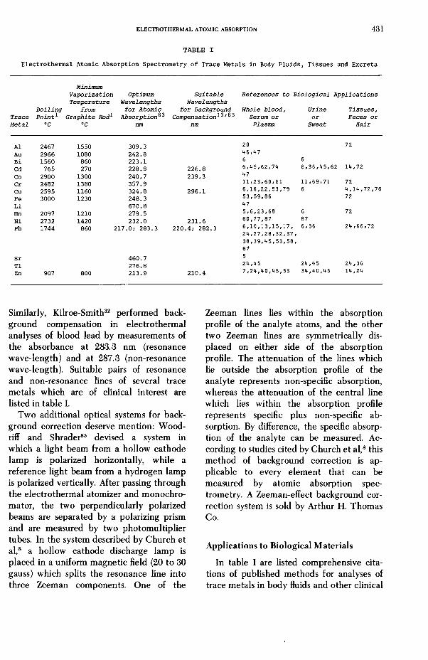
T A B L E I
MinimumVaporization Optimum Suitable References to Biological ApplicationsTemperature Wavelengths Wavelengths
Boiling from for Atomic for Background Whole blood, Urine Tissues,Trace Point* Graphite Rod1 Absorption**^ Compensation*^'^3 Serum or or Feces orMetal °C °C nm nm Plasma Sweat Hair
Al 2467 1550 309.3 20 72Au 2966 1080 242.8 46*47Bi 1560 860 223.1 6 6Cd 765 270 228.8 226.8 6,45/62/74 6,36,45,62 14,72Co 2900 1300 240.7 239.3 47Cr 2482 1380 357.9 11*23/60/61 11/69/71 72Cu 2595 1160 324.8 296.1 6,16,22,53,79 6 4,14,72,76Fe 3000 1230 248.3 53,59/86 72Li 670.8 47Mn 2097 1210 279.5 5/6/23/68 6 72Ni 2732 1420 232.0 231.6 60,77,87 87Pb 1744 860 217.0; 283.3 220.4; 282.3 6,10,13,15,17, 6/36 24 ,66,72
24/27,28/32,37,38/39/45/53,58,67
Sr 460.7 5T1 276.8 24,45 24,45 24,36Zn 907 800 213.9 210.4 7,24,40,45,53 34,40,45 14,24
Similarly, Kilroe-Smith32 performed background compensation in electrothermal analyses of blood lead by measurements of the absorbance at 283.3 nm (resonance wave-length) and at 287.3 (non-resonance wave-length). Suitable pairs of resonance and non-resonance lines of several trace metals which are of clinical interest are listed in table I.
Two additional optical systems for background correction deserve mention: Wood- riff and Shrader85 devised a system in which a light beam from a hollow cathode lamp is polarized horizontally, while a reference light beam from a hydrogen lamp is polarized vertically. After passing through the electrothermal atomizer and monochromator, the two perpendicularly polarized beams are separated by a polarizing prism and are measured by two photomultiplier tubes. In the system described by Church et al,8 a hollow cathode discharge lamp is placed in a uniform magnetic field (20 to 30 gauss) which splits the resonance line into three Zeeman components. One of the
Zeeman lines lies within the absorption profile of the analyte atoms, and the other two Zeeman lines are symmetrically displaced on either side of the absorption profile. The attenuation of the lines which lie outside the absorption profile of the analyte represents non-specific absorption, whereas the attenuation of the central line which lies within the absorption profile represents specific plus non-specific absorption. By difference, the specific absorption of the analyte can be measured. According to studies cited by Church et al,8 this method of background correction is applicable to every element that can be measured by atomic absorption spectrometry. A Zeeman-effect background correction system is sold by Arthur H. Thomas Co.
Applications to Biological Materials
In table I are listed comprehensive citations of published methods for analyses of trace metals in body fluids and other clinical
4 3 2 SUNDERM AN
specimens by means of electrothermal atomic absorption spectrometry. Readers are cautioned that many of the early methods that are cited in table I have become outmoded, owing to improvements in instrumentation for electrothermal atomic absorption spectrometry. All of the published methods need to be critically evaluated in the prospective analyst’s laboratory before they can be confidently employed for diagnostic measurements of trace metals in body fluids. Despite these caveats, the author believes that table I should be helpful as a guide to the growing literature on clinical and biological applications of electrothermal atomic absorption spectrometry.
References
1. Aggett, J. and Sprott, A. ].: Non-flame atomization in atomic absorption spectrometry. Anal. Chim. Acta 72:49-56,1974.
2. Amos, M. D., Bennett, P. A., Brodie, K. G., Lung, P. W. Y., and Matousek, J. P. : Carbon rod atomizer in atomic absorption and fluorescence spectrometry and its clinical application. Anal. Chem. 43:211- 215, 1971.
3. Aspila, K. I., Chakrabarti, C. L., and Bratzel, M. P., Jr.: Pyrolytic graphite-tube micro-furnace for trace analysis by atomic absorption spectrometry. Anal. Chem. 44:1718-1720,1972.
4. Barnett, W. B. and Kahn, H. L.: Determination of copper in fingernails by atomic absorption with the graphite furnace. Clin. Chem. 18:923-927, 1972.
5. Bek, F., Janouskova, J., and Moldan, B. : Direct determination of manganese and strontium in human blood serum by means of the graphite furnace, Perkin-Elmer HGA-70. Chem. Listy 66:867-875,1972.
6. Bourdon, R., Galliot, M., and Prouillet, F. : Dosage du cuivre, du plomb, du manganèse, du bismuth, du cadmium et de l’or dans les liquides biologiques par spectrométrie d’absorption atomique sans flamme. Ann. Biol. Clin. 32:413-422, 1974.
7. Chooi, M. K., Todd, J. K., and Boyd, N. D.: Effect of carbon cup aging on plasma zinc determination by flameless atomic absorption spectrometry. Clin. Chem. 21:632-634, 1975.
8. Church, D. A., Hadeishi, T., Leong, L., McLaughlin, R. D., and Zak, B. : Two-chamber furnace for flameless atomic absorption spectrometry. Anal. Chem. 46:1352-1355,1974.
9. Clyburn, S. A., Kantor, T., and Veillon, C. : Pyrolysis treatment for graphite atomization systems. Anal. Chem. 46:2213-2215, 1974.
10. Cruz, R. B. and Loon, J. C. van: A critical study of the application of graphite-furnace non-flame atomic absorption spectrometry to the determi
nation of trace base metals in complex heavy-matrix sample solutions. Anal. Chim. Acta 72:231-243,1974.
11. Davidson, I. W. F. and Secrest, W. L.: Determination of chromium in biological materials by atomic absorption spectrometry using a graphite furnace atomizer. Anal. Chem. 44:1808-1813, 1972.
12. Donega, H. M. and Burgess, T. E.: Atomic absorption analysis by flameless atomization in a controlled atmosphere. Anal. Chem. 42:1521-1524,1970.
13. Ealy, J. A., Bolton, N. E., McElheny, R. J., and Morrow, R. W.: Determination of lead in whole blood by graphite furnace atomic absorption spectrophotometry. Amer. Ind. Hyg. Assoc. J. 35:566- 570, 1974.
14. Evenson, M. A. and Anderson, C. T., Jr.: Ultramicro analysis for copper, cadmium and zinc in human liver tissue by use of atomic absorption spectrophotometry and the heated graphite tube atomizer. Clin. Chem. 4J.-537-543, 1975.
15. Evenson, M. A. and Pendergast, D. D.: Rapid ultramicro direct determination of erythrocyte lead concentration by atomic absorption spectrophotometry with use of a graphite tube furnace. Clin. Chem. 20.163-171,1974.
16. Evenson, M. A. and Warren, B. L.: Determination of serum copper by atomic absorption, with use of the graphite cuvette. Clin. Chem. 21:619-625,1975.
17. Fernandez, F. J.: Micromethod for lead determination in whole blood by atomic absorption, with use of the graphite furnace. Clin. Chem. 21:558- 561, 1975.
18. Findlay, W. J., Zdrojewski, A., and Quickert, N.: Temperature measurements of a graphite furnace used in flameless atomic absorption. Spectrosc. Letters 7:63-72,1974.
19. Findlay, W. J., Zdrojewski, A. and Quickert, N.: Preatomization losses in flameless atomic absorption spectroscopy. Spectrosc. Letters 7:355- 364, 1974.
20. Fuchs, C., Brasche, M., Paschen, K. Nord beck, H., and Quellhorst, E.: Aluminum-Bestimmung im Serum mit flammenloser Atomabsorption. Clin. Chim. Acta 52:71-80, 1974.
21. Fuller, C. W.: A simple standards additions technique using the Model 306 atomic absorption spectrophotometer. Atomic Abs. Newslett. 11:65-66,1972.
22. Glenn, M., Savory, J., Hart, L., Glenn, T., and Winefordner, J . : Determination of copper in serum with a graphite rod atomizer for atomic absorption spectrophotometry. Anal. Chim. Acta 57:263-269,1971.
23. Grafflage, B., Buttgereit, G., Kubier, W. and Mertens, H-M.: Die Messung der Spurenelemente Chrom und Mangan im Serum mittels flammenloser Atomabsorption. Z. Klin. Chem. Klin. Biochem. 12:287-293, 1974.
24. Hauck, G.: Erfahrungen mit der flammenlosen Atomabsorption bei der Untersuchung biologischen Materials auf Spuren von Schwermetallen. Z. Anal. Chem. 267:337-341, 1973.
ELECTRO TH ERM A L ATOMIC ABSORPTION 4 3 3
25. Hohnadel, D. C., Sunderman, F. W., Jr., Nechay, M. W., and McNeely, M. D. : Atomic absorption spectrometry of nickel, copper, zinc and lead in sweat from healthy subjects during sauna bathing. Clin. Chem. 29:1288-1292, 1973.
26. Hwang, J. Y., Mokeler, C. J., and Ullucci, P. A.: Maximization of sensitivities in tantalum ribbon flameless atomic absorption spectrometry. Anal. Chem. 44:2018-2121, 1972.
27. Hwang, J. Y., Ullucci, P. A., and Mokeler, C. J.: Direct flameless atomic absorption determination of lead in blood. Anal. Chem. 45:795-798, 1973.
28. Hwang, J. Y., Ullucci, P. A., Smith, S. B., Jr., and Malenfant, A. L. : Microdetermination of lead in blood by flameless atomic absorption spectrometry. Anal. Chem. 43:1319-1321, 1971.
29. Ingle, J. D., Jr.: Precision of atomic absorption spectrometric measurements. Anal. Chem. 46: 2161-2171, 1974.
30. Kantor, T., Clyburn, S. A., and Veillon, C.: Continuous sample introduction with graphite atomization systems for atomic absorption spectrometry. Anal. Chem. 46:2205-2213, 1974.
31. Kerber, J. D., Russo, A. J., Peterson, G. E., and Ediger, R. D. : Performance improvements with the graphite furnace. Atomic Abs. Newslett. 12:106-108, 1973.
32. Kilroe-Smith, T. A. : Linear working graphs in blood lead determinations with the Beckman flameless atomic absorption cuvet. Clin. Chem. 21:630-632,1975.
33. King, J. S.: Editorial: A burgeoning branch of clinical analysis. Clin. Chem. 21:467, 1975.
34. Kirkbright, G. F. : The application of non-flame atom cells in atomic absorption and atomic fluorescence spectroscopy. A review. Analyst 96: 609-623,1971.
35. Koirtyohann, S. R., Sievers, H., and Pickett, E. E. : The electrically heated furnace in atomic absorption. Trace Substances in Environmental Health—V, Hemphill, D. D., ed. University of Missouri Press, Columbia, MO, pp 463-471, 1972.
36. Kubasik, N. P. and Volosin, M. T. : A simplified determination of urinary cadmium, lead, and thallium, with use of carbon rod atomization and atomic absorption spectrophotometry. Clin. Chem. 19:954-958, 1973.
37. Kubasik, N. P. and Volosin, M. T.: Use of the carbon rod atomizer for direct analysis of lead in blood. Clin. Chem. 20:300-301, 1974.
38. Kubasik, N. P. Volosin, M. T., and Murray, M. H. : A quantitative micro technique for the analysis of lead in blood by carbon rod atomization and atomic absorption spectrophotometry. Clin. Biochem. 5:266-270, 1972.
39. Kubasik, N. P., Volosin, M. T., and Murray, M. H. : Carbon rod atomizer applied to measurement of lead in whole blood by atomic absorption spectrophotometry. Clin. Chem. 28:410-412, 1972.
40. Kurz, D., Roach, J., and Eyring, E. J.: Determination of zinc by flameless atomic absorption spectrophotometry. Anal. Biochem. 53:586-593, 1973.
41. Lisk, D- J : Recent developments in the analysis of toxic elements. Science 284:1137-1141, 1974.
42. Lundgren, G. and Johansson, G.: A temperature- controlled graphite tube furnace for the determination of trace metals in solid biological tissue. Talanta 21 .-257-264, 1974.
43. Lundgren, G., Lundmark, L., and Johansson, G. : Temperature controlled heating of the graphite tube atomizer in flameless atomic absorption spectrometry. Anal. Chem. 46:1028-1031, 1974.
44. L’vov, B. V.: The analytical use of atomic absorption spectra. Spectrochim. Acta (Part B) 17:761-770, 1961.
45. Machata, G. and Binder, R. : The determination of lead, thallium, zinc and cadmium traces in biological material with flameless atomic absorption. Z. Rechtsmedizin 73:29-34, 1973.
46. Maessen, F. J. M. J. and Posma, F. D. : Fundamental aspects of flameless atomic absorption using the mini-Massmann carbon rod atomizer. Anal. Chem. 46:1439-1444, 1974.
47. Maessen, F. J. M. J., Posma, F. D., and Balke, J. : Direct determination of gold, cobalt, and lithium in blood plasma using the mini-Massmann carbon rod atomizer. Anal. Chem. 46:1445-1449, 1974.
48. McIntyre, N. S., Cook, M. G., and Boase, D. G.: Flameless atomic absorption determination of cobalt, nickel and copper. A comparison of tantalum and molybdenum evaporation surfaces. Anal. Chem. 46:1983-1987, 1974.
49. Manning, D. C.: Using the Perkin-Elmer deuterium background correction system. Atomic Absorp. Newsltr. 22:112-113,1972.
50. Manning, D. C. and Fernandez, F. : Atomization for atomic absorption using a heated graphite tube. Atomic Absorp. Newsltr. 9:65-70, 1970.
51. Massmann, H. : Heutiger Stand der Atomabsorp- tionsspektrometrie. Chimia 21 .-217-226, 1967.
52. Massmann, H. : Vergleich von Atomabsorption und Atomfluoreszenz in der Graphitkiivette. Spectrochim. Acta (Part B) 23:215-226, 1968.
53. Matousek, J. P. and Stevens, B. J.: Biological applications of the carbon rod atomizer in atomic absorption spectroscopy. Clin. Chem. 27:363-368,1971.
54. Montaser, A. and Crouch, S. R. : Analytical applications of the graphite braid nonflame atomizer. Anal. Chem. 46:1817-1820, 1974.
55. Montaser, A. and Crouch, S. R. : New methods for programmed heating of electrically heated nonflame atomic vapor cells. Anal. Chem. 47:38-45,1975.
56. Montaser, A., Goode, S. R., and Crouch, S. R. : Graphite braid atomizer for atomic absorption and atomic fluorescence spectrometry. Anal. Chem. 46:599-601, 1974.
57. Norris, J. D. and West, T. S.: Some applications of spectral overlap in atomic absorption spectrometry. Anal. Chem. 46:1423-1425, 1974.
58. Norval, E. and Butler, L. R. P.: The determination of lead in blood by atomic absorption with the high temperature graphite tube. Anal. Chim. Acta 58:47-57, 1972.
59. Olsen, E. D., Jatlow, P. I., Fernandez, F. J., and Kahn, H. L. : Ultramicro method for determination
4 3 4 SUNDERMAN
of iron in serum with the graphite furnace. Clin. Chem. 19:326-329, 1973.
60. Pekarek, R. S. and Hauer, E. C.: Direct determination of serum chromium and nickel by an atomic absorption spectrophotometer with a heated graphite atomizer. Fed. Proc. 3! :700 (Abs), 1972.
61. Pekarek, R. S., Hauer, E. C., Wannemacher, R. W., Jr., and Beisel, W. R.: The direct determination of serum chromium by an atomic absorption spectrophotometer with a heated graphite atomizer. Anal. Biochem. 59:283-292, 1974.
62. Perry, E. F., Koirtyohann, S. R., and Perry, H. M., Jr.: Determination of cadmium in blood and urine by graphite furnace atomic absorption spectrophotometry. Clin. Chem. 22:626-629, 1975.
63. Price, W. J .: Analytical Atomic Absorption Spectrometry. Heyden and Son, Ltd., London, 1972, pp 1-239.
64. Reeves, R. D., Patel, B. M., Molnar, C. J., and Winefordner, J. D.: Decay of atom populations following graphite rod atomization in atomic absorption spectrometry. Anal. Chem. 45:246-249, 1973.
65. Reinhold, J. G.: Trace elements: A selective suryey. Clin. Chem. 21: 476-500, 1975.
66. Renshaw, G. D., Pounds, C. A., and Pearson, E. F .: Variation in lead concentration along single hairs as measured by non-flame atomic absorption spectrophotometry. Nature 238:162-163, 1972.
67. Rosen, J. F. and Trinidad, E. E.: The microdetermination of blood lead in children by flameless atomic absorption: The carbon rod atomizer. J. Lab. Clin. Med. 80:567-576, 1972.
68. Ross, R. T. and Gonzalez, J. G.: Direct determination of trace quantities of manganese in blood and serum samples using selective volatilization and graphite tube reservoir atomic absorption spectrophotometry. Bull. Environ. Contam. Toxicol. 22:470-474, 1974.
69. Ross, R. T., Gonzalez, J. G., and Segar, D. A.: The direct determination of chromium in urine by selective volatilization with atom reservoir atomic absorption. Anal. Chim. Acta 63:205-209, 1973.
70. Sarbeck, J. R. and Landgraf, W. C.: Automated peak discrimination and integration for nonflame atomic absorption analysis at nanogram levels. J. Pharm. Sci. 63:929-930, 1974.
71. Schaller, K-H., Essing, H-G., Valentin, H., and Schacke, G.: Quantitative Chrombestimmung im Ham mit flammenloser Atomabsorptions-Spek- trometrie, Z. Klin. Chem. Klin. Biochem. 20:434- 437, 1972.
72. Schramel, P.: Determination of eight metals in the International Biological Standard by flameless
atomic absorption spectrometry. Anal. Chim. Acta 67:69-77,1973.
73. Schramel, P. : The application of peak integration in flameless atomic absorption spectrometry. Anal. Chim. Acta 72:414-418, 1974.
74. Schumacher, E. and Umland, F.: Improved fast destruction method for the determination of cadmium in body fluids using a graphite tube atomizer. Z. Anal. Chem. 270:285-286, 1974.
75. Sommerfeld, M. R., Love, T. D., and Olsen, R. D.: Trace metal contamination of disposable pipet tips. Atomic Absorp. Newsltr. 14:31-32, 1975.
76. Stevens, B. J. : Biological applications of the carbon rod atomizer in atomic absorption spectroscopy. 2. Determination of copper in small samples of tissue. Clin. Chem. 28:1379-1384, 1972.
77. Sunderman, F. W., Jr.: Atomic absorption spectrometry of trace metals in clinical pathology. Human Pathol. 4:549-582, 1973.
78. Torsi, G. and Tessari, G.: Influence of heating rate on analytical response in flameless atomic absorption spectrometry. Anal. Chem. 45:1812-1816, 1973.
79. Welz, B. and Wiedeking, E.: Bestimmung von Spurenelementen im Serum und Urin mit flammenloser Atomisierung. Z. Anal. Chem. 252:111-117, 1970.
80. West, C. D. : Relative effect of molecular absorption on atomic absorption and atomic fluorescence. Anal. Chem. 46:797-799, 1974.
81. West, T. S. and Williams, X. K. : Atomic absorption and fluorescence spectroscopy with a carbon filament atom reservoir. Anal. Chim. Acta 45:27-41, 1969.
82. Williams, M. and Piepmeier, E. H.: Commercial tungsten filament atomizer for analytical atomic spectrometry. Anal. Chem. 44:1342-1344, 1972.
83. Woodriff, R.: Atomization chambers for atomic absorption spectrochemical analysis. A review. Appl. Spectrosc. 28:413-416,1974.
84. WoodrifF, R. and Ramelow, G. : Atomic absorption spectroscopy with a high-temperature furnace. Spectrochim. Acta23B:665-671, 1968.
85. Woodriff, R. and Shrader, D.: Furnace atomic absorption with reference channel. Anal. Chem. 43:1918-1920, 1971.
86. Yeh, Y-Y and Zee, P. : Micromethod for determining total iron-binding capacity by flameless atomic absorption spectrophotometry. Clin. Chem. 20: 360-364, 1974.
87. Zachariasen, H., Andersen, I., Kostol, C., and Barton, R.: Technique for determining nickel in blood by flameless atomic absorption spectrophotometry. Clin. Chem. 21:562-567, 1975.