Embed Size (px)
Citation preview
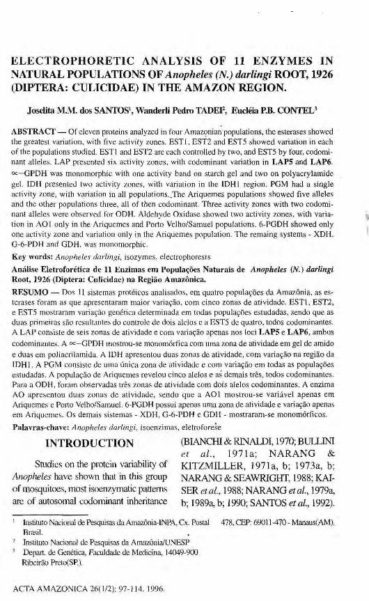
ELECTROPHORETIC ANALYSIS OF 11 ENZYMES IN NATURAL POPULATIONS OF Anopheles (N.) darlingi ROOT, 1926 (DIPTERA: CULICIDAE) IN THE AMAZON REGION.
Jaselita M.M. dos SANTOS1, Wanderli Pedro TADEP, Eucléia P.B. CONTEL3
ABSTRACT — Of eleven proteins analyzed in four Amazonian populations, the esterases showed the greatest variation, with five activity zones. EST1, EST2 and EST5 showed variation in each of the populations studied. EST1 and EST2 are each controlled by two, and EST5 by four, codomi-nant alleles. LAP presented six activity zones, with codominant variation in LAP5 and LAP6. oc—GPDH was monomorphic with one activity band on starch gel and two on polyacrylamide gel. 1DH presented two activity zones, with variation in the IDHl region. PGM had a single activity zone, with variation in all populations. The Ariquemes populations showed five alleles and the other populations three, all of then codominant. Three activity zones with two codominant alleles were observed for ODH. Aldehyde Oxidase showed two activity zones, with variation in AOl only in the Ariquemes and Porto Velho/Samuel populations. 6-PGDH showed only one activity zone and variation only in the Ariquemes population. The remaing systems - XDH, G-6-PDH and GDH. was m o n o m o r p h i c .
Key words: Anopheles darlingi, isozymes, electrophoresis
Análise Eletroforética de 11 Enzimas era Populações Naturais de Anopheles (Ν.) darlingi Root, 1926 (Diptera: Cuiicidae) na Região Amazônica.
RESUMO — Dos 11 sistemas protéicos analisados, em quatro populações da Amazônia, as esterases foram as que apresentaram maior variação, com cinco zonas de atividade. EST1, EST2, e EST5 mostraram variação genética determinada em todas populações estudadas, sendo que as duas primeiras são resultantes do controle de dois alelos e a EST5 de quatro, todos codominantes. A LAP consiste de seis zonas de atividade e com variação apenas nos loci LAP5 e LAP6, ambos codominantes. A GPDH mostrou-se monomórfica com uma zona de atividade em gel de amido e duas em poliacrilamida. A IDH apresentou duas zonas de atividade, com variação na região da IDH1. A PGM consiste de uma única zona de atividade e com variação em todas as populações estudadas. A população de Ariquemes revelou cinco alelos e as demais três, todos codominantes. Para a ODH, foram observadas três zonas de atividade com dois alelos codominantes. A enzima AO apresentou duas zonas de atividade, sendo que a AOl mostrou-se variável apenas em Ariquemes e Porto Velho/Samuel. 6-PGDH possui apenas uma zona de atividade e variação apenas em Ariquemes. Os demais sistemas - XDH, G-6-PDH e GDH - mostraram-se monomórficos.
Palavras-chave: Anopheles darlingi, isoenzimas, eletroforese
INTRODUCTION
Studies on the protein variability of Anopheles have shown that in this group of mosquitoes, most isoenzymatic patterns are of autosomal codominant inheritance
(BIANCHI & RINALDI, 1970; BOLLINI et al., 1971a; NARANG & KITZMILLER, 197la, b; 1973a, b; NARANG & SEAWRIGHT, 1988; KAISER etai, 1988; NARANG etal, 1979a, b; 1989a, b; 1990; SANTOS et al., 1992).
1 Instituto Nacional de Pesquisas da Amazônia-INPA, Cx. Postal 478, CEP: 69011 -470 - Manaus(AM), Brasil.
2 Instituto Nacional de Pesquisas da Amazônia/UNESP
3 Depart. de Genética, Faculdade de Medicina, 14049-900 Ribeirão Preto(SP).
ACTA AMAZÔNICA 26(1/2): 97-114. 1996.
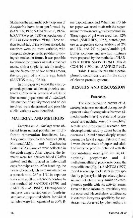
Studies on the enzymatic polymoiphism of Anopheles have been performed by (SANTOS, 1979; NARANG et ai, 1979a, b; SANTOS et ai, 1985) in populations of BR-174 (Manaus/Boa Vista). These authors found that, of the systems studied, the esterases were the most variable, with complex electrophoretic profiles involving six molecular forms. It was possible to estimate the number of males that had mated with a single female by analyzing the frequency of two alleles among the progeny of a single egg batch (SANTOS et ai, 1981a).
In this paper we report the electrophoretic patterns of eleven proteins analyzed in 4th-instar larvae and adults of four natural populations of A. darlingi. The number of activity zones and of loci involved were determined and possible allelic variants were identified.
MATERIAL A N D M E T H O D S
Samples on A. darlingi were obtained from natural populations of different Amazonian localities, i.e., Ariquemes, Porto Velho/ Samuel (RO), Manaus(AM), and Cachoeira Porteira(PA). Samples were collected in the adult stages. After capture, the females were fed chicken blood (Gallus gallus) and then placed in individuali cups for oviposition. After hatching, the larvae of each clutch were maintained in a sectarium at 26° ± l°C in separate trays until adult emergence according to the method of SANTOS (1979) and SANTOS et al. ( 1981 b). Electrophoretic analyses were carried out on fourth-in-star larvae, pupae and adults. Individual samples were homogenized in 0,5% β-
mercaptoethanol and Whatman n°3 filter paper was used to absorb the supernatant for horizontal gel electrophoresis. Three types of gel were used, i.e., 12% starch (SMITHIES, 1955), starch-agar-ose at respective concentrations of 2% and 1%, and 7% poly aery lamide gel. Buffer solutions and reaction mixtures were prepared by the methods of HARRIS & HOPKINSON (1976) LIMA & CONTEL (1990) and SANTOS (1992).
Table 1 summarizes the electrophoretic conditions used for the study of eleven protein systems.
RESULTS A N D DISCUSSION
Esterases
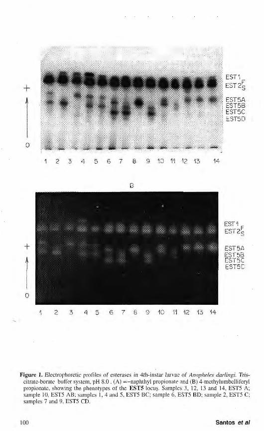
The electrophoretic pattern of A. darlingi esterases obtained during development using fluorogenic substrates (4-methylumbelliferyl acetate and propionate) and naphthol ester ( oc—naphthyl acetate and propionate) revealed five electrophoretic activity zones being the esterases 1,2 and 5 more deeply stained during the larval stages. Esterase 3 and 4 were characteristic of pupae and adult. The isozyme profiles obtained with the four substrates were quite similar, cc— naphthyl propionate and 4-methylumbelliferyl propionate being the most efficient (Fig. 1). SANTOS (1979) tested seven naphthol esters in this species by polyacrylamide gel electrophoresis and detected a very similar electrophoretic profile with six activity zones. Even on these substrates, specificity was not observed. Nevertheless, a difference in esterases isozymes specificity for substrates was observed by other authors in
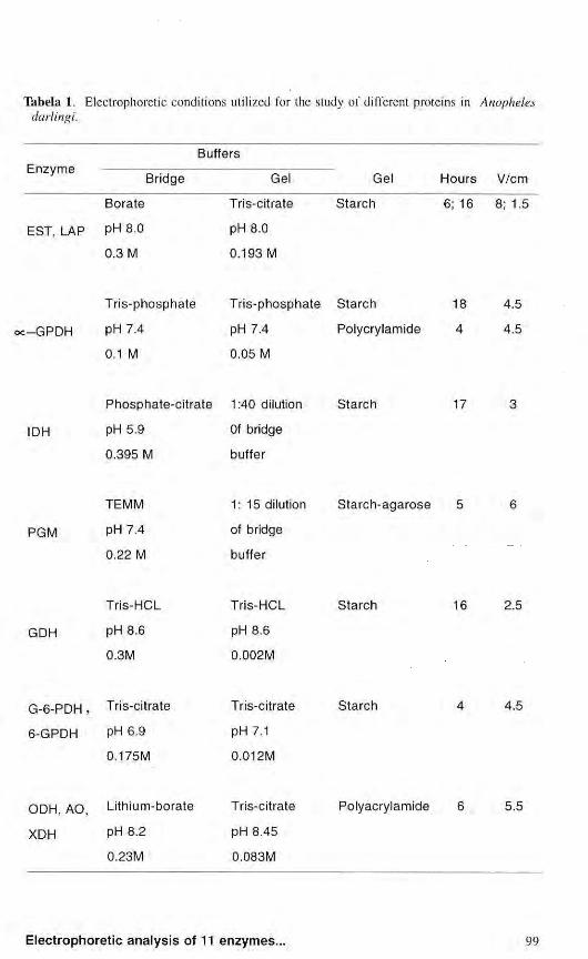
Tabela I. Electrophoretic conditions utilized for the study of different proteins in Anopheles darlingi.
Buffers E n z y m e
Bridge Gel Gel Hours V/cm
Bora te Tris-citrate S t a r ch 6; 16 8; 1.5
EST, LAP PH 8.0 pH 8.0 0.3 Μ 0.193 Μ
: - G P D H
Tr i s -phospha te
pH 7.4
0.1 Μ
Tr i s -phospha t e S ta rch
pH 7.4
0.05 Μ
Polycrylamide
4.5
4.5
IDH
Phospha te -c i t r a t e 1:40 dilution
pH 5.9
0.395 Μ
Of bridge
buffer
S ta rch 17
PGM
TEMM
pH 7.4
0.22 Μ
1 : 1 5 dilution
of bridge
buffer
Starch-agarose
GDH
Tris-HCL
pH 8.6
0.3M
Tris-HCL
pH 8.6
0.002M
S ta rch 16 2.5
G - 6 - P D H , Tris-citrate
6-GPDH PH 6.9
0.175M
Tris-citrate
pH 7.1
0.012M
Starch 4.5
ODH, AO Lithium-borate Tris-citrate
XDH PH 8.2 pH 8.45
0.23M 0.083M
Polyacrylamide 5.5
E5T5A EST5B EST5C EST5D
1 2 3 4 5 6 7 9 10 11 12 13 14
+
À
EST1 EST 2 S
EST5A EST 5 3 EST5C EST5C
1 2 3 4 5 6 7 8 9 13 14
Figure 1. Electrophoretic profiles of esterases in 4ih-instar larvae of Anopheles darlingi. Tris-citrate-borate buffer system, pH 8.0. (A) «=-naphthyl propionate and (B) 4-methylumbelliferyl propionate, showing the phenotypes of the EST5 locus. Samples 3, 12, 13 and 14, EST5 A; sample 10, EST5 AB; samples 1, 4 and 5, EST5 BC; sample 6, EST5 BD; sample 2, EST5 C; samples 7 and 9, EST5 CD.
insects and other organisms, TREBATOSKI & HAYNES (1969) observed this difference in twelve species of mosquitoes, HOPKINSON et al. (1973) and CO ATES et al. (1975) in human erythrocytes, FALCÃO & CONTEL (1990) in species of stingless bees, and LIMA & CONTEL (1990) in Spodoptera frugiperda.
Of the five isozymes detected, EST1, EST2 and EST5 showed variation in four populations analyzed. The electrophoretic phenotypes observed for EST1 and EST2 suggest that each is genetically controlled by a locus with two codominant alleles (EST1*F and EST1*S, EST2*F and EST2*S). The heterozygous individuals presented two bands for the two loci, suggesting that these enzymes have a monomeric structure.
Similar results were observed for the EST1, EST2 and EST4 loci of Anopheles triannulatus (SANTOS et ai, 1992), EST1 and EST2 of Anopheles nuneztovari (SCARPASSA, 1988) and EST2 of the A. darlingi population along the Manaus - Boa Vista Highway/BR-174 (SANTOS et ai, 1985) when the authors reported a genetic mechanism for each locus constituted by two codominant alleles.
The electrophoretic profiles for EST5 showed variation in the population studies with ten phenotypes. Of these possible phenotypes, EST5 C, EST5 CD and EST5 D were not detected in the Manaus population. Analysis of the data suggests that locus EST5 has four alleles - EST5*A, EST5*R, EST5*C and EST5*D - of codominant action. The pattern of two
bands observed in the heterozygotes suggests that EST5 presents a monomeric structure. A similar mechanism was found in the EST3 and EST5 loci of A. triannulatus (SANTOS et al, 1992) and in the EST6 locus of A. nuneztovari (SCARPASSA, 1988). The authors detected six phenotypes for these loci resulting from the action of three codominant alleles.
Leucine aminopeptidase
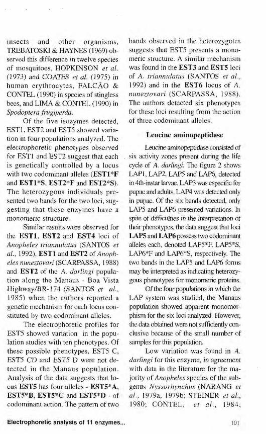
Leucine aminopeptidase consisted of six activity zones present during the life cycle of A. darlingi. The figure 2 shows LAPI, LAP2, LAP5 and LAP6, detected in 4th-instar larvae. LAP3 was especific for pupae and adults, LAP4 was detected only in pupae. Of the six bands detected, only LAP5 and LAP6 presented variations. In spite of difficulties in the interpretation of their phenotypes, the data suggest that loci LAP5 and LAP6 possess two codominant alleles each, denoted LAP5*F, LAP5*S, LAP6*F and LAP6*S, respectively. The two bands in the LAP5 and LAP6 forms may be interpreted as indicating heterozygous phenotypes for monomeric proteins.
Of the four populations in which the LAP system was studied, the Manaus population showed apparent monomor-phism for the six loci analyzed. However, the data obtained were not sufficiently conclusive because of the small number of samples for this population.
Low variation was found in A. darlingi for this enzyme, in agreement with data in the literature for the majority of Anopheles species of the subgenus Nyssorhynchus (NARANG et ai, 1979a, 1979b; STEINER et ai, 1980; CONTEL, et al., 1984;
Η™ •Μ
L A P 1
L A P 2
p L A P 5 LAP 6
Ο
3 4 6 7 8 9 40 12 13
Figure 2. Electrophoretic profiles of leucine-aminopeptidase in 4th-instar larvae of Anopheles darlingi showing low variation. Tris-citrate-borate buffer system, pH 8.0 .
SCARPASSA et al., 1992). For Ariquemes (RO) and High
way PA-422 (PA) populat ions, CONTEL et al. (1984) reported that the three loci detected were monomorphic in two populations. In a population of A. nuneztovari from Tucurui (PA), SCARPASSA et al. (1992) detected variability only for LAP5. STEINER et al. (1980) reported only the LAP-F locus in adult stage of this species in populations from Surinam and Venezuela with variations in both populat ions wich correspond the LAP3 detected by SCARPASSA (1992). In Anopheles aquasalis, four loci were described, with variation one for them (NARANG et ai, 1979b).
©^-Glycerophosphate dehydrogenase
The electrophoretic °c-GPDH patterns of A. darlingi showed differences in adults according to the type
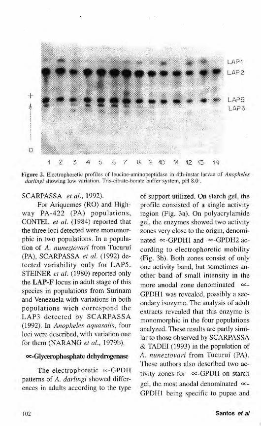
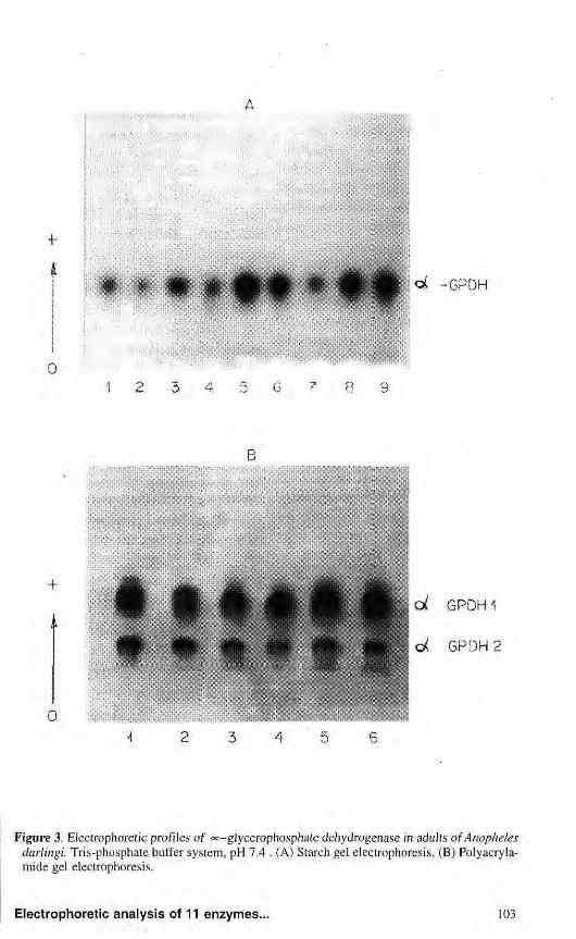
of support utilized. On starch gel, the profile consisted of a single activity region (Fig. 3a). On poly aery lamide gel, the enzymes showed two activity zones very close to the origin, denominated oc-GPDHl and °<-GPDH2 according to electrophoretic mobility (Fig. 3b). Both zones consist of only one activity band, but sometimes another band of small intensity in the more anodal zone denominated «=-GPDH1 was revealed, possibly a secondary isozyme. The analysis of adult extracts revealed that this enzyme is monomorphic in the four populations analyzed. These results are partly similar to those observed by SCARPASSA & TADEI (1993) in the population of A. nuneztovari from Tucurui (PA). These authors also described two activity zones for °<:-GPDH on starch gel, the most anodal denominated °c-GPDH1 being specific to pupae and
A
Figure 3. Electrophoretic profiles of oc-gjycerophosphate dehydrogenase in adults of Anopheles darlingi. Tris-phosphate buffer system, pH 7.4 . (A) Starch gel electrophoresis, (B) Polyacryla-mide gel electrophoresis.
adults. NARANG era!. (1979a) studying this same species in populations from Manaus, also found two «=-GPDH zones in adults by polyacryla-mide gel electrophoresis and reported that each one was coded by independent loci. The same was observed in a population of A. darlingi from BR-174 analysed on polyacrylamide gel. In this population, NARANG et al. (1979a) detected only one activity zone in adult stage. STEINER et al. (1980) also found one activity zone for the A. nuneztovari adult populations from Surinam and Venezuela.
The oc-GPDH enzyme of A. darlingi was monomorphic in the four populations studied. Considering the data in the literature, this enzyme varies little (JOHNSON, 1974; POWELL, 1975; LIMA & CONTEL, 1990; MACHADO & CONTEL, 1991). In A. nuneztovari, SCARPASSA & TADEI (1993) detected only three individuals with the ©c-GPDH
S/ oc-GPDH F phenotype out of 50 analyzed, and none showed the °c-GPDH F/ oc-GPDHF phenotype.
Isocitrate dehydrogenase
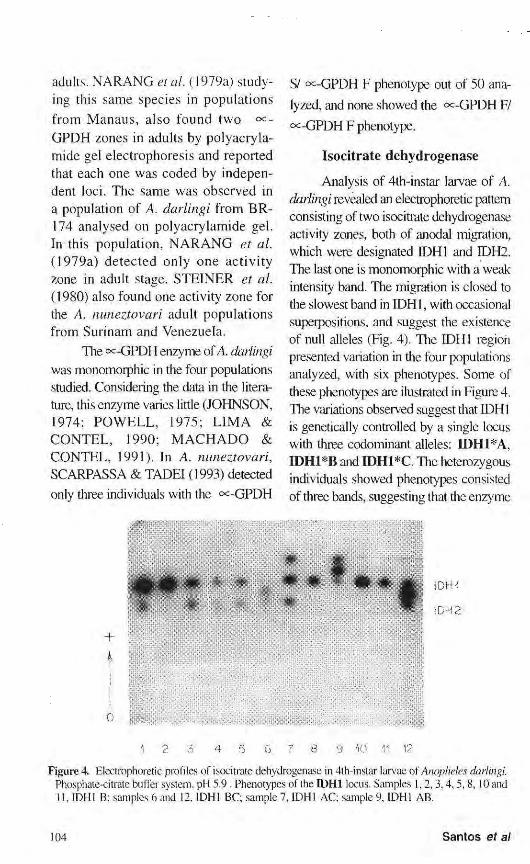
Analysis of 4th-instar larvae of A. darlingi revealed an electrophoretic pattern consisting of two isocitrate dehydrogenase activity zones, both of anodal migration, which were designated IDH1 and IDH2. The last one is monomorphic with a weak intensity band. The migration is closed to the slowest band in IDH1, with occasional superpositions, and suggest the existence of null alleles (Fig. 4). The IDH1 region presented variation in the four populations analyzed, with six phenotypes. Some of these phenotypes are ilustrated in Figure 4. The variations observed suggest that IDH1 is genetically controlled by a single locus with three codominant alleles: Π)Η1*Α, IDH1*B and IDH1*C. The heterozygous individuals showed phenotypes consisted of three bands, suggesting that the enzyme
mm 1DH2
Figure 4. Electrophoretic profiles of isocitrate dehydrogenase in 4th-instar larvae of Anopheles darlingi. Phosphate-citrate buffer system, pH 5.9. Phenotypes of the IDH1 locus. Samples 1,2,3,4,5,8, 10 and 11,1DH1 B; samples 6 and 12, IDH1 BC; sample 7, IDH1 AC; sample 9, IDH1 AB.
may be dimeric. In the analysis of the Porto Velho/
Samuel population, the presence of allele Π)Η1*Α was not detected. In the Cachoeira Porteira and Manaus populations the IDH1 A and IDH1 C pheno-types were not detected, whereas in Ariquemes only phenotype IDH1 A was not observed. Similar results were found in populations of Anopheles stephensi from Pakistan by VAN DRIEL et al. (1987) when 11 progenies were analyzed and the allele IDH1*A was not detected in four of them. The IDH2 region was monomorphic in all populations studied and consisted of only one weakly staining band located very close to the slowest IDH1 band. Analysis of these two regions was also hampered by occasional overlapping.
These results disagree, in part, with those obtained by ROSA-FREITAS et al. (1992) for three A. darlingi populations, the authors reported three alleles for the IDH2 locus, two of them having low frequencies (0.0455 and 0.1591).
Phenotypic variants of IDH1 and IDH2 have been observed in the populations of A. nuneztovari from BR-174 (SCARPASSA, personal communication) and from Manaus for the IDH2 locus (NARANG et al. 1979a).
Phosphoglucomutase
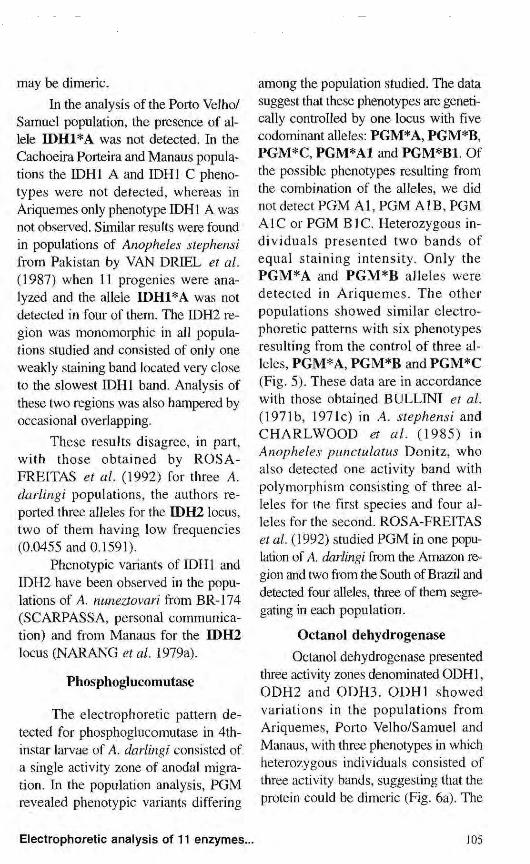
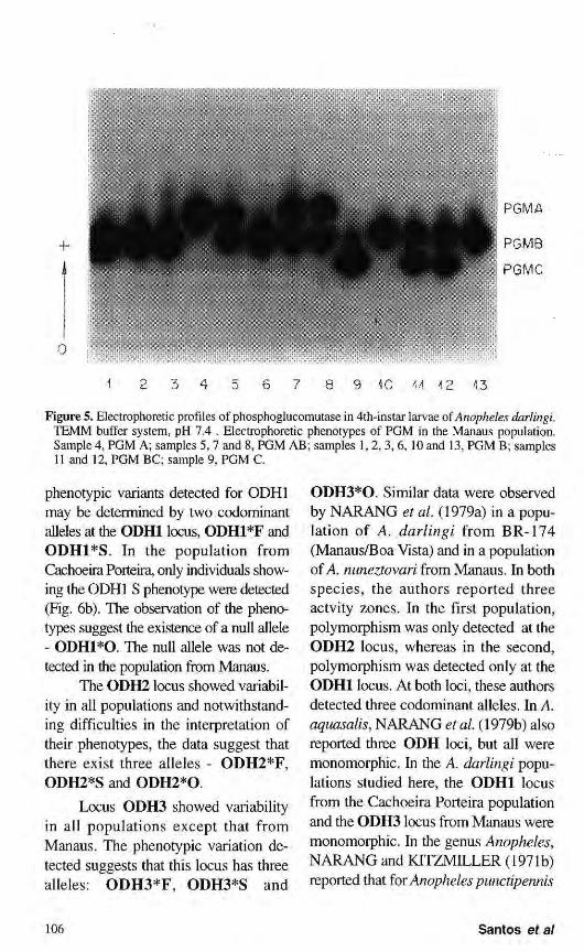
The electrophoretic pattern detected for phosphoglucomutase in 4th-instar larvae of A. darlingi consisted of a single activity zone of anodal migration. In the population analysis, PGM revealed phenotypic variants differing
among the population studied. The data suggest that these phenotypes are genetically controlled by one locus with five codominant alleles: PGM*A, PGM*B, PGM*C, PGM*A1 and PGM*B1 Of the possible phenotypes resulting from the combination of the alleles, we did not detect PGM Al , PGM A IB, PGM A1C or PGM B1C. Heterozygous individuals presented two bands of equal staining intensity. Only the PGM*A and PGM*B alleles were detected in Ariquemes. The other populations showed similar electrophoretic patterns with six phenotypes resulting from the control of three alleles, PGM*A, PGM*B and PGM*C (Fig. 5). These data are in accordance with those obtained BULLINI et al. (1971b, 1971c) in A. stephensi and CHARLWOOD et al. (1985) in Anopheles punctulatus Donitz, who also detected one activity band with polymorphism consisting of three alleles for the first species and four alleles for the second. ROSA-FREITAS et al. (1992) studied PGM in one population of A darlingi from the Amazon region and two from the South of Brazil and detected four alleles, three of them segregating in each population.
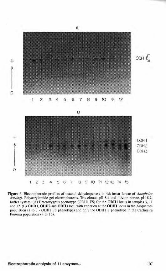
Octanol dehydrogenase Octanol dehydrogenase presented
three activity zones denominated ODH1, ODH2 and ODH3. ODH1 showed variations in the populations from Ariquemes, Porto Velho/Samuel and Manaus, with three phenotypes in which heterozygous individuals consisted of three activity bands, suggesting that the protein could be dimeric (Fig. 6a). The
1 2 3 4 5 6 7 8 9 10 U 12 Ί 3
Figure 5. Electrophoretic profiles of phosphoglucomutase in 4th-instar larvae of Anopheles darlingi. TEMM buffer system, pH 7.4 . Electrophoretic phenotypes of PGM in the Manaus population. Sample 4, PGM A; samples 5,7 and 8, PGM AB; samples 1,2, 3, 6, 10 and 13, PGM B; samples 11 and 12, PGM BC; sample 9, PGM C.
phenotypic variants detected for ODH1 may be determined by two codominant alleles at the ODH1 locus, ODHl*F and ODHl*S. In the population from Cachoeira Porteira, only individuals showing the ODH1 S phenotype were detected (Fig. 6b). The observation of the phenotypes suggest the existence of a null allele - 0DH1*0. The null allele was not detected in the population from Manaus.
The ODH2 locus showed variability in all populations and notwithstanding difficulties in the interpretation of their phenotypes, the data suggest that there exist three alleles - ODH2*F, ODH2*S and ODH2*0.
Locus ODH3 showed variability in all populations except that from Manaus. The phenotypic variation detected suggests that this locus has three alleles: ODH3*F, ODH3*S and
ODH3*0. Similar data were observed by NARANG et al. (1979a) in a population of A. darlingi from BR-174 (Manaus/Boa Vista) and in a population of A. nuneztovari from Manaus. In both species, the authors reported three actvity zones. In the first population, polymorphism was only detected at the ODH2 locus, whereas in the second, polymorphism was detected only at the ODH1 locus. At both loci, these authors detected three codominant alleles. In A. aqimsalis, NARANG et al. (1979b) also reported three ODH loci, but all were monomorphic. In the A. darlingi populations studied here, the ODH1 locus from the Cachoeira Porteira population and the ODH3 locus from Manaus were monomorphic. In the genus Anopheles, NARANG and KITZMILLER (1971b) reported that for Anopheles punctipennis
Β
1 2 3 4 5 6 7 8 9 40 11 12 13 14 45
Figure 6. Electrophoretic profiles of octanol dehydrogenase in 4th-instar larvae of Anopheles darlingi. Polyacrylamide gel electrophoresis. Tris-citrate, pH 8.4 and lithium-borate, pH 8.2, buffer system. (A) Heterozygous phenotype (ODH1 FS) for the ODH1 locus in samples 3, 11 and 12. (B) ODH1, ODH2 and ODH3 loci, with variation at the ODH1 locus in the Ariquemes population (1 to 7 - ODH1 FS phenotype) and only the ODH1 S phenotype in the Cachoeira Porteira population (8 to 15).
the electrophoretic profile of ODH consisted of only one activity band with three phenotypes controlled by two codominant alleles. Similar results were found in Anopheles culicifacies by AHMAD et al. (1978).
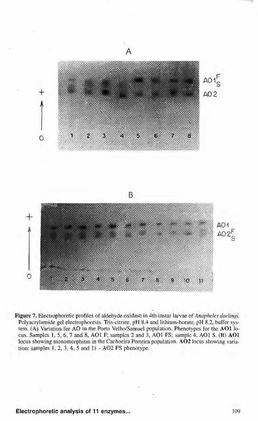
Aldehyde oxidase Aldehyde oxidase consisted of
two main activity zones: AOl and A02. A third zone was detected in the Ariquemes and Cachoeira Porteira populations, but it was not considered in the populations studied.
AOl showed variability only in Ariquemes and Porto Velho/Samuel, and consisted of three phenotypes. The phenotypic variants detected suggest that this locus may have two codominant alleles, A01*F and A01*S, with heterozygous individuals showing three bands suggesting a dimeric structure (Fig. 7a). Null alleles were also detected at this locus. Analysis of the Cachoeira Porteira and Manaus populations did not show variations for the A O l locus, but only the A01*F allele was observed (Fig. 7b).
A02 showed variations in all populations analyzed, with a pattern consisting of two or three bands in the heterozygotes, which may be interpreted as secondary changes, i.e., post-translational changes. Analysis of these phenotypes suggests that the genetic control of this enzyme occurs through two codominant alleles - A02*F and A02*S. A similar pattern was observed in a population of A. darlingi from BR-174 (Manaus/Boa Vista) and a population of A. nuneztovari from Manaus (NARANG et ai, 1979a). In both species, the authors reported three activity zones,
with variations, in which the most anodic (AOl) had low activity, which was visualized only after a prolonged time. In A. darlingi studied here, it was the A03 zone that presented low activity and that was not visualized in most analyses.
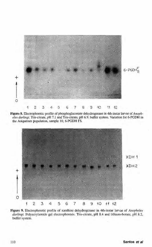
6-Phosphogluconate dehydrogenase
A single activity zone of phosphogluconate dehydrogenase was detected (Fig. 8). Variation for this enzyme was found only in the Ariquemes population. The phenotypes observed suggest the existence of two alleles: 6-PGDH*F and 6-PGDH*S. The populations from Porto Velho/Samuel, Cachoeira Porteira and Manaus were monomorphic.
The data of variation in heterozygotes showed three bands, indicating that this protein has a dimeric structure, the same as that reported by CHENG & HACKER (1976) in Culex p. quinquefasciatus. SCARPASSA (personal communication) detected three alleles for the 6-PGDH locus with segregation for only two in each population of A. nuneztovari from amazon region.
Xanthine dehydrogenase
Under the electrophoretic conditions used, two activity zones of xanthine dehydrogenase were observed: XDH1 and XDH2. These two regions were monomorphic in all populations analyzed and were represented by only one band (Fig. 9). XDH1 presented low activity and was detected after a prolongated period. XDH2 is the most prominent and presents a band of more intense activity. These results are in agreement with those reported by NARANG et al. (1979a)
A
Figure 7. Electrophoretic profiles of aldehyde oxidase in 4th-instar larvae of Anopheles darlingi. Polyacrylamide gel electrophoresis. Tris-citrate, pH 8.4 and lithium-borate, pH 8.2, buffer system. (A) Variation for AO in the Porto Velho/Samuel population. Phenotypes for the AOl locus. Samples 1, 5, 6, 7 and 8, AOl F; samples 2 and 3, AOl FS; sample 4, AOl S. (B) AOl locus showing monomorphism in the Cachoeira Porteira population. A 0 2 locus showing variation: samples 1, 2, 3, 4, 5 and 11 - A 0 2 FS phenotype.
o 1 6 7 11 12
Figure 8. Electrophoretic profile of phosphoglucoriate dehydrogenase in 4th-instar larvae of Anopheles darlingi. Tris-citrate, pH 7.1 and Tris-citrate, pH 6.9, buffer system. Variation for 6-PGDH in the Ariquemes population, sample 10, 6-PGDH FS.
X D H 1
X D H 2
0
9 1 0 ÍÍ 42
Figure 9. Electrophoretic profile of xanthine dehydrogenase in 4th-instar larvae of Anopheles darlingi. Polyacrylamide gel electrophoresis. Tris-citrate, pH 8.4 and lithium-borate, pH 8.2, buffer system.
for the A. darlingi population from BR-174. These authors detected three activity zones without variation, in which only the XDH2 zone was cons idered, since XDH1 and X D H 3 showed low activity.
The other systems analyzed in the present study - glucose dehydrogenase and glucose-6-phosphate dehydrogenase - revealed only one activity band, without variations in the populations.
A C K N O W L E D G M E N T S
The authors are grateful to Juracy de Freitas Maia for technical assistance. This research was supported in part by POLONOROESTE/ CNPq/ELETRONORTR
LITERATURE C I T E D
AHMAD, M.; SAKAI, R.K.; BAKER, R.H.; AINSLEY, R.W. 1978. Genetic analysis of two enzyme polymorphisms in a malaria vector mosquito. J. Hered, 69:155-158.
BIANCHI, V.; RINALDI, A. 1970. New Gene enzyme system in Anopheles atroparvus. Ocurrence and frequencies of four alleles at the EST6 locus. Can. J. Genet. Cytol, 12:233-330.
BULLINI, L., COLUZZI, M.; BULLINI, A.P.; BIANCHI, U.; BLEINER, G. 1971a. Phosphoglucomutase polymorphism in Culex pipiens (Diptera:Cul ic idae) . Parasitologia, 13:349-443.
BULLINI, L.; CANCRINI, G.; BULLINI, A.P.; BIANCHI, U.; DECO, M. 1971b. Further studies on the phophoglucomutase gene in Anopheles stephensi: evidence for a fourth allele (Diptera:Culicidae). Parasitologia, 13:435-438.
BULLINI, L.; COLUZZI, M.; CANCRINI, G.; SANTOLAMAZZA, C. 1971c. Multiple phosphoglucomutase alleles in Anopheles stephensi. Heredity, 26:476-478.
CHARLWOOD, J.D.; DAGORO, H.; PARU, R.
1985. Blood-infeeding and resting behaviour in the Anopheles punctulatus Donitz complex (Diptera: Culicidae) from coastal Papua New Guinea. Bull, Ent. Res., 75:463-475.
CHENG, M.; HACKER, C.S. 1976. Intheritance of 6-phosphogluconate dehydrogenase variants in Culex pipiens quinquefasciatus Say. J. Hered., 67:215-219.
COATES, P.M.; MESTRINER, M.A.; HOPKINSON, D.A. 1975. A preliminary genetic interpretation of the esterases isozymes of human tissues. Ann. Hum. Genet., 39:1-20.
CONTEL, E.P.B.; SANTOS, J.M.M.; TADEI, W.R 1984. Biologia de Anofelinos Amazônicos VI. Variabilidade enzimàtica em Anopheles darlingi Root (Diptera: Culicidae). Acta Amazônica, 14(1/2):238-243.
FALCÃO, T.M.M.A.; CONTEL, E.P.B. 1990. Genetic variability in natural populations of Brazilian social bees. I. Isozyme patterns and polymorphism for esterases and total protein. Rev. Brasil. Genet., 13(4):731-754.
HARRIS, H.; H O P K I N S O N , D.A. 1976. Handbook of enzyme electrophoresis in human genetics. North Holland.
HOPKINSON, D.A.; MESTRINER, M.A.: CORTNER, J. 1973. Esterase D: a new human po lymorph i sm. Ann. Hum. Genet., 37:119-137.
JOHNSON, G.B. 1974. Enzyme polymorphism and metabolism. Science, 184:28-37.
KAISER, R E ; NARANG, S.K.; SEAWRIGHT, J.A.; KLINE, D.L. 1988. A new member of the Anopheles quadrimaculatus complex, species C. J. Am. Mos. Control Assoc., 4 (4):494-499.
LIMA, L.M.K.S; CONTEL, E.P.B. 1990 - Electrophoretic analysis of 12 proteins in natural populations of Spodoptera frugiperda (Lepidoptera: Noctuidae). Rev. Brasil. Genet., 13 (4):711-729.
MACHADO, M.F.P.S.; CONTEL, E.P.B. 1991. Glycerol-Phosphate Dehydrogenase (G-3-PDH; EC 1.1.1.8) variation in Brazilian singless bees and in wasp species .
Biochem. Genet., 29 (5/6):255-260. NARANG, S.; KITZMILLER, J.B. 1971a. Es
terase polymorphism in a natural population of Anopheles punctipennis. II. Analysis of the EST-C system. Can. J. Genet.
CytoL, 13:771-776.
NARANG, S.; KITZMILLER, J.B. 1971b. Dehydrogenase polymorphism in Anopheles punctipennis (Diptera:Cuiicidae). Genetics of xanthine and octanol dehydrogenases. Ami. Ent. Soc. Am., 65 (4):798-804.
NARANG, S.; KITZMILLER, J.B. 1973a. Esterase polymorphism in a natural population of Anopheles punctipennis. Genetic analysis of the esterase E system. Rev. Bras. Pesq. Med. Biol., 6 (6):361-365.
NARANG, S.; KITZMILLER, J.B. 1973b. Esterase polymorphism in a natural population of Anopheles punctipennis. VI. Genetic analysis of the EST F system. Ciem. e Cult., 25 (11):1085-1088.
NARANG, S.; SANTOS, J.M.M.; GARCIA, J.C.; CRISTAKOU. H.D.; NARANG, N. 1979a. Genét ica de populações de anofelinos IV. Estudos eletroforéticos das populações naturais de Anopheles nuneztovari e Anopheles darlingi. Correlação genética entre espécies. Acta Amazônica, 9(3):529-542.
NARANG, S.K.; KITZMILLER, J .B. ; GALLER, R.; RIOS, I.R.; NARANG, N. 1979b. Genét ica de populações de anofelinos III. Análise eletroforética de Anopheles aquasalis. Rev. Bras. Pes. Med. Biol., 12 (4/5):303-309.
NARANG, S.K.; SEAWRIGHT, J.A. 1988. Electrophoretic method for recognition of sibling species of anopheline mosquitoes. A praticai approach. Proc. Symp. New Technologies for Taxonomic Ident if icat ion of de Ar th ropods . Pia. Entomol, 71:303-311.
N A R A N G , S.K.; K A I S E R , P.E.; SEAWRIGHT, J.A. 1989a. Dichotomus electrophoretic taxonomic key for identification of sibling species A, Β and C of the Anopheles quadrimaculatus complex (Diptera :Cul ic idae) . J.Med.
Entomol., 26 (2):94-99.
N A R A N G , S.K.; T O N I O L O , S.R.; SEAWRIGHT, J .A.; KAISER, P.E. 1989b. Genetic differentiation among s ibl ing spec ies A, Β and C of the Anopheles quadrimaculatus complex (Diptera:Culicidae). Ann. Ent. Soc. Am., 82 (4):508-515.
NARANG, S.K.; SEAWRIGHT, J.A.; KAISER, P.E. 1990. Evidence for microgeographic genetic subdivision of Anopheles quadrimaculatus species C. J. Am. Mosq. Control Assoc., 6 (2):179-187.
POWELL, J.R. 1975. Protein variation in natural populations of animals. Evolutionary Biology, 18:79-119.
ROSA-FREITAS, M.G.; BROOMFIELD, G.; PRIESTMAN, Α.; MILLIGAN, P.J.M.; Μ OMEN, H.; MOLYNEUX, D.H. 1992. Cuticular hydrocarbons isoenzymes and behavior of three populations of Anopheles darlingi from Brazil. Mem. Inst. Oswaldo Cruz, 85 (3):275-289.
SANTOS, J.M.M. 1979. Aspectos biológicos e isoenzimálicos de Anopheles (Ν.) darlingi Root, 1926 (Diptera: Culicidae). Dissertação de Mestrado, INPA/FUA, Manaus, Amazonas, 87 p.
SANTOS, J.M.M.; CONTEL, E.P.B.; KERR, W.E. 1981a. Biologia de Anofelinos Amazônicos. II. Fêmeas de Anopheles darlingi produzem filhos de um só macho. Acta Amazônica, 11 (2):413-414.
SANTOS, J.M.M.; CONTEL, E.P.B.; KERR, W.E. 1981b. Biologia de Anofelinos Amazônicos 1 - Ciclo biológico, postura e estádios larvais de Anopheles darlingi Root, 1926 (Diptera: Cul ic idae) da Rodovia Manaus /Boa Vista. Acta Amazônica, 11 (4):789-797.
SANTOS, J.M.M.; CONTEL, E.P.B.; KERR, W.E. 1985. Biology of amazonian mosqui toes . III. Esterases i sozymes in Anopheles darlingi. Acta Amazônica, 15 (1/2):167-177.
S A N T O S , J .M.M. 1992. Variabilidade genética em populações naturais de Anopheles (Nyssorhynchus) darlingi Root, 1926 (Diptera: Culicidae). Tese
de Doutoramento. INPA/FUA, Manaus, Amazonas, 150 p.
SANTOS, J.M.M.; TADEI, W.P.; CONTEL, E.RB. 1992. Biologia de Anofelinos Amazônicos XIV. Isoenzimas de esterase em Anopheles triannulatus (Neiva & Pinto, 1922). Acta Amazônica, 22(2):219-228.
SCARPASSA, V.M. 1988. Estudo do ciclo biològico e de isoenzimas na ontogênese de Anopheles (Nyssorhynchus) nuneztovari Gabaldon, 1940 (Diptera; Culicidae). Dissertação de Mestrado, INPA/FUA, Manaus, Amazonas, 172 p.
SCARPASSA, V.M.; TADEI, W.P.; CONTEL, E.P.B. 1992. Biologia de Anofelinos Amazônicos. XV Leucina aminopeptidase em Anopheles (Nyssoríiynchus) nuneztovari: ontogenia e variação genética. Acta Amazônica, 22 (2):229-238.
SCARPASSA, V.M.; TADEI, W.P 1993. Biology of Amazonian Anopheline. XIX. « -glycerophosphate dehydrogenase in Anopheles (Nyssorhynchus) nurteztovari: ontogeny and genetic variation. Rev. Brasil. Genet., 16 (2):297-306.
SMITHIES, O. 1955. Zone electrophoresis in starch gels: Group variation in the serum prote ins of normal human adult. Biochem. J., 61:629-641.
STEINER, W.W.M.; KITZMILLER, J.B.; OSTHERBUR, D.L. 1980. Gene differentiation in chromosome races of Anopheles nuneztovari (Gabaldon). Mosq. Syst., 12 (3):306-319.
TREBATOSKI, A.M.; HAYNES, J.F. 1969. Comparati on of enzymes of twelve species of mosquitoes. Ann. Entomol. Soc. Am., 62 (2):327-335.
VAN DRIEL, J.W.; SLUITERS, J.F.; VAN DER KAAY, H.J. 1987. Allozyme variation in Anopheles stephensi Liston from Pakistan (Diptera: Culicidae). Biochem. Genet., 25 (11/12):789-802.
Aceito para publicação em 28.02.96
Elec t rophore t ic ana lys is of 11 e n z y m e s . . .